 Raminta Vaiciuleviciute1†
 Jolita Pachaleva1†
 Eiva Bernotiene1,2
 Gabija Kugaudaite1
 Ignas Lebedis1
 Edvinas Krugly3
 Ilona Uzieliene1,3*
- 1Department of Regenerative Medicine, Innovative Medicine Centre, Vilnius, Lithuania
- 2Department of Chemistry and Bioengineering, VilniusTech Faculty of Fundamental Sciences, Vilnius Gediminas Technical University, Vilnius, Lithuania
- 3Department of Environmental Technology, Kaunas University of Technology, Kaunas, Lithuania
Menstrual blood-derived mesenchymal stromal cells (MenSCs) have emerged as a novel source for regenerative medicine, offering a unique alternative to traditional stem cell types, including adipose-derived and bone marrow-derived mesenchymal stromal cells. MenSCs are characterized by their pluripotency, multi-lineage differentiation potential and immunomodulatory properties, which enable them to contribute to the regeneration of various tissues such as skin, uterus, muscle, connective tissues and nerves. Extracellular vesicles (EVs) secreted by MenSCs contain biologically active molecules, including proteins, lipids, and miRNAs, which play a key role in mediating these regenerative effects. Compared to other MSC-derived EVs, MenSC-EVs offer distinct advantages due to their enhanced regenerative capabilities and lower immunogenicity. Moreover, MenSC-EVs are a promising source for disease biomarkers in various diseases, including female reproductive system issues such as infertility. This manuscript reviews the latest findings on MenSCs and their EVs, highlighting their cargo composition, regenerative potential and as a source of biomarkers across multiple tissues, comparing their cargo profiles with EVs derived from other MSC sources.
1 Introduction
Over the past few decades, mesenchymal stromal cells (MSCs) have gained significant attention in regenerative medicine. Traditional sources of MSCs, such as adipose tissue and bone marrow, have been studied and applied in various models due to their multipotency, immunomodulatory properties, and secretion of bioactive molecules, establishing their regenerative potential (Lu et al., 2023; Lei et al., 2013; Tran et al., 2011; Baghaei et al., 2017; Maslennikov and Maksym, 2023; Lee et al., 2013; Asadian et al., 2021; Sober et al., 2023; Xie et al., 2009). However, alternative and relatively less studied sources for MSCs can offer unique advantages over conventional MSC sources. Menstrual blood, accessible without invasive procedures, provides an abundant reservoir of menstrual blood MSCs (MenSCs) possessing multipotency and even pluripotency-like features, including multi-lineage differentiation potential and the ability to promote regeneration of different tissues including the skin, uterus, bones, and muscles (Aleahmad et al., 2018; Rahimi et al., 2018; Mou et al., 2013; Sheikholeslami et al., 2021; Akhavan-Tavakoli et al., 2017; Meng et al., 2007). These features make MenSCs a valuable and a potential candidate for cellular therapy.
MSC extracellular vesicles (EVs), nano-sized particles that encapsulate bioactive molecules such as proteins, lipids and RNA have attracted attention from both scientists and clinicians. Among them, MenSC-EVs have been studied the least. These EVs are key mediators of the regenerative and therapeutic effects of MenSCs, facilitating cellular communication and modulating immune responses (Robalo Cordeiro et al., 2024; Chen et al., 2021; de Pedro et al., 2023). Compared to EVs derived from other MSC sources, MenSCs-EVs exhibit enhanced regenerative properties, a lower immunogenic profile and a greater potential for personalized therapeutic applications. It was shown that MenSC-EVs possess potential wound healing properties, including cardiac, neural, liver tissue repair (Dalirfardouei et al., 2019; Lopez-Verrilli et al., 2016; Wang et al., 2017; Chen et al., 2017) and most importantly–hold promise in female reproductive tissue regeneration (Robalo Cordeiro et al., 2024; Marinaro et al., 2018; Zhang et al., 2021b). Furthermore, the cargo within MenSC-EVs is a potential source for disease biomarkers, offering new strategies in diagnostics and treatment for issues in female infertility and more. For instance, MenSC-EVs can be used for evaluation of endometriosis and endometriosis-related infertility compared to healthy donors (Cordeiro et al., 2023; Zhou et al., 2020). Additionally, undefined female infertility biomarkers can be detected and validated by MenSC-EVs (Vaiciuleviciute et al., 2025).
This review explores the characteristics and functions of MenSCs, comparing them to pluripotency-possessing embryonic stem cells and classical MSCs, focusing on their EVs as a novel therapeutic and diagnostic tool in regenerative medicine. By comparing MenSC-EV cargo to those from other MSC sources, we aim to highlight the unique properties of MenSCs in personalized therapy, tissue regeneration, and disease management, with an emphasis on different disease conditions, such as reproductive system, heart, liver and skin degeneration. Through this review, we illustrate the need for continued research to fully understand the potential of MenSC-EVs, aiming for improved clinical outcomes in the future.
2 Menstrual blood-derived mesenchymal stromal cells and their pluripotent-like properties
Endometrial cells exhibiting stemness were first discovered in 2004 (Gargett, 2004) and further characterized as a menstrual-blood stromal cell population in 2007 (firstly referred to as endometrial regenerative cells). MenSCs are collected from menstrual blood, which contains cellular material shed from the functionalis layer of the endometrium during the menstrual phase. This includes endometrial stromal cells and progenitor-like populations with mesenchymal and pluripotency-like features. Unlike amniotic fluid-derived MSCs, which have been shown to originate, at least in part, from exfoliated fetal kidney cells during nephrogenesis and deposited via fetal urine (Rahman et al., 2018), MenSCs represent an adult-derived MSC source from hormonally regulated, cyclic endometrial tissue of two major zones: the functional layer as well as a supportive stroma (Achmad and Götte, 2014).
It was shown that MenSCs possess more advantageous properties compared to BMMSCs, as they are easy to harvest, differentiate into a variety of tissue cells, have a high proliferative rate (doubling every 19.4 h, compared to around 40–45 h for BMMSCs) (Meng et al., 2007) and low immunogenicity (Chen et al., 2019; Gargett et al., 2016; Tabatabaei and Ai, 2017; Liu et al., 2018; Alcayaga-Miranda et al., 2015).
Furthermore, a great advantage of MenSCs is the ability to collect them repeatedly throughout the lifetime during menstruation, presenting potential use for autologous transplantation, and lack of ethical concerns compared to sourcing other types of stem cells. Menstrual blood can be kept at 4°C for up to 3 days with no changes in MenSC morphology, marker expression, proliferation capacity or differentiation potential, adding to the convenience of sourcing them from donors and transporting them prior to isolation and expansion (Liu et al., 2018). Also, an important part of MenSCs is their secretome, which has gained interest as a potential cell-free therapy, while retaining the immunomodulatory, stimulatory and paracrine effects of the cells themselves (Uzieliene et al., 2018; Uzieliene et al., 2023).
Over the last few decades, therapeutic potential of MenSCs has been considered in multiple in vitro studies, such as neural, cardiac, liver, lung, endometrium and cartilage diseases (Mou et al., 2013; Toyoda et al., 2007; Azedi et al., 2017; Uzieliene et al., 2023; Skliutė et al., 2021; Valatkaitė et al., 2021). In vivo studies also revealed positive results of MenSCs transplantation in the reproductive system. MenSCs transplanted to mice uterus, after endometrial-factor induced infertility, presented a positive impact on endometrium restoration and outcomes (Bausyte et al., 2023). Additionally, it was shown MenSCs increased fertility, number of offspring and restored the estrous cycle of mice after chemotherapy that resulted in ovarian degeneration, indicating the restoration of fertility and ovarian function (Lai et al., 2015). Likewise, a clinical trial with 36 poor ovarian responder women of mature age (>40) was carried out in 2018–2019, implanting autologous MenSCs into the ovaries. The therapy improved oocyte numbers and quality, fertility and overall success of pregnancy (Zafardoust et al., 2020), showing consistent results from MenSC therapy in ovarian health and fertility improvement even in human trials.
2.1 Phenotypic profile and differentiation capacity of MenSCs
MenSCs possess a typical MSC phenotypic profile (surface marker expression) compared to other MSCs, although they also express unique, pluripotency-related surface markers. The phenotypic analysis of in vitro expanded MenSCs revealed a positive expression for the surface markers CD44, CD73, CD90, and CD105 and negative for CD14, CD34, CD45, CD80, and HLA-DR, while endometrial MSCs have positive expression for CD73, CD90, CD105, CD13, CD29, CD44 markers and the absence of expression of the hematopoietic cell surface antigens CD19, CD34, CD45, CD117, CD130 and HLA-DR (class II) (Zemelko et al., 2012). Moreover, MenSCs possess pluripotency markers, such as Oct-4, SOX2, NANOG, and SALL-4, which make them a unique, MSC type, as compared to other sources MSCs (Borlongan et al., 2010). However, the expression of some pluripotency-associated markers in MenSCs does not equate to the full functional capacity of embryonic stem cells or induced pluripotent stem cells (iPSCs). To date, no definitive evidence has demonstrated the ability of MenSCs to differentiate into all three germ layers in vivo, which is a critical hallmark of true pluripotency. Thus, more comprehensive studies, including comparative transcriptomic and functional analyses are needed to validate MenSCs pluripotency, while currently MenSCs remain classified as multipotent.
A MenSC surface marker panel, including positive and negative markers (expressed and non-expressed) as well as differentiation capabilities, is provided in Table 1 and summarized in Figure 1, comparing them to embryonic stem cells, BMMSCs, umbilical cord (UC)/Wharton’s jelly, adipose tissue (ATMSCs), amniotic fluid and placental MSCs.
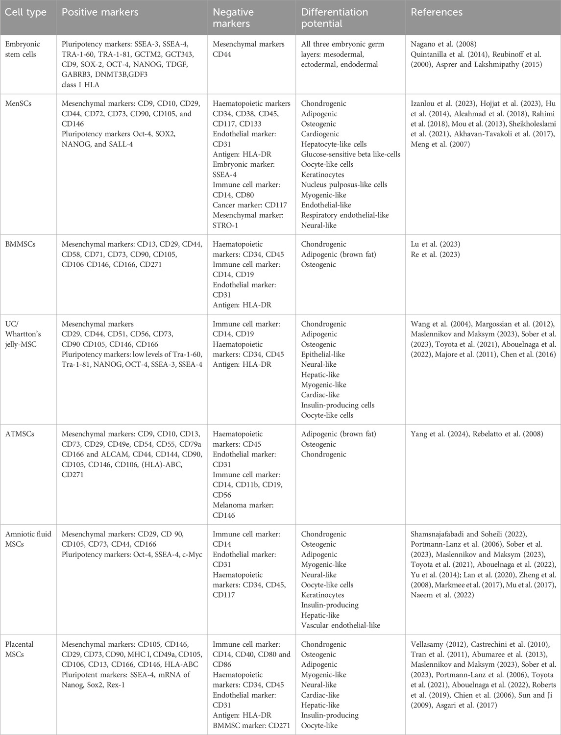
Table 1. MenSC surface marker expression and differentiation potential, as compared to embryonic, BMMSC, ATMSC, UC/Wharton jelly MSC, amniotic fluid and placental MSCs.
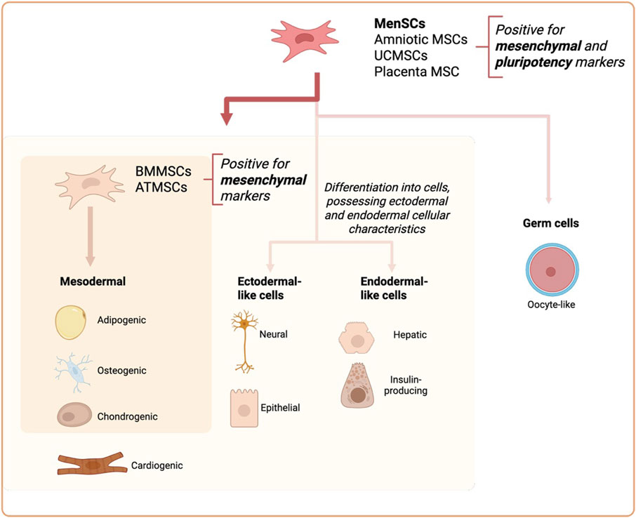
Figure 1. Comparison of MenSCs with Amniotic MSCs, umbilical cord MSCs (UCMSCs), Placenta MSCs, Adipose tissue MSCs (ATMSC) and Bone Marrow MSCs (BMMSC) differentiation potential.
Beside phenotypical differences with other types of stem cells, MenSCs also differ in their differentiation capabilities. It is known that MenSCs differentiate into a wide range of cell types, and are even able to differentiate into cardiomyocytes with the functions of spontaneously beating cells after induction, resulting in the decreased myocardial infarction area in a rat model (Hida et al., 2008; Ikegami et al., 2010). Furthermore, it has been shown that MenSCs are capable of differentiation into neural, epidermal-like cells (Azedi et al., 2017; Faramarzi et al., 2016; Chen et al., 2015; Toyoda et al., 2007), functional hepatocytes (Mou et al., 2013) and even oocyte-like cells (Asgari et al., 2017) which suggest a superior spectrum of their differentiation potential compared to other tissue MSCs.
2.2 MenSCs secretome
MenSCs secrete large amounts of paracrine factors, including growth factors responsible for endometrium regeneration, which may be a potential co-stimulant for other tissue regeneration purposes (Chen et al., 2019; Liu et al., 2018). MenSCs also secrete angiogenic factors VEGF, HGF, ANG and MMP-1 and different cytokines (IL-6, IL-8 and IFN-gamma), and most importantly, MenSCs were shown to be safe to transplant due to their low tumorigenicity (Liu et al., 2018). The secretome of MenSCs also includes EVs, containing proteins or miRNAs (more information in section 2). Different studies reported secretion of various growth factors by MenSCs. Table 2 summarizes all current findings on MenSCs secretome, including protein family, functions and comparison to other types of MSCs.
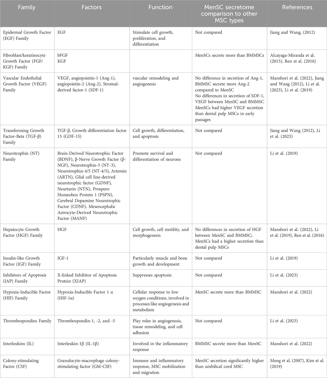
Table 2. MenSCs secreted proteins and comparison to other types of MSCs.
Factors secreted by MenSCs already showed positive immunomodulatory, cardioprotective, angiogenic and regenerative effects. Paracrine effects of MenSCs were analysed in numerous studies and their effects were proposed being more superior to BMMSCs (Alcayaga-Miranda et al., 2015). For instance, MenSC paracrine factors possessed promising results in rat model of myocardial infarction by reducing apoptosis of cells and stimulating endogenous regeneration, while transplantation of MenSCs achieved significantly better cardiac performance than BMMSCs or ATMSCs (Jiang et al., 2013; Wang et al., 2017). Important to note, BMMSCs were shown to secrete higher concentrations of IL-1β (Jiang and Wang, 2012; Manshori et al., 2022). Also, it was revealed that MenSC secrete higher amounts of EGF, FGF and HIF-1α, as compared to BMMCS, while no differences were observed in VEGF or angiopoietin secretion, which were higher in the MenSC secretome compared to UC and dental pulp MSCs. Moreover, MenSC secrete higher levels of HGF than dental pulp MSCs and higher levels of GM-CSF compared to UCMSCs.
Noteworthy, MenSC secretome can be modulated by different environmental conditions. For instance, under hypoxic conditions MenSCs secreted significantly higher levels of VEGF, while EGF and TGF-β secretion was not affected (Jiang and Wang, 2012; Alcayaga-Miranda et al., 2015). Hypoxia can also enhance the release of EVs, as previously shown in UCMSCs (Zhang et al., 2012). Moreover, it was demonstrated that endometrial MSC MiRNAs: miR-148a-3p, hsa-miR-378a-3p (related to angiogenesis, wound healing), hsa-miR-424-5p (associated with angiogenesis), hsa-miR-23a-3p, and hsa-miR-let-7a-5p (related to immune modulation) were the most widely expressed in acute hypoxic conditions (0.1%-1%), while hsa-miR-34a-5p (reduces expression of VEGF), hsa-miR-532-5p, hsa-miR-221-3p, hsa-miR-93-5p (regulating cell cycle and proliferation) were detected only under normoxic conditions (de Pedro et al., 2023). These results are directly associated with MenSC physiological behavior in vivo and differences obtained in vitro.
In order to stimulate MenSC immunomodulator or regenerative properties, MenSCs can be additionally stimulated by external factors using different cultivation conditions. MenSCs increase IDO1 secretion and EVs release under treatment with IFN-γ and TNF-α (de Pedro et al., 2021). bFGF and 5-aza increased the levels of VEGF, SDF-1, HIF-1α, IL-1β, and ANG-1 secretion from MenSCs (Manshori et al., 2022). Moreover, MenSCs may help protect insulin-producing pancreatic β-cells from autoimmune attack in type 1 diabetic mice. By modulating immune responses, these cells could potentially slow disease progression and preserve insulin production (Wu et al., 2014).
3 MenSC EVs and their cargo
EVs isolated from human bodily fluids, such as blood, urine, saliva, or cell culture supernatants have emerged as a promising approach for non-invasive therapies and diagnostics, also known as “liquid biopsy” because of their selectively packed cargo, including proteins, lipids and nucleic acids (Yokoi et al., 2015; Jia et al., 2014; Ciferri et al., 2021; Matsuzaka and Yashiro, 2022). The cargo of EVs is essential for cellular responses and can regulate various physiological and pathological processes, as well as serve as potential biomarkers for diagnosis (Mir and Goettsch, 2020). MenSC-EVs have demonstrated regenerative properties, primarily due to their capacity to transport cargo to recipient cells and modulate key signaling pathways associated with cell survival, differentiation, and proliferation (Robalo Cordeiro et al., 2024; Chen et al., 2021; de Pedro et al., 2023). A schematic representation of MenSC-EV composition is presented in Figure 2.
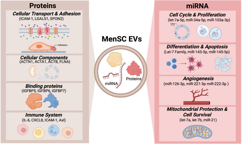
Figure 2. MenSC-EV cargo.
The composition of EV cargo is highly specific and depends on the cell type, metabolic state, and presence of disease. Furthermore, the cargo of EVs is the main factor that defines their mechanism of action, application possibilities, and therapeutic effects (Mir and Goettsch, 2020; Figueroa-Valdés et al., 2021).
3.1 MenSCs and other MSC EV protein cargo
Mainly, MenSC-EVs carry proteins related to processes such as cellular transport, including vesicle-mediated transport or cell adhesion and migration. An additional group of proteins is related to cellular components, including extracellular organelles, membrane components and parts of the cytosol. MenSC-EVs are also enriched with different binding proteins. Upon evaluation of the functional properties of the most abundant proteins in MenSC-EVs, it was determined that the majority are associated with immune system processes and extracellular matrix (ECM) organization (de Pedro et al., 2023). Additionally, MenSC-EVs contain various bioactive molecules, including cytokines. A comparative analysis of MenSCs and MenSC-EVs revealed that the latter contain higher concentrations of IL-6 and IL-8, intercellular cell adhesion molecule-1 (ICAM-1), angiopoietin-2, Axl, angiogenin, insulin-like growth factor-binding protein 6 (IGFBP-6), and osteoprotegerin (Chen et al., 2017). Moreover, it was reported that MenSC-EV are enriched with E3 ubiquitin ligase (UBR4), which inhibited fibrosis of rat endometrial stromal cells by affecting YAP activity (Qi et al., 2023).
The culturing conditions of MenSCs significantly alter the cargo and the EV-associated proteome. Proinflammatory conditions were found to downregulate proteins related to wound healing, adhesion and migration processes and upregulate proteins involved in angiogenesis and inflammatory responses. As an example, MenSCs cultured under physioxic conditions (1%–2% O2) secreted EVs enriched with proteins related to cell adhesion and intracellular transport. Acute hypoxia (<1% O2) had different effects on EV cargo–it upregulated proteins associated with cell adhesion, cell migration and angiogenesis pathways (de Pedro et al., 2023).
At present, the available information regarding MenSC-EV cargo is relatively limited in relation to MSC-derived EVs from alternative sources, such as ATMSCs, BMMSCs, and UCMSCs. BMMSC-EVs contain proteins involved in ion and other protein transport. In addition, proteins associated with cell cycle regulation, transcription and translation regulation, cell adhesion and lipid metabolism, apoptosis and inflammation were identified in BMMSCs. Upon classification of proteins according to cellular components, the majority of proteins were found to be associated with the cell membrane, nucleus, cytoplasm, mitochondria and endoplasmic reticulum (McBride et al., 2021).
ATMSC-EVs encompass a multitude of proteins which play crucial roles in various biological processes. These processes include cellular migration, modulation of immune responses, proliferation of cells, formation of new blood vessels, metabolism of osteocytes, and regeneration of nerve tissue (Alonso-Alonso et al., 2022).
Human UCMSC exosomes are enriched with proteins related to different mechanisms and signaling pathways. The majority of proteins detected in UCMSC exosomes play roles in modulating various biological processes, including complement response, HIF-1, MAPK signaling, metabolic pathways, NF-κB pathway, and microbial infection. Additionally, proteins related to PI3K-AKT, cholesterol metabolism, IgA production, VEGF, and B-cell receptor signaling pathways were detected (Bi et al., 2022). Table 3 presents a more detailed categorization of cargo proteins in different MSC-derived EVs and their respective functions.
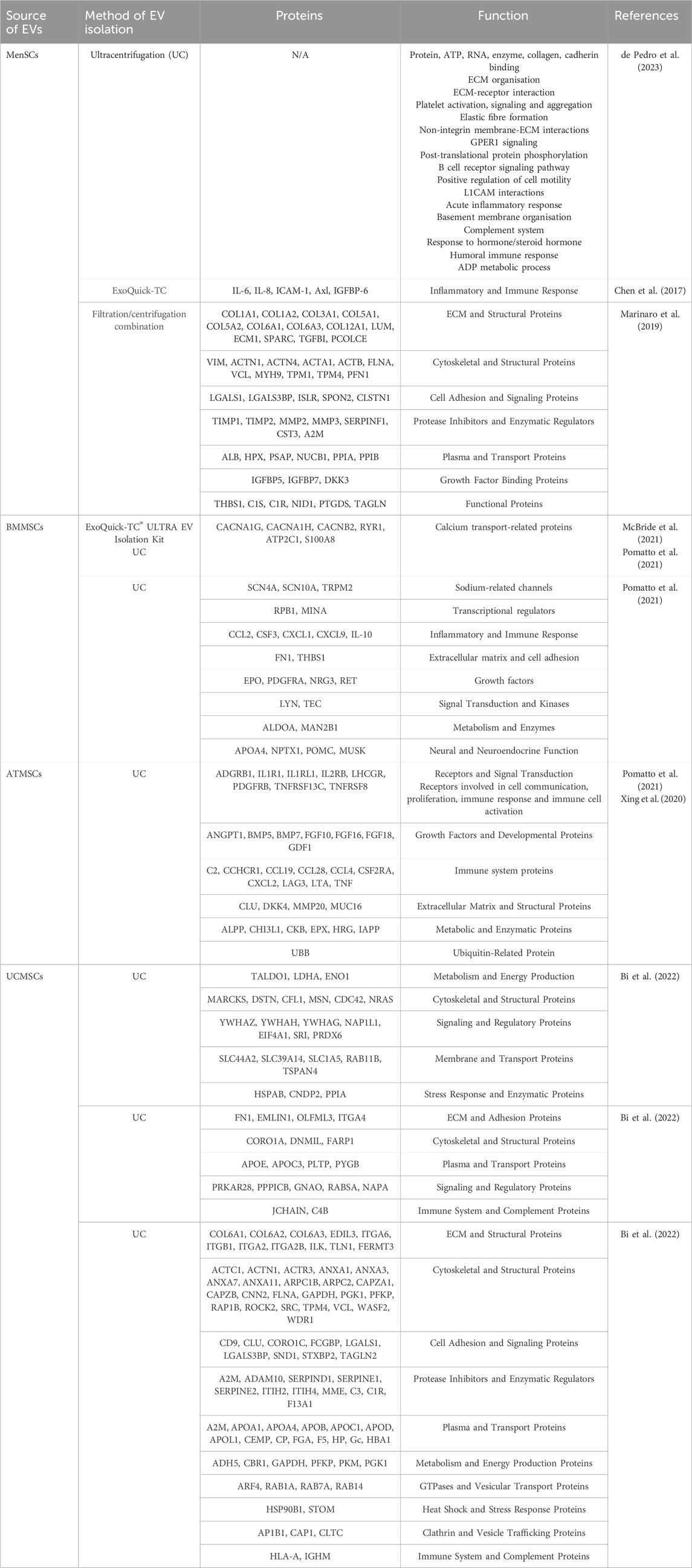
Table 3. Comparison of EVs protein cargo from different sources of MSCs.
3.2 MenSCs and other MSC EV miRNA cargo
MenSC-derived EVs contain a broad range of microRNAs (miRNAs). The identified mi RNAs in MenSC-EVs included let-7a-5p, miR-143-3p, miR-21-5p, let-7b-5p, let-7f-5p, miR-16-5p, miR-199a-3p, miR-199b-3p, miR-126-3p, let-7i-5p, miR-26a-5p, which are involved in the regulation of cell cycle, proliferation, differentiation, apoptosis and angiogenesis (Marinaro et al., 2019). Let-7 and miR-21 play crucial roles in controlling mitochondrial-DNA damage, promoting cell survival and proposed to provide superior cardioprotection and alleviate pulmonary fibrosis (Wang et al., 2017; Sun et al., 2019). Cargo of MenSC-EVs also contain information related to certain diseases and their predispositions to them. The elevated levels of miR-4443 found in MenSC-EVs were discovered to play a role in the progression of endometriosis. This specific miRNA was found to suppress ACSS2 expression and as a result activate the PI3K/AKT signaling pathway. This activation resulted in enhanced migration and proliferation of endometrial stem cells (Ji et al., 2024).
An analysis of EVs from MenSCs, BMMSCs, and ATMSCs revealed that MenSC-EVs exhibited the highest levels of miR-21 among the three sources of EVs. Additionally, the paracrine effect of MenSCs on rat myocardial infarction was compared to that of BMMSCs and ATMSCs. MenSCs were found to enhance cardioprotection through the transfer of miR-21 via EVs. miR-21 from menstrual blood EVs downregulated phosphatase and tensin homolog (PTEN), enhancing Akt survival kinase activity, resulting in reduced apoptosis in cardiomyocytes and improved angiogenesis in endothelial cells (Wang et al., 2017). This finding demonstrates the superior cardioprotective effect of MenSC-EV cargo compared to BMMSC or ATMSC EVs. Additionally, EVs derived from MenSCs attenuate severe pulmonary inflammation and damage through the transmission of miRNA-671. This miRNA is known to target the kinase AAK1 for post-transcriptional degradation. AAK1 positively regulates the NF-κB signaling pathway (Lian et al., 2023).
MiRNAs highly expressed in BMMSC-EVs were found to be associated with cellular proliferation, death, metabolism and immune regulation (miRs-21, miR-22, miR-26a, miR-10b, miR-99b, miR-125b, and miR-148a) (Vaka et al., 2023; Baglio et al., 2015). In comparison with BMMSC-EVs, UCMSC-EVs are enriched with miRNA related to regenerative processes, aging and cell proliferation (Vaka et al., 2023). Several studies reported, that the most abundant miRNA in UCMSC-EVs are miR-16, miR-21, miR-23, miR-34, miR-146a and miR-222, which are associated with cell proliferation and immune regulation (Jothimani et al., 2022). MenSC-EV and other MSC EV miRNAs are presented in Table 4.
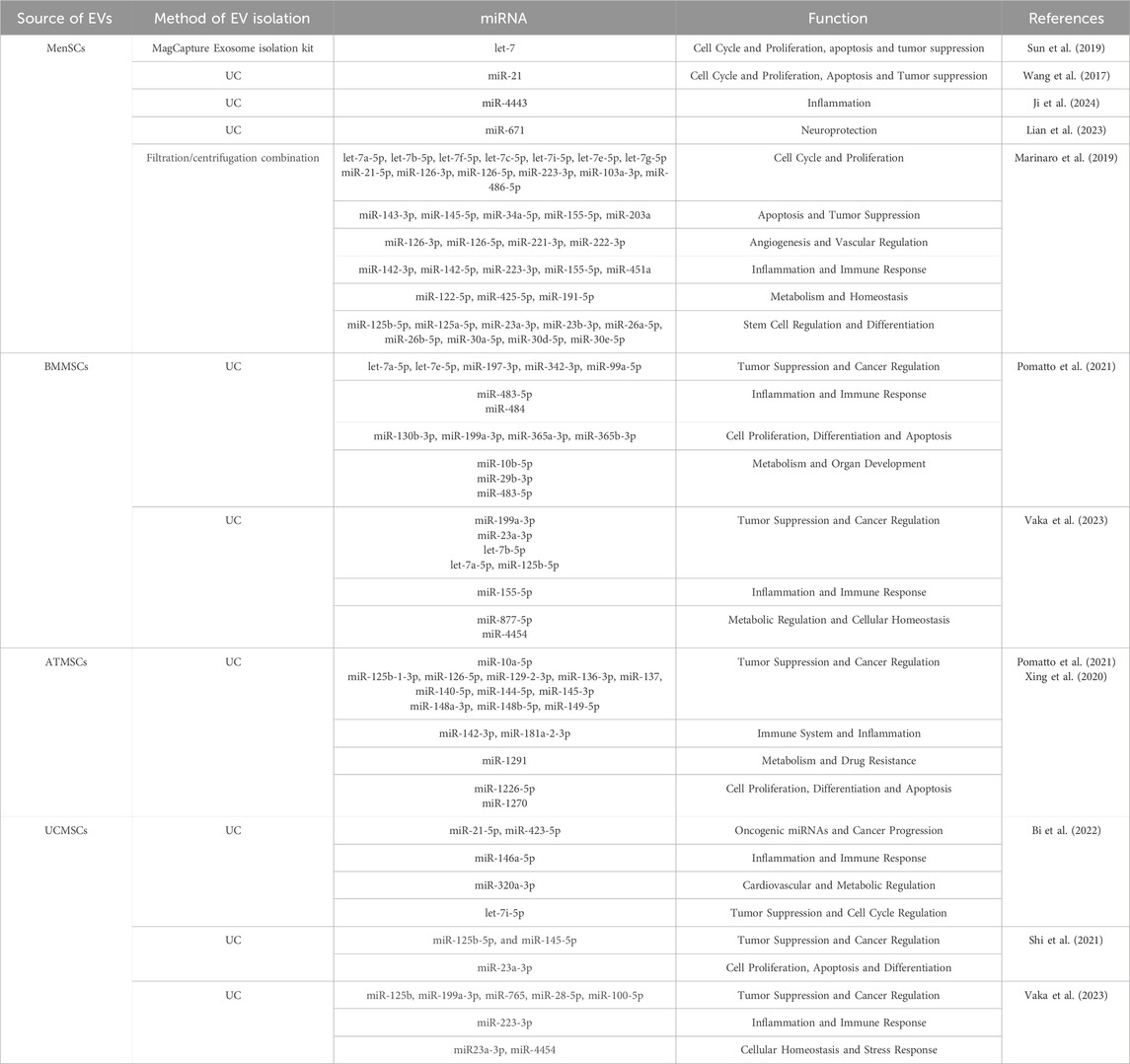
Table 4. Comparison of EVs miRNA cargo from different sources of MSCs.
4 MenSC-EV potential in tissue regeneration
MenSC-EVs showed promising therapeutic potential for regeneration of different tissues, but the most significant effects were demonstrated for the regeneration of female reproductive system tissues, including endometrium and ovaries in diseases, such as Premature Ovarian Insufficiency (POI) and Intrauterine Adhesion (IUA). In addition to that, numerous studies have also indicated the MenSC-EV therapeutic potential on wound healing, neural, liver, heart tissue repair and more.
4.1 MenSCs-EV therapeutic effect on female reproductive tissues
In a rat model of POI induced by chemotherapeutic agents, MenSC-EVs restored ovarian function by increasing ovarian weight, follicle numbers at various developmental stages, and serum estrogen levels. The therapeutic effects were associated with activation of the PI3K/AKT signaling pathway, inhibition of apoptosis, and overall enhancement of ovarian microenvironment stability (Robalo Cordeiro et al., 2024). MenSC-EVs contribute to endometrial repair by enhancing cell proliferation and stimulating VEGF production, which promotes angiogenesis (Marinaro et al., 2018). A study by Zhang et al. demonstrated that MenSC-EVs had the effect of promoting ovarian cell proliferation, inhibiting apoptosis and regulating the ovarian extracellular matrix, while increasing the expression of follicle markers DAZL and FOXL2 in rat ovaries. Also, MenSC exosome injections restored the female rat estrous cycle and increased fertility, as treated subjects exhibited increased endometrial thickness, improved glandular formation and reduced fibrosis. Notably, repeated EV administration enhanced endometrial receptivity and improved embryo implantation rates, suggesting potential clinical applications in infertility treatment (Zhang et al., 2021b).
4.2 MenSCs-EV therapeutic effect on other tissues
MenSC-EVs have demonstrated efficacy in wound healing by promoting fibroblast proliferation, collagen synthesis, and reducing oxidative stress (Zhang et al., 2023). MiRNA cargo, including miR-21 and miR-29, facilitates keratinocyte migration and differentiation, accelerating skin repair. In models of skin injury, MenSC-EVs have been shown to accelerate wound by promoting the growth of new skin cells (keratinocytes and fibroblasts), increasing collagen production. MenSC-EV treatment promoted re-epithelialization, increased angiogenesis, and modulated inflammation, leading to improved healing outcomes in a diabetic mouse model (Dalirfardouei et al., 2019). These effects suggest their potential use in treating chronic wounds or burns.
MenSC-EVs have also shown ability to promote axonal regeneration and functional recovery following neural injury, further underscoring their broad therapeutic utility (Lopez-Verrilli et al., 2016). Moreover, MenSC-EVs can alleviate fulminant hepatic failure. In experimental models, these EVs reduced liver inflammation and promoted hepatocyte proliferation, leading to improved liver function (Chen et al., 2017). Additionally, MenSC-EVs have been shown to promote angiogenesis and reduce scarring in heart tissue, ultimately improving heart function and reducing long-term damage (Wang et al., 2017). And as mentioned before, MenSC-EVs may help slow down fibrosis by reducing the activity of fibroblasts (cells that contribute to scarring) and lowering levels of fibrotic markers, leading to improved lung function as well (Chen et al., 2021).
In cancer therapy, it was shown that MenSC-EVs block tumor associated angiogenesis and could be used as a tool for cancer treatment. MenSC-EVs reduced the secretion of VEGF and NF-κB activity in human prostate PC3 tumor cells (Alcayaga-Miranda et al., 2015). Other studies additionally emphasize the pro-angiogenic effect of MenSC-EVs (Chang et al., 2021a; Wang et al., 2017). Therefore, the precise mechanism of this targeted action of MenSC-EVs remains unclear.
MenSC-EVs also exhibit immunomodulatory properties by regulating T-cell proliferation, macrophage polarization, and inflammatory cytokine production (Song et al., 2023; Qi et al., 2023). This suggests potential therapeutic applications in autoimmune diseases, inflammatory disorders, and systemic tissue repair.
5 MenSC and menstrual blood EVs–a source for disease biomarkers and future diagnostic strategies
EVs show a great potential in diagnostics of different pathologies with leading studies related to early cancer detection, monitoring tumor progression and response to treatment (Weng J. et al., 2021; Kumar et al., 2024). EVs also showed promising results in detection of neurodegenerative diseases (Parkinson’s disease, Alzheimer disease) as they had increased levels of tau proteins, contributed to the diagnosis of cardiac diseases (cardiac fibrosis, ischemic heart disease, heart failure and others) with increased levels of miR-133a, miR-499, miR-199a, pregnancy disorders with higher numbers of circulating EVs in preeclamptic and eclamptic women (Ciferri et al., 2021; Liu et al., 2021; Zhang et al., 2022; Smith and Russell, 2022). EVs could potentially improve diagnostic accuracy and further treatment decisions. The main advantages of EVs for diagnostic approaches include stability in circulation and ability to protect their cargo (Kodam and Ullah, 2021). Nevertheless, challenges remain in EV isolation, especially from human biofluids. Their characterization needs advanced analysis methods, such as digital PCR, mass spectrometry, also in addition to proper storage to keep them intact for clinical application (Weng Z. et al., 2021; Jia et al., 2014; Kodam and Ullah, 2021). Despite these challenges, EVs hold significant promise for improving disease diagnosis and monitoring.
EVs isolated from various reproductive biofluids, including follicular fluid, uterine fluid, peritoneal fluid and serum, alongside the endometrium and endometrial lesions have exhibited significant alterations in miRNAs in pathological conditions such as PCOS premature ovarian insufficiency, endometriosis, and recurrent spontaneous abortion (Duval et al., 2024; Esfandyari et al., 2021). However, conventional diagnostic procedures, such as endometrial biopsies and follicular fluid collection are often invasive, painful, and associated with potential complications (Terzic et al., 2022), while MenSC or menstrual blood serum EVs could be used for the analysis of uterine lesions and abnormalities.
Beyond the regenerative capabilities of MenSC-EVs, these vesicles also hold promise in disease diagnostics and biomarker discovery. Their cargo can provide valuable insights into the molecular changes associated with aging and disease progression. Also, the use of MenSCs-EVs for diagnostic purposes could enable earlier detection and more precise targeting of therapies, leading to more personalized and effective treatment strategies. MenSC-EVs represent a promising source of biomarkers for female reproductive disorders. For instance, we demonstrated that MenSC-EVs can be used as a source of biomarkers of unexplained infertility (uIF) (Vaiciuleviciute et al., 2025). These EVs were compared between healthy and uIF female groups and detected differences included alterations in cell adherence, inflammatory processes, protein metabolism of uIF patients, as compared to healthy controls, which are promising for further uIF validation in women who are not able to conceive for at least a year. Also, menstrual blood serum was characterized as a less invasive source of infertility biomarkers, where EMILIN1, TRIP6, LAMB1, LAMC1, NID1, APOB, APOA4 were detected as the main differences in uIF patients as compared to healthy controls (Brennan et al., 2025). Both MenSCs and MenSC-EVs already showed alterations in endometriosis and endometriosis-related infertility compared to healthy donors (Cordeiro et al., 2023; Zhou et al., 2020). MenSC-EVs even indicated decidual response that is critical for embryo implantation. Additionally, EVs derived from uterine fluid may serve as biomarkers for endometrial receptivity assessment in assisted reproductive technologies (Giacomini et al., 2021).
MenSC-EV-based diagnostics could offer a non-invasive alternative with significant potential for the monitoring of endometrial receptivity and pathology diagnostics of female reproductive diseases. MenSC-EVs not only share the inherent advantages of EV-based diagnostics but also offer additional benefits derived from their cellular origin. Menstrual blood collection is a non-invasive, easily accessible, and repeatable process, eliminating ethical concerns associated with other sources of reproductive tract-derived EVs. Importantly, because menstrual blood is collected during the same phase of the menstrual cycle, it minimizes variability related to hormonal fluctuations and serves as a highly localized source of biomarkers, providing a direct reflection of endometrial status (Zaheer et al., 2024).
The therapeutic and diagnostic potential of MenSC- EVs is schematically visualized in Figure 3, presenting current in vitro and in vivo study discoveries.
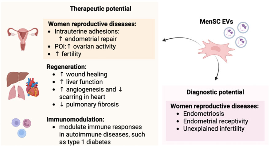
Figure 3. MenSC-EV therapeutic and diagnostic potential for different types of diseases and immunomodulation.
6 Discussion and future directions
The uterine endometrium is a unique, fast-regenerating tissue, which plays an essential role in the female reproductive system. It has been considered as an easy-accessible source for stem cells decades ago (Borlongan et al., 2010). The endometrium undergoes over 400 cycles of regeneration during a woman’s reproductive life cycle, allowing for pregnancy, and can be continued to regenerate after menopause using hormone therapy (Tabatabaei and Ai, 2017). Endometrial stromal cells–MenSCs, have drawn attention in modern research, relating to evidence of their pluripotent-like and therapeutic properties. They offer a non-invasive alternative to traditional MSC sources and hold promise for regenerative applications, particularly through EVs, which enhance tissue repair (Chen et al., 2017; Asl et al., 2023). Even though EVs from all types of MSCs have positive effects on tissue regeneration–for instance, BMMSC-EVs showed increased muscle regeneration in a rat sarcopenia model, restored bone mass and strength in a mice osteoporosis model, regenerated cartilage, restored heart function in myocardial infarction in rat models and others (Guo et al., 2024; Wang et al., 2023; Bian et al., 2013) – MenSC-EVs show exceptional therapeutic potential in the female reproductive system, wound healing, neural, liver regeneration, and more, as discussed previously.
The potential of MenSC-EVs as a therapeutic tool for postmenopausal women is a promising field in regenerative medicine. As women age, particularly after menopause, they face a range of health challenges such as osteoporosis, muscle degeneration, skin aging, and decreased regenerative capacity across various tissues. The application of MenSCs and their EVs presents a novel approach to mitigate these age-related conditions by enhancing tissue regeneration and reducing the effects of chronic inflammation and immunosenescence, which are often observed in postmenopausal women. MenSC-EVs have shown potential in promoting tissue repair and regeneration through their cargo, which includes growth factors, cytokines, lipids, and RNAs that regulate cell survival, proliferation, and differentiation, as described above. These bioactive molecules help to modulate immune responses, stimulate tissue repair and enhance the functionality of damaged cells. Noteworthy, the ability to collect menstrual blood for autologous treatment with MenSCs is progressively reduced in elderly women, representing a limitation for their therapeutic applications. On the other hand, if these cells could be collected and cryopreserved in advance, there will always be an opportunity to use them later in the donor’s lifetime. Additionally, the concept of biobanking MenSCs, particularly from younger women, holds significant potential for future therapeutic applications. Cryopreserving MenSCs could provide a ready and accessible resource for regenerative therapies in elderly populations. Such biobanks would enable the use of autologous MenSCs and their EVs for personalized medicine in later years, overcoming the limitations associated with age-related declines in stem cell function and regenerative capacity. This approach could be particularly advantageous for postmenopausal women, as it offers the possibility of utilizing young, high-quality MenSCs for future therapies targeting conditions such as age-related diseases.
6.1 Limitations
Despite the promising potential of MenSC-EVs in regenerative medicine and disease diagnostics, several limitations of the current source and EVs should be acknowledged. First of all, there is a lack of long-term safety and efficacy data in all of the published studies, as most of them focus on short-term outcomes. Long-term effect of MenSC-EVs on tissue homeostasis or potential off-target response remain largely unexplored, where rigous in vivo studies are essential to ensure translational relevance and clinical safety. Moreover, there is a significant variability in EV isolation and characterization protocols across studies, which is an important aspect to bear in mind working with various EV sources, not only MenSC. Differences in EV isolation, filtration methods, their parameters, quantification techniques, instruments used contribute to inconsistencies in EV purity, yield and functional content. Even if the protocols are normalized, refined according to the consensus guidelines as minimal information for studies of EVs (MISEV) (Welsh et al., 2024), the variability between batch-to-batch samples is also a significant issue adapting EVs for therapeutic purposes.
Also, the use of MenSC-EVs faces regulatory, manufacturing, and bioethical challenges that need to be addressed to ensure their safe and effective use in clinical applications. On the regulatory side, the absence of specific guidelines and the complexity of proving safety, efficacy, and pharmacokinetics make clinical approvals difficult (Stawarska et al., 2024; Wang et al., 2024). Manufacturing these EVs at scale while maintaining consistency, stability, and quality remains a major obstacle (Claridge et al., 2021; Wang et al., 2024). Ethically, while menstrual blood is a non-invasive source, it has a significant ethical advantage over other stem cell sources. Issues such as informed consent and donor privacy must be carefully managed (Achmad and Götte, 2014; Savary et al., 2023). Therefore, in order to ensure reproducibility and clinical applicability, future research should prioritize standardized methodologies, explore the mechanisms underlying MenSC-EV therapeuitc actions and conduct controlled in vivo studies with long-term follow ups to support their safety and integration into clinical therapies.
6.2 Conclusion
In conclusion, MenSCs and their EVs represent a potential tool for advancing diagnostics and therapies. Their ability to promote tissue regeneration, provide diagnostic insights, and enable personalized treatments holds immense potential for improving the quality of life for women of all ages. The development of biobanks for MenSCs could further enhance the accessibility and applicability of these cells and their EVs, offering a new hope for the development of innovative treatment strategies for different conditions.
Author contributions
RV: Writing – original draft, Visualization, Writing – review and editing. JP: Writing – review and editing, Investigation, Writing – original draft, Validation. EB: Writing – original draft, Supervision, Writing – review and editing, Investigation. GK: Writing – review and editing, Writing – original draft, Investigation. IL: Validation, Writing – review and editing, Writing – original draft, Investigation. EK: Writing – review and editing, Investigation, Formal Analysis, Writing – original draft. IU: Supervision, Writing – review and editing, Writing – original draft, Conceptualization.
Funding
The author(s) declare that financial support was received for the research and/or publication of this article. The study is funded by the Lithuanian Research Councils project, according to the postdoctoral fellowship program No. P-PD-24-167, agreement No. S-PD-24-114 (No. 33154).
Acknowledgments
The authors want to acknowledge Lithuanian Research Council for providing opportunities to conduct this review article. Also, the corresponding author wants to acknowledge Lithuanian Academy of Science for providing the award for young investigators on June 20, 2024.
Conflict of interest
The authors declare that the research was conducted in the absence of any commercial or financial relationships that could be construed as a potential conflict of interest.
Generative AI statement
The author(s) declare that no Generative AI was used in the creation of this manuscript.
Publisher’s note
All claims expressed in this article are solely those of the authors and do not necessarily represent those of their affiliated organizations, or those of the publisher, the editors and the reviewers. Any product that may be evaluated in this article, or claim that may be made by its manufacturer, is not guaranteed or endorsed by the publisher.
References
Abouelnaga, H., El-Khateeb, D., Moemen, Y., El-Fert, A., Mohamed, E., and Khalil, A. (2022). Characterization of mesenchymal stem cells isolated from wharton’s jelly of the human umbilical cord. Egypt. Liver J. 12 (1), 2. doi:10.1186/s43066-021-00165-w
Abumaree, M. H., Al Jumah, M. A., Kalionis, B., Jawdat, D., Al Khaldi, A., Abomaray, F. M., et al. (2013). Human placental mesenchymal stem cells (PMSCs) play a role as immune suppressive cells by shifting macrophage differentiation from inflammatory M1 to anti-inflammatory M2 macrophages. Stem Cell Rev. Rep. 9 (5), 620–641. doi:10.1007/s12015-013-9455-2
Achmad, N., and Götte, M. (2014). Characteristics and therapeutic potential of menstrual blood-derived stem cells. Regen. Med. Using Non-Fetal Sources Stem Cells, 1–285. doi:10.1007/978-1-4471-6542-2
Akhavan-Tavakoli, M., Fard, M., Khanjani, S., Zare, S., Edalatkhah, H., Mehrabani, D., et al. (2017). In vitro differentiation of menstrual blood stem cells into keratinocytes: a potential approach for management of wound healing. Biologicals 48, 66–73. doi:10.1016/j.biologicals.2017.05.005
Alcayaga-Miranda, F., Cuenca, J., Luz-Crawford, P., Aguila-Díaz, C., Fernandez, A., Figueroa, F. E., et al. (2015). Characterization of menstrual stem cells: angiogenic effect, migration and hematopoietic stem cell support in comparison with bone marrow mesenchymal stem cells. Stem Cell Res. and Ther. 6, 32. doi:10.1186/s13287-015-0013-5
Aleahmad, M., Ghanavatinejad, A., Bozorgmehr, M., Shokri, M. R., Nikoo, S., Tavakoli, M., et al. (2018). Menstrual blood-derived stromal stem cells augment CD4+ T cells proliferation. Avicenna J. Med. Biotechnol. 10 (3), 183–191.
Alonso-Alonso, M. L., García-Posadas, L., and Diebold, Y. (2022). Extracellular vesicles from human adipose-derived mesenchymal stem cells: a review of common cargos. Stem Cell Rev. Rep. 18 (3), 854–901. doi:10.1007/s12015-021-10155-5
Asadian, N., Jadidi, M., Safari, M., Jadidi, T., and Gholami, M. (2021). EMF frequency dependent differentiation of rat bone marrow mesenchymal stem cells to astrocyte cells. Neurosci. Lett. 744, 135587. doi:10.1016/j.neulet.2020.135587
Asgari, H. R., Akbari, M., Yazdekhasti, H., Rajabi, Z., Navid, S., Aliakbari, F., et al. (2017). Comparison of human amniotic, chorionic, and umbilical cord multipotent mesenchymal stem cells regarding their capacity for differentiation toward female germ cells. Cell. Reprogr. 19 (1), 44–53. doi:10.1089/cell.2016.0035
Asl, D., Saeideh Sahraei, S., Kalhor, N., Fazaeli, H., Sheykhhasan, M., Moud, S. S., et al. (2023). Promising effects of exosomes from menstrual blood-derived mesenchymal stem cells on endometriosis. Reprod. Biol. 23 (3), 100788. doi:10.1016/j.repbio.2023.100788
Asprer, J. S. T., and Lakshmipathy, U. (2015). Current methods and challenges in the comprehensive characterization of human pluripotent stem cells. Stem Cell Rev. Rep. 11 (2), 357–372. doi:10.1007/s12015-014-9580-6
Azedi, F., Kazemnejad, S., Hassan Zarnani, A., Soleimani, M., Shojaei, A., and Arasteh, S. (2017). Comparative capability of menstrual blood versus bone marrow derived stem cells in neural differentiation. Mol. Biol. Rep. 44 (1), 169–182. doi:10.1007/s11033-016-4095-7
Baghaei, K., Hashemi, S. M., Tokhanbigli, S., Ali, A. R., Assadzadeh-Aghdaei, H., Sharifian, A., et al. (2017). Isolation, differentiation, and characterization of mesenchymal stem cells from human bone marrow. Gastroenterology Hepatology Bed Bench 10 (3), 208–213. doi:10.22037/ghfbb.v0i0.1089
Baglio, S. R., Rooijers, K., Koppers-Lalic, D., Verweij, F. J., Pérez Lanzón, M., Zini, N., et al. (2015). Human bone marrow- and adipose-mesenchymal stem cells secrete exosomes enriched in distinctive MiRNA and TRNA species. Stem Cell Res. Ther. 6 (1), 127–20. doi:10.1186/s13287-015-0116-z
Bausyte, R., Vaigauskaite - Mazeikiene, B., Borutinskaite, V., Valatkaite, E., Besusparis, J., Barbora Valkiuniene, R., et al. (2023). Human endometrium-derived mesenchymal stem/stromal cells application in endometrial-factor induced infertility. Front. Cell Dev. Biol. 11, 1–17. doi:10.3389/fcell.2023.1227487
Bi, Y., Qiao, X., Liu, Q., Song, S., Zhu, K., Qiu, X., et al. (2022). Systemic proteomics and MiRNA profile analysis of exosomes derived from human pluripotent stem cells. Stem Cell Res. Ther. 13 (1), 449–19. doi:10.1186/s13287-022-03142-1
Bian, L., Hou, C., Tous, E., Rai, R., Mauck, R. L., and Burdick, J. A. (2013). The influence of hyaluronic acid hydrogel crosslinking density and macromolecular diffusivity on human MSC chondrogenesis and hypertrophy. Biomaterials 34 (2), 413–421. doi:10.1016/j.biomaterials.2012.09.052
Borlongan, C. V., Kaneko, Y., Maki, M., Yu, S. J., Ali, M., Allickson, J. G., et al. (2010). Menstrual blood cells display stem cell–like phenotypic markers and exert neuroprotection following transplantation in experimental stroke. Stem Cells Dev. 19 (4), 439–452. doi:10.1089/scd.2009.0340
Brennan, K., Vaiciuleviciute, R., Uzieliene, I., Pachaleva, J., Kasilovskiene, Z., Piesiniene, L., et al. (2025). Menstrual blood serum extracellular vesicles reveal novel molecular biomarkers and potential endotypes of unexplained infertility. Sci. Rep. 15 (1), 11974–15. doi:10.1038/s41598-025-95818-w
Castrechini, N. M., Murthi, P., Gude, N. M., Erwich, J. J. H. M., Gronthos, S., Zannettino, A., et al. (2010). Mesenchymal stem cells in human placental chorionic villi reside in a vascular niche. Placenta 31 (3), 203–212. doi:10.1016/j.placenta.2009.12.006
Chen, J., Du, X., Chen, Q., and Xiang, C. (2015). Effects of donors' age and passage number on the biological characteristics of menstrual blood-derived stem cells. Stem Cells 8 (11), 14584–14595.
Chen, S., Zhang, W., Wang, Ji M., Duan, H. T., Kong, J. H., Wang, Y. X., et al. (2016). Differentiation of isolated human umbilical cord mesenchymal stem cells into neural stem cells. Int. J. Ophthalmol. 9 (1), 41–47. doi:10.18240/ijo.2016.01.07
Chen, L., Xiang, B., Wang, X., and Xiang, C. (2017). Exosomes derived from human menstrual blood-derived stem cells alleviate fulminant hepatic failure. Stem Cell Res. Ther. 8 (1), 9–15. doi:10.1186/s13287-016-0453-6
Chen, L., Qu, J., Cheng, T., Chen, X., and Xiang, C. (2019). Menstrual blood-derived stem cells: toward therapeutic mechanisms, novel strategies, and future perspectives in the treatment of diseases. Stem Cell Res. Ther. 10 (1), 406–412. doi:10.1186/s13287-019-1503-7
Chen, L., Qu, J., Mei, Q., Chen, X., Fang, Y., Chen, L., et al. (2021). Small extracellular vesicles from menstrual blood-derived mesenchymal stem cells (MenSCs) as a novel therapeutic impetus in regenerative medicine. Stem Cell Res. Ther. 12 (1), 433–15. doi:10.1186/s13287-021-02511-6
Chien, C.-C., Yen, B. L., Lee, F.-K., Lai, T.-H., Chen, Y.-C., Chan, S.-H., et al. (2006). In vitro differentiation of human placenta-derived multipotent cells into hepatocyte-like cells. Stem Cells 24 (7), 1759–1768. doi:10.1634/stemcells.2005-0521
Ciferri, M. C., Quarto, R., and Tasso, R. (2021). Extracellular vesicles as biomarkers and therapeutic tools: from pre-clinical to clinical applications. Biology 10 (5), 359–14. doi:10.3390/biology10050359
Claridge, B., Lozano, J., Qi, H. P., and Greening, D. W. (2021). Development of extracellular vesicle therapeutics: challenges, considerations, and opportunities. Front. Cell Dev. Biol. 9, 734720. doi:10.3389/fcell.2021.734720
Cordeiro, M. R., Carvalhos, C. A., and Figueiredo-Dias, M. (2023). The emerging role of menstrual-blood-derived stem cells in endometriosis. Biomedicines 11 (1), 39–10. doi:10.3390/biomedicines11010039
Dalirfardouei, R., Jamialahmadi, K., Jafarian, A. H., and Mahdipour, E. (2019). Promising effects of exosomes isolated from menstrual blood-derived mesenchymal stem cell on wound-healing process in diabetic mouse model. J. Tissue Eng. Regen. Med. 13 (4), 555–568. doi:10.1002/term.2799
de Pedro, M. Á., Gómez-Serrano, M., Marinaro, F., López, E., Pulido, M., Preußer, C., et al. (2021). Ifn-gamma and tnf-alpha as a priming strategy to enhance the immunomodulatory capacity of secretomes from menstrual blood-derived stromal cells. Int. J. Mol. Sci. 22 (22), 12177. doi:10.3390/ijms222212177
de Pedro, M. Á., López, E., Manuel González-Nuño, F., Pulido, M., Álvarez, V., Marchena, A. M., et al. (2023). Menstrual blood-derived mesenchymal stromal cells: impact of preconditioning on the cargo of extracellular vesicles as potential therapeutics. Stem Cell Res. Ther. 14 (1), 1–20. doi:10.1186/s13287-023-03413-5
Duval, C., Wyse, B. A., Tsang, B. K., and Librach, C. L. (2024). Extracellular vesicles and their content in the context of polycystic ovarian syndrome and endometriosis: a review. J. Ovarian Res. 17 (1), 160. doi:10.1186/s13048-024-01480-7
Esfandyari, S., Elkafas, H., Chugh, R. M., Park, H. S., Navarro, A., and Al-Hendy, A. (2021). Exosomes as biomarkers for female reproductive diseases diagnosis and therapy. Int. J. Mol. Sci. 22 (4), 2165–26. doi:10.3390/ijms22042165
Faramarzi, H., Mehrabani, D., Fard, M., Akhavan, M., Zare, S., Bakhshalizadeh, S., et al. (2016). The potential of menstrual blood-derived stem cells in differentiation to epidermal lineage: a preliminary report. World J. Plastic Surg. 5 (1), 26–31. Available online at: https://pmc.ncbi.nlm.nih.gov/articles/PMC4904135/.
Figueroa-Valdés, A. I., Catalina de la, F., Hidalgo, Y., Vega-Letter, A. M., Tapia-Limonchi, R., Khoury, M., et al. (2021). A chemically defined, xeno- and blood-free culture medium sustains increased production of small extracellular vesicles from mesenchymal stem cells. Front. Bioeng. Biotechnol. 9, 1–14. doi:10.3389/fbioe.2021.619930
Gargett, C. E. (2004). Stem cells in gynaecology. Aust. N. Z. J. Obstetrics Gynaecol. 44 (5), 380–386. doi:10.1111/j.1479-828X.2004.00290.x
Gargett, C. E., Schwab, K. E., and Deane, J. A. (2016). Endometrial stem/progenitor cells: the first 10 years. Hum. Reprod. Update 22, 137–163. doi:10.1093/humupd/dmv051
Giacomini, E., Scotti, G. M., Vanni, V. S., Lazarevic, D., Makieva, S., Privitera, L., et al. (2021). Global transcriptomic changes occur in uterine fluid-derived extracellular vesicles during the endometrial window for embryo implantation. Hum. Reprod. 36 (8), 2249–2274. doi:10.1093/humrep/deab123
Guo, R., Wu, Z., Liu, Ao, Li, Q., Han, T., and Shen, C. (2024). Hypoxic preconditioning-engineered bone marrow mesenchymal stem cell-derived exosomes promote muscle satellite cell activation and skeletal muscle regeneration via the MiR-210-3p/KLF7 mechanism. Int. Immunopharmacol. 142, 113143. doi:10.1016/j.intimp.2024.113143
Hida, N., Nishiyama, N., Miyoshi, S., Kira, S., Segawa, K., Uyama, T., et al. (2008). Novel cardiac precursor-like cells from human menstrual blood-derived mesenchymal cells. Stem Cells 26 (7), 1695–1704. doi:10.1634/stemcells.2007-0826
Hojjat, A., Mansour, R. N., Enderami, S. E., Hassannia, H., Mahdavi, M., Mellati, A., et al. (2023). The differentiation and generation of glucose-sensitive beta like-cells from menstrual blood-derived stem cells using an optimized differentiation medium with platelet-rich plasma (PRP). Acta Histochem. 125 (3), 152025. doi:10.1016/j.acthis.2023.152025
Hu, X., Zhou, Y., Zheng, X., Tian, N., Xu, C., Wu, W., et al. (2014). Differentiation of menstrual blood-derived stem cells toward nucleus pulposus-like cells in a coculture system with nucleus pulposus cells. Spine 39 (9), 754–760. doi:10.1097/BRS.0000000000000261
Ikegami, Y., Miyoshi, S., Nishiyama, N., Hida, N., Okamoto, K., Miyado, K., et al. (2010). Serum-independent cardiomyogenic transdifferentiation in human endometrium-derived mesenchymal cells. Artif. Organs 34 (4), 280–288. doi:10.1111/j.1525-1594.2009.00859.x
Izanlou, S., Afshar, A., Zare, A., Zhilisbayeva, K. R., Bakhshalizadeh, S., Safaei, Z., et al. (2023). Enhancing differentiation of menstrual blood-derived stem cells into female germ cells using a bilayer amniotic membrane and nano-fibrous fibroin scaffold. Tissue Cell 85, 102215. doi:10.1016/j.tice.2023.102215
Ji, S., Qi, H., Yan, L., Zhang, D., Wang, Y., Mudanlifu, H., et al. (2024). MiR-4443 contained extracellular vesicles: a factor for endometriosis progression by PI3K/AKT/ACSS2 cascade in-Vitro. Int. J. Nanomedicine 19, 6085–6098. doi:10.2147/IJN.S456594
Jia, S., Zocco, D., Samuels, M. L., Chou, M. F., Chammas, R., Skog, J., et al. (2014). Emerging technologies in extracellular vesicle-based molecular diagnostics. Expert Rev. Mol. Diagnostics 14 (3), 307–321. doi:10.1586/14737159.2014.893828
Jiang, Z., and Wang, J. (2012). Menstrual blood stem cells improved myocardial survival after rat myocardial infarction by paracrine. Heart 98 (Suppl. 2), E170–E172. doi:10.1136/heartjnl-2012-302920j.31
Jiang, Z., Hu, X., Yu, H., Xu, Y., Wang, L., Chen, H., et al. (2013). Human endometrial stem cells confer enhanced myocardial salvage and regeneration by paracrine mechanisms. J. Cell. Mol. Med. 17 (10), 1247–1260. doi:10.1111/jcmm.12100
Jothimani, G., Pathak, S., Dutta, S., Duttaroy, A. K., and Banerjee, A. (2022). A comprehensive cancer-associated MicroRNA expression profiling and proteomic analysis of human umbilical cord mesenchymal stem cell-derived exosomes. Tissue Eng. Regen. Med. 19 (5), 1013–1031. doi:10.1007/s13770-022-00450-8
Kim, J., Kim, Na K., Park, So Ra, and Choi, B. H. (2019). GM-CSF enhances mobilization of bone marrow mesenchymal stem cells via a CXCR4-medicated mechanism. Tissue Eng. Regen. Med. 16 (1), 59–68. doi:10.1007/s13770-018-0163-5
Kodam, S. P., and Ullah, M. (2021). Diagnostic and therapeutic potential of extracellular vesicles. Technol. Cancer Res. Treat. 20, 15330338211041203–15330338211041210. doi:10.1177/15330338211041203
Kumar, M. A., Baba, S. K., Sadida, H. Q., Marzooqi, S. A., Jerobin, J., Altemani, F. H., et al. (2024). Extracellular vesicles as tools and targets in therapy for diseases. Signal Transduct. Target. Ther. 9 (1), 27. doi:10.1038/s41392-024-01735-1
Lai, D., Wang, F., Yao, X., Zhang, Q., Wu, X., and Xiang, C. (2015). Human endometrial mesenchymal stem cells restore ovarian function through improving the renewal of germline stem cells in a mouse model of premature ovarian failure. J. Transl. Med. 13 (1), 155–13. doi:10.1186/s12967-015-0516-y
Lan, D. T. P., Binh, P. T., Giang, N. T. Q., Van Mao, C., Chung, D. T., Van Diep, N., et al. (2020). Isolation and differentiation of amniotic membrane stem cells into keratinocytes. Cell Transplant. 29, 096368972096438–7. doi:10.1177/0963689720964381
Lee, Se Y., Ham, O., Ji Cha, M., Song, B. W., Choi, E., Kim, Il K., et al. (2013). The promotion of cardiogenic differentiation of HMSCs by targeting epidermal growth factor receptor using MicroRNA-133a. Biomaterials 34 (1), 92–99. doi:10.1016/j.biomaterials.2012.09.069
Lei, G., Yu, Y., Jiang, Y., Wang, S., Yan, M., Smith, A. J., et al. (2013). Differentiation of BMMSCs into odontoblast-like cells induced by natural dentine matrix. Archives Oral Biol. 58 (7), 862–870. doi:10.1016/j.archoralbio.2013.01.002
Li, H., Yahaya, B. H., Ng, W. H., Yusoff, N. M., and Lin, J. (2019). Conditioned medium of human menstrual blood-derived endometrial stem cells protects against MPP+-Induced cytotoxicity in vitro. Front. Mol. Neurosci. 12, 80–15. doi:10.3389/fnmol.2019.00080
Li, H., Wei, J., Zhang, Z., Li, J., Ma, Y., Zhang, P., et al. (2023). Menstrual blood-derived endometrial stem cells alleviate neuroinflammation by modulating M1/M2 polarization in cell and rat Parkinson’s disease models. Stem Cell Res. Ther. 14 (1), 85–18. doi:10.1186/s13287-023-03330-7
Lian, J., Zhu, X., Du, J., Huang, B., Zhao, F., Ma, C., et al. (2023). Extracellular vesicle-transmitted MiR-671-5p alleviates lung inflammation and injury by regulating the AAK1/NF-κb Axis. Mol. Ther. 31 (5), 1365–1382. doi:10.1016/j.ymthe.2023.01.025
Liu, Y., Niu, R., Yang, F., Yan, Y., Liang, S., Sun, Y., et al. (2018). Biological characteristics of human menstrual blood-derived endometrial stem cells. J. Cell. Mol. Med. 22 (3), 1627–1639. doi:10.1111/jcmm.13437
Liu, Q., Piao, H., Wang, Y., Zheng, D., and Wang, W. (2021). Circulating exosomes in ophthalmic disease: novel carriers of biological information circulating exosomes in ophthalmic disease. Eur. Rev. Med. Pharmacol. Sci. 25 (5), 2172–2181. doi:10.26355/eurrev_202103_25208
Lopez-Verrilli, M. A., Caviedes, A., Cabrera, A., Sandoval, S., Wyneken, U., and Khoury, M. (2016). Mesenchymal stem cell-derived exosomes from different sources selectively promote neuritic outgrowth. Neuroscience 320, 129–139. doi:10.1016/j.neuroscience.2016.01.061
Lu, Y., Xu, X., Liu, S., Wang, Z., Jiang, K., Gu, J., et al. (2023). Icariin has the potential to induce the differentiation of bone marrow mesenchymal stem cells into Brown fat cells via PDE5A inhibition. Heliyon 9 (12), e22487. doi:10.1016/j.heliyon.2023.e22487
Majore, I., Moretti, P., Stahl, F., Hass, R., and Kasper, C. (2011). Growth and differentiation properties of mesenchymal stromal cell populations derived from whole human umbilical cord. Stem Cell Rev. Rep. 7 (1), 17–31. doi:10.1007/s12015-010-9165-y
Manshori, M., Kazemnejad, S., Naderi, N., Darzi, M., Aboutaleb, N., and Golshahi, H. (2022). Greater angiogenic and immunoregulatory potency of BFGF and 5-aza-2ʹ-deoxycytidine pre-treated menstrual blood stem cells in compare to bone marrow stem cells in rat model of myocardial infarction. BMC Cardiovasc. Disord. 22 (1), 578–12. doi:10.1186/s12872-022-03032-7
Margossian, T., Reppel, L., Makdissy, N., Stoltz, J. F., Bensoussan, D., and Huselstein, C. (2012). Mesenchymal stem cells derived from wharton’s jelly: comparative phenotype analysis between tissue and in vitro expansion. Bio-Medical Mater. Eng. 22 (4), 243–254. doi:10.3233/BME-2012-0714
Marinaro, F., Pericuesta, E., Sánchez-Margallo, F. M., Casado, J. G., Álvarez, V., Matilla, E., et al. (2018). Extracellular vesicles derived from endometrial human mesenchymal stem cells improve IVF outcome in an aged murine model. Reproduction Domest. Animals 53, 46–49. doi:10.1111/rda.13314
Marinaro, F., Gómez-Serrano, M., Jorge, I., Silla-Castro, J. C., Vázquez, J., Sánchez-Margallo, F. M., et al. (2019). Unraveling the molecular signature of extracellular vesicles from endometrial-derived mesenchymal stem cells: potential modulatory effects and therapeutic applications. Front. Bioeng. Biotechnol. 7, 1–19. doi:10.3389/fbioe.2019.00431
Markmee, R., Aungsuchawan, S., Narakornsak, S., Tancharoen, W., Bumrungkit, K., Pangchaidee, N., et al. (2017). Differentiation of mesenchymal stem cells from human amniotic fluid to cardiomyocyte-like cells. Mol. Med. Rep. 16 (5), 6068–6076. doi:10.3892/mmr.2017.7333
Maslennikov, S., and Maksym, G. (2023). The most commonly used cell surface markers for determining mesenchymal stromal cells in stromal vascular fraction and bone marrow autologous concentrate: a systematic review. J. Biotech Res. 14, 85–94.
Matsuzaka, Y., and Yashiro, R. (2022). Therapeutic strategy of mesenchymal-stem-cell-derived extracellular vesicles as regenerative medicine. Int. J. Mol. Sci. 23 (12), 6480. doi:10.3390/ijms23126480
McBride, J. D., Rodriguez-Menocal, L., Wellington, G., Khan, A., Ciara, M., Liu, X., et al. (2021). Proteomic analysis of bone marrow-derived mesenchymal stem cell extracellular vesicles from healthy donors: implications for proliferation, angiogenesis, wnt signaling, and the basement membrane. Stem Cell Res. Ther. 12 (1), 1–11. doi:10.1186/s13287-021-02405-7
Meng, X., Ichim, T. E., Zhong, J., Rogers, A., Yin, Z., Jackson, J., et al. (2007). Endometrial regenerative cells: a novel stem cell population. J. Transl. Med. 5, 57. doi:10.1186/1479-5876-5-57
Mir, B., and Goettsch, C. (2020). Extracellular vesicles as delivery vehicles of specific cellular cargo. Cells 9 (7), 1601–1619. doi:10.3390/cells9071601
Mou, X. Z., Lin, J., Chen, J. Y., Li, Y. F., Wu, X., Xiang, B. Y., et al. (2013). Menstrual blood-derived mesenchymal stem cells differentiate into functional hepatocyte-like cells. J. Zhejiang Univ. Sci. B 14 (11), 961–972. doi:10.1631/jzus.B1300081
Mu, Xu P., Ren, Li Q., Yan, H. W., Zhang, X. M., Xu, T. M., Wei, An H., et al. (2017). Enhanced differentiation of human amniotic fluid-derived stem cells into insulin-producing cells in vitro. J. Diabetes Investigation 8 (1), 34–43. doi:10.1111/jdi.12544
Naeem, A., Gupta, N., Naeem, U., Khan, M. J., Elrayess, M. A., Cui, W., et al. (2022). A comparison of isolation and culture protocols for human amniotic mesenchymal stem cells. Cell Cycle 21 (15), 1543–1556. doi:10.1080/15384101.2022.2060641
Nagano, K., Yoshida, Y., and Isobe, T. (2008). Cell surface biomarkers of embryonic stem cells. Proteomics 8 (19), 4025–4035. doi:10.1002/pmic.200800073
Pomatto, M., Gai, C., Negro, F., Cedrino, M., Grange, C., Ceccotti, E., et al. (2021). Differential therapeutic effect of extracellular vesicles derived by bone marrow and adipose mesenchymal stem cells on wound healing of diabetic ulcers and correlation to their cargoes. Int. J. Mol. Sci. 22 (8), 3851–26. doi:10.3390/ijms22083851
Portmann-Lanz, C. B., Schoeberlein, A., Huber, A., Sager, R., Malek, A., Holzgreve, W., et al. (2006). Placental mesenchymal stem cells as potential autologous graft for pre- and perinatal neuroregeneration. Am. J. Obstetrics Gynecol. 194 (3), 664–673. doi:10.1016/j.ajog.2006.01.101
Qi, J., Zhang, X., Zhang, S., Wu, S., Lu, Y., Li, S., et al. (2023). P65 mediated UBR4 in exosomes derived from menstrual blood stromal cells to reduce endometrial fibrosis by regulating YAP ubiquitination. J. Nanobiotechnology 21 (1), 305–316. doi:10.1186/s12951-023-02070-3
Quintanilla, R. H., Asprer, J. S. T., Vaz, C., Tanavde, V., and Lakshmipathy, U. (2014). CD44 is a negative cell surface marker for pluripotent stem cell identification during human fibroblast reprogramming. PLoS ONE 9 (1), e85419. doi:10.1371/journal.pone.0085419
Rahimi, M., Hassan Zarnani, A., Mobini, S., Khorasani, S., Darzi, M., and Kazemnejad, S. (2018). Comparative effectiveness of three-dimensional scaffold, differentiation media and Co-culture with native cardiomyocytes to trigger in vitro cardiogenic differentiation of menstrual blood and bone marrow stem cells. Biologicals 54, 13–21. doi:10.1016/j.biologicals.2018.05.003
Rahman, Md S., Sebastian Spitzhorn, L., Wruck, W., Hagenbeck, C., Balan, P., Graffmann, N., et al. (2018). The presence of human mesenchymal stem cells of renal origin in amniotic fluid increases with gestational time. Stem Cell Res. and Ther. 9 (1), 113. doi:10.1186/s13287-018-0864-7
Re, F., Sartore, L., Borsani, E., Ferroni, M., Pasini, C., Pandini, S., et al. (2023). Potentiality of bone marrow-derived mesenchymal stromal cells (BM-MSCs) to differentiate into the osteogenic lineage in in vitro model of tissue regeneration. Blood 142 (Suppl. 1), 5626. doi:10.1182/blood-2023-177940
Rebelatto, C. K., Aguiar, A. M., Moretão, M. P., Senegaglia, A. C., Hansen, P., Barchiki, F., et al. (2008). Dissimilar differentiation of mesenchymal stem cells from bone marrow, umbilical cord blood, and adipose tissue. Exp. Biol. Med. 233 (7), 901–913. doi:10.3181/0712-RM-356
Ren, H., Sang, Y., Zhang, F., Liu, Z., Qi, N., and Chen, Y. (2016). Comparative analysis of human mesenchymal stem cells from umbilical cord, dental pulp, and menstrual blood as sources for cell therapy. Stem Cells Int., 3516574. doi:10.1155/2016/3516574
Reubinoff, B. E., Pera, M. F., Fong, C. Y., Trounson, A., and Bongso, A. (2000). Embryonic stem cell lines from human blastocysts: somatic differentiation in vitro. Nat. Biotechnol. 18 (4), 399–404. doi:10.1038/74447
Robalo Cordeiro, M., Roque, R., Laranjeiro, B., Carvalhos, C., and Figueiredo-Dias, M. (2024). Menstrual blood stem cells-derived exosomes as promising therapeutic tools in premature ovarian insufficiency induced by gonadotoxic systemic anticancer treatment. Int. J. Mol. Sci. 25 (15), 8468. doi:10.3390/ijms25158468
Roberts, E. G., Piekarski, B. L., Huang, K., Emani, S., Wong, J. Y., and Emani, S. M. (2019). Evaluation of placental mesenchymal stem cell sheets for myocardial repair and regeneration. Tissue Eng. - Part A 25 (11–12), 867–877. doi:10.1089/ten.tea.2018.0035
Savary, R., Negar, M. K., Mahdi, A., Seyed, M. R. S., and Seyyedeh, E. M. (2023). Extracellular Vesicles Isolated From Menstrual Blood-derived Mesenchymal Stem Cells in Regenerative Medicine. Jentashapir Journal of Cellular and Molecular Biology 14 (2). doi:10.5812/jjcmb-136652
Shamsnajafabadi, H., and Soheili, Z. S. (2022). Amniotic fluid characteristics and its application in stem cell therapy: a review. Int. J. Reproductive Biomed. 20 (8), 627–643. doi:10.18502/ijrm.v20i8.11752
Sheikholeslami, A., Kalhor, N., Sheykhhasan, M., Jannatifar, R., and Sahraei, S. S. (2021). Evaluating differentiation potential of the human menstrual blood-derived stem cells from infertile women into oocyte-like cells. Reprod. Biol. 21 (1), 100477. doi:10.1016/j.repbio.2020.100477
Shi, L., Ren, J., Li, J., Wang, D., Wang, Y., Qin, T., et al. (2021). Extracellular vesicles derived from umbilical cord mesenchymal stromal cells alleviate pulmonary fibrosis by means of transforming growth factor-β signaling inhibition. Stem Cell Res. Ther. 12 (1), 230–13. doi:10.1186/s13287-021-02296-8
Skliutė, G., Baušytė, R., Borutinskaitė, V., Valiulienė, G., Kaupinis, A., Valius, M., et al. (2021). Menstrual blood-derived endometrial stem cells’ impact for the treatment perspective of female infertility. Int. J. Mol. Sci. 22 (13), 6774. doi:10.3390/ijms22136774
Smith, T. I., and Russell, A. E. (2022). Extracellular vesicles in reproduction and pregnancy. Extracell. Vesicles Circulating Nucleic Acids 3 (3), 275–300. doi:10.20517/evcna.2022.27
Sober, S. A., Darmani, H., Alhattab, D., and Awidi, A. (2023). Flow cytometric characterization of cell surface markers to differentiate between fibroblasts and mesenchymal stem cells of different origin. Archives Med. Sci. 19 (5), 1487–1496. doi:10.5114/aoms/131088
Song, A., Zhang, S., Zhao, X., Wu, S., Qi, X., Gao, S., et al. (2023). Exosomes derived from menstrual blood stromal cells ameliorated premature ovarian insufficiency and granulosa cell apoptosis by regulating SMAD3/AKT/MDM2/P53 pathway via delivery of thrombospondin-1. Biomed. Pharmacother. 166, 115319. doi:10.1016/j.biopha.2023.115319
Stawarska, A., Bamburowicz-Klimkowska, M., Runden-Pran, E., Dusinska, M., Cimpan, M. R., Rios-Mondragon, I., et al. (2024). Extracellular vesicles as next-generation diagnostics and advanced therapy medicinal products. Int. J. Mol. Sci. 25 (12), 6533. doi:10.3390/ijms25126533
Sun, N. Z., and Ji, H. S. (2009). In vitro differentiation of human placenta-derived adherent cells into insulin-producing cells. J. Int. Med. Res. 37 (2), 400–406. doi:10.1177/147323000903700215
Sun, L., Zhu, M., Feng, W., Lin, Y., Yin, J., Jin, J., et al. (2019). Exosomal MiRNA let-7 from menstrual blood-derived endometrial stem cells alleviates pulmonary fibrosis through regulating mitochondrial DNA damage. Oxidative Med. Cell. Longev., 1–17. doi:10.1155/2019/4506303
Tabatabaei, F. S., and Ai, J. (2017). Mesenchymal endometrial stem/stromal cells for hard tissue engineering: a review of in vitro and in vivo evidence. Regen. Med. 12, 983–995. doi:10.2217/rme-2017-0029
Terzic, M., Aimagambetova, G., Ukybassova, T., Bapayeva, G., Kaiyrlykyzy, A., Foster, F., et al. (2022). Factors influencing on pain in patients undergoing pipelle endometrial biopsy for abnormal uterine bleeding: why a personalized approach should Be applied? J. Personalized Med. 12 (3), 431–439. doi:10.3390/jpm12030431
Toyoda, M., Cui, C., and Umezawa, A. (2007). Myogenic transdifferentiation of menstrual blood-derived cells. Acta Myol. 26, 176–178.
Toyota, A., Shinagawa, R., Mano, M., Tokioka, K., and Suda, N. (2021). Regeneration in experimental alveolar bone defect using human umbilical cord mesenchymal stem cells. Cell Transplant. 30, 963689720975391–15. doi:10.1177/0963689720975391
Tran, Tu C., Kimura, K., Nagano, M., Yamashita, T., Ohneda, K., Sugimori, H., et al. (2011). Identification of human placenta-derived mesenchymal stem cells involved in Re-endothelialization. J. Cell. Physiology 226 (1), 224–235. doi:10.1002/jcp.22329
Uzieliene, I., Urbonaite, G., Tachtamisevaite, Z., Mobasheri, A., and Bernotiene, E. (2018). The potential of menstrual blood-derived mesenchymal stem cells for cartilage repair and regeneration: novel aspects. Stem Cells Int., 1–10. doi:10.1155/2018/5748126
Uzieliene, I., Bialaglovyte, P., Miksiunas, R., Lebedis, I., Pachaleva, J., Vaiciuleviciute, R., et al. (2023). Menstrual blood-derived stem cell paracrine factors possess stimulatory effects on chondrogenesis in vitro and diminish the degradation of articular cartilage during osteoarthritis. Bioengineering 10 (9), 1001. doi:10.3390/bioengineering10091001
Vaiciuleviciute, R., Brennan, K., Uzieliene, I., Pachaleva, J., Kasilovskiene, Z., Piesiniene, L., et al. (2025). Proteomic signature of menstrual blood mesenchymal stromal cells and their extracellular vesicles in women with unexplained infertility. Reprod. Biomed. Online, 104980. doi:10.1016/j.rbmo.2025.104980
Vaka, R., Parent, S., Risha, Y., Khan, S., Courtman, D., Stewart, D. J., et al. (2023). Extracellular vesicle MicroRNA and protein cargo profiling in three clinical-grade stem cell products reveals key functional pathways. Mol. Ther. Nucleic Acids 32, 80–93. doi:10.1016/j.omtn.2023.03.001
Valatkaitė, E., Baušytė, R., Vitkevičienė, A., Ramašauskaitė, D., and Navakauskienė, R. (2021). Decidualization potency and epigenetic changes in human endometrial origin stem cells during propagation. Front. Cell Dev. Biol. 9, 765265–15. doi:10.3389/fcell.2021.765265
Vellasamy, S. (2012). Isolation and characterisation of mesenchymal stem cells derived from human Placenta tissue. World J. Stem Cells 4 (6), 53. doi:10.4252/wjsc.v4.i6.53
Wang, H. S., Hung, S. C., Peng, S. T., Huang, C. C., Wei, H. M., Guo, Y.-J., et al. (2004). Mesenchymal stem cells in the wharton’s jelly of the human umbilical cord. Stem Cells 22 (7), 1330–1337. doi:10.1634/stemcells.2004-0013
Wang, K., Jiang, Z., Webster, K. A., Chen, J., Hu, H., Zhou, Y., et al. (2017). Enhanced cardioprotection by human endometrium mesenchymal stem cells driven by exosomal MicroRNA-21. Stem Cells Transl. Med. 6, 209–222. doi:10.5966/sctm.2015-0386
Wang, X., Zou, C., Hou, C., Bian, Z., Jiang, W., Li, M., et al. (2023). Extracellular vesicles from bone marrow mesenchymal stem cells alleviate osteoporosis in mice through USP7-mediated YAP1 protein stability and the wnt/β-catenin pathway. Biochem. Pharmacol. 217, 115829. doi:10.1016/j.bcp.2023.115829
Wang, C. K., Tsai, T. H., and Lee, C. H. (2024). Regulation of exosomes as biologic medicines: regulatory challenges faced in exosome development and manufacturing processes. Clin. Transl. Sci. 17 (8), e13904–e13908. doi:10.1111/cts.13904
Welsh, J. A., Goberdhan, D. C. I., O’Driscoll, L., Buzas, E. I., Blenkiron, C., Bussolati, B., et al. (2024). Minimal information for studies of extracellular vesicles (MISEV2023): from basic to advanced approaches. J. Extracell. Vesicles 13 (2), e12404. doi:10.1002/jev2.12404
Weng, J., Xiang, X., Ding, L., Wong, A.Li A., Qi, Z., Sethi, G., et al. (2021). Extracellular vesicles, the cornerstone of next-generation cancer diagnosis? Seminars Cancer Biol. 74, 105–120. doi:10.1016/j.semcancer.2021.05.011
Weng, Z., Zhang, B., Wu, C., Yu, F., Han, B., Li, B., et al. (2021). Therapeutic roles of mesenchymal stem cell-derived extracellular vesicles in cancer. J. Hematol. Oncol. 14 (1), 136–22. doi:10.1186/s13045-021-01141-y
Wu, X., Luo, Y., Chen, J., Pan, R., Xiang, B., Du, X., et al. (2014). Transplantation of human menstrual blood progenitor cells improves hyperglycemia by promoting endogenous progenitor differentiation in type 1 diabetic mice. Stem Cells Dev. 23 (11), 1245–1257. doi:10.1089/scd.2013.0390
Xie, Q. P., Huang, H., Xu, B., Dong, X., Gao, S. L., Zhang, Bo, et al. (2009). Human bone marrow mesenchymal stem cells differentiate into insulin-producing cells upon microenvironmental manipulation in vitro. Differentiation 77 (5), 483–491. doi:10.1016/j.diff.2009.01.001
Xing, X., Han, S., Cheng, G., Ni, Y., Li, Z., and Li, Z. (2020). Proteomic analysis of exosomes from adipose-derived mesenchymal stem cells: a novel therapeutic strategy for tissue injury. BioMed Res. Int., 6094562. doi:10.1155/2020/6094562
Yang, H., Li, C., Che, M., Liang, J., Tian, X., Yang, G., et al. (2024). HDAC11 deficiency resists obesity by converting adipose-derived stem cells into Brown adipocyte-like cells. Int. J. Biol. Macromol. 258, 128852. doi:10.1016/j.ijbiomac.2023.128852
Yokoi, A., Yoshioka, Y., and Ochiya, T. (2015). Towards the realization of clinical extracellular vesicle diagnostics: challenges and opportunities. Expert Rev. Mol. Diagnostics 15 (12), 1555–1566. doi:10.1586/14737159.2015.1104249
Yu, X., Wang, N., Qiang, R., Wan, Q., Qin, M., Chen, S., et al. (2014). Human amniotic fluid stem cells possess the potential to differentiate into primordial follicle oocytes in vitro. Biol. Reproduction 90 (4), 73–11. doi:10.1095/biolreprod.113.112920
Zafardoust, S., Kazemnejad, S., Darzi, M., Fathi-Kazerooni, M., Rastegari, H., and Mohammadzadeh, A. (2020). Improvement of pregnancy rate and live birth rate in poor ovarian responders by intraovarian administration of autologous menstrual blood derived- mesenchymal stromal cells: phase I/II clinical trial. Stem Cell Rev. Rep. 16 (4), 755–763. doi:10.1007/s12015-020-09969-6
Zaheer, A., Komel, A., Bakr, M. B. A., Singh, A. K., Sam Saji, A., Kharal, M. M., et al. (2024). Potential for and challenges of menstrual blood as a non-invasive diagnostic specimen: current status and future directions. Ann. Med. and Surg. 86 (8), 4591–4600. doi:10.1097/ms9.0000000000002261
Zemelko, V. I., Grinchuk, T. M., Domnina, A. P., Artzibasheva, I. V., Zenin, V. V., Kirsanov, A. A., et al. (2012). Multipotent mesenchymal stem cells of desquamated endometrium: isolation, characterization, and application as a feeder layer for maintenance of human embryonic stem cells. Cell Tissue Biol. 6 (1), 1–11. doi:10.1134/S1990519X12010129
Zhang, H. C., Liu, X. B., Huang, S., Bi, X. Y., Wang, H. X., Xie, Li X., et al. (2012). Microvesicles derived from human umbilical cord mesenchymal stem cells stimulated by hypoxia promote angiogenesis both in vitro and in vivo. Stem Cells Dev. 21 (18), 3289–3297. doi:10.1089/scd.2012.0095
Zhang, S., Chang, Q., Li, P., Tong, X., Feng, Y., Xinyao, H., et al. (2021a). Concentrated small extracellular vesicles from menstrual blood-derived stromal cells improve intrauterine adhesion, a pre-clinical study in a rat model. Nanoscale 13 (15), 7334–7347. doi:10.1039/d0nr08942g
Zhang, S., Huang, B., Su, P., Chang, Q., Li, P., Song, A., et al. (2021b). Concentrated exosomes from menstrual blood-derived stromal cells improves ovarian activity in a rat model of premature ovarian insufficiency. Stem Cell Res. Ther. 12 (1), 178–16. doi:10.1186/s13287-021-02255-3
Zhang, X., Wu, Y., Cheng, Q., Bai, L., Huang, S., and Gao, J. (2022). Extracellular vesicles in cardiovascular diseases: diagnosis and therapy. Front. Cell Dev. Biol. 10, 875376–12. doi:10.3389/fcell.2022.875376
Zhang, X., Zhang, S., Qi, J., Zhao, F., Lu, Y., Li, S., et al. (2023). PDGFBB improved the biological function of menstrual blood-derived stromal cells and the anti-fibrotic properties of exosomes. Stem Cell Res. Ther. 14 (1), 113–114. doi:10.1186/s13287-023-03339-y
Zheng, Yu B., Gao, Z. L., Xie, C., Zhu, H. P., Peng, L., Chen, J. H., et al. (2008). Characterization and hepatogenic differentiation of mesenchymal stem cells from human amniotic fluid and human bone marrow: a comparative study. Cell Biol. Int. 32 (11), 1439–1448. doi:10.1016/j.cellbi.2008.08.015
Keywords: menstrual blood mesenchymal stromal cells, extracellular vesicles, MSCs, biomarkers, therapy, diagnostics
Citation: Vaiciuleviciute R, Pachaleva J, Bernotiene E, Kugaudaite G, Lebedis I, Krugly E and Uzieliene I (2025) Menstrual blood-derived mesenchymal stromal cell extracellular vesicles – a potential tool for tissue regeneration and disease detection. Front. Bioeng. Biotechnol. 13:1643408. doi: 10.3389/fbioe.2025.1643408
Received: 08 June 2025; Accepted: 11 July 2025;
Published: 08 August 2025.
Edited by:
Ranieri Cancedda, Independent Researcher, Genoa, ItalyReviewed by:
Roberta Tasso, University of Genoa, ItalyMd Shaifur Rahman, Institute of Tissue Banking and Biomaterial Research, Atomic Energy Research Establishment, Bangladesh
Copyright © 2025 Vaiciuleviciute, Pachaleva, Bernotiene, Kugaudaite, Lebedis, Krugly and Uzieliene. This is an open-access article distributed under the terms of the Creative Commons Attribution License (CC BY). The use, distribution or reproduction in other forums is permitted, provided the original author(s) and the copyright owner(s) are credited and that the original publication in this journal is cited, in accordance with accepted academic practice. No use, distribution or reproduction is permitted which does not comply with these terms.
*Correspondence: Ilona Uzieliene, aWxvbmEudXppZWxpZW5lQGltY2VudHJhcy5sdA==
†These authors have contributed equally to this work