In the published article, there was an error in Figure 3C as published. Figure 3C is a sampling diagram of the left ventricular global longitudinal strain (GLS) analysis of the four groups [(1) normal control group; (2) DAPA-control group; (3) diabetic group; (4) DAPA-diabetic group] at 8 weeks. In the process of combining the original figure (1)–(4) into Figure 3C, parts (3) and (4) were accidentally replaced with part(2). The corrected Figure 3C and its caption appear below.
Figure 3
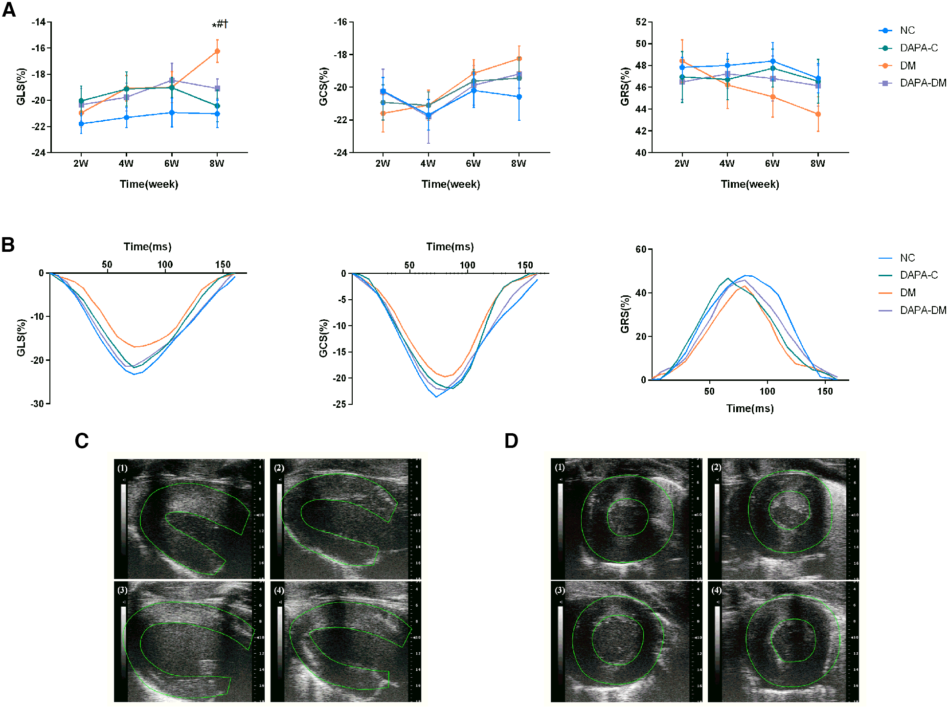
Myocardial strain assessed by 2D-STE. (A) GLS, GRS and GCS scores of the four groups at different time points. (B) Representative strain time curves of the four groups. (C) Representative long-axis strain images of the four groups at 8 weeks. (D) Representative short-axis strain images of the four groups at 8 weeks. (1) Normal control group, (2) DAPA-control group, (3) diabetic group, (4) DAPA-diabetic group. GLS, global peak longitudinal strain; GRS, global peak radial strain; GCS, global peak circumferential strain. NC, normal control group; DAPA-C, DAPA-control group; DM, diabetic group; DAPA-DM, DAPA-diabetic group. *p < 0.05 vs. normal control group; †p < 0.05 vs. DAPA-control group; #p < 0.05 vs. diabetic group at 2 weeks.
The authors apologize for this error and state that this does not change the scientific conclusions of the article in any way. The original article has been updated.
Publisher's note
All claims expressed in this article are solely those of the authors and do not necessarily represent those of their affiliated organizations, or those of the publisher, the editors and the reviewers. Any product that may be evaluated in this article, or claim that may be made by its manufacturer, is not guaranteed or endorsed by the publisher.
Summary
Keywords
early diabetes mellitus, microvascular strain, microvascular perfusion, speckle tracking echocardiography, myocardial contrast echocardiography, dapagliflozin
Citation
Liu J, Wang Y, Zhang J, Li X, Tan L, Huang H, Dai Y, Shang Y and Shen Y (2024) Corrigendum: Dynamic evolution of left ventricular strain and microvascular perfusion assessed by speckle tracking echocardiography and myocardial contrast echocardiography in diabetic rats: effect of dapagliflozin. Front. Cardiovasc. Med. 11:1452088. doi: 10.3389/fcvm.2024.1452088
Received
20 June 2024
Accepted
25 June 2024
Published
09 July 2024
Volume
11 - 2024
Edited and reviewed by
Grigorios Korosoglou, GRN Klinik Weinheim, Germany
Updates
Copyright
© 2024 Liu, Wang, Zhang, Li, Tan, Huang, Dai, Shen and Shang.
This is an open-access article distributed under the terms of the Creative Commons Attribution License (CC BY). The use, distribution or reproduction in other forums is permitted, provided the original author(s) and the copyright owner(s) are credited and that the original publication in this journal is cited, in accordance with accepted academic practice. No use, distribution or reproduction is permitted which does not comply with these terms.
* Correspondence: Yang Dai yutongwushe@163.com Yongning Shang ynshang@aliyun.com Ying Shen rjshenying@qq.com
†These authors have contributed equally to this work
Disclaimer
All claims expressed in this article are solely those of the authors and do not necessarily represent those of their affiliated organizations, or those of the publisher, the editors and the reviewers. Any product that may be evaluated in this article or claim that may be made by its manufacturer is not guaranteed or endorsed by the publisher.