 Bettina Kronsteiner1,2*
 Lydia M. Zopf3Patrick Heimel3,4,5
 Gunpreet Oberoi1Anne M. Kramer2
 Paul Slezak3,4
 Wolfgang J. Weninger6,7
 Bruno K. Podesser2
 Attila Kiss2
 Francesco Moscato1,2,4
- 1Center for Medical Physics and Biomedical Engineering, Medical University of Vienna, Vienna, Vienna, Austria
- 2Ludwig Boltzmann Cluster for Cardiovascular Research, Vienna, Vienna, Austria
- 3AUVA Research Centre, Ludwig Boltzmann Institute for Experimental and Clinical Traumatology, Vienna, Vienna, Austria
- 4Austrian Cluster for Tissue Regeneration, Vienna, Vienna, Austria
- 5Karl Donath Laboratory for Hard Tissue and Biomaterial Research, University Clinic of Dentistry, Medical University of Vienna, Vienna, Vienna, Austria
- 6Department of Anatomy, Center for Anatomy and Cell Biology, Medical University of Vienna, Vienna, Vienna, Austria
- 7Division of Anatomy, Center for Anatomy and Cell Biology, Medical University of Vienna, Vienna, Vienna, Austria
A Corrigendum on
Mapping the functional anatomy and topography of the cardiac autonomic innervation for selective cardiac neuromodulation using MicroCT
by Kronsteiner B, Zopf LM, Heimel P, Oberoi G, Kramer AM, Slezak P, Weninger WJ, Podesser BK, Kiss A and Moscato F (2022). Front. Cell Dev. Biol. 10:968870. doi: 10.3389/fcell.2022.968870
In the published article, there was an error in “Figure 1. Schematic overview of the surgical window in a pig” as published. The dashed line indicating the anatomical “position 4” was accidently moved to the wrong position. The correct position is right above the branching point of the vagal cardiac branch as correctly written in the main text. The corrected figure “Figure 1 Schematic overview of the surgical window in a pig” and its caption “Schematic overview of the surgical window in a pig. The VN can be distinguished from the ST by identification of the NG. In some individuals, the two nerves were observed to merge or split at the cervical level. In rabbits, the VN and the ST were travelling individually next to each other to the cardiac plexus. Five anatomical positions (pos 1-pos 5) were defined from cranial to caudal at which the fascicle number and nerve areas were measured. The superior CB was the branch used in this study (one superior CB per nerve) and harvested right above the level of the subclavian artery (pos.5); the inferior CB (indicated as dashed lines) was not used in this study since its anatomical position in the epicardial fat is surgically less feasible for selective stimulation than the superior CB. Pos. 1-3 label the upper to the mid-cervical level. Pos.4 indicates the cardiac branching point of the superior CB. NG, nodose ganglion; CCA, common carotid artery; VN, Vagus Nerve; ST, Sympathetic trunk; Pos, position; sup. cardiac branch, superior cardiac branch; inf. cardiac branch, inferior cardiac branch.” appear below.
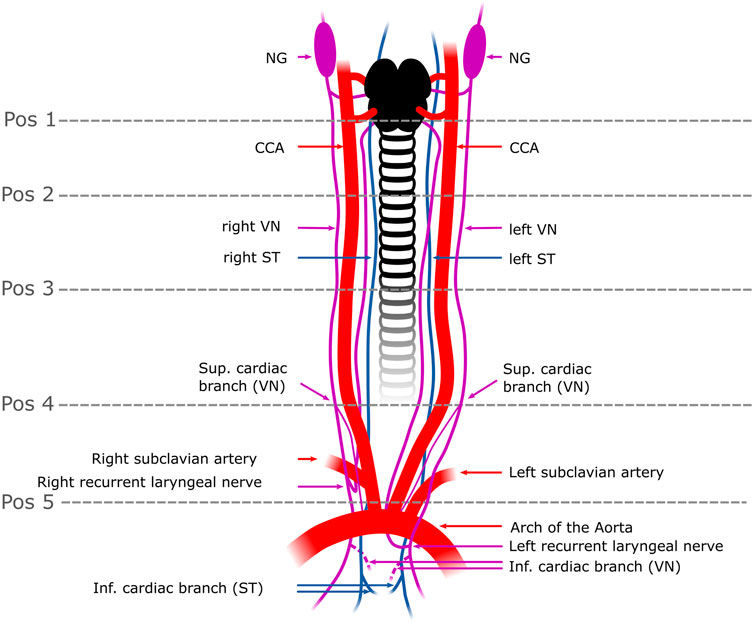
FIGURE 1. Schematic overview of the surgical window in a pig. The VN can be distinguished from the ST by identification of the NG. In some individuals, the two nerves were observed to merge or split at the cervical level. In rabbits, the VN and the ST were travelling individually next to each other to the cardiac plexus. Five anatomical positions (pos 1-pos 5) were defined from cranial to caudal at which the fascicle number and nerve areas were measured. The superior CB was the branch used in this study (one superior CB per nerve) and harvested right above the level of the subclavian artery (pos.5); the inferior CB (indicated as dashed lines) was not used in this study since its anatomical position in the epicardial fat is surgically less feasible for selective stimulation than the superior CB. Pos. 1-3 label the upper to the mid-cervical level. Pos. 4 indicates the cardiac branching point of the superior CB. NG, nodose ganglion; CCA, common carotid artery; VN, Vagus Nerve; ST, Sympathetic trunk; Pos, position; sup. cardiac branch, superior cardiac branch; inf. cardiac branch, inferior cardiac branch.
The authors apologize for this error and state that this does not change the scientific conclusions of the article in any way. The original article has been updated.
Publisher’s note
All claims expressed in this article are solely those of the authors and do not necessarily represent those of their affiliated organizations, or those of the publisher, the editors and the reviewers. Any product that may be evaluated in this article, or claim that may be made by its manufacturer, is not guaranteed or endorsed by the publisher.
Keywords: vagus nerve stimulation, cardiovascular diseases, fascicular anatomy, selective cardiac, neuromodulation, microcomputed tomography, 3D rendering
Citation: Kronsteiner B, Zopf LM, Heimel P, Oberoi G, Kramer AM, Slezak P, Weninger WJ, Podesser BK, Kiss A and Moscato F (2023) Corrigendum: Mapping the functional anatomy and topography of the cardiac autonomic innervation for selective cardiac neuromodulation using MicroCT. Front. Cell Dev. Biol. 11:1193013. doi: 10.3389/fcell.2023.1193013
Received: 24 March 2023; Accepted: 27 March 2023;
Published: 31 March 2023.
Approved by:
Frontiers Editorial Office, Frontiers Media SA, SwitzerlandCopyright © 2023 Kronsteiner, Zopf, Heimel, Oberoi, Kramer, Slezak, Weninger, Podesser, Kiss and Moscato. This is an open-access article distributed under the terms of the Creative Commons Attribution License (CC BY). The use, distribution or reproduction in other forums is permitted, provided the original author(s) and the copyright owner(s) are credited and that the original publication in this journal is cited, in accordance with accepted academic practice. No use, distribution or reproduction is permitted which does not comply with these terms.
*Correspondence: Bettina Kronsteiner, YmV0dGluYS5rcm9uc3RlaW5lckBtZWR1bml3aWVuLmFjLmF0