 Deepa Ponnaiyan
 Roshan R. Rughwani
 Ganesh Shetty
 Jaideep Mahendra
- 1SRM Dental College, Chennai, Tamil Nadu, India
- 2Namma Dentist Dental Clinic, Chennai, India
- 3Dental and Orthodontic Clinic, Bangalore, India
- 4Meenakshi Academy of Higher Education and Research, Chennai, Tamil Nadu, India
Periodontal regeneration involves the composite action of cell, scaffolds and signaling molecules. There are numerous autologous sources of regenerative cells which are present close to the vicinity of the periodontally debilitated site, the primary one being the periodontal ligament stem cell, which is believed to have a key role in regeneration. Various methods can be harnessed to optimize and enhance the regenerative potential of PDLSCs such as the application of LASERs. In the last few years there have been various studies which have evaluated the effect of different types of LASERs on PDLSCs and the present review summarizes the photo-biomodulative activity of LASERs in general and its beneficial role in the stimulation of PDLSC specifically.
Introduction
Periodontitis is a disease primarily characterized by inflammation and an interplay of bacteria and endotoxins which impacts the soft and hard tissues of the periodontium. The disease leads to significant cellular damage and tissue loss which eventually culminates in bone loss (Hajishengallis, 2022; Zenobia and Darveau, 2022; Vitkov et al., 2023). The extent of hard and soft tissue loss determines the treatment strategies; however, mechanical debridement remains the cornerstone of periodontal treatment which turns the inflammatory state of the periodontium to a resolving state (Albeshri and Greenstein, 2022; Laleman et al., 2022). Periodontal treatment not only involves elimination of inflammatory and bacterial component from the diseased tooth supporting tissues, but also comprises of regeneration of deteriorated periodontal structures in amenable cases which serves as the bedrock to principles of tissue engineering that engages application of appropriate cells, growth factors and scaffolds Tavelli et al. (2022), Yi et al. (2022), Sopi et al. (2023).
Dental stem cells owing to their distinct stemness, migration, differentiation and immunological modulation properties, have been perceived as a potential agent for regeneration (Nagata et al., 2022; Sun et al., 2023). Stomatognathic stem cells are placed in different niches and they can be further categorized as dental and periodontal stem cells based on their location within the oral complex (Ponnaiyan et al., 2022; Alarcón-Apablaza et al., 2023). Recently, research shows that periodontal ligament stem cells that are mesenchymal in nature and located within the periodontal ligament, offers substantial periodontal regeneration in comparison to the regenerative outputs offered by other kinds of stem cells (Li et al., 2020; Dubuc et al., 2022; Mendoza et al., 2023).
The regenerative output of PDLSCs is further enhanced by the adjuvant application of LASERs as evidenced by literature (Mylona et al., 2022; L; Wang et al., 2022). Hence, the current review aims to summarize the additional use of LASERs in regenerative and stem cell therapy in periodontics.
Periodontal ligament stem cells (PDLSCs)
PDLSCs are formed from ecto-mesenchymal cells that originate from the neural crest and are isolated from middle third of the root surface after extraction of permanent teeth (T. Wang et al., 2022). Root surface derived PDLSCs (r-PDLSCs) are stem cells isolated from root surface, while alveolar socket derived PDLSCs (a-PDLSCs) are stem cells separated from tissue taken from the bone surface (Rad et al., 2022). In comparison to r-PDLSCs, it has been found that a-PDLSCs maintain a higher proliferative capability as well as strong osteogenic and adipogenic potential (Nagata et al., 2022). PDLSCs can differentiate into peripheral nerves, blood vessels, alveolar bone, cementum and even periodontal ligament (Seo et al., 2004). It is interesting to know that PDLSCs phenotypically express various neural crest, embryonic and antigenic markers which contribute to its diverse multipotent differentiation characteristics (Iwayama et al., 2022). They express standard mesenchymal stem cells markers and are negative for hematopoietic markers (Song et al., 2023). Amongst embryonic markers they highly express Nanog, SRY (sex determining region Y)-box 2, also known as SOX2, SSEA4 (Stage specific embryonic Antigen) and express October 4 (Octamer) and Krüppel-like factor 4 (KLF4) in lower levels (Alves et al., 2023; Takahashi et al., 2023). However, the expression levels of these markers are highly dependent on the environment in which the PDLSC is residing as inflammation affects stem cells and those cells in turn have immune-modulatory effect in an inflammatory milieu (Zhang et al., 2021; Liu et al., 2022).
The equilibrium between the pro-inflammatory response and the stemness of PDLSCs is a key factor in determining whether tissue integrity and homeostasis is maintained or if disease progression occurs (Zhao et al., 2022). It has been observed that long-term stimulation of PDLSCs by P. gingivalis lipo-polysaccharide (LPS) resulted in an increase in cellular cytokine production and LPS also prevents the development of osteoblasts by impairing ALP activity and mineral synthesis in PDLSCs (Chen et al., 2021; Xu et al., 2023).
Conversely PDLSCs also have an immunomodulatory influence on periodontal regeneration (Andrukhov et al., 2019; Liu et al., 2022). PDLSCs also affect the innate immune response by increasing neutrophil proliferation and decreasing their capacity to undergo apoptosis. PDLSCs also promote CD-136, IL-10, and Arginase 1, which enhances the anti-inflammatory M2 macrophage phenotypic polarization in addition to inhibiting T cell proliferation (Shin et al., 2017; Li et al., 2022). These evidences suggest that, to harness the maximum regenerative potential of PDLSCs, it is imperative to maintain the state of controlled inflammation or homeostasis which favors repair which is possible only through the elimination of the pro-inflammatory components, bacteria and endotoxins via mechanical debridement thereby, declaring LASERs to be an adjunct to the mechanical therapy (Jiang et al., 2022; Lu et al., 2023).
LASERs
The LASER revolution in dentistry and the invention of ruby LASER by Theodore Maiman in 1960 has seen significant changes over the past few decades owing to advances and usability of LASERs in a multitude of dental and periodontal applications (Passanezi et al., 2015; Theodoro et al., 2021; Corbella et al., 2023). LASERs are clinically categorized into two types based on their wavelength: 1) a deeply penetrating type where the LASER light penetrates and scatters into the tissue deeply, such as the diode LASERs (810–980 nm) and neodymium-doped yttrium-aluminum-garnet (Nd:YAG) available for clinical application; and 2) a superficially absorbed type such are the erbium-doped yttrium-aluminum-garnet (Er:YAG) (2940 nm), carbon dioxide (CO2) (10,600 nm) and erbium, chromium: yttrium-scandium-gallium-garnet (Er, Cr:YSGG) (2780 nm) LASERs (Ohsugi et al., 2020; Fu and Wo, 2021).
When LASERs energy penetrates the tissue surface, it may be absorbed, dispersed, reflected, or transmitted to the cells in the vicinity and the amount of energy that is absorbed by the cells influences the therapeutic and photo-biomodulative property exhibited by the LASER (Glass, 2021; Lopes et al., 2022). Since the 20th Century, the photobiomodulative property of LASER has been discovered and this property has been harnessed for specific effect of LASERs on tissues and cells for achieving desired treatment outcomes (Arjmand et al., 2021). Photobiomodulation makes use of nonionizing light sources such as LASERs, light-emitting diodes and broad-spectrum light to encourage physiological changes and therapeutic effects which supra-adds to the stimulation of stem cells involved in the regeneration triad (Firoozi et al., 2022). The PBM property of LASER encompasses a wide array of properties such as the photothermal, photodynamic, bio stimulative, photo ablative, photo vaporolytic and photo plasmolytic properties which may alter the cellular dynamics of periodontal tissues when applied at different settings and for varying time periods (Glass, 2021; Parker et al., 2022).
Alternate and more compliant sources of light such as LASER and light-emitting diode (LED) sources emit a wide range of visible and infrared spectrum that are used in photo-biomodulation (PBM), which can be effectively used in the treatment of a number of diseases, wounds, and disorders (Dompe et al., 2020; Bunch, 2023). A well-accepted theory holds that the light energy given to tissues is absorbed by the cell chromophores, encouraging the generation of adenosine triphosphate (ATP) (Glass, 2021). Understanding the processes behind the effects of PBM has been of significant interest. Nevertheless, some researchers have observed favorable effect of ATP generation on oxidative stress, survival, and tissue regeneration (da Silva et al., 2023; Prado et al., 2023).
Effect of LASER on PDLSCs
The effect of LASERs in periodontal regeneration is well established. Initially, various studies were done in the past to identify the effects of LASERs on native PDL cells. Studies have showed that stretched periodontal ligament cells during orthodontic treatment demonstrated marked reduction in the level of pro-inflammatory mediators on the application of diode LASERs (Ozawa et al., 1997; Yamauchi et al., 2018).
With advances in orthodontic research, it is understood that tooth movement was a PDL phenomenon which occurred as a result of coupled resorptive and regenerative activities of the periodontium (Li et al., 2021). With the advent of stem cell isolation techniques, it was later seen that not only fully differentiated PDL cells but also these stem cells residing in the periodontal ligament, took part in regeneration (Costela-Ruiz et al., 2022). As it is known that regeneration involves the Melcher’s triad, the cells and in particular, the stem cells become an indispensable tool to achieve optimal regeneration (Ward, 2022; Yi et al., 2022). To expedite the regenerative potential of these stem cells, various light sources including LASERs and LEDs have been proven to be beneficial in the stimulation of the PDLSCs as mentioned in Table 1. It is interesting to know that the maximal beneficial role of PDLSCs can be achieved only at certain irradiation settings, thereby making the wavelength, irradiation time interval and the energy setting to be the key regulating factors in optimal stimulation of PDLSCs by light sources (Oyebode and Houreld, 2022; da Silva et al., 2023). LASER application causes the release of a photoreceptor called cyctochrome-c oxidase (CCO), which is found in the mitochondrial respiratory chain at unit IV. This raises the potential of the mitochondrial membrane and produces more ATP, which in turn causes cell division (Signaling in Photobiomodulation, 2018; Pan et al., 2022) (Figure 1).
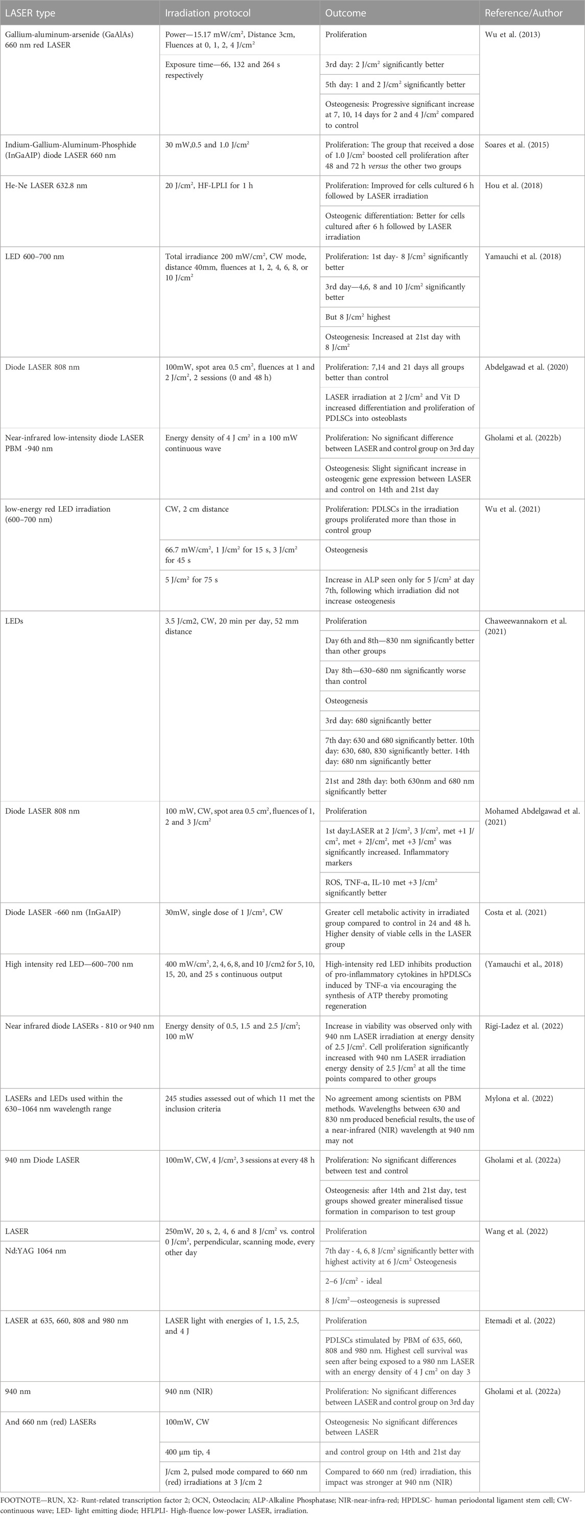
TABLE 1. Effect of different types of LASERs on PDLSCs.

FIGURE 1. Effects of LASER at the cellular level on the stimulation of PDLSCs.
Literature evidence shows that LASER PBM on PDLSCs significantly increases proliferation and osteogenesis. However, majority of studies show wide variation in the wavelength and energy settings for bio stimulation, and this difference in the amount of power and time settings makes the stem cell proliferation vary (Felician et al., 2023). Initially Soares et al. and Wu et al. have shown there is significant increase in proliferation when the power of 660 nm red diode LASER is increased from 1 to 5 J/cm2 and also when duration of exposure is increased. However, it has been noted that increasing the energy setting or duration of exposure doesn’t change the rate of proliferation or osteogenic gene expression (Gholami et al., 2022a; Gholami et al., 2022b). This signifies that energy settings, time of irradiation and follow up with subsequent irradiations makes difference when the optimal energy setting is applied. Still, there is no standardized protocol to stimulate PDSLCs to the maximum.
It was observed that using 600–700 nm LED in the power of 8 J/cm2 had the highest rate of proliferation and osteogenic differentiation of PDLSCs and increasing the duration to 3 weeks also proved to be optimal in maintaining regeneration capability (Yamauchi et al., 2018). This suggests that lower the wavelength of light source higher is the photo bio modulatory property.
Further, Gholami et al. compared 940 nm diode and 660 nm (red LASER) and showed that group receiving 940 nm irradiation showed better cell proliferation and differentiation on day 3 and also 3 weeks of LASER application. Similarly, Chaweewannakorn et al. showed that by using three different wavelengths of LEDs, there is a decline in proliferation on day 8th of irradiated groups and stated that inadequate wavelength can cause damage to cell viability of PDLSCs thereby emphasizing the importance of optimal wavelength. Rigi Ladez et al. stated that using a wavelength of diode LASER of 940 nm instead of 810 nm showed a better proliferation of PDLSCs which is in concurrence of Gholami et al. Wang et al. and Etemadi et al. who observed positive effects of LASER irradiation on PDLSCs on day 21 and day 5 respectively.
LASER PBM is a beneficial technique for tissue regeneration even in inflammatory regions and has a substantial anti-inflammatory impact by lowering pro-inflammatory cytokines (Yamauchi et al., 2018). In a systematic review on the effect of LASERs and LEDs on PDLSC proliferation by Mylona et al., it was observed that the stemness and differentiation abilities of periodontal ligament stem cells can be improved by photo biomodulation. On PBM techniques, such as duration, wavelength and energy settings, there is currently no consensus among experts. The usage of a near-infrared (NIR) wavelength at 940 nm may not have the same positive effects as wavelengths between 630 nm and 810 nm. It was said that the fluence shouldn’t be greater than 8 J/cm2 when utilizing LED therapy devices and that it shouldn’t be greater than 4 J/cm2 while using LASERs.
Conclusion
Photo-biomodulation, a property which stimulates PDLSCs and other stem cells in general, is a property specially owned by cold LASERs. With the above-mentioned evidences, it can be noted that by decreasing the wavelength of the LASER, better PBM can be achieved; however, there are various other factors such the spot size, time and mode of irradiation which decides optimal stimulation of PDLSCs. In the future, it can be seen that post periodontal treatments with Erbium group of LASERs which operate at a higher wavelength may still require a LASER of lower wavelength for bio-modulation, thereby giving rise to a dual LASER therapy. The above-mentioned studies in the table hold a lot of ambiguity as different types of LASERs are used with different wavelengths and power settings as studies are mostly done by individual research scholars often funded by commercial LASER companies. Hence, it is necessary for the governing bodies around the globe to come up with a consensus and a protocol to make the most of the stimulation of PDLSCs.
Author contributions
DP: Writing–original draft, Conceptualization, Data curation, Software. RR: Writing–original draft, Conceptualization, Software. GS: Data curation, Writing–review and editing. JM: Writing–review and editing, Supervision.
Funding
The author(s) declare that no financial support was received for the research, authorship, and/or publication of this article.
Conflict of interest
The authors declare that the research was conducted in the absence of any commercial or financial relationships that could be construed as a potential conflict of interest.
Publisher’s note
All claims expressed in this article are solely those of the authors and do not necessarily represent those of their affiliated organizations, or those of the publisher, the editors and the reviewers. Any product that may be evaluated in this article, or claim that may be made by its manufacturer, is not guaranteed or endorsed by the publisher.
References
Abdelgawad, L. M., Abdelaziz, A. M., Sabry, D., and Abdelgwad, M. (2020). Influence of photobiomodulation and vitamin D on osteoblastic differentiation of human periodontal ligament stem cells and bone-like tissue formation through enzymatic activity and gene expression. Biomol. Concepts 11 (1), 172–181. doi:10.1515/bmc-2020-0016
Alarcón-Apablaza, J., Prieto, R., Rojas, M., and Fuentes, R. (2023). Potential of oral cavity stem cells for bone regeneration: a scoping review. Cells 12 (10), 1392. doi:10.3390/cells12101392
Albeshri, S., and Greenstein, G. (2022). Efficacy of nonsurgical periodontal therapy for treatment of periodontitis: practical application of current knowledge. General Dent. 70 (5), 12–19.
Alves, L., Machado, V., Botelho, J., Mendes, J. J., Cabral, J. M. S., da Silva, C. L., et al. (2023). Enhanced proliferative and osteogenic potential of periodontal ligament stromal cells. Biomedicines 11 (5), 1352. doi:10.3390/biomedicines11051352
Andrukhov, O., Behm, C., Blufstein, A., and Rausch-Fan, X. (2019). Immunomodulatory properties of dental tissue-derived mesenchymal stem cells: implication in disease and tissue regeneration. World J. Stem Cells 11 (9), 604–617. doi:10.4252/wjsc.v11.i9.604
Arjmand, B., Khodadost, M., Jahani Sherafat, S., Rezaei Tavirani, M., Ahmadi, N., Hamzeloo Moghadam, M., et al. (2021). Low-level laser therapy: potential and complications. J. Lasers Med. Sci. 12, e42. doi:10.34172/jlms.2021.42
Bunch, J. (2023). Photobiomodulation (therapeutic lasers): an update and review of current literature. Small Anim. Pract. 53 (4), 783–799. doi:10.1016/j.cvsm.2023.02.010
Chaweewannakorn, C., Santiwong, P., Surarit, R., Sritanaudomchai, H., and Chintavalakorn, R. (2021). The effect of LED photobiomodulation on the proliferation and osteoblastic differentiation of periodontal ligament stem cells: in vitro. J. World Fed. Orthod. 10 (2), 79–85. doi:10.1016/j.ejwf.2021.03.003
Chen, Q., Liu, X., Wang, D., Zheng, J., Chen, L., Xie, Q., et al. (2021). Periodontal inflammation-triggered by periodontal ligament stem cell pyroptosis exacerbates periodontitis. Front. Cell. Dev. Biol. 9, 663037. doi:10.3389/fcell.2021.663037
Corbella, S., Calciolari, E., Donos, N., Alberti, A., Ercal, P., and Francetti, L. (2023). Laser treatments as an adjunct to non-surgical periodontal therapy in subjects with periodontitis and type 2 diabetes mellitus: a systematic review and meta-analysis. Clin. Oral Investig. 27 (4), 1311–1327. doi:10.1007/s00784-023-04873-y
Costa, E. H. P., Lira, J., Sabino, V., Pinheiro, J., Miguel, M., Silva, T., et al. (2021). Photobiomodulation increases the viability and proliferation of human periodontal ligament stem cells cultured on the surface of polylatic acid films. Cytotherapy 23 (4), 30. doi:10.1016/j.jcyt.2021.02.087
Costela-Ruiz, V. J., Melguizo-Rodríguez, L., Bellotti, C., Illescas-Montes, R., Stanco, D., Arciola, C. R., et al. (2022). Different sources of mesenchymal stem cells for tissue regeneration: A guide to identifying the most favorable one in orthopedics and dentistry applications. Int. J. Mol. Sci. 23 (11), 6356. doi:10.3390/ijms23116356
da Silva, T. G., Ribeiro, R. S., Mencalha, A. L., and de Souza Fonseca, A. (2023). Photobiomodulation at molecular, cellular, and systemic levels. Lasers Med. Sci. 38 (1), 136. doi:10.1007/s10103-023-03801-6
Dompe, C., Moncrieff, L., Matys, J., Grzech-Leśniak, K., Kocherova, I., Bryja, A., et al. (2020). Photobiomodulation-underlying mechanism and clinical applications. J. Clin. Med. 9 (6), 1724. doi:10.3390/jcm9061724
Dubuc, A., Planat-Bénard, V., Marty, M., Monsarrat, P., and Kémoun, P. (2022). Periodontal cell therapy: a systematic review and meta-analysis. Adv. Exp. Med. Biol. 1373, 377–397. doi:10.1007/978-3-030-96881-6_20
Etemadi, A., Faghih, A., and Chiniforush, N. (2022). Effects of photobiomodulation therapy with various laser wavelengths on proliferation of human periodontal ligament mesenchymal stem cells. Photochem. Photobiol. 98 (5), 1182–1189. doi:10.1111/php.13588
Felician, M. C., Belotto, R., Tardivo, J. P., Baptista, M. S., and Martins, W. K. (2023). Photobiomodulation: Cellular, molecular, and clinical aspects. J. Photochem. Photobiol., 100197. doi:10.1016/j.jpap.2023.100197
Firoozi, P., Amiri, M. A., Soghli, N., Farshidfar, N., Hakimiha, N., and Fekrazad, R. (2022). The role of photobiomodulation on dental-derived mesenchymal stem cells in regenerative dentistry: a comprehensive systematic review. Curr. Stem Cell Res. Ther. 19 (4), 559–586(28). doi:10.2174/1574888X17666220810141411
Fu, W., and Wo, C. (2021). The use of laser in dentistry: a narrative review. J. Biol. Regul. Homeost. Agents 35 (1), 11–18.
Gholami, L., Hendi, S. S., Saidijam, M., Mahmoudi, R., Tarzemany, R., Arkian, A., et al. (2022a). Near-infrared 940-nm diode laser photobiomodulation of inflamed periodontal ligament stem cells. Lasers Med. Sci. 37 (1), 449–459. doi:10.1007/s10103-021-03282-5
Gholami, L., Khorsandi, K., and Fekrazad, R. (2022b). Effect of red and near-infrared irradiation on periodontal ligament stem cells: ROS generation and cell cycle analysis. J. Biomol. Struct. Dyn. 41, 10051–10058. doi:10.1080/07391102.2022.2152869
Glass, G. E. (2021). Photobiomodulation: a review of the molecular evidence for low level light therapy. J. Plastic, Reconstr. Aesthetic Surg. JPRAS 74 (5), 1050–1060. doi:10.1016/j.bjps.2020.12.059
Hajishengallis, G. (2022). Interconnection of periodontal disease and comorbidities: evidence, mechanisms, and implications. Periodontol. 2000 89 (1), 9–18. doi:10.1111/prd.12430
Hou, T., Li, S., Zhang, G., and Li, Y. (2018). High-fluence low-power laser irradiation promotes odontogenesis and inflammation resolution in periodontitis by enhancing stem cell proliferation and differentiation. Int. J. Mol. Med. 42 (4), 2107–2119. doi:10.3892/ijmm.2018.3804
Iwayama, T., Sakashita, H., Takedachi, M., and Murakami, S. (2022). Periodontal tissue stem cells and mesenchymal stem cells in the periodontal ligament. Jpn. Dent. Sci. Rev. 58, 172–178. doi:10.1016/j.jdsr.2022.04.001
Jiang, Y., Feng, J., Du, J., Fu, J., Liu, Y., Guo, L., et al. (2022). Clinical and biochemical effect of laser as an adjunct to non-surgical treatment of chronic periodontitis. Oral. Dis. 28 (4), 1042–1057. doi:10.1111/odi.13847
Laleman, I., Seidel, L., Gagnot, G., Reners, M., and Lambert, F. (2022). Instrumentation during the second stage of periodontal therapy: a European survey. Clin. Oral. Investig. 26 (7), 4781–4787. doi:10.1007/s00784-022-04442-9
Li, Q., Yang, G., Li, J., Ding, M., Zhou, N., Dong, H., et al. (2020). Stem cell therapies for periodontal tissue regeneration: a network meta-analysis of preclinical studies. Stem Cell Res. Ther. 11 (1), 427. doi:10.1186/s13287-020-01938-7
Li, W., Huang, X., Yu, W., Xu, Y., Huang, R., Park, J., et al. (2022). Activation of functional somatic stem cells promotes endogenous tissue regeneration. J. Dent. Res. 101 (7), 802–811. doi:10.1177/00220345211070222
Li, Y., Zhan, Q., Bao, M., and Yi, J. (2021). Biomechanical and biological responses of periodontium in orthodontic tooth movement: up-date in a new decade. Int. J. Oral Sci. 13 (1), 20. doi:10.1038/s41368-021-00125-5
Liu, J., Wang, H., Zhang, L., Li, X., Ding, X., Ding, G., et al. (2022). Periodontal ligament stem cells promote polarization of M2 macrophages. J. Leukoc. Biol. 111 (6), 1185–1197. doi:10.1002/JLB.1MA1220-853RR
Lopes, C. D. C. A., Limirio, J. P. J. O., Zanatta, L. S. A., Simamoto, V. R. N., Dechichi, P., and Limirio, A. P. H. J. O. (2022). Effectiveness of photobiomodulation therapy on human bone healing in dentistry: a systematic review. Photobiomodulation, Photomed. Laser Surg. 40 (7), 440–453. doi:10.1089/photob.2021.0092
Lu, J. W., Huang, S. H., Lei, X. X., Deng, L., and Luo, L. J. (2023). Clinical outcomes of diode laser as an adjunct to nonsurgical periodontal therapy for residual periodontal pockets in mandibular second molars-a randomized controlled clinical trial. Clin. Oral. Investig. 27 (8), 4493–4501. doi:10.1007/s00784-023-05071-6
Mendoza, A. H., Balzarini, D., Alves, T., Holzhausen, M., and Rovai, E. S. (2023). Potential of mesenchymal stem cell sheets on periodontal regeneration: a systematic review of pre-clinical studies. Curr. Stem Cell Res. Ther. 18 (7), 958–978. doi:10.2174/1574888X17666220706092520
Mohamed Abdelgawad, L., Abd El-Hamed, M. M., Sabry, D., and Abdelgwad, M. (2021). Efficacy of photobiomodulation and metformin on diabetic cell line of human periodontal ligament stem cells through keap1/nrf2/Ho-1 pathway. Rep. Biochem. Mol. Biol. 10 (1), 30–40. doi:10.52547/rbmb.10.1.30
Mylona, V., Anagnostaki, E., Chiniforush, N., Barikani, H., Lynch, E., and Grootveld, M. (2022). Photobiomodulation effects on periodontal ligament stem cells: a systematic review of in-vitro studies. Curr. Stem Cell Res. Ther. 19 (4), 544–558(15). doi:10.2174/1574888X17666220527090321
Nagata, M., English, J. D., Ono, N., and Ono, W. (2022). Diverse stem cells for periodontal tissue formation and regeneration. Genes. (New York, N. Y. 2000) 60 (8–9), e23495. doi:10.1002/dvg.23495
Ohsugi, Y., Niimi, H., Shimohira, T., Hatasa, M., Katagiri, S., Aoki, A., et al. (2020). In vitro cytological responses against laser photobiomodulation for periodontal regeneration. Int. J. Mol. Sci. 21 (23), 9002. doi:10.3390/ijms21239002
Oyebode, O. A., and Houreld, N. N. (2022). Photobiomodulation at 830 nm stimulates migration, survival and proliferation of fibroblast cells. Diabetes. Metab. Syndr. Obes. 15, 2885–2900. doi:10.2147/DMSO.S374649
Ozawa, Y., Shimizu, N., and Abiko, Y. (1997). Low-energy diode laser irradiation reduced plasminogen activator activity in human periodontal ligament cells. Lasers Surg. Med. 21 (5), 456–463. doi:10.1002/(sici)1096-9101(1997)21:5<456::aid-lsm7>3.0.co;2-p
Pan, L. C., Hang, N. L., Colley, M. M. S., Chang, J., Hsiao, Y. C., Lu, L. S., et al. (2022). Single cell effects of photobiomodulation on mitochondrial membrane potential and reactive oxygen species production in human adipose mesenchymal stem cells. Cells. 11 (6), 972. doi:10.3390/cells11060972
Parker, S., Cronshaw, M., and Grootveld, M. (2022). Photobiomodulation delivery parameters in dentistry: an evidence-based approach. Photobiomodulation, Photomed. Laser Surg. 40 (1), 42–50. doi:10.1089/photob.2021.0116
Passanezi, E., Damante, C. A., de Rezende, M. L., and Greghi, S. L. (2015). Lasers in periodontal therapy. Periodontol 2000. 67 (1), 268–291. doi:10.1111/prd.12067
Ponnaiyan, D., Rughwani, R. R., Victor, D. J., and Shetty, G. (2022). Stem cells in the periodontium-anatomically related yet physiologically diverse. Eur. J. Dent. [Epub ahead of print]. doi:10.1055/s-0042-1759487
Prado, T. P., Zanchetta, F. C., Barbieri, B., Aparecido, C., Melo Lima, M. H., and Araujo, E. P. (2023). Photobiomodulation with blue light on wound healing: a scoping review. Life 13 (2), 575. doi:10.3390/life13020575
Rad, M. R., Atarbashi-Moghadam, F., Khodayari, P., and Sijanivandi, S. (2022). Periodontal ligament stem cell isolation protocol: a systematic review. Curr. Stem Cell Res. Ther. 17 (6), 537–563. doi:10.2174/1574888X17666220128114825
Rigi-Ladez, M. A., Hendi, S. S., Mirzaei, A., Gholami, L., and Fekrazad, R. (2022). Near infrared laser photobiomodulation of periodontal ligament stem cells. Chin. J. Dent. Res. 25 (1), 57–65. doi:10.3290/j.cjdr.b2752657
Seo, B. M., Miura, M., Gronthos, S., Bartold, P. M., Batouli, S., Brahim, J., et al. (2004). Investigation of multipotent postnatal stem cells from human periodontal ligament. Lancet 364 (9429), 149–155. doi:10.1016/S0140-6736(04)16627-0
Shin, C., Kim, M., Han, J. A., Choi, B., Hwang, D., Do, Y., et al. (2017). Human periodontal ligament stem cells suppress T-cell proliferation via down-regulation of non-classical major histocompatibility complex-like glycoprotein CD1b on dendritic cells. J. Periodontal Res. 52 (1), 135–146. doi:10.1111/jre.12378
Signaling in Photobiomodulation (2018). Photochem. photobiol. 94 (2), 199–212. doi:10.1111/php.12864
Soares, D. M., Ginani, F., Henriques, Á. G., and Barboza, C. A. G. (2015). Effects of laser therapy on the proliferation of human periodontal ligament stem cells. Lasers Med. Sci. 30 (3), 1171–1174. doi:10.1007/s10103-013-1436-9
Song, W.-P., Jin, L. Y., Zhu, M. D., Wang, H., and Xia, D. S. (2023). Clinical trials using dental stem cells: 2022 update. World J. Stem Cells 15 (3), 31–51. doi:10.4252/wjsc.v15.i3.31
Sopi, M., Koçani, F., Bardhoshi, M., and Meqa, K. (2023). The effect of periodontal therapy on the level of MMP-8 in patients with chronic periodontitis. Eur. J. Dent. 17 (1), 70–75. doi:10.1055/s-0041-1742132
Sun, L., Du, X., Kuang, H., Sun, H., Luo, W., and Yang, C. (2023). Stem cell-based therapy in periodontal regeneration: a systematic review and meta-analysis of clinical studies. BMC Oral Health 23 (1), 492. doi:10.1186/s12903-023-03186-6
Takahashi, Y., Yasuhara, R., Tanaka, J., Nakano, H., Maki, K., and Mishima, K. (2023). Transcriptome profiles associated with human periodontal ligament differentiation. J. Oral Biosci. 65 (1), 40–46. doi:10.1016/j.job.2023.01.005
Tavelli, L., Chen, C. Y. J., Barootchi, S., and Kim, D. M. (2022). Efficacy of biologics for the treatment of periodontal infrabony defects: an American Academy of Periodontology best evidence systematic review and network meta-analysis. J. Periodontology 93 (12), 1803–1826. doi:10.1002/JPER.22-0120
Theodoro, L. H., Marcantonio, R. A. C., Wainwright, M., and Garcia, V. G. (2021). LASER in periodontal treatment: is it an effective treatment or science fiction? Braz. Oral Res. 35 (2), e099. doi:10.1590/1807-3107bor-2021.vol35.0099
Vitkov, L., Singh, J., Schauer, C., Minnich, B., Krunić, J., Oberthaler, H., et al. (2023). Breaking the gingival barrier in periodontitis. Int. J. Mol. Sci. 24 (5), 4544. doi:10.3390/ijms24054544
Wang, L., Liu, C., and Wu, F. (2022). Low-level laser irradiation enhances the proliferation and osteogenic differentiation of PDLSCs via BMP signaling. Lasers Med. Sci. 37 (2), 941–948. doi:10.1007/s10103-021-03338-6
Ward, E. (2022). A review of tissue engineering for periodontal tissue regeneration. J. Veterinary Dent. 39 (1), 49–62. doi:10.1177/08987564211065137
Wu, J.-Y., Chen, C. H., Yeh, L. Y., Yeh, M. L., Ting, C. C., and Wang, Y. H. (2013). Low-power laser irradiation promotes the proliferation and osteogenic differentiation of human periodontal ligament cells via cyclic adenosine monophosphate. Int. J. Oral Sci. 5 (2), 85–91. doi:10.1038/ijos.2013.38
Wu, Y., Zhu, T., Yang, Y., Gao, H., Shu, C., Chen, Q., et al. (2021). Irradiation with red light-emitting diode enhances proliferation and osteogenic differentiation of periodontal ligament stem cells. Lasers Med. Sci. 36 (7), 1535–1543. doi:10.1007/s10103-021-03278-1
Xu, J., Yang, F., Luo, S., Gao, Y., Huang, D., and Zhang, L. (2023). The role of SDF-1α-CXCR4/CXCR7 in migration of human periodontal ligament stem cells. Int. J. Stem Cells 16 (2), 180–190. doi:10.15283/ijsc22053
Yamauchi, N., Taguchi, Y., Kato, H., and Umeda, M. (2018). High-power, red-light-emitting diode irradiation enhances proliferation, osteogenic differentiation, and mineralization of human periodontal ligament stem cells via ERK signaling pathway. J. Periodontology 89 (3), 351–360. doi:10.1002/JPER.17-0365
Yi, Y., Liu, Y., Men, Y., Wang, J., and Zhao, H. (2022). Advances in periodontal stem cells and the regulating niche: From in vitro to in vivo. Genesis. 60 (8–9), e23494. doi:10.1002/dvg.23494
Zenobia, C., and Darveau, R. P. (2022). Does oral endotoxin contribute to systemic inflammation? Front. Oral health 3, 911420. doi:10.3389/froh.2022.911420
Zhang, Z., Deng, M., Hao, M., and Tang, J. (2021). Periodontal ligament stem cells in the periodontitis niche: inseparable interactions and mechanisms. J. Leukoc. Biol. 110 (3), 565–576. doi:10.1002/JLB.4MR0421-750R
Keywords: PDLSCs (periodontal ligament stem cells), regeneration, periodontal regeneration, stem cell, dental stem cell, photobiomodulation, laser
Citation: Ponnaiyan D, Rughwani RR, Shetty G and Mahendra J (2024) The effect of adjunctive LASER application on periodontal ligament stem cells. Front. Cell Dev. Biol. 11:1341628. doi: 10.3389/fcell.2023.1341628
Received: 20 November 2023; Accepted: 18 December 2023;
Published: 12 January 2024.
Edited by:
Marco Tatullo, University of Bari Medical School, ItalyReviewed by:
Simona Delle Monache, University of L’Aquila, ItalyCopyright © 2024 Ponnaiyan, Rughwani, Shetty and Mahendra. This is an open-access article distributed under the terms of the Creative Commons Attribution License (CC BY). The use, distribution or reproduction in other forums is permitted, provided the original author(s) and the copyright owner(s) are credited and that the original publication in this journal is cited, in accordance with accepted academic practice. No use, distribution or reproduction is permitted which does not comply with these terms.
*Correspondence: Deepa Ponnaiyan, ZGVlcGFfcG9ubmFpeWFuQHlhaG9vLmNvLmlu; Roshan R. Rughwani, cm9zaGFucnVnaHdhbmlAZ21haWwuY29t