 Hayder M. Al-kuraishy1
 Ghassan M. Sulaiman2*
 Hamdoon A. Mohammed3
 Mohammed H. Abu-Alghayth4
 Salim Albukhaty5
 Majid S. Jabir2
 Ali K. Albuhadily1Ali I. Al-Gareeb6
 Daniel J. Klionsky7*Mosleh M. Abomughaid4
- 1Department of Clinical Pharmacology and Medicine, College of Medicine, Mustansiriyah University, Baghdad, Iraq
- 2Division of Biotechnology, Department of Applied Sciences, University of Technology, Baghdad, Iraq
- 3Department of Medicinal Chemistry and Pharmacognosy, College of Pharmacy, Qassim University, Qassim, Saudi Arabia
- 4Department of Medical Laboratory Sciences, College of Applied Medical Sciences, University of Bisha, Bisha, Saudi Arabia
- 5Department of Laboratory Techniques, Al-Manara College for Medical Sciences, Maysan, Iraq
- 6Department of Clinical Pharmacology and Medicine, Jabir Ibn Hayyan Medical University, Najaf, Iraq
- 7Life Sciences Institute, University of Michigan, Ann Arbor, MI, United States
Graves disease (GD), an autoimmune disease affects the thyroid gland, results in hyperthyroidisms and goiter. The main cause of GD is not clearly defined; however, stimulating autoantibodies for thyroid stimulating hormone receptor (TSHR) known as thyroid-stimulating immunoglobulins (TSIs) are the primary proposed mechanism. The TSI activation of TSHRs of thyroid gland results in excessive release of thyroid hormones with the subsequent development of hyperthyroidism and goiter. The cellular process of macroautophagy/autophagy is implicated in the pathogenesis of GD and other thyroid diseases. Autophagy plays a critical role in many thyroid diseases and in different stages of the same disease through modulation of immunity and the inflammatory response. In addition, autophagy is also implicated in the pathogenesis of thyroid-associated ophthalmopathy (TAO). However, the exact role of autophagy in GD is not well explained. Therefore, this review discusses how autophagy is intricately involved in the pathogenesis of GD regarding its protective and harmful effects.
Introduction
Graves disease (GD) is an autoimmune disease that affects the thyroid gland resulting in hyperthyroidisms and goiter (enlargement of the thyroid gland); therefore, GD is also known as a toxic diffuse goiter (Davies et al., 2020). The name of GD is derived from the Irish doctor Robert James Graves who described a case of GD in 1835. Subsequently, in 1840, the German physician Karl Adolph von Basedow independently described the same features of GD (Weetman, 2003), thus leading to the alternate name of Basedow disease (Leporati et al., 2015) among others. The basic clinical feature of GD includes the symptoms of hyperthyroidism such as weight loss, sweating, tachycardia, and diarrhea (Leporati et al., 2015). Approximately 25%–30% of patients with GD have extra-thyroidal manifestations such as exophthalmos (eye bulging) or Graves opthalmopathy; accordingly, GD is also known as exophthalmic goiter (Cao et al., 2022). The pathognomonic features of GD are hyperthyroidism, goiter, exophthalmos, and pretibial myxedema that not present in other types of hyperthyroidism (Manni et al., 2020). The thyroid-associated opthalmopathy (TAO) develops in GD due to stimulation of thyroid stimulating hormone receptor (TSHR) of fibroblasts and hypertrophy of muscles around the eyes (Neag and Smith, 2022).
The incidence of GD is about 7.5 times more common in women than men. It is more frequent in the age of 40–60 years, although it can happen at any age (Hussain et al., 2017). GD is regarded as the most common cause of hyperthyroidism in the United States (Bartalena, 2013). In addition, GD is often associated with other autoimmune diseases such as rheumatoid arthritis and type 1 diabetes suggesting the immune disorders of GD (Ferrari et al., 2019; Al-Kuraishy et al., 2024b). Of note, genetic causes are also involved in the pathogenesis of GD (Brix et al., 2001). For example, HLA-DRB3/DR3 (major histocompatibility complex, class II, DR beta 3) increases the susceptibility for the induction of GD (Inaba et al., 2016). As well, single gene defects are also linked with the induction of GD. For example, PTPN22 (protein tyrosine phosphatase non-receptor type 22) and CTLA4 (cytotoxic T-lymphocyte associated protein 4) gene mutations are implicated in the pathogenesis of GD (Pujol-Borrell et al., 2015). Moreover, the autoimmunity of GD is triggered by viral and bacterial infections due to antigenic mimicry. For example, Yersinia enterocolitica has a structural similarity with human TSH receptor and infection by this organism results in the induction of GD (Hargreaves et al., 2013). Likewise; Epstein-Barr virus is considered as a potential trigger of GD (Pyzik et al., 2019).
Although the main cause of GD is not clearly defined, the primary proposed mechanism involves autoantibodies that activate TSHRs; hence, these autoantibodies are known as TSIs (thyroid-stimulating immunoglobulins) (Mathew et al., 2021). TSIs activate TSHRs of the thyroid gland resulting in excessive release of thyroid hormones with subsequent development of hyperthyroidism and goiter (Mathew et al., 2021).
The underlying mechanisms for the development of autoimmunity in GD are related to the autoactivation of T and B cells with subsequent generation of autoantibodies against TSHRs (Liu et al., 2018). Of note, both cellular and humoral immunity are intricately involved in the pathogenesis of GD. Type 1 T helper (Th1) and Th2 cells are highly involved in the induction of abnormal immune response and the pathogenesis of GD (Antonelli et al., 2020). Th1 through activation of cytotoxic lymphocytes and macrophages affect the proliferation of thyroid follicular cells (Pierman et al., 2021). However, Th2 triggers the production and activation of B lymphocytes and plasma cells resulting in the generation of TSIs against TSHRs (He et al., 2020). In GD, the stimulatory activity of TSI is mainly present in the IgG1 subclass, which is chiefly activated by Th1 cells (Li et al., 2021d). As well, Th1 promotes the generation of TSIs from B lymphocyte via an IL10-dependent pathway (Pierman et al., 2021). Moreover, exaggerated Th17 also activates abnormal TSI in GD (Torimoto et al., 2022). However, regulatory T/Treg cells, which downregulate the abnormal immune response are highly reduced leading to the induction of an abnormal immune response in GD (Fang et al., 2021). In addition, in GD the B lymphocytes are autoactivated due to the downregulation of regulatory B/Breg lymphocytes, also leading to an abnormal immune response and activating the release of TSIs (Wang et al., 2021). Therefore, an abnormal immune response and the development of autoimmunity are involved in the pathogenesis of GD (Figure 1).
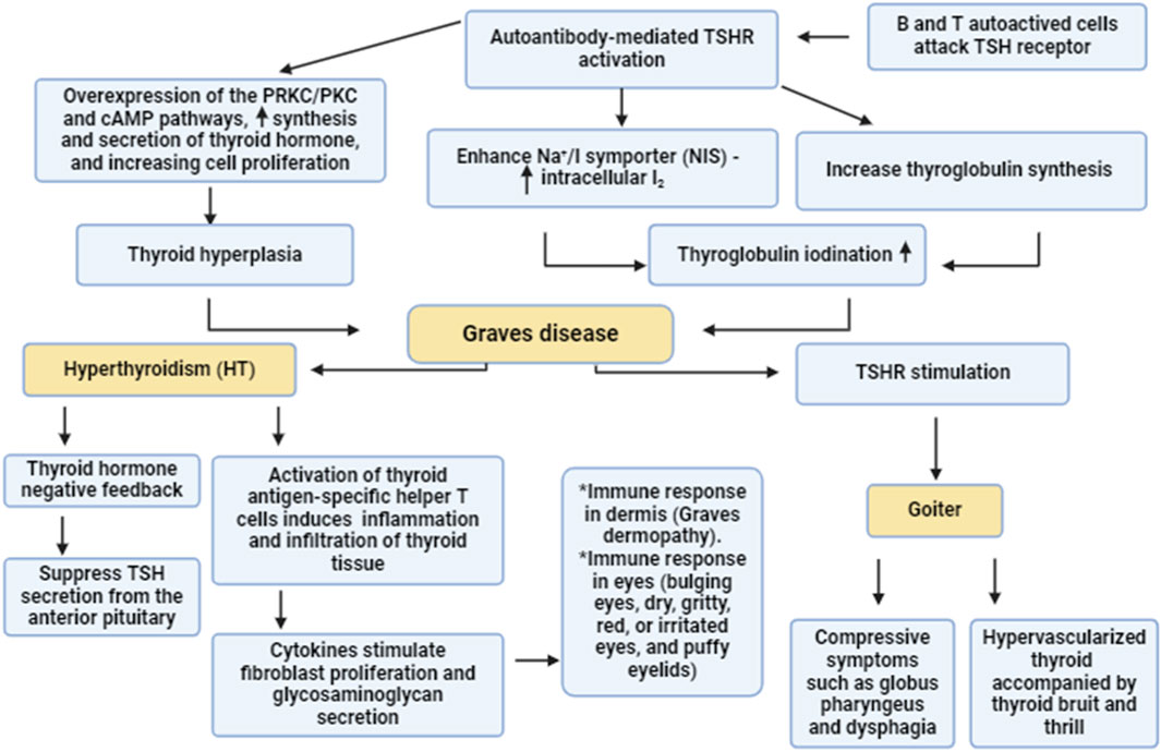
Figure 1. Pathogenesis of GD. Autoreactive B and T cells induce the formation of autoantibodies which activate thyroid stimulating hormone receptors (TSHR). Activated TSHR activates the synthesis and release of thyroid hormones via PKC/cAMP which promote thyroid hyperplasia. In addition, activated TSHR stimulates thyroglobuline synthesis and enhances intracellular accumulation of I2 through activation of Na+/I symporter. These changes trigger the development of GD which causes goiter and hyperthyroidism.
Different studies highlight a potential crosstalk between autophagy and the immune response, and the former plays a critical role in autoimmunity (Wu and Adamopoulos, 2017). Autophagy is intricately involved in the expression of intracellular genes, the initial immune response, and cytokine release (Bhattacharya and Eissa, 2013). Autophagy inhibition ameliorates many autoimmune diseases such systemic lupus erythematosus/SLE and rheumatoid arthritis. Conversely, autophagy inhibition exacerbates Crohn disease and psoriasis (Bhattacharya and Eissa, 2013; Wu and Adamopoulos, 2017). It has been reported that autophagy is implicated in the pathogenesis of GD and other thyroid diseases through the modulation of immunity and the inflammatory response (Duan et al., 2019; Chen et al., 2023). In addition, autophagy is also implicated in the pathogenesis of TAO (Duan et al., 2019; Chen et al., 2023). However, the exact role of autophagy in GD is not well explained. Therefore, this review discusses how autophagy plays an integral role in the pathogenesis of GD regarding its protective and harmful effects.
Autophagy and molecular signaling
Autophagy is an evolutionarily conserved cellular process that promotes the survival of eukaryotic cells in response to different exogenous and endogenous stimuli such as starvation (Ali et al., 2023; Marzoog et al., 2023; Al-Kuraishy et al., 2024c; Sulaiman et al., 2024). Autophagy is involved in the elimination of damaged or superfluous organelles and proteins, recycling the breakdown products that result from their degradation for cellular nutrition (Li et al., 2021b; Al-Kuraishy et al., 2024a). Autophagy plays critical roles in different biological functions in normal and disease states. This process is crucial in the regulation of inflammation, immunity, stress adaptation, cancer, aging and neurodegenerative diseases (Luo et al., 2020). A key feature of autophagy is that degradation occurs through the lysosomal pathway (Cao et al., 2021). There are various types of autophagy that differ in terms of the mechanism as well as substrates (Galluzzi et al., 2017). The predominant form of autophagy, macroautophagy (hereafter autophagy) is initiated by the formation of a phagophore which is sequesters cytoplasmic components and then matures into a double-membrane autophagosome (Broggi et al., 2020). The autophagosome fuses with an endosome and/or a lysosome to form an autolysosome where the contents are degraded; the resulting macromolecules are then released back into the cytosol (Broggi et al., 2020) (Figure 2).
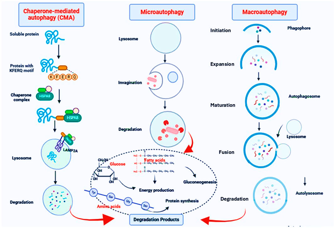
Figure 2. The autophagy pathway. Macroautophagy is initiated by the formation of a phagophore which is sequesters cytoplasmic components and then matures into a double-membrane autophagosome. The autophagosome fuses with an endosome and/or a lysosome to form an autolysosome where the contents are degraded; the resulting macromolecules are then released back into the cytosol. In microautophagy, the limiting membrane protrudes or invaginates at the surface of the lysosome/vacuole by the lateral segregation of lipids and local exclusion of large transmembrane proteins, which is conducted at the small smooth areas with a very low content of transmembrane proteins. In CMA, proteins, the only cargo degraded by this pathway, cross the lysosomal membrane one by one. Not all proteins can undergo degradation via CMA. To be CMA substrates, proteins must contain a specific targeting motif in their amino acid sequence. This motif binds to a cytosolic chaperone (HSPA8), which brings the unfolded substrate protein to the lysosomal surface for internalization and rapid intralysosomal degradation.
Autophagy is coordinated by ATG (autophagy related) proteins. Phagophore formation is initiated by ULK1 (unc-51 like autophagy activating kinase 1)/ULK2 which forms a complex with ATG13, RB1CC1 and ATG101 (Li et al., 2020). This step also requires the class III phosphatidylinositol 3-kinase complex that includes PIK3C3/VPS34, PIK3R4/VPS15, BECN1, ATG14 and NRBF2 (Pavlinov et al., 2020). In addition, BECN1 interacts with other binding proteins such as UVRAG (UV radiation resistance associated), AMBRA1 (autophagy and beclin 1 regulator 1) and SH3GLB1/BIF-1 (SH3 domain containing GRB2 like, endophilin B1) which form various class III complexes (Chang and Zou, 2020). Two ubiquitin-like conjugation systems involving the ATG12–ATG5-ATG16L1 complex, and MAP1LC3/LC3 (microtubule associated protein 1 light chain 3) along with the ATG2-ATG9 lipid transferase-scramblase complex are essential for the expansion of the phagophore (Iriondo et al., 2023); the conversion of LC3-I to the LC3-II lipidated form reflects autophagosome formation (Ueno and Komatsu, 2020). Fusion of autophagosomes with lysosomes requires different components such as UVRAG, VPS proteins and RAB7 (Lőrincz and Juhász, 2020). Autophagy is highly regulated to maintain optimal levels-either too little or too much can be deleterious to cellular physiology. For example, stress and nutrient depletion activate adenosine monophosphate-activated protein kinase/AMPK and inhibit MTOR (mechanistic target of rapamycin kinase) resulting in the activation of the ULK1 and phosphatidylinositol 3-kinase complexes (He, 2022) (Figure 3).
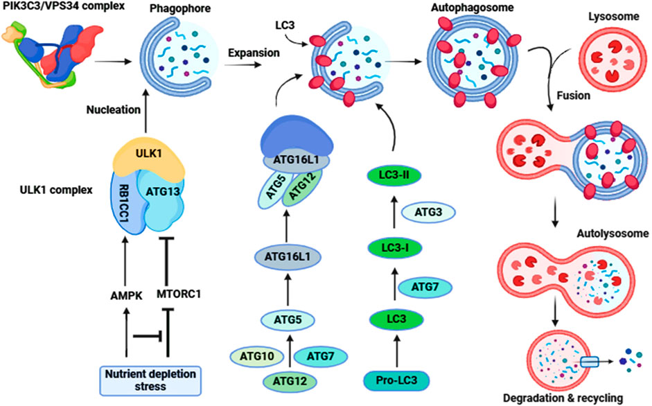
Figure 3. Molecular signaling of autophagy. The ULK1-ATG13-RB1CC1-ATG101 complex is activated to induce autophagy (Initiation). Following the induction of autophagy, an omegasome is formed from the ER by association with ZFYVE1/DFCP1. Next is the formation of a phagophore, which expands to engulf cytoplasmic components, including mitochondria and endoplasmic reticulum (Expansion). Association with the ATG12–ATG5-ATG16L1 complex forms the phagophore. LC3-II localizes to the phagophore membrane at the latter step of autophagosome formation, while the ATG12–ATG5-ATG16L1 complex dissociates from it. Finally, the phagophore membrane is enclosed to form an autophagosome (Maturation). After autophagosome formation, the lysosome fuses with the autophagosome (autophagosome-lysosome Fusion) to form an autolysosome. Intra-autophagosomal contents are degraded by lysosomal hydrolases (Degradation). After formation of the autolysosome, the lysosomal hydrolases degrade the intra-autophagosomal contents, including LC3-II.
Role of autophagy in GD
Of note, thyrocyte basal autophagy is essential for the survival of thyroid follicular cells (Kurashige et al., 2019). Kurashige et al. (2019), found that atg5 gene knockout mice experience abnormal morphology and function of thyrocytes with progressive apoptosis. As well, an imbalance of autophagy and apoptosis triggers the development of thyroid damage in rats (Li et al., 2021c). A reduction of autophagy and augmentation of apoptosis are observed in patients with hyperthyroidism due to excess iodine intake (Xu et al., 2016), and induction of the development and progression of autoimmune thyroid disease (AITD) in animal models is mediated by inhibition of autophagy (Duan et al., 2019). Di-isononyl phthalate/DINP-induced AITD occurs through inhibition of normal autophagy via an MTOR-AKT-dependent pathway. Supporting this finding, inhibition of the MTOR pathway by rapamycin attenuates the development of AITD (Duan et al., 2019). GD is regarded as one of most common AITDs and its pathogenesis is highly affected by the MTOR-AKT pathway (Li et al., 2015). In a GD mouse model, the MTOR-AKT pathway is exaggerated and correlates with signs of hyperthyroidism. Treatment in this model with the antithyroid medication methimazole reduces activity of the MTOR-AKT pathway and mitigates GD pathology (Li et al., 2015; Al-Kuraishy et al., 2021; AlAnazi et al., 2023). Zhang et al. (2023) observed that rapamycin mitigates TAO in patients with GD by inhibiting cytotoxic T lymphocytes, which have an upregulated MTOR pathway. Importantly, isolated IgG from GD patients induces the chemoattractant activity of cytotoxic T lymphocytes via the MTOR pathway (Zhang et al., 2023).
Of note, stimulatory TSIs induce survival and proliferation of thyrocytes, whereas blocking TSIs leads to the inactivation of thyrocytes. Moreover, neutral TSIs, which induce apoptosis of thyrocytes can activate the MTOR pathway and cause a downstream decrease in the proliferation of thyrocytes (Morshed et al., 2010). These findings suggest that activation of the MTOR pathway in AITDs such as GD may be responsible for the inhibition of thyroid autophagy and the progression in the pathogenesis of GD. Thus, restoration of thyroid autophagy may reduce the pathogenesis of GD. Indeed, findings from preclinical studies illustrate that the addition of TSH or the antioxidant N-acetyl-L-cysteine/NAC to rat thyroid FRTL-5 cells activates autophagy and attenuates apoptosis (Morshed et al., 2022). The evidence for autophagy activation is shown by an increase in the levels of SQSTM1/p62, ULK1, LC3A, LC3B and BECN1 as well as PRKN- and PINK-related proteins (Morshed et al., 2022). Therefore, enhancement of thyrocyte autophagy prevents TSI-induced apoptosis in GD. Conversely, Faustino et al. (Faustino et al., 2018) showed that IFNA/IFN-α (interferon alpha) induces AITD through induction of autophagy and lysosomal-dependent degradation of TG (thyroglobulin) in human thyroid cells. Moreover, defective thyrocyte autophagy induces apoptosis of thyroid follicular cells by activating the generation of reactive oxygen species/ROS in patients with Hashimoto disease (Lu et al., 2018). These findings indicate that thyroid autophagy is dysregulated in AITDs including GD.
The physiological level of TSH activates thyrocyte autophagy in PCCL3 cells via cAMP-PRKA/PKA as evidenced by increasing levels of LC3 and SQSTM1; however, thyroid hormones T4 and T3 inhibit thyrocyte autophagy (Kurashige et al., 2020). Moreover, many studies confirmed that TSH activates autophagy in muscles, liver and adipose tissue (Sinha et al., 2015; Lesmana et al., 2016; Yau et al., 2019). However, Xin et al. (2017) illustrated that TSH inhibits autophagy in chondrocytes. Therefore, the effect of TSH on thyrocyte autophagy is difficult to interpret as TSH through activation of PRKA promotes the activation of MAPK/ERK, CREB and MTOR which differentially affect autophagy activation (Figure 4). These findings suggest that the action of TSH and TSI differs downstream as TSI promotes the MTOR pathway whereas TSH mainly activates MAPK/ERK and CREB (Kurashige et al., 2020).
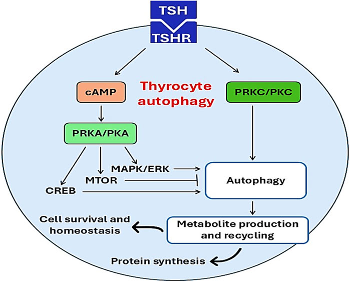
Figure 4. Effects of TSH on thyrocyte autophagy. Activation of TSHR by TSH triggers the activation of thyrocyte autophagy through activation of PKC/cAMP and other signaling pathways. Activated thyrocyte autophagy promotes protein synthesis, cell survival, and nomeostasis.
Conversely, GD-induced hyperthyroidism is associated with augmentation of the circulating levels of T3 and T4 which may affect thyrocyte autophagy. In vivo and ex-vivo findings demonstrate that thyroid hormones stimulate liver fatty acid β-oxidation through induction of autophagy, and blockade of hepatic autophagy by siRNA which targets ATG5 inhibits fatty acid β-oxidation (Sinha et al., 2012). Chi et al. (2019) highlighted that thyroid hormones activate hepatic autophagy in different liver diseases such as non-alcoholic fatty liver disease. Therefore, thyroid hormone-induced autophagy can be a compensatory mechanism to control dysregulated autophagy in GD. Remarkably, polymorphism of the autophagy-related gene IRGM (immunity related GTPase M) is associated with the risk of GD and other AITDs (Yao et al., 2018) signifying a potential link between GD and autophagy dysfunction. IRGM plays a critical role in regulating inflammation by increasing engulfment of apoptotic cells by autophagy (Yao et al., 2018). A case-control study showed that the T allele of rs10065172, A allele of rs4958847, and C allele of rs13361189 are higher in GD patients (Yao et al., 2018). Furthermore, exaggeration of thyroid hormones in GD is linked with the development of oxidative stress, which activates the autophagy flux (Kiffin et al., 2004; Londzin-Olesik et al., 2020). Of interest, oxidative stress is strongly implicated in the pathogenesis of GD (Žarković, 2012). For example, oxidative stress markers are higher in GD patients compared to controls (Ademoğlu et al., 2006). Many studies indicate that oxidative stress through activation of NFKB triggers the development of an autoimmune response in hyperthyroidism (Nandakumar et al., 2008; Makay et al., 2009). NFKB is essential for activation of autophagy (Min et al., 2018) and increases GD risk by 39% (Niyazoglu et al., 2014). Supporting this claim, treatment with the antioxidant selenium reduces the disease severity in GD patients (Marcocci et al., 2011).
These findings proposed that thyrocyte autophagy is dysregulated in GD due to direct effects of TSI and thyroid hormones, and indirectly by oxidative stress and NFKB activation. Moreover, thyrocyte autophagy seems to be inhibited in early GD and activated in late GD to mitigate the inflammatory and oxidative stress disorders.
Autophagy and GD ophthalmopathy
GD ophthalmopathy (GO) is the most common extrathyroidal manifestation of GD characterized by unilateral (10%) or bilateral (90%) eye proptosis. GO develops due to activation of T cells and TSI directed against retro-orbital tissues, which share antigenic epitopes with thyrocytes (Nabi and Rafiq, 2020). GO as an autoimmune disease leads to inflammation and injury of extraocular muscles and orbital adipose tissues (Bahn, 2010). The occurrence of GO may precede GD in 23% of cases, coexist with GD in 39% and follow GD in 37% (Claytor and Li, 2021). T cell-mediated activation and the Th1 immune response are activated in the early stage of GO, although the Th2 immune response and antibody production are stimulated in the late stage (Chen et al., 2023). These immune responses activate orbital inflammation and differentiation of adipocytes and myofibroblasts (Li et al., 2021a). These immunoinflammatory changes trigger autophagy, which may induce beneficial or detrimental effects according to the disease stage.
The potential role of autophagy in GO had been discussed in different studies; however, the precise role of autophagy in GO was not fully elucidated (Yoon et al., 2015a). In early GO there is marked inflammatory reactions, which induce aberrant autophagy activation (Guo et al., 2020). GO-associated inflammation is linked with autophagy activation as evidenced by increases of ATG5 and BECN1 and higher conversion of LC3-I to LC3-II (Yoon et al., 2015a; Guo et al., 2020). It has been illustrated that autophagy promotes adipogenesis in patients with GO. A case-control study confirmed that ATG5, LC3 and SQSTM1 are increased in orbital fat from GO patients compared to controls (Yoon et al., 2015a) proposing that activated autophagy is implicated in the pathogenesis of GO. Therefore, inhibition of autophagy may attenuate the progression of GO. In fact, it has been established that autophagy inhibitors chloroquine or hydroxycholoroquine attenuate adipogenesis by inhibiting autophagy of orbital fibroblasts (Guo et al., 2020). Similarly, the autophagy inhibitor bafilomycin A1 or deletion of ATG5 inhibits adipogenesis in orbital fibroblasts (Yoon et al., 2015b). Moreover, astragaloside and icariin suppress orbital fibroblasts and adipogenesis through inhibition of autophagy (Li et al., 2017; Li et al., 2018). These findings indicate that autophagy inhibitors are helpful in the management of GO.
Conversely, the MTOR inhibitor rapamycin, which activates autophagy, produces beneficial effects against GO (Roos and Murthy, 2019; Zhang et al., 2023). Rapamycin improves ocular restriction by inhibiting the differentiation of ocular myofibroblasts (Roos and Murthy, 2019). Indeed, the MTOR pathway is upregulated in patients with GO resulting in the induction of inflammation, fibrosis, and adipogenesis. Of interest, low-dose rapamycin mitigates diplopia/double vision in patients with refractory GO by inhibiting CD4-induced inflammation in GO (Zhang et al., 2023). Of note, rapamycin is also effective in different autoimmune disorders such as systemic sclerosis, systemic lupus erythematosus and rheumatoid arthritis (Bruyn et al., 2008; Su et al., 2009). Recently, it has been shown that rapamycin is more effective than steroids in the management of GO (Lanzolla et al., 2022). Therefore, autophagy activators may be effective in the management of GO. These findings highlight the fact that autophagy plays a double-edged sword role in the pathogenesis of GO—it may be protective or harmful according to the different stages of GO.
Taken together, autophagy may be protective against thyroid GD, but it has dual protective and harmful effects. Therefore, additional preclinical and clinical studies are recommended in this regard.
Conclusion
GD is the most common autoimmune disease of the thyroid gland and is characterized by hyperthyroidism and goiter due to production of TSI. TSI activates TSH receptors of the thyroid gland resulting in excessive release of thyroid hormones with subsequent development of hyperthyroidism and goiter. Of note, autophagy plays a critical role in many thyroid diseases and in different stages of the same disease through modulation of immunity and the inflammatory response. In addition, autophagy is also implicated in the pathogenesis of TAO. Thus this review has focused on how autophagy is involved in the pathogenesis of GD regarding its protective and harmful effects. Thyrocyte autophagy is dysregulated in GD due to direct effects of TSI and thyroid hormones, and indirectly by oxidative stress and NFKB activation. Moreover, thyrocyte autophagy seems to be inhibited in early GD and activated in late GD to mitigate the inflammatory and oxidative stress disorders. Importantly, autophagy plays a double-edged sword role in the pathogenesis of GO, where it may be protective or harmful according to the different stages of the disease. Further preclinical and clinical studies are recommended in this regard.
Author contributions
HA-K: Conceptualization, Writing–original draft, Writing–review and editing. GS: Conceptualization, Writing–original draft, Writing–review and editing. HM: Software, Writing–review and editing. MA-A: Investigation, Writing–review and editing. SA: Data curation, Writing–review and editing. MJ: Investigation, Writing–review and editing. AA: Writing–original draft, Writing–review and editing. AA-G: Writing–original draft, Writing–review and editing. DK: Conceptualization, Writing–original draft, Writing–review and editing.
Funding
The author(s) declare that financial support was received for the research, authorship, and/or publication of this article. The authors are thankful to the Deanship of Graduate Studies and Scientific Research at University of Bisha for supporting this work through the Fast-Track Research Support Program.
Conflict of interest
The authors declare that the research was conducted in the absence of any commercial or financial relationships that could be construed as a potential conflict of interest.
The author(s) declared that they were an editorial board member of Frontiers, at the time of submission. This had no impact on the peer review process and the final decision.
Publisher’s note
All claims expressed in this article are solely those of the authors and do not necessarily represent those of their affiliated organizations, or those of the publisher, the editors and the reviewers. Any product that may be evaluated in this article, or claim that may be made by its manufacturer, is not guaranteed or endorsed by the publisher.
Abbreviations
AITD, autoimmune thyroid disease; GD, Graves disease; GO, GD ophthalmopathy; TAO, thyroid-associated opthalmopathy; Th1, type 1 T helper; TSH, thyroid stimulating hormone; TSIs, thyroid-stimulating immunoglobulins.
References
Ademoğlu, E., Özbey, N., Erbil, Y., Tanrikulu, S., Barbaros, U., Yanik, B. T., et al. (2006). Determination of oxidative stress in thyroid tissue and plasma of patients with Graves’ disease. Eur. J. Intern. Med. 17, 545–550. doi:10.1016/j.ejim.2006.04.013
AlAnazi, F. H., Al-Kuraishy, H. M., Alexiou, A., Papadakis, M., Ashour, M. H. M., Alnaaim, S. A., et al. (2023). Primary hypothyroidism and alzheimer’s disease: a tale of two. Cell. Mol. Neurobiol. 43, 3405–3416. doi:10.1007/s10571-023-01392-y
Ali, N. H., Al-Kuraishy, H. M., Al-Gareeb, A. I., Alnaaim, S. A., Alexiou, A., Papadakis, M., et al. (2023). Autophagy and autophagy signaling in Epilepsy: possible role of autophagy activator. Mol. Med. 29, 142. doi:10.1186/s10020-023-00742-2
Al-Kuraishy, H. M., Al-Bdulhadi, M. H., and Al-Gareeb, A. I. (2021). Neuropeptide Y-Agouti related peptide ratio (NAR) in patients with idiopathic primary hypothyroidism: nudge and Risk. J. Pak Med. Assoc. 71 (Suppl. 8), S27–S31.
Al-Kuraishy, H. M., Jabir, M. S., Al-Gareeb, A. I., Klionsky, D. J., and Albuhadily, A. K. (2024a). Dysregulation of pancreatic β-cell autophagy and the risk of type 2 diabetes. Autophagy 18, 2361–2372. doi:10.1080/15548627.2024.2367356
Al-Kuraishy, H. M., Jabir, M. S., Al-Gareeb, A. I., Saad, H. M., Batiha, G. E.-S., and Klionsky, D. J. (2024b). The beneficial role of autophagy in multiple sclerosis: yes or no? Autophagy 20, 259–274. doi:10.1080/15548627.2023.2259281
Al-Kuraishy, H. M., Sulaiman, G. M., Jabir, M. S., Mohammed, H. A., Al-Gareeb, A. I., Albukhaty, S., et al. (2024c). Defective autophagy and autophagy activators in myasthenia gravis: a rare entity and unusual scenario. Autophagy 20, 1473–1482. doi:10.1080/15548627.2024.2315893
Antonelli, A., Fallahi, P., Elia, G., Ragusa, F., Paparo, S. R., Ruffilli, I., et al. (2020). Graves’ disease: clinical manifestations, immune pathogenesis (cytokines and chemokines) and therapy. Best Pract. and Res. Clin. Endocrinol. and Metabolism 34, 101388. doi:10.1016/j.beem.2020.101388
Bahn, R. S. (2010). Graves’ ophthalmopathy. N. Engl. J. Med. 362, 726–738. doi:10.1056/NEJMra0905750
Bartalena, L. (2013). Diagnosis and management of Graves disease: a global overview. Nat. Rev. Endocrinol. 9, 724–734. doi:10.1038/nrendo.2013.193
Bhattacharya, A., and Eissa, N. T. (2013). Autophagy and autoimmunity crosstalks. Front. Immunol. 4, 88. doi:10.3389/fimmu.2013.00088
Brix, T. H., Kyvik, K. O., Christensen, K., and Hegedüs, L. (2001). Evidence for a major role of heredity in Graves’ disease: a population-based study of two Danish twin cohorts. J. Clin. Endocrinol. and Metabolism 86, 930–934. doi:10.1210/jcem.86.2.7242
Broggi, G., Ieni, A., Russo, D., Varricchio, S., Puzzo, L., Russo, A., et al. (2020). The macro-autophagy-related protein Beclin-1 immunohistochemical expression correlates with tumor cell type and clinical behavior of uveal Melanoma. Front. Oncol. 10, 589849. doi:10.3389/fonc.2020.589849
Bruyn, G. A. W., Tate, G., Caeiro, F., Maldonado-Cocco, J., Westhovens, R., Tannenbaum, H., et al. (2008). Everolimus in patients with rheumatoid arthritis receiving concomitant methotrexate: a 3-month, double-blind, randomised, placebo-controlled, parallel-group, proof-of-concept study. Ann. rheumatic Dis. 67, 1090–1095. doi:10.1136/ard.2007.078808
Cao, J., Su, Y., Chen, Z., Ma, C., and Xiong, W. (2022). The risk factors for Graves’ ophthalmopathy. Graefe’s Archive Clin. Exp. Ophthalmol. 260, 1043–1054. doi:10.1007/s00417-021-05456-x
Cao, W., Li, J., Yang, K., and Cao, D. (2021). An overview of autophagy: mechanism, regulation and research progress. Bull. Du. cancer 108, 304–322. doi:10.1016/j.bulcan.2020.11.004
Chang, H., and Zou, Z. (2020). Targeting autophagy to overcome drug resistance: further developments. J. Hematol. and Oncol. 13, 159. doi:10.1186/s13045-020-01000-2
Chen, Y.-Q., Gao, L.-D., Liu, Y.-L., Shen, Y., Diao, J.-L., Yang, W.-H., et al. (2023). Autophagy in graves’ ophthalmopathy. Front. Cell. Dev. Biol. 11, 1158279. doi:10.3389/fcell.2023.1158279
Chi, H.-C., Tsai, C.-Y., Tsai, M.-M., Yeh, C.-T., and Lin, K.-H. (2019). Molecular functions and clinical impact of thyroid hormone-triggered autophagy in liver-related diseases. J. Biomed. Sci. 26, 24–15. doi:10.1186/s12929-019-0517-x
Claytor, B., and Li, Y. (2021). Challenges in diagnosing coexisting ocular myasthenia gravis and thyroid eye disease. Muscle and Nerve 63, 631–639. doi:10.1002/mus.27118
Davies, T. F., Andersen, S., Latif, R., Nagayama, Y., Barbesino, G., Brito, M., et al. (2020). Graves’ disease. Nat. Rev. Dis. Prim. 6, 52. doi:10.1038/s41572-020-0184-y
Duan, J., Deng, T., Kang, J., and Chen, M. (2019). DINP aggravates autoimmune thyroid disease through activation of the Akt/mTOR pathway and suppression of autophagy in Wistar rats. Environ. Pollut. 245, 316–324. doi:10.1016/j.envpol.2018.10.108
Fang, S., Lu, Y., Huang, Y., Zhou, H., and Fan, X. (2021). Mechanisms that underly T cell immunity in graves’ orbitopathy. Front. Endocrinol. 12, 648732. doi:10.3389/fendo.2021.648732
Faustino, L. C., Lombardi, A., Madrigal-Matute, J., Owen, R. P., Libutti, S. K., and Tomer, Y. (2018). Interferon-α triggers autoimmune thyroid diseases via lysosomal-dependent degradation of thyroglobulin. J. Clin. Endocrinol. and Metabolism 103, 3678–3687. doi:10.1210/jc.2018-00541
Ferrari, S. M., Fallahi, P., Ruffilli, I., Elia, G., Ragusa, F., Benvenga, S., et al. (2019). The association of other autoimmune diseases in patients with Graves’ disease (with or without ophthalmopathy): review of the literature and report of a large series. Autoimmun. Rev. 18, 287–292. doi:10.1016/j.autrev.2018.10.001
Galluzzi, L., Baehrecke, E. H., Ballabio, A., Boya, P., Bravo-San Pedro, J. M., Cecconi, F., et al. (2017). Molecular definitions of autophagy and related processes. EMBO J. 36, 1811–1836. doi:10.15252/embj.201796697
Guo, Y., Li, H., Chen, X., Yang, H., Guan, H., He, X., et al. (2020). Novel roles of chloroquine and hydroxychloroquine in Graves’ orbitopathy therapy by targeting orbital fibroblasts. J. Clin. Endocrinol. and Metabolism 105, 1906–1917. doi:10.1210/clinem/dgaa161
Hargreaves, C. E., Grasso, M., Hampe, C. S., Stenkova, A., Atkinson, S., Joshua, G. W. P., et al. (2013). Yersinia enterocolitica provides the link between thyroid-stimulating antibodies and their germline counterparts in Graves’ disease. J. Immunol. 190, 5373–5381. doi:10.4049/jimmunol.1203412
He, C. (2022). Balancing nutrient and energy demand and supply via autophagy. Curr. Biol. 32, R684–R696. doi:10.1016/j.cub.2022.04.071
He, K., Jiang, P., Liu, B., Liu, X., Mao, X., and Hu, Y. (2020). Intrathyroid injection of dexamethasone inhibits Th2 cells in Graves’ disease. Archives Endocrinol. metabolism 64, 243–250. doi:10.20945/2359-3997000000244
Hussain, Y. S., Hookham, J. C., Allahabadia, A., and Balasubramanian, S. P. (2017). Epidemiology, management and outcomes of Graves’ disease—real life data. Endocrine 56, 568–578. doi:10.1007/s12020-017-1306-5
Inaba, H., De Groot, L. J., and Akamizu, T. (2016). Thyrotropin receptor epitope and human leukocyte antigen in Graves’ disease. Front. Endocrinol. 7, 218181. doi:10.3389/fendo.2016.00120
Iriondo, M. N., Etxaniz, A., Varela, Y. R., Ballesteros, U., Lázaro, M., Valle, M., et al. (2023). Effect of ATG12–ATG5-ATG16L1 autophagy E3-like complex on the ability of LC3/GABARAP proteins to induce vesicle tethering and fusion. Cell. Mol. Life Sci. 80, 56. doi:10.1007/s00018-023-04704-z
Kiffin, R., Christian, C., Knecht, E., and Cuervo, A. M. (2004). Activation of chaperone-mediated autophagy during oxidative stress. Mol. Biol. Cell. 15, 4829–4840. doi:10.1091/mbc.e04-06-0477
Kurashige, T., Nakajima, Y., Shimamura, M., Matsuyama, M., Yamada, M., Nakashima, M., et al. (2019). Basal autophagy deficiency causes thyroid follicular epithelial cell death in mice. Endocrinology 160, 2085–2092. doi:10.1210/en.2019-00312
Kurashige, T., Nakajima, Y., Shimamura, M., Yamada, M., and Nagayama, Y. (2020). Hormonal regulation of autophagy in thyroid PCCL3 cells and the thyroids of male mice. J. Endocr. Soc. 4, bvaa054. doi:10.1210/jendso/bvaa054
Lanzolla, G., Maglionico, M. N., Comi, S., Menconi, F., Piaggi, P., Posarelli, C., et al. (2022). Sirolimus as a second-line treatment for Graves’ orbitopathy. J. Endocrinol. Investigation 45, 2171–2180. doi:10.1007/s40618-022-01862-y
Leporati, P., Groppelli, G., Zerbini, F., Rotondi, M., and Chiovato, L. (2015). Etiopathogenesis of Basedow's disease. Trends and current aspects. Nuklearmedizin-NuclearMedicine 54, 204–210. doi:10.3413/Nukmed-0739-15-04
Lesmana, R., Sinha, R. A., Singh, B. K., Zhou, J., Ohba, K., Wu, Y., et al. (2016). Thyroid hormone stimulation of autophagy is essential for mitochondrial biogenesis and activity in skeletal muscle. Endocrinology 157, 23–38. doi:10.1210/en.2015-1632
Li, H., Gao, L., Min, J., Yang, Y., and Zhang, R. (2021a). Neferine suppresses autophagy-induced inflammation, oxidative stress and adipocyte differentiation in Graves’ orbitopathy. J. Cell. Mol. Med. 25, 1949–1957. doi:10.1111/jcmm.15931
Li, H., Yuan, Y., Zhang, Y., Zhang, X., Gao, L., and Xu, R. (2017). Icariin inhibits AMPK-dependent autophagy and adipogenesis in adipocytes in vitro and in a model of graves’ orbitopathy in vivo. Front. physiology 8, 223122. doi:10.3389/fphys.2017.00045
Li, H., Zhang, Y., Min, J., Gao, L., Zhang, R., and Yang, Y. (2018). Astragaloside IV attenuates orbital inflammation in Graves’ orbitopathy through suppression of autophagy. Inflamm. Res. 67, 117–127. doi:10.1007/s00011-017-1100-0
Li, Q. M., Wei, J. P., Li, M., and Meng, S. H. (2015). Effect of jiakangning capsule on thyroid function and akt/mTOR signal pathway of graves’ disease mice: an experimental study. Chin. J. Integr. Traditional West. Med. 35, 1119–1124.
Li, W., He, P., Huang, Y., Li, Y.-F., Lu, J., Li, M., et al. (2021b). Selective autophagy of intracellular organelles: recent research advances. Theranostics 11, 222–256. doi:10.7150/thno.49860
Li, X., He, S., and Ma, B. (2020). Autophagy and autophagy-related proteins in cancer. Mol. cancer 19, 12. doi:10.1186/s12943-020-1138-4
Li, Y., Zhang, W., Liu, M., Zhang, Q., Lin, Z., Jia, M., et al. (2021c). Imbalance of autophagy and apoptosis induced by oxidative stress may be involved in thyroid damage caused by sleep deprivation in rats. Oxidative Med. Cell. Longev. 2021, 2021. doi:10.1155/2021/5645090
Li, Y., Zhao, C., Zhao, K., Yu, N., Li, Y., Yu, Y., et al. (2021d). Glycosylation of anti-thyroglobulin IgG1 and IgG4 subclasses in thyroid diseases. Eur. Thyroid J. 10, 114–124. doi:10.1159/000507699
Liu, Y., Yuan, X., Li, X., Cui, D., and Xie, J. (2018). Constitutive changes in circulating follicular helper T cells and their subsets in patients with Graves’ disease. J. Immunol. Res. 2018, 8972572. doi:10.1155/2018/8972572
Londzin-Olesik, M., Kos-Kudła, B., Nowak, A., Wielkoszyński, T., and Nowak, M. (2020). The effect of thyroid hormone status on selected antioxidant parameters in patients with Graves’ disease and active thyroid-associated orbitopathy. Endokrynol. Pol. 71, 418–424. doi:10.5603/EP.a2020.0049
Lőrincz, P., and Juhász, G. (2020). Autophagosome-lysosome fusion. J. Mol. Biol. 432, 2462–2482. doi:10.1016/j.jmb.2019.10.028
Lu, Q., Luo, X., Mao, C., Zheng, T., Liu, B., Dong, X., et al. (2018). Caveolin-1 regulates autophagy activity in thyroid follicular cells and is involved in Hashimoto’s thyroiditis disease. Endocr. J. 65, 893–901. doi:10.1507/endocrj.EJ18-0003
Luo, F., Sandhu, A. F., Rungratanawanich, W., Williams, G. E., Akbar, M., Zhou, S., et al. (2020). Melatonin and autophagy in aging-related neurodegenerative diseases. Int. J. Mol. Sci. 21, 7174. doi:10.3390/ijms21197174
Makay, B., Makay, O., Yenisey, C., Icoz, G., Ozgen, G., Unsal, E., et al. (2009). The interaction of oxidative stress response with cytokines in the thyrotoxic rat: is there a link? Mediat. Inflamm. 2009, 391682. doi:10.1155/2009/391682
Manni, A., Quarde, A., Manni, A., and Quarde, A. (2020). Thyroid gland signs. Endocr. Pathophysiol. A Concise Guide Phys. Exam, 27–50. doi:10.1007/978-3-030-49872-6_2
Marcocci, C., Kahaly, G. J., Krassas, G. E., Bartalena, L., Prummel, M., Stahl, M., et al. (2011). Selenium and the course of mild Graves’ orbitopathy. N. Engl. J. Med. 364, 1920–1931. doi:10.1056/NEJMoa1012985
Marzoog, T. R., Jabir, M. S., Ibraheem, S., Jawad, S. F., Hamzah, S. S., Sulaiman, G. M., et al. (2023). Bacterial extracellular vesicles induced oxidative stress and mitophagy through mTOR pathways in colon cancer cells, HT-29: implications for bioactivity. Biochimica Biophysica Acta (BBA)-Molecular Cell. Res. 1870, 119486. doi:10.1016/j.bbamcr.2023.119486
Mathew, A. A., Papaly, R., Maliakal, A., and Chandra, L. (2021). Elevated graves’ disease-specific thyroid-stimulating immunoglobulin and thyroid stimulating hormone receptor antibody in a patient with subacute thyroiditis. Cureus 13. doi:10.7759/cureus.19448
Min, Y., Kim, M.-J., Lee, S., Chun, E., and Lee, K.-Y. (2018). Inhibition of TRAF6 ubiquitin-ligase activity by PRDX1 leads to inhibition of NFKB activation and autophagy activation. Autophagy 14, 1347–1358. doi:10.1080/15548627.2018.1474995
Morshed, S., Latif, R., and Davies, T. F. (2022). Rescue of thyroid cells from antibody induced cell death via induction of autophagy. J. Autoimmun. 126, 102746. doi:10.1016/j.jaut.2021.102746
Morshed, S. A., Ando, T., Latif, R., and Davies, T. F. (2010). Neutral antibodies to the TSH receptor are present in Graves’ disease and regulate selective signaling cascades. Endocrinology 151, 5537–5549. doi:10.1210/en.2010-0424
Nabi, T., and Rafiq, N. (2020). Factors associated with severity of orbitopathy in patients with Graves’ disease. Taiwan J. Ophthalmol. 10, 197–202. doi:10.4103/tjo.tjo_10_20
Nandakumar, D. N., Koner, B. C., Vinayagamoorthi, R., Nanda, N., Negi, V. S., Goswami, K., et al. (2008). Activation of NF-kappaB in lymphocytes and increase in serum immunoglobulin in hyperthyroidism: possible role of oxidative stress. Immunobiology 213, 409–415. doi:10.1016/j.imbio.2007.10.005
Neag, E. J., and Smith, T. J. (2022). 2021 update on thyroid-associated ophthalmopathy. J. Endocrinol. investigation 45, 235–259. doi:10.1007/s40618-021-01663-9
Niyazoglu, M., Baykara, O., Koc, A., Aydoğdu, P., Onaran, I., Dellal, F. D., et al. (2014). Association of PARP-1, NF-κB, NF-κBIA and IL-6, IL-1β and TNF-α with graves disease and graves ophthalmopathy. Gene 547, 226–232. doi:10.1016/j.gene.2014.06.038
Pavlinov, I., Salkovski, M., and Aldrich, L. N. (2020). Beclin 1–ATG14L protein–protein interaction inhibitor selectively inhibits autophagy through disruption of VPS34 complex I. J. Am. Chem. Soc. 142, 8174–8182. doi:10.1021/jacs.9b12705
Pierman, G., Delgrange, E., and Jonas, C. (2021). Recurrence of Graves’ disease (a Th1-type cytokine disease) following SARS-CoV-2 mRNA vaccine administration: a simple coincidence? Eur. J. case Rep. Intern. Med. 8, 002807. doi:10.12890/2021_002807
Pujol-Borrell, R., Giménez-Barcons, M., Marín-Sánchez, A., and Colobran, R. (2015). Genetics of Graves’ disease: special focus on the role of TSHR gene. Hormone Metabolic Res. 47, 753–766. doi:10.1055/s-0035-1559646
Pyzik, A., Grywalska, E., Matyjaszek-Matuszek, B., Ludian, J., Kiszczak-Bochyńska, E., Smoleń, A., et al. (2019). Does the epstein–barr virus play a role in the pathogenesis of graves’ disease? Int. J. Mol. Sci. 20, 3145. doi:10.3390/ijms20133145
Roos, J. C. P., and Murthy, R. (2019). Sirolimus (rapamycin) for the targeted treatment of the fibrotic sequelae of Graves’ orbitopathy. Eye 33, 679–682. doi:10.1038/s41433-019-0340-3
Sinha, R. A., Singh, B. K., Zhou, J., Wu, Y., Farah, B. L., Ohba, K., et al. (2015). Thyroid hormone induction of mitochondrial activity is coupled to mitophagy via ROS-AMPK-ULK1 signaling. Autophagy 11, 1341–1357. doi:10.1080/15548627.2015.1061849
Sinha, R. A., You, S.-H., Zhou, J., Siddique, M. M., Bay, B.-H., Zhu, X., et al. (2012). Thyroid hormone stimulates hepatic lipid catabolism via activation of autophagy. J. Clin. investigation 122, 2428–2438. doi:10.1172/JCI60580
Su, T. K., Khanna, D., Furst, D. E., Danovitch, G., Burger, C., Maranian, P., et al. (2009). Rapamycin versus methotrexate in early diffuse systemic sclerosis: results from a randomized, single-blind pilot study. Arthritis and Rheumatism Official J. Am. Coll. Rheumatology 60, 3821–3830. doi:10.1002/art.24986
Sulaiman, G. M., Al-Kuraishy, H. M., Jabir, M. S., Mohammed, H. A., Al-Gareeb, A. I., Albukhaty, S., et al. (2024). Defective autophagy and autophagy activators in Myasthenia Gravis: a rare entity and unusual scenario. Autophagy 20, 1473–1482. doi:10.1080/15548627.2024.2315893
Torimoto, K., Okada, Y., Nakayamada, S., Kubo, S., Kurozumi, A., Narisawa, M., et al. (2022). Comprehensive immunophenotypic analysis reveals the pathological involvement of Th17 cells in Graves’ disease. Sci. Rep. 12, 16880. doi:10.1038/s41598-022-19556-z
Ueno, T., and Komatsu, M. (2020). Monitoring autophagy flux and activity: principles and applications. Bioessays 42, 2000122. doi:10.1002/bies.202000122
Wang, X., Huang, J., Zhang, A., Fang, C., Ma, Q., and Jiang, P. (2021). Altered expression profile of BAFF receptors on peripheral blood B lymphocytes in Graves’ disease. BMC Endocr. Disord. 21, 88. doi:10.1186/S12902-021-00752-3
Wu, D. J., and Adamopoulos, I. E. (2017). Autophagy and autoimmunity. Clin. Immunol. 176, 55–62. doi:10.1016/j.clim.2017.01.007
Xin, W., Yu, Y., Ma, Y., Gao, Y., Xu, Y., Chen, L., et al. (2017). Thyroid-stimulating hormone stimulation downregulates autophagy and promotes apoptosis in chondrocytes. Endocr. J. 64, 749–757. doi:10.1507/endocrj.EJ16-0534
Xu, C., Wu, F., Mao, C., Wang, X., Zheng, T., Bu, L., et al. (2016). Excess iodine promotes apoptosis of thyroid follicular epithelial cells by inducing autophagy suppression and is associated with Hashimoto thyroiditis disease. J. Autoimmun. 75, 50–57. doi:10.1016/j.jaut.2016.07.008
Yao, Q., Zhu, Y., Wang, W., Song, Z., Shao, X., Li, L., et al. (2018). Polymorphisms in autophagy-related gene IRGM are associated with susceptibility to autoimmune thyroid diseases. BioMed Res. Int. 2018, 7959707. doi:10.1155/2018/7959707
Yau, W. W., Singh, B. K., Lesmana, R., Zhou, J., Sinha, R. A., Wong, K. A., et al. (2019). Thyroid hormone (T3) stimulates brown adipose tissue activation via mitochondrial biogenesis and MTOR-mediated mitophagy. Autophagy 15, 131–150. doi:10.1080/15548627.2018.1511263
Yoon, J. S., Lee, H. J., Chae, M. K., and Lee, E. J. (2015a). Autophagy is involved in the initiation and progression of Graves’ orbitopathy. Thyroid 25, 445–454. doi:10.1089/thy.2014.0300
Yoon, J. S., Lee, H. J., Chae, M. K., and Lee, E. J. (2015b). Autophagy is involved in the initiation and progression of Graves’ orbitopathy. Thyroid 25, 445–454. doi:10.1089/thy.2014.0300
Žarković, M. (2012). The role of oxidative stress on the pathogenesis of Graves’ disease. J. Thyroid Res. 2012, 302537. doi:10.1155/2012/302537
Keywords: autoantibodies, autophagy, Graves disease, pathogenesis, thyroid-associated ophthalmopathy
Citation: Al-kuraishy HM, Sulaiman GM, Mohammed HA, Abu-Alghayth MH, Albukhaty S, Jabir MS, Albuhadily AK, Al-Gareeb AI, Klionsky DJ and Abomughaid MM (2025) The role of autophagy in Graves disease: knowns and unknowns. Front. Cell Dev. Biol. 12:1480950. doi: 10.3389/fcell.2024.1480950
Received: 14 August 2024; Accepted: 12 December 2024;
Published: 06 January 2025.
Edited by:
Craig Michael Walsh, University of California, Irvine, United StatesReviewed by:
Sangam Rajak, Sanjay Gandhi Post Graduate Institute of Medical Sciences (SGPGI), IndiaAlessio Lepore, University of Leeds, United Kingdom
Copyright © 2025 Al-kuraishy, Sulaiman, Mohammed, Abu-Alghayth, Albukhaty, Jabir, Albuhadily, Al-Gareeb, Klionsky and Abomughaid. This is an open-access article distributed under the terms of the Creative Commons Attribution License (CC BY). The use, distribution or reproduction in other forums is permitted, provided the original author(s) and the copyright owner(s) are credited and that the original publication in this journal is cited, in accordance with accepted academic practice. No use, distribution or reproduction is permitted which does not comply with these terms.
*Correspondence: Ghassan M. Sulaiman, Z2hhc3Nhbi5tLnN1bGFpbWFuQHVvdGVjaG5vbG9neS5lZHUuaXE=; Daniel J. Klionsky, a2xpb25za3lAdW1pY2guZWR1