There was a mistake in Figure 3E as published. Due to the multiple rounds of revision and the large number of panels, we inadvertently overlooked a mistake in Figure 3E during proofing: the images depicting the viability of IFPSCs and SDSCs on PDLLA were incorrectly presented. The corrected Figure 3 appears below.
FIGURE 3
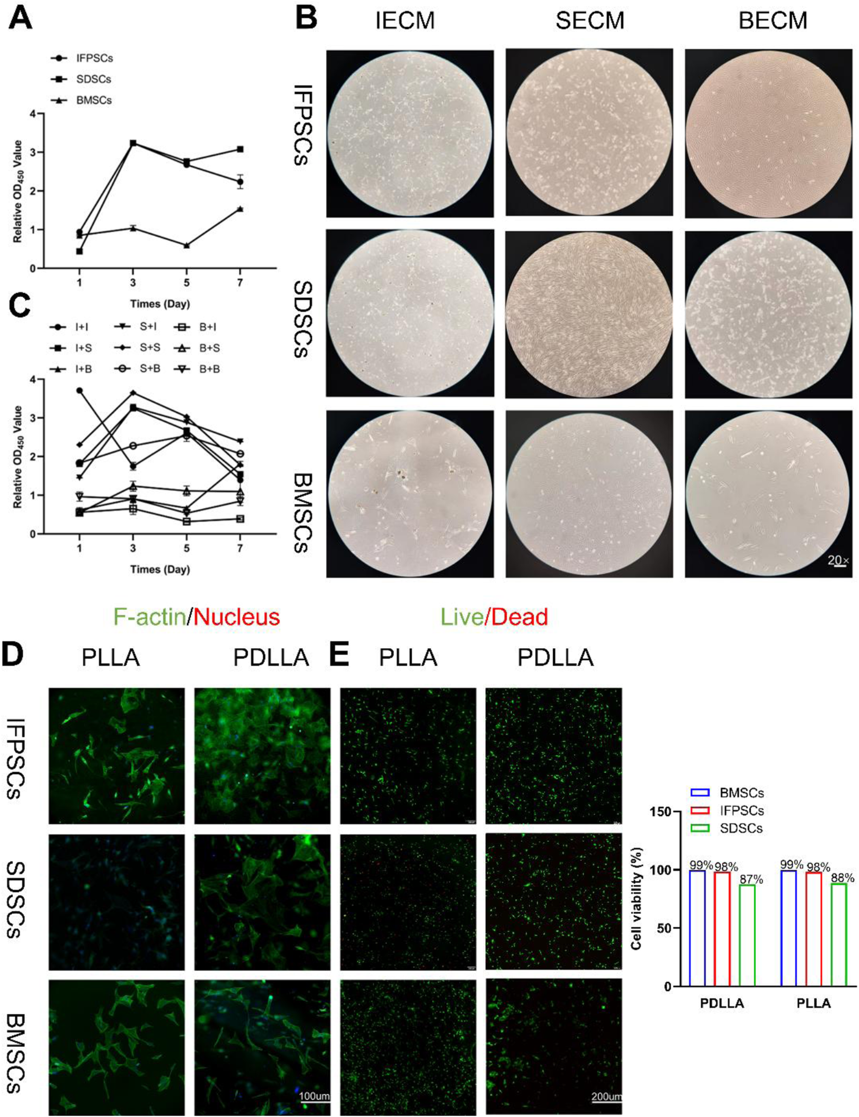
(A) Original stem cell proliferation rates were measured using a CCK-8 kit at 1, 3, 5, and 7 days. (B) Images of cells on dECM. (C) dECM pretreated-stem cell proliferation rates were measured using a CCK-8 kit at 1, 3, 5, and 7 days. (Key; I + I: IFPSCs + IECM (dECM deposited by IFPSCs); I + S: IFPSCs + SECM (dECM deposited by SDSCs); I + B: IFPSCs + BECM (dECM deposited by BMSCs); S + I: SDSCs + IECM; S + S: SDSCs + SECM; S + B: SDSCs + BECM; B + I: BMSCs + IECM; B + S: BMSCs + SECM; B + B: and BMSCs + BECM). (D) Cell adhesion morphology on PLLA and PDLLA nanofiber scaffolds. (E) Cell survival on electrospun nanofiber scaffolds made from PLLA and PDLLA. The cell viability was quantified using ImageJ software.
The original article has been updated.
Statements
Generative AI statement
Any alternative text (alt text) provided alongside figures in this article has been generated by Frontiers with the support of artificial intelligence and reasonable efforts have been made to ensure accuracy, including review by the authors wherever possible. If you identify any issues, please contact us.
Publisher’s note
All claims expressed in this article are solely those of the authors and do not necessarily represent those of their affiliated organizations, or those of the publisher, the editors and the reviewers. Any product that may be evaluated in this article, or claim that may be made by its manufacturer, is not guaranteed or endorsed by the publisher.
Summary
Keywords
mesenchymal stem cells, decellularized extracellular matrix (dECM), electrospun nanofiber scaffolds, osteogenesis, adipogenesis, chondrogenesis
Citation
Wei Z, Yu Q, Xie Q, Liao D, Gou X and Chen S (2026) Correction: A comparative analysis of stem cells derived from young rabbit knee joints: the potentially superior performance of decellularized extracellular matrix pretreated infrapatellar fat pad stem cells on nanofiber scaffolds. Front. Cell Dev. Biol. 14:1779159. doi: 10.3389/fcell.2026.1779159
Received
01 January 2026
Revised
27 January 2026
Accepted
28 January 2026
Published
06 February 2026
Volume
14 - 2026
Edited and reviewed by
Valerie Kouskoff, The University of Manchester, United Kingdom
Updates
Copyright
© 2026 Wei, Yu, Xie, Liao, Gou and Chen.
This is an open-access article distributed under the terms of the Creative Commons Attribution License (CC BY). The use, distribution or reproduction in other forums is permitted, provided the original author(s) and the copyright owner(s) are credited and that the original publication in this journal is cited, in accordance with accepted academic practice. No use, distribution or reproduction is permitted which does not comply with these terms.
*Correspondence: Song Chen, chensongchinese@163.com; Xue Gou, xgou@swjtu.edu.cn
Disclaimer
All claims expressed in this article are solely those of the authors and do not necessarily represent those of their affiliated organizations, or those of the publisher, the editors and the reviewers. Any product that may be evaluated in this article or claim that may be made by its manufacturer is not guaranteed or endorsed by the publisher.