 Erik Bényei
 András Laki1
 Miklós Tóth
 Judit Tőke
- 1Faculty of Medicine, Department of Internal Medicine and Oncology, Semmelweis University, Budapest, Hungary
- 2Faculty of Medicine, Medical Imaging Centre, Semmelweis University, Budapest, Hungary
- 3Faculty of Medicine, Medical Imaging Centre, Department of Nuclear Imaging, Semmelweis University, Budapest, Hungary
Introduction: Pheochromocytomatosis, defined as the implantation of pheochromocytoma cells to the intraoperatively opened surfaces during surgical manipulation, is an infrequent complication of surgical intervention of pheochromocytomas. Only a handful of pheochromocytomatosis cases have been reported since the first case was described in 2001.
Case report: In 2011, a 33-year-old male patient presented with episodic palpitations and hypertensive surges triggered by physical activity. Imaging revealed a left adrenal tumor, which showed intense radiopharmaceutical uptake on 131I-metaiodobenzylguanidine ([131I]MIBG) scintigraphy. Urinary analysis of metanephrines confirmed pheochromocytoma, and laparoscopic left-sided adrenalectomy was performed. Owing to the large tumor size, intraoperative fragmentation was necessary for removal. The patient remained asymptomatic for five years. In 2016, recurrent paroxysmal symptoms prompted imaging, revealing a lesion at the left renal hilum. During the reoperation in 2017, multiple peritoneal tumor deposits were observed and later confirmed histologically. Over the following years, the patient received conservative, symptomatic treatment with tolerable paroxysmal symptoms. In 2023, worsening symptoms led to the decision to commence three cycles of ([131I]MIBG) therapy, followed by alleviation of symptoms, and a decrease in biochemical parameters.
Discussion: An extensive literature search for publications from the past 25 years identified 22 pheochromocytomatosis cases whose details were also summarized and analyzed. This condition appears to have a longer recurrence-free survival compared to patients’ cohorts with metastatic pheochromocytomas. Pheochromocytomatosis is usually characterized by a prolonged asymptomatic postsurgical interval, emphasizing the need for long-term follow-up with close biochemical and radiological surveillance. Treatment strategies parallel those used for advanced/metastatic pheochromocytomas.
1 Introduction
Pheochromocytomas and paragangliomas are catecholamine-producing tumors developing from the enterochromaffin cells of the adrenal medulla and the sympathetic ganglia. These tumors are characterized by the paroxysmal symptoms caused by these catecholamines, such as palpitations, sweating and hypertensive surges. The first-line treatment is surgical removal, which can provide a curative solution in cases with localized disease. Since all pheochromocytomas have metastatic potential, the term “malignant” is no longer used; instead, metastatic pheochromocytoma is applied when enterochromaffin tissue appears extra-adrenally at the time of diagnosis or during follow-up (1). Pheochromocytomatosis – defined as multifocal nodular implantation of the pheochromocytoma cells to the intraoperatively opened surfaces without the signs of distant metastases – is a rare, iatrogenic event caused by mechanical damage to the tumor capsule during surgery. This phenomenon was first defined in 2001 (2), and 22 cases have been reported in the literature since then.
In the past 25 years, we have treated and followed over 200 patients with pheochromocytomas at our endocrine referral center. Here, we report a patient’s history with pheochromocytomatosis who presented us with a therapeutic challenge during long-term management.
2 Case report
A 33-year-old male patient presented with episodic palpitations and hypertensive surges triggered by physical activity in 2011. During the diagnostic work-up, an abdominal MRI revealed a 4 x 4,8 x 6 cm tumor in the left adrenal gland, which exhibited significant radiopharmaceutical uptake on 131I-metaiodobenzylguanidine ([131I]MIBG) scintigraphy. Urinary analysis showed elevated 24-hour metanephrine levels at 8860 µg/24h (normal range: 64 – 302 µg/24h) and normetanephrine levels at 7164 µg/24h (normal range: 162 – 527 µg/24h), alongside a serum chromogranin A level of 800 ng/mL (normal range: 19,4 – 98,1 ng/mL). The timeline of biochemical markers is shown in Figure 1. Based on these results, pheochromocytoma was diagnosed, and laparoscopic left adrenalectomy was performed in 2011. According to the surgical report, the large tumor could not be placed into the endobag; removal required deliberate fragmentation and manual extraction through an enlarged port. Histological investigation confirmed the diagnosis of pheochromocytoma. For the first five years after surgery, the patient remained asymptomatic and radiological follow-up showed no signs of recurrence.
![Graph A shows metanephrine and normetanephrine levels from 2011 to 2025, with a sharp decreases after surgeries in 2011 and 2017 and after three cycles of ([131-I]MIBG therapy in 2024 and 2025. Graph B shows chromogranin A levels, stable until 2017, then rising steeply after 2021, peaking in 2025 and decreasing sharply after three cycles of ([131-I]MIBG therapy.](https://www.frontiersin.org/files/Articles/1679629/fendo-16-1679629-HTML/image_m/fendo-16-1679629-g001.jpg)
Figure 1. (A) Urinary metanephrine and normetanephrine excretion over time. (B) Serum chromogranin A concentrations between 2011 and 2024. Black arrows indicate surgical interventions, and white arrows mark the three cycles of ([131I]MIBG) therapy.
In 2016, the patient presented with recurrent paroxysmal episodes with palpitations and hypertensive surges. Abdominal MRI and ([131I]MIBG) scintigraphy revealed a lesion at the left renal hilum, consistent with a tumor recurrence (Figure 2), which was further verified by the elevated urinary excretions of metanephrine (6641 ug/24h), normetanephrine (2882 µg/24h) and serum chromogranin A (910 ng/mL) levels (Figure 1). During reoperation in June 2017, a massive perisplenic invasion was observed. Cytoreductive surgery was performed, including splenectomy and distal pancreatic resection. The surgeon noted multiple 2–3 mm tumor deposits forming only a partially resectable tumor-like mass in the left hypochondrium. Histological analysis confirmed peritoneal and retroperitoneal pheochromocytoma deposits. Postoperative ([131I]MIBG) scintigraphy indicated persisting multifocal peritoneal foci. Next-generation sequencing (ENDOGEN panel, Illumina MiSeq device) of DNA prepared from peripheral blood leukocytes revealed no pathogenic mutations in genes associated with hereditary pheochromocytoma/paraganglioma syndromes (SDHA, SDHB, SDHC, KIF1B, EGLN1, FH, SDHAF2, MAX, SDHD, RET exon 10,11, VHL, TMEM127).
![Comparison of CT and [131-I]MIBG SPECT scans from 2017 and 2019. Image A shows 2017 scans with a highlighted area in yellow and green indicating low radiopharmaceutical uptake. Image B from 2019 shows increased radiopharmaceutical uptake iindicating with highlighted area in red and green, circled on the SPECT scan, indicating a change over time.](https://www.frontiersin.org/files/Articles/1679629/fendo-16-1679629-HTML/image_m/fendo-16-1679629-g002.jpg)
Figure 2. ([131I]MIBG) scintigraphy SPECT/CT images in (A) 2017 and (B) 2019 show multiple peritoneal lesions with significant radiopharmaceutical uptake in the left hypochondrium. No distant metastases were detected.
Over the following years, the patient received symptomatic drug treatment using alpha- and beta-blockers with tolerable paroxysmal symptoms. Regular radiological follow-ups revealed no new lesions compared to the imaging done in 2017; however, mild growth of previously described deposits was noted. Somatostatin-receptor scintigraphy turned out to be negative. In 2023, the patient’s symptoms became more frequent and intense, accompanied by radiological and biochemical progression (Figure 1). In 2024 and 2025, three cycles of ([131I]MIBG) therapies were administered (3579 MBq, 3468 MBq and 3326 MBq). Post-treatment imaging showed a mild increase in lesions’ size and number. However, urinary metanephrine and normetanephrine excretions as well as serum chromogranin A concentration exhibited an unambiguous decrease following treatments, and the patient reported an alleviation of symptoms.
3 Literature review
A literature search was conducted using MEDLINE, EMBASE, and Web of Science databases. We identified 11 full-text publications including 22 patients with pheochromocytomatosis reported between January 2000 and December 2024 (2–12). Although a few case reports describing pheochromocytomatosis date back nearly fifty years, these were excluded due to limited data quantity and/or quality and the substantial evolution in diagnostic and therapeutic procedures over time (13, 14). Publications were identified through keyword searches using the terms “pheochromocytomatosis”, {[“pheochromocytoma” OR “paraganglioma”] AND “recurrence”}, as well as by citation chasing. Only English-language publications with full-text availability were considered, and inclusion was based on a detailed full-text evaluation.
The clinicopathological characteristics of these 22 patients completed with our presented case are summarized in Table 1, while treatment and follow-up details are presented in Table 2. The mean age at the time of initial diagnosis was 42 ± 13.7 years. Pheochomocytomatosis was diagnosed predominantly in females (60.9%). Mean primary tumor size was 6.0 ± 2.2 cm and the majority of adrenalectomies (69.6%) was performed laparoscopically. Three patients were reported to have MEN2A syndrome, and one to have neurofibromatosis type 1. The surgical or the pathological reports typically described inadvertent tumor fragmentation or capsule rupture. All publications reported patients to be tumor-free after initial surgery, confirmed with resolution of clinical symptoms and normalization of biochemical parameters. Median (minimum – maximum) recurrence-free survival across all cases was 72 (23 – 180) months. Diagnostic workup for recurrence, prompted by positive biochemical follow-up, was conducted in only 5 patients (21.7% of cases), whereas in 12 patients (52.2% of cases) pheochromocytomatosis diagnoses were initiated due to recurrent symptoms. Most frequently performed imaging procedures were CT and ([131I]MIBG) scintigraphy, followed by abdominal MR scans. All patients underwent a second surgical intervention. At least 13 (56.5%) of them required further surgical interventions or additional therapies. The postoperative follow-up duration was highly variable. At the time of publication, 6 (26.1%) patients were reported to be in remission, 8 (34.7%) to have stable disease, and 2 (8.7%) died from tumor progression. Notably, out of the 23 patients, only 2 (8.7%) were reported to be tumor-free at least one year following the second surgery (Table 2).
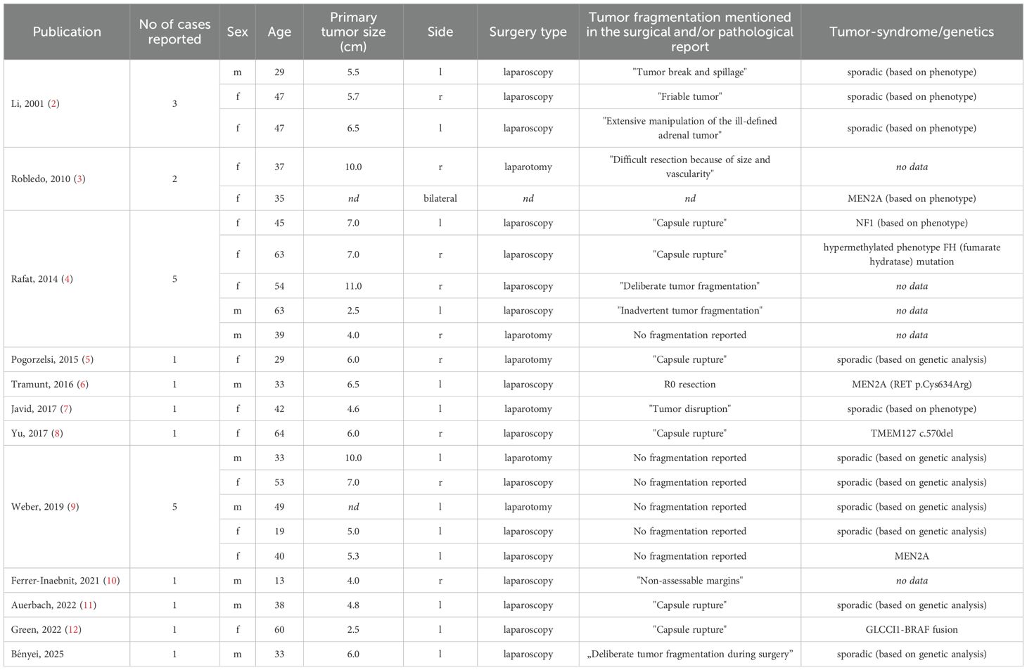
Table 1. Clinicopathological characteristics at the time of initial surgery for all patients reported with pheochromocytomatosis.
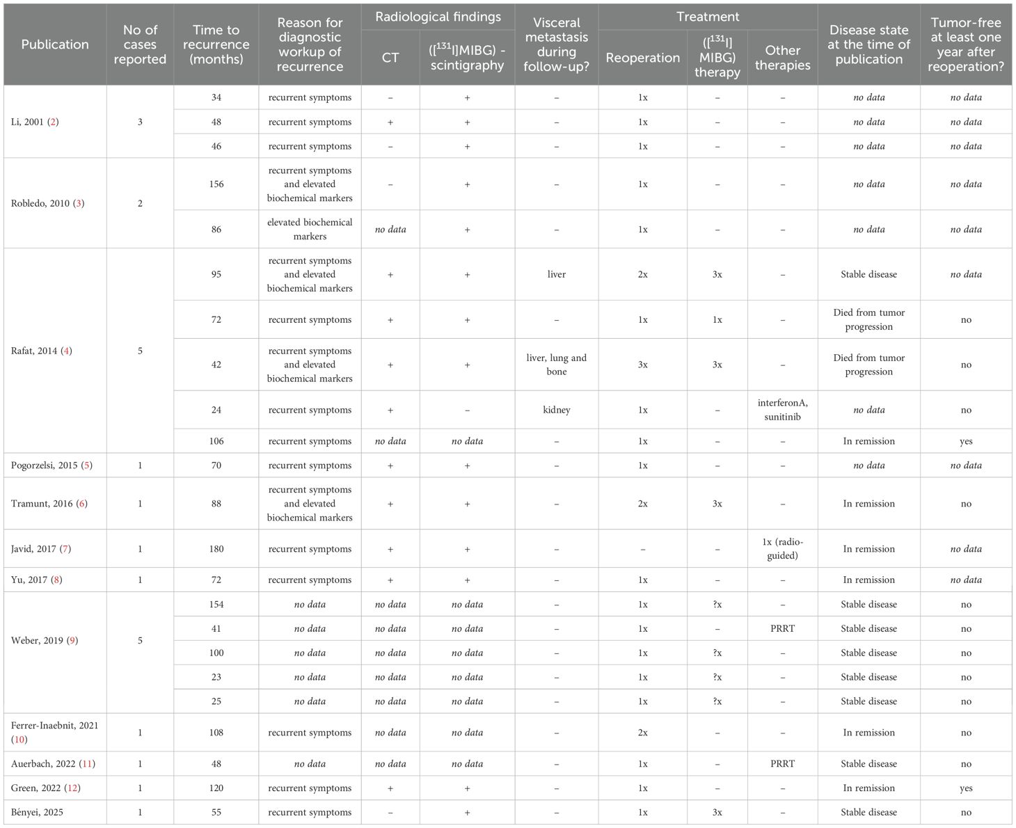
Table 2. Tumor recurrence and follow-up for all patients reported with pheochromocytomatosis.
4 Discussion
In addition to local recurrence and distant metastases, characteristic of malignant pheochromocytomas, pheochromocytomatosis represents another, infrequently reported type of tumor progression, which does not indicate malignancy. The removal of pheochromocytomas always poses a surgical challenge due to the tumor’s fragility and frequently soft consistency, with a rare complication being peritoneal tumor cell dissemination following damage to the tumor capsule (2). To characterize this condition better, we performed an extensive literature search.
Recurrence of pheochromocytoma after surgical removal is not considered rare, occurring in approximately 6.5 – 16.5% of cases, depending on the length of follow-up. It may be significantly more common in the presence of certain specific mutations (1, 15). Recurrence-free survival in pheochromocytoma patients is generally reported to be between 30 and 50 months (15–18), which is considerably shorter than the 72 months in pheochromocytomatosis patients of our meta-analysis. Pheochromocytomatosis case reports often describe a long, latent period – typically several years – following the initial surgery, during which patients appear biochemically and radiologically tumor-free. In most cases, the diagnostic investigation for pheochromocytomatosis was initiated after the recurrence of symptoms, despite documented tumor capsule rupture in nearly all cases. This highlights the critical importance of meticulous follow-up for these patients, encompassing regular radiological and biochemical evaluations while closely monitoring paroxysmal symptoms characteristic of pheochromocytomas. Due to the rarity and uncertain incidence of pheochromocytomatosis, robust survival data are lacking; however, one study reported better overall survival for patients with pheochromocytomatosis compared to those with metastatic pheochromocytomas (9).
Therapeutic approaches of pheochromocytomatosis largely corresponds to those used for metastatic pheochromocytomas. A „watch and wait” approach may spare patients the risks and side effects of other therapies in asymptomatic and radiologically stable disease cases. Cytoreductive (debulking) surgery can alleviate symptoms by reducing tumor burden and catecholamine excess. However, as repeated surgery led to remission in only a few cases, surgical interventions alone are unlikely to eliminate the long-term need for additional therapies. For tumors with sufficient radiopharmaceutical uptake, peptide receptor radionuclide therapy using radiolabeled somatostatin analogue or ([131I]MIBG) treatment may result in disease stabilization. Like in advanced pheochromocytoma management, somatostatin analogue therapy may also be a treatment option for tumors expressing somatostatin receptors. The efficacy of tyrosine kinase inhibitor sunitib, already approved for treating neuroendocrine tumors, has also been confirmed in patients with metastatic pheochromocytoma (FIRSTMAPP study) (19). Although Rafat et al. reported the inclusion of a patient with pheochromocytomatosis in this trial, the efficacy of sunitinib treatment remains unclear (4).
In recent years, two studies have examined somatic mutations of tumor cells in patients with pheochromocytomatosis. For the gene TMEM127, previously linked to pheochromocytomas (20), a new, likely pathogenic mutation (c. 570delC) was identified. In 2022, Green et al. proposed targeted systematic therapy with MEK and/or BRAF inhibitors following the identification of a GLCCI1-BRAF fusion gene (12). Identifying therapeutic targets could provide additional treatment options for therapy-resistant tumors.
A key strength of our case report lies in its detailed presentation of a rare and poorly understood condition, supported by comprehensive radiological and biochemical data. Another notable strength is the careful contextualization achieved by analyzing of similar cases reported in the literature. A limitation of our case report is the incomplete documentation of certain clinical details from the earlier years of follow-up. Regarding the literature review, a significant limitation is the heterogeneity in the pheochromocytomatosis management and follow-up across studies, which restricts the strength of conclusions drawn. Furthermore, the previously published cases span over two and a half decades, during which the clinical management of pheochromocytoma has undergone substantial changes, further limiting direct comparisons.
In conclusion, pheochromocytomatosis is an infrequent complication of pheochromocytoma surgery. Cautious intraabdominal handling of the tumor is key to preventing this adverse event. It is recommended that the surgery be performed by an experienced surgeon in a center specializing in adrenal surgery. In case of capsule rupture, rigorous radiological and biochemical follow-up is critical for the timely diagnosis and treatment of peritoneal dissemination, which may arise even several years after adrenalectomy. Analogously to the treatment of advanced, metastatic pheochromocytomas, therapeutic options to achieve stable disease include tumor debulking surgery, PRRT, somatostatin analogues and targeted systemic therapies. Adjuvant treatments are necessary to achieve stable disease.
Author contributions
EB: Data curation, Methodology, Writing – original draft, Writing – review & editing. AL: Investigation, Writing – review & editing. GK: Investigation, Writing – review & editing. ZV: Investigation, Writing – review & editing. MT: Conceptualization, Investigation, Supervision, Writing – original draft, Writing – review & editing. JT: Investigation, Supervision, Writing – review & editing.
Funding
The author(s) declare that no financial support was received for the research and/or publication of this article.
Acknowledgments
We are grateful to our patient, who gave written consent to publish his case. The Department of Internal Medicine and Oncology, Semmelweis University is a Reference Centre of the ENDO-ERN: European Reference Network on Rare Endocrine Conditions.
Conflict of interest
The authors declare that the research was conducted in the absence of any commercial or financial relationships that could be construed as a potential conflict of interest.
Generative AI statement
The author(s) declare that no Generative AI was used in the creation of this manuscript.
Any alternative text (alt text) provided alongside figures in this article has been generated by Frontiers with the support of artificial intelligence and reasonable efforts have been made to ensure accuracy, including review by the authors wherever possible. If you identify any issues, please contact us.
Publisher’s note
All claims expressed in this article are solely those of the authors and do not necessarily represent those of their affiliated organizations, or those of the publisher, the editors and the reviewers. Any product that may be evaluated in this article, or claim that may be made by its manufacturer, is not guaranteed or endorsed by the publisher.
References
1. Neumann HPH, Young WF Jr., and Eng C. Pheochromocytoma and paraganglioma. N Engl J Med. (2019) 381:552–65. doi: 10.1056/NEJMra1806651
2. Li ML, Fitzgerald PA, Price DC, and Norton JA. Iatrogenic pheochromocytomatosis: A previously unreported result of laparoscopic adrenalectomy. Surgery. (2001) 130:1072–7. doi: 10.1067/msy.2001.118373
3. Robledo AB, Ponce Marco JL, Ibáñez TB, Meseguer Anastasio MF, and Gómez-Gavara C. Pheochromocytomatosis: A risk after pheochromocytoma surgery. Am Surg. (2010) 76:122–4. doi: 10.1177/000313481007600810
4. Rafat C, Zinzindohoue F, Hernigou A, Hignette C, Favier J, Tenenbaum F, et al. Peritoneal implantation of pheochromocytoma following tumor capsule rupture during surgery. J Clin Endocrinol Metab. (2014) 99:E2681–E5. doi: 10.1210/jc.2014-1975
5. Pogorzelski R, Toutounchi S, Fiszer P, Krajewska E, Łoń I, Zapała Ł, et al. The local spread of pheochromocytoma after adrenalectomy with a rupture of the tumor capsule at the time of the surgery. Open Med. (2015) 10:335–7. doi: 10.1515/med-2015-0049
6. Tramunt B, Buffet A, Grunenwald S, Vezzosi D, Bennet A, Huyghe E, et al. Local recurrence of pheochromocytoma in multiple endocrine neoplasia type 2A: a diagnostic and therapeutic challenge. Clin Case Rep. (2016) 4:298–300. doi: 10.1002/ccr3.498
7. Javid M, Callender GG, Baregamian N, and Carling T. Pheochromocytomatosis treated by radio-guided surgery. AACE Clin Case Rep. (2017) 3:e170–e5. doi: 10.4158/EP151053.CR
8. Yu R, Sharaga D, Donner C, Palma Diaz MF, Livhits MJ, and Yeh MW. Pheochromocytomatosis associated with a novel TMEM127 mutation. Endocrinology Diabetes Metab Case Rep. (2017) 2017:17–0026. doi: 10.1530/EDM-17-0026
9. Weber F, Belker J, Unger N, Lahner H, Theurer S, Schmid KW, et al. Phäochromozytomatose nach Adrenalektomie: Metastasierung oder Zellverschleppung? Der Chirurg. (2020) 91:345–53. doi: 10.1007/s00104-019-01070-0
10. Ferrer-Inaebnit E, Segura-Sampedro JJ, Alfonso-García M, González-Argente X, and Morales-Soriano R. Cytoreductive surgery in functioning peritoneal pheochromocytomatosis. Cir Esp (Engl Ed). (2021) 99:73–6. doi: 10.1016/j.ciresp.2020.03.010
11. Auerbach MS, Livhits MJ, and Yu R. Pheochromocytomatosis treated with peptide receptor radionuclide therapy. Clin Nucl Med. (2022) 47:e276–e8. doi: 10.1097/RLU.0000000000003973
12. Green BL, Grant RRC, Richie CT, Chatterjee B, Sampaio De Melo M, Barr FG, et al. Novel GLCCI1-BRAF fusion drives kinase signaling in a case of pheochromocytomatosis. Eur J Endocrinology. (2022) 187:185–96. doi: 10.1530/EJE-21-0797
13. Sellwood RA, Wapnick S, Breckenridge A, Williams ED, and Welbourn RB. Recurrent phaeochromocytoma. BJS (British J Surgery). (1970) 57:309–12. doi: 10.1002/bjs.1800570419
14. Brennan MF and Kaiser HR. Persistent and recurrent pheochromocytoma: The role of surgery. World J Surg. (1982) 6:397–401. doi: 10.1007/BF01657665
15. Press D, Akyuz M, Dural C, Aliyev S, Monteiro R, Mino J, et al. Predictors of recurrence in pheochromocytoma. Surgery. (2014) 156:1523–8. doi: 10.1016/j.surg.2014.08.044
16. Parasiliti-Caprino M, Lucatello B, Lopez C, Burrello J, Maletta F, Mistrangelo M, et al. Predictors of recurrence of pheochromocytoma and paraganglioma: a multicenter study in Piedmont, Italy. Hypertens Res. (2020) 43:500–10. doi: 10.1038/s41440-019-0339-y
17. Li Z, Lai D, Jia Y, Luo J, Ma X, Zhang X, et al. Predictors of postoperative recurrence of pheochromocytoma: a monocentric study. BMC Surg. (2025) 25:179. doi: 10.1186/s12893-025-02824-w
18. Holscher I, van den Berg TJ, Dreijerink KMA, Engelsman AF, and Nieveen van Dijkum EJM. Recurrence rate of sporadic pheochromocytomas after curative adrenalectomy: A systematic review and meta-analysis. J Clin Endocrinol Metab. (2021) 106:588–97. doi: 10.1210/clinem/dgaa794
19. Baudin E, Goichot B, Berruti A, Hadoux J, Moalla S, Laboureau S, et al. Sunitinib for metastatic progressive phaeochromocytomas and paragangliomas: results from FIRSTMAPPP, an academic, multicentre, international, randomised, placebo-controlled, double-blind, phase 2 trial. Lancet. (2024) 403:1061–70. doi: 10.1016/S0140-6736(23)02554-0
Keywords: pheochromocytoma, paraganglioma, pheochromocytomatosis, recurrence, peritoneal implantation
Citation: Bényei E, Laki A, Kiss G, Varga Z, Tóth M and Tőke J (2025) Peritoneal implantation of pheochromocytoma – pheochromocytomatosis: a case report and mini review. Front. Endocrinol. 16:1679629. doi: 10.3389/fendo.2025.1679629
Received: 04 August 2025; Accepted: 21 October 2025;
Published: 03 November 2025.
Edited by:
Sujit Kumar Chowdhary, Indraprastha Apollo Hospitals, IndiaReviewed by:
Mehmet Haciyanli, Izmir Katip Celebi University, TürkiyeCopyright © 2025 Bényei, Laki, Kiss, Varga, Tóth and Tőke. This is an open-access article distributed under the terms of the Creative Commons Attribution License (CC BY). The use, distribution or reproduction in other forums is permitted, provided the original author(s) and the copyright owner(s) are credited and that the original publication in this journal is cited, in accordance with accepted academic practice. No use, distribution or reproduction is permitted which does not comply with these terms.
*Correspondence: Miklós Tóth, dG90aC5taWtsb3NAc2VtbWVsd2Vpcy5odQ==
†These authors have contributed equally to this work and share last authorship