 Denise Battaglini1,2,3
 Miquéias Lopes-Pacheco4
 Hugo C. Castro-Faria-Neto5
 Paolo Pelosi1,2
 Patricia R. M. Rocco4,6,7*
- 1Anesthesia and Intensive Care, San Martino Policlinico Hospital, Instituto di Ricovero e Cura a Carattere Scientifico (IRCCS) for Oncology and Neuroscience, Genoa, Italy
- 2Department of Surgical Science and Integrated Diagnostics (DISC), University of Genoa, Genoa, Italy
- 3Department of Medicine, University of Barcelona, Barcelona, Spain
- 4Laboratory of Pulmonary Investigation, Carlos Chagas Filho Biophysics Institute, Federal University of Rio de Janeiro, Rio de Janeiro, Brazil
- 5Laboratory of Immunopharmacology, Oswaldo Cruz Institute - Fiocruz, Rio de Janeiro, Brazil
- 6COVID-19 Virus Network from Brazilian Council for Scientific and Technological Development, Brasília, Brazil
- 7COVID-19 Virus Network from Foundation Carlos Chagas Filho Research Support of the State of Rio de Janeiro, Rio de Janeiro, Brazil
Severe acute respiratory syndrome-coronavirus 2 (SARS-CoV-2) causes a wide spectrum of clinical manifestations, with progression to multiorgan failure in the most severe cases. Several biomarkers can be altered in coronavirus disease 2019 (COVID-19), and they can be associated with diagnosis, prognosis, and outcomes. The most used biomarkers in COVID-19 include several proinflammatory cytokines, neuron-specific enolase (NSE), lactate dehydrogenase (LDH), aspartate transaminase (AST), neutrophil count, neutrophils-to-lymphocytes ratio, troponins, creatine kinase (MB), myoglobin, D-dimer, brain natriuretic peptide (BNP), and its N-terminal pro-hormone (NT-proBNP). Some of these biomarkers can be readily used to predict disease severity, hospitalization, intensive care unit (ICU) admission, and mortality, while others, such as metabolomic and proteomic analysis, have not yet translated to clinical practice. This narrative review aims to identify laboratory biomarkers that have shown significant diagnostic and prognostic value for risk stratification in COVID-19 and discuss the possible clinical application of novel analytic strategies, like metabolomics and proteomics. Future research should focus on identifying a limited but essential number of laboratory biomarkers to easily predict prognosis and outcome in severe COVID-19.
Introduction
Severe acute respiratory syndrome-coronavirus-2 (SARS-CoV-2) causes a wide spectrum of clinical manifestations, from mild respiratory symptoms to pneumonia and, in more severe cases, multiple organ failure (1). The mechanisms underlying multisystem involvement may include an unbalanced immune response that facilitates the progression of coronavirus disease-2019 (COVID-19). This hypothesis has been confirmed by laboratory biomarker alterations, showing greater potential for abnormal immune response, mainly an increase in neutrophil counts and a substantial reduction in lymphocyte counts, thus altering the neutrophil-to-lymphocyte ratio. Such an abnormal immune response is driven by an increased serum concentration of many pro-inflammatory mediators. These include interleukin (IL)-1β, IL-2, IL-6, IL-8, interferon (IFN)-γ-induced protein 10, granulocyte colony-stimulating factor, monocyte chemoattractant protein 1, macrophage inflammatory protein-1α, and tumor necrosis factor-α, among others (2–5). Nevertheless, the inflammatory cytokine storm in patients with COVID-19 is less injurious than that observed in patients with sepsis or acute respiratory distress syndrome (ARDS) but without COVID-19 (6), thus raising questions regarding the mechanisms underlying multiorgan involvement in COVID-19.
Several biomarkers other than cytokines have been found altered in COVID-19, and are associated with diagnosis, prognosis and outcomes (7). Some of these biomarkers can be easily used to predict disease severity, hospitalization, intensive care unit (ICU) admission, and mortality, while others, like metabolomic and proteomic analysis, are still of purely investigational concern and difficult to translate into clinical practice, despite their prognostic potential (8–10).
The aim of this narrative review is to identify laboratory biomarkers that have shown significant diagnostic and prognostic value for risk stratification in COVID-19 and to discuss the possible clinical application of novel analytic strategies, such as metabolomics and proteomics.
Potential for Multiorgan Involvement in COVID-19
SARS-CoV-2 is an enveloped, single-stranded ribonucleic acid (ssRNA) virus. The SARS-CoV-2 genome is composed of two polypeptides encoded between two open-reading frames that are processed by viral proteases to produce nonstructural proteins (11). These proteins are involved in viral replication and suppression of host innate immune defense. On the other hand, structural proteins of SARS-CoV-2 include the spike (S), envelope (E), and nucleocapsid (N) protein, as well as the membrane (M) glycoprotein. The S protein is a transmembrane glycoprotein that is located on the viral surface and cleaved by host-cell proteases. After anchoring the S protein, SARS-CoV-2 enters host cells via angiotensin receptor-2 (ACE2), thus activating transmembrane serine protease 2 (TMPRSS2), cathepsin B and L. The E protein is a glycoprotein involved in virion maturation and pathogenesis, while the M protein is involved in viral assembly and delineates the shape of the viral envelope; finally, the N protein binds directly to viral RNA (11). The pathogenic mechanisms of SARS-CoV-2 include 1) direct epithelial damage, 2) dysregulated immune response, 3) ACE2 dysregulation and downregulation of the renin-angiotensin- aldosterone system (RAAS), 4) direct endothelial damage, and, possibly, 5) tissue fibrosis (11). Hence, patients with severe COVID-19 are at high risk of multiple organ involvement and, ultimately, death. Indeed, the virus has been identified in multiple tissues, including endothelial, liver, kidney, pulmonary, and neuronal cells, suggesting direct invasion as possible pathological mechanism underlying systemic effects (1). Therefore, laboratory biomarkers of organ damage play a key role in the diagnosis, prediction, and prognosis of patients at high risk of multiorgan involvement, and their use should be implemented in clinical practice (1). Table 1 summarizes the most investigated biomarkers in COVID-19, while Figure 1 depicts possible multiorgan involvement in COVID-19. In the following section, we will describe individual organ systems and how they can be affected by severe COVID-19, associated laboratory and clinical biomarkers of damage, severity, and outcome, and their potential utility for patient management.
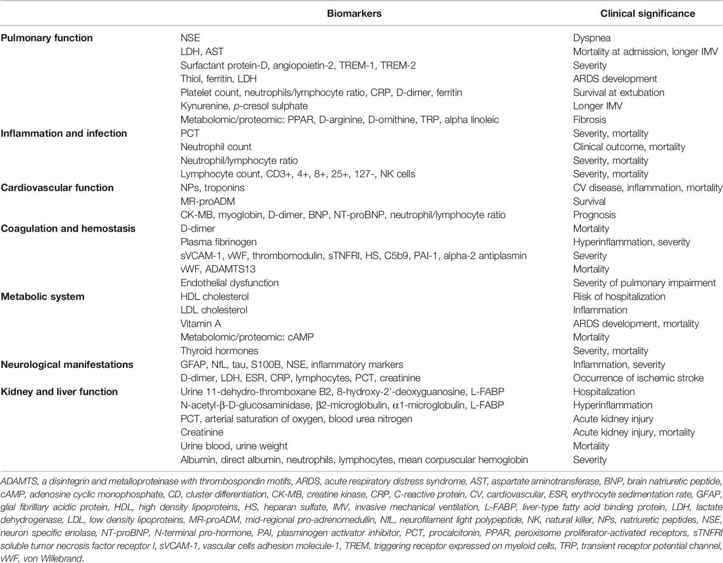
Table 1 Laboratory biomarkers in COVID-19.
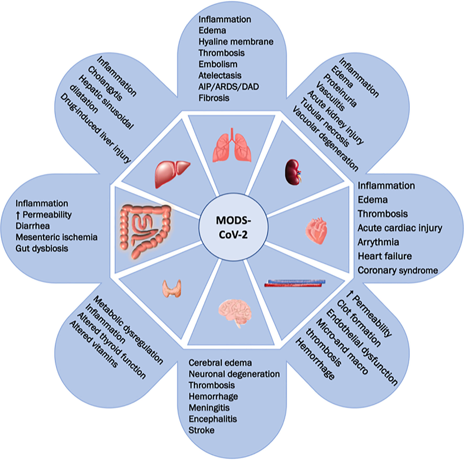
Figure 1 COVID-19 multiple organ dysfunction. This figure shows the potential for multiorgan involvement in COVID-19. Respiratory (AIP, acute interstitial pneumonia; ARDS, acute respiratory distress syndrome; DAD, diffuse alveolar damage), renal, cardiovascular, coagulative/hemostatic, liver, gastrointestinal, metabolic/endocrine, and cerebral functions and systems, as well as their possible alterations, are presented.
Diagnostic and Prognostic Value of Biomarkers
Biomarkers reflecting multiple organ involvement and/or pharmacological effects have been widely examined in critically ill patients. Some of these biomarkers are also used to monitor dysfunction in distinct organs at the same time, due to their redundancy or non-specificity. However, the most appropriate biomarkers to be studied in critically ill patients with COVID-19 have yet to be defined. Figure 2 depicts a proposed algorithm for critical care management which includes the investigation of biomarkers in severe COVID-19 patients at ICU admission.
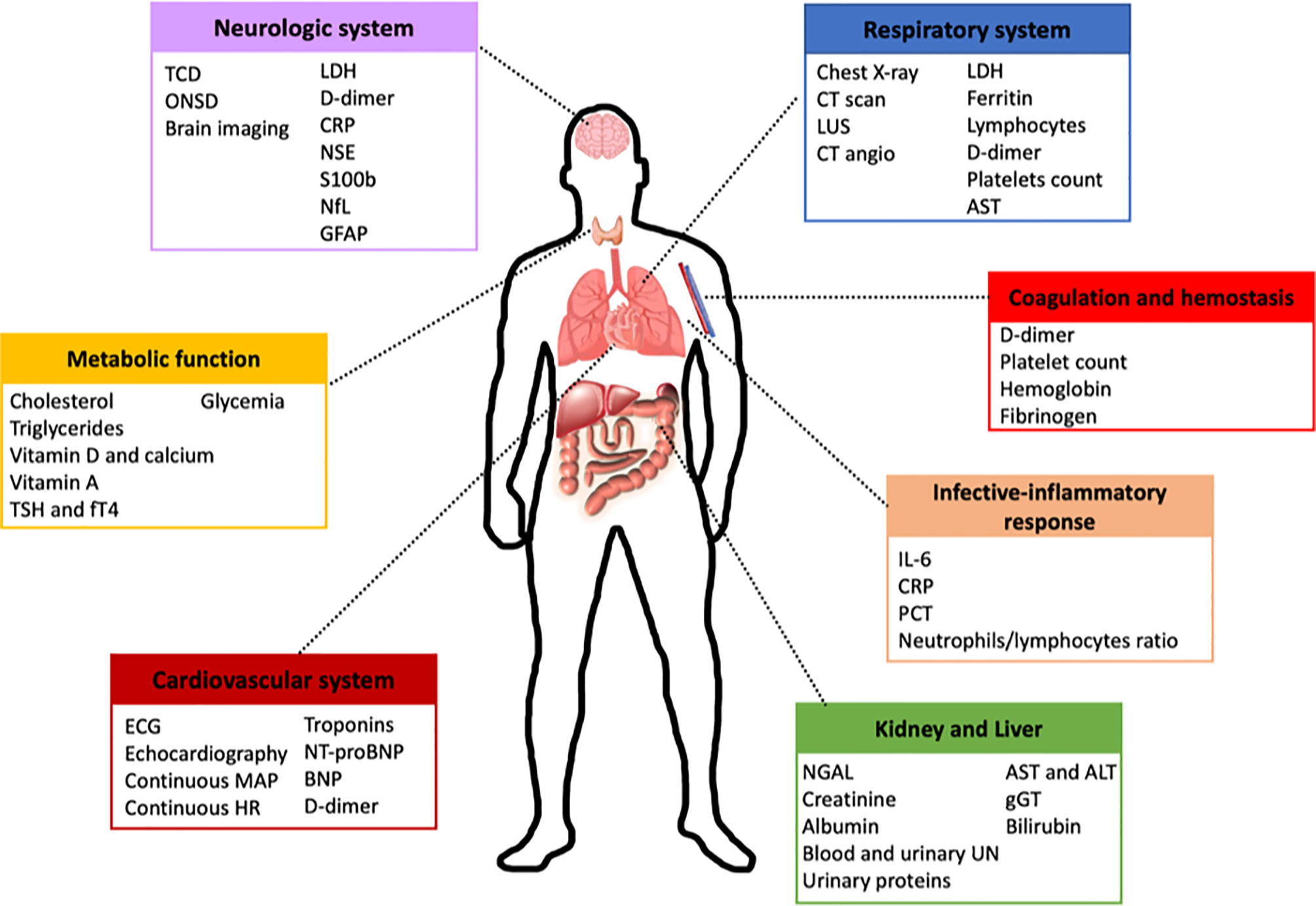
Figure 2 Proposed algorithm for the management of patients with COVID-19 at ICU admission. This figure shows a potential algorithm for initial patient management at ICU admission, including the most useful biomarkers to be used in the COVID-19 critical care setting. Neurological system: sequential transcranial doppler (TCD) and/or optic nerve sheath diameter (ONSD) in sedated patients for whom conventional neurological evaluation is impossible. Cardiovascular system: electrocardiogram and echocardiography, as well as continuous monitoring of mean arterial pressure (MAP) and heart rate (HR), are suggested on ICU admission. Respiratory system: computed tomography (CT) scan is the gold standard; if not feasible, chest X-ray, CT angiography, and/or lung ultrasound should be performed. Lactate dehydrogenase (LDH), C-reactive protein (CRP), neuron specific enolase (NSE), neurofilament light polypeptide (NfL), glial fibrillary acidic protein (GFAP), thyrotropic stimulating hormone (TSH), NGAL, aspartate transaminase (AST), alanine aminotransferase (ALT), gamma-glutamyl transferase (γGT), interleukin-6 (IL-6). BNP, brain natriuretic peptide; UN, urea nitrogen; NT-proBNP, N-terminal pro-hormone.
Respiratory System
The lungs are usually the organs affected primarily by SARS-CoV-2, due to their large and highly vascularized surface area (11). The pathogenesis of COVID-19 in the lung includes an initial phase of local inflammation, endothelial cell damage, and antifibrinolytic activation in the upper and lower respiratory tracts, followed by repair mechanisms that can elicit the restoration of normal pulmonary architecture. Inflammation is followed by platelet recruitment with degranulation, clot formation, altered vessel permeability, and accumulation of leukocytes in the injury site, leading to the recruitment of other inflammatory cells with the involvement of specific cytokines (i.e., IL-4, IL-13, transforming growth factor-β) that are also responsible for pro-fibrotic activity (12).
SARS-CoV-2 lung infection causes a wide variety of clinical manifestations and symptoms, from asymptomatic, mild, and moderate disease to severe COVID-19. Severe and critical illness accounts for up to 14% and 5% of cases, respectively, with the ARDS occurring in 10-20% of patients; multiorgan failure and death may supervene (13, 14). Various phenotypes have been identified by computed tomography (CT) (15, 16), including phenotype L or 1, which is characterized by low compliance, altered ventilation and perfusion, and shunting with focal hypo/hyper-perfused ground-glass opacities; and phenotype H or 2, which is identified by an inhomogeneous distribution of atelectasis with a patchy ARDS-like pattern (17, 18). Progressive evolution of COVID-19 (19) may lead to phenotype F, caused by mechanical stretch of lung epithelial cells and pathological fibro-proliferation and remodeling of the extracellular matrix, with increased expression of pro-fibrotic markers, as is mainly typical of severe forms of lung disease (20).
Although not specific to pulmonary disease, several biomarkers of different stages of lung involvement in COVID-19 have been identified and have been associated with pulmonary and systemic hyperinflammation and fibrotic damage (12). In the early disease course, neuron-specific enolase (NSE) can be used to differentiate patients who are going to develop dyspnea (21). On admission, higher lymphocyte and platelet counts and lower ferritin, D-dimer, lactate dehydrogenase (LDH), and aspartate transaminase (AST) have all been associated with lower risk of mortality in COVID-19 patients who ultimately required intubation and mechanical ventilation (22). Surfactant protein-D, angiopoietin-2, triggering receptor expressed on myeloid cell (TREM)-1, and TREM-2 levels were found to be higher in mild/moderate and severe/critical COVID-19 pneumonia than in asymptomatic and uncomplicated cases. Moreover, these biomarkers correlated well with clinical severity (23, 24). In severe COVID-19 cases, total thiol, ferritin, and LDH were identified as prognostic biomarkers for ARDS development (25). At extubation, COVID-19 survivors had higher platelet counts and neutrophil-to-lymphocyte ratios and lower C-reactive protein (CRP), D-dimer, ferritin, LDH, and AST (22).
Infection and Systemic Inflammatory Response
Following SARS-CoV-2 invasion of the host cells, the virus replicates at the infection site, thus triggering activation of the innate and adaptive immune responses (26). Neutrophils are rapidly recruited to infection foci, while innate cells recognize the virus and secrete multiple cytokines. Antigen-presenting cells recognize viral antigens which are carried to the local lymph nodes, while activating the T-helper cell response, which is also responsible for stimulating B cells to secrete antibodies (27). The systemic immune-inflammatory response is activated; if left unchecked, this may progress to multiorgan illness (28).
Patients with severe COVID-19 are highly susceptible to superimposed bacterial, fungal, and viral infections, including ventilator-associated pneumonia and bloodstream infection, among others (29, 30). As for systemic biomarkers of infection, procalcitonin is a predictor of disease severity (31), and can be useful to guide antimicrobial stewardship (32, 33). Another study found an association between procalcitonin and mortality in COVID-19 patients more than 75 years old (34). Neutrophil count was also predictive of clinical outcome in hospitalized COVID-19 patients (35), while the neutrophil-to-lymphocyte ratio was strongly associated with severity and mortality in COVID-19 (36). Additionally, total lymphocyte count, cluster differentiation (CD)3+, CD4+, CD8+, CD25+, CD127– T cells, and natural killer (NK) cells were found to be depressed in severe COVID-19 (37), whereas C-reactive protein, erythrocyte sedimentation rate, and IL-6 – common markers of inflammation – were elevated (38).
Cardiovascular System
SARS-CoV-2 can directly trigger endothelial dysfunction, causing a status known as COVID-19-associated coagulopathy. After viral entry into the cells, increased vascular permeability and tissue factor expression in subendothelial cells, with activation of platelets and leukocytes, may trigger the coagulation cascade. Endothelial damage and a generalized inflammatory state are drivers of thrombosis, which can contribute to cardiovascular manifestations (39).
Cardiovascular manifestations of COVID-19 are frequently reported (2, 40). Acute heart failure and exacerbation of chronic heart failure are reported in up to 20-30% of hospitalized patients, and carry high mortality rates, especially in patients with severe comorbidities (41–43). Acute coronary syndrome has been reported in a high proportion of patients, probably because of plaque rupture, coronary spasm, or microthrombi triggered by systemic inflammation and cytokine storm (44). In general, the mechanisms underlying cardiovascular manifestations include increased cardiac workload, hypoxemia, hypervolemia, myocardial injury, arrhythmias, myocarditis, stress-induced cardiomyopathy, acute kidney injury, and, as noted above, systemic inflammatory response with the release of several cytokines and chemokines (45). Triggering mechanisms may be attributed to an imbalance between heightened cardiac workload and reduced oxygen supply secondary to systemic conditions, with possible type-2 myocardial infarction (46).
Cardiac biomarkers (47), electrocardiography (ECG), and transthoracic echocardiography (TTE) play a pivotal role in risk stratification and early detection of cardiovascular complications, as well as to guide treatment (48, 49). Recent evidence confirmed that cardiac biomarkers, including natriuretic peptides (NPs) and troponins, may reflect cardiovascular involvement and inflammation in COVID-19, and are strongly associated with poor prognosis and mortality (41, 50–53). In some cases, troponin elevation in COVID-19 has been associated with ECG changes (54), ICU admission, and in-hospital death (55, 56). However, despite the confirmed prognostic impact of troponins, routine testing is still a matter of debate, because of several other variables that have been associated with outcome and prognosis (48). Additionally, pre-existing cardiac disease and/or acute stress injury may justify mild elevations in cardiac troponins, while myocarditis, Takotsubo syndrome, type 2 myocardial infarction triggered by severe respiratory failure, systemic hypoxemia, or shock are mostly associated with more marked increase in troponins (44, 57, 58). Other cardiac and non-cardiac biomarkers are common findings in COVID-19-associated cardiovascular disease, including creatine kinase (CK)-MB, myoglobin, D-dimer, brain natriuretic peptide (BNP) and its N-terminal pro-hormone (NT-proBNP), and neutrophil-to-lymphocyte ratio (55, 59–61). Myoglobin seems to offer higher prognostic accuracy than other cardiac-specific biomarkers (troponins and CK-MB) in COVID-19 (62). Moreover, mid-regional pro-adrenomedullin (MR-proADM) levels were found to be associated with endothelial dysfunction and mortality in COVID-19, potentially making it an optimal biomarker for the prediction of survival in this patient population (63). Nevertheless, only limited evidence exists so far to define any of these biomarkers as an independent predictor of prognosis in COVID-19 (48, 64).
Coagulation and Hemostasis
Coagulation derangement is a well-known systemic effect of COVID-19 that can originate from direct or indirect viral impact on the endothelium, or from immunothrombosis (65). COVID-19 can cause alterations in the coagulation cascade, with imbalance of the regulatory mechanisms of coagulation and fibrinolysis, altered platelet function, and a hyperinflammatory response (11, 65). In this context, D-dimer has been identified among the first altered coagulation biomarkers in COVID-19, and is predictive of mortality on admission (66). Similarly, plasma fibrinogen appears to be associated with hyperinflammation and disease severity in COVID-19 (67). A coagulopathy signature diagnostic of COVID-19 has been identified, including elevated levels of soluble vascular cell adhesion molecule (sVCAM)-1 (68), von Willebrand Factor (vWF), thrombomodulin, soluble tumor necrosis factor (TNF) receptor I (sTNFRI), heparan sulfate, C5b9 complement, plasminogen activator inhibitor (PAI)-1, and alpha-2 antiplasmin, among others. Some of these markers, such as sVCAM-1, vWF, sTNFRI, and heparan sulfate, were also associated with disease severity (69). Fibrinogen, thrombin peak, vWF, and ADAMTS13 at admission and elevated vWF : Ag to ADAMTS13 activity ratio were associated with severity and higher risk of death (70, 71). Endothelial dysfunction seems to be persistent after resolution of COVID-19, and directly associated with the severity of pulmonary impairment (72).
Metabolic Function
Sphingolipid metabolism regulates the inflammation and immune response through the conversion of sphingosine to sphingosine 1-phosphate, increasing the release of lymphocytes into the blood, with subsequent systemic inflammation and release of cytokines and chemokines in COVID-19 (73). Like lipid metabolism, fat-soluble vitamins such as vitamin D have been implicated in suppressing the cytokine storm and enhancing the immune response (74). Investigating lipid metabolism and its biomarkers could thus be of diagnostic and prognostic value in COVID-19.
Metabolic comorbidities including obesity, diabetes, cardiovascular, and hypertension have been associated with poor prognosis in COVID-19 (75). A certain degree of metabolic dysregulation has been found in COVID-19, possibly due to immune-triggered inflammation and hypercoagulability, as well as microbial changes in host physiology (10, 76). Indeed, COVID-19 patients with lower levels of high-density lipoprotein (HDL) cholesterol are more susceptible to hospitalization, while low-density lipoprotein (LDL) cholesterol was associated with higher inflammation (77). Critically ill patients with COVID-19 showed significantly lower levels of vitamin A than non-critical ones, and this was associated with higher inflammation (78). Vitamin A levels below 0.2 mg/L were significantly associated with the developments of ARDS and higher mortality (78). Vitamin D, a well-known regulator of phosphate and calcium metabolism with immunomodulatory functions, seems to not influence mortality or hospital length of stay in COVID-19 (79, 80). Finally, thyroid hormones showed marked association with disease severity and mortality, suggesting the importance of early assessment of thyroid function – and, when necessary, initiation of treatment – in hospitalized COVID-19 patients (81).
Neurologic Involvement
Pathogenetic mechanisms of SARS-CoV-2 neurologic manifestations include possible spreading of the virus across the blood-brain barrier via leukocyte migration or sluggish movement of blood within the microcirculation, thus binding to endothelial cells. Cells which may present ACE2 receptors, including neurons, astrocytes, and oligodendrocytes, can all be affected directly by viral entry and activate the local immune response. As a consequence of neuronal involvement, several biomarkers of neuroinflammation and damage can be detected (82).
Although COVID-19 rarely affects the brain as a primary manifestation, neurological complications are common in this patient population (82–84). Patients with neurological complications, compared to those without, may experience longer hospital stays, and the duration of mechanical ventilation can be associated with the risk of developing new neurological complications (84, 85). CT and magnetic resonance imaging (MRI) are considered the gold standard for detecting cerebral derangements, although the use of methods which involve exposure to ionizing radiation in non-primarily brain-injured patients can only be justified in case of high suspicion of neurological complications (86). The use of multimodal neuromonitoring has received increasing attention as a means of identifying patients at higher risk of brain derangement because of its low cost, speed, safety, and ready availability. However, the use of neuromonitoring tools is still mainly limited to specific settings (i.e., ICU) and patient populations (i.e., those with primary brain injury) (84).
Other than imaging, blood biomarkers can detect brain damage and predict prognosis efficiently. Blood biomarkers for the study of brain derangements include glial fibrillary acidic protein (GFAP), neurofilament light polypeptide (NfL), tau, S100B calcium binding protein, NSE, and inflammatory markers. Increased GFAP staining has been found in postmortem analysis of brain tissue from patients with COVID-19 (87), and NfL was significantly associated with COVID-19 status (88). Another study reported that GFAP was increased in both moderate and severe COVID-19 cases, whereas serum NfL was increased only in severe cases compared to controls (89). However, another study reported that serum NfL, although elevated across patients hospitalized with COVID-19, was not associated with neurological manifestations. Additionally, the usual close correlation between cerebrospinal fluid and serum NfL was not found, suggesting serum NfL elevation in the non-neurological patients may reflect peripheral nerve damage in response to severe illness (90). In COVID-19 patients with altered NfL and GFAP, values of these markers had normalized in all individuals at 6-month follow-up, suggesting that post-COVID-19 neurological sequelae may be not accompanied by ongoing brain injury (91). Inflammatory and coagulatory markers like D-dimer, LDH, erythrocyte sedimentation rate (ESR), and CRP were independently associated with the occurrence of ischemic stroke in COVID-19 (92, 93), while higher age, diabetes mellitus, and hypertension were found not to be significant predictors of stroke in this population, despite being known predictors of non-COVID-19 stroke (93). Levels of lymphocytes, procalcitonin, and creatinine were higher in COVID-19 stroke patients (94). S100B was higher in patients with mild and severe COVID-19 than in healthy controls, and may be a marker of disease severity (95). Antiphospholipid antibodies (i.e., anti-phosphatidylserine/prothrombin) were higher in COVID-19 patients, particularly those with neurological manifestations, than in controls. In contrast, anticardiolipin antibodies were not associated with neurologic involvement in COVID-19 (96).
Kidney and Liver
COVID-19 may cause kidney and liver injury by either direct infection of cells, via host immune clearance and immune tolerance disorders, endothelium-associated vasculitis, thrombus formation, metabolism and glucose disorder, or tissue hypoxia. As a consequence, biomarkers of endothelial, renal, hepatic, vascular, or hypoxic damage can help in the detection of new organ involvement and assist in determining prognosis (97).
As part of multiorgan involvement in COVID-19, kidney function might be altered directly by viral invasion or may occur secondary to multiple organ failure due to systemic inflammation or aggressive therapies (98). Around 25% of patients hospitalized with COVID-19 were reported to develop acute kidney injury, including low molecular weight proteinuria, Fanconi syndrome, and tubular injury (98). Moreover, regional inflammation, endothelial injury, and microthrombi have been identified as major causative factors of renal pathology in COVID-19. This is also sustained by the fact that anti-inflammatory drugs, such as steroids, play a key role in limiting renal disease progression (98). Classic diagnostic biomarkers of kidney damage include creatinine, neutrophil gelatinase-associated lipocalin (NGAL), cystatin C, kidney injury molecule-1 (KIM-1), blood and urinary urea nitrogen, and urinary proteins (99, 100).
Novel urinary biomarkers have been proposed in COVID-19, including urine 11-dehydro-thromboxane B2, 8-hydroxy-2′-deoxyguanosine, and liver-type fatty acid binding protein (L-FABP) levels, all of which were higher in this patient cohort at the time of hospitalization (101). N-acetyl-β-D-glucosaminidase, β2-microglobulin, α1-microglobulin, and L-FABP, which are all markers of tubular injury, were significantly associated with inflammation, as were IL-6 levels (102). Indeed, another observational study confirmed the association between pro-inflammatory cytokines, urinary cytokines, and urinary kidney injury markers (103). Procalcitonin was associated with acute kidney injury in COVID-19, and a score including simple and easily accessible variables such as procalcitonin, arterial saturation of oxygen, and blood urea nitrogen was shown to be predictive of acute kidney injury (104).
Altered serum creatinine levels with decreased kidney function at admission and up to 24 hours thereafter were significantly associated with acute kidney injury and in-hospital mortality (105). Additionally, urine blood >0.03 mg/dL and urine specific gravity >1.026 were associated with acute kidney injury, ICU admission, and higher mortality (106).
Abnormal liver and hepatobiliary function have been also identified in COVID-19 (107). A systematic review and meta-analysis showed a cumulative prevalence of liver disease of 24% in COVID-19, with possible alterations in albuminemia, liver enzymes, and total bilirubin (108). Recent findings showed that some liver and renal biomarkers, including albumin, direct bilirubin, neutrophil and lymphocyte counts, and mean corpuscular hemoglobin, are associated with risk of developing severe COVID-19 (107). Moreover, the presence of pre-existing liver fibrosis with silent liver injury significantly influenced mortality in COVID-19 (109).
Future Perspectives: Metabolomic and Proteomic Biomarkers and Machine Learning Models
Given the significant immune dysregulation of COVID-19 patients, the interplay between metabolism and immunity may play a pivotal role in the disease course (110). Additionally, oxygen deprivation may affect homeostasis in tissues and organs such as the lung, brain, kidney, and liver. The modulation of oxygen homeostasis and response to hypoxia is mainly mediated by glycolysis and the lactate cycle. This has increased research interest in proteomic and metabolomic methods to investigate pathways linked to energy production and amino acid metabolism in patients with SARS-CoV-2 infections (110). Metabolomic analyses in COVID-19 patients with and without pulmonary fibrosis revealed that pathways including the peroxisome proliferator-activated receptor (PPAR), D-arginine and D-ornithine metabolism, inflammatory tryptophan metabolic pathway (TRP), and alpha-linolenic acid metabolism were significantly increased in fibrotic lungs, thus suggesting that PPAR signaling is one of the main pathways involved in the formation and development of lung fibrosis in COVID-19 (9). A proteomic and metabolomic analysis identified hypoxanthine and betaine as predictors of ICU stay, and early ICU admission, elevated creatinine, and D-dimer were found to be associated with these pathways (8). Longer duration of invasive mechanical ventilation was associated with the kynurenine and p-cresol sulfate pathways (8). Several markers of metabolic function identified via metabolomic analysis were associated with in-hospital mortality, including cyclic adenosine monophosphate (cAMP), which plays a role in SARS-CoV2 endocytosis in the initial phase of the disease (10). Another major signature of the serum metabolome in COVID-19 was lactic acid, as well as spermidine and spermine. Many other metabolites were commonly increased, including glutamate, aspartate, phenylalanine, β-alanine, ornithine, arachidonic acid, choline, and xanthine (110). Recent machine learning models have been developed to support decision making and risk stratification in COVID-19. Most predictive models rely on demographic and clinical variables. However, biomarkers have recently shown good correlation with severity of disease and mortality in COVID-19 modeling (111). One example was a large study of 2,895 consecutive patients with COVID-19 in whom three biomarkers measured at admission were found to reflect pathobiological axes of myocardial injury, altered coagulation, and inflammation. The machine learning model concluded that patients with low levels of these biomarkers were at lower risk of critical disease and in-hospital mortality (112). In conclusion, the alterations found in the serum metabolome of patients with COVID-19 may reflect a more complex systemic derangement affecting carbon and nitrogen liver metabolism, but further research is needed to completely understand the impact of these alterations on routine clinical practice. Machine learning models can be promising in risk stratification in COVID-19. However, further investigations are needed to develop mathematical models that can help clinicians select the right parameters and interpret results.
Conclusions
Laboratory biomarkers have shown significant diagnostic and prognostic value for risk stratification in COVID-19. Furthermore, novel analytic strategies including metabolomics and proteomics offer interesting insights for early detection of patients at higher risk of severe disease and death. However, their limited availability restricts their widespread clinical use. Further investigations are warranted to identify a core set of laboratory biomarkers which can be used in daily clinical practice to easily predict prognosis and outcome in hospitalized patients with severe COVID-19.
Author Contributions
DB and ML-P: review, design, writing, editing. HC-F-N and PP: editing. PR: review, design, editing, senior contribution. All authors contributed to the article and approved the submitted version.
Funding
This work was supported by the Brazilian Council for Scientific and Technological Development (COVID-19-CNPq; 401700/2020-8 and 403485/2020-7); Rio de Janeiro State Research Foundation (COVID-19-FAPERJ; E-26/210.181/2020); and Funding Authority for Studies and Projects (01200008.00), Brazil.
Conflict of Interest
The authors declare that the research was conducted in the absence of any commercial or financial relationships that could be construed as a potential conflict of interest.
Publisher’s Note
All claims expressed in this article are solely those of the authors and do not necessarily represent those of their affiliated organizations, or those of the publisher, the editors and the reviewers. Any product that may be evaluated in this article, or claim that may be made by its manufacturer, is not guaranteed or endorsed by the publisher.
Acknowledgments
The authors express their gratitude to Mrs. Moira Elizabeth Schottler and Mr Filippe Vasconcellos for their assistance in editing the paper.
Abbreviations
ACE2, angiotensin receptor 2; ARDS, acute respiratory distress syndrome; AST, aspartate transaminase; BNP, brain natriuretic peptide; cAMP, cyclic adenosine monophosphate; CD, cluster differentiation; CK, creatine kinase; COVID-19, coronavirus disease 2019; CRP, C-reactive protein; CT, computed tomography; ECG, electrocardiography; ESR, erythrocyte sedimentation rate; GFAP, glial fibrillary acidic protein; HDL, high-density lipoprotein; ICU, intensive care unit; IL, interleukin; KIM, kidney injury molecule; L-FABP, liver-type fatty acid binding protein; LDH, lactate dehydrogenase; LDL, low-density lipoprotein; MR-proADM, mid-regional pro-adrenomedullin; MRI, magnetic resonance imaging; NfL, neurofilament light polypeptide; NGAL, neutrophil gelatinase-associated lipocalin; NK, natural killer; NP, natriuretic peptides; NSE, neuron-specific enolase; NT-proBNP, N-terminal pro-hormone BNP; PPAR, peroxisome proliferator-activated receptor; RASS, renin-angiotensin- aldosterone system; SARS-CoV-2, severe acute respiratory syndrome-coronavirus-2; ssRNA, single-stranded ribonucleic acid; sTNFRI, soluble TNF receptor I; sVCAM, soluble vascular cell adhesion molecule; TMPRSS2, transmembrane serine protease 2; TNF, tumor necrosis factor; TREM, triggering receptor expressed on myeloid cell; TRP, tryptophan metabolic pathway; TTE, transthoracic electrocardiography; vWF, von Willebrand Factor.
References
1. Robba C, Battaglini D, Pelosi P, Rocco PRM. Multiple Organ Dysfunction in SARS-CoV-2: MODS-CoV-2. Expert Rev Respir Med (2020) 14:865–8. doi: 10.1080/17476348.2020.1778470
2. Huang C, Wang Y, Li X, Ren L, Zhao J, Hu Y, et al. Clinical Features of Patients Infected With 2019 Novel Coronavirus in Wuhan, China. Lancet (2020) 395:497–506. doi: 10.1016/S0140-6736(20)30183-5
3. Zhang C, Wu Z, Li J-W, Zhao H, Wang G-Q. Cytokine Release Syndrome in Severe COVID-19: Interleukin-6 Receptor Antagonist Tocilizumab May Be the Key to Reduce Mortality. Int J Antimicrob Agents (2020) 55:105954. doi: 10.1016/j.ijantimicag.2020.105954
4. Liu J, Li S, Liu J, Liang B, Wang X, Wang H, et al. Longitudinal Characteristics of Lymphocyte Responses and Cytokine Profiles in the Peripheral Blood of SARS-CoV-2 Infected Patients. EBioMedicine (2020) 55:102763. doi: 10.1016/j.ebiom.2020.102763
5. Ackermann M, Verleden SE, Kuehnel M, Haverich A, Welte T, Laenger F, et al. Pulmonary Vascular Endothelialitis, Thrombosis, and Angiogenesis in Covid-19. N Engl J Med (2020) 383:120–8. doi: 10.1056/NEJMoa2015432
6. Leisman DE, Ronner L, Pinotti R, Taylor MD, Sinha P, Calfee CS, et al. Cytokine Elevation in Severe and Critical COVID-19: A Rapid Systematic Review, Meta-Analysis, and Comparison With Other Inflammatory Syndromes. Lancet Respir Med (2020) 8:1233–44. doi: 10.1016/S2213-2600(20)30404-5
7. Andrianto, Al-Farabi MJ, Nugraha RA, Marsudi BA, Azmi Y. Biomarkers of Endothelial Dysfunction and Outcomes in Coronavirus Disease 2019 (COVID-19) Patients: A Systematic Review and Meta-Analysis. Microvasc Res (2021) 138:104224. doi: 10.1016/j.mvr.2021.104224
8. Taleb S, Yassine HM, Benslimane FM, Smatti MK, Schuchardt S, Albagha O, et al. Predictive Biomarkers of Intensive Care Unit and Mechanical Ventilation Duration in Critically-Ill Coronavirus Disease 2019 Patients. Front Med (2021) 8:733657. doi: 10.3389/fmed.2021.733657
9. Yang J, Chen C, Chen W, Huang L, Fu Z, Ye K, et al. Proteomics and Metabonomics Analyses of Covid-19 Complications in Patients With Pulmonary Fibrosis. Sci Rep (2021) 11:14601. doi: 10.1038/s41598-021-94256-8
10. Saccon E, Bandera A, Sciumè M, Mikaeloff F, Lashari AA, Aliberti S, et al. Distinct Metabolic Profile Associated With a Fatal Outcome in COVID-19 Patients During the Early Epidemic in Italy. Microbiol Spectr (2021) 9:e00549–21. doi: 10.1128/Spectrum.00549-21
11. Lopes-Pacheco M, Silva PL, Cruz FF, Battaglini D, Robba C, Pelosi P, et al. Pathogenesis of Multiple Organ Injury in COVID-19 and Potential Therapeutic Strategies. Front Physiol (2021) 12:593223. doi: 10.3389/fphys.2021.593223
12. Vianello A, Guarnieri G, Braccioni F, Lococo S, Molena B, Cecchetto A, et al. The Pathogenesis, Epidemiology and Biomarkers of Susceptibility of Pulmonary Fibrosis in COVID-19 Survivors. Clin Chem Lab Med (2022) 60:307–16. doi: 10.1515/cclm-2021-1021
13. Chen N, Zhou M, Dong X, Qu J, Gong F, Han Y, et al. Epidemiological and Clinical Characteristics of 99 Cases of 2019 Novel Coronavirus Pneumonia in Wuhan, China: A Descriptive Study. Lancet (2020) 395:507–13. doi: 10.1016/S0140-6736(20)30211-7
14. Zhou M, Zhang X, Qu J. Coronavirus Disease 2019 (COVID-19): A Clinical Update. Front Med (2020) 14:126–35. doi: 10.1007/s11684-020-0767-8
15. Orlandi D, Battaglini D, Robba C, Viganò M, Bergamaschi G, Mignatti T, et al. COVID-19 Phenotypes, Lung Ultrasound, Chest Computed Tomography, and Clinical Features in Critically Ill Mechanically Ventilated Patients. Ultrasound Med Biol (2021) 41(12):3323–32. doi: 10.1016/j.ultrasmedbio.2021.07.014.
16. Pelosi P, Ball L, Barbas CSV, Bellomo R, Burns KEA, Einav S, et al. Personalized Mechanical Ventilation in Acute Respiratory Distress Syndrome. Crit Care (2021) 25:250. doi: 10.1186/s13054-021-03686-3
17. Gattinoni L, Chiumello D, Caironi P, Busana M, Romitti F, Brazzi L, et al. COVID-19 Pneumonia: Different Respiratory Treatments for Different Phenotypes? Intensive Care Med (2020) 46:1099–102. doi: 10.1007/s00134-020-06033-2
18. Robba C, Battaglini D, Ball L, Patroniti N, Loconte M, Brunetti I, et al. Distinct Phenotypes Require Distinct Respiratory Management Strategies in Severe COVID-19. Respir Physiol Neurobiol (2020) 279:103455. doi: 10.1016/j.resp.2020.103455
19. Tonelli R, Marchioni A, Tabbì L, Fantini R, Busani S, Castaniere I, et al. Spontaneous Breathing and Evolving Phenotypes of Lung Damage in Patients With COVID-19: Review of Current Evidence and Forecast of a New Scenario. J Clin Med (2021) 10:975. doi: 10.3390/jcm10050975
20. George PM, Wells AU, Jenkins RG. Pulmonary Fibrosis and COVID-19: The Potential Role for Antifibrotic Therapy. Lancet Respir Med (2020) 8:807–15. doi: 10.1016/S2213-2600(20)30225-3
21. Cione E, Siniscalchi A, Gangemi P, Cosco L, Colosimo M, Longhini F, et al. Neuron-Specific Enolase Serum Levels in COVID-19 Are Related to the Severity of Lung Injury. PLoS One (2021) 16:e0251819. doi: 10.1371/journal.pone.0251819
22. Topp G, Bouyea M, Cochran-Caggiano N, Ata A, Torres P, Jacob J, et al. Biomarkers Predictive of Extubation and Survival of COVID-19 Patients. Cureus (2021) 13:e15462. doi: 10.7759/cureus.15462
23. Alay H, Laloglu E. The Role of Angiopoietin-2 and Surfactant Protein-D Levels in SARS-CoV-2-Related Lung Injury: A Prospective, Observational, Cohort Study. J Med Virol (2021) 93:6008–15. doi: 10.1002/jmv.27184
24. Kerget F, Kerget B, İba Yılmaz S, Kızıltunç A. Evaluation of the Relationship Between TREM-1/TREM-2 Ratio and Clinical Course in COVID-19 Pneumonia. Int J Clin Pract (2021) 75(10):e14697. doi: 10.1111/ijcp.14697
25. Martinez Mesa A, Cabrera César E, Martín-Montañez E, Sanchez Alvarez E, Lopez PM, Romero-Zerbo Y, et al. Acute Lung Injury Biomarkers in the Prediction of COVID-19 Severity: Total Thiol, Ferritin and Lactate Dehydrogenase. Antioxidants (2021) 10:1221. doi: 10.3390/antiox10081221
26. V’kovski P, Kratzel A, Steiner S, Stalder H, Thiel V. Coronavirus Biology and Replication: Implications for SARS-CoV-2. Nat Rev Microbiol (2021) 19:155–70. doi: 10.1038/s41579-020-00468-6
27. Rouse BT, Sehrawat S. Immunity and Immunopathology to Viruses: What Decides the Outcome? Nat Rev Immunol (2010) 10:514–26. doi: 10.1038/nri2802
28. Hamming I, Timens W, Bulthuis M, Lely A, Navis G, van Goor H. Tissue Distribution of ACE2 Protein, the Functional Receptor for SARS Coronavirus. A First Step Understanding SARS Pathogen J Pathol (2004) 203:631–7. doi: 10.1002/path.1570
29. Ippolito M, Misseri G, Catalisano G, Marino C, Ingoglia G, Alessi M, et al. Ventilator-Associated Pneumonia in Patients With COVID-19: A Systematic Review and Meta-Analysis. Antibiotics (2021) 10:545. doi: 10.3390/antibiotics10050545
30. Ippolito M, Simone B, Filisina C, Catalanotto FR, Catalisano G, Marino C, et al. Bloodstream Infections in Hospitalized Patients With COVID-19: A Systematic Review and Meta-Analysis. Microorganisms (2021) 9:2016. doi: 10.3390/microorganisms9102016
31. Shen Y, Cheng C, Zheng X, Jin Y, Duan G, Chen M, et al. Elevated Procalcitonin Is Positively Associated With the Severity of COVID-19: A Meta-Analysis Based on 10 Cohort Studies. Med (B Aires) (2021) 57:594. doi: 10.3390/medicina57060594
32. Waris A, Din M, Iqbal N, Yar L, Khalid A, Nawaz M, et al. Evaluation of Serum Procalcitonin Level as a Biomarker for Disease Severity in COVID-19 Patients. New Microbes New Infect (2021) 43:100922. doi: 10.1016/j.nmni.2021.100922
33. Calderon M, Li A, Bazo-Alvarez JC, Dennis J, Baker KF, Schim van der Loeff I, et al. Evaluation of Procalcitonin-Guided Antimicrobial Stewardship in Patients Admitted to Hospital With COVID-19 Pneumonia. JAC-Antimicrobial Resist (2021) 3(3):dlab133. doi: 10.1093/jacamr/dlab133
34. Ticinesi A, Nouvenne A, Prati B, Guida L, Parise A, Cerundolo N, et al. The Clinical Significance of Procalcitonin Elevation in Patients Over 75 Years Old Admitted for COVID-19 Pneumonia. Mediators Inflamm (2021) 2021:1–10. doi: 10.1155/2021/5593806
35. Kåsine T, Dyrhol-Riise AM, Barratt-Due A, Kildal AB, Olsen IC, Nezvalova-Henriksen K, et al. Neutrophil Count Predicts Clinical Outcome in Hospitalized COVID-19 Patients: Results From the NOR-Solidarity Trial. J Intern Med (2021) 291(12):241–3. doi: 10.1111/joim.13377
36. Ulloque-Badaracco JR, Ivan Salas-Tello W, Al-kassab-Córdova A, Alarcón-Braga EA, Benites-Zapata VA, Maguiña JL, et al. Prognostic Value of Neutrophil-to-Lymphocyte Ratio in COVID-19 Patients: A Systematic Review and Meta-Analysis. Int J Clin Pract (2021) 75:e14596. doi: 10.1111/ijcp.14596
37. Liu K, Yang T, Peng X, Lv S, Ye X, Zhao T, et al. A Systematic Meta-Analysis of Immune Signatures in Patients With COVID-19. Rev Med Virol (2021) 31:e2195. doi: 10.1002/rmv.2195
38. Iwamura APD, Tavares da Silva MR, Hümmelgen AL, Soeiro Pereira PV, Falcai A, Grumach AS, et al. Immunity and Inflammatory Biomarkers in COVID-19: A Systematic Review. Rev Med Virol (2021) 31:e2199. doi: 10.1002/rmv.2199
39. Gorog DA, Storey RF, Gurbel PA, Tantry US, Berger JS, Chan MY, et al. Current and Novel Biomarkers of Thrombotic Risk in COVID-19: A Consensus Statement From the International COVID-19 Thrombosis Biomarkers Colloquium. Nat Rev Cardiol (2022), 1–21. doi: 10.1038/s41569-021-00665-7
40. Wang D, Hu B, Hu C, Zhu F, Liu X, Zhang J, et al. Clinical Characteristics of 138 Hospitalized Patients With 2019 Novel Coronavirus–Infected Pneumonia in Wuhan, China. JAMA (2020) 323:1061. doi: 10.1001/jama.2020.1585
41. Zhou F, Yu T, Du R, Fan G, Liu Y, Liu Z, et al. Clinical Course and Risk Factors for Mortality of Adult Inpatients With COVID-19 in Wuhan, China: A Retrospective Cohort Study. Lancet (2020) 395:1054–62. doi: 10.1016/S0140-6736(20)30566-3
42. Yang J, Zheng Y, Gou X, Pu K, Chen Z, Guo Q, et al. Prevalence of Comorbidities and its Effects in Patients Infected With SARS-CoV-2: A Systematic Review and Meta-Analysis. Int J Infect Dis (2020) 94:91–5. doi: 10.1016/j.ijid.2020.03.017
43. Zhu Z, Wang M, Lin W, Cai Q, Zhang L, Chen D, et al. Cardiac Biomarkers, Cardiac Injury, and Comorbidities Associated With Severe Illness and Mortality in Coronavirus Disease 2019 (COVID-19): A Systematic Review and Meta-Analysis. Immun Inflammation Dis (2021) 9(4):1071–100. doi: 10.1002/iid3.471
44. Schiavone M, Gobbi C, Biondi-Zoccai G, D’Ascenzo F, Palazzuoli A, Gasperetti A, et al. Acute Coronary Syndromes and Covid-19: Exploring the Uncertainties. J Clin Med (2020) 9:1683. doi: 10.3390/jcm9061683
45. Nishiga M, Wang DW, Han Y, Lewis DB, Wu JC. COVID-19 and Cardiovascular Disease: From Basic Mechanisms to Clinical Perspectives. Nat Rev Cardiol (2020) 17:543–58. doi: 10.1038/s41569-020-0413-9
46. Jabri A, Kalra A, Kumar A, Alameh A, Adroja S, Bashir H, et al. Incidence of Stress Cardiomyopathy During the Coronavirus Disease 2019 Pandemic. JAMA Netw Open (2020) 3:e2014780. doi: 10.1001/jamanetworkopen.2020.14780
47. De Marzo V, Di Biagio A, Della Bona R, Vena A, Arboscello E, Emirjona H, et al. Prevalence and Prognostic Value of Cardiac Troponin in Elderly Patients Hospitalized for COVID-19. J Geriatr Cardiol (2021) 18:338–45. doi: 10.11909/j.issn.1671-5411.2021.05.004
48. The European Society for Cardiology. ESC Guidance for the Diagnosis and Management of CV Disease During the COVID-19 Pandemic. (2020). Avalable at: https://www.escardio.org/Education/Covid-19-and-Cardiology/ESC-COVID-19-(update: 10 June 2020).
49. Gilad V, De Marzo V, Guglielmi G, Della BR, Giovinazzo S, Pescetelli F, et al. Cardiac Point-of-Care Ultrasound in Hospitalized Coronavirus Disease-2019 Patients: Findings and Association With Outcome. J Cardiovasc Med (2022) 23:e3–7. doi: 10.2459/JCM.0000000000001177
50. Metkus TS, Sokoll LJ, Barth AS, Czarny MJ, Hays AG, Lowenstein CJ, et al. Myocardial Injury in Severe COVID-19 Compared With Non–COVID-19 Acute Respiratory Distress Syndrome. Circulation (2021) 143:553–65. doi: 10.1161/CIRCULATIONAHA.120.050543
51. Shi S, Qin M, Shen B, Cai Y, Liu T, Yang F, et al. Association of Cardiac Injury With Mortality in Hospitalized Patients With COVID-19 in Wuhan, China. JAMA Cardiol (2020) 5:802. doi: 10.1001/jamacardio.2020.0950
52. Guan W, Ni Z, Hu Y, Liang W, Ou C, He J, et al. Clinical Characteristics of Coronavirus Disease 2019 in China. N Engl J Med (2020) 382:1708–20. doi: 10.1056/NEJMoa2002032
53. Ni W, Yang X, Liu J, Bao J, Li R, Xu Y, et al. Acute Myocardial Injury at Hospital Admission Is Associated With All-Cause Mortality in COVID-19. J Am Coll Cardiol (2020) 76:124–5. doi: 10.1016/j.jacc.2020.05.007
54. Doyen D, Moceri P, Ducreux D, Dellamonica J. Myocarditis in a Patient With COVID-19: A Cause of Raised Troponin and ECG Changes. Lancet (2020) 395:1516. doi: 10.1016/S0140-6736(20)30912-0
55. Cipriani A, Capone F, Donato F, Molinari L, Ceccato D, Saller A, et al. Cardiac Injury and Mortality in Patients With Coronavirus Disease 2019 (COVID-19): Insights From a Mediation Analysis. Intern Emerg Med (2021) 16:419–27. doi: 10.1007/s11739-020-02495-w
56. Lombardi CM, Carubelli V, Iorio A, Inciardi RM, Bellasi A, Canale C, et al. Association of Troponin Levels With Mortality in Italian Patients Hospitalized With Coronavirus Disease 2019. JAMA Cardiol (2020) 5:1274. doi: 10.1001/jamacardio.2020.3538
57. Cameli M, Pastore MC, Mandoli GE, D’Ascenzi F, Focardi M, Biagioni G, et al. COVID-19 and Acute Coronary Syndromes: Current Data and Future Implications. Front Cardiovasc Med (2021) 7:593496. doi: 10.3389/fcvm.2020.593496
58. Chieffo A, Stefanini GG, Price S, Barbato E, Tarantini G, Karam N, et al. EAPCI Position Statement on Invasive Management of Acute Coronary Syndromes During the COVID-19 Pandemic. Eur Heart J (2020) 41:1839–51. doi: 10.1093/eurheartj/ehaa381
59. Chung MK, Zidar DA, Bristow MR, Cameron SJ, Chan T, Harding CV, et al. COVID-19 and Cardiovascular Disease. Circ Res (2021) 128:1214–36. doi: 10.1161/CIRCRESAHA.121.317997
60. Zinellu A, Sotgia S, Fois AG, Mangoni AA, Serum CK-MB. COVID-19 Severity and Mortality: An Updated Systematic Review and Meta-Analysis With Meta-Regression. Adv Med Sci (2021) 66:304–14. doi: 10.1016/j.advms.2021.07.001
61. Zhan L, Liu Y, Cheng Y, Guo W, Yang J. Predictive Value of Neutrophil/Lymphocyte Ratio (NLR) on Cardiovascular Events in Patients With COVID-19. Int J Gen Med (2021) 14:3899–907. doi: 10.2147/IJGM.S317380
62. Yu J-S, Chen R-D, Zeng L-C, Yang H-K, Li H. Myoglobin Offers Higher Accuracy Than Other Cardiac-Specific Biomarkers for the Prognosis of COVID-19. Front Cardiovasc Med (2021) 8:686328. doi: 10.3389/fcvm.2021.686328
63. García de Guadiana-Romualdo L, Martínez Martínez M, Rodríguez Mulero MD, Esteban-Torrella P, Hernández Olivo M, Alcaraz García MJ, et al. Circulating MR-proADM Levels, as an Indicator of Endothelial Dysfunction, for Early Risk Stratification of Mid-Term Mortality in COVID-19 Patients. Int J Infect Dis (2021) 111:211–8. doi: 10.1016/j.ijid.2021.08.058
64. Sorrentino S, Cacia M, Leo I, Polimeni A, Sabatino J, Spaccarotella CAM, et al. B-Type Natriuretic Peptide as Biomarker of COVID-19 Disease Severity—A Meta-Analysis. J Clin Med (2020) 9:2957. doi: 10.3390/jcm9092957
65. Robba C, Battaglini D, Ball L, Valbusa A, Porto I, Della Bona R, et al. Coagulative Disorders in Critically Ill COVID-19 Patients With Acute Distress Respiratory Syndrome: A Critical Review. J Clin Med (2021) 10:140. doi: 10.3390/jcm10010140
66. Poudel A, Poudel Y, Adhikari A, Aryal BB, Dangol D, Bajracharya T, et al. D-Dimer as a Biomarker for Assessment of COVID-19 Prognosis: D-Dimer Levels on Admission and Its Role in Predicting Disease Outcome in Hospitalized Patients With COVID-19. PLoS One (2021) 16:e0256744. doi: 10.1371/journal.pone.0256744
67. Sui J, Noubouossie DF, Gandotra S, Cao L. Elevated Plasma Fibrinogen Is Associated With Excessive Inflammation and Disease Severity in COVID-19 Patients. Front Cell Infect Microbiol (2021) 11:734005. doi: 10.3389/fcimb.2021.734005
68. Blot M, de Maistre E, Bourredjem A, Quenot J-P, Nguyen M, Bouhemad B, et al. Specific Features of the Coagulopathy Signature in Severe COVID-19 Pneumonia. Front Med (2021) 8:675191. doi: 10.3389/fmed.2021.675191
69. Fernández S, Moreno-Castaño AB, Palomo M, Martinez-Sanchez J, Torramadé-Moix S, Téllez A, et al. Distinctive Biomarker Features in The Endotheliopathy of COVID-19 and Septic Syndromes. Shock (2021) 57(1):95–105. doi: 10.1097/SHK.0000000000001823. Online ahead of print.
70. Billoir P, Alexandre K, Duflot T, Roger M, Miranda S, Goria O, et al. Investigation of Coagulation Biomarkers to Assess Clinical Deterioration in SARS-CoV-2 Infection. Front Med (2021) 8:670694. doi: 10.3389/fmed.2021.670694
71. Joly BS, Darmon M, Dekimpe C, Dupont T, Dumas G, Yvin E, et al. Imbalance of Von Willebrand Factor and ADAMTS13 Axis Is Rather a Biomarker of Strong Inflammation and Endothelial Damage Than a Cause of Thrombotic Process in Critically Ill COVID-19 Patients. J Thromb Haemost (2021) 19:2193–8. doi: 10.1111/jth.15445
72. Ambrosino P, Calcaterra I, Molino A, Moretta P, Lupoli R, Spedicato GA, et al. Persistent Endothelial Dysfunction in Post-Acute COVID-19 Syndrome: A Case-Control Study. Biomedicines (2021) 9:957. doi: 10.3390/biomedicines9080957
73. Janneh AH, Kassir MF, Dwyer CJ, Chakraborty P, Pierce JS, Flume PA, et al. Alterations of Lipid Metabolism Provide Serologic Biomarkers for the Detection of Asymptomatic Versus Symptomatic COVID-19 Patients. Sci Rep (2021) 11:14232. doi: 10.1038/s41598-021-93857-7
74. Razdan K, Singh K, Singh D. Vitamin D Levels and COVID-19 Susceptibility: Is There Any Correlation? Med Drug Discov (2020) 7:100051. doi: 10.1016/j.medidd.2020.100051
75. Fakhroo AD, Al Thani AA, Yassine HM. Markers Associated With COVID-19 Susceptibility, Resistance, and Severity. Viruses (2020) 13:45. doi: 10.3390/v13010045
76. Battaglini D, Robba C, Fedele A, Trancǎ S, Sukkar SG, Di Pilato V, et al. The Role of Dysbiosis in Critically Ill Patients With COVID-19 and Acute Respiratory Distress Syndrome. Front Med (2021) 8:671714. doi: 10.3389/fmed.2021.671714
77. Alcántara-Alonso E, Molinar-Ramos F, González-López JA, Alcántara-Alonso V, Muñoz-Pérez MA, Lozano-Nuevo JJ, et al. High Triglyceride to HDL-Cholesterol Ratio as a Biochemical Marker of Severe Outcomes in COVID-19 Patients. Clin Nutr ESPEN (2021) 44:437–44. doi: 10.1016/j.clnesp.2021.04.020
78. Tepasse P-R, Vollenberg R, Fobker M, Kabar I, Schmidt H, Meier JA, et al. Vitamin A Plasma Levels in COVID-19 Patients: A Prospective Multicenter Study and Hypothesis. Nutrients (2021) 13:2173. doi: 10.3390/nu13072173
79. Murai IH, Fernandes AL, Sales LP, Pinto AJ, Goessler KF, Duran CSC, et al. Effect of a Single High Dose of Vitamin D 3 on Hospital Length of Stay in Patients With Moderate to Severe COVID-19. JAMA (2021) 325:1053. doi: 10.1001/jama.2020.26848
80. Zelzer S, Prüller F, Curcic P, Sloup Z, Holter M, Herrmann M, et al. Vitamin D Metabolites and Clinical Outcome in Hospitalized COVID-19 Patients. Nutrients (2021) 13:2129. doi: 10.3390/nu13072129
81. Beltrão FE de L, Beltrão DC de A, Carvalhal G, Beltrão FE de L, Brito A da S, da Capistrano KHR, et al. Thyroid Hormone Levels During Hospital Admission Inform Disease Severity and Mortality in COVID-19 Patients. Thyroid (2021) 31:1639–49. doi: 10.1089/thy.2021.0225
82. Battaglini D, Brunetti I, Anania P, Fiaschi P, Zona G, Ball L, et al. Neurological Manifestations of Severe SARS-CoV-2 Infection: Potential Mechanisms and Implications of Individualized Mechanical Ventilation Settings. Front Neurol (2020) 11:845. doi: 10.3389/fneur.2020.00845
83. Huth SF, Cho S-M, Robba C, Highton D, Battaglini D, Bellapart J, et al. Neurological Manifestations of Coronavirus Disease 2019: A Comprehensive Review and Meta-Analysis of the First 6 Months of Pandemic Reporting. Front Neurol (2021) 12:664599. doi: 10.3389/fneur.2021.664599
84. Battaglini D, Santori G, Chandraptham K, Iannuzzi F, Bastianello M, Tarantino F, et al. Neurological Complications and Noninvasive Multimodal Neuromonitoring in Critically Ill Mechanically Ventilated COVID-19 Patients. Front Neurol (2020) 11:602114. doi: 10.3389/fneur.2020.602114
85. Barbosa-Silva MC, Lima MN, Battaglini D, Robba C, Pelosi P, Rocco PRM, et al. Infectious Disease-Associated Encephalopathies. Crit Care (2021) 25:236. doi: 10.1186/s13054-021-03659-6
86. Lee B, Newberg A. Neuroimaging in Traumatic Brain Imaging. NeuroRX (2005) 2:372–83. doi: 10.1602/neurorx.2.2.372
87. Reichard RR, Kashani KB, Boire NA, Constantopoulos E, Guo Y, Lucchinetti CF. Neuropathology of COVID-19: A Spectrum of Vascular and Acute Disseminated Encephalomyelitis (ADEM)-Like Pathology. Acta Neuropathol (2020) 140:1–6. doi: 10.1007/s00401-020-02166-2
88. Ameres M, Brandstetter S, Toncheva AA, Kabesch M, Leppert D, Kuhle J, et al. Association of Neuronal Injury Blood Marker Neurofilament Light Chain With Mild-to-Moderate COVID-19. J Neurol (2020) 267:3476–8. doi: 10.1007/s00415-020-10050-y
89. Kanberg N, Ashton NJ, Andersson L-M, Yilmaz A, Lindh M, Nilsson S, et al. Neurochemical Evidence of Astrocytic and Neuronal Injury Commonly Found in COVID-19. Neurology (2020) 95:e1754–9. doi: 10.1212/WNL.0000000000010111
90. Paterson RW, Benjamin LA, Mehta PR, Brown RL, Athauda D, Ashton NJ, et al. Serum and Cerebrospinal Fluid Biomarker Profiles in Acute SARS-CoV-2-Associated Neurological Syndromes. Brain Commun (2021) 3:fcab099. doi: 10.1093/braincomms/fcab099
91. Kanberg N, Simrén J, Edén A, Andersson L-M, Nilsson S, Ashton NJ, et al. Neurochemical Signs of Astrocytic and Neuronal Injury in Acute COVID-19 Normalizes During Long-Term Follow-Up. EBioMedicine (2021) 70:103512. doi: 10.1016/j.ebiom.2021.103512
92. Esenwa C, Cheng NT, Luna J, Willey J, Boehme AK, Kirchoff-Torres K, et al. Biomarkers of Coagulation and Inflammation in COVID-19–Associated Ischemic Stroke. Stroke (2021) 52:e706–709. doi: 10.1161/STROKEAHA.121.035045
93. Goyal N, Sodani AK, Jain R, Ram H. Do Elevated Levels of Inflammatory Biomarkers Predict the Risk of Occurrence of Ischemic Stroke in SARS-CoV2 ?: An Observational Study. J Stroke Cerebrovasc Dis (2021) 30:106063. doi: 10.1016/j.jstrokecerebrovasdis.2021.106063
94. Yassin A, Ghzawi A, Al-Mistarehi A-H, El-Salem K, Y Benmelouka A, M Sherif A, et al. Mortality Rate and Biomarker Expression Within COVID-19 Patients Who Develop Acute Ischemic Stroke: A Systematic Review and Meta-Analysis. Futur Sci OA (2021) 7:FSO713. doi: 10.2144/fsoa-2021-0036
95. Mete E, Sabirli R, Goren T, Turkcuer I, Kurt O, Koseler A. Association Between S100b Levels and COVID-19 Pneumonia: A Case Control Study. Vivo (Brooklyn) (2021) 35:2923–8. doi: 10.21873/invivo.12583
96. Benjamin LA, Paterson RW, Moll R, Pericleous C, Brown R, Mehta PR, et al. Antiphospholipid Antibodies and Neurological Manifestations in Acute COVID-19: A Single-Centre Cross-Sectional Study. EClinicalMedicine (2021) 39:101070. doi: 10.1016/j.eclinm.2021.101070
97. Faour WH, Choaib A, Issa E, El Choueiry F, Shbaklo K, Alhajj M, et al. Mechanisms of COVID-19-Induced Kidney Injury and Current Pharmacotherapies. Inflammation Res (2022) 71:39–56. doi: 10.1007/s00011-021-01520-8
98. Legrand M, Bell S, Forni L, Joannidis M, Koyner JL, Liu K, et al. Pathophysiology of COVID-19-Associated Acute Kidney Injury. Nat Rev Nephrol (2021) 17:751–64. doi: 10.1038/s41581-021-00452-0
99. Liu Y, Xia P, Cao W, Liu Z, Ma J, Zheng K, et al. Divergence Between Serum Creatine and Cystatin C in Estimating Glomerular Filtration Rate of Critically Ill COVID-19 Patients. Ren Fail (2021) 43:1104–14. doi: 10.1080/0886022X.2021.1948428
100. Yasar E, Ozger HS, Yeter HH, Yildirim C, Osmanov Z, Cetin TE, et al. Could Urinary Kidney Injury Molecule-1 be a Good Marker in Subclinical Acute Kidney Injury in Mild to Moderate COVID-19 Infection? Int Urol Nephrol (2021) 54(3):627–36. doi: 10.1007/s11255-021-02937-0
101. Tantry US, Bliden KP, Cho A, Walia N, Dahlen JR, Ens G, et al. First Experience Addressing the Prognostic Utility of Novel Urinary Biomarkers in Patients With COVID-19. Open Forum Infect Dis (2021) 8:ofab274. doi: 10.1093/ofid/ofab274
102. Fukao Y, Nagasawa H, Nihei Y, Hiki M, Naito T, Kihara M, et al. COVID-19-Induced Acute Renal Tubular Injury Associated With Elevation of Serum Inflammatory Cytokine. Clin Exp Nephrol (2021) 25:1240–6. doi: 10.1007/s10157-021-02101-z
103. Gradin A, Andersson H, Luther T, Anderberg SB, Rubertsson S, Lipcsey M, et al. Urinary Cytokines Correlate With Acute Kidney Injury in Critically Ill COVID-19 Patients. Cytokine (2021) 146:155589. doi: 10.1016/j.cyto.2021.155589
104. Wang RR, He M, Kang Y. A Risk Score Based on Procalcitonin for Predicting Acute Kidney Injury in COVID-19 Patients. J Clin Lab Anal (2021) 35:e23805. doi: 10.1002/jcla.23805
105. Alfano G, Ferrari A, Fontana F, Mori G, Ligabue G, Giovanella S, et al. Twenty-Four-Hour Serum Creatinine Variation Is Associated With Poor Outcome in the Novel Coronavirus Disease 2019 (COVID-19) Patients. Kidney Res Clin Pract (2021) 40:231–40. doi: 10.23876/j.krcp.20.177
106. Morell-Garcia D, Ramos-Chavarino D, Bauça JM, Argente del Castillo P, Ballesteros-Vizoso MA, García de Guadiana-Romualdo L, et al. Urine Biomarkers for the Prediction of Mortality in COVID-19 Hospitalized Patients. Sci Rep (2021) 11:11134. doi: 10.1038/s41598-021-90610-y
107. Wang K, Qu M, Ding L, Shi X, Wang C, Cheng S, et al. Liver and Kidney Function Biomarkers, Blood Cell Traits and Risk of Severe COVID-19: A Mendelian Randomization Study. Front Genet (2021) 12:647303. doi: 10.3389/fgene.2021.647303
108. Kumar- MP, Mishra S, Jha DK, Shukla J, Choudhury A, Mohindra R, et al. Coronavirus Disease (COVID-19) and the Liver: A Comprehensive Systematic Review and Meta-Analysis. Hepatol Int (2020) 14:711–22. doi: 10.1007/s12072-020-10071-9
109. Romero-Cristóbal M, Clemente-Sánchez A, Piñeiro P, Cedeño J, Rayón L, del Río J, et al. Possible Unrecognised Liver Injury Is Associated With Mortality in Critically Ill COVID-19 Patients. Ther Adv Gastroenterol (2021) 14:175628482110234. doi: 10.1177/17562848211023410
110. Caterino M, Costanzo M, Fedele R, Cevenini A, Gelzo M, Di Minno A, et al. The Serum Metabolome of Moderate and Severe COVID-19 Patients Reflects Possible Liver Alterations Involving Carbon and Nitrogen Metabolism. Int J Mol Sci (2021) 22:9548. doi: 10.3390/ijms22179548
111. Lasso G, Khan S, Allen SA, Mariano M, Florez C, Orner EP, et al. Longitudinally Monitored Immune Biomarkers Predict the Timing of COVID-19 Outcomes. PLoS Comput Biol (2022) 18:e1009778. doi: 10.1371/journal.pcbi.1009778
Keywords: biomarkers, COVID-19, inflammation, metabolomics, proteomics
Citation: Battaglini D, Lopes-Pacheco M, Castro-Faria-Neto HC, Pelosi P and Rocco PRM (2022) Laboratory Biomarkers for Diagnosis and Prognosis in COVID-19. Front. Immunol. 13:857573. doi: 10.3389/fimmu.2022.857573
Received: 18 January 2022; Accepted: 31 March 2022;
Published: 27 April 2022.
Edited by:
Xin-Xin Zhang, Shanghai Public Health Clinical Center, ChinaReviewed by:
Barbara Bottazzi, University of Milan, ItalyChun Fang Gao, Shanghai University of Traditional Chinese Medicine, China
Copyright © 2022 Battaglini, Lopes-Pacheco, Castro-Faria-Neto, Pelosi and Rocco. This is an open-access article distributed under the terms of the Creative Commons Attribution License (CC BY). The use, distribution or reproduction in other forums is permitted, provided the original author(s) and the copyright owner(s) are credited and that the original publication in this journal is cited, in accordance with accepted academic practice. No use, distribution or reproduction is permitted which does not comply with these terms.
*Correspondence: Patricia R. M. Rocco, cHJtcm9jY29AZ21haWwuY29t