 Shintaro Seto
 Shiho Omori1
 Hajime Nakamura
 Minako Hijikata
 Naoto Keicho
- 1Department of Pathophysiology and Host Defense, The Research Institute of Tuberculosis, Japan Anti-Tuberculosis Association, Tokyo, Japan
- 2Department of Basic Mycobacteriosis, Nagasaki University Graduate School of Biomedical Sciences, Nagasaki, Japan
- 3The Research Institute of Tuberculosis, Japan Anti-Tuberculosis Association, Tokyo, Japan
Tuberculosis (TB) pathology involves complex immune responses within granulomatous lesions. Using single-cell RNA sequencing, we characterized the cellular compositions of necrotizing granulomatous lesions that developed in the lungs of Mycobacterium tuberculosis-infected C3HeB/FeJ mice. We identified 11 distinct major cell types, including phagocytes such as neutrophils and macrophages, and T cells, natural killer cells, B cells, dendritic cells, and plasmacytoid dendritic cells. Among T cells, particularly, Pdcd1+ γδ T cells were detected in necrotizing granulomatous lesions, suggesting their potential role in the pathogenicity of M. tuberculosis. Within the macrophage populations, we identified a cluster with significantly higher Plin2 expression compared to other clusters, whose transcriptomic profile was consistent with that of foamy macrophages. A subset of the Plin2-expressing macrophages was identified as a major source of Ifnb1 and Cxcl1, suggesting their involvement in type I interferon signaling and neutrophil recruitment. Furthermore, we identified Flrt2, Hyal1, and Mmp13 as novel molecular markers of Plin2-expressing macrophages, which were localized to the peripheral rim regions of necrotizing granulomas. In conclusion, our results provide the immune landscape of necrotizing granulomas and reveal novel functional states of macrophages contributing to TB pathogenesis.
Introduction
Mycobacterium tuberculosis infects approximately a quarter of the global population and remains the causative agent of tuberculosis (TB), which is one of the leading causes of death worldwide (1). Over a lifetime, 5–10% of infected individuals eventually develop active TB disease. Understanding the immunological conditions leading to TB progression is crucial for the development of new vaccines, early diagnostics, and host-directed therapies.
Upon inhalation, M. tuberculosis bacilli are phagocytosed by alveolar macrophages, followed by their migration into the interstitial space in the lungs (2). Due to their inability to control the intracellular replication of M. tuberculosis (3), infected macrophages secrete cytokines and chemokines that recruit lymphocytes and additional macrophages from blood vessels. The resulting aggregation of immune cells leads to the formation of granulomas (4). Despite the heterogeneity of granulomatous lesions, necrotizing granulomas are a pathological hallmark in TB patients (5–7). Moreover, foamy macrophages play critical roles in granuloma formation, development, maintenance, and dissemination of infection (5, 8). These foamy macrophages serve as a niche for M. tuberculosis replication (9), and their cell death is believed to contribute to the formation of necrotic cores within granulomas (5, 8).
Recent studies on TB pathology have focused on the cellular heterogeneity of granulomas by analyzing their cellular composition and transcriptomic profiles. Advanced technologies such as single-cell RNA sequencing (scRNA-seq) and spatial transcriptomics have been applied to lung tissues from TB patients, and M. tuberculosis-infected non-human primates and mice (10–28). Although transcriptomic analyses combined with laser microdissection have revealed significantly elevated Plin2 expression in foamy macrophages within necrotizing granulomas in the lungs of both TB patients and C3HeB/FeJ mice (29, 30), single-cell transcriptomic profiling specifically targeting foamy macrophages remains limited.
In this study, we employed a TB mouse model using C3HeB/FeJ mice, which develop necrotizing granulomas upon M. tuberculosis infection and closely recapitulate the pathological features observed in human TB (7). Using scRNA-seq, we characterized the cellular composition and transcriptomic profiles of necrotizing granulomatous lesions in the lungs of this mouse model. The findings of this study will reveal unique transcriptional signatures of foamy macrophages, highlighting their potential as targets for novel TB diagnostics and host-directed therapeutics.
Results
Single cell transcriptomics reveals the cellular landscape of necrotizing granulomatous lesions
C3HeB/FeJ mice infected with M. tuberculosis via aerosol exposure developed necrotizing granulomatous lesions in the lungs at 12 weeks postinfection (p.i.) (Supplementary Figure 1), which is consistent with previous reports (30–34). We collected lung lesions with necrotic granulomas and prepared single-cell suspensions. To optimize the resolution of cellular transcriptomics, cell suspensions were further separated using Ficoll-Paque density gradient centrifugation, as necrotizing granulomas are primarily composed of abundant neutrophils and dead cell debris (30, 33, 35, 36). Cells collected from the interface layers of Ficoll-Paque solution were subjected to scRNA-seq (Figures 1A, B). Among 30,159 cells isolated from necrotizing granulomatous lesions, we manually annotated 11 major cell types. These included phagocytotic cells such as neutrophils and macrophages; dendritic cells (DCs) including conventional DCs (cDCs) and plasmacytoid DCs (pDCs); T cells including αβ T cell and γδ T cell subsets; natural killer (NK) cells; and B cells.
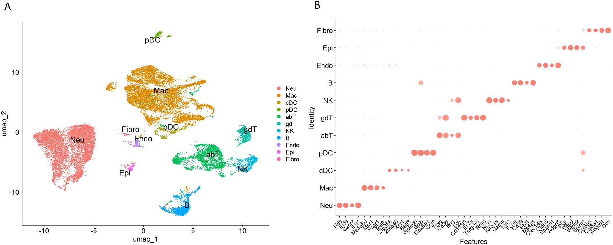
Figure 1. Single-cell transcriptomic landscape of necrotizing granulomatous lesions developed in Mycobacterium tuberculosis-infected lungs of C3HeB/FeJ mice. (A) Uniform manifold approximation and projection (UMAP) plot of 30,159 cells isolated from necrotizing granulomas, showing 11 major cell types. Neu, neutrophil; Mac, macrophage, cDC, conventional dendritic cell; pDC, plasmacytoid DC; abT, αβ T cell; gdT, γδ T cell; NK, natural killer cell; B, B cell; Endo, endothelial cell, Epi, epithelial cell, Fibro, fibroblast. (B) Dot plot showing the distinct expression of selected marker genes in each cell cluster.
Assessment of T cell populations in necrotizing granulomas lesions
Next, we assessed the subclusters of T cells within the necrotizing granulomatous lesions (Figure 2, Supplementary Figure 2). Among CD4+ T cells, three clusters were identified: effector T cells, naïve T cells, and regulatory T cells (Tregs). For CD8+ T cells, two clusters were observed: cytotoxic and exhausted CD8+ T cells. Furthermore, we identified tissue-resident memory T cells (TRM) either expressing CD4 or CD8 within necrotizing granulomas.
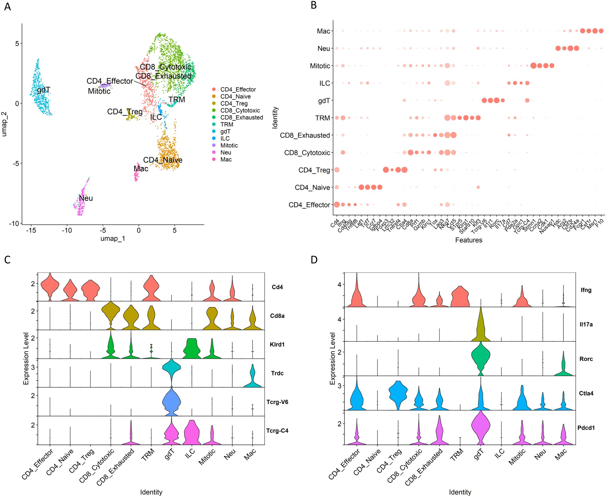
Figure 2. T cell clusters within necrotizing granulomatous lesions. T cell populations from necrotizing granulomas were further filtered to remove doubles and subsequently re-clustered. (A) UMAP plot showing T cell populations, including αβ T cells and γδ T cells. CD4_Effector, effector CD4+ T cell; CD4_Naive, naïve CD4+ T cell; CD4_Treg, regulatory CD4+ T cell; CD8_Cytotoxic, cytotoxic CD8+ T cell, CD8_exhausted, exhausted CD8+ T cell; TRM, tissue-resident memory T cell; gdT, γδ T cell; ILC, innate lymphoid cell; Mitotic, mitotic cell; Neu, neutrophil; Mac, macrophage. (B) Dot plot displaying the expression of selected marker genes for identified T cell clusters. (C, D) Violin plots displaying gene expression levels of T cell receptors and co-receptor molecules (C) and cytokines and immune checkpoint molecules (D).
During M. tuberculosis infection, γδ T cells participate in the immune response in the lungs (37). In particular, IL-17-mediated immunity is critical for γδ T cells to perform their function in host defense in TB murine models (38). In necrotizing granulomas, γδ T cells expressing Il17a were identified (Figures 2C, D). These γδ T cells also expressed Pdcd1, which encodes programmed cell death-1 (PD-1), an immune checkpoint molecule inhibiting immune responses (39).
Among other T cell types, we identified a unique population co-expressing Tcrg (T cell receptor gamma) and Klrd1 (encoding an NK cell receptor) (Figures 2B, C). This population also expressed Gata3, Tbx21, Il7r, Cd7, and Xcl1 (Supplementary Figures 2A, C), suggesting the presence of certain innate lymphoid cells (ILCs) expressing these markers.
Following the removal of doublet cells from the scRNA-seq data, we observed clusters expressing both T cell markers (such as CD3e) and characteristic genes of neutrophils or macrophages (Figure 2B, Supplementary Figure 2C). This observation may reflect biological processes such as the phagocytosis of T cell-derived material; however, further analyses are required to confirm this phenomenon.
Macrophage populations in necrotizing granulomatous lesions
To investigate the hallmark of macrophage populations within necrotizing granulomas, we analyzed their gene expression profiles. Given that Plin2 expression is significantly upregulated in foamy macrophages within necrotizing granulomas (29, 30), we identified the cluster exhibiting significantly elevated Plin2 expression compared to other clusters (Figure 3A). To characterize the remaining clusters, we analyzed their gene expression profiles and found that macrophages expressing Cxcl10, Clec4e, C1qc, Ighm, S100a9, or Siglecf also represented distinct macrophage populations within the necrotizing granulomatous lesions (Figure 3A). Moreover, we confirmed that the expression levels of the corresponding genes in their respective clusters were significantly higher than those in other clusters (Figure 3B).
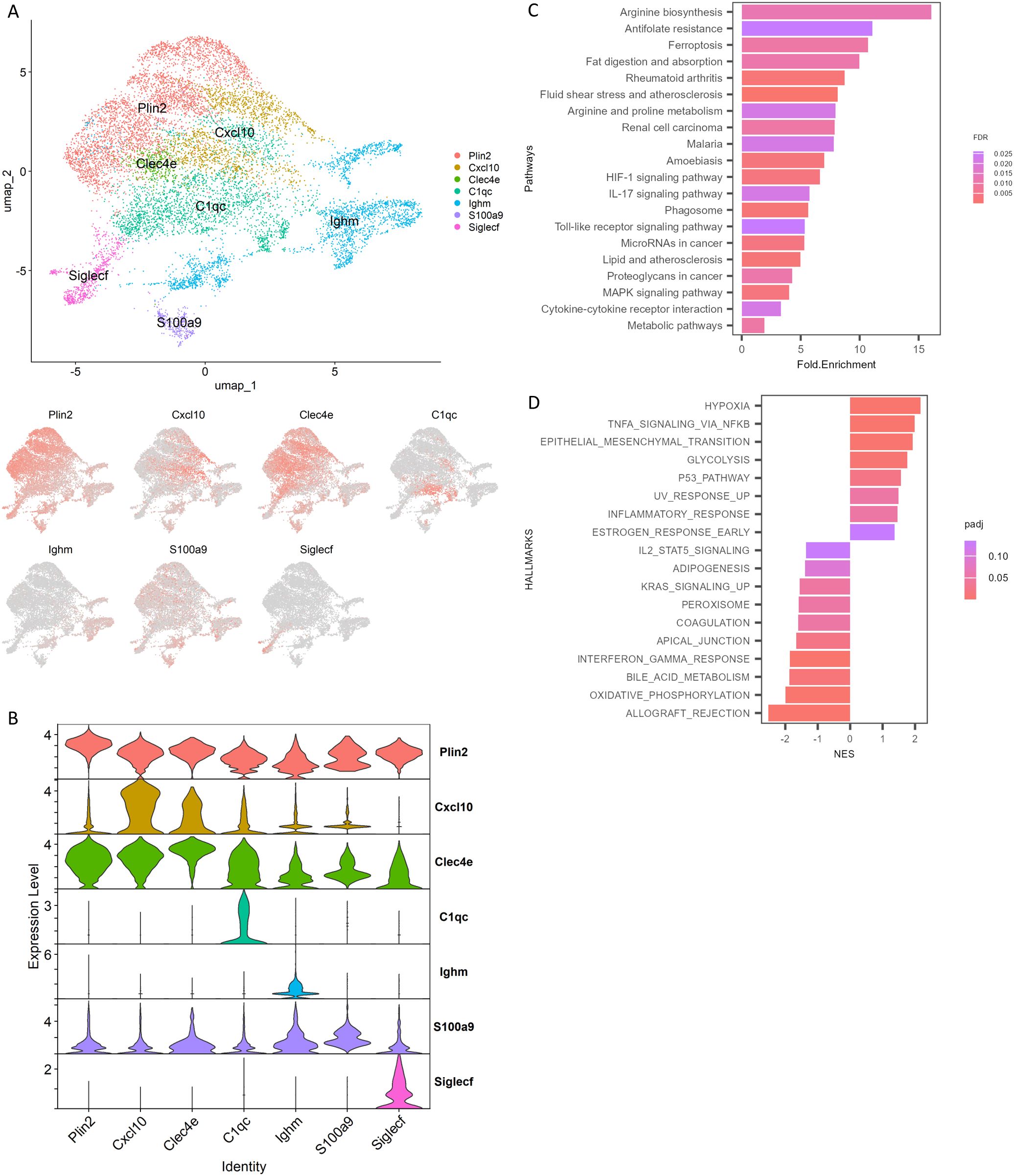
Figure 3. Identification of foamy macrophage associated subclusters. Macrophage populations in necrotizing granulomas were re-clustered and analyzed. (A) UMAP plot of macrophage clusters in necrotizing granulomatous lesions, characterized by signature genes. The expression patterns of signature genes are also depicted. (B) Violin plot showing significantly elevated expression of the indicated genes in specific clusters. (C) Bar plot showing gene ontology analysis of differentially expressed genes upregulated in the Plin2-expressing macrophage cluster. Top 20 enriched KEGG pathways are indicated, along with fold of enrichment (Fold.Enrichment) and false discovery rate (FDR). (D) Gene set enrichment analysis (GSEA) of Plin2-expressing macrophage cluster. Significantly enriched hallmarks are depicted, representing both activated and suppressed hallmarks in the Plin2+ cluster compared with other clusters.
We investigated the differentially expressed genes between the Plin2-expressing (Plin2+) cluster and other clusters within the macrophage population. Genes that were significantly upregulated in the Plin2+ cluster were selected (Supplementary Table 1) and subjected to Gene Ontology (GO) enrichment analysis (Figure 3C). Genes related to fat digestion and absorption, as well as lipid and atherosclerosis, were found to be upregulated. To further investigate the gene expression profile of Plin2+ cluster, we performed gene set enrichment analysis (GSEA) (Figure 3D). GSEA revealed that genes associated with hypoxia, TNF-α signaling, glycolysis, and inflammatory responses were upregulated, whereas those associated with oxidative phosphorylation and type II interferon (IFN) responses were downregulated in the Plin2+ cluster compared to other macrophage clusters.
We investigated the gene expression profiles of additional macrophage clusters within necrotizing granulomatous lesions using GSEA (Supplementary Figure 3). Clusters expressing Cxcl10 or Clec4e exhibited gene expression signatures characteristic of pro-inflammatory macrophages. In contrast, clusters expressing C1qc, Ighm, or Siglecf showed gene expression profiles associated with anti-inflammatory macrophages. A more detailed analysis revealed that Ighm+ macrophages expressed several anti-inflammatory genes, including Cd244a, Nr4a1, Clec4a1, and Clec4a2 (Supplementary Figure 4A). Since Siglecf is a well-established marker of alveolar macrophages (40), the Siglecf+ cluster was identified as alveolar macrophages. Siglecf-expressing macrophages expressed Alox5, a gene that promotes anti-inflammatory polarization and contributes to increased susceptibility to M. tuberculosis infection (41, 42) (Supplementary Figure 4B). Trem2 has been shown to act as a receptor for mycolic acid from mycobacteria and to limit anti-mycobacterial macrophage activation (43). The expression of Trem2 was mainly observed in the Siglecf+ cluster (Supplementary Figure 4B). These results suggest that macrophages in the Siglecf+ cluster may contribute to a permissive environment for M. tuberculosis infection within necrotizing granulomatous lesions.
Novel polarization of Plin2-expressing macrophages
Nos2 and Arg1, well-established markers of macrophage polarization, play key roles in regulating immune responses (44, 45). We have previously demonstrated that Plin2-expressing macrophages express Nos2 or Arg1 in necrotizing granulomas (30), suggesting that the polarization of foamy macrophages exhibits either pro-inflammatory or anti-inflammatory characteristics. Accordingly, scRNA-seq profiling revealed that Nos2 was highly expressed in pro-inflammatory macrophage clusters including the Plin2+ cluster (Figure 4A). Moreover, Arg1 expression was predominantly observed in the Plin2+ cluster. Feature plots revealed the presence of Plin2+ macrophages co-expressing Nos2 and Arg1 (Figure 4B). Further, we investigated the transcriptional factors associated with the regulation of Nos2 or Arg1 expression in macrophages within necrotizing granulomas. Among them, we found that Irf7 expression was correlated with Nos2 expression patterns in macrophage clusters (Figure 4A). Irf7 regulates macrophage polarization toward both pro-inflammatory and anti-inflammatory states (46). Fosl1 encodes a transcription factor that promotes pro-inflammatory polarization by repressing Arg1 expression (47). We observed Fosl1 expression in a subset of Arg1+ macrophages within the Plin2+ cluster (Figure 4C), suggesting that Plin2+ macrophages may consist of heterogeneous subpopulations with distinct or transitional polarization states. These findings suggest that Irf7 and Fosl1 may contribute to the dynamic regulation of macrophage polarization within necrotizing granulomas.
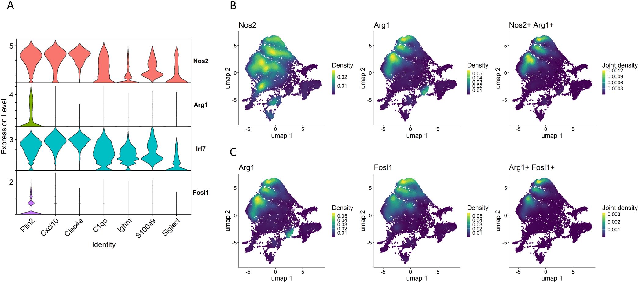
Figure 4. Novel polarization of the Plin2+ macrophage cluster. (A) Violin plots showing the gene expression of Nos2 and Arg1 and their transcriptional factors Irf7 and Fosl1 in macrophage clusters. (B) UMAP plot illustrating expression and co-expression of Nos2 and Arg1 in macrophages. (C) Expression and co-expression of Arg1 and Fosl1 in macrophages.
We further analyzed polarization states of macrophages derived from myeloid cell lineages in the whole lungs of M. tuberculosis-infected Sp140 knockout (KO) mice, using the data from Kotov et al. (28). In C3HeB/FeJ mice, the reduced gene expression of both Sp110 and Sp140 in Sst1 locus contributes to increased susceptibility to M. tuberculosis infection (48–50). In addition, similar to C3HeB/FeJ mice, Sp140 KO mice also exhibit increased susceptibility to M. tuberculosis infection, indicating that Sp140 is a key determinant of host vulnerability to the infection (51). Macrophage populations derived from M. tuberculosis-infected Sp140 KO mice were re-clustered based on the original annotations (Figure 5A). Subsequently, Plin2 expression was found to be elevated in the cluster of interferon-stimulated gene-positive (ISG+) interstitial macrophages (IMs) compared to other macrophage clusters. This result suggests that ISG+ IMs correspond to the Plin2+ cluster identified in our study (Figure 5B). This was further supported by a comparative analysis of macrophage populations between C3HeB/FeJ mice and Sp140 KO mice (Supplementary Figures 5A, B). Notably, Nos2 was expressed in ISG+ IM and IM populations, whereas Arg1 was dominantly expressed in ISGs+ IMs (Figure 5B, Supplementary Figure 5C). Moreover, macrophages co-expressing Nos2 and Arg1 were observed in a subset of the ISG+ IM cluster (Figure 5C). These results suggest the emergency of a novel macrophage polarization state in ISG + IMs derived from M. tuberculosis-infected Sp140 KO mice, corresponding to the Plin2+ cluster of C3HeB/FeJ mice identified in our study.
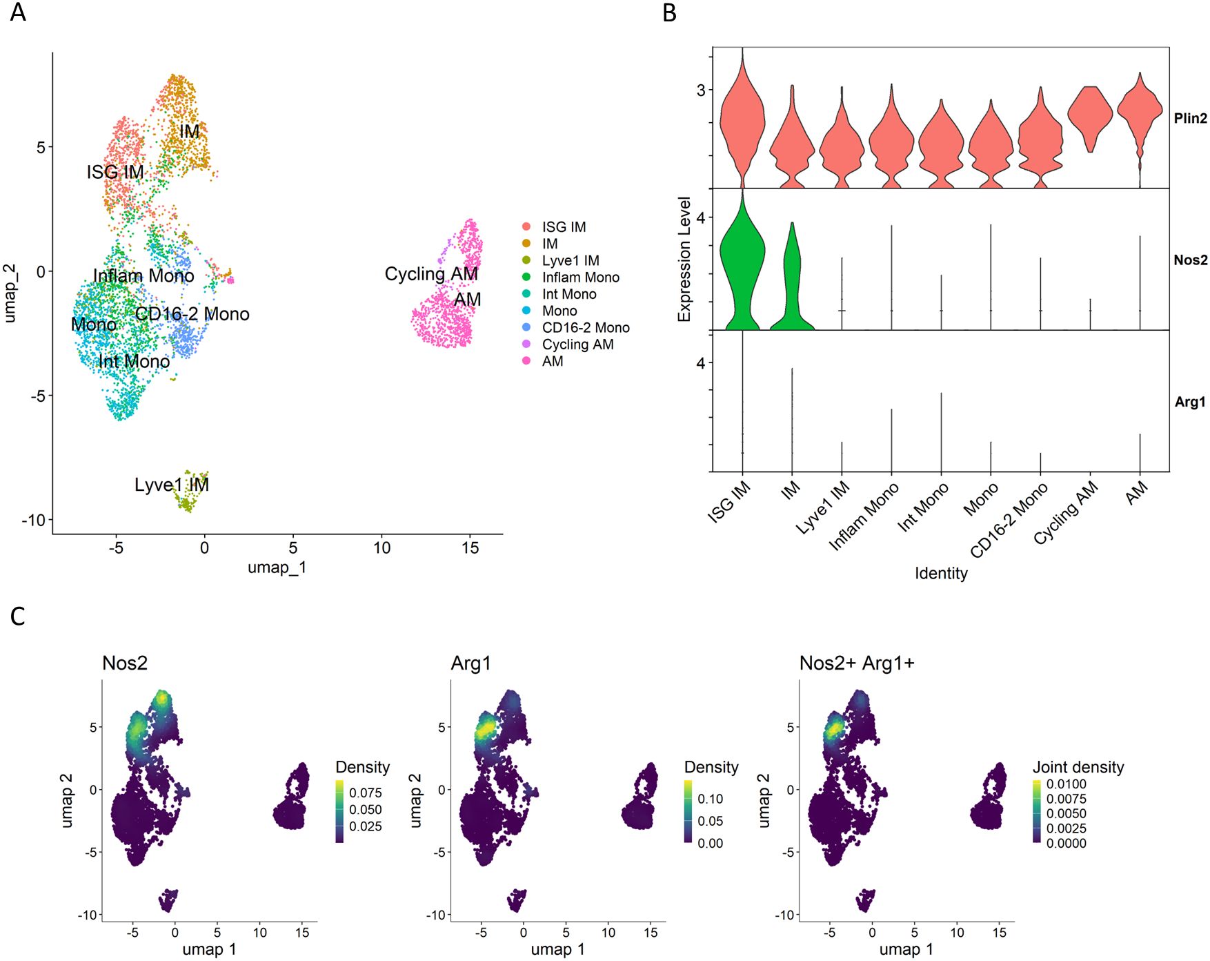
Figure 5. Macrophages derived from M. tuberculosis-infected Sp140-deficient mouse lungs. Macrophage populations of the GSE216023 data from Kotov et al. (28) were analyzed. (A) UMAP plot of macrophage populations from Sp140 knockout (KO) mouse lungs infected with M. tuberculosis. (B) Volin plot showing expression levels of Plin2, Nos2 and Arg1 in macrophage clusters derived from Sp140 KO mice. (C) UMAP plot showing expression and co-expression of Nos2 and Arg1 in macrophages derived from Sp140 KO mice.
Gene expression characteristic of Plin2-expressing macrophages
Type I IFNs and neutrophils contribute to TB exacerbation (35, 52). Accordingly, we investigated the expression of Ifnb1, a type I IFN gene, and Cxcl1, a chemokine gene involved in neutrophil recruitment, in macrophage populations (Figure 6). Both genes were specifically expressed in the Plin2+ cluster, consistent with previous reports (28, 35).
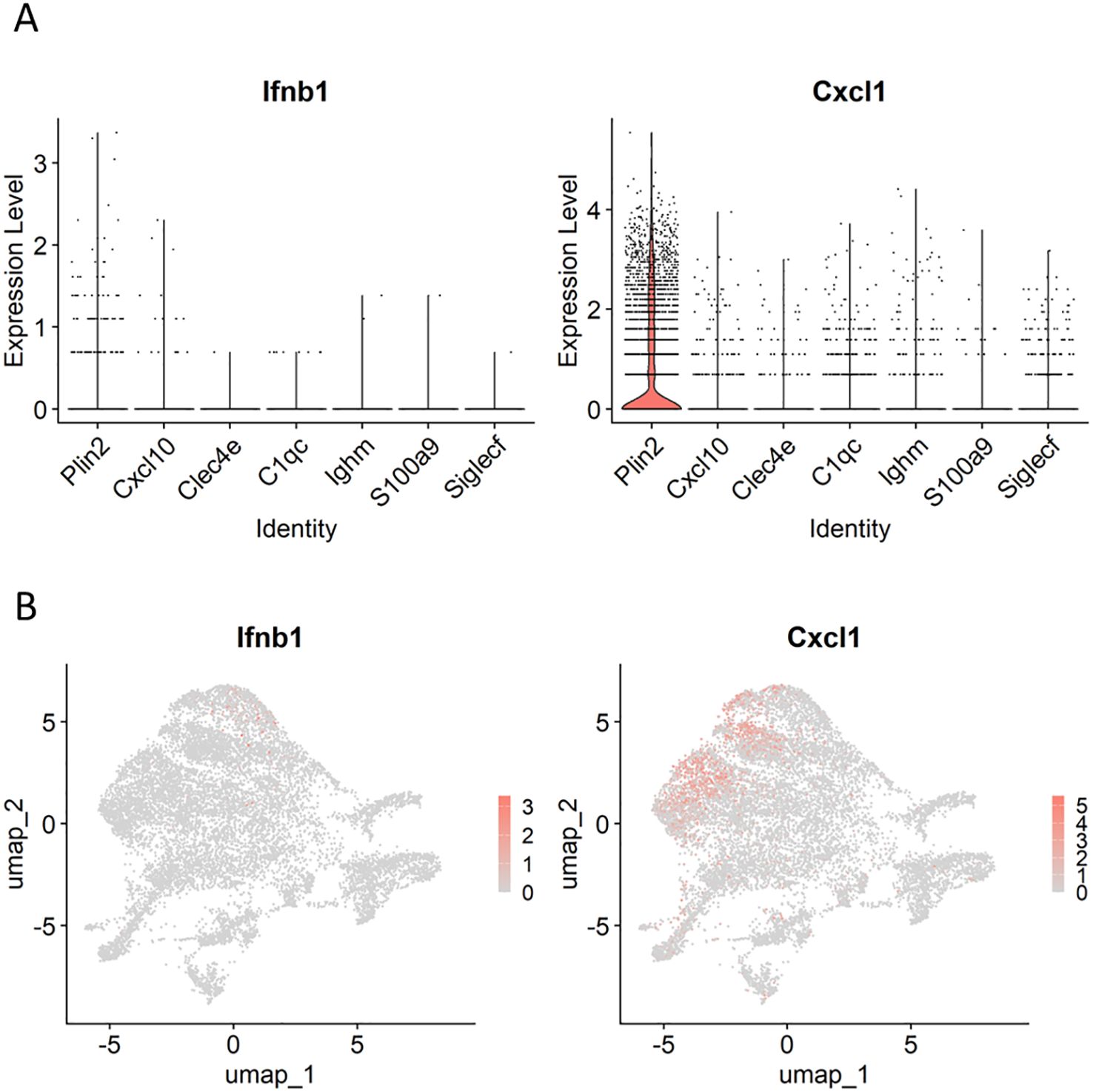
Figure 6. Expression profiles of genes involved in tuberculosis (TB) exacerbation in macrophages. Violin (A) and UMAP (B) plots demonstrating Ifnb1 and Cxcl1 expression in macrophage clusters.
We investigated novel gene expression signatures characteristic of the Plin2+ cluster within necrotizing granulomas. Among the genes expressed in the Plin2+ cluster, Flrt2, Hyal1, and Mmp13 were selected for further examination, due to their specific and enriched expression patterns (Figures 7A, B). IHC revealed that these proteins were localized to the rim regions of necrotizing granulomas, consistent with Plin2 localization (Figure 7C). Immunofluorescence microscopy (IFM) showed co-localization of all the three proteins with Plin2+ cells. Notably, the fluorescent signals of Flrt2, Hyal1, and Mmp13 displayed distinct subcellular localization patterns, whereas Plin2 was predominantly localized to the cytosolic regions of the same vacuolated cells (Figure 7D). These results suggest that Flrt2, Hyal1, and Mmp13 are novel molecular markers characterizing Plin2+ macrophages in necrotizing granulomas.
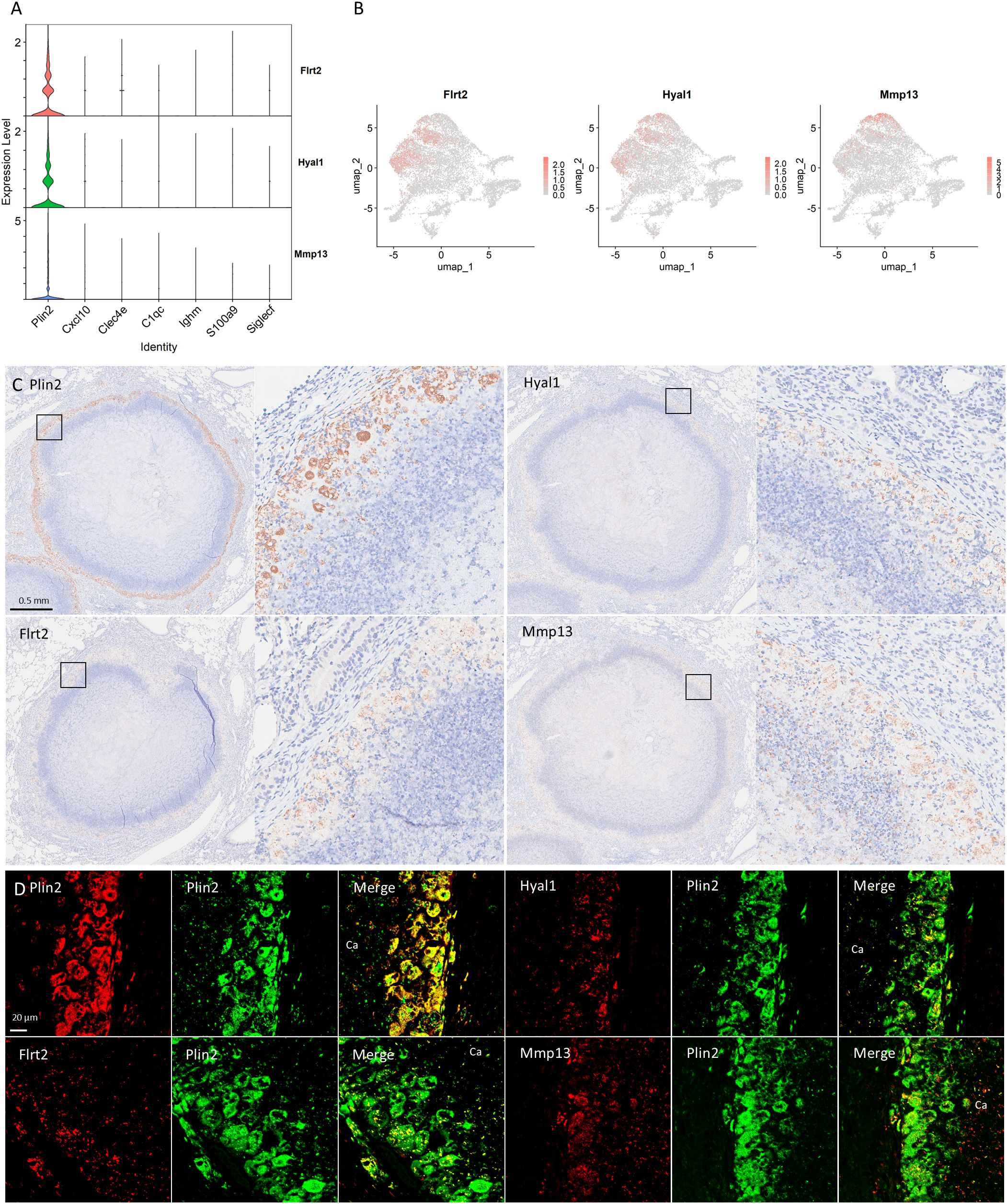
Figure 7. Novel signature of foamy macrophages in necrotizing granulomas. (A) Violin plots demonstrating the expression of Flrt2, Hyal1, and Mmp13, specifically expressed in the Plin2+ cluster. (B) UMAP plots demonstrating the expression of Flrt2, Hyal1, and Mmp13, specifically associated with the Plin2+ cluster. (C) Immunohistochemistry images showing the localization of Flrt2, Hyal1, and Mmp13 in necrotizing granulomas. The marker for foamy macrophages, Plin2 is also shown. For each protein localization, the right panel displays an enlarged view of the area indicated by a square in the corresponding left panel. (D) Immunofluorescence microscopic images demonstrating co-localization of Plin2 with the indicated proteins in necrotizing granulomas. Ca, caseous necrosis region within necrotizing granulomas.
pDC expressed immunomodulatory and cytotoxic factors in necrotizing granulomatous lesions
pDCs regulate viral infections by producing a large amount of type I IFNs (53). In the context of M. tuberculosis infection, the depletion of pDCs results in increased bacterial burdens in the lungs (28). Lee et al. demonstrated that M. tuberculosis-infected neutrophils can stimulate pDCs to produce type I IFNs (54). Therefore, we assessed the expression levels of Ifnb1 in pDCs (Figures 8A, B). However, Ifnb1 expression were predominantly detected in macrophages rather than pDCs. In contrast, pDCs expressed Il34 and Kmo. Il34 encodes an anti-inflammatory cytokine that regulates the expression of pro-inflammatory cytokines and promotes macrophage polarization toward an anti-inflammatory phenotype (55). Kmo encodes kynurenine 3-monooxygenase, which catalyzes the conversion of kynurenine to 3-hydroxykynurenine (3-HK), a metabolite inducing cellular damage and apoptosis via oxidative stress (56). These results suggest that pDCs are localized to necrotizing granulomatous lesions and contribute to the pathogenicity of M. tuberculosis infection via the expression of immunomodulatory and cytotoxic factors.
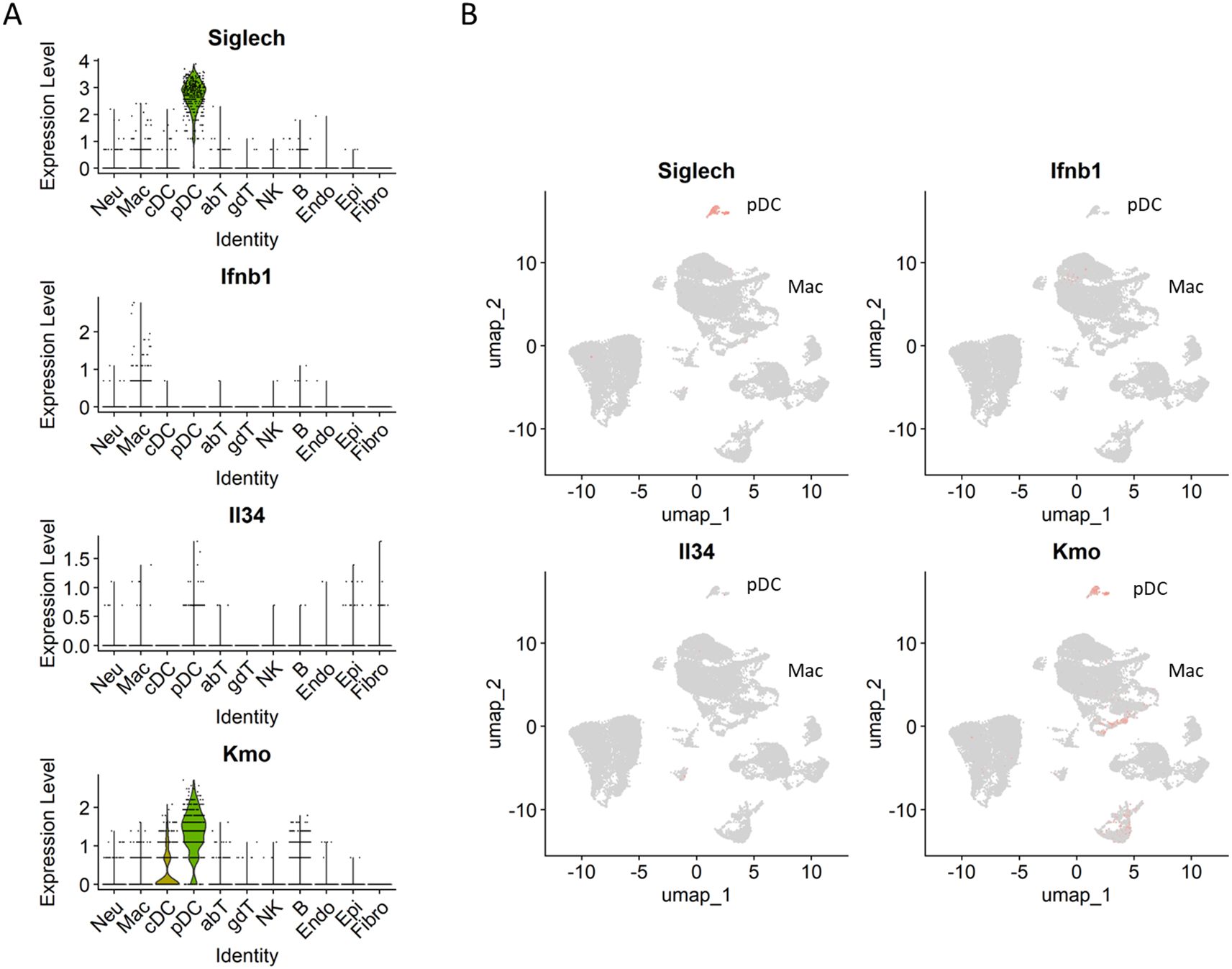
Figure 8. Expression of Il34 and Kmo in necrotizing granulomas of M. tuberculosis-infected C3HeB/FeJ mice. Violin (A) and UMAP (B) plots showing the gene expression of Ifnb1, Il34, Kmo and Siglech (pDC marker) in necrotizing granulomas derived from M. tuberculosis-infected C3HeB/FeJ mice.
Discussion
scRNA-seq has been widely utilized to evaluate the cellular transcriptomics of tissues or blood samples from TB patients, as well as from M. tuberculosis-infected non-human primates, mice and other experimental models (57). However, the detailed cellular composition of necrotizing granulomatous lesions, a hallmark of TB pathology (5, 7), remains incompletely characterized. In this study, we isolated single-cell suspensions from necrotizing granulomatous lesions developed in the lungs of M. tuberculosis-infected C3HeB/FeJ mice, and performed scRNA-seq to comprehensively investigate their cellular landscape. We identified 11 major cell type, including immune cells such as neutrophils, macrophages, dendritic cells, T cells, NK cell, and B cells (Figure 1). To prepare single-cell suspensions from necrotizing granulomatous lesions, we performed Ficoll-Paque density gradient centrifugation to reduce the number of dead cells and neutrophils. However, due to the heterogeneity and activation states of neutrophils, their density can vary, resulting in partial retention at the interface of the Ficoll-Paque gradient.
Among T cells, CD4+ T cells, particularly central memory Mtb-specific CD4+ T cells, play a crucial role in protective immunity against Mtb infection (58). In this study, we identified CD4+ T cells in necrotizing granulomatous lesions as naïve, effector or TRM types (Figure 2, Supplementary Figure 2). However, central memory CD4+ T cells (Cd44+, Sell+, and Ccr7+) were not identified, suggesting that these cells do not localize to necrotizing granulomatous lesions. Naïve CD4+ T cells were enriched in the lesions, consistent with previous reports in non-human primate and murine models of M. tuberculosis infection (25–27). Moreover, Pdcd1+ γδ T cells were identified within necrotizing granulomatous lesions (Figure 2). Although IL-17-producing γδ T cells play a protective role in the lungs of M. tuberculosis-infected mice (59), the presence of Pdcd1+ γδ T cells has not been previously reported in this context. In the experimental autoimmune encephalomyelitis mouse model, Pdcd1+ γδ T cells have been implicated in promoting disease pathogenesis (60). These findings suggest that Pdcd1+ γδ T cells may similarly contribute to the immunopathology of necrotizing granulomas during M. tuberculosis infection.
In macrophage populations within necrotizing granulomas, the Plin2+ cluster was identified based on Plin2 expression in the clusters (Figure 3). Because Plin2 is the marker of foamy macrophages in the lungs of TB patients and M. tuberculosis-infected C3HeB/FeJ mice (29, 30), we referred to the Plin2+ cluster as the foamy macrophage population. Furthermore, differential gene expression analysis and GSEA revealed that gene expression profiles of the Plin2+ cluster were consistent with the characteristics of foamy macrophages in necrotizing granulomas (29, 30).
Previously, we have demonstrated that foamy macrophages in necrotizing granulomas express typical macrophage polarization markers, either Nos2 or Arg1, suggesting their differentiation into a pro-inflammatory or anti-inflammatory state (30). scRNA-seq revealed that Plin2+ macrophages express Nos2 or Arg1 (Figure 4). Moreover, we identified a subset of Plin2+ macrophages co-expressing both Nos2 and Arg1, suggesting a novel polarization state of foamy macrophages in necrotizing granulomas. A similar polarization profile was also observed in macrophage populations isolated from the lungs of M. tuberculosis-infected Sp140 KO mice (28) (Figure 5). Foamy macrophages exhibiting the dual expression profile may represent a transitional state between pro- and anti-inflammatory phenotypes. To investigate this possibility, we performed a comparative analysis of macrophage populations between C3HeB/FeJ and Sp140 KO mice (Supplementary Figure 5). This analysis revealed a distinct correspondence between the Plin2+ cluster in C3HeB/FeJ mice and ISG+ IMs in Sp140 KO mice, as well as between the C1qc+ cluster and IMs, the Ighm+ cluster and monocyte clusters, the Siglecf+ cluster and alveolar macrophage clusters. Together, these results suggest that monocytes infiltrating infected lungs differentiate into IMs and ISG+ IMs, subsets of which further progress toward a foamy macrophage phenotype.
Type I IFNs and neutrophils are key exacerbating factors in TB, contributing to disease progression by promoting inflammation and inducing the release of neutrophil extracellular traps (NETs) (35, 51, 52). In particular, neutrophils and their remnants resulting from NETosis accumulate in the caseous necrosis regions of necrotizing granulomas. We found a subset of Plin2+ macrophages expressing Ifnb1 or Cxcl1 (Figure 6). scRNA-seq revealed that ISG+ IMs and IMs express type I IFNs in the lungs of M. tuberculosis-infected Sp140 KO mice (28). Chowdhury et al. demonstrated that type I IFNs are primarily expressed by epithelioid cells proximal to the caseous necrosis regions in necrotizing granulomas developed in C3HeB/FeJ mice and non-human primate models (35), which is consistent with our findings. Moreover, Chowdhury et al. demonstrated that pDCs within necrotizing granulomatous lesions are not significantly associated with type I IFN expression. In agreement with this, our study revealed that pDCs within necrotizing granulomatous lesions do not predominantly express Ifnb1, but rather express other immunosuppressive genes, such as Il34 and Kmo (Figure 8). These results suggest that Cxcl1, secreted by foamy macrophages in necrotizing granulomas, contribute to the recruitment of neutrophils into the caseous necrosis regions, where NETosis may be subsequently induced by type I IFNs.
We investigated unique molecular signatures of the Plin2+ cluster in necrotizing granulomas. We found that three proteins, Flrt2, Hyal1, and Mmp13, were specifically localized to Plin2+ macrophages (Figure 7). Flrt2 regulates macrophage differentiation and activate Akt/mTOR signaling (61). Given the involvement of mTORC1 signaling in the differentiation of foamy macrophages during M. tuberculosis infection (62), Flrt2 may contribute to foamy macrophage differentiation in necrotizing granulomas. Hyal1 encodes a lysosomal hyaluronidase that digests extracellular matrix (63). We found that a subset of Cxcl1-expressing Plin2+ macrophages also expressed Hyal1 (Supplementary Figure 4C), suggesting that Hyal1 facilitates neutrophil recruitment into the caseous necrosis regions. Mmp13 encodes a metalloprotease involved in collagen remodeling in atherosclerotic plaques (64). Foamy macrophages exhibit a characteristic arrangement around the necrotic core within necrotizing granulomas, suggesting that Mmp13 expressed by foamy macrophages may regulate cell–cell interaction or tissue remodeling. Taken together, these identified molecules are involved in the development of necrotizing granulomas through the regulation of foamy macrophage differentiation, localization, and function.
In conclusion, we conducted an in-depth single-cell transcriptomic analysis of necrotizing granulomatous lesions using a TB mouse model that closely recapitulates the pathological features observed in TB patients. Our results revealed novel cellular signatures within necrotizing granulomas, with particular emphasis on the characterization of foamy macrophage. These insights into the cellular and molecular landscape of necrotizing granulomas advance our understanding of TB pathogenesis and will facilitate the development of novel diagnostic tool and host-directed therapeutic drugs for TB.
Materials and methods
Ethics statement
All animal experiments in this study were approved by the Animal Care and Use Committee of The Research Institute of Tuberculosis (RIT) (permission number ID 2022-02) and conducted in accordance with the RIT ethical guidelines for animal care and use.
Mouse model and infection
C3HeB/FeJ mice were purchased from Jackson Laboratory and housed in a filtered-air, laminar-flow cabinet under specific pathogen-free conditions at the animal facility of the RIT. Mice were provided with sterile bedding, water, and mouse chow. Specific pathogen-free status was verified by monitoring sentinel mice housed within the colony. Mice aged 6–10 weeks were transferred to the biosafety level III animal facility of the RIT. For M. tuberculosis infection, frozen stocks of the M. tuberculosis Erdman strain stored at -80°C was used as previously described (65). Mice were infected with approximately 100 CFU of M. tuberculosis bacilli via aerosol using an infection exposure system (Glas-Col).
Preparation of single cells from necrotizing granulomatous lesions
At 12 weeks p.i., infected mice were euthanized by exsanguination under anesthesia with 0.75 mg/kg medetomidine, 4.0 mg/kg midazolam, and 5.0 mg/kg butorphanol via the intraperitoneal route. The lungs were excised and subsequently dissected to collect infected lesions including necrotizing granulomas (Supplementary Figure 1). For the preparation of single-cell suspensions, infected lungs from three or four mice were pooled to collect more than 10 lesions containing necrotizing granulomas. Necrotizing granulomatous lesions were minced and incubated in a collagenase/hyaluronidase/DNase I solution (Stemcell) with RNase inhibitor at 0.2 U/μL. Following red blood cell lysis, the dissociated cells were resuspended in PBS containing 2% fetal bovine serum and RNase inhibitor at 0.2 U/μL. To remove dead cells and a large proportion of neutrophils, cell suspensions were subjected to Ficoll-Paque density gradient centrifugation, followed by the collection of cells from the interface layer according to the manufacturer’s instructions (Cytiva).
Construction of scRNA-seq libraries and sequencing
Isolated cell suspensions from necrotizing granulomatous lesions were subjected to scRNA-seq library constructions using Chromium Fixed RNA Profiling Reagent Kit (10x Genomics). Cells were fixed with a fixation solution containing formaldehyde for 24 h at 4°C. Inactivation of M. tuberculosis in the fixed samples was confirmed by CFU assay. Fixed cells were further processed to construct scRNA-seq libraries according to the manufacturer’s protocol. The resulting libraries were sequenced on NextSeq 1000 (Illumina).
Data analysis
Raw sequencing reads were aligned against the mouse reference genome (mm10) using Cellranger version 7.0.1 (10x Genomics). Subsequent analyses were performed in R version 4.4.1 using the Seurat package version 5.2.1 (66). Data from four independent samples were filtered to include cells with 500–6000 genes and less than 20% mitochondrial reads. Data were normalized using the NormalizeData function, followed by the identification of highly variable features using the FindVariableFeatures function with the variance stabilizing transformation method, selecting the top 2000 most variable genes. Principal component analysis (PCA) was performed using the RunPCA function. To correct for batch effects, the Harmony algorithm was applied using the RunHarmony function. Following batch correction, uniform manifold approximation and projection (UMAP) was computed using the first 30 dimensions. A shared nearest neighbor (SNN) graph was then constructed using the FindNeighbors function with the same set of dimensions. Clustering was performed using the FindClusters function with a resolution parameter set to 0.7. The resulting dataset was processed with scDblFinder version 1.18.0 (67) to remove doublet cells. The filtrated data were reprocessed through data scaling, PCA, UMAP, followed by the construction of SNN graph and clustering. Cell clusters were manually annotated based on specific gene expression patterns of the respective cell types.
Differential expression analysis was performed using Seurat FindMarkers function. Gene expression analysis, GO analysis, and GSEA were performed using Nebulosa version 1.14.0 (68), ShinyGO version 0.82 (69), and, fGSEA version 1.30.00 (70), respectively.
Immunohistochemistry and immunofluorescence microscopy
IHC and IFM were performed as previously described (30, 36). Antibodies used in this study are listed in Supplementary Table 2. IHC and IFM samples were visualized using a NanoZoomer S60 (Hamamatsu Photonics) and a FV4000 (Evident), respectively.
Data availability statement
The datasets presented in this study can be found in online repositories. The names of the repository/repositories and accession number(s) can be found below: https://ddbj.nig.ac.jp/search/entry/bioproject/PRJDB20543. https://ddbj.nig.ac.jp/public/ddbj_database/gea/experiment/E-GEAD-1000/E-GEAD-1082/.
Ethics statement
The animal study was approved by the Animal Care and Use Committee of The Research Institute of Tuberculosis (permission number ID 2022-02). The study was conducted in accordance with the local legislation and institutional requirements.
Author contributions
SS: Funding acquisition, Writing – review & editing, Conceptualization, Writing – original draft, Data curation, Formal Analysis. SO: Writing – review & editing, Methodology. HN: Writing – review & editing, Methodology. MH: Data curation, Conceptualization, Writing – original draft, Funding acquisition, Writing – review & editing, Formal Analysis. NK: Data curation, Conceptualization, Funding acquisition, Formal Analysis, Writing – original draft, Writing – review & editing.
Funding
The author(s) declare that financial support was received for the research and/or publication of this article. This study was supported by the Emerging/Re-emerging Infectious Diseases Project of the Japan Agency for Medical Research and Development (JP23wm0225028, JP23gm1610013, JP23fk0108673, JP23fk0108674, JP23fk0108703, JP25fk0108730), and Grants-in-Aid for Scientific Research, Japan Society for the Promotion of Science (20KK0197, 22K07065).
Acknowledgments
We thank Ms. Miyako Seto and Ms. Mariko Ogasawara in Department of Pathophysiology and Host Defense for technical supports.
Conflict of interest
The authors declare that the research was conducted in the absence of any commercial or financial relationships that could be construed as a potential conflict of interest.
Generative AI statement
The author(s) declare that no Generative AI was used in the creation of this manuscript.
Publisher’s note
All claims expressed in this article are solely those of the authors and do not necessarily represent those of their affiliated organizations, or those of the publisher, the editors and the reviewers. Any product that may be evaluated in this article, or claim that may be made by its manufacturer, is not guaranteed or endorsed by the publisher.
Supplementary material
The Supplementary Material for this article can be found online at: https://www.frontiersin.org/articles/10.3389/fimmu.2025.1624072/full#supplementary-material
Supplementary Figure 1 | Macroscopic image of C3HeB/FeJ mouse lung with necrotizing granulomatous lesions at 12 weeks postinfection (p.i.). Arrowheads indicate necrotizing granulomas. Scale bar, 1 cm.
Supplementary Figure 2 | Violin plots of transcriptional factors regulating T cell differentiation (A), and markers representing memory or effector type (B), and innate lymphoid cells (C).
Supplementary Figure 3 | GSEA of macrophage clusters characterized by the expression of Cxcl10 (A), Clec4e (B), C1qc (C), Ighm (D), S100a9 (E), and Siglecf (F). Significantly enriched hallmarks are depicted, representing both activated and suppressed hallmarks in each cluster compared with other clusters.
Supplementary Figure 4 | Violin plots displaying the characteristic gene expression in the Ighm+ cluster (A) and the Siglecf+ cluster (B). Gene expression levels of Cxcl1 and Hyal1 in macrophage populations are also shown (C).
Supplementary Figure 5 | Comparative analysis of macrophage populations between C3HeB/FeJ and Sp140-deficient mice. Macrophage populations from this study and the GSE216023 dataset from Kotov et al. (28) were integrated and re-clustered. (A) Comparative UMAP plots of macrophage clusters between C3HeB/FeJ (FeJ) and Sp140-deficient mice (SP140KO), with designated cluster names. (B, C) Violin plots showing expression levels of the indicated genes in macrophage clusters from FeJ and SP140KO.
Supplementary Table 1 | List of differentially expressed genes of the Plin2+ cluster in the macrophage population.
Supplementary Table 2 | List of antibodies for IHC and IFM.
References
1. WHO. Tuberculosis(2025). Available online at: https://www.who.int/news-room/fact-sheets/detail/tuberculosis (Accessed March 14, 2025).
2. Cohen SB, Gern BH, Delahaye JL, Adams KN, Plumlee CR, Winkler JK, et al. Alveolar macrophages provide an early mycobacterium tuberculosis niche and initiate dissemination. Cell Host Microbe. (2018) 24:439–46.e4. doi: 10.1016/j.chom.2018.08.001
3. Bussi C and Gutierrez MG. Mycobacterium tuberculosis infection of host cells in space and time. FEMS Microbiol Rev. (2019) 43:341–61. doi: 10.1093/femsre/fuz006
4. Pagán AJ and Ramakrishnan L. Immunity and immunopathology in the tuberculous granuloma. Cold Spring Harbor Perspect Med. (2014) 5:a018499. doi: 10.1101/cshperspect.a018499
5. Sarathy JP and Dartois V. Caseum: A niche for mycobacterium tuberculosis drug-tolerant persisters. Clin Microbiol Rev. (2020) 33:e00159-19. doi: 10.1128/cmr.00159-19
6. Cadena AM, Fortune SM, and Flynn JL. Heterogeneity in tuberculosis. Nat Rev Immunol. (2017) 17:691–702. doi: 10.1038/nri.2017.69
7. Lenaerts A, Barry CE 3rd, and Dartois V. Heterogeneity in tuberculosis pathology, microenvironments and therapeutic responses. Immunol Rev. (2015) 264:288–307. doi: 10.1111/imr.12252
8. Russell DG, Cardona PJ, Kim MJ, Allain S, and Altare F. Foamy macrophages and the progression of the human tuberculosis granuloma. Nat Immunol. (2009) 10:943–8. doi: 10.1038/ni.1781
9. Walter ND, Born SEM, Robertson GT, Reichlen M, Dide-Agossou C, Ektnitphong VA, et al. Mycobacterium tuberculosis precursor Rrna as a measure of treatment-shortening activity of drugs and regimens. Nat Commun. (2021) 12:2899. doi: 10.1038/s41467-021-22833-6
10. Carow B, Hauling T, Qian X, Kramnik I, Nilsson M, and Rottenberg ME. Spatial and temporal localization of immune transcripts defines hallmarks and diversity in the tuberculosis granuloma. Nat Commun. (2019) 10:1823. doi: 10.1038/s41467-019-09816-4
11. Kumar R, Kolloli A, Subbian S, Kaushal D, Shi L, and Tyagi S. Imaging the architecture of granulomas induced by mycobacterium tuberculosis infection with single-molecule fluorescence in situ hybridization. J Immunol (Baltimore Md: 1950). (2024) 213:526–37. doi: 10.4049/jimmunol.2300068
12. Magoulopoulou A, Qian X, Pediatama Setiabudiawan T, Marco Salas S, Yokota C, Rottenberg ME, et al. Spatial resolution of mycobacterium tuberculosis bacteria and their surrounding immune environments based on selected key transcripts in mouse lungs. Front Immunol. (2022) 13:876321. doi: 10.3389/fimmu.2022.876321
13. Qiu X, Zhong P, Yue L, Li C, Yun Z, Si G, et al. Spatial transcriptomic sequencing reveals immune microenvironment features of mycobacterium tuberculosis granulomas in lung and omentum. Theranostics. (2024) 14:6185–201. doi: 10.7150/thno.99038
14. McCaffrey EF, Donato M, Keren L, Chen Z, Delmastro A, Fitzpatrick MB, et al. The immunoregulatory landscape of human tuberculosis granulomas. Nat Immunol. (2022) 23:318–29. doi: 10.1038/s41590-021-01121-x
15. Bromley JD, Ganchua SKC, Nyquist SK, Maiello P, Chao M, Borish HJ, et al. Cd4(+) T cells re-wire granuloma cellularity and regulatory networks to promote immunomodulation following Mtb reinfection. Immunity. (2024) 57:2380–98.e6. doi: 10.1016/j.immuni.2024.08.002
16. Cinco IR, Napier EG, Rhoades NS, Davies MH, Allison DB, Kohama SG, et al. Immunological and microbial shifts in the aging rhesus macaque lung during nontuberculous mycobacterial infection. mBio. (2024) 15:e0082924. doi: 10.1128/mbio.00829-24
17. Pisu D, Johnston L, Mattila JT, and Russell DG. The frequency of Cd38(+) alveolar macrophages correlates with early control of M. Tuberculosis in the murine lung. Nat Commun. (2024) 15:8522. doi: 10.1038/s41467-024-52846-w
18. Carow B, Muliadi V, Skålén K, Yokota C, Kathamuthu GR, Setiabudiawan TP, et al. Immune mapping of human tuberculosis and sarcoidosis lung granulomas. Front Immunol. (2023) 14:1332733. doi: 10.3389/fimmu.2023.1332733
19. Zheng W, Borja M, Dorman LC, Liu J, Zhou A, Seng A, et al. Single-cell analysis reveals mycobacterium tuberculosis esx-1-mediated accumulation of permissive macrophages in infected mouse lungs. Sci Adv. (2025) 11:eadq8158. doi: 10.1126/sciadv.adq8158
20. Yang Q, Qi F, Ye T, Li J, Xu G, He X, et al. The interaction of macrophages and cd8 T cells in bronchoalveolar lavage fluid is associated with latent tuberculosis infection. Emerging Microbes infections. (2023) 12:2239940. doi: 10.1080/22221751.2023.2239940
21. Winchell CG, Nyquist SK, Chao MC, Maiello P, Myers AJ, Hopkins F, et al. Cd8+ Lymphocytes are critical for early control of tuberculosis in macaques. J Exp Med. (2023) 220:e20230707. doi: 10.1084/jem.20230707
22. Wen Z, Wang L, Ma H, Li L, Wan L, Shi L, et al. Integrated single-cell transcriptome and T cell receptor profiling reveals defects of T cell exhaustion in pulmonary tuberculosis. J infection. (2024) 88:106158. doi: 10.1016/j.jinf.2024.106158
23. Pisu D, Huang L, Narang V, Theriault M, Lê-Bury G, Lee B, et al. Single cell analysis of M. Tuberculosis phenotype and macrophage lineages in the infected lung. J Exp Med. (2021) 218:e20210615. doi: 10.1084/jem.20210615
24. Wang L, Ma H, Wen Z, Niu L, Chen X, Liu H, et al. Single-cell rna-sequencing reveals heterogeneity and intercellular crosstalk in human tuberculosis lung. J infection. (2023) 87:373–84. doi: 10.1016/j.jinf.2023.09.004
25. Gideon HP, Hughes TK, Tzouanas CN, Wadsworth MH 2nd, Tu AA, Gierahn TM, et al. Multimodal profiling of lung granulomas in macaques reveals cellular correlates of tuberculosis control. Immunity. (2022) 55:827–46.e10. doi: 10.1016/j.immuni.2022.04.004
26. Akter S, Chauhan KS, Dunlap MD, Choreño-Parra JA, Lu L, Esaulova E, et al. Mycobacterium tuberculosis infection drives a type I Ifn signature in lung lymphocytes. Cell Rep. (2022) 39:110983. doi: 10.1016/j.celrep.2022.110983
27. Esaulova E, Das S, Singh DK, Choreño-Parra JA, Swain A, Arthur L, et al. The immune landscape in tuberculosis reveals populations linked to disease and latency. Cell Host Microbe. (2021) 29:165–78.e8. doi: 10.1016/j.chom.2020.11.013
28. Kotov DI, Lee OV, Fattinger SA, Langner CA, Guillen JV, Peters JM, et al. Early cellular mechanisms of type I interferon-driven susceptibility to tuberculosis. Cell. (2023) 186:5536–53.e22. doi: 10.1016/j.cell.2023.11.002
29. Kim MJ, Wainwright HC, Locketz M, Bekker LG, Walther GB, Dittrich C, et al. Caseation of human tuberculosis granulomas correlates with elevated host lipid metabolism. EMBO Mol Med. (2010) 2:258–74. doi: 10.1002/emmm.201000079
30. Seto S, Nakamura H, Guo TC, Hikichi H, Wakabayashi K, Miyabayashi A, et al. Spatial multiomic profiling reveals the novel polarization of foamy macrophages within necrotic granulomatous lesions developed in lungs of C3heb/Fej mice infected with mycobacterium tuberculosis. Front Cell infection Microbiol. (2022) 12:968543. doi: 10.3389/fcimb.2022.968543
31. Driver ER, Ryan GJ, Hoff DR, Irwin SM, Basaraba RJ, Kramnik I, et al. Evaluation of a mouse model of necrotic granuloma formation using C3heb/Fej mice for testing of drugs against mycobacterium tuberculosis. Antimicrobial Agents chemotherapy. (2012) 56:3181–95. doi: 10.1128/aac.00217-12
32. Harper J, Skerry C, Davis SL, Tasneen R, Weir M, Kramnik I, et al. Mouse model of necrotic tuberculosis granulomas develops hypoxic lesions. J Infect Dis. (2012) 205:595–602. doi: 10.1093/infdis/jir786
33. Irwin SM, Driver E, Lyon E, Schrupp C, Ryan G, Gonzalez-Juarrero M, et al. Presence of multiple lesion types with vastly different microenvironments in C3heb/Fej mice following aerosol infection with mycobacterium tuberculosis. Dis Models Mech. (2015) 8:591–602. doi: 10.1242/dmm.019570
34. Lanoix JP, Lenaerts AJ, and Nuermberger EL. Heterogeneous disease progression and treatment response in a C3heb/Fej mouse model of tuberculosis. Dis Models Mech. (2015) 8:603–10. doi: 10.1242/dmm.019513
35. Chowdhury CS, Kinsella RL, McNehlan ME, Naik SK, Lane DS, Talukdar P, et al. Type I Ifn-mediated net release promotes mycobacterium tuberculosis replication and is associated with granuloma caseation. Cell Host Microbe. (2024) 32:2092–111.e7. doi: 10.1016/j.chom.2024.11.008
36. Seto S, Morimoto K, Yoshida T, Hiramatsu M, Hijikata M, Nagata T, et al. Proteomic profiling reveals the architecture of granulomatous lesions caused by tuberculosis and mycobacterium avium complex lung disease. Front Microbiol. (2019) 10:3081. doi: 10.3389/fmicb.2019.03081
37. Hu Y, Hu Q, Li Y, Lu L, Xiang Z, Yin Z, et al. Γδ T cells: origin and fate, subsets, diseases and immunotherapy. Signal transduction targeted Ther. (2023) 8:434. doi: 10.1038/s41392-023-01653-8
38. Meraviglia S, El Daker S, Dieli F, Martini F, and Martino A. Γδ T cells cross-link innate and adaptive immunity in mycobacterium tuberculosis infection. Clin Dev Immunol. (2011) 2011:587315. doi: 10.1155/2011/587315
39. Okazaki T and Honjo T. PD-1 and PD-1 ligands: from discovery to clinical application. Int. Immunol.. (2007) 19:813–24. doi: 10.1093/intimm/dxm057
40. Misharin AV, Morales-Nebreda L, Mutlu GM, Budinger GR, and Perlman H. Flow cytometric analysis of macrophages and dendritic cell subsets in the mouse lung. Am J Respir Cell Mol Biol. (2013) 49:503–10. doi: 10.1165/rcmb.2013-0086MA
41. Bafica A, Scanga CA, Serhan C, MaChado F, White S, Sher A, et al. Host control of mycobacterium tuberculosis is regulated by 5-lipoxygenase-dependent lipoxin production. J Clin Invest. (2005) 115:1601–6. doi: 10.1172/jci23949
42. Marakalala MJ, Martinez FO, Plüddemann A, and Gordon S. Macrophage heterogeneity in the immunopathogenesis of tuberculosis. Front Microbiol. (2018) 9:1028. doi: 10.3389/fmicb.2018.01028
43. Iizasa E, Chuma Y, Uematsu T, Kubota M, Kawaguchi H, Umemura M, et al. Trem2 is a receptor for non-glycosylated mycolic acids of mycobacteria that limits anti-mycobacterial macrophage activation. Nat Commun. (2021) 12:2299. doi: 10.1038/s41467-021-22620-3
44. Bogdan C. Nitric oxide synthase in innate and adaptive immunity: an update. Trends Immunol. (2015) 36:161–78. doi: 10.1016/j.it.2015.01.003
45. Murray PJ, Allen JE, Biswas SK, Fisher EA, Gilroy DW, Goerdt S, et al. Macrophage activation and polarization: nomenclature and experimental guidelines. Immunity. (2014) 41:14–20. doi: 10.1016/j.immuni.2014.06.008
46. Qing F and Liu Z. Interferon regulatory factor 7 in inflammation, cancer and infection. Front Immunol. (2023) 14:1190841. doi: 10.3389/fimmu.2023.1190841
47. Hannemann N, Cao S, Eriksson D, Schnelzer A, Jordan J, Eberhardt M, et al. Transcription factor fra-1 targets arginase-1 to enhance macrophage-mediated inflammation in arthritis. J Clin Invest. (2019) 129:2669–84. doi: 10.1172/jci96832
48. Nakamura H, Hikichi H, Seto S, Hijikata M, and Keicho N. Transcriptional regulators Sp110 and Sp140 modulate inflammatory response genes in mycobacterium tuberculosis-infected human macrophages. Microbiol Spectr. (2024) 12:e0010124. doi: 10.1128/spectrum.00101-24
49. Kramnik I, Dietrich WF, Demant P, and Bloom BR. Genetic control of resistance to experimental infection with virulent mycobacterium tuberculosis. Proc Natl Acad Sci United States America. (2000) 97:8560–5. doi: 10.1073/pnas.150227197
50. Pan H, Yan BS, Rojas M, Shebzukhov YV, Zhou H, Kobzik L, et al. Ipr1 gene mediates innate immunity to tuberculosis. Nature. (2005) 434:767–72. doi: 10.1038/nature03419
51. Ji DX, Witt KC, Kotov DI, Margolis SR, Louie A, Chevée V, et al. Role of the transcriptional regulator Sp140 in resistance to bacterial infections via repression of type I interferons. eLife. (2021) 10:e67290. doi: 10.7554/eLife.67290
52. Moreira-Teixeira L, Stimpson PJ, Stavropoulos E, Hadebe S, Chakravarty P, Ioannou M, et al. Type I ifn exacerbates disease in tuberculosis-susceptible mice by inducing neutrophil-mediated lung inflammation and netosis. Nat Commun. (2020) 11:5566. doi: 10.1038/s41467-020-19412-6
53. Ngo C, Garrec C, Tomasello E, and Dalod M. The role of plasmacytoid dendritic cells (Pdcs) in immunity during viral infections and beyond. Cell Mol Immunol. (2024) 21:1008–35. doi: 10.1038/s41423-024-01167-5
54. Lee AM, Laurent P, Nathan CF, and Barrat FJ. Neutrophil-plasmacytoid dendritic cell interaction leads to production of type I Ifn in response to mycobacterium tuberculosis. Eur J Immunol. (2024) 54:e2350666. doi: 10.1002/eji.202350666
55. Guillonneau C, Bézie S, and Anegon I. Immunoregulatory properties of the cytokine Il-34. Cell Mol Life sciences: CMLS. (2017) 74:2569–86. doi: 10.1007/s00018-017-2482-4
56. Cervenka I, Agudelo LZ, and Ruas JL. Kynurenines: tryptophan’s metabolites in exercise, inflammation, and mental health. Sci (New York NY). (2017) 357:eaaf9794. doi: 10.1126/science.aaf9794
57. Pan J, Chang Z, Zhang X, Dong Q, Zhao H, Shi J, et al. Research progress of single-cell sequencing in tuberculosis. Front Immunol. (2023) 14:1276194. doi: 10.3389/fimmu.2023.1276194
58. Sakai S, Mayer-Barber KD, and Barber DL. Defining features of protective Cd4 T cell responses to mycobacterium tuberculosis. Curr Opin Immunol. (2014) 29:137–42. doi: 10.1016/j.coi.2014.06.003
59. Torrado E and Cooper AM. Il-17 and Th17 cells in tuberculosis. Cytokine Growth factor Rev. (2010) 21:455–62. doi: 10.1016/j.cytogfr.2010.10.004
60. Leane CM, Sutton CE, Moran B, and Mills KHG. Pd-1 regulation of pathogenic Il-17-secreting Γδ T cells in experimental autoimmune encephalomyelitis. Eur J Immunol. (2024) 54:e2451212. doi: 10.1002/eji.202451212
61. Fang Y, Ma K, Huang YM, Dang Y, Liu Z, Xu Y, et al. Fibronectin leucine-rich transmembrane protein 2 drives monocyte differentiation into macrophages via the Unc5b-Akt/Mtor axis. Front Immunol. (2023) 14:1162004. doi: 10.3389/fimmu.2023.1162004
62. Guerrini V, Prideaux B, Blanc L, Bruiners N, Arrigucci R, Singh S, et al. Storage lipid studies in tuberculosis reveal that foam cell biogenesis is disease-specific. PloS Pathog. (2018) 14:e1007223. doi: 10.1371/journal.ppat.1007223
63. Martin DC, Atmuri V, Hemming RJ, Farley J, Mort JS, Byers S, et al. A mouse model of human mucopolysaccharidosis ix exhibits osteoarthritis. Hum Mol Genet. (2008) 17:1904–15. doi: 10.1093/hmg/ddn088
64. Lee HS, Noh JY, Shin OS, Song JY, Cheong HJ, and Kim WJ. Matrix metalloproteinase-13 in atherosclerotic plaque is increased by influenza a virus infection. J Infect Dis. (2020) 221:256–66. doi: 10.1093/infdis/jiz580
65. Hikichi H, Seto S, Wakabayashi K, Hijikata M, and Keicho N. Transcription factor Mafb controls type I and ii interferon response-mediated host immunity in mycobacterium tuberculosis-infected macrophages. Front Microbiol. (2022) 13:962306. doi: 10.3389/fmicb.2022.962306
66. Hao Y, Stuart T, Kowalski MH, Choudhary S, Hoffman P, Hartman A, et al. Dictionary learning for integrative, multimodal and scalable single-cell analysis. Nat Biotechnol. (2024) 42:293–304. doi: 10.1038/s41587-023-01767-y
67. Germain PL, Lun A, Garcia Meixide C, Macnair W, and Robinson MD. Doublet identification in single-cell sequencing data using Scdblfinder. F1000Research. (2021) 10:979. doi: 10.12688/f1000research.73600.2
68. Alquicira-Hernandez J and Powell JE. Nebulosa recovers single-cell gene expression signals by kernel density estimation. Bioinf (Oxford England). (2021) 37:2485–7. doi: 10.1093/bioinformatics/btab003
69. Ge SX, Jung D, and Yao R. Shinygo: A graphical gene-set enrichment tool for animals and plants. Bioinf (Oxford England). (2020) 36:2628–9. doi: 10.1093/bioinformatics/btz931
Keywords: tuberculosis, Mycobacterium tuberculosis, C3HeB/FeJ TB model, necrotizing granuloma, foamy macrophage, scRNA-seq
Citation: Seto S, Omori S, Nakamura H, Hijikata M and Keicho N (2025) Single-cell transcriptomic profiling reveals a novel signature of necrotizing granulomatous lesions in the lungs of Mycobacterium tuberculosis-infected C3HeB/FeJ mice. Front. Immunol. 16:1624072. doi: 10.3389/fimmu.2025.1624072
Received: 06 May 2025; Accepted: 21 July 2025;
Published: 06 August 2025.
Edited by:
Nathalie Winter, Institut National de recherche pour l’agriculture, l’alimentation et l’environnement (INRAE), FranceReviewed by:
Ana Raquel Maceiras, Gulbenkian Institute for Molecular Medicine, PortugalMarina Cella, Washington University in St. Louis, United States
Copyright © 2025 Seto, Omori, Nakamura, Hijikata and Keicho. This is an open-access article distributed under the terms of the Creative Commons Attribution License (CC BY). The use, distribution or reproduction in other forums is permitted, provided the original author(s) and the copyright owner(s) are credited and that the original publication in this journal is cited, in accordance with accepted academic practice. No use, distribution or reproduction is permitted which does not comply with these terms.
*Correspondence: Shintaro Seto, cy1zZXRvQGphdGEub3IuanA=