 Jiangtao Zhang1,2†
 Jingting Li1,2†Shangfeng Yang2†Xiaoyan Tang1,2Chunze Wang1,2Jiaxing Lin1,2Qiancheng Chen2Hui Xu1Yuanyuan Ma3
 Xiaoling Gao1,2*
- 1The Clinical Laboratory Center, Hainan General Hospital, Hainan Affiliated Hospital of Hainan Medical University, Haikou, Hainan, China
- 2Hainan Medical University, Haikou, China
- 3Second Department of Critical Care Medicine, Xi’an Daxing Hospital, Shanxi, China
By Zhang J, Li J, Yang S, Tang X, Wang C, Lin J, Chen Q, Xu H, Ma Y and Gao X (2025). Front. Immunol. 16:1541491. doi: 10.3389/fimmu.2025.1541491
In the published article, there was an error in affiliation(s) 1. Instead of “The Clinical Laboratory Center, Hainan General Hospital, Hainan Affiliated Hospital of Hainan, Medical University, Haikou, Hainan, China.”, it should be “The Clinical Laboratory Center, Hainan General Hospital, Hainan Affiliated Hospital of Hainan Medical University, Haikou, Hainan, China.”.
In the published article, there was an error in the legend for Figure 7 as published. The Y-axis label in the statistical graph of Figure 7D should have the percentage symbol removed, and Figure 7B has been updated accordingly. The corrected legend appears below.
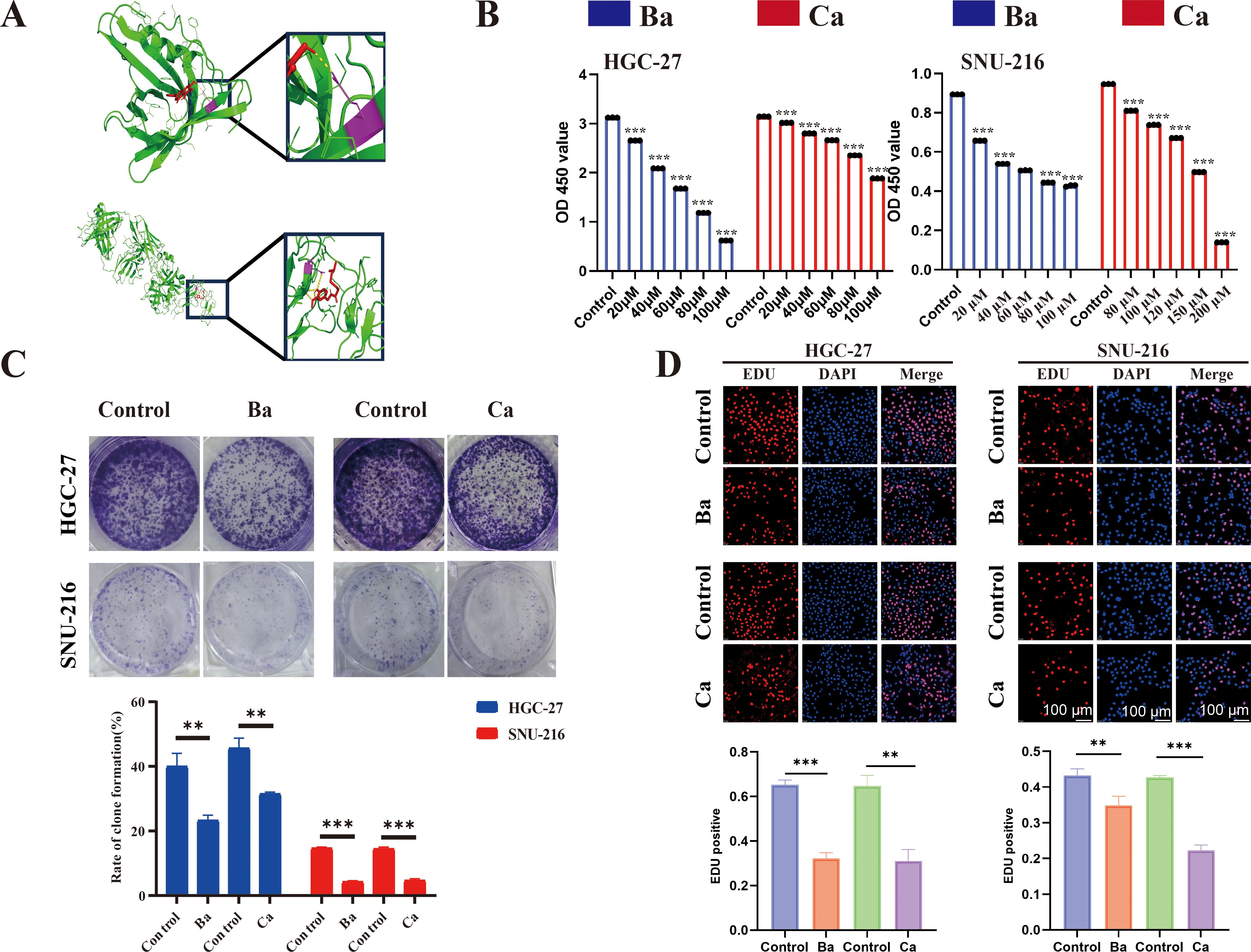
Figure 7. Molecular docking and drugs on GC cell proliferation. (A) Three-dimensional (3D) view of the optimal conformations for APOD and baicalein (up), PROC, and capsaicin (down). Green areas indicate target proteins, whereas red areas represent small molecules, yellow areas denote hydrogen bonds between the protein and the small molecule, and purple areas indicate binding sites. (B) CCK8 assay was performed to assess the proliferative ability of GC cells (HGC-27 and SNU-216) after drug treatment. (C) A colony formation assay was performed to assess the colony-forming ability of GC cell lines after drug treatment. (D) EdU assay for GC cells after drug intervention and matched quantitative analysis results. Ba, baicalein; Ca, capsaicin. ** for p < 0.01, and *** for p < 0.001. Error bars indicate SD; The error bars are short due to the low variance.
The authors apologize for this error and state that this does not change the scientific conclusions of the article in any way. The original article has been updated.
Publisher’s note
All claims expressed in this article are solely those of the authors and do not necessarily represent those of their affiliated organizations, or those of the publisher, the editors and the reviewers. Any product that may be evaluated in this article, or claim that may be made by its manufacturer, is not guaranteed or endorsed by the publisher.
Keywords: ARID1A, gastric cancer, molecular docking, immune gene risk model, prognosis
Citation: Zhang J, Li J, Yang S, Tang X, Wang C, Lin J, Chen Q, Xu H, Ma Y and Gao X (2025) Correction: Development and validation of an ARID1A-related immune genes risk model in evaluating prognosis and immune therapeutic efficacy for gastric cancer patients: a translational study. Front. Immunol. 16:1637370. doi: 10.3389/fimmu.2025.1637370
Received: 29 May 2025; Accepted: 30 May 2025;
Published: 13 June 2025.
Approved by:
Frontiers Editorial Office, Frontiers Media SA, SwitzerlandCopyright © 2025 Zhang, Li, Yang, Tang, Wang, Lin, Chen, Xu, Ma and Gao. This is an open-access article distributed under the terms of the Creative Commons Attribution License (CC BY). The use, distribution or reproduction in other forums is permitted, provided the original author(s) and the copyright owner(s) are credited and that the original publication in this journal is cited, in accordance with accepted academic practice. No use, distribution or reproduction is permitted which does not comply with these terms.
*Correspondence: Xiaoling Gao, Z2FveGwwMDhAaG90bWFpbC5jb20=
†These authors have contributed equally to this work