 Dominic S Nolan
 Arwen F Altenburg*
- Department of Pathology, University of Cambridge, Cambridge, United Kingdom
Major histocompatibility complex (MHC, human leukocyte antigen [HLA] in humans) class I molecules present peptides at the cell surface to cytotoxic immune cells. Peptides are generated and selected for MHC class I presentation in the antigen processing and presentation (APP) pathway. Recognition of a foreign peptide in the context of MHC class I by CD8+ T cells results in target cell lysis. While CD8+ T cells don’t provide sterile immunity, they are pivotal in viral infections by clearing infected cells and may impact disease duration and severity, and virus spread. Additionally, MHC class I molecules act as ligands for NK cell receptors, which similarly play an important role in the control of virus infections through cytotoxic activity and cytokine production. To evade immune recognition, viruses have developed strategies to modulate MHC class I levels by targeting MHC molecules directly or by disrupting components of the APP pathway. Herpesviruses, large DNA viruses that encode numerous immunoevasins, are notorious for disrupting virtually every stage of the MHC class I APP pathway. Over the years, it has become clear that a wide range of other viruses also have evolved targeted mechanisms to modulate MHC class I or components of the APP pathway to evade cytotoxic immune responses. Here, we review the literature on targeted viral manipulation of HLA class I, including non-classical HLA molecules, and modulation of components of the APP pathway by viruses infecting humans.
1 Introduction
1.1 The MHC class I antigen processing and presentation pathway
Presentation of immunogenic peptides on major histocompatibility complex (MHC, human leukocyte antigen [HLA] in humans) class I is crucial for immune detection and control of malignant or infected cells. MHC class I molecules consist of a heavy chain (HC) bound to β2-microglobulin (β2m). Assembly of these molecules requires several chaperones that aid in general protein folding and quality control in the endoplasmic reticulum (ER). The immunoglobulin-binding protein BiP (aliases HSPA5 or GRP78) stabilises polypeptides during folding, facilitates retrograde translocation of misfolded proteins for proteasomal degradation, and regulates the unfolded protein response (1, 2) (Figure 1.1). The chaperone calnexin recognises monoglycosylated N-linked oligosaccharides on folding intermediates, assists in folding, and releases the MHC class I HC when it assembles with β2m (1, 3–5) (Figure 1.2). ERp57 (aliases PDIA3 or GRP58) is recruited by calnexin and catalyses disulphide formation in the HC (5–7).

Figure 1. The MHC class I antigen processing and presentation pathway. MHC class I heavy chains (HC) are translated into the ER where BiP performs quality control (1) and folding is assisted by calnexin (CANX) and ERp57 (2). Proteins are degraded by the proteasome (3) and peptides are transported into the ER by transporter associated with antigen processing (TAP) (4) where they can be further trimmed by ER aminopeptidases (ERAP) (5). Tapasin loads a peptide onto MHC class I in the peptide loading complex (PLC) (6) and TAPBPR further optimises the peptide repertoire (7). Optimally loaded MHC class I molecules traffic to the cell surface for immune surveillance. When a CD8+ T cell recognises a foreign peptide in the context of MHC class I, it lyses the target cell (8). Additionally, MHC class I molecules serve as a ligand for NK cell receptors (9).
Immunogenic peptides for presentation on MHC class I are generated by proteasomal processing (Figure 1.3) of proteins that are degraded because of misfolding, as part of their natural turnover (‘retiree’), or are sourced from defective ribosomal products (DRiPs), which are defective polypeptides produced from errors occurring during translation (8, 9). Peptides are transported from the cytoplasm into the ER by the transporter associated with antigen processing (TAP) (10–13) (Figure 1.4), where they can be further trimmed to an optimal length of 9–12 amino acids by ER aminopeptidases (ERAPs) (14–17) (Figure 1.5).
To acquire peptides, MHC class I molecules are chaperoned by calreticulin, a soluble homologue of calnexin (18). Calreticulin recruits the HC:β2m heterodimer into the peptide loading complex (PLC), which additionally consists of tapasin docked onto TAP, and is stabilized by ERp57 (19–23) (Figure 1.6). Peptides are loaded onto MHC class I by tapasin in the PLC (24–28). MHC class I molecules can undergo further peptide editing by TAPBPR, a tapasin homologue that functions independent of the PLC (29–31). TAPBPR can perform direct peptide exchange or recruit UDP-glucose:glycoprotein glucosyltransferase 1 (UGT1), which causes peptide receptive MHC class I to recycle back to the PLC for peptide acquisition (32, 33) (Figure 1.7).
Stable peptide:MHC class I complexes traffic to the cell surface for immunosurveillance by cytotoxic immune cells. A foreign or modified peptide in the context of MHC class I activate cytotoxic CD8+ T cells which subsequently lyse the target cell (Figure 1.8). Additionally, MHC class I molecules regulate natural killer (NK) cell activity through immune receptors (Figure 1.9) and may bind similar receptors on other immune cell types. For example, reduction in MHC class I levels due to pathogen-mediated interference may decrease signalling through inhibitory NK cell receptors and therefore lower the threshold for NK cell activation.
1.2 Classical and non-classical HLA class I
HLA class I molecules are subdivided into two groups: classical HLA class Ia (HLA-A, B and C) and non-classical HLA class Ib (HLA-E, F and G). Classical HLA class I molecules are expressed on virtually every nucleated cell and are encoded by the most polymorphic genes in humans, with 5092 HLA-A, 6311 HLA-B, and 4858 HLA-C molecules described to date (IPD-IMGT/HLA Database, numbers from the latest version – 3.61 (2025–07)). Most polymorphisms are located near the peptide binding groove, which determines peptide specificity, allowing different HLA class I molecules to display different peptides for immunosurveillance. The polymorphic nature of the HLA class I genes enables selective advantage against the diversity of pathogens and antigens encountered by the population as a whole and have substantial impact on an individuals’ susceptibility to infectious diseases, cancer, and autoimmune conditions. For example, specific HLA class I alleles have been associated with susceptibility or protection from human immunodeficiency virus (HIV) disease progression (34, 35). Furthermore, several HLA-B*27 allotypes are associated with high susceptibility to ankylosing spondylitis (36–38).
Group HLA class Ib molecules display reduced genetic diversity in the human population compared to HLA class Ia molecules (IPD-IMGT/HLA Database). HLA-E is widely expressed (39), whereas HLA-F molecules are mostly expressed in lymphocytes (40–43) and HLA-G expression is restricted to human extravillous trophoblasts (EVT) (44, 45). HLA class Ib molecules are best known as NK cell ligands (46–52), however, the role and importance of non-classical HLA class I molecules on the outcome of viral infections remains largely unknown.
1.3 The HLA class I pathway in virus infections
While CD8+ T cells don’t provide sterile immunity, they are pivotal in viral infections by clearing infected cells and may impact disease duration, severity and spread. NK cells similarly play an important role in virus infections through cytotoxic activity and cytokine production (53). General disruption of host cell function during viral infections may affect HLA class I antigen processing and presentation (APP) and consequently antiviral immune responses. For example, viruses may cause host shut-off; halting synthesis of cellular proteins, including APP proteins, to favour production of viral proteins (54). Furthermore, viruses such as influenza A virus (IAV), poliovirus, coronaviruses, hepatitis C virus (HCV), and HIV-1 encode viroporins that integrate into the host (ER) membranes, thereby disrupting the ion homeostasis and inhibiting host trafficking through the secretory pathway (55). These disruptions of the infected cell may impact the abundance of peptide: HLA class I complexes at the cell surface and the quality of the peptide repertoire presented to immune cells. Many viruses have evolved targeted mechanisms to modulate HLA class I or components of the HLA class I APP pathway to evade cytotoxic immune responses. Here, we review the literature on targeted manipulation of proteins in the HLA class I APP pathway by viruses infecting humans.
2 Viral interference with classical HLA class I
HLA-A, -B, and -C are essential for the activation of anti-viral CD8+ T cells and regulate NK cell activity. While about a third of the HLA-A and -B allotypes can be recognised by killer cell immunoglobulin-like receptors (KIR), all HLA-C molecules serve as ligand for these NK cell receptors (56). These functional differences may explain selective virus-mediated downregulation of HLA-A and -B to mediate CD8+ T cell evasion whereas HLA-C expression remains unchanged to maintain an inhibitory signal to NK cells. Selective targeting of HLA-A and -B molecules has been reported for human cytomegalovirus (HCMV) proteins US2 & US11 (57–59), Kaposi’s sarcoma-associated herpesvirus (KSHV) protein K5 (60), human papillomavirus strain 16 (HPV-16) protein E5 (61), Epstein-Barr virus (EBV) protein BILF1 (62), adenovirus protein E3-19K (63, 64) and HIV-1 protein Nef (targeting HLA-A more than HLA-B) (65, 66). However, more recent work using primary HIV-1 strains instead of lab-adapted strains indicated that the Vpu protein of some strains can downregulate HLA-C (67, 68). Furthermore, HCMV proteins US3 and US6 expressed by recombinant vaccinia virus (rVACV) downregulate HLA-C on trophoblasts (69) and US10 was recently shown to downregulate tapasin-dependent HLA-B molecules and also HLA-C (70). KSHV also expresses a second HLA class I-modulating protein (K3) to downregulate HLA-C (60) resulting in reduced surface expression of all classical HLA class I molecules.
In contrast, VACV infection resulted in downregulation of HLA-C, while HLA-A and -B expression levels remained largely unchanged (71, 72). H3N2 influenza A virus (IAV) also seemed to cause stronger downregulation of HLA-C allotypes compared to HLA-A and -B, whereas influenza B virus (IBV) downregulated all three allotypes similarly (73). Finally, HLA-C molecules were also highly susceptible to modulation by herpes simplex virus-2 (HSV-2) protein ICP47, whereas HLA-B surface expression largely remained unaffected and varying results were shown for HLA-A (74, 75). It was proposed that HSV-2 utilises the NK cell response activated through downregulation of HLA-C to mediate killing of dendric cells and prevent activation of adaptive immunity (74).
Taken together, virus-mediated modulation of HLA class I expression is highly context dependent. Differential – e.g. time or cell type-dependent – expression of these viral proteins may regulate HLA class I modulation and therefore contribute to protection from immune recognition.
While HLA class I molecules are a common target for a broad range of viruses, there are differential mechanisms underlying viral manipulation (Table 1):

Table 1. Strategies for viral modulation of classical HLA class I.
2.1 Interference with HLA class I heavy chain synthesis
Transcription of the HLA class I heavy chain can be upregulated by immune receptor signalling, for example by binding of cytokines interferon (IFN)-γ and tumour necrosis factor (TNF)-α to their respective receptors at the cell surface. The subsequent intracellular signalling leads to degradation of IκB, releasing NFκB to translocate to the nucleus where it binds to the HLA class I HC promotor region (76, 77) (Figure 2). Additionally, phosphorylation and homodimerisation of STAT1 in the cytoplasm is triggered. The STAT1 homodimer translocates to the nucleus where it induces expression of interferon regulatory factor (IRF)-1 and NOD-like receptor family CARD domain containing (NLRC) 5. In turn, these function as transcription factors for the expression of proteins of the HLA class I APP pathway, including the HLA class I HC (76, 77) (Figure 2).

Figure 2. Viral interference with HLA class I HC transcription. Immune receptor signalling triggers production of transcription factors which translocate to the nucleus. NFκB, interferon regulatory factor (IRF)-1, and NOD-like receptor family CARD domain containing (NLRC) 5 regulate transcription of HLA class I genes. (1) IκB degradation is required to release NFκB, allowing it to translocate to the nucleus. HIV-1 Vpu stabilises IκB by inhibiting its degradation. (2) Ad12 E1A may disable NFκB binding and enable repressor activity. (3) HPV E7 may interfere with NFκB translocation (a) and repress chromatin activation (b). (4–5) KSHV inhibits HLA class I transcription by blocking IRF-1, and possibly NFκB, using viral IRF1 (vIRF1) (4). During the latent phase KSHV expresses vFLIP to stimulate NFκB and enhance HLA class I expression (5). (6) SARS-CoV-2 ORF6 inhibits translocation of STAT1 (a), IRF1 (b) and NLRC5 (c) into the nucleus. ISRE = interferon responsive element. DNA strand illustration from NIAID NIH BIOART Source (bioart.niaid.nih.gov/bioart/123).
Several viruses modulate different steps that regulate HLA class I transcription. For example, the HIV-1 protein Vpu, which is expressed at a late stage of infection, prevents the degradation of IκB and inhibits NFκB from translocating to the nucleus (78) (Figure 2.1). This results in a reduction of NFκB-dependent expression of IFN and IFN-stimulated genes, including HLA class I (78, 79). The HIV-1 Nef protein boosts NFκB expression early during the viral life cycle when Vpu is not yet expressed (78). This may explain why HIV-1 Nef presents an additional strategy to downregulate HLA class I molecules (more in section 2.4).
Oncogenic adenovirus type 12 (Ad12) encodes protein E1A that reduces MHC class I levels (80, 81). An initial report described that E1A in interferes with p105-NFκB1 processing, preventing the formation of NFkB dimers (82). Later publications describe NFκB activity in the nucleus, and that the N-terminus of Ad12 E1A prevents phosphorylation of the p65 subunit of NFκB to inhibit binding to the enhancer element (83, 84) (Figure 2.2). Additionally, E1A was suggested to associate with the enhancer region to recruit histone deacetylases (HDAC) 1 and HDAC8 to deacetylate histones and repress MHC class I transcription (85). The decrease in MHC class I expression due to one or more of these mechanisms may contribute to immune escape by Ad12-transformed cells.
Similar to Ad12 E1A, the oncoprotein E7 of selected HPV types was suggested to associate with the HLA class I promotor together with HDACs to repress chromatin activation (86–88) (Figure 2.3a). Additionally, it was reported that E7 impairs nuclear translocation of NFκB to downregulate HLA class I promotor activity (88) (Figure 2.3b). The E7-mediated interference with HLA class I expression may impact the susceptibility to NK cells (89).
Manipulation of HLA class I transcription is carefully balanced by oncovirus KSHV, also known as human herpesvirus 8 (HHV-8). After primary infection, herpesviruses enter a latent phase during which viral protein expression is minimal to evade immune recognition allowing the virus to persist for the lifetime of the host. The virus can reactivate from this latent phase and enter a lytic phase during which novel viral particles are produced, and cell lysis is triggered to facilitate virion release. The KSHV protein viral (v)IRF1 inhibits HLA class I expression by blocking the cellular IRF1 and possibly NFκB (90) (Figure 2.4). In contrast, viral FLICE inhibitory protein (vFLIP) enhances HLA class I transcription by stimulating NFκB (Figure 5.5), which may present a strategy to prevent uncontrolled viral dissemination in latency (90).
Severe Acute Respiratory Syndrome Coronavirus 2 (SARS-CoV-2) protein ORF6 targets the nuclear localisation of STAT1, IRF1 and NLRC5 by blocking karyopherin-mediated protein import (Figure 2.6). This results in reduced transcription and consequently lower HLA class I expression in SARS-CoV-2-infected cells (91). The related virus Middle East Respiratory Syndrome Coronavirus (MERS-CoV) downregulates RNA levels of genes involved in antigen presentation, both within the MHC locus and antigen presentation genes located on other chromosomes, resulting in downregulation of HLA class I proteins (92). It was proposed that this is caused by alterations in DNA methylation after MERS-CoV infection (92).
Similarly, avian H5N1 influenza virus (A/influenza/Vietnam/1203/2004) downregulated RNA expression of genes involved in antigen presentation, while seasonal H1N1 did not. In contrast to MERS-CoV infection, this led to downregulation of HLA-A and -C, but not HLA-B proteins (92). A combination of DNA methylation and histone alterations were suggested to underly the altered expression levels (92). Of note, these studies are based on -omics datasets and require further investigations to biochemically and mechanistically study altered protein expression.
2.2 Targeting of nascent HLA class I chains
After transcription, the mRNA encoding the HLA class I HC is translated into the ER. HCMV (or HHV-5, typically asymptomatic except in immunocompromised individuals or newborns) employs a range of strategies to evade CD8+ T cells and targets HLA class I HCs with US2 and US11 (58, 93–95). The US2:HC or US11:HC complexes are dislocated to the cytoplasm through the cellular Sec61 translocon where they are degraded by the proteasome (94–96) (Figure 3.1). A different strategy is employed by SARS-CoV-2, which encodes ORF7a to act as a β2m mimic to compete for HC binding (Figure 3.2). This prevents PLC formation and retains HLA class I molecules in the ER (97, 98).
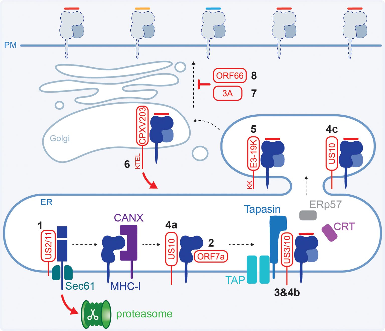
Figure 3. Viral interference with the HLA class I HC and transport. (1) HCMV US2 or US11 form a complex with the HC and target it for proteasomal degradation in the cytoplasm. (2) SARS-CoV-2 ORF7a mimics β2m and competes for HC binding. (3) HCMV US3 retains HLA class I in the ER trough tapasin binding. (4) HCMV US10 binds all HLA class I HCs (a) but only prevents β2m:HLA-B from interacting with the PLC (b) and interacts with selected β2m:HLA-C causing ER retention (c). (5) Adenovirus E3-19K retains HLA class I in the ER using a dilysine motif. (6) Cowpoxvirus CPXV203 has a C-terminal KTEL motif that is recognised by the cellular KDEL pathway for protein retrieval from the Golgi back to the ER. (7) Poliovirus and coxsackievirus B3 proteins 3A disrupt the Golgi and interfere with HLA class I trafficking. (8) VZV ORF66 retains HLA class I molecules in the Golgi.
2.3 Intracellular retention of HLA class I
Retention of HLA class I inside the cell reduces the available peptide:HLA class I complexes at the cell surface that are monitored by immune cells. Different stages of the egress pathway are targeted by viruses to downregulate surface HLA class I levels. For example, HCMV protein US3 causes ER retention of stable HLA class I heterodimers loaded with a peptide (99, 100). Distinct domains in US3 are involved in the interaction with HLA class I and binding to tapasin causing ER retention (101–103) (Figure 3.3). US3 is only expressed during the early phases of infection and therefore targets the existing mature pHLA class I complexes, while expression of US2 and US11 (section 2.2) peaks slightly later ensuring efficient downregulation of newly synthesised HCs. Co-expression of US3 and US2, but not US11, enhanced the association between US2 and HCs, thereby increasing their degradation (104, 105).
Additionally, HCMV protein US10 was shown to bind a diverse range of HLA class I allotypes and decreased surface expression of tapasin-dependent HLA-B, and selected HLA-C molecules (70, 106). In the proposed model, US10 interacts with all HLA class I HCs, but only tapasin-dependent HLA-B molecules are prevented from interacting with PLC components, while HLA-C:β2m heterodimers are stabilised and retained in the ER (70) (Figures 3.4a–c).
ER retention is also mediated by adenovirus protein E3-19K, which binds the luminal domain of MHC class I molecules (107–110). Inhibition of transport is mediated by a dilycine motif in the E3-19K cytoplasmic tail which, in concert with the transmembrane domain, enables ER retrieval for static ER retention (109, 111, 112) (Figure 3.5). This results in a decrease in surface HLA class I molecules and reduced CD8+ T cell recognition of infected cells (107, 108, 113, 114).
Using mouse cells, it was shown that cowpox virus (zoonotic infection that causes large blisters in the skin) protein CPXV203 interacts with MHC class I and has a C-terminal KTEL motif that resembles the KDEL motif in calreticulin (Figure 3.6). This enables CPXV203 to hijack the cellular pathway used for ER retention where calreticulin binds suboptimally loaded MHC class I in the Golgi and recruits the KDEL receptor to initiate retrograde transport (115). CPXV203-mediated downregulation of surface MHC class I levels impaired the antiviral CD8+ T cell response (116–118).
Even though picornaviruses are small RNA viruses, and have limited genomic capacity for immune modulatory proteins, they manipulate HLA class I molecules. While the surface expression of MHC class I was not affected, poliovirus (the causative agent of paralytic polio disease) protein 3A inhibits the secretory pathway and slows down transport of newly synthesised MHC class I molecules (119). Furthermore, coxsackievirus B3 (CVB3, does not cause serious disease in immunocompetent individuals but is more dangerous for newborns and can cause myocarditis) protein 3A disrupts the Golgi to inhibit anterograde transport (120) (Figure 3.7). This resulted in an impaired antiviral CD8+ T cell response in chimpanzee cells or mice, respectively (119, 121).
Herpesviruses other than HCMV manipulate the levels of surface HLA class I as well, although the exact molecular mechanisms have not been established. Varicella zoster virus (VZV, or HHV-3), the causative agent of chickenpox and shingles, encodes the ORF66 protein kinase that downregulates surface HLA class I by delaying maturation and mediates Golgi retention (122–124) (Figure 3.8). Furthermore, interference with the exocytic pathway and Golgi retention is reportedly mediated by EBV (or HHV-4, the most common cause of mononucleosis) protein BILF1, which impairs CD8+ T cell activation (125).
Downregulation of HLA class I from the cell surface by interference with the exocytic pathway and Golgi retention has also been reported to be mediated by protein E5 of selected HPV types (61, 126, 127). This impacted CD8+ T cell activation (128). Finally, it was proposed that during IBV infection, HLA class I molecules are retained in an undefined vesicular location enroute to the plasma membrane resulting in their proteasome-dependent downregulation (73). The molecular mechanism, biological consequence or in vivo relevance of the observed HLA class I modulation by HPV and IBV have not established thus far.
2.4 Internalisation of HLA class I
While CV3B protein 3A was shown to inhibit anterograde transport (section 2.3), this did not result in a complete block of HLA class I trafficking to the cell surface (120). To aid evasion of the CD8+ T cell response, the virus encodes proteins 2B and 2BC to enhance endocytosis resulting in removal of HLA class I from the cell surface (120, 121) (Figure 4.1). The mechanism underlying this surface downregulation and the fate of HLA class I after internalisation by CXV3B remain to be determined. As a result of HLA class I downregulation, very few virus-derived peptides are presented, and they display limited recognition by CD8+ T cells in CVB-positive individuals (129).

Figure 4. Viral proteins causing HLA class I internalisation, degradation or shielding. (1) CXV3B proteins 2B and 2BC enhance endocytosis of surface HLA class (I) (2) EBV protein BILF1 internalises surface HLA class I and directs it to the lysosome for degradation. (3) KSHV K3 and K5 ubiquitinate the HLA class I cytoplasmic tail which induces internalisation and trafficking to the lysosome. (4) HIV-1 Nef enhances HLA class I endocytosis and translocates it to the trans-Golgi network (TGN) where it accumulates and may be transported to the lysosome for degradation. It has also been proposed that Nef targets exocytic export of newly synthesised HLA class I to the lysosome. (5) SARS-CoV-2 ORF8 causes autophagy via the becillin-1 pathway, resulting in lysosomal HLA class I degradation. (6) HHV-6 and -7 encode protein U21 which redirects HLA class I to the lysosome. However, it is unknown if this occurs from the ER (a), Golgi (b) or cell surface (c). (7) IAV infection results in a global loss of HLA class I molecules which is dependent on the proteasome. The viral protein(s) involved has yet to be identified. (8) The heavily glycosylated EBOV GP and EBV gp150 shield surface HLA class I.
The EBV G protein-coupled receptor (GPCR) BILF1 similarly targets both endocytic (section 2.3) and exocytic pathways. Residues in the extracellular bridges between transmembrane domains of BILF1 were shown important for interaction with surface HLA class I, likely due to their role in maintaining BILF1 protein conformation (130). HLA class I downregulation by BILF1 is independent of its GPCR signalling properties (130, 131). BILF1 enhances HLA class I endocytosis via the DRY-like EKT motif, which is a highly conserved sequence in GPCRs located on the cytoplasmic side of transmembrane domain 3 (125). Subsequent lysosomal degradation was shown to depend on the BILF1 C-terminus and several residues in the HLA class I cytoplasmic tail (62, 125, 131) (Figure 4.2), although the exact motif and trafficking routes have not been identified thus far.
KSHV encodes proteins K3 and K5 that enhance endocytosis of surface HLA class I (60, 132, 133). These proteins are ubiquitin ligases that ubiquitinate a lysine in the HLA class I cytoplasmic tail, which acts as a signal for internalisation after which HLA is sorted to the late endosomal pathway where it is degraded (134) (Figure 4.3).
HLA class I manipulation by HIV-1 protein Nef has been extensively studied. Nef binds the HLA class I cytoplasmic tail and its acidic 62EEEE65 motif recruits cellular sorting proteins phosphofurin acidic cluster sorting protein (PACS)-1 and PACS-2 to promote retrieval from the cell surface (135–141). The methionine at position 20 in Nef prevents HLA class I from recycling back to the cell surface and redirects it to the trans-Golgi network (TGN), which acts as a sorting compartment (139). In the TGN, the Nef 72PXXP75 motif activates Src family tyrosine kinase (SFK) to stimulate phosphoinositide 3-kinase (PI3K) signalling to increase the rate of endocytosis of HLA class I from the cell surface via an ADP ribosylation factor 6 (ARF6)-regulated pathway (139, 140, 142). However, there are also reports that Nef-induced endocytosis of HLA class I is independent of PACS-1 or ARF6 and that PI3K-signalling is only required for Golgi retention rather than endocytosis (143, 144).
Binding of Nef to the HLA class I cytoplasmic tail compensates for an incomplete sorting motif in HLA class I, forming a docking site for the cellular clatherin adaptor protein (AP)-1 to sort proteins to lysosomal compartments (136, 138, 145–149). However, there are conflicting reports suggesting that the Nef:MHC class I complex is retained in the TGN without lysosomal degradation (139, 141) or may be held in a pre-lysosomal compartment but not the TGN (144). Additionally, it has been proposed that newly synthesised HLA class I, rather than molecules endocytosed from the cell surface, are trafficked from the TGN to the lysosome (146, 150). One model does not necessarily exclude the other, and Nef may indeed both internalise HLA class I and interfere with anterograde transport (151). The inconsistent reports on the source and fate of these HLA class I molecules may potentially be explained by the cell type and/or intracellular Nef concentrations used in the various studies (152, 153). Nonetheless, the reduction in surface HLA class I protects the infected cells from CD8+ T cell recognition (154) (Figure 4.4).
2.5 HLA class I degradation
Rather than internalising surface molecules, the SARS-CoV-2 protein ORF8 triggers lysosomal degradation of HLA class I through the induction of the Becilin-1 autophagy pathway (155) (Figure 4.5).
The U21 protein encoded by HHV-6 and -7 (causative agents of roseola, which usually mild and self-limiting in children) also directly binds HLA class I molecules and targets them to the lysosome, which is independent of the HLA class I cytoplasmic tail (156–159). U21 traffics to the lysosome even when HLA molecules are not associated and trafficking was proposed to involve a Golgi-derived vesicle that is clatherin-independent (158, 160). Trafficking may involve a currently unknown cellular protein (158). It remains to be established if U21 targets HLA class I molecules in the ER, Golgi or at the cell surface (158) (Figures 4.6a–c).
Infection with seasonal IAV resulted in a global loss of HLA class I expression and a 30-40% reduction at the cell surface (73). The mild reduction overall may potentially be due to the strong HLA-C downregulation, and more subtle downregulation of HLA-A and -B. HLA class I downregulation by IAV was shown to be dependent on the proteasome, however, the exact mechanism remains to be established (73) (Figure 4.7).
2.6 HLA class I masking
The Ebola virus (EBOV) glycoprotein (GP) was shown to sterically hinder detection of HLA class I on the cell surface (161–163) (Figure 4.8). The glycan-mediated shielding of surface HLA class I impaired the CD8+ T cell response (163). Of note, these studies were not performed in the context of EBOV infection and GP was expressed using a plasmid or adenovirus vector. Similar to EBOV, the EBV-encoded gp150 protein also causes a reduction in detection of surface HLA class I, and other antigen presentation molecules, potentially by shielding the molecules through its abundantly sialylated glycans (164). Thus, HLA class I molecules still reach the cell surface; however, activation of T cell responses was prevented through a glycan shield.
3 Viral manipulation of non-classical HLA class I
3.1 HLA-E
HLA-E is widely expressed and is recognised by inhibitory CD94/NKG2 heterodimers and LILRB1/2 receptors on NK cells, and other immune cells (46–48, 165). Generally, HLA-E presents self-peptides derived from the leader sequences of HLA-A, -B, and -C molecules, which allows the immune system to monitor HLA class I expression (46, 166, 167) (Figure 5). This is a control mechanism for (pathogen-mediated) interference with classical HLA class I as the loss of signal peptide reduces inhibitory HLA-E levels, resulting in NK cell activation. Furthermore, HLA-E can present HLA-G-derived leader peptides. While HLA-E:peptide complexes typically have higher affinity for inhibitory CD94/NKG2A receptors, presentation of the HLA-G leader peptide results in a higher affinity for the activating CD94/NKG2C (167). Additionally, MHC-E can present virus-derived peptides, such as from HCMV, HIV, influenza virus, hepatitis B virus (HBV), HCV, and SARS-CoV-2, which activate ‘unconventional’ CD8+ T cell responses (168–175) (Figure 5). Given the low genetic variation of HLA-E in the human population, HLA-E-restricted CD8+ T cells are of particular interest for vaccine development (173, 176, 177).

Figure 5. HLA-E in virus infections. HLA-E presents the signal peptide from classical HLA molecules and HLA-G, which provides an inhibitory signal to NK cells via CD94/NKG2A and LILRB1/2 receptors. HLA-E can also present peptides derived from viral proteins and activate ‘unconventional’ CD8+ T cell responses. (1) HCMV encodes a signal peptide mimic in the US40 protein, stabilising HLA-E at the cell surface and inhibiting NK cells (a). However, this can result in T cell activation (b). (2) HIV-1 Nef downregulates HLA-E via its cytoplasmic tail. (3) E7 of HPV was reported to downregulate HLA-E expression by hypermethylation of the HLA-E gene. DNA strand illustration from NIAID NIH BIOART Source (bioart.niaid.nih.gov/bioart/123).
Viral manipulation of HLA-E to evade immune cells (Table 2) is well-established in HCMV infection. HCMV encodes protein US40 which contains a signal peptide mimicking the signal peptide derived from cellular HLA class I molecules and is presented on HLA-E in a TAP-independent manner (178–181) (Figure 5.1). By stabilising HLA-E at the cell surface, HCMV compensates for the loss of HLA-A, -B, and -C expression after infection and maintains NK cell inhibitory signals (178–181). However, this can result in the activation of UL40-specific HLA-E-restricted T cell responses (168–170, 182).

Table 2. Viral manipulation of non-classical HLA class I.
In contrast, selected primary HIV-1 strains have been shown to reduce surface HLA-E expression, which was mediated by the Nef protein targeting the HLA-E cytoplasmic tail (183) (Figure 5.2). Modest HLA-E downregulation on B lymphoblastoid cell line (BLCL) has been reported after VACV infection, which may potentially impact NK cell activation (184). It is unclear if this is targeted downregulation mediated by VACV. Finally, HPV oncoprotein E7 of HPV16 and HPV18 strains with high oncogenic potential, but not of low-risk HPV6 and HPV11, may downregulate HLA-E expression by DNA hypermethylation of the HLA-E gene (185) (Figure 5.3). However, the effect of HLA-E downregulation on anti-HPV immune responses remains to be established.
3.2 HLA-F
In contrast to the highly polymorphic HLA class Ia molecules, only 122 HLA-F alleles have been described which encode 27 different HLA-F proteins (IPD-IMGT/HLA Database). The peptide binding groove of HLA-F in complex with β2m accommodates relatively long peptides (186–188). However, peptide loading is not essential for HLA-F trafficking and surface expression, and the protein can be expressed as an open conformer (OC) consisting of the HC only (41, 43, 189, 190). These OCs may also be expressed as a homodimer or a heterodimer with an HLA class Ia HC, potentially with a role in cross-presentation of exogenous antigens (191).
HLA-F resides mostly intracellularly but can be expressed at the cell surface of activated lymphocytes as ligands for activating and inhibitory KIR and LILR receptors on immune cells (40–43, 49, 50). For example, the activating NK cell receptor KIR3DS1 recognises HLA-F OCs while the HLA-F:β2m:peptide complex binds inhibitory receptor LILRB1/2 (49, 186, 192). HLA-F OCs are also recognised by inhibitory receptors KIR3DL1 and KIR3DL2, although they bind with lower affinity compared to KIR3DS1 (50, 191). Conflicting results have been published regarding HLA-F OCs functioning as a ligand for KIR2DS4 (50, 191). It is currently unknown if viral peptides can be presented by HLA-F to regulate NK or T cell responses.
Increased HLA-F expression has been reported after infection with Japanese Encephalitis virus, HIV-1 (early in infection), HCV, and BK polyomavirus (BKpV) (50, 193–195). For the latter three, this was shown to result in enhanced recognition by KIR3DS1 and increased NK cell activation in vitro (50, 194–196). Furthermore, KIR3DS1 has been associated with delayed progression of disease caused by HIV-1, and the HLA-F*01:03 polymorphism was associated decreased levels of HBV DNA (50, 197). Taken together, this suggests that HLA-F may play a role in infection control.
Evidence for manipulation of HLA-F by viruses to escape immune responses is limited. HLA-F expression may be reduced on CD4+ T cells late in infection with HIV-1, impacting KIR3DS1 binding and NK cell activation (50, 196). However, the mechanism and impact on antiviral immune responses remain elusive.
3.3 HLA-G
HLA-G expression is restricted to EVT cells at the maternal-foetal interface where it mediates tolerance (51, 198–200). HLA-G is the only HLA class I molecule described to form β2m-associated homodimers, which interact with the inhibitory LILR receptors (200–202). HLA-G has a number of additional unusual features including the lack of an endosomal recycling motif (203), presence of an ER retrieval motif (204), and it presents a restricted peptide repertoire (205, 206). Several HLA-G isoforms have been described, including soluble versions, although the biological role and relevance of the alternative transcripts in vivo has yet to be confirmed (207).
A recent review summarised neoexpression of HLA-G in cells infected with HPV, HBV, HCV, HCMV, EBV, HIV, human lymphotropic virus (HTLV)-1, IAV or SARS-CoV-2 (208). However, this should be interpreted with caution as i) most antibodies against HLA-G are poorly characterised and may cross-react with classical HLA molecules, ii) experimental setup may be lacking controls or an Fc receptor block, which is essential particularly when staining immune cells, and iii) the biological relevance of soluble HLA-G in vivo remains controversial (207). The role of HLA-G in virus infections therefore remains elusive.
Targeted manipulation of HLA-G (Table 2) has been reported for human herpesvirus 6 (HHV-6), which in the placenta predisposes the mother to pre-eclampsia (209, 210). HHV-6 protein U94 acts on the human transcription factor cyclic AMP-dependent transcription factor (ATF)3 and reportedly increases expression of both membrane and soluble HLA-G isoforms (211). Given the role of HLA-G in tolerance, increased HLA-G expression levels may result in suppression of the antiviral immune response. Of note, these studies did not include biochemical analyses and the 87G antibody that was used may cross-react with HLA class Ia molecules (207).
HCMV protein US10 downregulates HLA-G via a tri-leucine motif in the cytoplasmic tail in a proteasome-dependent manner (212). However, a more recent study proposed US10 binding to HLA-G to mediate ER retention without destabilisation (70). US10-mediated interference with HLA-G hampered NK cell inhibition (212). HCMV proteins US3 (sections 2.3 & 7) and US6 (more in section 5) were also reported to interfere with HLA-G expression (69). Conflicting results have been reported for US2, which have been attributed to experimental setup (57, 69, 213). All these studies include biochemical analysis detecting the smaller 39kDa HLA-G HC, opposed to the 45kDa classical HLA HC, providing confidence that the results are indeed HLA-G-specific. It has been hypothesised that HCMV-mediated downregulation of HLA-G as a ligand for inhibitory NK receptors may play a role in release of intracellular viral particles through cytolysis or that the activated NK cells may produce a more favourable cytokine environment for the virus (212).
4 Viral interference with peptide generation
The proteasome plays a pivotal role in the generation of antigenic peptides for presentation on MHC class I molecules through the regulated degradation of virtually all proteins in the cell (214). The proteasome core consists of four stacked heptameric rings – two α (outer) and two β (inner) rings – that together form a barrel-shape. This 20S core is present with or without regulatory subunits capping either end. The catalytic activity of the core is restricted to β1 (caspase-like), β2 (trypsin-like), and β5 (chymotrypsin-like) subunits (215). Three alternative β subunits (β1i/LMP2, β2i/MECL1, β5i/LMP7) are part of the specialised immunoproteasome that displays altered peptide-cleave properties (216). The immunoproteasome, with 11S regulatory subunit, is abundantly expressed in hematopoietic cells and can be induced in non-immune cells in inflammatory conditions such as viral infections (216).
Two herpesviruses have been described to modulate MHC class I antigen presentation by reducing the pool of available peptides in cis, e.g. reducing peptides derived from these two herpesvirus proteins (Table 3). EBV nuclear antigen 1 (EBNA1) protein expressed during latency contains a long repetitive sequence consisting exclusively of glycine and alanine residues. Initially, it was described that this prevents its degradation by the proteasome (217). However, the absence or low level of EBNA-1-derived peptides was later attributed to inhibition of messenger RNA translation in cis to interfere with formation of EBNA1 DRiPs (218, 219) (Figure 6.1). The reduced pool of EBNA1-derived peptides available for MHC class I presentation limits the EBNA1-specific CD8+ T cell response (220, 221). Similarly, the EBNA1 homologue latency-associated nuclear antigen 1 (LANA1) encoded by KSHV contains a QED-rich central repeat (CR) domain that inhibits translation in cis and impacts presentation of LANA1-derived MHC class I antigens (222, 223) (Figure 6.2).

Table 3. Viral interference with antigen processing.

Figure 6. Viral interference with antigen processing. (1–2) EBV EBNA1 and KSHV LANA1 inhibit translation in cis to reduce the pool of peptides derived from these proteins. (3) HIV-1 Gag p24 downregulates the β2i and β5i subunits from the immunoproteasome and the 11S regulatory subunit that can associate with either the constitutive or immunoproteasome. (4) HIV-1 Tat prevents interaction of 11S with the 20S core (a), downregulates β1i transcription (b) and stabilises the β2i and β5i subunits (c). (5–6) Adenovirus E1A and HPV E7 downregulate β1i transcription. (7) HCV NS3 interferes with β5i to reduce immunoproteasome activity.
Virus-mediated manipulation of proteasome subunits can impact antigen processing (Table 3) and may result in changes of the peptide repertoire presented at the cell surface. The HIV-1 Gag p24 protein was described to downregulate the β2i and β5i subunits of the immunoproteasome and components of the 11S regulatory subunit (alias PA28) in dendritic cells (224) (Figure 6.3). More established is proteasome modulation by the HIV-1 Tat protein, which binds the α4 and α7 subunits to prevent the interaction of 11S with the 20S core (225–227) (Figure 6.4a). Additionally, Tat mediates downregulation of β1i at transcriptional level through interference with the formation of the STAT1-IRF-1 complex, inhibiting IRF-1 binding to the β1i promotor (228) (Figure 6.4b). Tat also interacts with six β subunits of the constitutive 20S core, and stabilises β2i and β5i, leading to a modified composition of the (immuno)proteasome (227, 229) (Figure 6.4c).
Similar to HIV-1 Tat, adenovirus protein E1A and protein E7 from high-risk HPV18 and low-risk strain 6b were reported to downregulate β1i expression at transcription level to interfere with the presentation of viral antigens (86, 230) (Figure 6.5, 6). Furthermore, HCV protein NS3 binds to β5i to reduce immunoproteasome activity (231), which may interfere with processing of viral antigens and impact antiviral immune responses (Figure 6.7).
HBV infection causes viral hepatitis which can lead to hepatocellular carcinoma. While HBxAg encoded by HBV is a substrate for proteasome degradation, it binds the α4 subunit of the 20S proteasome core and the PSMC1 component of the 19S regulatory subunit (232–234). As described for HIV-1 Tat, the interaction between HBxAg and α4 may inhibit binding of the 11S regulatory subunit and explain the observed proteasome inhibition (234, 235). The interactions between HBxAg and components of the proteasome have predominantly been described in the context of its role as a transactivator. The impact of HBxAg on the presented HLA class I peptide repertoire and the antiviral CD8+ T cell response has thus far not been reported.
5 Viral modulation of peptide transport by TAP
The TAP transporter shuttles peptides from the cytoplasm into the ER and is an essential component of the PLC (Figure 1.4). Subunits TAP1 and TAP2 each consist of a series of transmembrane domains (TMD) with an ATP-binding C-terminal nucleotide-binding domain (NBD) in the cytoplasm (236). When the peptide binding pocket faces the cytoplasm, the NBDs are separated. Peptide and ATP bind TAP independently, and when both are bound the NBDs dimerise. These conformational rearrangements are relayed to the TMDs, exposing the peptide binding pocket to the ER lumen where the peptide is released. Upon ATP hydrolysis, the NBDs dissociate and TAP switches back to a cytoplasm (‘inward’)-facing conformation (236) (Figure 7A).

Figure 7. TAP-mediated peptide transport in virus infections. (A) Schematic overview of peptide transport by TAP. (B) (1) HCMV US6 and CPXV protein CPXV012 bind TAP in the ER lumen to block ATP binding, likely through distal allosteric effects. (2) EBV protein BILF2a prevents both ATP and peptide binding to the ‘inward’ facing conformation of TAP. (3) HSV-1/2 protein ICP47 binds TAP on the cytosolic side to block peptide binding by competitive inhibition.
TAP1 is expressed using a bidirectional promotor that is shared with the β1i gene. Although the transcription factor requirement for the two genes is not identical, viral proteins manipulating β1i expression at transcriptional level (section 4) may similarly affect TAP1. For example, protein E7 of selected HPV strains was suggested to inhibit peptide transport by TAP and decrease TAP1, but not TAP2, levels potentially by transcriptional regulation (86, 237, 238).
Blocking of peptide transport from the cytoplasm into the ER is a common mechanism by which viruses, particularly herpesviruses, interfere with HLA class I antigen presentation (Table 4). HCMV protein US6 binds TAP in the ER lumen and prevents the conformational changes required for ATP to bind TAP1 but does not interfere with peptide binding (239–241) (Figure 7B.1). A similar mechanism was described for CPXV transmembrane protein CPXV012 (118, 242), resulting in reduced MHC class I surface expression and affects the antiviral CD8+ T cell response (117, 118).

Table 4. Viral manipulation of TAP.
The 60 amino acid EBV protein BILF2a also interacts with TAP in the ER lumen and interferes with ATP and peptide binding to TAP (243, 244) (Figure 7B.2). Finally, HSV-1 and 2 (or HHV-1&2) encode the 88 amino acid protein ICP47 that binds TAP1/2 on the cytoplasmic side and blocks peptide binding by competitive inhibition (245–248) (Figure 7B.3). While ATP can still bind, ICP47 freezes TAP in the ‘inward’ facing state with the NBDs unable to dimerise, which is required for ATP hydrolysis (247–249). For all these herpesviruses, reduction of the peptide supply in the ER by interference with TAP function resulted in decreased HLA class I surface expression and inhibition of recognition by CD8+ T cells (243, 244, 250, 251).
6 Viral inhibition of ERAP-mediated peptide trimming
Within the ER, ERAPs trim N-terminal extensions of peptide precursors to generate optimal length peptides for presentation on HLA class I (252). Humans encode ERAP1 and ERAP2, which share 49% sequence identity and exhibit preferential differences for their peptide substrate (14, 15, 253). Peptides of 9–16 amino acids with hydrophobic N-terminal extensions, excluding proline, have a higher affinity for trimming by ERAP1, whilst shorter peptides with basic N-termini are more efficiently trimmed by ERAP2 (253–255). The molecules can also trim peptides synergistically as a heterodimer, however, functional ERAP2 may not be expressed by 25% of individuals due to a single nucleotide polymorphism (SNP) resulting in alternative splicing and a truncated protein (15, 255, 256). Addition of recombinant ERAP2 to peripheral blood mononuclear cells (PBMCs) from healthy individuals was able to reduce HIV infection in vitro (257). Furthermore, ERAP1 exerts selective pressure on HIV-1 in individuals expressing HLA-B*57, which commonly results in a mutation that prevents ERAP1 from trimming an immunodominant epitope. This was associated with a 22-fold increase in viral load (258). Taken together, ERAP activity strongly impacts the (viral) repertoire presented on HLA class I molecules.
While virus-mediated manipulation of ERAP2 has thus far not been described, HCMV downregulates ERAP1 during infection using two microRNAs, miR-US4–1 and miR-UL122-5p, that target the 3’ untranslated region (UTR) of ERAP1 (259, 260) (Table 5). This results in recognition by the RNA-induced silencing complex, followed by degradation of the transcript (259). Decreased ERAP1 expression in HCMV-infected cells reduced trimming of virus-derived peptides and impairs T cell recognition of the infected cells (259, 260). Naturally occurring SNPs within the microRNA binding site of ERAP1 can render it resistant to downregulation by HCMV, which may be counteracted by heterogeneity within the HCMV microRNAs (260, 261).

Table 5. Viral manipulation of proteins impacting the peptide repertoire.
7 Viral modulation of peptide editing by tapasin
Tapasin is a central component of the PLC, bridging TAP with peptide-receptive HLA class I molecules to facilitate the association of high-affinity peptides (19, 25, 26, 262–264). The loss of tapasin results in an altered peptide repertoire and (antiviral) CD8+ T cell response (265, 266). HLA class I allotypes exhibit varying degrees of reliance on tapasin binding for their association with high-affinity peptide cargo (267). Therefore, viral modulation of tapasin (Table 5) may result in a reduction in surface expression of tapasin-dependent HLA class I molecules and may not affect other allotypes.
The HCMV protein US3 binds tapasin not only to retain HLA class I molecules in the cell (section 2.3) but also prevents peptide loading on tapasin-dependent HLA class I molecules (103). HCMV may regulate tapasin inhibition using a truncated isoform of US3, which functions as a dominant negative regulator of full-length US3 activity thereby abolishing the initial disruption of tapasin-mediated peptide optimisation (268). Furthermore, HCMV infection also reduces synthesis of new tapasin molecules (269).
In addition to its role in retaining HLA class I in the ER (section 2.3), adenovirus E3-19K prevents tapasin from optimising the HLA class I peptide repertoire by independently binding both HCs and TAP, preventing the formation of the PLC (270). This allows E3-19K to affect antigen presentation by HLA class I molecules that it only moderately affects by direct retention (270).
Molluscum cantagiosum virus (MCV, an endemic human poxvirus that can cause skin lesions) encodes MC80, an MHC class I-like protein that associates with β2m to interact with tapasin and TAP. This leads to the recruitment of ER-associated protein degradation (ERAD) complexes targeting tapasin for ubiquitination and proteasomal degradation (271). Consequently, TAP levels are also downregulated and there is a reduction in classical HLA class I and HLA-E levels (271, 272). MC80 expression from an adenovirus backbone modulated NK cell activity and promoted evasion of CD8+ T cells (272).
8 Discussion
Manipulation of HLA class I and the APP pathway by large DNA viruses such as herpesviruses has been well established over the last four decades. This work was complemented by studies on MHC evasion by non-human viruses, for example murine CMV, equine herpesvirus, and bovine herpesvirus. It has become clear that numerous different strategies to modulate MHC class I levels are used by a wide range of viruses – both DNA and RNA viruses, and viruses causing acute or chronic infections. This highlights the importance of the APP pathway in the antiviral immune response.
While for example HLA class I modulation in HCMV infection is well-established, different mechanisms have been proposed to underly other viral manipulation strategies – e.g. HLA class I modulation by the HIV Nef protein. The seemingly contradicting findings could potentially be due to the use of primary vs. laboratory-adapted strains. Alternatively, they may simply reflect temporal regulation as immune evasion requirements early in infection may differ from those at a late stage of infection. Similarly, different manipulation mechanisms may be required depending on the cell type infected or influences of the tissue environment.
Although many viruses use more than one strategy to widely modulate HLA class I expression, allotype-specific manipulation has been reported for several viral proteins. The HLA molecule-specific manipulation by viral proteins may not only be due to direct targeting of specific allotypes. It may also reflect the dependency of a given HLA molecules on components of the APP pathway, e.g. TAP or tapasin, for peptide acquisition. Therefore, viral inhibitors of the HLA class I pathway serve as useful research tools. Indeed, the discovery and characterisation of viral proteins targeting the HLA class I pathway have greatly enhanced our understanding of APP in health and disease.
Nonetheless, the role and importance of non-classical HLA class I molecules, particularly HLA-F and -G, in virus infections remains largely elusive. Historically, lack of reagents with sufficient specificity has complicated investigations into these proteins. For example, several HLA-G antibodies have been shown to cross-react with classical HLA molecules over the years (207). Furthermore, the absence of a murine HLA-F homologue has also hampered research into its role and function, including in virus infections. Excitingly, recent work on HLA-E demonstrates the tremendous potential of these molecules in vaccine development due to the non-polymorphic nature of non-classical HLA class I (173, 176, 177). Therefore, it is pivotal that we continue to enhance our understanding of the various HLA class I molecules and components of the antigen processing and presentation pathway.
Viruses such as adenoviruses, poxviruses, HSV-1 and HCMV are of interest as vector vaccine or oncolytic therapeutics. The efficiency of these viral vectors may be enhanced by strategically altering the expression of viral vector backbone proteins manipulating the APP pathway. Continued characterisation of interactions between viruses and the HLA class I APP components is therefore highly relevant for the development of novel vaccines and antivirals, with potential applications in development of cancer and autoimmunity therapeutics.
Author contributions
DSN: Writing – original draft. AFA: Conceptualization, Funding acquisition, Project administration, Supervision, Visualization, Writing – original draft, Writing – review & editing.
Funding
The author(s) declared that financial support was received for this work and/or its publication. DSN and AFA were supported by a Wellcome Early-Career Award awarded to AFA (225511/Z/22/Z).
Acknowledgments
The authors would like to thank Prof Ashley Moffett and Prof Louise Boyle for helpful discussions and insights. The authors used Notebook LM and M365 Copilot to enhance the writing and refine grammar of selected subsections.
Conflict of interest
The authors declare that the research was conducted in the absence of any commercial or financial relationships that could be construed as a potential conflict of interest.
Generative AI statement
The author(s) declare that Generative AI was used in the creation of this manuscript. The authors used Notebook LM and M365 Copilot to enhance the writing and refine grammar of selected subsections. After using this tool, the authors reviewed and edited the text and take full responsibility for the content of this manuscript.
Any alternative text (alt text) provided alongside figures in this article has been generated by Frontiers with the support of artificial intelligence and reasonable efforts have been made to ensure accuracy, including review by the authors wherever possible. If you identify any issues, please contact us.
Publisher’s note
All claims expressed in this article are solely those of the authors and do not necessarily represent those of their affiliated organizations, or those of the publisher, the editors and the reviewers. Any product that may be evaluated in this article, or claim that may be made by its manufacturer, is not guaranteed or endorsed by the publisher.
References
1. Paulsson KM, Wang P, Anderson PO, Chen S, Pettersson RF, and Li S. Distinct differences in association of MHC class I with endoplasmic reticulum proteins in wild-type, and beta 2-microglobulin- and TAP-deficient cell lines. Int Immunol. (2001) 13:1063–73. doi: 10.1093/intimm/13.8.1063
2. Wang J, Lee J, Liem D, and Ping P. HSPA5 Gene encoding Hsp70 chaperone BiP in the endoplasmic reticulum. Gene. (2017) 618:14–23. doi: 10.1016/j.gene.2017.03.005
3. Vassilakos A, CohenDoyle MF, Peterson PA, Jackson MR, and Williams DB. The molecular chaperone calnexin facilitates folding and assembly of class I histocompatibility molecules. EMBO J. (1996) 15:1495–506. doi: 10.1002/j.1460-2075.1996.tb00493.x
4. Kozlov G and Gehring K. Calnexin cycle - structural features of the ER chaperone system. FEBS J. (2020) 287:4322–40. doi: 10.1111/febs.15330
5. Trowitzsch S and Tampe R. Multifunctional chaperone and quality control complexes in adaptive immunity. Annu Rev Biophys. (2020) 49:135–61. doi: 10.1146/annurev-biophys-121219-081643
6. Lindquist JA, Jensen ON, Mann M, and Hammerling GJ. ER-60, a chaperone with thiol-dependent reductase activity involved in MHC class I assembly. EMBO J. (1998) 17:2186–95. doi: 10.1093/emboj/17.8.2186
7. Morrice NA and Powis SJ. A role for the thiol-dependent reductase ERp57 in the assembly of MHC class I molecules. Curr Biol. (1998) 8:713–6. doi: 10.1016/S0960-9822(98)70279-9
8. Yewdell JW, Anton LC, and Bennink JR. Defective ribosomal products (DRiPs): a major source of antigenic peptides for MHC class I molecules? J Immunol. (1996) 157:1823–6. doi: 10.4049/jimmunol.157.5.1823
9. Yewdell JW. DRiPs solidify: progress in understanding endogenous MHC class I antigen processing. Trends Immunol. (2011) 32:548–58. doi: 10.1016/j.it.2011.08.001
10. Spies T and DeMars R. Restored expression of major histocompatibility class I molecules by gene transfer of a putative peptide transporter. Nature. (1991) 351:323–4. doi: 10.1038/351323a0
11. Neefjes JJ, Momburg F, and Hammerling GJ. Selective and ATP-dependent translocation of peptides by the MHC-encoded transporter. Science. (1993) 261:769–71. doi: 10.1126/science.8342042
12. Shepherd JC, Schumacher TN, Ashton-Rickardt PG, Imaeda S, Ploegh HL, Janeway CA Jr., et al. TAP1-dependent peptide translocation in vitro is ATP dependent and peptide selective. Cell. (1993) 74:577–84. doi: 10.1016/0092-8674(93)80058-M
13. Androlewicz MJ, Anderson KS, and Cresswell P. Evidence that transporters associated with antigen processing translocate a major histocompatibility complex class I-binding peptide into the endoplasmic reticulum in an ATP-dependent manner. Proc Natl Acad Sci U S A. (1993) 90:9130–4. doi: 10.1073/pnas.90.19.9130
14. Saric T, Chang SC, Hattori A, York IA, Markant S, Rock KL, et al. An IFN-gamma-induced aminopeptidase in the ER, ERAP1, trims precursors to MHC class I-presented peptides. Nat Immunol. (2002) 3:1169–76. doi: 10.1038/ni859
15. Saveanu L, Carroll O, Lindo V, Del Val M, Lopez D, Lepelletier Y, et al. Concerted peptide trimming by human ERAP1 and ERAP2 aminopeptidase complexes in the endoplasmic reticulum. Nat Immunol. (2005) 6:689–97. doi: 10.1038/ni1208
16. Serwold T, Gonzalez F, Kim J, Jacob R, and Shastri N. ERAAP customizes peptides for MHC class I molecules in the endoplasmic reticulum. Nature. (2002) 419:480–3. doi: 10.1038/nature01074
17. Hammer GE, Gonzalez F, Champsaur M, Cado D, and Shastri N. The aminopeptidase ERAAP shapes the peptide repertoire displayed by major histocompatibility complex class I molecules. Nat Immunol. (2006) 7:103–12. doi: 10.1038/ni1286
18. Gao B, Adhikari R, Howarth M, Nakamura K, Gold MC, Hill AB, et al. Assembly and antigen-presenting function of MHC class I molecules in cells lacking the ER chaperone calreticulin. Immunity. (2002) 16:99–109. doi: 10.1016/S1074-7613(01)00260-6
19. Sadasivan B, Lehner PJ, Ortmann B, Spies T, and Cresswell P. Roles for calreticulin and a novel glycoprotein, tapasin, in the interaction of MHC class I molecules with TAP. Immunity. (1996) 5:103–14. doi: 10.1016/S1074-7613(00)80487-2
20. Ortmann B, Copeman J, Lehner PJ, Sadasivan B, Herberg JA, Grandea AG, et al. A critical role for tapasin in the assembly and function of multimeric MHC class I-TAP complexes. Science. (1997) 277:1306–9. doi: 10.1126/science.277.5330.1306
21. Hughes EA and Cresswell P. The thiol oxidoreductase ERp57 is a component of the MHC class I peptide-loading complex. Curr Biol. (1998) 8:709–12. doi: 10.1016/S0960-9822(98)70278-7
22. Blees A, Januliene D, Hofmann T, Koller N, Schmidt C, Trowitzsch S, et al. Structure of the human MHC-I peptide-loading complex. Nature. (2017) 551:525–8. doi: 10.1038/nature24627
23. Garbi N, Tanaka S, Momburg F, and Hammerling GJ. Impaired assembly of the major histocompatibility complex class I peptide-loading complex in mice deficient in the oxidoreductase ERp57. Nat Immunol. (2006) 7:93–102. doi: 10.1038/ni1288
24. Suh WK, Derby MA, Cohen-Doyle MF, Schoenhals GJ, Fruh K, Berzofsky JA, et al. Interaction of murine MHC class I molecules with tapasin and TAP enhances peptide loading and involves the heavy chain alpha3 domain. J Immunol. (1999) 162:1530–40. doi: 10.4049/jimmunol.162.3.1530
25. Williams AP, Peh CA, Purcell AW, McCluskey J, and Elliott T. Optimization of the MHC class I peptide cargo is dependent on tapasin. Immunity. (2002) 16:509–20. doi: 10.1016/S1074-7613(02)00304-7
26. Chen M and Bouvier M. Analysis of interactions in a tapasin/class I complex provides a mechanism for peptide selection. EMBO J. (2007) 26:1681–90. doi: 10.1038/sj.emboj.7601624
27. Muller IK, Winter C, Thomas C, Spaapen RM, Trowitzsch S, and Tampe R. Structure of an MHC I-tapasin-ERp57 editing complex defines chaperone promiscuity. Nat Commun. (2022) 13:5383. doi: 10.1038/s41467-022-32841-9
28. Jiang J, Taylor DK, Kim EJ, Boyd LF, Ahmad J, Mage MG, et al. Structural mechanism of tapasin-mediated MHC-I peptide loading in antigen presentation. Nat Commun. (2022) 13:5470. doi: 10.1038/s41467-022-33153-8
29. Boyle LH, Hermann C, Boname JM, Porter KM, Patel PA, Burr ML, et al. Tapasin-related protein TAPBPR is an additional component of the MHC class I presentation pathway. Proc Natl Acad Sci U S A. (2013) 110:3465–70. doi: 10.1073/pnas.1222342110
30. Hermann C, van Hateren A, Trautwein N, Neerincx A, Duriez PJ, Stevanovic S, et al. TAPBPR alters MHC class I peptide presentation by functioning as a peptide exchange catalyst. Elife. (2015) 4:1–22. doi: 10.7554/eLife.09617
31. Morozov GI, Zhao H, Mage MG, Boyd LF, Jiang J, Dolan MA, et al. Interaction of TAPBPR, a tapasin homolog, with MHC-I molecules promotes peptide editing. Proc Natl Acad Sci U S A. (2016) 113:E1006–15. doi: 10.1073/pnas.1519894113
32. Neerincx A, Hermann C, Antrobus R, van Hateren A, Cao H, Trautwein N, et al. TAPBPR bridges UDP-glucose:glycoprotein glucosyltransferase 1 onto MHC class I to provide quality control in the antigen presentation pathway. Elife. (2017) 6:1–25. doi: 10.7554/eLife.23049
33. Sagert L, Winter C, Ruppert I, Zehetmaier M, Thomas C, and Tampe R. The ER folding sensor UGGT1 acts on TAPBPR-chaperoned peptide-free MHC I. Elife. (2023) 12:1–17. doi: 10.7554/eLife.85432
34. Kaslow RA, Carrington M, Apple R, Park L, Munoz A, Saah AJ, et al. Influence of combinations of human major histocompatibility complex genes on the course of HIV-1 infection. Nat Med. (1996) 2:405–11. doi: 10.1038/nm0496-405
35. International HIVCS, Pereyra F, Jia X, McLaren PJ, Telenti A, de Bakker PI, et al. The major genetic determinants of HIV-1 control affect HLA class I peptide presentation. Science. (2010) 330:1551–7. doi: 10.1126/science.1195271
36. Caffrey MF and James DC. Human lymphocyte antigen association in ankylosing spondylitis. Nature. (1973) 242:121. doi: 10.1038/242121a0
37. Brewerton DA, Hart FD, Nicholls A, Caffrey M, James DC, and Sturrock RD. Ankylosing spondylitis and HL-A 27. Lancet. (1973) 1:904–7. doi: 10.1016/S0140-6736(73)91360-3
38. Schlosstein L, Terasaki PI, Bluestone R, and Pearson CM. High association of an HL-A antigen, W27, with ankylosing spondylitis. N Engl J Med. (1973) 288:704–6. doi: 10.1056/NEJM197304052881403
39. Boegel S, Lower M, Bukur T, Sorn P, Castle JC, and Sahin U. HLA and proteasome expression body map. BMC Med Genomics. (2018) 11:36. doi: 10.1186/s12920-018-0354-x
40. Lepin EJ, Bastin JM, Allan DS, Roncador G, Braud VM, Mason DY, et al. Functional characterization of HLA-F and binding of HLA-F tetramers to ILT2 and ILT4 receptors. Eur J Immunol. (2000) 30:3552–61. doi: 10.1002/1521-4141(200012)30:12<3552::AID-IMMU3552>3.0.CO;2-L
41. Lee N and Geraghty DE. HLA-F surface expression on B cell and monocyte cell lines is partially independent from tapasin and completely independent from TAP. J Immunol. (2003) 171:5264–71. doi: 10.4049/jimmunol.171.10.5264
42. Lee N, Ishitani A, and Geraghty DE. HLA-F is a surface marker on activated lymphocytes. Eur J Immunol. (2010) 40:2308–18. doi: 10.1002/eji.201040348
43. Wainwright SD, Biro PA, and Holmes CH. HLA-F is a predominantly empty, intracellular, TAP-associated MHC class Ib protein with a restricted expression pattern. J Immunol. (2000) 164:319–28. doi: 10.4049/jimmunol.164.1.319
44. Kovats S, Main EK, Librach C, Stubblebine M, Fisher SJ, and DeMars R. A class I antigen, HLA-G, expressed in human trophoblasts. Science. (1990) 248:220–3. doi: 10.1126/science.2326636
45. McMaster MT, Librach CL, Zhou Y, Lim KH, Janatpour MJ, DeMars R, et al. Human placental HLA-G expression is restricted to differentiated cytotrophoblasts. J Immunol. (1995) 154:3771–8. doi: 10.4049/jimmunol.154.8.3771
46. Braud VM, Allan DS, O’Callaghan CA, Soderstrom K, D’Andrea A, Ogg GS, et al. HLA-E binds to natural killer cell receptors CD94/NKG2A, B and C. Nature. (1998) 391:795–9. doi: 10.1038/35869
47. Lee N, Llano M, Carretero M, Ishitani A, Navarro F, Lopez-Botet M, et al. HLA-E is a major ligand for the natural killer inhibitory receptor CD94/NKG2A. Proc Natl Acad Sci U S A. (1998) 95:5199–204. doi: 10.1073/pnas.95.9.5199
48. Middelburg J, Ghaffari S, Schoufour TAW, Sluijter M, Schaap G, Goynuk B, et al. The MHC-E peptide ligands for checkpoint CD94/NKG2A are governed by inflammatory signals, whereas LILRB1/2 receptors are peptide indifferent. Cell Rep. (2023) 42:113516. doi: 10.1016/j.celrep.2023.113516
49. Burian A, Wang KL, Finton KA, Lee N, Ishitani A, Strong RK, et al. HLA-F and MHC-I open conformers bind natural killer cell ig-like receptor KIR3DS1. PloS One. (2016) 11:e0163297. doi: 10.1371/journal.pone.0163297
50. Garcia-Beltran WF, Holzemer A, Martrus G, Chung AW, Pacheco Y, Simoneau CR, et al. Open conformers of HLA-F are high-affinity ligands of the activating NK-cell receptor KIR3DS1. Nat Immunol. (2016) 17:1067–74. doi: 10.1038/ni.3513
51. Chumbley G, King A, Robertson K, Holmes N, and Loke YW. Resistance of HLA-G and HLA-A2 transfectants to lysis by decidual NK cells. Cell Immunol. (1994) 155:312–22. doi: 10.1006/cimm.1994.1125
52. Navarro F, Llano M, Bellon T, Colonna M, Geraghty DE, and Lopez-Botet M. The ILT2(LIR1) and CD94/NKG2A NK cell receptors respectively recognize HLA-G1 and HLA-E molecules co-expressed on target cells. Eur J Immunol. (1999) 29:277–83. doi: 10.1002/(SICI)1521-4141(199901)29:01<277::AID-IMMU277>3.0.CO;2-4
53. Bjorkstrom NK, Strunz B, and Ljunggren HG. Natural killer cells in antiviral immunity. Nat Rev Immunol. (2022) 22:112–23. doi: 10.1038/s41577-021-00558-3
54. Jan E, Mohr I, and Walsh D. A cap-to-tail guide to mRNA translation strategies in virus-infected cells. Annu Rev Virol. (2016) 3:283–307. doi: 10.1146/annurev-virology-100114-055014
55. Gargan S and Stevenson NJ. Unravelling the immunomodulatory effects of viral ion channels, towards the treatment of disease. Viruses. (2021) 13:1–32. doi: 10.3390/v13112165
56. Parham P, Norman PJ, Abi-Rached L, and Guethlein LA. Human-specific evolution of killer cell immunoglobulin-like receptor recognition of major histocompatibility complex class I molecules. Philos Trans R Soc Lond B Biol Sci. (2012) 367:800–11. doi: 10.1098/rstb.2011.0266
57. Schust DJ, Tortorella D, Seebach J, Phan C, and Ploegh HL. Trophoblast class I major histocompatibility complex (MHC) products are resistant to rapid degradation imposed by the human cytomegalovirus (HCMV) gene products US2 and US11. J Exp Med. (1998) 188:497–503. doi: 10.1084/jem.188.3.497
58. Gewurz BE, Wang EW, Tortorella D, Schust DJ, and Ploegh HL. Human cytomegalovirus US2 endoplasmic reticulum-lumenal domain dictates association with major histocompatibility complex class I in a locus-specific manner. J Virol. (2001) 75:5197–204. doi: 10.1128/JVI.75.11.5197-5204.2001
59. Barel MT, Pizzato N, Le Bouteiller P, Wiertz EJ, and Lenfant F. Subtle sequence variation among MHC class I locus products greatly influences sensitivity to HCMV US2- and US11-mediated degradation. Int Immunol. (2006) 18:173–82. doi: 10.1093/intimm/dxh362
60. Ishido S, Wang C, Lee BS, Cohen GB, and Jung JU. Downregulation of major histocompatibility complex class I molecules by Kaposi’s sarcoma-associated herpesvirus K3 and K5 proteins. J Virol. (2000) 74:5300–9. doi: 10.1128/JVI.74.11.5300-5309.2000
61. Ashrafi GH, Haghshenas MR, Marchetti B, O’Brien PM, and Campo MS. E5 protein of human papillomavirus type 16 selectively downregulates surface HLA class I. Int J Cancer. (2005) 113:276–83. doi: 10.1002/ijc.20558
62. Griffin BD, Gram AM, Mulder A, Van Leeuwen D, Claas FH, Wang F, et al. EBV BILF1 evolved to downregulate cell surface display of a wide range of HLA class I molecules through their cytoplasmic tail. J Immunol. (2013) 190:1672–84. doi: 10.4049/jimmunol.1102462
63. Liu H, Fu J, and Bouvier M. Allele- and locus-specific recognition of class I MHC molecules by the immunomodulatory E3-19K protein from adenovirus. J Immunol. (2007) 178:4567–75. doi: 10.4049/jimmunol.178.7.4567
64. Fu J, Li L, and Bouvier M. Adenovirus E3-19K proteins of different serotypes and subgroups have similar, yet distinct, immunomodulatory functions toward major histocompatibility class I molecules. J Biol Chem. (2011) 286:17631–9. doi: 10.1074/jbc.M110.212050
65. Cohen GB, Gandhi RT, Davis DM, Mandelboim O, Chen BK, Strominger JL, et al. The selective downregulation of class I major histocompatibility complex proteins by HIV-1 protects HIV-infected cells from NK cells. Immunity. (1999) 10:661–71. doi: 10.1016/S1074-7613(00)80065-5
66. Rajapaksa US, Li D, Peng YC, McMichael AJ, Dong T, and Xu XN. HLA-B may be more protective against HIV-1 than HLA-A because it resists negative regulatory factor (Nef) mediated down-regulation. Proc Natl Acad Sci U S A. (2012) 109:13353–8. doi: 10.1073/pnas.1204199109
67. Apps R, Del Prete GQ, Chatterjee P, Lara A, Brumme ZL, Brockman MA, et al. HIV-1 vpu mediates HLA-C downregulation. Cell Host Microbe. (2016) 19:686–95. doi: 10.1016/j.chom.2016.04.005
68. Korner C, Simoneau CR, Schommers P, Granoff M, Ziegler M, Holzemer A, et al. HIV-1-mediated downmodulation of HLA-C impacts target cell recognition and antiviral activity of NK cells. Cell Host Microbe. (2017) 22:111–9 e4. doi: 10.1016/j.chom.2017.06.008
69. Jun Y, Kim E, Jin M, Sung HC, Han H, Geraghty DE, et al. Human cytomegalovirus gene products US3 and US6 down-regulate trophoblast class I MHC molecules. J Immunol. (2000) 164:805–11. doi: 10.4049/jimmunol.164.2.805
70. Gerke C, Bauersfeld L, Schirmeister I, Mireisz CN, Oberhardt V, Mery L, et al. Multimodal HLA-I genotype regulation by human cytomegalovirus US10 and resulting surface patterning. Elife. (2024) 13:1–29. doi: 10.7554/eLife.85560
71. Kirwan S, Merriam D, Barsby N, McKinnon A, and Burshtyn DN. Vaccinia virus modulation of natural killer cell function by direct infection. Virology. (2006) 347:75–87. doi: 10.1016/j.virol.2005.11.037
72. Depierreux DM, Altenburg AF, Soday L, Fletcher-Etherington A, Antrobus R, Ferguson BJ, et al. Selective modulation of cell surface proteins during vaccinia infection: A resource for identifying viral immune evasion strategies. PloS Pathog. (2022) 18:e1010612. doi: 10.1371/journal.ppat.1010612
73. Koutsakos M, McWilliam HEG, Aktepe TE, Fritzlar S, Illing PT, Mifsud NA, et al. Downregulation of MHC class I expression by influenza A and B viruses. Front Immunol. (2019) 10:1158. doi: 10.3389/fimmu.2019.01158
74. Elboim M, Grodzovski I, Djian E, Wolf DG, and Mandelboim O. HSV-2 specifically down regulates HLA-C expression to render HSV-2-infected DCs susceptible to NK cell killing. PloS Pathog. (2013) 9:e1003226. doi: 10.1371/journal.ppat.1003226
75. Sethumadhavan S, Barth M, Spaapen RM, Schmidt C, Trowitzsch S, and Tampe R. Viral immune evasins impact antigen presentation by allele-specific trapping of MHC I at the peptide-loading complex. Sci Rep. (2022) 12:1516. doi: 10.1038/s41598-022-05000-9
76. Kobayashi KS and van den Elsen PJ. NLRC5: a key regulator of MHC class I-dependent immune responses. Nat Rev Immunol. (2012) 12:813–20. doi: 10.1038/nri3339
77. Neerincx A, Castro W, Guarda G, and Kufer TA. NLRC5, at the heart of antigen presentation. Front Immunol. (2013) 4:397. doi: 10.3389/fimmu.2013.00397
78. Sauter D, Hotter D, Van Driessche B, Sturzel CM, Kluge SF, Wildum S, et al. Differential regulation of NF-kappaB-mediated proviral and antiviral host gene expression by primate lentiviral Nef and Vpu proteins. Cell Rep. (2015) 10:586–99. doi: 10.1016/j.celrep.2014.12.047
79. Kerkau T, Bacik I, Bennink JR, Yewdell JW, Hunig T, Schimpl A, et al. The human immunodeficiency virus type 1 (HIV-1) Vpu protein interferes with an early step in the biosynthesis of major histocompatibility complex (MHC) class I molecules. J Exp Med. (1997) 185:1295–305. doi: 10.1084/jem.185.7.1295
80. Vasavada R, Eager KB, Barbanti-Brodano G, Caputo A, and Ricciardi RP. Adenovirus type 12 early region 1A proteins repress class I HLA expression in transformed human cells. Proc Natl Acad Sci U S A. (1986) 83:5257–61. doi: 10.1073/pnas.83.14.5257
81. Vaessen RT, Houweling A, Israel A, Kourilsky P, and van der Eb AJ. Adenovirus E1A-mediated regulation of class I MHC expression. EMBO J. (1986) 5:335–41. doi: 10.1002/j.1460-2075.1986.tb04217.x
82. Schouten GJ, van der Eb AJ, and Zantema A. Downregulation of MHC class I expression due to interference with p105-NF kappa B1 processing by Ad12E1A. EMBO J. (1995) 14:1498–507. doi: 10.1002/j.1460-2075.1995.tb07136.x
83. Liu X, Ge R, and Ricciardi RP. Evidence for the involvement of a nuclear NF-kappa B inhibitor in global down-regulation of the major histocompatibility complex class I enhancer in adenovirus type 12-transformed cells. Mol Cell Biol. (1996) 16:398–404. doi: 10.1128/MCB.16.1.398
84. Jiao J, Guan H, Lippa AM, and Ricciardi RP. The N terminus of adenovirus type 12 E1A inhibits major histocompatibility complex class I expression by preventing phosphorylation of NF-kappaB p65 Ser276 through direct binding. J Virol. (2010) 84:7668–74. doi: 10.1128/JVI.02317-09
85. Zhao B and Ricciardi RP. E1A is the component of the MHC class I enhancer complex that mediates HDAC chromatin repression in adenovirus-12 tumorigenic cells. Virology. (2006) 352:338–44. doi: 10.1016/j.virol.2006.04.036
86. Georgopoulos NT, Proffitt JL, and Blair GE. Transcriptional regulation of the major histocompatibility complex (MHC) class I heavy chain, TAP1 and LMP2 genes by the human papillomavirus (HPV) type 6b, 16 and 18 E7 oncoproteins. Oncogene. (2000) 19:4930–5. doi: 10.1038/sj.onc.1203860
87. Li H, Ou X, Xiong J, and Wang T. HPV16E7 mediates HADC chromatin repression and downregulation of MHC class I genes in HPV16 tumorigenic cells through interaction with an MHC class I promoter. Biochem Biophys Res Commun. (2006) 349:1315–21. doi: 10.1016/j.bbrc.2006.08.182
88. Li H, Zhan T, Li C, Liu M, and Wang QK. Repression of MHC class I transcription by HPV16E7 through interaction with a putative RXRbeta motif and NF-kappaB cytoplasmic sequestration. Biochem Biophys Res Commun. (2009) 388:383–8. doi: 10.1016/j.bbrc.2009.08.019
89. Bottley G, Watherston OG, Hiew YL, Norrild B, Cook GP, and Blair GE. High-risk human papillomavirus E7 expression reduces cell-surface MHC class I molecules and increases susceptibility to natural killer cells. Oncogene. (2008) 27:1794–9. doi: 10.1038/sj.onc.1210798
90. Lagos D, Trotter MW, Vart RJ, Wang HW, Matthews NC, Hansen A, et al. Kaposi sarcoma herpesvirus-encoded vFLIP and vIRF1 regulate antigen presentation in lymphatic endothelial cells. Blood. (2007) 109:1550–8. doi: 10.1182/blood-2006-05-024034
91. Yoo JS, Sasaki M, Cho SX, Kasuga Y, Zhu B, Ouda R, et al. SARS-CoV-2 inhibits induction of the MHC class I pathway by targeting the STAT1-IRF1-NLRC5 axis. Nat Commun. (2021) 12:6602. doi: 10.1038/s41467-021-26910-8
92. Menachery VD, Schafer A, Burnum-Johnson KE, Mitchell HD, Eisfeld AJ, Walters KB, et al. MERS-CoV and H5N1 influenza virus antagonize antigen presentation by altering the epigenetic landscape. Proc Natl Acad Sci U S A. (2018) 115:E1012–E21. doi: 10.1073/pnas.1706928115
93. Jones TR, Hanson LK, Sun L, Slater JS, Stenberg RM, and Campbell AE. Multiple independent loci within the human cytomegalovirus unique short region down-regulate expression of major histocompatibility complex class I heavy chains. J Virol. (1995) 69:4830–41. doi: 10.1128/jvi.69.8.4830-4841.1995
94. Wiertz EJ, Jones TR, Sun L, Bogyo M, Geuze HJ, and Ploegh HL. The human cytomegalovirus US11 gene product dislocates MHC class I heavy chains from the endoplasmic reticulum to the cytosol. Cell. (1996) 84:769–79. doi: 10.1016/S0092-8674(00)81054-5
95. Wiertz EJ, Tortorella D, Bogyo M, Yu J, Mothes W, Jones TR, et al. Sec61-mediated transfer of a membrane protein from the endoplasmic reticulum to the proteasome for destruction. Nature. (1996) 384:432–8. doi: 10.1038/384432a0
96. Story CM, Furman MH, and Ploegh HL. The cytosolic tail of class I MHC heavy chain is required for its dislocation by the human cytomegalovirus US2 and US11 gene products. Proc Natl Acad Sci U S A. (1999) 96:8516–21. doi: 10.1073/pnas.96.15.8516
97. Zhang F, Zang TM, Stevenson EM, Lei X, Copertino DC, Mota TM, et al. Inhibition of major histocompatibility complex-I antigen presentation by sarbecovirus ORF7a proteins. Proc Natl Acad Sci U S A. (2022) 119:e2209042119. doi: 10.1073/pnas.2209042119
98. Arshad N, Laurent-Rolle M, Ahmed WS, Hsu JC, Mitchell SM, Pawlak J, et al. SARS-CoV-2 accessory proteins ORF7a and ORF3a use distinct mechanisms to down-regulate MHC-I surface expression. Proc Natl Acad Sci U S A. (2023) 120:e2208525120. doi: 10.1073/pnas.2208525120
99. Ahn K, Angulo A, Ghazal P, Peterson PA, Yang Y, and Fruh K. Human cytomegalovirus inhibits antigen presentation by a sequential multistep process. Proc Natl Acad Sci U S A. (1996) 93:10990–5. doi: 10.1073/pnas.93.20.10990
100. Jones TR, Wiertz EJ, Sun L, Fish KN, Nelson JA, and Ploegh HL. Human cytomegalovirus US3 impairs transport and maturation of major histocompatibility complex class I heavy chains. Proc Natl Acad Sci U S A. (1996) 93:11327–33. doi: 10.1073/pnas.93.21.11327
101. Lee S, Yoon J, Park B, Jun Y, Jin M, Sung HC, et al. Structural and functional dissection of human cytomegalovirus US3 in binding major histocompatibility complex class I molecules. J Virol. (2000) 74:11262–9. doi: 10.1128/JVI.74.23.11262-11269.2000
102. Lee S, Park B, and Ahn K. Determinant for endoplasmic reticulum retention in the luminal domain of the human cytomegalovirus US3 glycoprotein. J Virol. (2003) 77:2147–56. doi: 10.1128/JVI.77.3.2147-2156.2003
103. Park B, Kim Y, Shin J, Lee S, Cho K, Fruh K, et al. Human cytomegalovirus inhibits tapasin-dependent peptide loading and optimization of the MHC class I peptide cargo for immune evasion. Immunity. (2004) 20:71–85. doi: 10.1016/S1074-7613(03)00355-8
104. Noriega VM and Tortorella D. Human cytomegalovirus-encoded immune modulators partner to downregulate major histocompatibility complex class I molecules. J Virol. (2009) 83:1359–67. doi: 10.1128/JVI.01324-08
105. Noriega VM, Hesse J, Gardner TJ, Besold K, Plachter B, and Tortorella D. Human cytomegalovirus US3 modulates destruction of MHC class I molecules. Mol Immunol. (2012) 51:245–53. doi: 10.1016/j.molimm.2012.03.024
106. Furman MH, Dey N, Tortorella D, and Ploegh HL. The human cytomegalovirus US10 gene product delays trafficking of major histocompatibility complex class I molecules. J Virol. (2002) 76:11753–6. doi: 10.1128/JVI.76.22.11753-11756.2002
107. Burgert HG and Kvist S. An adenovirus type 2 glycoprotein blocks cell surface expression of human histocompatibility class I antigens. Cell. (1985) 41:987–97. doi: 10.1016/S0092-8674(85)80079-9
108. Andersson M, Paabo S, Nilsson T, and Peterson PA. Impaired intracellular transport of class I MHC antigens as a possible means for adenoviruses to evade immune surveillance. Cell. (1985) 43:215–22. doi: 10.1016/0092-8674(85)90026-1
109. Hermiston TW, Tripp RA, Sparer T, Gooding LR, and Wold WS. Deletion mutation analysis of the adenovirus type 2 E3-gp19K protein: identification of sequences within the endoplasmic reticulum lumenal domain that are required for class I antigen binding and protection from adenovirus-specific cytotoxic T lymphocytes. J Virol. (1993) 67:5289–98. doi: 10.1128/jvi.67.9.5289-5298.1993
110. Li L, Muzahim Y, and Bouvier M. Crystal structure of adenovirus E3-19K bound to HLA-A2 reveals mechanism for immunomodulation. Nat Struct Mol Biol. (2012) 19:1176–81. doi: 10.1038/nsmb.2396
111. Cox JH, Bennink JR, and Yewdell JW. Retention of adenovirus E19 glycoprotein in the endoplasmic reticulum is essential to its ability to block antigen presentation. J Exp Med. (1991) 174:1629–37. doi: 10.1084/jem.174.6.1629
112. Sester M, Ruszics Z, Mackley E, and Burgert HG. The transmembrane domain of the adenovirus E3/19K protein acts as an endoplasmic reticulum retention signal and contributes to intracellular sequestration of major histocompatibility complex class I molecules. J Virol. (2013) 87:6104–17. doi: 10.1128/JVI.03391-12
113. Burgert HG and Kvist S. The E3/19K protein of adenovirus type 2 binds to the domains of histocompatibility antigens required for CTL recognition. EMBO J. (1987) 6:2019–26. doi: 10.1002/j.1460-2075.1987.tb02466.x
114. Andersson M, McMichael A, and Peterson PA. Reduced allorecognition of adenovirus-2 infected cells. J Immunol. (1987) 138:3960–6. doi: 10.4049/jimmunol.138.11.3960
115. Byun M, Wang X, Pak M, Hansen TH, and Yokoyama WM. Cowpox virus exploits the endoplasmic reticulum retention pathway to inhibit MHC class I transport to the cell surface. Cell Host Microbe. (2007) 2:306–15. doi: 10.1016/j.chom.2007.09.002
116. Dasgupta A, Hammarlund E, Slifka MK, and Fruh K. Cowpox virus evades CTL recognition and inhibits the intracellular transport of MHC class I molecules. J Immunol. (2007) 178:1654–61. doi: 10.4049/jimmunol.178.3.1654
117. Byun M, Verweij MC, Pickup DJ, Wiertz EJ, Hansen TH, and Yokoyama WM. Two mechanistically distinct immune evasion proteins of cowpox virus combine to avoid antiviral CD8 T cells. Cell Host Microbe. (2009) 6:422–32. doi: 10.1016/j.chom.2009.09.012
118. Alzhanova D, Edwards DM, Hammarlund E, Scholz IG, Horst D, Wagner MJ, et al. Cowpox virus inhibits the transporter associated with antigen processing to evade T cell recognition. Cell Host Microbe. (2009) 6:433–45. doi: 10.1016/j.chom.2009.09.013
119. Deitz SB, Dodd DA, Cooper S, Parham P, and Kirkegaard K. MHC I-dependent antigen presentation is inhibited by poliovirus protein 3A. Proc Natl Acad Sci U S A. (2000) 97:13790–5. doi: 10.1073/pnas.250483097
120. Cornell CT, Kiosses WB, Harkins S, and Whitton JL. Coxsackievirus B3 proteins directionally complement each other to downregulate surface major histocompatibility complex class I. J Virol. (2007) 81:6785–97. doi: 10.1128/JVI.00198-07
121. Kemball CC, Harkins S, Whitmire JK, Flynn CT, Feuer R, and Whitton JL. Coxsackievirus B3 inhibits antigen presentation in vivo, exerting a profound and selective effect on the MHC class I pathway. PloS Pathog. (2009) 5:e1000618. doi: 10.1371/journal.ppat.1000618
122. Cohen JI. Infection of cells with varicella-zoster virus down-regulates surface expression of class I major histocompatibility complex antigens. J Infect Dis. (1998) 177:1390–3. doi: 10.1086/517821
123. Abendroth A, Lin I, Slobedman B, Ploegh H, and Arvin AM. Varicella-zoster virus retains major histocompatibility complex class I proteins in the Golgi compartment of infected cells. J Virol. (2001) 75:4878–88. doi: 10.1128/JVI.75.10.4878-4888.2001
124. Eisfeld AJ, Yee MB, Erazo A, Abendroth A, and Kinchington PR. Downregulation of class I major histocompatibility complex surface expression by varicella-zoster virus involves open reading frame 66 protein kinase-dependent and -independent mechanisms. J Virol. (2007) 81:9034–49. doi: 10.1128/JVI.00711-07
125. Zuo J, Quinn LL, Tamblyn J, Thomas WA, Feederle R, Delecluse HJ, et al. The Epstein-Barr virus-encoded BILF1 protein modulates immune recognition of endogenously processed antigen by targeting major histocompatibility complex class I molecules trafficking on both the exocytic and endocytic pathways. J Virol. (2011) 85:1604–14. doi: 10.1128/JVI.01608-10
126. Cartin W and Alonso A. The human papillomavirus HPV2a E5 protein localizes to the Golgi apparatus and modulates signal transduction. Virology. (2003) 314:572–9. doi: 10.1016/S0042-6822(03)00509-9
127. Ashrafi GH, Brown DR, Fife KH, and Campo MS. Down-regulation of MHC class I is a property common to papillomavirus E5 proteins. Virus Res. (2006) 120:208–11. doi: 10.1016/j.virusres.2006.02.005
128. Campo MS, Graham SV, Cortese MS, Ashrafi GH, Araibi EH, Dornan ES, et al. HPV-16 E5 down-regulates expression of surface HLA class I and reduces recognition by CD8 T cells. Virology. (2010) 407:137–42. doi: 10.1016/j.virol.2010.07.044
129. Vecchio F, Carre A, Korenkov D, Zhou Z, Apaolaza P, Tuomela S, et al. Coxsackievirus infection induces direct pancreatic beta cell killing but poor antiviral CD8(+) T cell responses. Sci Adv. (2024) 10:eadl1122. doi: 10.1126/sciadv.adl1122
130. Fares S, Spiess K, Olesen ETB, Zuo J, Jackson S, Kledal TN, et al. Distinct roles of extracellular domains in the epstein-barr virus-encoded BILF1 receptor for signaling and major histocompatibility complex class I downregulation. mBio. (2019) 10:1–15. doi: 10.1128/mBio.01707-18
131. Zuo J, Currin A, Griffin BD, Shannon-Lowe C, Thomas WA, Ressing ME, et al. The Epstein-Barr virus G-protein-coupled receptor contributes to immune evasion by targeting MHC class I molecules for degradation. PloS Pathog. (2009) 5:e1000255. doi: 10.1371/journal.ppat.1000255
132. Coscoy L and Ganem D. Kaposi’s sarcoma-associated herpesvirus encodes two proteins that block cell surface display of MHC class I chains by enhancing their endocytosis. Proc Natl Acad Sci U S A. (2000) 97:8051–6. doi: 10.1073/pnas.140129797
133. Haque M, Ueda K, Nakano K, Hirata Y, Parravicini C, Corbellino M, et al. Major histocompatibility complex class I molecules are down-regulated at the cell surface by the K5 protein encoded by Kaposi’s sarcoma-associated herpesvirus/human herpesvirus-8. J Gen Virol. (2001) 82:1175–80. doi: 10.1099/0022-1317-82-5-1175
134. Hewitt EW, Duncan L, Mufti D, Baker J, Stevenson PG, and Lehner PJ. Ubiquitylation of MHC class I by the K3 viral protein signals internalization and TSG101-dependent degradation. EMBO J. (2002) 21:2418–29. doi: 10.1093/emboj/21.10.2418
135. Schwartz O, Marechal V, Le Gall S, Lemonnier F, and Heard JM. Endocytosis of major histocompatibility complex class I molecules is induced by the HIV-1 Nef protein. Nat Med. (1996) 2:338–42. doi: 10.1038/nm0396-338
136. Greenberg ME, Iafrate AJ, and Skowronski J. The SH3 domain-binding surface and an acidic motif in HIV-1 Nef regulate trafficking of class I MHC complexes. EMBO J. (1998) 17:2777–89. doi: 10.1093/emboj/17.10.2777
137. Piguet V, Wan L, Borel C, Mangasarian A, Demaurex N, Thomas G, et al. HIV-1 Nef protein binds to the cellular protein PACS-1 to downregulate class I major histocompatibility complexes. Nat Cell Biol. (2000) 2:163–7. doi: 10.1038/35004038
138. Crump CM, Xiang Y, Thomas L, Gu F, Austin C, Tooze SA, et al. PACS-1 binding to adaptors is required for acidic cluster motif-mediated protein traffic. EMBO J. (2001) 20:2191–201. doi: 10.1093/emboj/20.9.2191
139. Blagoveshchenskaya AD, Thomas L, Feliciangeli SF, Hung CH, and Thomas G. HIV-1 Nef downregulates MHC-I by a PACS-1- and PI3K-regulated ARF6 endocytic pathway. Cell. (2002) 111:853–66. doi: 10.1016/S0092-8674(02)01162-5
140. Atkins KM, Thomas L, Youker RT, Harriff MJ, Pissani F, You H, et al. HIV-1 Nef binds PACS-2 to assemble a multikinase cascade that triggers major histocompatibility complex class I (MHC-I) down-regulation: analysis using short interfering RNA and knock-out mice. J Biol Chem. (2008) 283:11772–84. doi: 10.1074/jbc.M707572200
141. Dirk BS, Pawlak EN, Johnson AL, Van Nynatten LR, Jacob RA, Heit B, et al. HIV-1 Nef sequesters MHC-I intracellularly by targeting early stages of endocytosis and recycling. Sci Rep. (2016) 6:37021. doi: 10.1038/srep37021
142. Hung CH, Thomas L, Ruby CE, Atkins KM, Morris NP, Knight ZA, et al. HIV-1 Nef assembles a Src family kinase-ZAP-70/Syk-PI3K cascade to downregulate cell-surface MHC-I. Cell Host Microbe. (2007) 1:121–33. doi: 10.1016/j.chom.2007.03.004
143. Larsen JE, Massol RH, Nieland TJ, and Kirchhausen T. HIV Nef-mediated major histocompatibility complex class I down-modulation is independent of Arf6 activity. Mol Biol Cell. (2004) 15:323–31. doi: 10.1091/mbc.e03-08-0578
144. Lubben NB, Sahlender DA, Motley AM, Lehner PJ, Benaroch P, and Robinson MS. HIV-1 Nef-induced down-regulation of MHC class I requires AP-1 and clathrin but not PACS-1 and is impeded by AP-2. Mol Biol Cell. (2007) 18:3351–65. doi: 10.1091/mbc.e07-03-0218
145. Le Gall S, Erdtmann L, Benichou S, Berlioz-Torrent C, Liu L, Benarous R, et al. Nef interacts with the mu subunit of clathrin adaptor complexes and reveals a cryptic sorting signal in MHC I molecules. Immunity. (1998) 8:483–95. doi: 10.1016/S1074-7613(00)80553-1
146. Roeth JF, Williams M, Kasper MR, Filzen TM, and Collins KL. HIV-1 Nef disrupts MHC-I trafficking by recruiting AP-1 to the MHC-I cytoplasmic tail. J Cell Biol. (2004) 167:903–13. doi: 10.1083/jcb.200407031
147. Noviello CM, Benichou S, and Guatelli JC. Cooperative binding of the class I major histocompatibility complex cytoplasmic domain and human immunodeficiency virus type 1 Nef to the endosomal AP-1 complex via its mu subunit. J Virol. (2008) 82:1249–58. doi: 10.1128/JVI.00660-07
148. Singh RK, Lau D, Noviello CM, Ghosh P, and Guatelli JC. An MHC-I cytoplasmic domain/HIV-1 Nef fusion protein binds directly to the mu subunit of the AP-1 endosomal coat complex. PloS One. (2009) 4:e8364. doi: 10.1371/journal.pone.0008364
149. Jia X, Singh R, Homann S, Yang H, Guatelli J, and Xiong Y. Structural basis of evasion of cellular adaptive immunity by HIV-1 Nef. Nat Struct Mol Biol. (2012) 19:701–6. doi: 10.1038/nsmb.2328
150. Schaefer MR, Wonderlich ER, Roeth JF, Leonard JA, and Collins KL. HIV-1 Nef targets MHC-I and CD4 for degradation via a final common beta-COP-dependent pathway in T cells. PloS Pathog. (2008) 4:e1000131. doi: 10.1371/journal.ppat.1000131
151. Yi L, Rosales T, Rose JJ, Chowdhury B, Knutson JR, and Venkatesan S. HIV-1 Nef binds a subpopulation of MHC-I throughout its trafficking itinerary and down-regulates MHC-I by perturbing both anterograde and retrograde trafficking. J Biol Chem. (2010) 285:30884–905. doi: 10.1074/jbc.M110.135947
152. Liu X, Schrager JA, Lange GD, and Marsh JW. HIV Nef-mediated cellular phenotypes are differentially expressed as a function of intracellular Nef concentrations. J Biol Chem. (2001) 276:32763–70. doi: 10.1074/jbc.M101025200
153. Kasper MR and Collins KL. Nef-mediated disruption of HLA-A2 transport to the cell surface in T cells. J Virol. (2003) 77:3041–9. doi: 10.1128/JVI.77.5.3041-3049.2003
154. Collins KL, Chen BK, Kalams SA, Walker BD, and Baltimore D. HIV-1 Nef protein protects infected primary cells against killing by cytotoxic T lymphocytes. Nature. (1998) 391:397–401. doi: 10.1038/34929
155. Zhang Y, Chen Y, Li Y, Huang F, Luo B, Yuan Y, et al. The ORF8 protein of SARS-CoV-2 mediates immune evasion through down-regulating MHC-Iota. Proc Natl Acad Sci U.S.A. (2021) 118:1–12. doi: 10.1073/pnas.2024202118
156. Hudson AW, Howley PM, and Ploegh HL. A human herpesvirus 7 glycoprotein, U21, diverts major histocompatibility complex class I molecules to lysosomes. J Virol. (2001) 75:12347–58. doi: 10.1128/JVI.75.24.12347-12358.2001
157. Glosson NL and Hudson AW. Human herpesvirus-6A and -6B encode viral immunoevasins that downregulate class I MHC molecules. Virology. (2007) 365:125–35. doi: 10.1016/j.virol.2007.03.048
158. Glosson NL, Gonyo P, May NA, Schneider CL, Ristow LC, Wang Q, et al. Insight into the mechanism of human herpesvirus 7 U21-mediated diversion of class I MHC molecules to lysosomes. J Biol Chem. (2010) 285:37016–29. doi: 10.1074/jbc.M110.125849
159. May NA, Wang Q, Balbo A, Konrad SL, Buchli R, Hildebrand WH, et al. Human herpesvirus 7 U21 tetramerizes to associate with class I major histocompatibility complex molecules. J Virol. (2014) 88:3298–308. doi: 10.1128/JVI.02639-13
160. Dirck AT, Whyte ML, and Hudson AW. HHV-7 U21 exploits Golgi quality control carriers to reroute class I MHC molecules to lysosomes. Mol Biol Cell. (2020) 31:196–208. doi: 10.1091/mbc.E19-07-0363
161. Simmons G, Wool-Lewis RJ, Baribaud F, Netter RC, and Bates P. Ebola virus glycoproteins induce global surface protein down-modulation and loss of cell adherence. J Virol. (2002) 76:2518–28. doi: 10.1128/jvi.76.5.2518-2528.2002
162. Sullivan NJ, Peterson M, Yang ZY, Kong WP, Duckers H, Nabel E, et al. Ebola virus glycoprotein toxicity is mediated by a dynamin-dependent protein-trafficking pathway. J Virol. (2005) 79:547–53. doi: 10.1128/JVI.79.1.547-553.2005
163. Francica JR, Varela-Rohena A, Medvec A, Plesa G, Riley JL, and Bates P. Steric shielding of surface epitopes and impaired immune recognition induced by the ebola virus glycoprotein. PloS Pathog. (2010) 6:e1001098. doi: 10.1371/journal.ppat.1001098
164. Gram AM, Oosenbrug T, Lindenbergh MF, Bull C, Comvalius A, Dickson KJ, et al. The Epstein-Barr Virus Glycoprotein gp150 Forms an Immune-Evasive Glycan Shield at the Surface of Infected Cells. PloS Pathog. (2016) 12:e1005550. doi: 10.1371/journal.ppat.1005550
165. Vales-Gomez M, Reyburn HT, Erskine RA, Lopez-Botet M, and Strominger JL. Kinetics and peptide dependency of the binding of the inhibitory NK receptor CD94/NKG2-A and the activating receptor CD94/NKG2-C to HLA-E. EMBO J. (1999) 18:4250–60. doi: 10.1093/emboj/18.15.4250
166. Lee N, Goodlett DR, Ishitani A, Marquardt H, and Geraghty DE. HLA-E surface expression depends on binding of TAP-dependent peptides derived from certain HLA class I signal sequences. J Immunol. (1998) 160:4951–60. doi: 10.4049/jimmunol.160.10.4951
167. Llano M, Lee N, Navarro F, Garcia P, Albar JP, Geraghty DE, et al. HLA-E-bound peptides influence recognition by inhibitory and triggering CD94/NKG2 receptors: preferential response to an HLA-G-derived nonamer. Eur J Immunol. (1998) 28:2854–63. doi: 10.1002/(SICI)1521-4141(199809)28:09<2854::AID-IMMU2854>3.0.CO;2-W
168. Pietra G, Romagnani C, Mazzarino P, Falco M, Millo E, Moretta A, et al. HLA-E-restricted recognition of cytomegalovirus-derived peptides by human CD8+ cytolytic T lymphocytes. Proc Natl Acad Sci U S A. (2003) 100:10896–901. doi: 10.1073/pnas.1834449100
169. Mazzarino P, Pietra G, Vacca P, Falco M, Colau D, Coulie P, et al. Identification of effector-memory CMV-specific T lymphocytes that kill CMV-infected target cells in an HLA-E-restricted fashion. Eur J Immunol. (2005) 35:3240–7. doi: 10.1002/eji.200535343
170. Jouand N, Bressollette-Bodin C, Gerard N, Giral M, Guerif P, Rodallec A, et al. HCMV triggers frequent and persistent UL40-specific unconventional HLA-E-restricted CD8 T-cell responses with potential autologous and allogeneic peptide recognition. PloS Pathog. (2018) 14:e1007041. doi: 10.1371/journal.ppat.1007041
171. Bansal A, Gehre MN, Qin K, Sterrett S, Ali A, Dang Y, et al. HLA-E-restricted HIV-1-specific CD8+ T cell responses in natural infection. J Clin Invest. (2021) 131:1–15. doi: 10.1172/JCI148979
172. Hogan MJ, Maheshwari N, Begg BE, Nicastri A, Hedgepeth EJ, Muramatsu H, et al. Cryptic MHC-E epitope from influenza elicits a potent cytolytic T cell response. Nat Immunol. (2023) 24:1933–46. doi: 10.1038/s41590-023-01644-5
173. Burwitz BJ, Hashiguchi PK, Mansouri M, Meyer C, Gilbride RM, Biswas S, et al. MHC-E-restricted CD8(+) T cells target hepatitis B virus-infected human hepatocytes. J Immunol. (2020) 204:2169–76. doi: 10.4049/jimmunol.1900795
174. Schulte D, Vogel M, Langhans B, Kramer B, Korner C, Nischalke HD, et al. The HLA-E(R)/HLA-E(R) genotype affects the natural course of hepatitis C virus (HCV) infection and is associated with HLA-E-restricted recognition of an HCV-derived peptide by interferon-gamma-secreting human CD8(+) T cells. J Infect Dis. (2009) 200:1397–401. doi: 10.1086/605889
175. Yang H, Sun H, Brackenridge S, Zhuang X, Wing PAC, Quastel M, et al. HLA-E-restricted SARS-CoV-2-specific T cells from convalescent COVID-19 patients suppress virus replication despite HLA class Ia down-regulation. Sci Immunol. (2023) 8:eabl8881. doi: 10.1126/sciimmunol.abl8881
176. Hansen SG, Wu HL, Burwitz BJ, Hughes CM, Hammond KB, Ventura AB, et al. Broadly targeted CD8(+) T cell responses restricted by major histocompatibility complex E. Science. (2016) 351:714–20. doi: 10.1126/science.aac9475
177. Hansen SG, Marshall EE, Malouli D, Ventura AB, Hughes CM, Ainslie E, et al. A live-attenuated RhCMV/SIV vaccine shows long-term efficacy against heterologous SIV challenge. Sci Transl Med. (2019) 11:1–15. doi: 10.1126/scitranslmed.aaw2607
178. Tomasec P, Braud VM, Rickards C, Powell MB, McSharry BP, Gadola S, et al. Surface expression of HLA-E, an inhibitor of natural killer cells, enhanced by human cytomegalovirus gpUL40. Science. (2000) 287:1031. doi: 10.1126/science.287.5455.1031
179. Ulbrecht M, Martinozzi S, Grzeschik M, Hengel H, Ellwart JW, Pla M, et al. Cutting edge: the human cytomegalovirus UL40 gene product contains a ligand for HLA-E and prevents NK cell-mediated lysis. J Immunol. (2000) 164:5019–22. doi: 10.4049/jimmunol.164.10.5019
180. Wang EC, McSharry B, Retiere C, Tomasec P, Williams S, Borysiewicz LK, et al. UL40-mediated NK evasion during productive infection with human cytomegalovirus. Proc Natl Acad Sci U S A. (2002) 99:7570–5. doi: 10.1073/pnas.112680099
181. Prod’homme V, Tomasec P, Cunningham C, Lemberg MK, Stanton RJ, McSharry BP, et al. Human cytomegalovirus UL40 signal peptide regulates cell surface expression of the NK cell ligands HLA-E and gpUL18. J Immunol. (2012) 188:2794–804. doi: 10.4049/jimmunol.1102068
182. Romagnani C, Pietra G, Falco M, Mazzarino P, Moretta L, and Mingari MC. HLA-E-restricted recognition of human cytomegalovirus by a subset of cytolytic T lymphocytes. Hum Immunol. (2004) 65:437–45. doi: 10.1016/j.humimm.2004.02.001
183. van Stigt Thans T, Akko JI, Niehrs A, Garcia-Beltran WF, Richert L, Sturzel CM, et al. Primary HIV-1 strains use nef to downmodulate HLA-E surface expression. J Virol. (2019) 93:1–21. doi: 10.1128/JVI.00719-19
184. Brooks CR, Elliott T, Parham P, and Khakoo SI. The inhibitory receptor NKG2A determines lysis of vaccinia virus-infected autologous targets by NK cells. J Immunol. (2006) 176:1141–7. doi: 10.4049/jimmunol.176.2.1141
185. Cicchini L, Blumhagen RZ, Westrich JA, Myers ME, Warren CJ, Siska C, et al. High-risk human papillomavirus E7 alters host DNA methylome and represses HLA-E expression in human keratinocytes. Sci Rep. (2017) 7:3633. doi: 10.1038/s41598-017-03295-7
186. Dulberger CL, McMurtrey CP, Holzemer A, Neu KE, Liu V, Steinbach AM, et al. Human leukocyte antigen F presents peptides and regulates immunity through interactions with NK cell receptors. Immunity. (2017) 46:1018–29 e7. doi: 10.1016/j.immuni.2017.06.002
187. Ho GT, Heinen FJ, Blasczyk R, Pich A, and Bade-Doeding C. HLA-F allele-specific peptide restriction represents an exceptional proteomic footprint. Int J Mol Sci. (2019) 20:1–11. doi: 10.3390/ijms20225572
188. Ho GT, Heinen FJ, Huyton T, Blasczyk R, and Bade-Doding C. HLA-F*01:01 presents peptides with N-terminal flexibility and a preferred length of 16 residues. Immunogenetics. (2019) 71:353–60. doi: 10.1007/s00251-019-01112-1
189. Boyle LH, Gillingham AK, Munro S, and Trowsdale J. Selective export of HLA-F by its cytoplasmic tail. J Immunol. (2006) 176:6464–72. doi: 10.4049/jimmunol.176.11.6464
190. Goodridge JP, Burian A, Lee N, and Geraghty DE. HLA-F complex without peptide binds to MHC class I protein in the open conformer form. J Immunol. (2010) 184:6199–208. doi: 10.4049/jimmunol.1000078
191. Goodridge JP, Lee N, Burian A, Pyo CW, Tykodi SS, Warren EH, et al. HLA-F and MHC-I open conformers cooperate in a MHC-I antigen cross-presentation pathway. J Immunol. (2013) 191:1567–77. doi: 10.4049/jimmunol.1300080
192. Kiani Z, Dupuy FP, Bruneau J, Lebouche B, Zhang CX, Jackson E, et al. HLA-F on HLA-null 721.221 cells activates primary NK cells expressing the activating killer ig-like receptor KIR3DS1. J Immunol. (2018) 201:113–23. doi: 10.4049/jimmunol.1701370
193. Kumar G, Date OS, Kim KS, and Manjunath R. Infection of human amniotic and endothelial cells by Japanese encephalitis virus: Increased expression of HLA-F. Virology. (2014) 471-473:29–37. doi: 10.1016/j.virol.2014.09.022
194. Lunemann S, Schobel A, Kah J, Fittje P, Holzemer A, Langeneckert AE, et al. Interactions between KIR3DS1 and HLA-F activate natural killer cells to control HCV replication in cell culture. Gastroenterology. (2018) 155:1366–71 e3. doi: 10.1053/j.gastro.2018.07.019
195. Koyro TF, Kraus E, Lunemann S, Holzemer A, Wulf S, Jung J, et al. Upregulation of HLA-F expression by BK polyomavirus infection induces immune recognition by KIR3DS1-positive natural killer cells. Kidney Int. (2021) 99:1140–8. doi: 10.1016/j.kint.2020.12.014
196. Kiani Z, Bruneau J, Geraghty DE, and Bernard NF. HLA-F on autologous HIV-infected cells activates primary NK cells expressing the activating killer immunoglobulin-like receptor KIR3DS1. J Virol. (2019) 93:1–14. doi: 10.1128/JVI.00933-19
197. Laaribi AB, Hannachi N, Ben Yahia H, Marzouk M, Mehri A, Belhadj M, et al. Human leukocyte antigen (HLA-F) polymorphism is associated with chronic HBV infection. 3 Biotech. (2018) 8:49. doi: 10.1007/s13205-017-1079-9
198. Pazmany L, Mandelboim O, Vales-Gomez M, Davis DM, Reyburn HT, and Strominger JL. Protection from natural killer cell-mediated lysis by HLA-G expression on target cells. Science. (1996) 274:792–5. doi: 10.1126/science.274.5288.792
199. Soderstrom K, Corliss B, Lanier LL, and Phillips JH. CD94/NKG2 is the predominant inhibitory receptor involved in recognition of HLA-G by decidual and peripheral blood NK cells. J Immunol. (1997) 159:1072–5. doi: 10.4049/jimmunol.159.3.1072
200. Apps R, Gardner L, Sharkey AM, Holmes N, and Moffett A. A homodimeric complex of HLA-G on normal trophoblast cells modulates antigen-presenting cells via LILRB1. Eur J Immunol. (2007) 37:1924–37. doi: 10.1002/eji.200737089
201. Boyson JE, Erskine R, Whitman MC, Chiu M, Lau JM, Koopman LA, et al. Disulfide bond-mediated dimerization of HLA-G on the cell surface. Proc Natl Acad Sci U S A. (2002) 99:16180–5. doi: 10.1073/pnas.212643199
202. Gonen-Gross T, Achdout H, Gazit R, Hanna J, Mizrahi S, Markel G, et al. Complexes of HLA-G protein on the cell surface are important for leukocyte Ig-like receptor-1 function. J Immunol. (2003) 171:1343–51. doi: 10.4049/jimmunol.171.3.1343
203. Davis DM, Reyburn HT, Pazmany L, Chiu I, Mandelboim O, and Strominger JL. Impaired spontaneous endocytosis of HLA-G. Eur J Immunol. (1997) 27:2714–9. doi: 10.1002/eji.1830271035
204. Park B, Lee S, Kim E, Chang S, Jin M, and Ahn K. The truncated cytoplasmic tail of HLA-G serves a quality-control function in post-ER compartments. Immunity. (2001) 15:213–24. doi: 10.1016/S1074-7613(01)00179-0
205. Clements CS, Kjer-Nielsen L, Kostenko L, Hoare HL, Dunstone MA, Moses E, et al. Crystal structure of HLA-G: a nonclassical MHC class I molecule expressed at the fetal-maternal interface. Proc Natl Acad Sci U S A. (2005) 102:3360–5. doi: 10.1073/pnas.0409676102
206. Ishitani A, Sageshima N, Lee N, Dorofeeva N, Hatake K, Marquardt H, et al. Protein expression and peptide binding suggest unique and interacting functional roles for HLA-E, F, and G in maternal-placental immune recognition. J Immunol. (2003) 171:1376–84. doi: 10.4049/jimmunol.171.3.1376
207. Apps R, Gardner L, and Moffett A. A critical look at HLA-G. Trends Immunol. (2008) 29:313–21. doi: 10.1016/j.it.2008.02.012
208. Jasinski-Bergner S, Schmiedel D, Mandelboim O, and Seliger B. Role of HLA-G in viral infections. Front Immunol. (2022) 13:826074. doi: 10.3389/fimmu.2022.826074
209. Caselli E, Campioni D, Cavazzini F, Gentili V, Bortolotti D, Cuneo A, et al. Acute human herpesvirus-6A infection of human mesothelial cells modulates HLA molecules. Arch Virol. (2015) 160:2141–9. doi: 10.1007/s00705-015-2490-3
210. Gaccioli F, Lager S, de Goffau MC, Sovio U, Dopierala J, Gong S, et al. Fetal inheritance of chromosomally integrated human herpesvirus 6 predisposes the mother to pre-eclampsia. Nat Microbiol. (2020) 5:901–8. doi: 10.1038/s41564-020-0711-3
211. Rizzo R, D’Accolti M, Bortolotti D, Caccuri F, Caruso A, Di Luca D, et al. Human Herpesvirus 6A and 6B inhibit in vitro angiogenesis by induction of Human Leukocyte Antigen G. Sci Rep. (2018) 8:17683. doi: 10.1038/s41598-018-36146-0
212. Park B, Spooner E, Houser BL, Strominger JL, and Ploegh HL. The HCMV membrane glycoprotein US10 selectively targets HLA-G for degradation. J Exp Med. (2010) 207:2033–41. doi: 10.1084/jem.20091793
213. Barel MT, Ressing M, Pizzato N, van Leeuwen D, Le Bouteiller P, Lenfant F, et al. Human cytomegalovirus-encoded US2 differentially affects surface expression of MHC class I locus products and targets membrane-bound, but not soluble HLA-G1 for degradation. J Immunol. (2003) 171:6757–65. doi: 10.4049/jimmunol.171.12.6757
214. Rock KL, Gramm C, Rothstein L, Clark K, Stein R, Dick L, et al. Inhibitors of the proteasome block the degradation of most cell proteins and the generation of peptides presented on MHC class I molecules. Cell. (1994) 78:761–71. doi: 10.1016/S0092-8674(94)90462-6
215. Baumeister W, Walz J, Zuhl F, and Seemuller E. The proteasome: paradigm of a self-compartmentalizing protease. Cell. (1998) 92:367–80. doi: 10.1016/S0092-8674(00)80929-0
216. van den Eshof BL, Medfai L, Nolfi E, Wawrzyniuk M, and Sijts A. The function of immunoproteasomes-an immunologists’ Perspective. Cells. (2021) 10:1–16. doi: 10.3390/cells10123360
217. Levitskaya J, Coram M, Levitsky V, Imreh S, Steigerwald-Mullen PM, Klein G, et al. Inhibition of antigen processing by the internal repeat region of the Epstein-Barr virus nuclear antigen-1. Nature. (1995) 375:685–8. doi: 10.1038/375685a0
218. Yin Y, Manoury B, and Fahraeus R. Self-inhibition of synthesis and antigen presentation by Epstein-Barr virus-encoded EBNA1. Science. (2003) 301:1371–4. doi: 10.1126/science.1088902
219. Apcher S, Daskalogianni C, Manoury B, and Fahraeus R. Epstein Barr virus-encoded EBNA1 interference with MHC class I antigen presentation reveals a close correlation between mRNA translation initiation and antigen presentation. PloS Pathog. (2010) 6:e1001151. doi: 10.1371/journal.ppat.1001151
220. Khanna R, Burrows SR, Kurilla MG, Jacob CA, Misko IS, Sculley TB, et al. Localization of Epstein-Barr virus cytotoxic T cell epitopes using recombinant vaccinia: implications for vaccine development. J Exp Med. (1992) 176:169–76. doi: 10.1084/jem.176.1.169
221. Blake N, Lee S, Redchenko I, Thomas W, Steven N, Leese A, et al. Human CD8+ T cell responses to EBV EBNA1: HLA class I presentation of the (Gly-Ala)-containing protein requires exogenous processing. Immunity. (1997) 7:791–802. doi: 10.1016/S1074-7613(00)80397-0
222. Zaldumbide A, Ossevoort M, Wiertz EJ, and Hoeben RC. In cis inhibition of antigen processing by the latency-associated nuclear antigen I of Kaposi sarcoma herpes virus. Mol Immunol. (2007) 44:1352–60. doi: 10.1016/j.molimm.2006.05.012
223. Kwun HJ, da Silva SR, Shah IM, Blake N, Moore PS, and Chang Y. Kaposi’s sarcoma-associated herpesvirus latency-associated nuclear antigen 1 mimics Epstein-Barr virus EBNA1 immune evasion through central repeat domain effects on protein processing. J Virol. (2007) 81:8225–35. doi: 10.1128/JVI.00411-07
224. Steers NJ, Peachman KK, McClain SR, Alving CR, and Rao M. Human immunodeficiency virus type 1 Gag p24 alters the composition of immunoproteasomes and affects antigen presentation. J Virol. (2009) 83:7049–61. doi: 10.1128/JVI.00327-09
225. Seeger M, Ferrell K, Frank R, and Dubiel W. HIV-1 tat inhibits the 20 S proteasome and its 11 S regulator-mediated activation. J Biol Chem. (1997) 272:8145–8. doi: 10.1074/jbc.272.13.8145
226. Huang X, Seifert U, Salzmann U, Henklein P, Preissner R, Henke W, et al. The RTP site shared by the HIV-1 Tat protein and the 11S regulator subunit alpha is crucial for their effects on proteasome function including antigen processing. J Mol Biol. (2002) 323:771–82. doi: 10.1016/S0022-2836(02)00998-1
227. Apcher GS, Heink S, Zantopf D, Kloetzel PM, Schmid HP, Mayer RJ, et al. Human immunodeficiency virus-1 Tat protein interacts with distinct proteasomal alpha and beta subunits. FEBS Lett. (2003) 553:200–4. doi: 10.1016/S0014-5793(03)01025-1
228. Remoli AL, Marsili G, Perrotti E, Gallerani E, Ilari R, Nappi F, et al. Intracellular HIV-1 Tat protein represses constitutive LMP2 transcription increasing proteasome activity by interfering with the binding of IRF-1 to STAT1. Biochem J. (2006) 396:371–80. doi: 10.1042/BJ20051570
229. Gavioli R, Gallerani E, Fortini C, Fabris M, Bottoni A, Canella A, et al. HIV-1 tat protein modulates the generation of cytotoxic T cell epitopes by modifying proteasome composition and enzymatic activity. J Immunol. (2004) 173:3838–43. doi: 10.4049/jimmunol.173.6.3838
230. Chatterjee-Kishore M, van Den Akker F, and Stark GR. Adenovirus E1A down-regulates LMP2 transcription by interfering with the binding of stat1 to IRF1. J Biol Chem. (2000) 275:20406–11. doi: 10.1074/jbc.M001861200
231. Khu YL, Tan YJ, Lim SG, Hong W, and Goh PY. Hepatitis C virus non-structural protein NS3 interacts with LMP7, a component of the immunoproteasome, and affects its proteasome activity. Biochem J. (2004) 384:401–9. doi: 10.1042/BJ20040858
232. Huang J, Kwong J, Sun EC, and Liang TJ. Proteasome complex as a potential cellular target of hepatitis B virus X protein. J Virol. (1996) 70:5582–91. doi: 10.1128/jvi.70.8.5582-5591.1996
233. Zhang Z, Torii N, Furusaka A, Malayaman N, Hu Z, and Liang TJ. Structural and functional characterization of interaction between hepatitis B virus X protein and the proteasome complex. J Biol Chem. (2000) 275:15157–65. doi: 10.1074/jbc.M910378199
234. Stohwasser R, Holzhutter HG, Lehmann U, Henklein P, and Kloetzel PM. Hepatitis B virus HBx peptide 116–138 and proteasome activator PA28 compete for binding to the proteasome alpha4/MC6 subunit. Biol Chem. (2003) 384:39–49. doi: 10.1515/BC.2003.005
235. Hu Z, Zhang Z, Doo E, Coux O, Goldberg AL, and Liang TJ. Hepatitis B virus X protein is both a substrate and a potential inhibitor of the proteasome complex. J Virol. (1999) 73:7231–40. doi: 10.1128/JVI.73.9.7231-7240.1999
236. Lehnert E and Tampe R. Structure and dynamics of antigenic peptides in complex with TAP. Front Immunol. (2017) 8:10. doi: 10.3389/fimmu.2017.00010
237. Vambutas A, DeVoti J, Pinn W, Steinberg BM, and Bonagura VR. Interaction of human papillomavirus type 11 E7 protein with TAP-1 results in the reduction of ATP-dependent peptide transport. Clin Immunol. (2001) 101:94–9. doi: 10.1006/clim.2001.5094
238. Li W, Deng XM, Wang CX, Zhang X, Zheng GX, Zhang J, et al. Down-regulation of HLA class I antigen in human papillomavirus type 16 E7 expressing HaCaT cells: correlate with TAP-1 expression. Int J Gynecol Cancer. (2010) 20:227–32. doi: 10.1111/IGC.0b013e3181cceec5
239. Ahn K, Gruhler A, Galocha B, Jones TR, Wiertz EJ, Ploegh HL, et al. The ER-luminal domain of the HCMV glycoprotein US6 inhibits peptide translocation by TAP. Immunity. (1997) 6:613–21. doi: 10.1016/S1074-7613(00)80349-0
240. Hengel H, Koopmann JO, Flohr T, Muranyi W, Goulmy E, Hammerling GJ, et al. A viral ER-resident glycoprotein inactivates the MHC-encoded peptide transporter. Immunity. (1997) 6:623–32. doi: 10.1016/S1074-7613(00)80350-7
241. Hewitt EW, Gupta SS, and Lehner PJ. The human cytomegalovirus gene product US6 inhibits ATP binding by TAP. EMBO J. (2001) 20:387–96. doi: 10.1093/emboj/20.3.387
242. Luteijn RD, Hoelen H, Kruse E, van Leeuwen WF, Grootens J, Horst D, et al. Cowpox virus protein CPXV012 eludes CTLs by blocking ATP binding to TAP. J Immunol. (2014) 193:1578–89. doi: 10.4049/jimmunol.1400964
243. Hislop AD, Ressing ME, van Leeuwen D, Pudney VA, Horst D, Koppers-Lalic D, et al. A CD8+ T cell immune evasion protein specific to Epstein-Barr virus and its close relatives in Old World primates. J Exp Med. (2007) 204:1863–73. doi: 10.1084/jem.20070256
244. Horst D, van Leeuwen D, Croft NP, Garstka MA, Hislop AD, Kremmer E, et al. Specific targeting of the EBV lytic phase protein BNLF2a to the transporter associated with antigen processing results in impairment of HLA class I-restricted antigen presentation. J Immunol. (2009) 182:2313–24. doi: 10.4049/jimmunol.0803218
245. Hill A, Jugovic P, York I, Russ G, Bennink J, Yewdell J, et al. Herpes simplex virus turns off the TAP to evade host immunity. Nature. (1995) 375:411–5. doi: 10.1038/375411a0
246. Fruh K, Ahn K, Djaballah H, Sempe P, van Endert PM, Tampe R, et al. A viral inhibitor of peptide transporters for antigen presentation. Nature. (1995) 375:415–8. doi: 10.1038/375415a0
247. Ahn K, Meyer TH, Uebel S, Sempe P, Djaballah H, Yang Y, et al. Molecular mechanism and species specificity of TAP inhibition by herpes simplex virus ICP47. EMBO J. (1996) 15:3247–55. doi: 10.1002/j.1460-2075.1996.tb00689.x
248. Tomazin R, Hill AB, Jugovic P, York I, van Endert P, Ploegh HL, et al. Stable binding of the herpes simplex virus ICP47 protein to the peptide binding site of TAP. EMBO J. (1996) 15:3256–66. doi: 10.1002/j.1460-2075.1996.tb00690.x
249. Oldham ML, Grigorieff N, and Chen J. Structure of the transporter associated with antigen processing trapped by herpes simplex virus. Elife. (2016) 5:1–16. doi: 10.7554/eLife.21829
250. Lehner PJ, Karttunen JT, Wilkinson GW, and Cresswell P. The human cytomegalovirus US6 glycoprotein inhibits transporter associated with antigen processing-dependent peptide translocation. Proc Natl Acad Sci U S A. (1997) 94:6904–9. doi: 10.1073/pnas.94.13.6904
251. York IA, Roop C, Andrews DW, Riddell SR, Graham FL, and Johnson DC. A cytosolic herpes simplex virus protein inhibits antigen presentation to CD8+ T lymphocytes. Cell. (1994) 77:525–35. doi: 10.1016/0092-8674(94)90215-1
252. Blanchard N, Kanaseki T, Escobar H, Delebecque F, Nagarajan NA, Reyes-Vargas E, et al. Endoplasmic reticulum aminopeptidase associated with antigen processing defines the composition and structure of MHC class I peptide repertoire in normal and virus-infected cells. J Immunol. (2010) 184:3033–42. doi: 10.4049/jimmunol.0903712
253. Tanioka T, Hattori A, Masuda S, Nomura Y, Nakayama H, Mizutani S, et al. Human leukocyte-derived arginine aminopeptidase. The third member of the oxytocinase subfamily of aminopeptidases. J Biol Chem. (2003) 278:32275–83. doi: 10.1074/jbc.M305076200
254. Chang SC, Momburg F, Bhutani N, and Goldberg AL. The ER aminopeptidase, ERAP1, trims precursors to lengths of MHC class I peptides by a “molecular ruler” mechanism. Proc Natl Acad Sci U S A. (2005) 102:17107–12. doi: 10.1073/pnas.0500721102
255. Chen H, Li L, Weimershaus M, Evnouchidou I, van Endert P, and Bouvier M. ERAP1-ERAP2 dimers trim MHC I-bound precursor peptides; implications for understanding peptide editing. Sci Rep. (2016) 6:28902. doi: 10.1038/srep28902
256. Andres AM, Dennis MY, Kretzschmar WW, Cannons JL, Lee-Lin SQ, Hurle B, et al. Balancing selection maintains a form of ERAP2 that undergoes nonsense-mediated decay and affects antigen presentation. PloS Genet. (2010) 6:e1001157. doi: 10.1371/journal.pgen.1001157
257. Saulle I, Ibba SV, Torretta E, Vittori C, Fenizia C, Piancone F, et al. Endoplasmic reticulum associated aminopeptidase 2 (ERAP2) is released in the secretome of activated MDMs and reduces in vitro HIV-1 infection. Front Immunol. (2019) 10:1648. doi: 10.3389/fimmu.2019.01648
258. Draenert R, Le Gall S, Pfafferott KJ, Leslie AJ, Chetty P, Brander C, et al. Immune selection for altered antigen processing leads to cytotoxic T lymphocyte escape in chronic HIV-1 infection. J Exp Med. (2004) 199:905–15. doi: 10.1084/jem.20031982
259. Kim S, Lee S, Shin J, Kim Y, Evnouchidou I, Kim D, et al. Human cytomegalovirus microRNA miR-US4–1 inhibits CD8(+) T cell responses by targeting the aminopeptidase ERAP1. Nat Immunol. (2011) 12:984–91. doi: 10.1038/ni.2097
260. Romania P, Cifaldi L, Pignoloni B, Starc N, D’Alicandro V, Melaiu O, et al. Identification of a genetic variation in ERAP1 aminopeptidase that prevents human cytomegalovirus miR-UL112-5p-mediated immunoevasion. Cell Rep. (2017) 20:846–53. doi: 10.1016/j.celrep.2017.06.084
261. Melaiu O, D’Amico S, Tempora P, Lucarini V, and Fruci D. Impact of Natural Occurring ERAP1 Single Nucleotide Polymorphisms within miRNA-Binding Sites on HCMV Infection. Int J Mol Sci. (2020) 21:1–19. doi: 10.3390/ijms21165861
262. Wearsch PA and Cresswell P. Selective loading of high-affinity peptides onto major histocompatibility complex class I molecules by the tapasin-ERp57 heterodimer. Nat Immunol. (2007) 8:873–81. doi: 10.1038/ni1485
263. Praveen PV, Yaneva R, Kalbacher H, and Springer S. Tapasin edits peptides on MHC class I molecules by accelerating peptide exchange. Eur J Immunol. (2010) 40:214–24. doi: 10.1002/eji.200939342
264. Lehner PJ, Surman MJ, and Cresswell P. Soluble tapasin restores MHC class I expression and function in the tapasin-negative cell line. 220. Immun. (1998) 8:221–31. doi: 10.1016/S1074-7613(00)80474-4
265. Garbi N, Tan P, Diehl AD, Chambers BJ, Ljunggren HG, Momburg F, et al. Impaired immune responses and altered peptide repertoire in tapasin-deficient mice. Nat Immunol. (2000) 1:234–8. doi: 10.1038/79775
266. Grandea AG 3rd, Golovina TN, Hamilton SE, Sriram V, Spies T, Brutkiewicz RR, et al. Impaired assembly yet normal trafficking of MHC class I molecules in Tapasin mutant mice. Immunity. (2000) 13:213–22. doi: 10.1016/S1074-7613(00)00021-2
267. Bashirova AA, Viard M, Naranbhai V, Grifoni A, Garcia-Beltran W, Akdag M, et al. HLA tapasin independence: broader peptide repertoire and HIV control. Proc Natl Acad Sci U.S.A. (2020) 117:28232–8. doi: 10.1073/pnas.2013554117
268. Shin J, Park B, Lee S, Kim Y, Biegalke BJ, Kang S, et al. A short isoform of human cytomegalovirus US3 functions as a dominant negative inhibitor of the full-length form. J Virology. (2006) 80:5397–404. doi: 10.1128/jvi.02397-05
269. Halenius A, Hauka S, Dolken L, Stindt J, Reinhard H, Wiek C, et al. Human cytomegalovirus disrupts the major histocompatibility complex class I peptide-loading complex and inhibits tapasin gene transcription. J Virol. (2011) 85:3473–85. doi: 10.1128/JVI.01923-10
270. Bennett EM, Bennink JR, Yewdell JW, and Brodsky FM. Cutting edge: adenovirus E19 has two mechanisms for affecting class I MHC expression. J Immunol. (1999) 162:5049–52. doi: 10.4049/jimmunol.162.9.5049
271. Harvey IB, Wang X, and Fremont DH. Molluscum contagiosum virus MC80 sabotages MHC-I antigen presentation by targeting tapasin for ER-associated degradation. PloS Pathog. (2019) 15:e1007711. doi: 10.1371/journal.ppat.1007711
272. Elasifer H, Wang ECY, Prod’homme V, Davies J, Forbes S, Stanton RJ, et al. Downregulation of HLA-I by the molluscum contagiosum virus mc080 impacts NK-cell recognition and promotes CD8(+) T-cell evasion. J Gen Virol. (2020) 101:863–72. doi: 10.1099/jgv.0.001417
Keywords: HLA, immune evasion, virus, infection, antigen presentation, CD8+ T cells, NK cells, MHC
Citation: Nolan DS and Altenburg AF (2025) Viral manipulation of the HLA class I antigen processing and presentation pathway. Front. Immunol. 16:1694498. doi: 10.3389/fimmu.2025.1694498
Received: 28 August 2025; Accepted: 20 November 2025; Revised: 19 November 2025;
Published: 05 December 2025.
Edited by:
Niels A. W. Lemmermann, University Hospital Bonn, GermanyReviewed by:
Natalio Garbi, University of Bonn, GermanyDavid H. Margulies, National Institute of Allergy and Infectious Diseases (NIH), United States
Copyright © 2025 Nolan and Altenburg. This is an open-access article distributed under the terms of the Creative Commons Attribution License (CC BY). The use, distribution or reproduction in other forums is permitted, provided the original author(s) and the copyright owner(s) are credited and that the original publication in this journal is cited, in accordance with accepted academic practice. No use, distribution or reproduction is permitted which does not comply with these terms.
*Correspondence: Arwen F Altenburg, YWZhMzRAY2FtLmFjLnVr