- 1College of Life Sciences, Ningxia University, Yinchuan, Ningxia, China
- 2Key Laboratory of Prevention and Control for African Swine Fever and Other Major Pig Diseases, Ministry of Agriculture and Rural Affairs, Changchun, Jilin, China
- 3Chinese Academy of Agricultural Sciences Changchun Veterinary Research Institute, Changchun, Jilin, China
- 4Institute of Rare Diseases, West China Hospital of Sichuan University, Chengdu, Sichuan, China
Introduction: African swine fever (ASF) is an infectious disease that causes considerable economic losses in pig farming. The agent of this disease, African swine fever virus (ASFV), is a double-stranded DNA virus with a capsid membrane and a genome that is 170-194 kb in length encoding over 150 proteins. In recent years, several live attenuated strains of ASFV have been studied as vaccine candidates, including the SY18ΔL7-11. This strain features deletion of L7L, L8L, L9R, L10L and L11L genes and was found to exhibit significantly reduced pathogenicity in pigs, suggesting that these five genes play key roles in virulence.
Methods: Here, we constructed and evaluated the virulence of ASFV mutations with SY18ΔL7, SY18ΔL8, SY18ΔL9, SY18ΔL10, and SY18ΔL11L.
Results: Our findings did not reveal any significant differences in replication efficiency between the single-gene deletion strains and the parental strains. Pigs inoculated with SY18ΔL8L, SY18ΔL9R and SY18ΔL10L exhibited clinical signs similar to those inoculated with the parental strains. Survival rate of pigs inoculated with 103.0TCID50 of SY18ΔL7L was 25%, while all pigs inoculated with 103.0TCID50 of SY18ΔL11L survived, and 50% inoculated with 106.0TCID50 SY18ΔL11L survived.
Discussion: The results indicate that L8L, L9R and L10L do not affect ASFV SY18 virulence, while the L7L and L11L are associated with virulence.
1 Introduction
African swine fever virus (ASFV) is the sole member of the Asfarviridae family and Asfivirus genus (Iyer et al., 2006). ASFV is a double-stranded DNA virus that replicates in the cytoplasm, and has a genome approximately 170–194 kb in length, containing 150–167 open reading frames (ORFs), and encoding over 150 proteins (Chapman et al., 2011). Various genes in ASFV genome cause clinical differences, resulting in chronic, subacute, acute, and hyperacute forms of infection in pigs, with domestic pigs typically exhibiting pronounced clinical signs (Gómez-Villamandos et al., 2013). Pigs with acute ASF infection generally succumb within about 10 days, displaying symptoms like high fever, anorexia, prostration, and cyanosis. In contrast, pigs with hyperacute ASF infection show no symptoms, and die within a week (Bosch-Camós et al., 2020). The mortality rate for subacute and chronic ASF is less than 100%. Subacute and chronic forms of the disease are mainly characterized by less virulent strains with a longer course of disease and Chronic disease signs (include intermittent fever, arthritis, weight loss, and skin ulcers; Sun et al., 2021a).
ASF was first identified in Kenya in 1921 (Wardley et al., 1983). Genotype I ASFV, initially transmitted from Africa in 1957, has been eradicated from all countries and regions except Sardinia, Italy, and Africa, through aggressive prevention and control measures (Mur et al., 2016; Dixon et al., 2019). Since its introduction from Africa to the Republic of Georgia in 2007, genotype II ASFV has continued to spread to neighboring countries (Rowlands et al., 2008). In 2018, ASFV emerged in China and several other Asian countries, and posed a significant threat to the global pig industry (Zhou et al., 2018; Ankhanbaatar et al., 2021; Mai et al., 2021). Endemic in China for nearly 5 years, ASFV was later detected in surveillance studies, which have identified naturally mutated low-virulence genotype II strains in swine farms (Sun et al., 2021b). Additionally, genotype I ASFV has been isolated from farms in China’s Shandong and Henan provinces (Sun et al., 2021a). The emergence of low-virulence and genotype I ASFV presents new challenges in the diagnosis, treatment, prevention, and control of ASF in China (Urbano and Ferreira, 2022).
Researchers worldwide have strived to develop effective vaccines to prevent and control ASFV infections (Borca et al., 2020; Zhang Y. et al., 2021). In these endeavors, live attenuated vaccines (LAVs) have become a significant focus. LAVs are created by deleting one or more specific genes in ASFV through homologous recombination, leading to attenuated virulence and resistance to highly virulent parental strains. Examples include ASFV-G-ΔI177L (Borca et al., 2020), SY18∆I226R (Zhang Y. et al., 2021), ASFV-GZ∆I73R (Liu et al., 2023a), ASFV-G-∆A137R (Gladue et al., 2021), ASFV-ΔQP509L/QP383R (Li et al., 2022), ASFV-G-ΔMGF (Deutschmann et al., 2022), ASFV-G-ΔI177L/ΔLVR (Borca et al., 2021), ASFV SY18∆L7-11 (Zhang J. et al., 2021). ASFV SY18ΔL7–11 is an attenuated strain obtained by deleting the ORFs of the L7L (alternative name I7L), L8L (I8L), L9R (I9R), L10L (I10L), and L11L genes in the highly virulent strain ASFV SY18. The deletion of L7L-L11L from ASFV SY18 genome was not found to alter the ability of the virus to replicate in vitro. After intramuscular injection of animals with SY18ΔL7–11 at 103.0TCID50 and 106.0TCID50, all pigs, except for one in the 103.0TCID50-inoculated group, died during a 21-day observation period. Thus, L7L-L11L genes were not associated with replication, but instead virulence. Therefore, L7L, L8L, L9R, L10L and L11L were separately deleted from ASFV SY18 genome here to identify which of these genes were related to virulence. Our results showed that pigs inoculated with the virus at 103.0TCID50 had overall survival rates of 25 and 100% upon deletion of L7L and L11L, respectively. The overall survival rate was 0% in the remaining three groups. Thus, deletion of the L7L and L11L genes reduced the virulence of the virus.
2 Materials and methods
2.1 Cells and viruses
Porcine primary alveolar macrophages (PAMs) were prepared as described previously (Zhang Y. et al., 2021). The viral titer assay was performed according to the method described by Reed and Muench. The strain ASFV SY18 (GenBank number: MH766894) used is a porcine-derived isolate from the Changchun Institute of Veterinary Medicine, Chinese Academy of Agricultural Sciences (Zhou et al., 2018).
2.2 Multiple sequence alignment of L7L-L11 gene sequences
Twelve ASFV strains of seven genotypes were selected to perform a multiple sequence alignment of L7L, L8L, L9R, L10L, and L11L fragments. For this purpose, the MAFFT website was used, and the Jalview was used to visualize alignment results.
2.3 Construction of ASFV mutants
The recombinant transfer vector (p72EGFPΔL7L) contained a fragment of about 1,200 bp of the gene flanking the L7L gene, and the p72 promoter EGFP gene cassette. p72EGFPΔL8L, p72EGFPΔL9R, p72EGFPΔL10L, and p72EGFPΔL11L were obtained identically. PAM cells (2 × 10⁶) PAM cells were spread into a 6-well plate, the recombinant transfer vector was added to the cells using Jet-Macrophage (Polyplus) transfection reagent, 1 MOI of ASFV SY18 was added after 4 h, and fluorescence was observed after 24 h. The fluorescence intensity was screened, and the purified virus was obtained using the limited dilution method. Polymerase chain reaction was used to identify the ASFV mutants, and specific sequences are shown in Table 1. Recombinant viral DNA was extracted and sent to Novogene (Tianjin, China) for next-generation sequencing.
2.4 Viral growth curves
A total of 2 × 10⁶ PAM cells were spread into 6-well plates, and infected with ASFV SY18, SY18ΔL7L, SY18ΔL8L, SY18ΔL9R, SY18ΔL10L, SY18ΔL11L at an amount of 0.1 MOI. The samples were collected at 2, 12, 24, 48, 72, and 96 h after infection. The samples were then freeze-thawed three times, and the TCID50 of each sample was determined.
2.5 Animal experiments
Experiments were conducted according to standard procedures approved by the Animal Welfare and Ethics Committee of the Changchun Veterinary Research Institute and the Animal Biosafety Level 3 (ABSL-3) Laboratory.
2.5.1 Experiment 1
The virulences of SY18ΔL7L, SY18ΔL8L, SY18ΔL9R, SY18ΔL10L, SY18ΔL11L with respect to ASFV SY18 were assessed using commercial pigs weighing around 15 kg. Each group of pigs (n = 4) was inoculated intramuscularly (i.m.) with 103.0TCID50 of the virus. Clinical symptoms and temperature changes were recorded daily throughout the experiment, and blood was collected on days 0, 3, 7, 10, 14, and 21 for nucleic acid and antibody detection. The experimental observation period was 21 days, and surviving animals were euthanized. Viral loads in the tissues and organs of each test animal were determined.
2.5.2 Experiment 2
Using commercial pigs around 15 kg, each group of pigs (n = 4) was injected intramuscularly with 103.0TCID50 SY18ΔL11L, 106.0TCID50 SY18ΔL11L and an equal amount of saline as a negative control. After 28 days after inoculation, each group of animals was inoculated intramuscularly with 103.0TCID50 ASFV SY18. Clinical performance and temperature changes were detected daily during the test period, and blood was collected every 7 days for nucleic acid and antibody detection. At the end of the test, the surviving animals were euthanized and the viral load in the tissues and organs of the test animals was detected.
2.6 Anti-African swine fever p54 antibody assay
Indirect ELISA was used to detect anti-p54 antibodies. Anti-p54 antibodies were detected in each serum sample as previously described, and sample optical density (OD) values were measured at 450 nm. Samples were considered antibody positive when the sample OD450 nm/positive control OD450 nm (S/P) was greater than 0.25.
3 Results
3.1 Genetic diversity of L7L-L11L of ASFV strains
A total of 309 bp in L7L encodes 102-amino-acids, 312 bp in L8L encodes 103, 291 bp in L9R encodes 96, 531 bp in L10L encodes 170, and 282 bp in L11L encodes 93 (Figure 1). They were located between nucleotide positions 180,724 and 181,032 of the reverse strand, 181,246 and 181,557 of the antisense strand, 181,752 and 182,042 of the plus strand, 182,118 and 182,630 of the antisense strand, and 182,869 and 183,150 of the antisense strand of the ASFV SY18 genome (Figure 2). Degree of conservation of L7L, L8L, L9R, L10L, and L11L in ASFV isolates of multiple genotypes. A comparison of the amino acid sequences of the isolate ASFV SY18 with those of genotype II strains isolated from Heilongjiang, China (Pig/HLJ/2018), Georgia (Georgia/2007/1), and Estonia (Estonia/2014) revealed that the amino acid sequences of L7L-L11L were completely identical. The amino acid sequence of L8L was identical in genotype I and II isolates. The amino acid sequences of L7L, L8L, L9R, L10L, and L11L were identical in the same genotype, and a large gap was observed between the different genotypes. The L7L sequence in the Pretoriuskop/96/4 strain (genotype X) was significantly different than those in other strains. The ASFV genotype II is prevalent globally. Positions 1, 17, 21, 24, and 67 of L11L in genotype II ASFV SY18, Pig/HLJ/2018, Georgia/2007/1, and Estonia/2014 included a methionine. However, L11L of OURT88/3, NHV, Benin97/1 (genotype I) had 77 amino acids, L11L of Malawi Lil/20/1 (genotype VIII) has 78, L11L of R8 (genotype IX) had 78, L11L of Pretoriuskop/96/4 (genotype XX) had 78 amino acids. These strains did not include the first 16 amino acids found in L11L of ASFV SY18, with the 17th amino acid in the L11L of ASFV SY18 being the most common initial amino acid (the second occurrence of methionine in the L11L of genotype II). The L11L of Tengani/62 (genotype V) contained 93 amino acids. The L11L of warm baths (genotype III) had the same sequence of amino acids between positions 3 and16 compared to L11L of ASFV SY18, except that the first amino acid was not methionine. Therefore, the first 16 amino acids of L11L in warm baths were also non-functional.
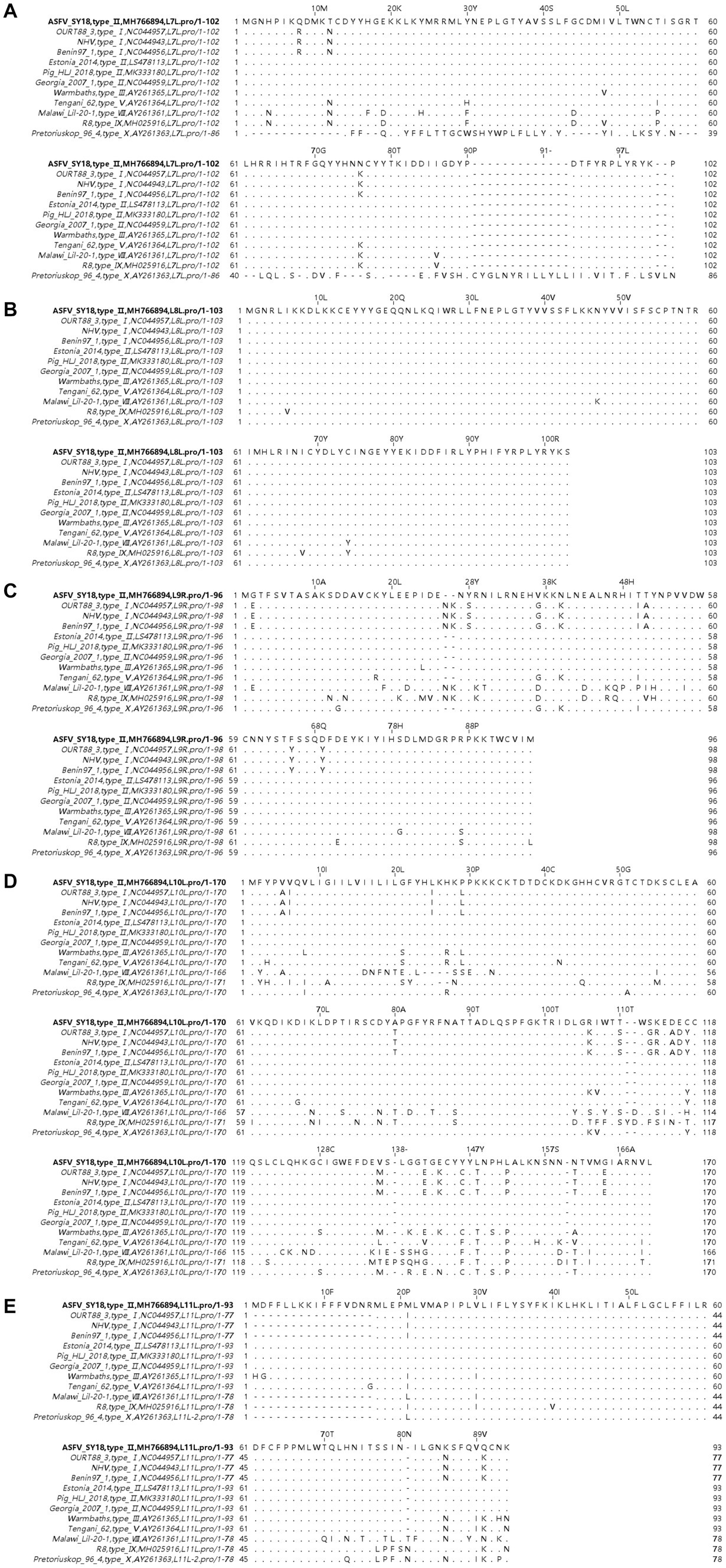
Figure 1. Multiple sequence alignment of amino acid sequences of L7L-L11L. Panels (A–E) show sequences of L7L, L8L, L9R, L10L, and L11L, respectively. The sequence of ASFV SY18 is placed on the top row for reference. Amino acids are indicated by their respective single letter codes. Matches are indicated with “. “, and gaps are denoted with “-.” Sequences were aligned using the MAFFT algorithm and Jalview software (Accessed 5 October 2023).
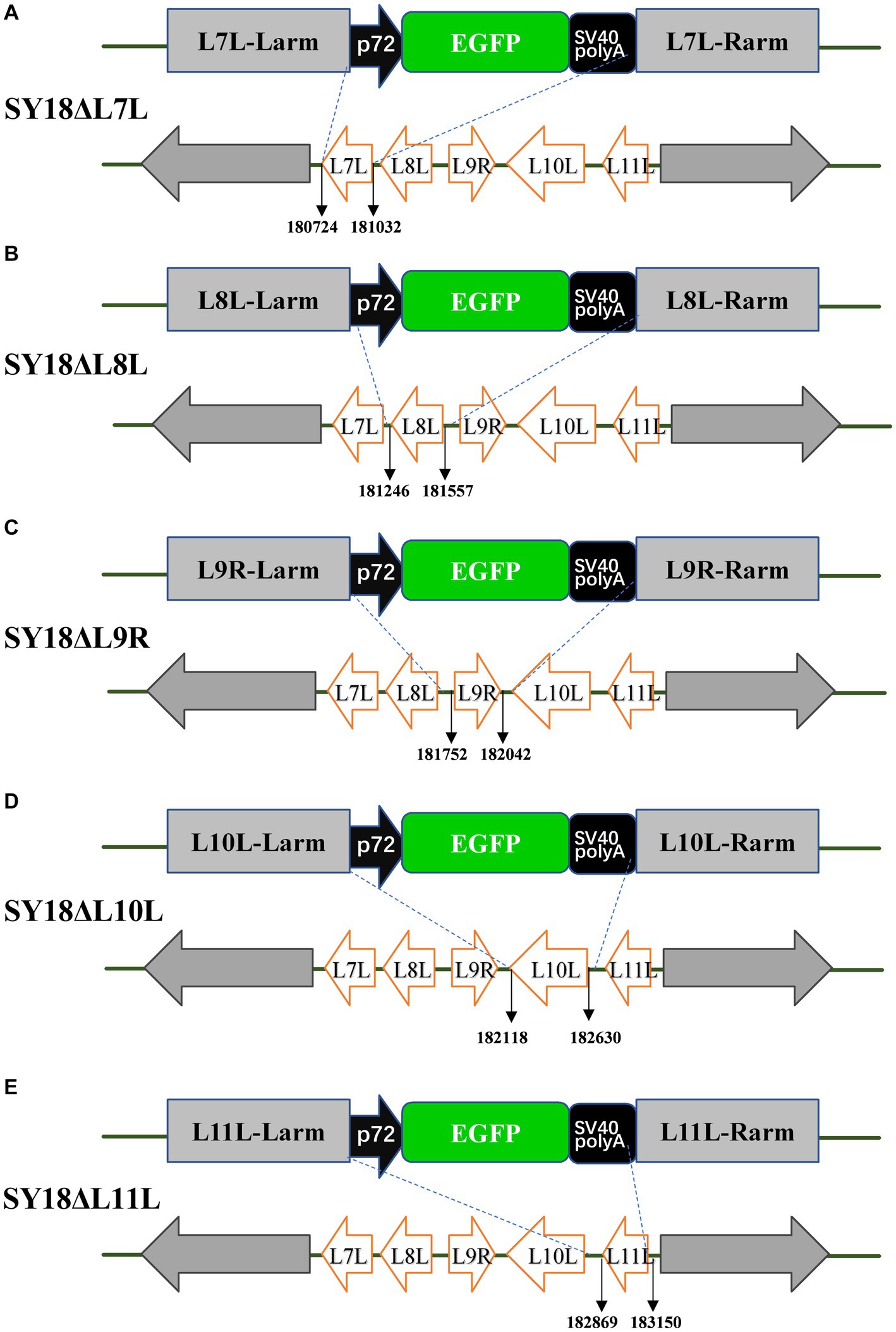
Figure 2. Schematic diagrams of recombinant viral constructs. (A) A schematic diagram of the SY18ΔL7L construct ASFV SY18 in which the L7L was replaced by a green fluorescent protein expression cassette. Panels (B–E) indicate the schematic diagrams of the constructs of SY18ΔL8L, SY18ΔL9R, SY18ΔL10L, and SY18ΔL11L, respectively, where the target genes were replaced by an EGFP expression cassette.
3.2 Creation of ASFV mutants
In order to identify the specific virulence genes among L7L-L11L, we designed and constructed single-gene deletion strains (SY18ΔL7L, SY18ΔL8L, SY18ΔL9R, SY18ΔL10L, and SY18ΔL11L). The construction method is shown in Figure 2. Target genes were replaced with a green fluorescent gene cassette containing the p72 promoter using a recombinant method. The recombinant viruses were purified using fluorescence activity. The PCR assay determined that the target bands of the gene were not detected, whereas the ASFV SY18 control showed the target bands, confirming that single-gene deletion viruses missing each target gene were successfully obtained (Figure 3). Comparison of the whole-genome sequences of the ASFV mutant and parental ASFV SY18 using second-generation sequencing revealed that, apart from design changes, no unwanted mutations were detected in the genomes of the constructed mutants.
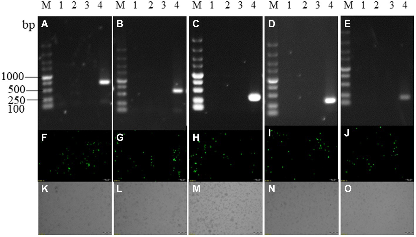
Figure 3. Recombinant virus identification. Panels (A–E) are PCR identification and purification results of SY18ΔL7L, SY18ΔL8L, SY18ΔL9R, SY18ΔL10L, SY18ΔL11L, panels (F–J) are the fluorescence maps of each single-gene deletion viruses, and panels (K–O) are the bright field of each single-gene deletion viruses. M: DL5000 marker; a1~a3, b1~b3, c1~c3, d1~d3, e1~e3: each single-gene deletion virus detection samples; 4: ASFV SY18 control.SY18ΔL7L and SY18ΔL8L amplified a 643 bp fragment in the presence of ASFV SY18; SY18ΔL9R amplified a 280 bp fragment in the presence of ASFV SY18; SY18ΔL10L, and SY18ΔL11L amplified a 200 bp fragment in the presence of ASFV SY18.
3.3 Replication of ASFV mutants in porcine macrophages
Macrophages were infected with each deletion strain at an MOI of 0.1, and the parental ASFV SY18 was used as a control. Samples were collected at 2, 12, 24, 48, 72, and 96 h post-infection (hpi). The results shown in Figure 4 revealed no significant differences between the growth curves of single-gene deletion viruses in target cells than those of the parental strains in vitro. There was also no difference in the replication pattern between the single-gene deletion strains, all of which reached the highest titer at 72 hpi. These findings demonstrate that the deletion of L7L, L8L, L9R, L10L, and L11L did not affect the proliferative ability of ASFV in macrophages in vitro.
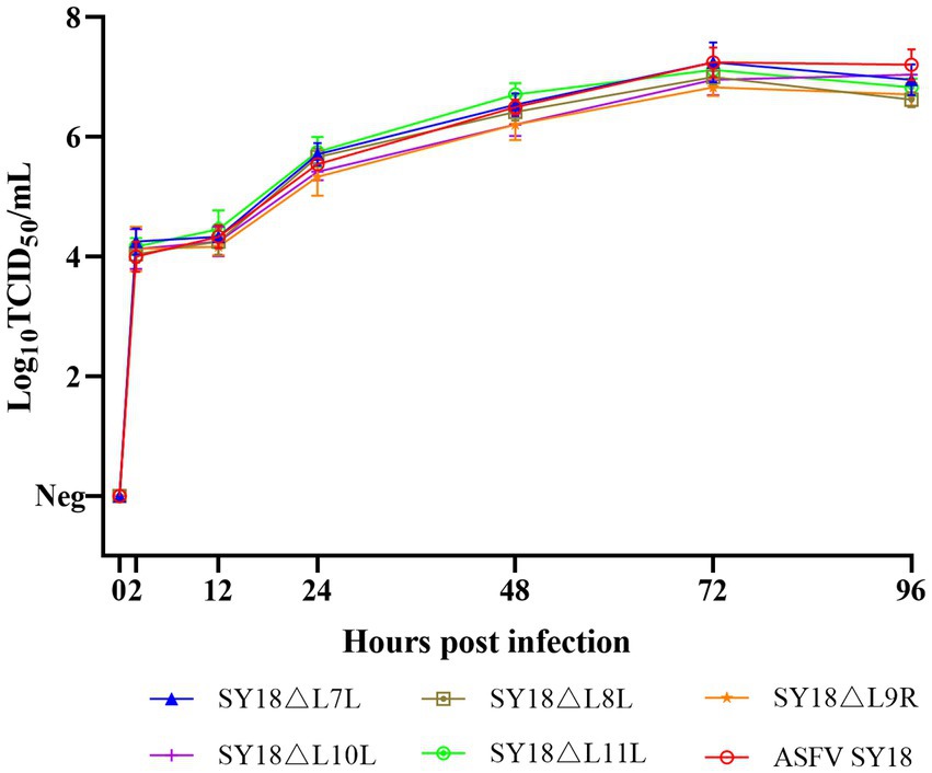
Figure 4. In vitro growth characteristics of recombinant viruses. PAM was infected with ASFV SY18, SY18ΔL7L, SY18ΔL8L, SY18ΔL9R, SY18ΔL10L, SY18ΔL11L at 0.1 MOI, and the viral titers of three independent experimental samples were determined at different times of infection. y-axis represents the viral titer expressed as log10 TCID50/mL, and the x-axis represents the time after infection (hours).
3.4 Evaluation of the virulence of ASFV mutants
To assess the virulence of SY18ΔL7L, SY18ΔL8L, SY18ΔL9R, SY18ΔL10L, SY18ΔL11L, and ASFV SY18 (positive control group) for pigs, each group was injected intramuscularly with 103.0 TCID50 of virus. The survival and body temperature results are shown in Figures 5A,B and Table 2. Pigs in the ASFV SY18 group became febrile on day 4 after infection, and showed typical symptoms of ASF, and died on day 8 post infection. Pigs in the SY18ΔL7L group showed persistent fever between days 1 and 5. Three of these pigs died on the 9th, 12th, and 13th dpi, and one pig (S7-1) survived during the observation period. Pigs in the SY18ΔL8L group died on days 7 and 8 post-infection, which was consistent with the timing of death in the control group. However, only one animal in the SY18ΔL8L group showed elevated body temperature, whereas the remaining three animals had normal body temperatures. Pigs in the SY18ΔL9R group died 10 to 16 days and pigs in the SY18ΔL10L group died 10 to 13 days post infection, respectively. Deaths in both groups delayed backwards compared with the control. All pigs in the SY18ΔL11L group survived the 21 days of observation, with three pigs experiencing a transient increase in body temperature and one returning to normal temperature after 6 days of fever.
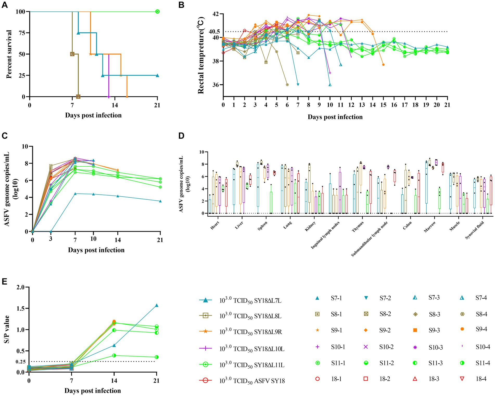
Figure 5. Survival, rectal temperature, viraemia, viral load in tissues and anti-p54 antibody levels in pigs inoculated with 103.0 TCID50 ASFV recombinant virus. (A) Survival of animals in each group. (B) Each line represents temperature data for individual animals. The dashed line indicates the fever threshold of 40.5°C. (C) Detection of ASFV genomic DNA in porcine blood. (D) Detection of ASFV genomic DNA in porcine tissues. (E) Anti-p54 antibody level in pig serum. y-axis represents the S/P ratio (sample OD450nm/positive control OD450nm), x-axis represents the number of days of immunization, and the dotted line represents the critical value of 0.25.
The viral loads in the blood of the experimental animals are shown in Figure 5C. Viral nucleic acids were detected in the blood of all animals on day 3 after infection, except for one pig in the SY18ΔL7L group. On the 3rd day of infection, all pigs in the ASFV SY18 group showed high viral loads in their blood (106.19–106.86 copies/mL), and pigs in the SY18ΔL8L and SY18ΔL9R groups similarly showed high viremia (106.72–107.72 copies/mL and 106.16–107.13 copies/mL). On the 3rd day of infection, pigs in the SY18ΔL7L, SY18ΔL10L, and SY18ΔL11L groups had lower viral loads in their blood compared to the control group (0–105.08 copies/mL, 103.58–107.12 copies/mL, and 103.37–105.42 copies/mL, respectively). The highest viral levels in the blood of all animals were reached on the 7th dpi. Subsequently, the viral load in the blood of surviving animals tended to decrease. The presence of virus was not detected in the blood of 1 pig without fever (S7-1) in the SY18ΔL7L group on the 3rd day, and the presence of the virus was detected on the 7th day, after which and the virus level was consistently low (103.56–104.44 copies/mL).
3.5 Viral load of pig tissues after infection with ASFV mutants
The pigs used in Experiment 1 were dissected, and organs including the heart, liver, spleen, lungs, kidneys, inguinal lymph nodes, thymus, submandibular lymph nodes, colon, bone marrow, muscles, and joint fluids were collected and tested for the presence of the virus. Figure 5D indicates that most animals had the virus in their organs, with the spleen, liver, and bone marrow showing the highest detection rates. In the SY18ΔL11L group, both the detection rate and viral load in the organs were lower compared to other groups. In the SY18ΔL7L group of non-febrile pigs (S7-1), post-dissection testing of tissues showed no viral nucleic acid except in the muscle tissue (102.68 copies/mL).
Specific antibody responses in pigs infected with ASFV mutants. To assess the specific immune response, we determined the levels of anti-p54 antibodies in the serum. As shown in Figure 5E, the sera of each animal were negative for anti-p54 antibodies on day 7 of immunization, and those that survived until day 14 (all pigs in the S9-1, S9-4, S7-1 and SY18ΔL11L groups) were positive for anti-p54 antibodies. Pigs S9-1 and S9-4 produced higher levels of anti-p54 antibodies (S/p values of 1.123 and 1.197, respectively), yet died on days 15th and 16th days. The S11-4 pigs in the SY18ΔL11L group expressed lower anti-p54 antibodies than S9-1, S9-4, but the S11-4 pigs survived the observation period. This finding demonstrates that the expression of ASF-specific antibodies is not a marker for animal survival.
3.6 Assessment of protective capacity of SY18ΔL11L mutant
To assess the degree of attenuation of SY18ΔL11L, a low dose (103.0TCID50) and a high dose (106.0TCID50) of SY18ΔL11L were inoculated intramuscularly. Another group of pigs was injected with an equal amount of saline as control (Figures 6A,B; Table 3). All pigs in the low-dose SY18ΔL11L-infected group survived, with two pigs experiencing a transient increase in body temperature and two pigs having persistent fever for 4–5 days. All animals in the high-dose SY18ΔL11L-infected group showed elevated body temperatures (>40.5°C), with symptoms of ASF in H-2 and H-3, which were euthanized on days 11th and 15th days post-inoculated due to the severity of the disease in both animals. Pigs in the control group showed no adverse reactions. To determine the response of the surviving pigs when challenged with the parental virus, a virus provocation test was performed on all pigs that survived the inoculation observation period (Figures 6C,D; Table 4). Each pig was intramuscularly injected with 103.0TCID50 of SY18 cells, and observed for 28 days. During the viral challenge, all pigs in the control group had elevated temperatures, developed an ASF clinical reaction, and were euthanized by the 9th day post-challenge (dpc). Pigs inoculated with SY18ΔL11L all survived during the challenge.
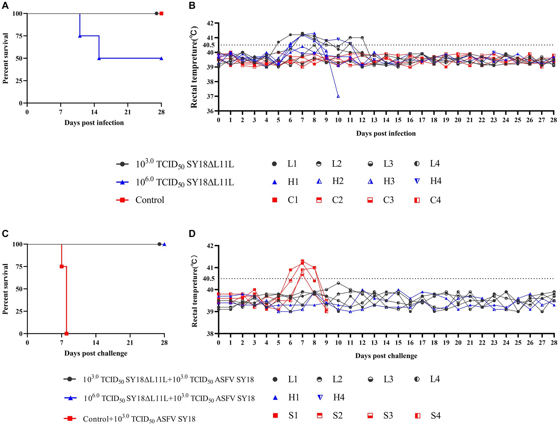
Figure 6. Survival and body temperature of pigs after immunization and attack challenge. Survival (A) and rectal temperature (B) of pigs immunized with 103.0 TCID50 SY18ΔL11L, 106.0 TCID50 SY18ΔL11L and control saline. Survival (C) and rectal temperature (D) of pigs challenged with 103.0 TCID50 ASFV SY18 tapping. Each line in the graph represents body temperature data for a single animal.
Animals inoculated with SY18ΔL11L strain developed viremia on day 7 after immunization (Figure 7A), with the viral load in blood reaching its maximum on day 7 or 14 post-infection, followed by a slow decrease. After ASFV SY18 challenge, control animals exhibited high viral loads in their blood. SY18ΔL11L-infected animals challenged with the parental virus exhibited a steady decline in blood viral load after an increased on day 7. Then, no virus in blood of all but 1 pig on the 28th dpc was be detected (Figure 7B). The ASFV load in the tissues and organs of the animals was measured at the end of the observation period. The results showed that pigs in the control group had higher viral detection in tissues and higher viral loads (Figure 7C). Pigs that died during the immunization period (H2 and H3) had relatively higher virus detection in tissues compared to other pigs immunized with SY18ΔL11L.
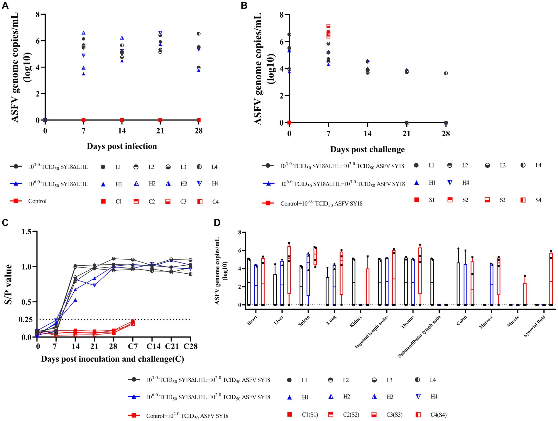
Figure 7. Viraemia, changes in antibody levels and tissue viral content in pigs after immunization and attack attacks. Detection of ASFV genomic DNA in the blood of pigs after immunization (A) and attack challenge (B). y values denote log10 copies/mL and x values denote time of inoculation and challenge. (C) Viral loads in tissues of immunized pigs after takedown challenge. Each symbol represents each pig. Values represent log10 copies/mL. H2 and H3 died during immunization and were not challenged with the parental virus; H2 and H3 only represent viral loads in tissues of immunized pigs. (D) Anti-p54 antibodies in pig sera after immunization and challenge. y-axis represents the S/P (sample OD450nm/positive control OD450nm) ratio and x-axis represents the number of days of immunization. The threshold of 0.25 is indicated by the dashed line.
The host antibody responses in animals inoculated with SY18ΔL11L are shown in Figure 7D. Animals inoculated with SY18ΔL11L were weakly positive for anti-p54 antibodies in sera of L3 and H4 on day 7 of immunization, and positive for antibodies on day 14, followed by high levels of antibodies in sera thereafter. H3 pigs in the high-dose SY18ΔL11L group died on day 15 even though they were positive for antibodies in serum on day 14 of immunization. After the takedown challenge, pigs in the control group remained negative, and antibodies in pigs inoculated with SY18ΔL11L remained at high levels.
4 Discussion
African swine fever continues to adversely impact the global pig farming industry, and is expanding into new endemic areas. Vaccination remains one of the most effective methods to prevent the spread of this virus. However, the effectiveness of inactivated vaccines has been unsatisfactory (Blome et al., 2014; Cadenas-Fernández et al., 2021). Subunit vaccines have seen limited research due insufficient knowledge on viral proteins (Liu et al., 2023). Effective subunit vaccines are still being investigated (Goatley et al., 2020). Live attenuated virus vaccines, which are produced by passaging tissues or cell cultures to generate ASFV and genetically-engineered and attenuated ASFV, induce strong immunity against ASFV-related strains. After years of ASF epidemics in some areas, a decrease in pig mortality has been observed, leading to isolation of naturally attenuated ASFV that cause subacute, chronic, or subclinical forms of the disease (Arias et al., 2017). However, these have very poor safety profiles, and are prone to causing persistent viremia in pigs, along with symptoms of chronic ASF infection (Gallardo et al., 2018). When ASFV grows in maladapted cells, significant portions of the virus can be lost or mutated, resulting in cell-adapted attenuated ASFV (Koltsova et al., 2021). The number of passages of cell-adapted attenuated ASFV is unpredictable, and immunogenicity decreases when the number of passages is excessive (Krug et al., 2015). Currently, genetically engineered live ASFV with reduced virulence is the most promising ASF vaccine; however, it may not completely clear the virus from pigs, and there is a risk of virulence reversal. Therefore, the construction of ASFV mutants by genetic engineering and the selection of rational virulence-related genes are essential for the development of future ASF vaccines.
ASFV has a genome 170–194 kb in size, containing more than 150 open reading frames (ORFs). Functions of only a small fraction of these ORFs have been characterized. The L7L-L11L ORF is located in the right variable region of the ASFV genome, where L10L is homologous to KP177R (encoding the p22 protein), and L7L and L8L genes are thought to be members of MGF110 (Vydelingum et al., 1993; Zhang J. et al., 2021; Cackett et al., 2022). We found that the replication efficiency of the 5 ASFV-mutants was similar to that of the parental strains during in vitro experiments. Therefore, L7L-L11L are not replication-associated genes. Current research on live attenuated ASFV vaccines has focused on the rational attenuation of virulent viruses through deletion of immunosuppressive genes (Liu et al., 2023b). In a previous study, deletion of the L7L-L11L ORF in the ASFV SY18 strain (genotype II) was found to significantly attenuate virulence (Zhang J. et al., 2021). Therefore, virulence-related genes are found in the ORF of L7L-L11L. Here, we found that inoculation of pigs with deletion strains of the L8L, L9R, and L10L led to deaths of all the pigs in each group. However, compared with the parental strains, the death time of pigs infected with the deletion strains of the L9R and L10L genes was delayed to a certain extent, one pig survived after being infected by the deletion strain of the L7L gene, and all pigs infected with the deletion strain of the L11L gene survived during the observation period, despite showing fever. We reason that the reduced pathogenicity of SY18ΔL7–11 is the result of the combined deletion of the L7L and L11L genes.
The virulence of ASFV SY18 was not significantly altered by deletion of L8L, and deletion of the L11L significantly reduced the pathogenicity of ASFV SY18. Vuono et al. performed a deletion of the I8L (L8L) alone in the Georgia2010 strain (genotype II), and reported no effects on viral replication or virulence, which is in agreement with our findings (Vuono et al., 2021). Kleiboeker et al. deleted L11L from Malawi Lil-20/1 strain, which resulted in the death of all three pigs inoculated with 102.0HAD50, and the L11L deletion did not reduce the virulence of Malawi Lil-20/1 (Kleiboeker et al., 1998). Malawi Lil-20/1 is a highly virulent strain isolated in Malawi, Africa, whose p72 type belongs to genotype VIII, whereas the epidemic strain in China is genotype II. The deletion of the same gene in different ASFVs may thus result in different virulence-reducing effects. The CD2v gene affects the virulence of BA71, ASFV-Kenya-IX-1033, and ASFV HLJ/18, yet has no effect on the virulence of Lil-20/1 in Malawi (Borca et al., 1998; Hemmink et al., 2022). Deletion of the DP148R reduced the virulence of Benin 97/1 but had no effect on the virulence of ASFV Georgia 2007/1 and ASFV HLJ18 (Reis et al., 2017; Chen et al., 2020; Rathakrishnan et al., 2021). This phenomenon also suggests that studies on ASFV genes should take the background of the strains and the differences between them into consideration. Because of space constraints we conducted trials using young strains (weaner pigs or grower pigs). The resistance and immunity of young pigs is lower compared to that finisher pigs. ASF causes abortions in sows in late pregnancy (Lohse et al., 2022). We demonstrated the virulence of five ASFV-mutants only in young pigs. However, the specific effects of these ASFV mutants on finishing pigs or gestating sows are uncertain and will require further experimental studies.
5 Conclusion
Previous studies have shown that deletion of L7L-L11L genes in ASFV SY18 significantly reduces viral virulence, and that virulence-related genes exist among L7L-L11L. In this study, we constructed five recombinant viruses with single-gene deletions using homologous recombination technology, 103.0TCID50 doses of the recombinant viruses were used to immunize pigs. All pigs in the SY18ΔL8L, SY18ΔL9R, and SY18ΔL10L immunization groups died during the observation period, while one pig survived in the SY18ΔL7L group, and all pigs in the SY18ΔL11L group survived. The results demonstrated that L8L, L9R, and L10L are not ASFV SY18 virulence genes, whereas L7L and L11L are related virulence. Pigs were immunized with 103.0TCID50 or 106.0TCID50 doses of recombinant virus SY18ΔL11L, and virus challenge protection experiments were performed. The results showed that SY18ΔL11L could not be completely detoxified by pigs after immunization, and large doses of immunization could still lead to death. However, surviving pigs could resist attack by the parental strain. Here, we found that SY18ΔL11L is an attenuated strain that protects against the parental strain, but not suitable as a vaccine candidate. L11L is an ASFV SY18 virulence gene, whereas L7L has a slight effect on virus virulence. An in-depth analysis of the functions of this gene may identify new targets for the development of effective vaccines in the future.
Data availability statement
The datasets presented in this study can be found in online repositories. The names of the repository/repositories and accession number(s) can be found at: (Genbank: OR944087), SY18ΔL8L (Genbank: OR944088), SY18ΔL9R (Genbank: OR944089), SY18ΔL10L (Genbank: OR944090), and SY18ΔL11L (Genbank: OR944091).
Ethics statement
The animal study was approved by Animal Welfare and Ethics Committee of the Changchun Veterinary Research Institute. The study was conducted in accordance with the local legislation and institutional requirements.
Author contributions
JF: Methodology, Validation, Writing – original draft. JZ: Validation, Writing – review & editing. FW: Validation, Writing – review & editing. FM: Validation, Writing – review & editing. HZ: Validation, Writing – review & editing. YJ: Validation, Writing – review & editing. YQ: Validation, Writing – review & editing. YZ: Validation, Writing – review & editing. LH: Validation, Writing – review & editing. DZ: Validation, Writing – review & editing. HY: Validation, Writing – review & editing. XZ: Validation, Writing – review & editing. QL: Validation, Writing – review & editing. YW: Validation, Writing – review & editing. TC: Funding acquisition, Writing – review & editing. RH: Funding acquisition, Writing – review & editing.
Funding
The author(s) declare financial support was received for the research, authorship, and/or publication of this article. This work was funded by the National Natural Science Foundation of China (no. 32102656) and National Key Research and Development Program of China (grant nos. 2021YFD1800100 and 2021YFD1801204).
Acknowledgments
The authors thank all the teachers of the Animal Biosafety Level 3 (ABSL-3) Laboratory of the Changchun Veterinary Research Institute for their help, and we are grateful to Editage for their help in revising the article.
Conflict of interest
The authors declare that the research was conducted in the absence of any commercial or financial relationships that could be construed as a potential conflict of interest.
Publisher’s note
All claims expressed in this article are solely those of the authors and do not necessarily represent those of their affiliated organizations, or those of the publisher, the editors and the reviewers. Any product that may be evaluated in this article, or claim that may be made by its manufacturer, is not guaranteed or endorsed by the publisher.
References
Ankhanbaatar, U., Sainnokhoi, T., Khanui, B., Ulziibat, G., Jargalsaikhan, T., Purevtseren, D., et al. (2021). African swine fever virus genotype II in Mongolia, 2019. Transbound. Emerg. Dis. 68, 2787–2794. doi: 10.1111/tbed.14095
Arias, M., Torre, A., Dixon, L., Gallardo, C., Jori, F., Laddomada, A., et al. (2017). Approaches and perspectives for development of African swine fever virus vaccines. Vaccines 5:35. doi: 10.3390/vaccines5040035
Blome, S., Gabriel, C., and Beer, M. (2014). Modern adjuvants do not enhance the efficacy of an inactivated African swine fever virus vaccine preparation. Vaccine 32, 3879–2882. doi: 10.1016/j.vaccine.2014.05.051
Borca, M., Carrillo, C., Zsak, L., Laegreid, W., Kutish, G., Neilan, J., et al. (1998). Deletion of a CD2-like gene, 8-DR, from African swine fever virus affects viral infection in domestic swine. J. Virol. 72, 2881–2889. doi: 10.1128/JVI.72.4.2881-2889.1998
Borca, M., Rai, A., Ramirez-Medina, E., Silva, E., Velazquez-Salinas, L., Vuono, E., et al. (2021). A cell culture-adapted vaccine virus against the current African swine fever virus pandemic strain. J. Virol. 95:e0012321. doi: 10.1128/JVI.00123-21
Borca, M., Ramirez-Medina, E., Silva, E., Vuono, E., Rai, A., Pruitt, S., et al. (2020). Development of a highly effective African swine fever virus vaccine by deletion of the I177L gene results in sterile immunity against the current epidemic Eurasia strain. J. Virol. 94, e02017–e02019. doi: 10.1128/JVI.02017-19
Bosch-Camós, L., López, E., and Rodriguez, F. (2020). African swine fever vaccines: a promising work still in progress. Porcine Health Manag. 6:17. doi: 10.1186/s40813-020-00154-2
Cackett, G., Portugal, R., Matelska, D., Dixon, L., and Werner, F. (2022). African swine fever virus and host response: transcriptome profiling of the Georgia 2007/1 strain and porcine macrophages. J. Virol. 96:e0193921. doi: 10.1128/jvi.01939-21
Cadenas-Fernández, E., Sánchez-Vizcaíno, J., Born, E., Kosowska, A., Kilsdonk, E., Fernández-Pacheco, P., et al. (2021). High doses of inactivated African swine fever virus are safe, but do not confer protection against a virulent challenge. Vaccines 9:242. doi: 10.3390/vaccines9030242
Chapman, D. A. G., Darby, A. C., Silva, M. D., Upton, C., Radford, A. D., and Dixon, L. D. (2011). Genomic analysis of highly virulent Georgia 2007/1 isolate of African swine fever virus. Emerg. Infect. Dis. 17, 599–605. doi: 10.3201/eid1704.101283
Chen, W., Zhao, D., He, X., Liu, R., Wang, Z., Zhang, X., et al. (2020). A seven-gene-deleted African swine fever virus is safe and effective as a live attenuated vaccine in pigs. Sci. China Life Sci. 63, 623–634. doi: 10.1007/s11427-020-1657-9
Deutschmann, P., Carrau, T., Sehl-Ewert, J., Forth, J., Viaplana, E., Mancera, J., et al. (2022). Taking a promising vaccine candidate further: efficacy of ASFV-G-ΔMGF after intramuscular vaccination of domestic pigs and Oral vaccination of wild boar. Pathogens. 11:996. doi: 10.3390/pathogens11090996
Dixon, L., Sun, H., and Roberts, H. (2019). African swine fever. Antiviral Res. 165, 34–41. doi: 10.1016/j.antiviral.2019.02.018
Gallardo, C., Sánchez, E., Pérez-Núñez, D., Nogal, M., León, P., Carrascosa, Á., et al. (2018). African swine fever virus (ASFV) protection mediated by NH/P68 and NH/P68 recombinant live-attenuated viruses. Vaccine 36, 2694–2704. doi: 10.1016/j.vaccine.2018.03.040
Gladue, D., Ramirez-Medina, E., Vuono, E., Silva, E., Rai, A., Pruitt, S., et al. (2021). Deletion of the A137R gene from the pandemic strain of African swine fever virus attenuates the strain and offers protection against the virulent pandemic virus. J. Virol. 95:e0113921. doi: 10.1128/JVI.01139-21
Goatley, L., Reis, A., Portugal, R., Goldswain, H., Shimmon, G., Hargreaves, Z., et al. (2020). A Pool of eight virally vectored African swine fever antigens protect pigs against fatal disease. Vaccines 8:234. doi: 10.3390/vaccines8020234
Gómez-Villamandos, J. C., Bautista, M. J., Sánchez-Cordón, P. J., and Carrasco, L. (2013). Pathology of African swine fever: the role of monocyte-macrophage. Virus Res. 173, 140–149. doi: 10.1016/j.virusres.2013.01.017
Hemmink, J., Khazalwa, E., Abkallo, H., Oduor, B., Khayumbi, J., Svitek, N., et al. (2022). Deletion of the CD2v gene from the genome of ASFV-Kenya-IX-1033 partially reduces virulence and induces protection in pigs. Viruses 14:1917. doi: 10.3390/v14091917
Iyer, L. M., Balaji, S., Koonin, E. V., and Aravind, L. (2006). Evolutionary genomics of nucleo-cytoplasmic large DNA viruses. Virus Res. 117, 156–184. doi: 10.1016/j.virusres.2006.01.009
Kleiboeker, S., Kutish, G., Neilan, J., Lu, Z., Zsak, L., and Rock, D. (1998). A conserved African swine fever virus right variable region gene, l11L, is non-essential for growth in vitro and virulence in domestic swine. J. Gen. Virol. 79, 1189–1195. doi: 10.1099/0022-1317-79-5-1189
Koltsova, G., Koltsov, A., Krutko, S., Kholod, N., Tulman, E., and Kolbasov, D. (2021). Growth kinetics and protective efficacy of attenuated ASFV strain Congo with deletion of the EP402 gene. Viruses 13:1259. doi: 10.3390/v13071259
Krug, P., Holinka, L., O'Donnell, V., Reese, B., Sanford, B., Fernandez-Sainz, I., et al. (2015). The progressive adaptation of a georgian isolate of African swine fever virus to vero cells leads to a gradual attenuation of virulence in swine corresponding to major modifications of the viral genome. J. Virol. 89, 2324–2332. doi: 10.1128/JVI.03250-14
Li, D., Wu, P., Liu, H., Feng, T., Yang, W., Ru, Y., et al. (2022). A QP509L/QP383R-deleted African swine fever virus is highly attenuated in swine but does not confer protection against parental virus challenge. J. Virol. 96:e0150021. doi: 10.1128/jvi.01210-22
Liu, W., Li, H., Liu, B., Lv, T., Yang, C., Chen, S., et al. (2023). A new vaccination regimen using adenovirus-vectored vaccine confers effective protection against African swine fever virus in swine. Emerg Microbes Infect 12:2233643. doi: 10.1080/22221751.2023.2233643
Liu, Y., Shen, Z., Xie, Z., Song, Y., Li, Y., Liang, R., et al. (2023a). African swine fever virus I73R is a critical virulence-related gene: a potential target for attenuation. Proc. Natl. Acad. Sci. U. S. A. 120:e2210808120. doi: 10.1073/pnas.2302161120
Liu, Y., Xie, Z., Li, Y., Song, Y., di, D., Liu, J., et al. (2023b). Evaluation of an I177L gene-based five-gene-deleted African swine fever virus as a live attenuated vaccine in pigs. Emerg Microbes Infect. 12:2148560. doi: 10.1080/22221751.2022.2148560
Lohse, L., Nielsen, J., Uttenthal, Å., Olesen, A. S., Strandbygaard, B., Rasmussen, T. B., et al. (2022). Experimental infections of pigs with African swine fever virus (genotype II); studies in young animals and pregnant sows. Viruses 14:1387. doi: 10.3390/v14071387
Mai, N., Vu, X., Nguyen, T., Nguyen, V., Trinh, T., Kim, Y., et al. (2021). Molecular profile of African swine fever virus (ASFV) circulating in Vietnam during 2019-2020 outbreaks. Arch. Virol. 166, 885–890. doi: 10.1007/s00705-020-04936-5
Mur, L., Atzeni, M., Martínez-López, B., Feliziani, F., Rolesu, S., and Sanchez-Vizcaino, J. M. (2016). Thirty-five-year presence of African swine fever in Sardinia: history, evolution and risk factors for disease maintenance. Transbound. Emerg. Dis. 63, e165–e177. doi: 10.1111/tbed.12264
Rathakrishnan, A., Reis, A., Goatley, L., Moffat, K., and Dixon, L. (2021). Deletion of the K145R and DP148R genes from the virulent ASFV Georgia 2007/1 isolate delays the onset, but does not reduce severity, of clinical signs in infected pigs. Viruses 13:1473. doi: 10.3390/v13081473
Reis, A., Goatley, L., Jabbar, T., Sanchez-Cordon, P., Netherton, C., Chapman, D., et al. (2017). Deletion of the African swine fever virus gene DP148R does not reduce virus replication in culture but reduces virus virulence in pigs and induces high levels of protection against challenge. J. Virol. 91, e01428–e01417. doi: 10.1128/JVI.01428-17
Rowlands, R., Michaud, V., Heath, L., Hutchings, G., Oura, C., Vosloo, W., et al. (2008). African swine fever virus isolate, Georgia, 2007. Emerg. Infect. Dis. 14, 1870–1874. doi: 10.3201/eid1412.080591
Sun, E., Huang, L., Zhang, X., Zhang, J., Shen, D., Zhang, Z., et al. (2021a). Genotype I African swine fever viruses emerged in domestic pigs in China and caused chronic infection. Emerg Microbes Infect. 10, 2183–2193. doi: 10.1080/22221751.2021.1999779
Sun, E., Zhang, Z., Wang, Z., He, X., Zhang, X., Wang, L., et al. (2021b). Emergence and prevalence of naturally occurring lower virulent African swine fever viruses in domestic pigs in China in 2020. Sci. China Life Sci. 64, 752–765. doi: 10.1007/s11427-021-1904-4
Urbano, A. C., and Ferreira, F. (2022). African swine fever control and prevention: an update on vaccine development. Emerg Microbes Infect. 11, 2021–2033. doi: 10.1080/22221751.2022.2108342
Vuono, E., Ramirez-Medina, E., Pruitt, S., Rai, A., Silva, E., Espinoza, N., et al. (2021). Evaluation in swine of a recombinant Georgia 2010 African swine fever virus lacking the I8L gene. Viruses 13:39. doi: 10.3390/v13010039
Vydelingum, S., Baylis, S., Bristow, C., Smith, G., and Dixon, L. (1993). Duplicated genes within the variable right end of the genome of a pathogenic isolate of African swine fever virus. J. Gen. Virol. 74, 2125–2130. doi: 10.1099/0022-1317-74-10-2125
Wardley, R., Andrade, C. M., Black, D. N., Portugal, F. L. C., Enjuanes, L., Hess, W. R., et al. (1983). African swine fever virus. Brief review. Arch. Virol. 76, 73–90.
Zhang, Y., Ke, J., Zhang, J., Yang, J., Yue, H., Zhou, X., et al. (2021). African swine fever virus bearing an I226R gene deletion elicits robust immunity in pigs to African swine fever. J. Virol. 95:e0119921. doi: 10.1128/JVI.01199-21
Zhang, J., Zhang, Y., Chen, T., Yang, J., Yue, H., Wang, L., et al. (2021). Deletion of the L7L-L11L genes attenuates ASFV and induces protection against homologous challenge. Viruses 13:225. doi: 10.3390/v13020255
Keywords: African swine fever virus, L7L, L11L, virulence, recombinant virus
Citation: Fan J, Zhang J, Wang F, Miao F, Zhang H, Jiang Y, Qi Y, Zhang Y, Hui L, Zhang D, Yue H, Zhou X, Li Q, Wang Y, Chen T and Hu R (2024) Identification of L11L and L7L as virulence-related genes in the African swine fever virus genome. Front. Microbiol. 15:1345236. doi: 10.3389/fmicb.2024.1345236
Edited by:
Lihua Wang, Kansas State University, United StatesReviewed by:
Fernando Costa Ferreira, University of Lisbon, PortugalMir Mubashir Khalid, Gladstone Institutes, United States
Copyright © 2024 Fan, Zhang, Wang, Miao, Zhang, Jiang, Qi, Zhang, Hui, Zhang, Yue, Zhou, Li, Wang, Chen and Hu. This is an open-access article distributed under the terms of the Creative Commons Attribution License (CC BY). The use, distribution or reproduction in other forums is permitted, provided the original author(s) and the copyright owner(s) are credited and that the original publication in this journal is cited, in accordance with accepted academic practice. No use, distribution or reproduction is permitted which does not comply with these terms.
*Correspondence: Teng Chen, Y3RjeDE5OTFAMTYzLmNvbQ==; Rongliang Hu, cm9uZ2xpYW5naHVAaG90bWFpbC5jb20=
†These authors have contributed equally to this work
 Jiaqi Fan1,2,3†
 Rongliang Hu


