 Daria Jurković Žilić1
 Šimun Naletilić2†Željko Mihaljević2†
 Ema Gagović1
 Silvio Špičić3Irena Reil3†
 Sanja Duvnjak3Maja Zdelar Tuk3†
 Adnan Hodžić4
 Relja Beck1*
- 1Laboratory for Parasitology, Department for Bacteriology and Parasitology, Croatian Veterinary Institute, Zagreb, Croatia
- 2Laboratory for Pathology, Department of Pathology, Croatian Veterinary Institute, Zagreb, Croatia
- 3Laboratory for Bacterial Zoonoses and Molecular Diagnostics of Bacterial Diseases, Department for Bacteriology and Parasitology, Croatian Veterinary Institute, Zagreb, Croatia
- 4Centre for Microbiology and Environmental Systems Science (CMESS), Division of Microbial Ecology (DoME), Department of Microbiology and Ecosystem Science, University of Vienna, Vienna, Austria
Objective: Hemotropic pathogens of the genera Anaplasma, Babesia, Theileria, and hemotropic Mycoplasma are significant infectious agents in domestic ruminants, most commonly associated with vector-borne transmission. However, their potential for transplacental transmission and their contribution to reproductive disorders remains poorly understood. This study aimed to investigate the presence of hemopathogens in aborted fetuses of cattle, sheep, and goats in Croatia, and to evaluate their potential role in transplacental transmission.
Methods: Molecular analyses were conducted on tissue samples from 651 aborted fetuses collected between 2016 and 2019 as part of national abortion surveillance programs. PCR screening followed by sequencing were used to detect Anaplasmataceae, Babesia, Theileria, and hemotropic Mycoplasma.
Results: Thirteen hemopathogens were detected in 94 of 651 fetuses (14.44%), including Anaplasma marginale, Anaplasma ovis, Anaplasma phagocytophilum, Theileria orientalis, Theileria ovis, Theileria sp. OT3, Babesia ovis, Babesia canis, Babesia vulpes, Mycoplasma wenyonii, Mycoplasma haemobos, Mycoplasma ovis, and Mycoplasma haemominutum. The highest infection rates were observed in cattle (17.27%) and sheep (15.85%), while goats showed significantly lower prevalence (5.3%). A. marginale and A. ovis were the most frequently detected pathogens in bovine and ovine fetuses, respectively. Hemotropic mycoplasmas were reported for the first time in Croatia, with the first Western Balkan record of ‘Candidatus M. haemobos’. Our study represents the first molecular documentation of a wide array of hemopathogens in aborted ruminant fetuses in Croatia, strongly indicating the possibility of transplacental transmission. The detection of species-specific patterns and the unexpected identification of protozoan species typically associated with canines highlight complex epidemiological dynamics.
Conclusion: Vertical transmission of the detected pathogens may play a role in abortion in endemic regions and should be integrated into differential diagnostic protocols for reproductive failure investigations.
Introduction
A wide range of pathogens can cause diseases in domestic ruminants, leading to substantial economic losses. Among these, hemopathogens, including genera such as Anaplasma, Ehrlichia, Babesia, Theileria, and hemotropic Mycoplasma represent a particularly important group (Neimark et al., 2001; Bock et al., 2004; Messick, 2004; Kocan et al., 2010; Rar et al., 2021; World Organisation for Animal Health [WOAH], 2021). These infections may be subclinical or range in severity from mild to fatal, depending on the specific pathogen involved and the host’s immune status. Clinical signs often include hemolytic anemia, fever, weight loss, lethargy, and jaundice. Beyond direct health impacts, such diseases contribute to decreased productivity (milk and meat yield) and reproductive disorders, including abortion. Although traditionally associated with tropical and subtropical climates, tick-borne diseases are increasingly affecting livestock in temperate, industrialized regions due to the geographic expansion of tick vectors (Eskezia and Desta, 2016). While ticks remain the primary vectors for most hemopathogens, transmission of hemotropic Mycoplasma species more commonly occurs via iatrogenic or mechanical routes, involving contaminated instruments or biting insects (Arendt et al., 2024). Alternative transmission routes, including intrauterine or transplacental transmission, have increasingly been recognized in recent years (Hornok et al., 2011; Niethammer et al., 2018; Andrade et al., 2024). While there is growing scientific interest, the epidemiological significance of transplacental transmission for hemopathogens remains poorly understood. However, emerging evidence suggests that several pathogens within this group may be transmitted vertically and may contribute to reproductive complications, including abortion (Henker et al., 2020).
Transplacental transmission has been confirmed for various Theileria species such as Theileria equi in horses (Allsopp et al., 2007; Georges et al., 2011; Françoso et al., 2018), Theileria lestoquardi in small ruminants (Zakian et al., 2014; Esmaeilnejad et al., 2018), and T. annulata in cattle (Sudan et al., 2015; Selim et al., 2021). In cattle, transplacental transmission of T. orientalis is also well documented (Lawrence et al., 2016; Swilks et al., 2017; Mekata et al., 2018), and experimental studies have demonstrated a 100% abortion rate in pregnant cows infected via tick bites (Baek et al., 2003), highlighting its potential as a reproductive threat. Similarly, vertical transmission of Babesia caballi in horses (Bartolomé del Pino et al., 2023) and Babesia bigemina and Babesia bovis in cattle has been reported (Grau et al., 2013; Costa et al., 2016), with neonatal fatalities associated with B. bovis (Henker et al., 2020).
Among Anaplasma species, Anaplasma marginale is the most extensively studied regarding transplacental transmission, with evidence from both experimental and longitudinal field studies (Norton et al., 1983; Potgieter and Van Rensburg, 1987; Ribeiro et al., 1995; Silva and Fonseca, 2014.; Silva et al., 2015). Anaplasma phagocytophilum has also been shown to cross the placenta in both cattle (Pusterla et al., 1997; Henniger et al., 2013) and sheep (Reppert et al., 2013; Stuen et al., 2018) and has been detected in 2.4 and 1.9% ovine and caprine abortion cases (Gouvias et al., 2024).
Although vertical transmission of hemotropic Mycoplasma is considered rare, molecular evidence suggests that it can occur (Fujihara et al., 2011). Candidatus M. haemobos and M. wenyonii, have been detected in aborted fetuses and neonatal calves from infected cows (Hornok et al., 2011; Girotto-Soares et al., 2016), indicating that vertical transmission may serve as a route of hemoplasma infection (Sasaoka et al., 2015; Niethammer et al., 2018).
In many cases of abortion, the underlying cause is not identified, as abortions often arise from multiple factors. However, infectious agents – particularly those requiring advanced molecular diagnostic tools – are frequently implicated (Moeller, 2001; Hecker et al., 2023). Given the scarcity of research on hemopathogens in aborted ruminant fetuses, the present study aimed to investigate their potential intrauterine transmission using molecular diagnostics in fetuses of cattle, sheep, and goats submitted as part of abortion surveillance programs in Croatia.
Materials and methods
Tissue sampling from aborted fetuses
This study analyzed tissue samples from 651 aborted fetuses of domestic ruminants, including 336 cattle, 183 sheep, and 132 goats. The fetuses were submitted by veterinary practitioners to the Croatian Veterinary Institute as part of national abortion surveillance programs conducted between 2016 and 2019. The primary aim of these programs was to monitor pathogens such as Brucella spp. (bacteriological testing according to World Organization for Animal Health [WOAH], 2022, Terrestrial Manual, Chapter Brucellosis), Coxiella burnetii (Berri et al., 2000), and Chlamydia spp. (Berri et al., 2009). Additionally, the presence of Toxoplasma gondii was assessed in sheep and goat fetuses, and Neospora caninum in cattle. Detection was performed using PCR and sequencing, as described by Hughes et al. (2006) and Jones et al. (2000). Tissue samples collected for molecular analysis included the liver, lungs, placenta, and lochia from cattle; the liver, lungs, and placenta from sheep; and the liver and lungs from goats. All necropsies were performed under strict biosafety conditions to prevent cross-contamination. Separate sets of sterile instruments were used for each fetus, and work surfaces were disinfected between each procedure. The simultaneous reception of multiple fetuses was uncommon. An official submission form provided by the attending veterinarian, containing farm origin and relevant epidemiological data, accompanied each sample.
Molecular analysis
DNA was extracted from 20 mg of tissue (liver, lungs, placenta or lochia) using either the QIAcube automated DNA isolation system with the QIAamp DNA Mini Kit (Qiagen, Hilden, Germany) or, in high-throughput settings, the KingFisher™ Flex automated instrument (Thermo Fisher Scientific, Waltham, MA, United States) with the MagMAX™ CORE Nucleic Acid Purification Kit (Applied Biosystems).
All samples were screened by conventional PCR for the presence of Babesia/Theileria, Anaplasmataceae, and hemotropic Mycoplasma spp. Detection of Anaplasmataceae was performed using primers ehr16SR/ehr16SD targeting a 345 bp fragment of the 16S rRNA gene (Parola et al., 2000). Positive samples corresponding to A. marginale, A. ovis, or A. capra were further amplified using primers targeting an 842 bp fragment of the msp4 gene (Hornok et al., 2007). Samples positive for A. phagocytophilum were additionally screened for a 1,256 bp portion of the groESL operon (Petrovec et al., 1999). Babesia and Theileria species were detected using primers targeting a ~ 560 bp region of the 18S rRNA gene (Beck et al., 2009). Hemotropic Mycoplasma spp. were amplified using primers targeting a ~600 bp fragment of the 16S rRNA gene (Varanat et al., 2011). All primers used are listed in Table 1.
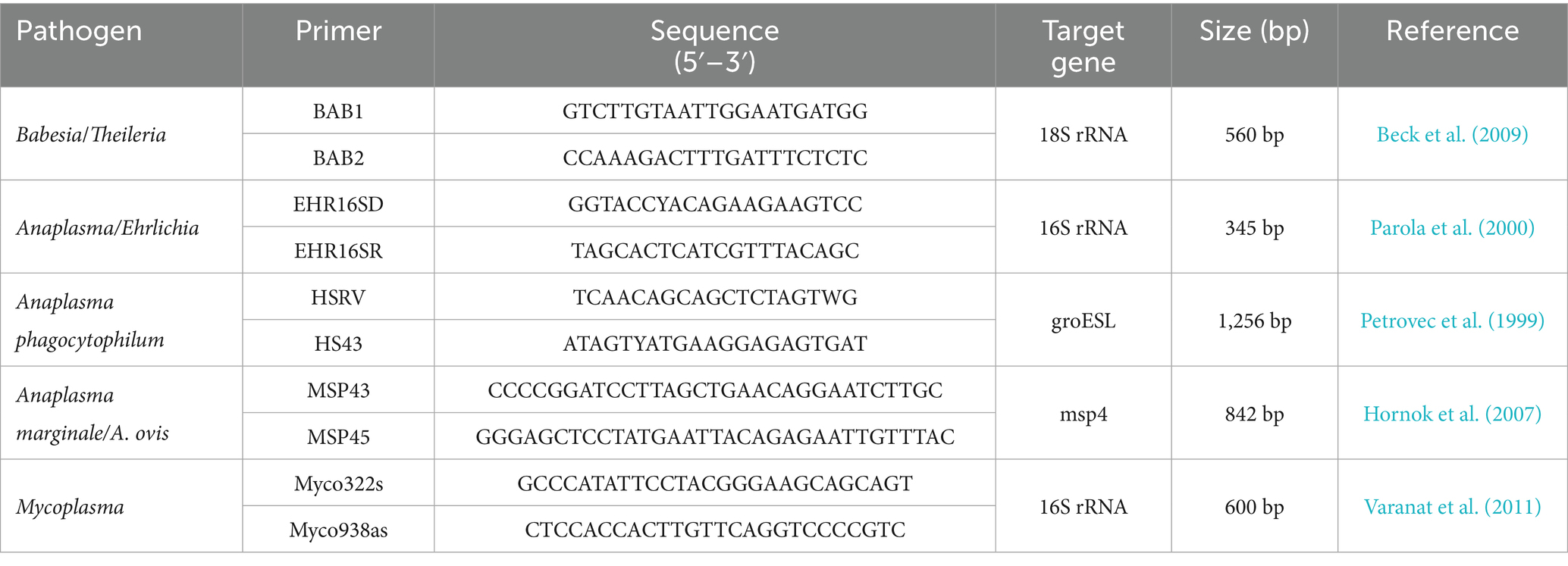
Table 1. Primers used for the molecular detection of hemotropic pathogens in aborted ruminant fetuses.
PCR reactions (20 μL) included 10 μL of GoTaq® G2 Master Mix (Promega, Madison, WI, United States), 7.2 μL of nuclease-free water, 0.4 μL of each primer (10 pmol/μL), and 2 μL of template DNA. Positive controls (A. marginale, A. phagocytophilum, B. divergens, T. orientalis, and M. wenyonii) and DNase/RNase-free water was included as a negative extraction control in each run.
Amplified products were visualized using QIAxcel capillary electrophoresis (Qiagen), employing the QIAxcel DNA Fast Analysis Kit and standard alignment/size markers. PCR products were purified using the ExoSAP-IT™ PCR Clean-up Reagent (Applied Biosystems) and sequenced by Macrogen Europe BV (Amsterdam, Netherlands) using the corresponding primer sets.
Sequence data were assembled and edited using SeqMan Pro 17 and SeqBuilder Pro 17 (DNASTAR, Madison, WI, USA), and identities confirmed by BLAST analysis1.
Statistical analysis
Statistical analysis was conducted using one-way ANOVA to assess differences in prevalence across species and tissues. In case where the assumptions of normality (as determined by Shapiro–Wilk test) and homogeneity of variance (Levene’s test) were not met, non-parametric Kruskal–Wallis test was employed.
Results
Out of 651 examined aborted fetuses, molecular evidence of infection with tick-borne or hemotropic pathogens was detected in 94 cases, corresponding to an overall prevalence of 14.44% (95% CI: 11.83–17.38). In this study, a total of 13 different pathogens were identified, including six piroplasm species, three Anaplasma species, and four hemotropic Mycoplasma species: Babesia ovis, Theileria ovis, Theileria sp. OT3, Theileria orientalis, Babesia canis, Babesia vulpes, Anaplasma phagocytophilum, Anaplasma ovis, Anaplasma marginale, Mycoplasma ovis, Candidatus Mycoplasma haemominutum, Mycoplasma wenyonii, and Candidatus Mycoplasma haemobos (Figure 1). Of these, 76 cases were single infections (11.67%, CI: 9.31–14.39), while co-infections were observed in 2.76% of cases (CI: 1.65–4.34). The highest prevalence was found in bovine fetuses, with 58 out of 336 infected (17.27%, CI: 13.38–21.74). Within this group, 48 out of 336 cases (14.29%, CI: 10.72–18.49) were found to be infected with a single pathogen, and 10 out of 336 cases (2.98%, CI: 1.44–5.41) were co-infected. In sheep fetuses, single infections were detected in 21 of 183 cases (11.48%, CI: 7.25–17.01), while co-infections occurred in eight of 183 cases (4.37%, CI: 1.91–8.43), resulting in an overall prevalence of 15.85% (CI: 10.88–21.96), with 29 of 183 cases (Table 2). When grouped by taxonomic affiliation, Anaplasma spp. were the most frequently detected pathogens identified in 59 out of 651 fetuses (9.06%; CI: 6.97–11.53), followed by piroplasms, which were detected in 32 fetuses (4.91%; CI: 3.39–6.87), and hemotropic Mycoplasma spp., which were confirmed in 22 cases (3.49%; CI: 2.20–5.23).
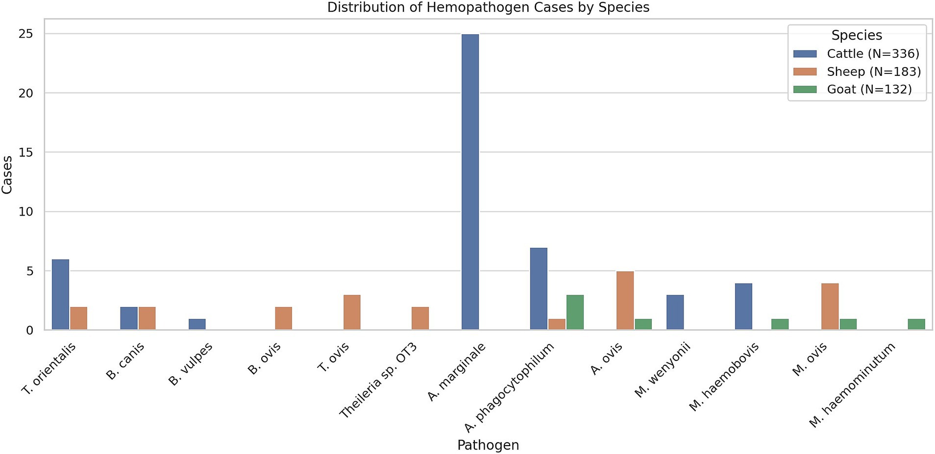
Figure 1. Distribution of identified hemopathogens in aborted fetuses of cattle, sheep, and goats. The bar chart shows the number of positive cases for each pathogen detected by molecular methods across the three ruminant species.
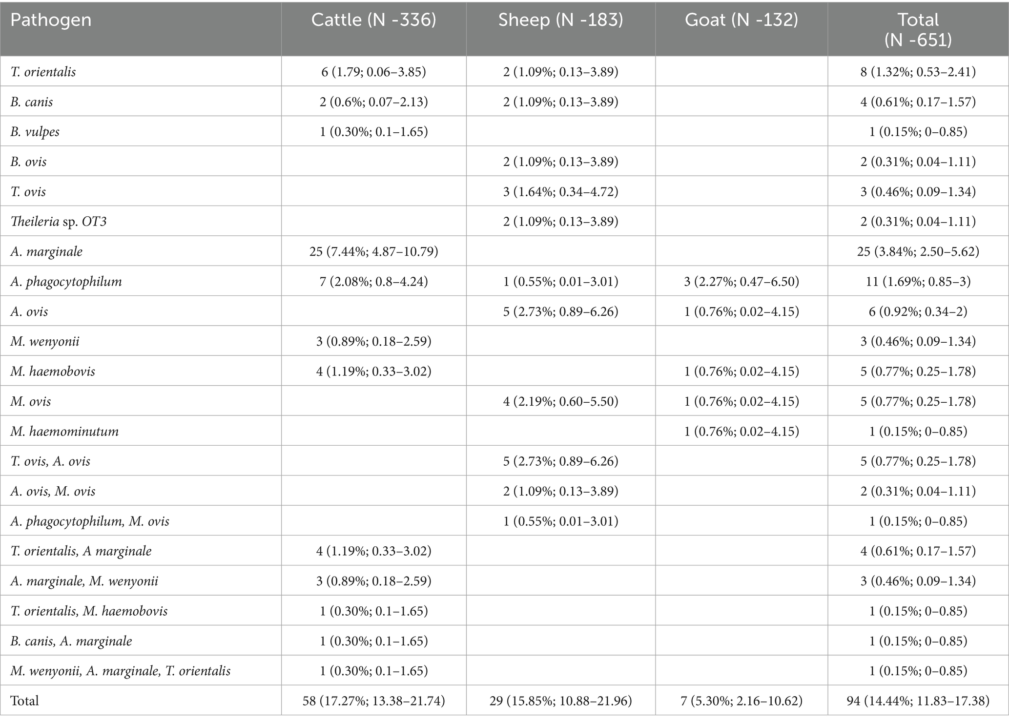
Table 2. Prevalence and coinfection rates of hemopathogens in aborted fetuses of cattle, sheep, and goats.
Seven pathogens were detected in bovine fetuses, including T. orientalis, B. vulpes, B. canis, A. marginale, A. phagocytophilum, M. wenyonii, and Candidatus M. haemobos. Anaplasma spp. had the highest prevalence, detected in 41 of 336 samples (12.2%, CI: 8.90–16.19), followed by piroplasms in 16 of 336 samples (4.76%, CI: 2.75–7.62) and hemotropic Mycoplasma in 12 of 336 samples (3.57%, CI: 1.86–6.16) (Table 2). The most frequently identified pathogen was A. marginale, which was detected in 34 of 336 samples (10.12%, CI: 7.11–13.87), followed by T. orientalis, which was found in 12 of 336 samples (3.58%, CI: 1.86–6.16). In addition, A. phagocytophilum and M. wenyonii were each identified in seven of 336 samples (2.08%, CI: 0.8–4.24), while Candidatus M. haemobos was detected in five of 336 samples (1.49%, CI: 0.49–3.44). Furthermore, B. canis was detected in three of 336 samples (0.89%, CI: 0.18–2.59), and B. vulpes in one of 336 samples (0.29%, CI: 0.01–1.65). Several co-infections were observed in bovine fetuses, including the combination of T. orientalis and A. marginale in four cases (1.19%), A. marginale and M. wenyonii in three cases (0.89%), and one case each involving T. orientalis and M. haemobos, B. canis and A. marginale, and a triple infection with M. wenyonii, A. marginale, and T. orientalis.
In sheep fetuses, the diversity of detected pathogens was the greatest among all studied animal species with the identification of eight different pathogenic agents, namely B. ovis, T. ovis, Theileria sp. OT3, T. orientalis, B. canis, A. ovis, A. phagocytophilum, and M. ovis (Table 2). Piroplasms were the most frequently detected pathogens, occurring in 16 of 183 samples (8.74%, CI: 5.08–13.81), followed by Anaplasma spp. in 14 of 183 samples (7.65%, CI: 4.25–12.50) and hemotropic Mycoplasma in seven of 138 samples (3.82%, CI: 1.55–7.72). Anaplasma ovis was identified as the most common pathogen, occurring in 12 of 183 samples (6.56%, CI: 3.43–11.17). This was followed by T. ovis, which was detected in eight of 183 samples (4.38%, CI: 1.91–8.43), and M. ovis, which was found in seven of 183 samples (3.82%, CI: 1.55–7.72). Other pathogens, including T. orientalis, Theileria sp. OT3, B. canis, B. ovis, and A. phagocytophilum, were each detected in two of 183 samples (1.09%, CI: 0.13–3.89). Several co-infections were observed in sheep, including T. ovis and A. ovis in five cases (2.73%), A. ovis and M. ovis in two cases (1.09%), and a single case with A. phagocytophilum and M. ovis (0.55%).
In goats, the overall prevalence of infection was lower than in cattle and sheep. Five pathogens were confirmed in goats: A. ovis, A. phagocytophilum, M. ovis, Candidatus M. haemominutum, and Candidatus M. haemobos (Table 2). Seven fetuses tested positive (5.30%; CI: 2.16–10.62), and only Anaplasma and hemotropic Mycoplasma species were detected. Anaplasma phagocytophilum was the most prevalent, confirmed in three fetuses (2.27%; CI: 0.47–6.50), while A. ovis, Candidatus M. haemobos, Candidatus M. haemominutum, and M. ovis were each identified in one fetus (0.76%; CI: 0.02–4.15). No piroplasms or co-infections were recorded in goats.
Mapping of positive cases revealed a widespread geographic distribution of hemopathogens across continental and coastal regions of Croatia in all three ruminant species. However, the density and diversity of pathogens varied by host species.
In cattle (Figure 2), most pathogen-positive cases clustered in central and eastern regions, with A. marginale, T. orientalis, and M. wenyonii detected most frequently. Several co-infections were also documented in this area, suggesting higher transmission pressure or improved detection due to population density and veterinary surveillance.
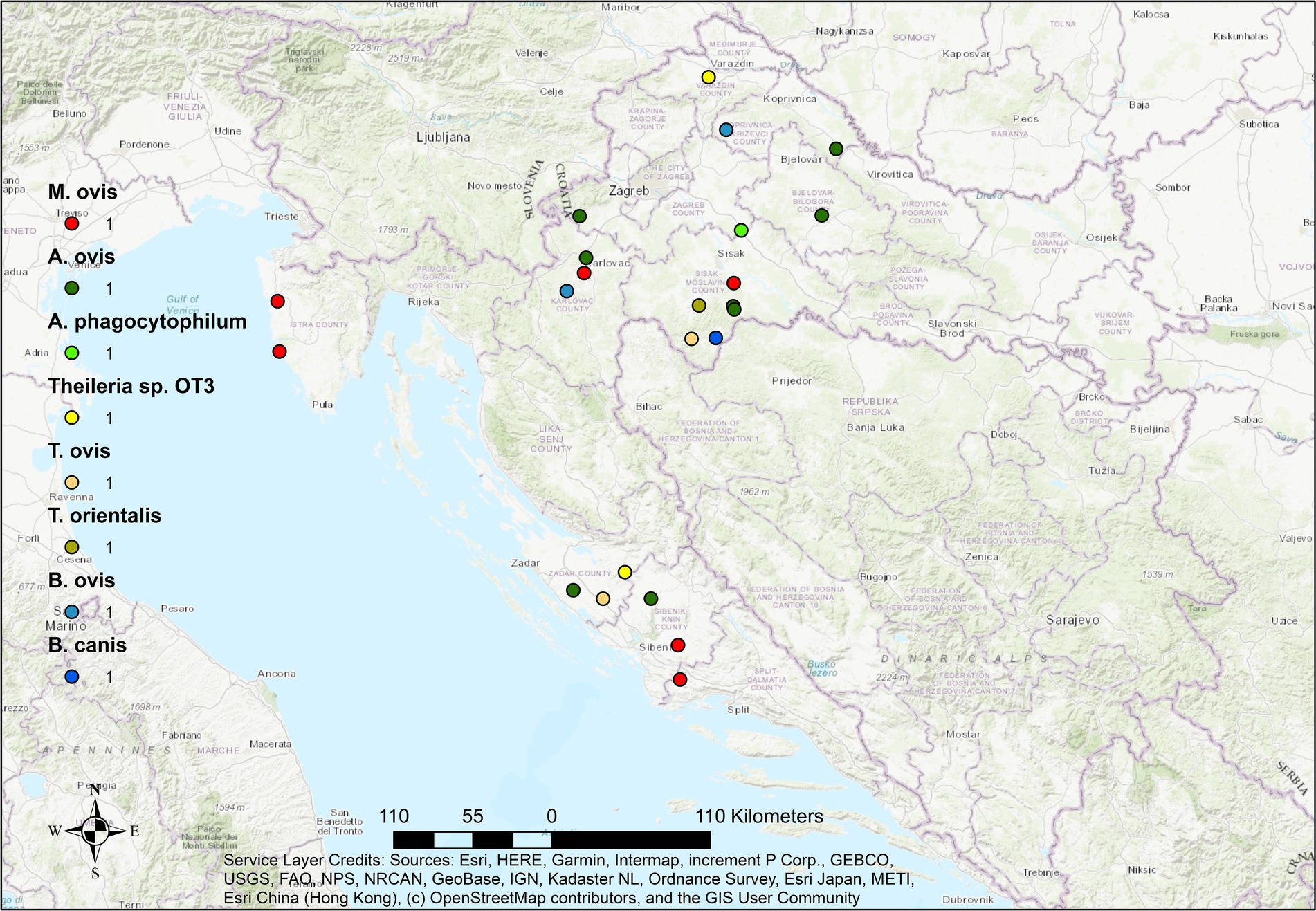
Figure 2. Geographical distribution of hemopathogens in aborted cattle fetuses in Croatia. Dots represent locations of individual cases. Color coding indicates haemopathogen, while size or density of symbols reflects number and diversity of pathogens detected at each site.
In sheep (Figure 3), the spatial distribution of pathogens was more scattered but still showed notable clustering in northern and central regions. A high diversity of pathogens, including T. ovis, A. ovis, and M. ovis, was recorded, consistent with the observed high pathogen diversity in this species. Co-infections were observed in several localities, particularly where T. ovis and A. ovis overlapped, potentially indicating ecological compatibility or co-transmission by shared tick vectors.
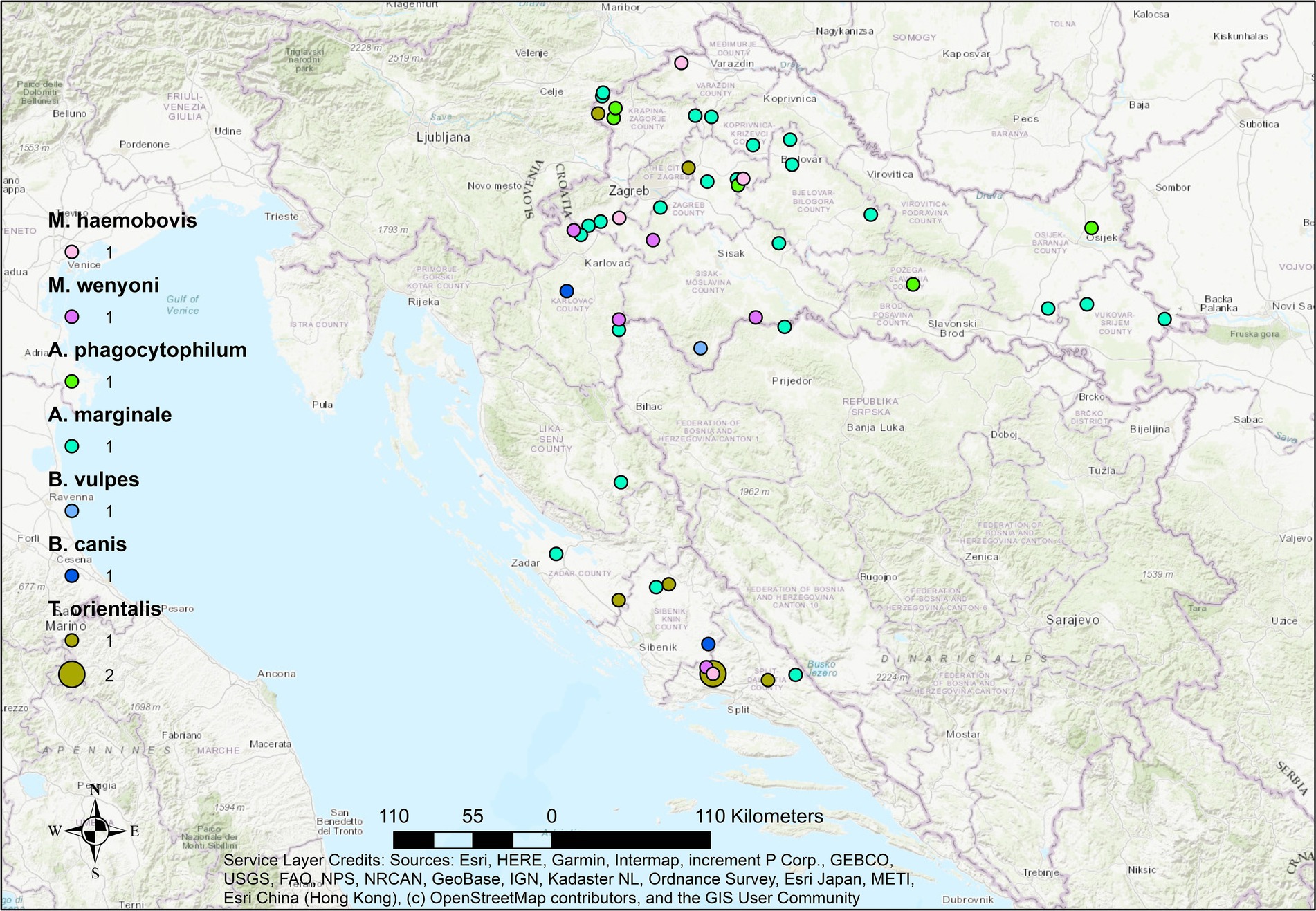
Figure 3. Geographical distribution of hemopathogens detected in aborted sheep fetuses in Croatia. Dots represent locations of individual cases.
In goats (Figure 4), the geographic distribution of positive cases was the most limited, with fewer locations and lower pathogen diversity. Only A. phagocytophilum, A. ovis, and hemotropic Mycoplasma spp. were detected, predominantly in coastal and south-central regions. The limited spatial footprint may reflect both the lower number of submitted caprine fetuses and the species’ differing exposure risk or management practices. Overall, there were no strong differences in pathogen presence between coastal and continental regions, suggesting that vector presence and pathogen circulation are not strictly region-dependent.
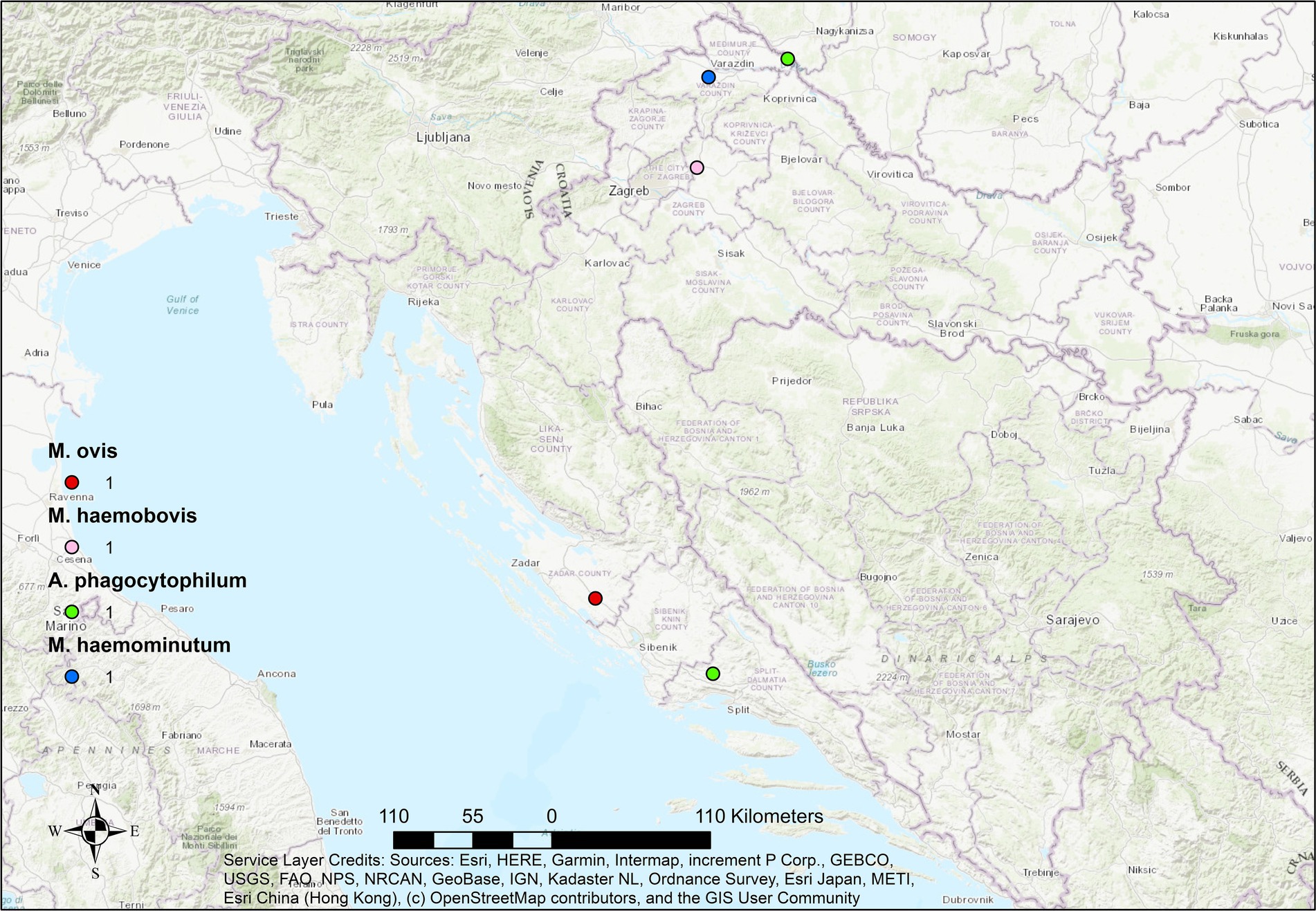
Figure 4. Geographical distribution of hemopathogens detected in aborted goat fetuses in Croatia. Dots represent locations of individual cases.
In the present study, no co-infections with C. burnetii, Brucella spp., or Chlamydia spp. were detected in any of the examined animal species. In cattle, co-infection with at least one hemopathogen and N. caninum was confirmed in 14 out of 336 aborted fetuses, corresponding to a prevalence of 4.17% (95% CI: 2.30–6.89). In sheep, co-infection with T. gondii was confirmed in two out of 183 fetuses, corresponding to a prevalence of 1.09% (95% CI: 0.13–3.89) (Table 3).
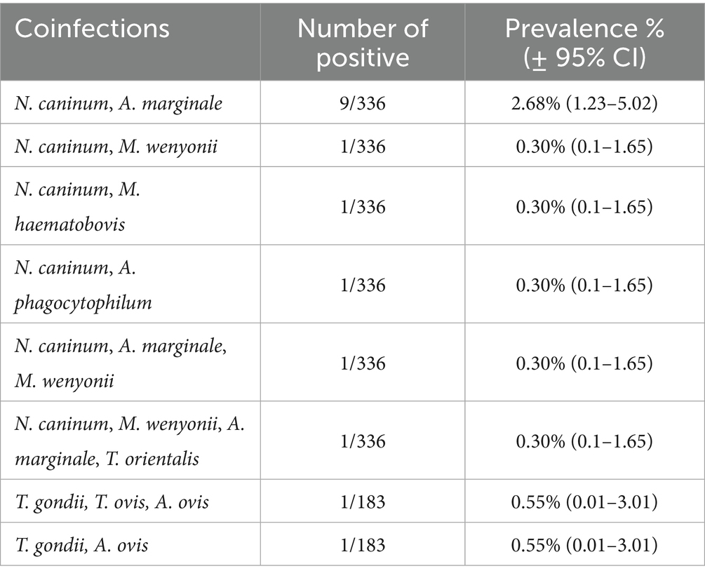
Table 3. Coinfections of hemopathogens with Neospora caninum in cattle and Toxoplasma gondii in sheep fetuses.
In addition to prevalence estimates, statistical analysis was conducted to assess whether the likelihood of detecting hemopathogens in aborted fetuses differed significantly between host species. A chi-square test of independence showed a significant association between host species and infection status (χ2 = 11.38, p = 0.0034), suggesting non-uniform distribution of infection risk. Pairwise comparisons using Fisher’s exact test revealed that aborted cattle fetuses had significantly higher odds of testing positive for hemopathogens compared to goats (OR = 3.73, 95% CI: 1.65–8.39, p < 0.001). Similarly, sheep fetuses had higher odds of infection than goat fetuses (OR = 3.36, 95% CI: 1.43–7.93, p = 0.0038). However, no statistically significant difference was observed between cattle and sheep (OR = 1.11, 95% CI: 0.68–1.80, p = 0.71). These findings suggest that cattle and sheep may be at greater risk of hemopathogen-associated abortion compared to goats.
Discussion
Hemopathogens represent an important group of infectious agents affecting domestic ruminants, particularly in tropical and subtropical regions. However, increasing evidence indicates that these pathogens may also be underrecognized in temperate areas, including Europe and Mediterranean Basin (Defaye et al., 2022). Despite historical reports of their presence in ruminants, recent molecular surveillance in this region has been limited (Kapo et al., 2025). Moreover, their potential role in reproductive disorders and fetal losses in ruminants has not been systematically investigated in Europe. This study provides novel insights into the transplacental occurrence of hemotropic pathogens in aborted ruminant fetuses in Croatia, using comprehensive molecular screening across three host species. Our findings reveal that infections with members of the genera Anaplasma, Babesia, Theileria, and hemotropic Mycoplasma are not only detectable in aborted fetal tissues but may also contribute to reproductive losses, including abortion.
The detection of A. phagocytophilum, A. marginale, and A. ovis was not unexpected, as these species have already been confirmed in Croatian ruminants (Jaarsma et al., 2019; Jurković et al., 2020; Šarić et al., 2022). Notably, A. phagocytophilum was the only hemopathogen detected in all three host species, albeit at low prevalence, consistent with findings from sheep flocks in Italy, Norway, Spain, the United Kingdom, and Greece (Jones and Davies, 1995; Garcia-Perez et al., 2003; Chianini et al., 2004; Lillini et al., 2006; Giudice et al., 2012; Gouvias et al., 2024), as well as goats in Greece (Chochlakis et al., 2020) and cattle (Van Loo et al., 2023). The low prevalence across species suggests a limited but potentially relevant role in reproductive disorders.
In contrast, A. marginale and A. ovis displayed a high degree of host specificity, being detected exclusively in cattle and small ruminants, respectively. These species were also the most commonly identified hemopathogens in their respective host species, both as single infections and in co-infections. High rates of transplacental transmission of A. marginale have been reported in Brazil and Venezuela, with prevalence in neonatal calves ranging from 10% to over 40% (Maldonado et al., 2012; Grau et al., 2013; Silva et al., 2014; Andrade et al., 2024). While vertical transmission is well documented, reports of abortion or fetal pathology directly linked to A. marginale remain scarce. Recent studies describing fetal lesions associated with A. marginale infection suggest that bovine anaplasmosis should be included in the differential diagnosis of fetal losses and neonatal mortality in endemic regions (Henker et al., 2020). Given that current study examined aborted fetuses, the observed association with A. marginale supports its potential role in reproductive failure, either as a primary cause or in conjunction with other pathogens.
In contrast, the association between A. ovis and fetal losses has not been established so far. In one experimental study, vertical transmission of A. ovis was not confirmed via direct parasitemia in neonatal lambs but was demonstrated by blood transfer to splenectomized recipients (Zaugg, 1987). Under natural conditions, A. ovis has been identified in neonatal elk calves (Hendrix et al., 2019), but not in domestic ruminants. Our detection of A. ovis in aborted sheep and a goat, both as a single agent and in co-infection with T. ovis and M. ovis, represents, to our knowledge, the first such finding suggesting possible intrauterine transmission and involvement in fetal pathology.
Interestingly, A. bovis, previously detected in deceased cattle in earlier study (Jurković et al., 2020), was not identified in any of the aborted bovine fetuses in this investigation. This observation may suggest that not all Anaplasma species possess the capacity for intrauterine transmission or contribute to fetal pathology.
Hemotropic Mycoplasma spp. were detected in domestic ruminants in Croatia for the first time in this study. Host-specific species were identified in their expected hosts, with M. wenyonii and Candidatus M. haemobos detected in cattle, and M. ovis in sheep and goats. Although intrauterine transmission of M. ovis has not been previously documented, parallels with other species—such as M. haemolamae in camelids—support the plausibility of vertical transmission (Tornquist et al., 2011).
Vertical transmission of bovine hemoplasmas is generally considered rare, but accumulating evidence suggests it is possible. Fujihara et al. (2011) first raised this possibility, while Hornok et al. (2011) demonstrated that 10.5% of neonatal beef calves born to infected dams were hemoplasma-positive. Subsequent studies by Sasaoka et al. (2015) and Niethammer et al. (2018) also supported the feasibility of vertical transmission. In one study by Tagawa et al. (2013), 14.1% of 71 dairy calves tested positive for bovine hemoplasmas; however, because blood sampling occurred up to a week after birth and neonatal animals were not sampled at birth, the route of transmission could not be definitively determined. Interestingly, M. wenyonii has previously been confirmed in neighboring Bosnia and Herzegovina (Stevanović et al., 2020), whereas the detection of Candidatus M. haemobos in our study represents the first confirmed case in the Western Balkan.
An unexpected finding was the detection of Candidatus M. haemominutum in a goat. Although typically regarded as a feline hemoplasma, this organism has also been identified in dogs (Zhuang et al., 2009) and wild canids such as wolves (Millán et al., 2018), raising the possibility of a broader host range and potential cross-species transmission.
The association between hemotropic Mycoplasma spp. and reproductive disorders remains poorly understood. However, our findings suggest that these organisms may contribute to fetal losses, either as sole infectious agents or in combination with other pathogens through synergistic co-infections.
Transplacental transmission of T. orientalis is of particular interest given the susceptibility of pregnant animals to abort. Prior studies indicate that vertical transmission of other Theileria spp. can occur in their respective hosts, including T. equi in horses (Phipps and Otter, 2004; Georges et al., 2011), T. lestoquardi in sheep (Zakian et al., 2014), and more rarely, T. annulata in cattle (Sudan et al., 2015). Experimental infection studies have demonstrated that transplacental transmission of T. orientalis can result in a 100% abortion rate in pregnant cows infected via ticks (Baek et al., 2003), while microscopic examinations in field settings in Japan suggested transplacental transmission at lower frequencies (Onoe et al., 1994). Conversely, a more recent study in New Zealand dairy herds experiencing T. orientalis outbreaks failed to confirm vertical transmission using highly sensitive qPCR assays (Lawrence et al., 2016).
In our study, it was identified in fetuses of both cattle and sheep, further supporting its abortifacient potential under field conditions (Baek et al., 2003; Swilks et al., 2017; Chisu et al., 2021). Additionally, the detection of T. ovis and Theileria sp. OT3 in aborted sheep fetuses expands the recognized diversity of Theileria species with the potential for intrauterine transmission. Alongside Babesia ovis, these findings indicate that a broader range of protozoan parasites may be implicated in small ruminant abortions than previously appreciated.
The presence of canine-origin piroplasms B. canis and B. vulpes in cattle and sheep, although unexpected, may reflect incidental spillover from dogs or wild canids, as both species have been previously reported in these hosts (Beck et al., 2009; Dežđek et al., 2010; Beck et al., 2017). Similar findings have been reported in Sardinia, where Ehrlichia canis, a canine tick-borne pathogen, was detected in aborted fetuses of sheep and goats (Chisu et al., 2021). However, the frequency of detection was substantially higher for E. canis than for canine piroplasms in the present study. Thus, it is likely that B. canis and B. vulpes detected here do not play a significant role in ruminant fetal pathology and represent incidental or non-pathogenic infections.
Importantly, no co-infections with classical abortifacient zoonotic pathogens such as Brucella spp., C. burnetii, or Chlamydia spp. were detected, suggesting that the hemopathogens identified in this study may act independently in causing fetal damage. However, co-infections with Neospora caninum in cattle and Toxoplasma gondii in sheep were observed, indicating the possibility of multifactorial causes of abortion.
The observed differences in pathogen prevalence and diversity underscore the complexity of vertical transmission dynamics in domestic ruminants. Notably, the significantly lower pathogen burden in goats suggests species-specific susceptibility. Although we cannot directly compute the probability of abortion due to infection (since we lack a control group of non-aborted fetuses), the observed infection prevalence in aborted fetuses may serve as a proxy indicator for potential vertical transmission and its relevance to reproductive loss.
Conclusion
To date, limited but growing evidence supports the possibility of transplacental transmission of hemoparasites in cattle. However, data on small ruminants remain sparse. This study represents the most extensive molecular investigation in Europe involving all three major ruminant species, and clearly demonstrates that transplacental transmission of hemopathogens is possible and likely underrecognized. Our results highlight the need to include such pathogens in the routine diagnostic workup of abortions, especially in endemic areas. Long-term, multidisciplinary studies that incorporate detailed histopathological, molecular, and clinical data are necessary to comprehensively assess the role of hemopathogens in reproductive losses of domestic ruminants.
Data availability statement
The datasets presented in this study can be found in online repositories. The names of the repository/repositories and accession number(s) can be found in the article/supplementary material.
Ethics statement
The animal study was approved by Ethic committee of Croatian Veterinary Institute. The study was conducted in accordance with the local legislation and institutional requirements.
Author contributions
DŽ: Writing – original draft, Investigation, Methodology. ŠN: Investigation, Methodology, Writing – review & editing. ŽM: Methodology, Data curation, Writing – original draft. EG: Methodology, Investigation, Writing – review & editing. SŠ: Investigation, Methodology, Writing – review & editing. IR: Investigation, Methodology, Writing – review & editing. SD: Investigation, Methodology, Writing – review & editing. MT: Investigation, Methodology, Writing – review & editing. AH: Writing – review & editing, Formal analysis, Writing – original draft. RB: Formal analysis, Writing – original draft, Writing – review & editing, Conceptualization, Funding acquisition, Resources, Supervision.
Funding
The author(s) declare that financial support was received for the research and/or publication of this article. This study was funded by the Project IP-2022-10-7842 “Apicomplexan and bacterial tick-borne pathogens in domestic ruminants, horses and haematophagous vectors” founded by Croatian Science Foundation (HRZZ).
Acknowledgments
The authors express their sincere gratitude to technicians Kristina Skrbin, Ada Vilić, Petra Cikoš, and Mario Maršić which significantly contributed to this study.
Conflict of interest
The authors declare that the research was conducted in the absence of any commercial or financial relationships that could be construed as a potential conflict of interest.
Generative AI statement
The authors declare that no Gen AI was used in the creation of this manuscript.
Publisher’s note
All claims expressed in this article are solely those of the authors and do not necessarily represent those of their affiliated organizations, or those of the publisher, the editors and the reviewers. Any product that may be evaluated in this article, or claim that may be made by its manufacturer, is not guaranteed or endorsed by the publisher.
Footnotes
References
Allsopp, M. T. E. P., Lewis, B. D., and Penzhorn, B. L. (2007). Molecular evidence for transplacental transmission of Theileria equi from carrier mares to their apparently healthy foals. Vet. Parasitol. 148, 130–136. doi: 10.1016/j.vetpar.2007.05.017
Andrade, L. S., De Souza, R. S., Carvalho de Araujo, A., Silva, S. O., Melo, M. N., Melo, F. G., et al. (2024). Hemopathogens in naturally infected bovine fetuses in Brazil. Ticks Tick Borne Dis. 15:102351. doi: 10.1016/j.ttbdis.2024.102351
Arendt, M., Stadler, J., Ritzmann, M., Ade, J., Hoelzle, K., and Hoelzle, L. E. (2024). Hemotrophic mycoplasmas —vector transmission in livestock. Microorganisms 12:1278. doi: 10.3390/microorganisms12071278
Baek, B. K., Soo, K. B., Kim, J. H., Hur, J., Lee, B. O., Jung, J. M., et al. (2003). Verification by polymerase chain reaction of vertical transmission of Theileria sergenti in cows. Can. J. Vet. Res. 67, 278–282.
Bartolomé del Pino, L. E., Meana, A., Zini, M., and Cersini, A. (2023). Evidence of transplacental transmission of equine piroplasms Theileria equi and Babesia caballi in an Italian breed mare. Folia Parasitol. 70:005. doi: 10.14411/fp.2023.005
Beck, A., Huber, D., Polkinghorne, A., Kurilj, A. G., Benko, V., Mrljak, V., et al. (2017). The prevalence and impact of Babesia canis and Theileria sp. in free-ranging grey wolf (Canis lupus) populations in Croatia. Parasit. Vectors 10:168. doi: 10.1186/s13071-017-2106-8
Beck, R., Vojta, L., Mrljak, V., Marinculić, A., Beck, A., Živičnjak, T., et al. (2009). Diversity of Babesia and Theileria species in symptomatic and asymptomatic dogs in Croatia. Int. J. Parasitol. 39, 843–848. doi: 10.1016/j.ijpara.2008.12.005
Berri, M., Laroucau, K., and Rodolakis, A. (2000). The detection of Coxiella burnetii from ovine genital swabs, milk and fecal damles by the use of a single touchdown polymerase chain reaction. Vet. Microbiol. 72, 285–293. doi: 10.1016/s0378-1135(99)00178-9
Berri, M., Rekiki, A., Boumedine, K. S., and Rodolakis, A. (2009). Simultaneous differential detection of Chlamydophila abortus, Chlamydophila pecorum and Coxiella burnetii from aborted ruminant's clinical samples using multiplex PCR. BMC Microbiol. 9:130. doi: 10.1186/1471-2180-9-130
Bock, R., Jackson, L., De Vos, A., and Jorgensen, W. (2004). Babesiosis of cattle. Parasitology 129, S247–S269. doi: 10.1017/S0031182004005190
Chianini, F., Adams, C., and Buxton, D. (2004). Neuropathological changes in ovine fetuses caused by tick-borne fever. Vet. Rec. 155, 805–806. doi: 10.1136/vr.155.25.805
Chisu, V., Loi, F., Mura, L., Tanda, A., Chessa, G., and Masala, G. (2021). Molecular detection of Theileria sergentii/orientalis/buffeli and Ehrlichia canis from aborted ovine and caprine products in Sardinia, Italy. Vet. Med. Sci. 7, 1762–1768. doi: 10.1002/vms3.510
Chochlakis, D., Giadinis, N. D., Petridou, E., Filioussis, G., Tselentis, Y., Psaroulaki, A., et al. (2020). Molecular evidence of Anaplasma phagocytophilum in aborted goat fetuses and placenta. Vet. Ital. 56, 302–303. doi: 10.12834/VetIt.1173.6516.2
Costa, S. C. L., Magalhães, V. C. S., Oliveira, U. V., Carvalho, F. S., Almeida, C. P., Machado, R. Z., et al. (2016). Transplacental transmission of bovine tick-borne pathogens: frequency, co-infections and fatal neonatal anaplasmosis in a region of enzootic stability in the northeast of Brazil. Ticks Tick Borne Dis. 7, 270–275. doi: 10.1016/j.ttbdis.2015.11.001
Defaye, B., Moutailler, S., Pasqualini, V., and Quilichini, Y. (2022). Distribution of tick-borne pathogens in domestic animals and their ticks in the countries of the Mediterranean basin between 2000 and 2021: a systematic review. Microorganisms 10:1236. doi: 10.3390/microorganisms10061236
Dežđek, D., Vojta, L., Ćurković, S., Lipej, Z., Mihaljević, Ž., Cvetnić, Ž., et al. (2010). Molecular detection of Theileria annae and Hepatozoon canis in foxes (Vulpes vulpes) in Croatia. Vet. Parasitol. 172, 333–336. doi: 10.1016/j.vetpar.2010.05.022
Eskezia, B. G., and Desta, A. H. (2016). Review on the impact of ticks on livestock health and productivity. J. Biol. Agric. Healthc. 6, 1–7.
Esmaeilnejad, B., Tavassoli, M., Samiei, A., and Hajipour, N. (2018). Molecular verification of transplacental transmission of Theileria lestoquardi in goat. Parasitol. Res. 117, 3315–3318. doi: 10.1007/s00436-018-6007-3
Françoso, R., Riccio, A. V., Fernandes, C. B., Alonso, M. A., and Belli, C. B. (2018). Transplacental transmission of Theileria equi in mules: should we worry? Vet. Parasitol. 264, 39–41. doi: 10.1016/j.vetpar.2018.10.017
Fujihara, Y., Sasaoka, F., Suzuki, J., Watanabe, Y., Fujihara, M., Ooshita, K., et al. (2011). Prevalence of hemoplasma infection among cattle in the western part of Japan. J. Vet. Med. Sci. 73, 1653–1655. doi: 10.1292/jvms.11-0269
Garcia-Perez, A. L., Barandika, J., Oporto, B., Povedano, J., and Juste, R. A. (2003). Anaplasma phagocytophila as an abortifacient agent in sheep farms from northern Spain. Ann. N. Y. Acad. Sci. 990, 429–432. doi: 10.1111/j.1749-6632.2003.tb07406.x
Georges, K. C., Ezeokoli, C. D., Sparagano, O., Pargass, I., Campbell, M., D’Abadie, R., et al. (2011). A case of transplacental transmission of Theileria equi in a foal in Trinidad. Vet. Parasitol. 175, 363–366. doi: 10.1016/j.vetpar.2010.10.019
Girotto-Soares, A., Soares, J. F., Bogado, A. L. G., De Macedo, C. A. B., Sandeski, L. M., Garcia, J. L., et al. (2016). ‘Candidatus Mycoplasma haemobos’: Transplacental transmission in dairy cows (Bos taurus). Vet. Microbiol. 195, 22–24. doi: 10.1016/j.vetmic.2016.08.020
Giudice, E., Giannetto, C., Furco, V., Alongi, A., and Torina, A. (2012). Anaplasma phagocytophilum seroprevalence in equids: a survey in Sicily (Italy). Parasitol. Res. 111, 951–955. doi: 10.1007/s00436-012-2854-5
Gouvias, I., Lysitsas, M., Batsidis, A., Malefaki, S., Bitchava, D., Tsara, A., et al. (2024). Molecular investigation of small ruminant abortions using a 10-plex HRM-qPCR technique: a novel approach in routine diagnostics. Microorganisms 12:1675. doi: 10.3390/microorganisms12081675
Grau, H. E. G., Da Cunha Filho, N. A., Pappen, F. G., and Da Rosa Farias, N. A. (2013). Transplacental transmission of Anaplasma marginale in beef cattle chronically infected in southern Brazil. Rev. Bras. Parasitol. Vet. 22, 189–193. doi: 10.1590/S1984-29612013000200038
Hecker, Y. P., González-Ortega, S., Cano, S., Ortega-Mora, L. M., and Horcajo, P. (2023). Bovine infectious abortion: a systematic review and meta-analysis. Front. Vet. Sci. 10:1249410. doi: 10.3389/fvets.2023.1249410
Hendrix, G. K., Brayton, K. A., and Burcham, G. N. (2019). Anaplasma ovis as the suspected cause of mortality in a neonatal elk calf. J. Vet. Diagn. Invest. 31, 267–270. doi: 10.1177/1040638719830456
Henker, L. C., Lorenzett, M. P., Fagundes-Moreira, R., Dalto, A. G. C., Sonne, L., Driemeier Driemeier, D., et al. (2020). Bovine abortion, stillbirths, and neonatal death associated with Babesia bovis and Anaplasma sp. infections in southern Brazil. Ticks Tick Borne Dis. 11:101443. doi: 10.1016/j.ttbdis.2020.101443
Henniger, T., Henniger, P., Grossmann, T., Distl, O., Ganter, M., and Von Loewenich, F. D. (2013). Congenital infection with Anaplasma phagocytophilum in a calf in northern Germany. Acta Vet. Scand. 55:38. doi: 10.1186/1751-0147-55-38
Hornok, S., Elk, V., De la Fuente, J., Naranjo, V., Farkas, R., Majoros, G., et al. (2007). First serological and molecular evidence on the endemicity of Anaplasma ovis and Anaplasma marginale in Hungary. Vet. Microbiol. 122, 316–322. doi: 10.1016/j.vetmic.2007.01.024
Hornok, S., Micsutka, A., Meli, M. L., Lutz, H., and Hofmann-Lehmann, R. (2011). Molecular investigation of transplacental and vector-borne transmission of bovine haemoplasmas. Vet. Microbiol. 152, 411–414. doi: 10.1016/j.vetmic.2011.04.031
Hughes, J. M., Williams, R. H., Morley, E. K., Cook, D. A. N., Terry, R. S., Murphy, R. G., et al. (2006). The prevalence of Neospora caninum and co-infection with toxoplasma gondii by PCR analysis in naturally occurring mammal populations. Parasitology 132, 29–36. doi: 10.1017/S0031182005008784
Jaarsma, R. I., Sprong, H., Takumi, K., Kazimirova, M., Silaghi, C., Mysterud, A., et al. (2019). Anaplasma phagocytophilum evolves in geographical and biotic niches of vertebrates and ticks. Parasit. Vectors 12:328. doi: 10.1186/s13071-019-3583-8
Jones, G. L., and Davies, I. H. (1995). An ovine abortion storm caused by infection with Cytoecetes phagocytophila. Vet. Rec. 136:127. doi: 10.1136/vr.136.5.127
Jones, C. D., Okhravi, N., Adamson, P., Tasker, S., and Lightman, S. (2000). Comparison of PCR detection methods for B1, P30, and 18S rDNA genes of T. Gondii in aqueous humor. Invest. Ophthalmol. Vis. Sci. 41, 634–644.
Jurković, D., Mihaljević, Ž., Duvnjak, S., Silaghi, C., and Beck, R. (2020). First reports of indigenous lethal infection with Anaplasma marginale, Anaplasma bovis and Theileria orientalis in Croatian cattle. Ticks Tick Borne Dis. 11:101469. doi: 10.1016/j.ttbdis.2020.101469
Kapo, N., Zuber Bogdanović, I., Gagović, E., Jurković Žilić, D., Sukara, R., Adžić, B., et al. (2025). Non-zoonotic tick-borne pathogens in Western Balkan. Parasit. Vectors 18:107. doi: 10.1186/s13071-025-06740-z
Kocan, K. M., De la Fuente, J., Blouin, E. F., Coetzee, J. F., and Ewing, S. A. (2010). The natural history of Anaplasma marginale. Vet. Parasitol. 167, 95–107. doi: 10.1016/j.vetpar.2009.09.012
Lawrence, K. E., Gedye, K., McFadden, A. M., Pulford, D. J., and Pomroy, W. E. (2016). An observational study of the vertical transmission of Theileria orientalis (Ikeda) in a New Zealand pastoral dairy herd. Vet. Parasitol. 218, 59–65. doi: 10.1016/j.vetpar.2016.01.003
Lillini, E., Macri, G., Proietti, G., and Scarpulla, M. (2006). New findings on anaplasmosis caused by infection with Anaplasma phagocytophilum. Ann. N. Y. Acad. Sci. 1081, 360–370. doi: 10.1196/annals.1373.053
Maldonado, J., Coronado, A., Kowalski, A., and Medina, J. (2012). Evidencia molecular de transmision transplacentaria de Anaplasma marginale em becerros neonatos cebú de Venezuela. Zootec. Trop. 30, 109–114.
Mekata, H., Minamino, T., Mikurino, Y., Yamamoto, M., Yoshida, A., Nonaka, N., et al. (2018). Evaluation of the natural vertical transmission of Theileria orientalis. Vet. Parasitol. 263, 1–4. doi: 10.1016/j.vetpar.2018.09.017
Messick, J. B. (2004). Hemotrophic mycoplasmas (hemoplasmas): a review and new insights into pathogenic potential. Vet. Clin. Pathol. 33, 2–13. doi: 10.1111/j.1939-165x.2004.tb00342.x
Millán, J., Velarde, R., Delicado, V., Negre, N., Ribas, A., Oleaga, A., et al. (2018). High diversity of hemotropic mycoplasmas in Iberian wild carnivores. Comp. Immunol. Microbiol. Infect. Dis. 60, 11–16. doi: 10.1016/j.cimid.2018.09.007
Moeller, R. B. (2001). Causes of caprine abortion: diagnostic assessment of 211 cases (1991–1998). J. Vet. Diagn. Invest. 13, 265–270. doi: 10.1177/104063870101300317
Neimark, H., Johansson, K. E., Rikihisa, Y., and Tully, J. G. (2001). Proposal to transfer some members of the genera Haemobartonella and Eperythrozoon to the genus Mycoplasma with descriptions of `Candidatus Mycoplasma haemofelis', `Candidatus Mycoplasma haemomuris', `Candidatus Mycoplasma haemosuis' and `Candidatus Mycoplasma wenyonii'. Int. J. Syst. Evol. Microbiol. 51, 891–899. doi: 10.1099/00207713-51-3-891
Niethammer, F. N., Ade, J., Hoelzle, L. E., and Schade, B. (2018). Hemotrophic mycoplasma in Simmental cattle in Bavaria: prevalence, blood parameters, and transplacental transmission of ‘Candidatus Mycoplasma haemobos’ and Mycoplasma wenyonii. Acta Vet. Scand. 60:74. doi: 10.1186/s13028-018-0428-y
Norton, J. H., Parker, R. J., and Forbes-Faulkner, J. C. (1983). Neonatal anaplasmosis in a calf. Aust. Vet. J. 60:348. doi: 10.1111/j.1751-0813.1983.tb02844.x
Onoe, S., Sugimoto, C., Tanaka, M., Kubota, H., Hirai, T., Yonemichi, H., et al. (1994). Prenatal infections with Theileria sergenti in calves. J. Protozool. Res. 4, 119–123.
Parola, P., Roux, V., Camicas, J. L., Baradji, I., Brouqui, P., and Raoult, D. (2000). Detection of ehrlichiae in African ticks by polymerase chain reaction. Trans. R. Soc. Trop. Med. Hyg. 94, 707–708. doi: 10.1016/s0035-9203(00)90243-8
Petrovec, M., Sumner, J. W., Nicholson, W. L., Childs, J. E., Strle, F., Barlic, J., et al. (1999). Identity of ehrlichial DNA sequences derived from Ixodes ricinus ticks with those obtained from patients with human granulocytic ehrlichiosis in Slovenia. J. Clin. Microbiol. 37, 209–210. doi: 10.1128/JCM.37.1.209-210.1999
Phipps, L. P., and Otter, A. (2004). Transplacental transmission of Theileria equi in two foals born and reared in the United Kingdom. Vet. Rec. 154, 406–408. doi: 10.1136/vr.154.13.406
Potgieter, F. T., and Van Rensburg, L. (1987). The persistence of colostral Anaplasma antibodies and incidence of in utero transmission of Anaplasma infection in calves under laboratory condition. Onderstepoort J. Vet. Res. 54, 557–560.
Pusterla, N., Braun, U., Wolfensberger, C., and Lutz, H. (1997). Intrauterine infection with Ehrlichia phagocytophila in a cow. Vet. Rec. 141, 101–102. doi: 10.1136/vr.141.4.101
Rar, V., Tkachev, S., and Tikunova, N. (2021). Genetic diversity of Anaplasma bacteria: twenty years later. Infect. Genet. Evol. 91:104833. doi: 10.1016/j.meegid.2021.104833
Reppert, E., Galindo, R. C., Breshears, M. A., Kocan, K. M., Blouin, E. F., and De la Fuente, J. (2013). Demonstration of transplacental transmission of a human isolate of Anaplasma phagocytophilum in an experimentally infected sheep. Transbound. Emerg. Dis. 60, 93–96. doi: 10.1111/tbed.12120
Ribeiro, M. F. B., Lima, J. D., Guimaräes, A. M., Scatamburlo, M. A., and Martins, N. E. (1995). Transmissäo congênita da anaplasmose bovina/congenital transmission of anaplasmosis in cattle. Arq. Bras. Med. Vet. Zootec. 47, 297–304.
Šarić, T., Beck, A., Taraš, I., Šuto, A., Orlović, D., Jurković, D., et al. (2022). The first description of ram infection with rickettsiae Anaplasma ovis in the Republic of Croatia. Vet. Stanica 53, 549–560. doi: 10.46419/VS.53.5.9
Sasaoka, F., Suzuki, J., Hirata, T. I., Ichijo, T., Furuhama, K., Harasawa, R., et al. (2015). Vertical transmission of Mycoplasma wenyonii in cattle, supported by analysis of the ribonuclease RNA gene—short communication. Acta Vet. Hung. 63, 271–274. doi: 10.1556/004.2015.025
Selim, A. M., Das, M., Senapati, S. K., Jena, G. R., Mishra, C., Mohanty, B., et al. (2021). Transplacental transmission of Theileria annulata in cattle confirmed by molecular techniques. J. Parasit. Dis. 45, 336–340. doi: 10.1007/s12639-021-01365-2
Silva, J. B., Castro, G. N. S., and Fonseca, A. H. (2014). Longitudinal study of risk factors for anaplasmosis and transplacental transmission in herd cattle. Semin. Agrar. 35, 2491–2500. doi: 10.5433/1679-0359.2014v35n4Supl1p2491
Silva, J. B., and Fonseca, A. H. (2014). Risk factors for anaplasmosis in dairy cows during the paripartum. Trop. Anim. Health Prod. 46, 461–465. doi: 10.1007/s11250-013-0514-0
Silva, J. B., Gonçalves, L. R., Varani, A. M., André, M. R., and Machado, R. Z. (2015). Genetic diversity and molecular phylogeny of Anaplasma marginale studied longitudinally under natural transmission conditions in Rio de Janeiro, Brazil. Ticks Tick Borne Dis. 6, 499–507. doi: 10.1016/j.ttbdis.2015.04.002
Stevanović, O., Jurković, D., Polkinghorne, A., Ćeleš, A., Ilić, T., Dimitrijević, S., et al. (2020). Molecular detection of Babesia divergens and Mycoplasma wenyonii infection in cattle from Bosnia and Herzegovina. Parasitol. Res. 119, 1423–1427. doi: 10.1007/s00436-020-06630-6
Stuen, S., Okstad, W., and Sagen, A. M. (2018). Intrauterine transmission of Anaplasma phagocytophilum in persistently infected lambs. Vet. Sci. 5:25. doi: 10.3390/vetsci5010025
Sudan, V., Singh, S. K., Jaiswal, A. K., Parashar, R., and Shanker, D. (2015). First molecular evidence of the transplacental transmission of Theileria annulata. Trop. Anim. Health Prod. 47, 1213–1215. doi: 10.1007/s11250-015-0835-2
Swilks, E., Fell, S. A., Hammer, J. F., Sales, N., Krebs, G. L., and Jenkins, C. (2017). Transplacental transmission of Theileria orientalis occurs at a low rate in field-affected cattle: infection in utero does not appear to be a major cause of abortion. Parasit. Vectors 10:227. doi: 10.1186/s13071-017-2166-9
Tagawa, M., Yamakawa, K., Aoki, T., Matsumoto, K., Ishii, M., and Inokuma, H. (2013). Effect of chronic hemoplasma infection on cattle productivity. J. Vet. Med. Sci. 75, 1271–1275. doi: 10.1292/jvms.13-0119
Tornquist, S. J., Boeder, L. J., Lubbers, S., and Cebra, C. K. (2011). Investigation of Mycoplasma haemolamae infection in crias born to infected dams. Vet. Rec. 168:380a. doi: 10.1136/vr.c6735
Van Loo, H., Pascottini, O. B., Hooyberghs, J., De Bleecker, K., Ribbens, S., Opsomer, G., et al. (2023). Detection of Anaplasma phagocytophilum in fetal and placental tissue of bovine abortions and perinatal mortalities. Vet. Rec. 193:e2880. doi: 10.1002/vetr.2880
Varanat, M., Maggi, R. G., Linder, K. E., and Breitschwerdt, E. B. (2011). Molecular prevalence of Bartonella, Babesia and hemotropic Mycoplasma sp. in dogs with splenic disease. J. Vet. Intern. Med. 25, 1284–1291. doi: 10.1111/j.1939-1676.2011.00811.x
World Organisation for Animal Health [WOAH] (2021). Available online at: Terrestrial manual. Chapter 3.4.2. Bovine babesiosis. https://www.woah.org/fileadmin/Home/eng/Health_standards/tahm/3.04.02 (Accessed January 24, 2025).
World Organization for Animal Health [WOAH] (2022). Terrestrial manual, chapter 3.1.4. Brucellosis (infection with B. abortus, B. Melitensis, and B. suis). Available online at: https://www.oie.int/en/what-we-do/standards/codes-and-manuals/terrestrial-manual-online-access (Accessed January 24, 2025).
Zakian, A., Nouri, M., Barati, F., Kahroba, H., Jolodar, A., and Rashidi, F. (2014). Vertical transmission of Theileria lestoquardi in sheep. Vet. Parasitol. 203, 322–325. doi: 10.1016/j.vetpar.2014.04.007
Zaugg, J. L. (1987). Ovine anaplasmosis: in utero transmission as it relates to stage of gestation. Am. J. Vet. Res. 48, 100–103. doi: 10.2460/ajvr.1987.48.01.100
Keywords: hemopathogens, transplacental transmission, abortion, ruminants, molecular diagnostics, Croatia
Citation: Žilić DJ, Naletilić Š, Mihaljević Ž, Gagović E, Špičić S, Reil I, Duvnjak S, Tuk MZ, Hodžić A and Beck R (2025) Hemotropic pathogens in aborted fetuses of domestic ruminants: transplacental transmission and implications for reproductive loss. Front. Microbiol. 16:1632135. doi: 10.3389/fmicb.2025.1632135
Edited by:
Leona Gilbert, Tezted Limited, FinlandReviewed by:
Hassan Y. A. H. Mahmoud, South Valley University, EgyptMansoor Azeem Siddiqui, National Institutes of Health (NIH), United States
Copyright © 2025 Žilić, Naletilić, Mihaljević, Gagović, Špičić, Reil, Duvnjak, Tuk, Hodžić and Beck. This is an open-access article distributed under the terms of the Creative Commons Attribution License (CC BY). The use, distribution or reproduction in other forums is permitted, provided the original author(s) and the copyright owner(s) are credited and that the original publication in this journal is cited, in accordance with accepted academic practice. No use, distribution or reproduction is permitted which does not comply with these terms.
*Correspondence: Relja Beck, YmVja0B2ZWluc3QuaHI=
†ORCID: Šimun Naletilić, https://orcid.org/0009-0002-5805-9892
Željko Mihaljević, https://orcid.org/0000-0002-3363-2341
Irena Reil, https://orcid.org/0000-0002-2198-557X
Maja Zdelar Tuk, https://orcid.org/0000-0002-9349-6177