Abstract
Background:
The increasing prevalence of antibiotic resistance genes (ARGs), such as the plasmid-mediated tigecycline-modifying enzyme tet(X), significantly hinders the treatment of infectious diseases in humans and animals. Livestock wastewater contributes to the transmission of these ARGs.
Methods:
Between June 2023 and December 2024, 140 wastewater samples from 15 swine farms in Shandong, China, were screened for tet(X)-positive strains using PCR and 16S rRNA sequencing. Raoultella ornithinolytica SD8 was assessed for antimicrobial susceptibility, plasmid stability, conjugation, fitness cost, and pathogenicity in a BALB/c mouse model. Furthermore, this strain was subjected to whole-genome sequencing.
Results:
tet(X4) was found to be located on a 78,159 bp IncFII(pCRY)-like plasmid (pSD8-1-2) in R. ornithinolytica SD8-1, exhibiting high stability (92% retention after 20 days) and conjugative transfer to Escherichia coli C600 and blaNDM-producing E218 at frequencies of 1.6 × 10⁻⁵ and 4.3 × 10⁻⁶, respectively, with minimal fitness cost. Studies in mice showed that R. ornithinolytica SD8-1 caused severe organ damage. pSD8-1-2 led to tigecycline treatment failure, unlike the plasmid-cured strain. Database analysis identified pSD8-1-2-like plasmids or fragments were identified predominantly in Klebsiella pneumoniae, indicating a potential risk of dissemination.
Conclusion:
The tet(X4)-carrying plasmid pSD8-1-2 in R. ornithinolytica SD8-1 exhibits high stability and cross-species transferability, exacerbating tigecycline resistance and treatment failure. Based on the “One Health” concept, the spread of this plasmid into humans in clinical settings should be closely monitored.
Introduction
The rise in antibiotic resistance presents a significant threat to human health. By 2035, resistance to last-resort antibiotics is projected to be 2.1 times higher than that in 2005. Currently, around 7.7 million people die each year from bacterial infections, of which antibiotic-resistant bacteria cause 4.95 million deaths (Okeke et al., 2024). Livestock farms, a major sector for antibiotic application, are critical sites for the evolution and spread of antibiotic resistance genes (ARGs) (Gupta et al., 2019). Due to their high abundance in farm manure, ARGs can spread into the environment through wastewater or be transferred to crops via fertilization and irrigation. Contamination of agricultural products with livestock feces or wastewater may introduce resistant or pathogenic bacteria into the food chain, posing potential risks to humans (Chu et al., 2024; Holvoet et al., 2013; Onalenna and Rahube, 2022). Improper use of antibiotics in the livestock industry most likely contributed to the emergence of genes, such as mobilized colistin resistance (mcr) and tet(X), which cause resistance to last-resort antibiotics, colistin and tigecycline, respectively (Aminov, 2021). After China officially banned the use of colistin as a prophylactic growth-promoting feed additive in 2017, the detection rate of mcr in all ecological niches, including clinical settings, dropped sharply, providing direct evidence for this view (Sun et al., 2023; Wang et al., 2020).
Since the discovery of novel tet(X) variants in China in 2019, it has attracted attention due to its association with tigecycline resistance (He et al., 2019; Sun et al., 2019; Zhang et al., 2020). Currently, tet(X)-positive strains are widely distributed across diverse sources, including food, meat, vegetables, wild birds, and human clinical and intestinal samples (Cui et al., 2020, 2022; Fan et al., 2024; Fang et al., 2020). However, they are predominantly detected in farms and surrounding environments. Among its variants, tet(X4) stands out because it has a clear preference for Enterobacteriaceae, mainly Escherichia coli, and has a higher activity. In addition to E. coli, tet(X) variants have been sporadically detected in K. pneumoniae and Salmonella (Liu X. et al., 2024; Yue et al., 2024; Zhang et al., 2024), but fortunately, its prevalence is not widespread in these species. tet(X4) is usually present on plasmids such as IncX1, IncQ, IncFII, IncFIB and mixed plasmids, some of which have a broad host range, facilitating its spread (Fang et al., 2020; Li et al., 2020).
R. ornithinolytica is a common bacterium primarily found in soil and aquatic environments, with additional reports in insects and fish. Although it was originally classified in the genus Klebsiella, advances in molecular techniques resulted in its re-classification into a newly formed genus Raoultella in 2001 (Drancourt et al., 2001). It can cause histamine poisoning, due to its ability to convert histidine into histamine (Hajjar et al., 2020). Although R. ornithinolytica is relatively rare, its infection rate has been steadily rising. R. ornithinolytica causes infections in various organs, including the respiratory tract, bloodstream, and soft tissues in the urinary tract (Hajjar et al., 2020; Jones et al., 2024). Moreover, this species often exhibits multidrug resistance (MDR) similar to that of K. pneumoniae, with reports of key resistance genes such as blaKPC–2, mcr (Xedzro et al., 2023), and tmexCD-toprJ (Zhu et al., 2023), posing a substantial threat to patients with underlying diseases or compromised immunity.
In this study, a tet(X4)-producing R. ornithinolytica strain was identified during routine large-scale wastewater surveillance at pig farms. Given the close phylogenetic relationship between R. ornithinolytica and the clinically significant pathogen K. pneumoniae, the antimicrobial resistance, pathogenicity, and plasmid characteristics of this strain were comprehensively analyzed.
Materials and methods
Sample collection and identification of tet(X)-positive strains
From June 2023 to December 2024, we collected a total of 140 non-repetitive sewage samples during the monitoring of sewage resistance in swine farms in Shandong Province. A total of 140 sewage samples were obtained from 15 different farms distributed across five cities, with each farm contributing between 7 and 14 samples. A total of 50 μl of sewage sample was transferred to 800 μl of LB broth and cultured overnight at 37 degrees with shaking. Then, overnight cultures were inoculated onto LB agar plates containing 2 mg/L tigecycline using an inoculation loop. Detection of common tigecycline resistance genes using PCR in resistant colonies. 16S DNA and Sanger sequencing were used for strain identification.
Antimicrobial susceptibility testing
Antimicrobial susceptibility of strains was determined by agar dilution method according to Clinical and Laboratory Standards Institute (CLSI) guidelines (Humphries et al., 2018). A panel of 14 antimicrobial drugs was tested: amikacin (AMK), gentamicin (GEN), fosfomycin (FOS), rifampicin (RIF), tetracycline (TET), ampicillin (AMP), cefoxitin (FOX), cefotaxime (CTX), meropenem (MEM), trimethoprim-sulfamethoxazole (SXT), and florfenicol (FFC). The MICs of tigecycline (TGC) and colistin (CS) were examined by the broth microdilution method. The breakpoints for tigecycline and colistin were interpreted as resistant at > 0.5 and > 2 mg/L, respectively, according to the European Committee on Antimicrobial Susceptibility Testing (European Committee on Antimicrobial Susceptibility Testing [EUCAST], 2025). E. coli ATCC 25922 served as the quality control strain.
Conjugation experiments
To verify the transferability of the pSD8-1-2 plasmid, filter mating experiment was performed using the E. coli C600 and the clinical isolates of meropenem-resistant K. pneumoniae K251 and K242 as recipient strains. The donor strain and the recipient strain were mixed in a ratio of 1:3 and inoculated into an antibiotic-free LB agar with a 0.22 μm pore size sterile filter membrane. Subsequently, the LB agar medium was incubated at 37 °C for 24 h. Binding compounds were screened on two-drug plates, with TGC (4 mg/L) and streptomycin (2,000 mg/L) added for E. coli C600, and TGC (4 mg/L) and MEM (4 mg/L) added for clinical K. pneumoniae. PCR and Sanger sequencing were used to confirm whether the transconjugant emerged. Conjugation frequency was calculated as the ratio of transconjugant colony-forming units (CFUs) to recipient CFUs. Briefly, after incubation, cells were collected, serially diluted, and plated on double-antibiotic plates to enumerate transconjugants and on recipient-selective plates to enumerate recipients.
Plasmid stability
The stability of pSD8-1-2 was investigated by conventional plate count method. Briefly, a single colony of freshly cultured SD8-1 was inoculated into 4 ml of antibiotic-free LB broth and cultured at 37 °C with shaking. The culture was then subcultured in antibiotic-free LB broth at a ratio of 1:1000, subcultured every 12 h, twice a day, for 25 consecutive days. The culture was taken every day, serially diluted, and then inoculated onto antibiotic-free LB agar plates. Following overnight incubation, 50 colonies were randomly selected for PCR amplification of the tet(X4) gene. The plasmid retention rate was calculated as the number of tet(X4)-positive colonies divided by the total number of colonies.
Growth curve measurement and statistical analysis
Single colonies of all strains to be tested were selected and inoculated into LB broth, and cultured overnight at 37 °C and 200 rpm with shaking. After adjusting the overnight culture to the 0.5 McFarland turbidity standard, it was diluted 1:100 in LB broth. The optical density was measured at a wavelength of 600 nm (OD600) using a microplate reader, recorded every hour, and bacterial growth was continuously monitored for 12 h. Bacterial growth curves were plotted based on the average OD600 value. The growth rate (μ) was calculated from the OD600 data during the logarithmic growth phase (2–4 h) using the formula:
The area under the curve (AUC) of the growth curve was calculated using the trapezoidal rule with the formula:
All data are presented as mean ± standard deviation (n = 3). Statistical analysis was performed using GraphPad Prism software (version 8.3.0). Unpaired t-test was used to compare the growth rate and AUC between the two groups, and the significance level was set at p < 0.05.
Mouse bloodstream infection model
To verify the pathogenicity of the SD8-1 strain and the effect of the pSD8-1-2 plasmid on tigecycline treatment, we constructed a bloodstream infection model in mice. All experiments were performed in accordance with the principles and procedures in the Guide for the Care and Use of Laboratory Animals of the National Institutes of Health and approved by the Experimental Animal Ethics Committee of Wenzhou Medical University. Male BALB/c mice aged 7 weeks were housed and had free access to food and water. Neutropenia was induced in mice by intraperitoneal injection of cyclophosphamide twice, the first dose was 150 mg/kg and the second dose was 100 mg/kg, injected 3 days and 1 day before infection, respectively. Each mouse was injected with 1 × 10⁶ CFU of SD8-1 and ΔSD8-1 (ΔpSD8-1-2) through the tail vein, with six mice in each group. Tigecycline 15 mg/kg was injected subcutaneously 2 h after infection, and tigecycline 7.5 mg/kg was injected subcutaneously every 12 h thereafter. All surviving mice were killed after 48 h, and liver, lung and kidney tissues were obtained for HE staining to observe tissue pathological changes.
Whole-genome sequencing
Genomic DNA from a tet(X)-positive R. ornithinolytica strain was extracted using the TIANamp Bacteria DNA Kit (Tiangen, China). Sequencing was performed on the Illumina HiSeq 2500 platform (Bionova Biotech Co.), followed by de novo assembly using SPAdes v3.12.0. Antibiotic resistance genes were identified with ResFinder v3.1,1 while transposons and insertion sequence (IS) elements were detected using ISfinder.2 Additionally, integrative and conjugative elements (ICEs) were predicted with ICEberg 2.0 (Liu et al., 2019). Functional annotation was conducted using the NCBI Prokaryotic Genome Annotation Pipeline (PGAP) and RAST server. For strain SD8-1, long-read sequencing was performed using the Nanopore system, with assembly carried out using Unicycler v0.4.1. Plasmid collinearity was visualized and compared using Easyfig. The 545 R. ornithinolytica genome sequences analyzed in this study were retrieved from the NCBI database, with data collected up to 1 March 2025.
Results
Identification and antimicrobial susceptibility testing of R. ornithinolytica SD8-1
From June 2023 to December 2024, 38 tet(X)-positive bacterial strains were identified from 141 wastewater samples collected across eight pig farms by a swine wastewater resistance monitoring project in Shandong Province, China. These comprised 32 E. coli, 2 Enterobacter cloacae, 2 Myroides spp., 1 Proteus mirabilis, and 1 R. ornithinolytica (designated SD8-1) strains, which were verified via PCR and 16S rRNA gene sequencing. This study focused on SD8-1, and the other strains will be described in detail in subsequent studies. Antibiotic susceptibility testing revealed that SD8-1 exhibited resistance to tigecycline (MIC 4 mg/L), fosfomycin (> 256 mg/L), ampicillin (> 256 mg/L), and cephalosporins (e.g., ceftriaxone and cefotaxime). However, it remained susceptible to colistin, meropenem, and ciprofloxacin (Supplementary Table 1).
Genomic analysis of R. ornithinolytica SD8-1
Whole-genome sequencing of SD8-1 was performed using short- and long-read approaches. The genome comprises a 5,489,588 bp chromosome and four plasmids: 78,159, 75,774, 33,222, and 10,755 bp. Average nucleotide identity analysis confirmed its identity as R. ornithinolytica (99.48% identity to R. ornithinolytica RoM27LC23, GCF_030505655.1). SD8-1 harbors multiple ARGs, including those conferring resistance to aminoglycosides [aadA16, aac(6’)-Ib-cr], β-lactam (blaPLA, blaTEM–1B), fosfomycin (fosA), chloramphenicol (floR, catA2), ciprofloxacin (qnrB6), rifamycin (ARR-3), and folate pathway antagonists (sul1, dfrA27), and tetracyclines [tet(X4), tet(D)]. Of these, blaPLA and fosA are chromosomally encoded, whereas tet(X4) is located on a 78,159 bp IncFII(pCRY)-like plasmid (81% replicon similarity to pCRY), which was designated as pSD8-1-2. The remaining 11 resistance genes were found on a 75,774 bp multidrug-resistant IncR-IncFIA(HI1) hybrid plasmid, named pSD8-1-3 (Supplementary Table 2). The resistance phenotype of SD8-1 closely corresponds to its genotype. Additionally, the chromosome of SD8-1 encodes several virulence genes, including fimH (type 1 fimbriae D-mannose-specific adhesin, mediating bacterial adhesion), fyuA (siderophore yersiniabactin receptor, facilitating iron uptake), irp2 (yersiniabactin non-ribosomal peptide synthetase, involved in iron acquisition), mchF (ABC transporter, aiding nutrient transport), and nlpI (lipoprotein, contributing to membrane stability), which collectively enhance adhesion, iron acquisition, and transport capabilities.
Characterization of tet(X4)-bearing plasmid pSD8-1-2
The IncFII(pCRY)-like plasmid pSD8-1-2, harboring tet(X4), is 78,159 bp-long with a GC content of 53%, and encodes 97 open reading frames. It comprises a 60.0 kb core region, harboring a type IV secretion system associated with plasmid conjugation, and an 18.2 kb variable region containing multiple insertion sequences (Figure 1). Based on searches on the National Center for Biotechnology Information (NCBI) database, pSD8-1-2 was found to share 100% query coverage and ≥ 99.99% identity with the tet(X)-positive plasmids in Klebsiella spp., including pNTT31XS-tetX4 (CP077430.1), pSDP9R-tetX4 (MW940621.1), pL3995-3 (CP135167.1), and pYZ-58-tetX (CP109771.1). Additionally, it exhibited 99.99% identity and 80%–90% coverage with tet(X4)-negative IncFII plasmids, with differences restricted to the variable regions. These IncFII-type plasmids typically carry 1–3 resistance genes, such as mcr-3.21, catA2, blaTEM–1, blaLAP–2, and tet(A), in their variable regions (Figure 1). A total of 84 pSD8-1-2-like plasmids and contigs (coverage > 80%) were identified, all within Klebsiella spp., with 80/84 in K. pneumoniae. The 18.2 kb variable region of pSD8-1-2, flanked by a recombinase gene upstream and TnAS1 downstream, contains an ISkra4-like element, IS26, and two ISCR2 elements in the same orientation. Notably, tet(X4) resides within a conserved genetic cassette flanked by two ISCR2 elements, a structure linked to the dissemination of tet(X) variants (Cui et al., 2020, 2022; Jin et al., 2024). Thus, pSD8-1-2 was likely formed via at least two recombination events: the assembly of the variable region and the ISCR2-mediated insertion of tet(X4).
FIGURE 1
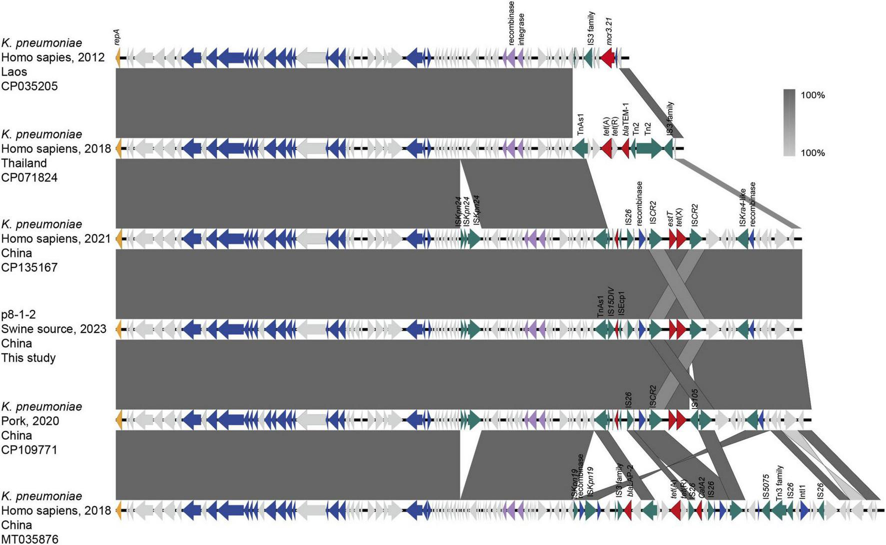
Linear sequence comparison of pSD8-1-2. Regions of homology are marked by shading. Arrows show the direction of transcription of open reading frames. The delta (Δ) symbol indicates a truncated gene. T4SS component, antimicrobial resistance, transposases, etc., are marked according to the colors in the legend to indicate their functions.
Given the dissemination of pSD8-1-2 and related plasmids in K. pneumoniae, their stability and transferability were further investigated. Stability tests showed that pSD8-1-2 was fully retained in the host strain SD8-1 after 10 days of serial subculturing without selective pressure. Plasmid loss began after 13 days, with 8% loss by day 20, increasing to 84% by day 24 (Figure 2a). This indicates this plasmid is highly stable and can persist for an extended period in the absence of antibiotic selective pressure in SD8-1. The conjugation experiments demonstrated that the plasmid can spread across bacterial species, successfully transferring into E. coli C600 and the clinical blaNDM-producing pathogenic strain E218, with conjugation frequencies of 1.6 × 10⁻⁵ and 4.3 × 10⁻⁶, respectively. Despite multiple attempts, pSD8-2-1 could not be transferred into the K. pneumoniae strains O41 and O32. After acquiring pSD8-1-2, the minimum inhibitory concentration (MIC) of the recipient strains for tigecycline increased significantly by 32-fold. To assess the potential fitness cost of pSD8-1-2 to the host strains, growth curves were constructed for all tested strains, which showed that pSD8-1-2 did not significant affect the logarithmic phase growth rate of all tested strains (E. coli E218, E. coli C600, and R. ornithinolytica SD8-1) (p > 0.05) (Figures 2b, c). However, area under the curve (AUC) analysis revealed a statistically significant reduction in total growth yield for E218 (p < 0.05), while no significant effect was observed for SD8-1 and C600 (p = 0.831) (Figure 2d).
FIGURE 2

Analysis of bacterial growth and plasmid stability. (a) Plasmid stability of pSD8-1-2 in SD8-1, assessed by the retention rate of the tet(X4) gene over 25 days in antibiotic-free LB broth, with subculturing every 12 h and PCR confirmation of 50 colonies daily. (b) Growth curves of E218, E218-p, ΔSD8-1, SD8-1, C600, and C600-p strains, measured as OD600 over 12 h in LB broth at 37 °C. (c) Growth rates (μ) of the strains during the logarithmic phase (2–4 h). (d) Area under the curve (AUC) of the growth curves, computed using the trapezoidal rule. statistical significance was determined by unpaired t-test (*p < 0.05; ns, not significant).
Effect of pSD8-1-2 on in vivo tigecycline efficacy
To assess the pathogenicity of R. ornithinolytica SD8-1 and the impact of pSD8-1-2 on the efficacy of tigecycline in vivo, a bloodstream infection model was established using BALB/c mice. Wild-type SD8-1 and plasmid-cured ΔSD8-1 strains (106 CFU) were injected into the mice via the tail vein. Two hours’ post-inoculation, tigecycline was administered intraperitoneally daily for 2 days. During treatment, 50% of mice (3/6) in the wild-type group died, whereas all mice in the ΔSD8-1 group survived. After 2 days, the surviving mice were euthanized, and their lung, liver, and kidney tissues were collected for hematoxylin-eosin (H&E) staining to examine histopathological changes. In the wild-type group, the pulmonary tissues exhibited thickened alveolar septa, neutrophil and mononuclear cell infiltration, and proteinaceous exudates, indicative of acute inflammation. In contrast, the ΔSD8-1 group showed markedly reduced inflammatory cell infiltration and interstitial thickening, with partial restoration of alveolar architecture (Figure 3a). Hepatic tissues from the wild-type group displayed focal hepatocellular necrosis, Kupffer cell activation, and sinusoidal dilation with inflammatory cell infiltration. In contrast, the ΔSD8-1 group exhibited fewer Kupffer cells, reduced inflammatory infiltration, and partial restoration of liver lobule structure (Figure 3b). Similarly, tubular epithelial cell swelling, vacuolar degeneration, cell detachment, interstitial inflammation, and vascular congestion were observed in the renal tissues in the wild-type group; these changes were significantly alleviated in the ΔSD8-1 group, with minimal tubular injury, reduced inflammatory edema, and restored tubular lumens (Figure 3c). These survival and histopathological findings suggest that the tet(X4)-harboring pSD8-1-2 contributes to tigecycline treatment failure in vivo.
FIGURE 3

Histopathological analysis of mouse tissues in a bloodstream infection model with tigecycline treatment. (a) Lung tissue sections stained with hematoxylin and eosin (HE) from control, SD8-1, and ASD8-1 groups, showing alveolar structure and inflammatory changes. (b) Liver tissue sections from the same groups, highlighting hepatic architecture and potential lesions. (c) Kidney tissue sections, demonstrating renal histology and pathological alterations. Scale bar = 100 μm.
Global distribution and resistance gene/replicon profiles of R. ornithinolytica
To investigate the prevalence and distribution of antibiotic resistance in R. ornithinolytica, we analyzed 545 non-duplicate R. ornithinolytica genome sequences from the NCBI database, isolated from 41 countries across six continents. The majority originated from the United States (45.5%, 248/545), China (17.2%, 94/545), the United Kingdom (5.9%, 32/545), Japan (3.9%, 21/545), and Canada (2.9%, 16/545), with 18.3% (100/545) from other countries and 6.2% (34/545) from unspecified sources (Figure 4a). Most strains were isolated from humans (n = 228), followed by food animals (n = 20), wild animals (n = 18), and the environment (n = 17) (Figure 4b). Approximately 40% of the genome sequences (n = 230) were submitted between 2019 and 2024 (Figure 4c). Bioinformatic analysis revealed that four antibiotic resistance genes (oqxA, oqxB, fosA, blaPLA) were present in over 98% of R. ornithinolytica strains, conferring resistance to quinolones (oqxAB), fosfomycin (fosA), and β-lactams (blaPLA). The incidence rate of the 5th to 10th most common resistance genes, blaOXA, blaTEM, aph(6)-Id, blaKPC, sul1, aph(3’)-Ia, ranged from 24% to 35%, which conferred resistance to β-lactams, aminoglycosides, and sulfonamides (Figure 4d). Other resistance genes, with incidence rates from 8.81% (mdfA) to 23.67% (dfrA14), conferred resistance to multiple antibiotic classes, including quinolones (qnrS1) and trimethoprim (dfrA14). Additionally, R. ornithinolytica strains occasionally carried the colistin resistance gene mcr and the tigecycline RND-type efflux pump gene tmexCD-toprJ. Notably, 20% of strains exhibited MDR, carrying eight or more ARGs. Comparison of the plasmid replicon types between R. ornithinolytica and K. pneumoniae revealed that R. ornithinolytica strains predominantly carried Col(pHAD28), IncFII(pKP91), IncFIB(K), Col440II, IncFIA(HI1), IncFII(pAR0022), and IncFII(p14) replicons, with incidence rates ranging from 8% to 20% (Figure 4e). This result highlights a high prevalence of IncFII-type plasmids in R. ornithinolytica strains, underscoring their role in antibiotic resistance dissemination.
FIGURE 4
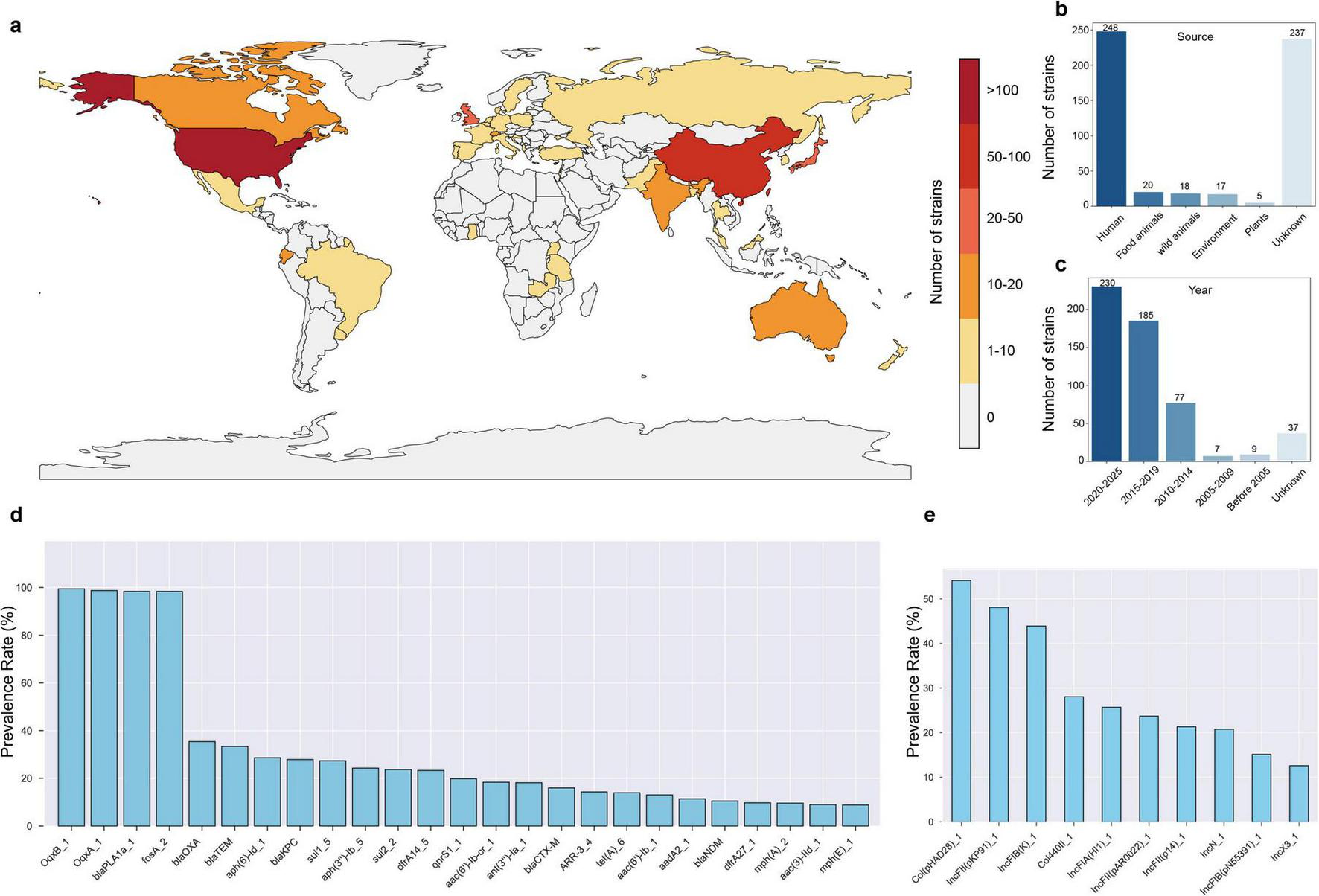
Global distribution and resistance profile of R. ornithinolytica strains. (a) World map showing the number of R. ornithinolytica strains isolated across 41 countries, colored by strain count (scale: 0 to > 100). (b) Source distribution of 545 strains. (c) Temporal distribution of genome submissions. (d) Carriage rates of the top 25 antibiotic resistance genes (ARGs). (e) Prevalence of the top 10 plasmid replicon types. Data are based on 545 non-duplicate genome sequences from the National Center for Biotechnology Information (NCBI) database, analyzed for antibiotic resistance genes and plasmid types.
Discussion
Livestock wastewater significantly contributes to the spread of resistant bacteria and ARGs from farms to the environment (Ibekwe et al., 2023; Zhu et al., 2020). Its abundant nutrients and residual antibiotics can increase the retention time of ARGs in the environment. The prevalence of ARGs, including those for β-lactamase (blaCTX–M), carbapenemases (blaNDM, blaKPC), and colistin resistance (mcr), is associated with the spread of their dominant plasmids (Bevan et al., 2017; Liu J. H. et al., 2024; Wu et al., 2019). Moreover, Alderliesten et al. (2020) found that conjugative plasmids can be transferred more efficiently between closely related species in a liquid environment. For example, compared with bacteria of the same species, plasmid conjugation frequency was reduced by about 0.37 times within the same family and 10 times within the same order (Alderliesten et al., 2020).
This study characterized the IncFII(pCRY)-like pSD8-1-2, carrying tet(X4), in R. ornithinolytica SD8-1. Using bioinformatic analysis, 84 pSD8-1-2-like plasmids or fragments were identified, predominantly in K. pneumoniae (80/84), across diverse ecological niches, including clinical human isolates, food animals, and meat products. These plasmids share a conserved backbone with pSD8-1-2, differing only in the variable regions harboring ARGs, such as tet(X4) within a conserved ISCR2-flanked cassette. Tigecycline is an important antibiotic for treating community- and hospital-acquired pneumonia, complicated intra-abdominal infections, and bloodstream infections caused by K. pneumoniae. It is typically used as a last-resort treatment for multidrug-resistant infections. Compared to its presence in bacteria like E. coli, the potential spread of tet(X4)-carrying pSD8-1-2 in K. pneumoniae poses a greater threat to clinical treatment, further limiting therapeutic options. The emergence of tet(X) genes in R. ornithinolytica suggests that the host range of tet(X) is expanding across species with the help of conjugative plasmids such as pSD8-1-2, which is exacerbated by the complex antibiotic environment in livestock wastewater.
pSD8-1-2 can transfer cross-species, successfully conjugating into E. coli C600 and clinical E218 with moderate efficiency (frequencies ranging from 1.6 × 10⁻⁵ to 4.3 × 10⁻⁶). Although the pSD8-1-2, with 100% coverage and 100% sequence identity, has been identified in the NCBI database (CP135167 and CP109771), our conjugation experiments failed to transfer pSD8-1-2 into the wild-type K. pneumoniae O41 and O32 strains, possibly due to interference from resident, incompatible IncFII-type plasmids in the recipient strains or the protective effects of cellular defense mechanisms such as restriction-modification and CRISPR–Cas systems (Siedentop et al., 2024; Womble et al., 1984). Stability assays revealed that pSD8-1-2 was retained in 92% of R. ornithinolytica SD8-1 after 20 days of serial passaging in an antibiotic-free environment. This high stability likely results from mechanisms such as partitioning systems, toxin-anti-toxin modules, and potential compensatory adaptations, which collectively ensure long-term plasmid maintenance. Notably, pSD8-1-2 imposed minimal metabolic burden, as it did not significantly affect the logarithmic growth phase rate across the tested strains (E. coli E218, E. coli C600, and R. ornithinolytica SD8-1) (p > 0.05). However, the total growth yield was slightly reduced in E. coli E218 (p < 0.05), suggesting a minor fitness cost. In contrast, R. ornithinolytica SD8-1 and E. coli C600 showed no significant reduction (p = 0.831), indicating that these strains were better adapted to hosting the plasmid, possibly due to optimized plasmid-host interactions. These results are consistent with the IncFII plasmid competition studies in K. pneumoniae (Li et al., 2021), underscoring the potential for widespread dissemination of pSD8-1-2 in diverse bacterial populations. Furthermore, this study confirmed the pathogenicity of R. ornithinolytica SD8-1 using an in vivo infection model and further demonstrated that pSD8-1-2 caused the failure of tigecycline treatment. Compared with the original SD8-1 group, the plasmid elimination group showed significant improvement in pathological signs.
In pSD8-1-2, tet(X4) is flanked by two 1,494-bp ISCR2 insertion sequences in the same orientation, forming the conserved ISCR2-aph-tet(X4)-ISCR2 structure. This structure, prevalent in E. coli, is capable of generating a ∼5 kb transposon intermediate that enhances tet(X4) mobility through horizontal gene transfer. ISCR2, the primary transposable element associated with tet(X), has been linked to all newly reported tet(X) variants, such as tet(X3) to tet(X7). Our recent studies have shown that in addition to assisting the spread of tet(X), ISCR2 also contributes to the recombination between tet(X) variants, promoting the formation of several variants, including tet(X5) (Jin et al., 2024). In addition to tet(X), genomic analysis also revealed that ISCR2 is associated with resistance to aminoglycosides, chloramphenicol, sulfonamides, and β-lactams (Che et al., 2021). These findings suggest that tet(X) may form an MDR cluster with these resistance genes through ISCR2, thereby creating favorable conditions for host bacteria to adapt to the complex antibiotic environment in farm wastewater.
Analysis of 545 non-duplicate R. ornithinolytica genome sequences from public databases highlighted their global prevalence and AMR profiles, providing insights into their epidemiological patterns and resistance mechanisms. These strains span 41 countries across six continents, with the United States (45.5%, n = 248) and China (17.2%, n = 94) being the primary contributors, likely reflecting differences in genome sequencing capacity, data submission frequency, and regional prevalence. The increased documentation of R. ornithinolytica strains from 2019 to 2024 (n = 230) corresponds with the advancements in sequencing technologies and enhanced data sharing. Host analysis revealed that humans are the primary source (n = 228), followed by food animals (n = 20), wild animals (n = 18), and environmental samples (n = 17), indicating R. ornithinolytica can transmit across the human-animal-environment interface, consistent with the “One Health” framework. The presence of environmental isolates suggests that the environment may serve as a reservoir and pathway for antibiotic-resistant bacteria, necessitating further investigation into its potential contribution to the transmission of ARGs. Multidrug-resistant phenotypes were also discovered in R. ornithinolytica, with high prevalence of carbapenem resistance genes, including blaKPC (n = 152, 27.9%) and blaOXA (n = 193, 35.4%), suggesting reduced efficacy of carbapenems. Additionally, R. ornithinolytica and K. pneumoniae were found to share highly prevalent plasmid replicon types, specifically IncFII(pKP91) and IncFIB(K), which are highly prevalent in both species (ranging from 40% to 50% in both) (Wyres et al., 2020). This shared replicon profile suggests that cross-species dissemination of resistance plasmids might occur between these two bacterial species.
Currently, the low prevalence of tet(X) genes in K. pneumoniae presents a critical window to curb their spread among clinically significant pathogens, such as carbapenem-resistant K. pneumoniae (CRKP) and hypervirulent K. pneumoniae (hvKP). Adopting a “One Health” approach that integrates human, animal, and environmental health, alongside coordinated cross-sectoral strategies, can significantly help mitigate this risk. In 2020, China banned the use of antibiotic growth promoters in animal feed (Ministry of Agriculture and Rural Affairs of the People’s Republic of China, 2020), markedly reducing the transmission of animal-derived antibiotic resistance to humans, representing a notable step forward in resistance control. However, continued use of tetracyclines, which remain the primary treatment options for bacterial infections in animal husbandry due to their affordability and broad-spectrum efficacy, may exert selection pressure. This might promote the spread of resistance genes, including tet(X) (Aminov, 2021), particularly through shared resistance plasmids between food animals and clinical isolates. Consequently, stricter regulations should be implemented on the non-essential use of older tetracyclines in agriculture to preserve the efficacy of tigecycline and next-generation tetracyclines, such as eravacycline and omadacycline. Furthermore, enhancing molecular surveillance of resistance genes, refining antibiotic stewardship guidelines, and promoting international collaboration in antibiotic resistance management can significantly benefit effective control (De Oliveira et al., 2020). These combined efforts are necessary to limit the spread of tet(X) and support the long-term sustainability of antibiotic therapies.
Statements
Data availability statement
All genomes have been deposited in GenBank under the BioProject PRJNA1255831.
Ethics statement
The animal study was approved by the Experimental Animal Ethics Committee of Wenzhou Medical University. The study was conducted in accordance with the local legislation and institutional requirements.
Author contributions
QJ: Writing – review & editing, Formal analysis, Data curation. HJ: Formal analysis, Data curation, Writing – review & editing. XJ: Resources, Writing – review & editing. XZ: Data curation, Writing – review & editing. CC: Supervision, Writing – review & editing, Funding acquisition, Writing – original draft.
Funding
The author(s) declare that financial support was received for the research and/or publication of this article. This work was supported by the Young Scientists Fund of the National Natural Science Foundation of China (Grant No. 32302927), and the Basic Public Welfare Research Project of Wenzhou Science and Technology Bureau (Grant No. 2023Y1770).
Conflict of interest
The authors. declare that the research was conducted in the absence of any commercial or financial relationships that could be construed as a potential conflict of interest.
Generative AI statement
The authors declare that no Generative AI was used in the creation of this manuscript.
Any alternative text (alt text) provided alongside figures in this article has been generated by Frontiers with the support of artificial intelligence and reasonable efforts have been made to ensure accuracy, including review by the authors wherever possible. If you identify any issues, please contact us.
Publisher’s note
All claims expressed in this article are solely those of the authors and do not necessarily represent those of their affiliated organizations, or those of the publisher, the editors and the reviewers. Any product that may be evaluated in this article, or claim that may be made by its manufacturer, is not guaranteed or endorsed by the publisher.
Supplementary material
The Supplementary Material for this article can be found online at: https://www.frontiersin.org/articles/10.3389/fmicb.2025.1642708/full#supplementary-material
References
1
Alderliesten J. B. Duxbury S. J. N. Zwart M. P. de Visser J. Stegeman A. Fischer E. A. J. (2020). Effect of donor-recipient relatedness on the plasmid conjugation frequency: A meta analysis.BMC Microbiol.20:135. 10.1186/s12866-020-01825-4
2
Aminov R. (2021). Acquisition and spread of antimicrobial resistance: A tet(X) case study.Int. J. Mol. Sci.22:3905. 10.3390/ijms22083905
3
Bevan E. R. Jones A. M. Hawkey P. M. (2017). Global epidemiology of CTX-M beta-lactamases: Temporal and geographical shifts in genotype.J. Antimicrob. Chemother.722145–2155. 10.1093/jac/dkx146
4
Che Y. Yang Y. Xu X. Brinda K. Polz M. F. Hanage W. P. et al (2021). Conjugative plasmids interact with insertion sequences to shape the horizontal transfer of antimicrobial resistance genes.Proc. Natl. Acad. Sci. U S A.118:e2008731118. 10.1073/pnas.2008731118
5
Chu Y. Ruan Y. X. Liu J. Q. Zhang Y. Wang M. G. Liao X. P. et al (2024). Population genomics of emerging multidrug-resistant Salmonella derby from pork and human in Guangdong, China.Lwt-Food Sci. Technol.205:116535. 10.1016/j.lwt.2024.116535
6
Cui C. Y. Li X. J. Chen C. Wu X. T. He Q. Jia Q. L. et al (2022). Comprehensive analysis of plasmid-mediated tet(X4)-positive Escherichia coli isolates from clinical settings revealed a high correlation with animals and environments-derived strains.Sci. Total Environ.806:150687. 10.1016/j.scitotenv.2021.150687
7
Cui C. Chen C. Liu B. He Q. Wu X. Sun R. et al (2020). Co-occurrence of plasmid-mediated tigecycline and carbapenem resistance in Acinetobacter spp. from waterfowls and their neighboring environment.Antimicrob. Agents Ch.64:e02502-19. 10.1128/AAC.02502-19
8
De Oliveira D. M. P. Forde B. M. Kidd T. J. Harris P. N. A. Schembri M. A. Beatson S. A. et al (2020). Antimicrobial resistance in ESKAPE pathogens. Clin. Microbiol. Rev.33:e00181-19. 10.1128/CMR.00181-19
9
Drancourt M. Bollet C. Carta A. Rousselier P. (2001). Phylogenetic analyses of Klebsiella species delineate Klebsiella and Raoultella gen. nov., with description of Raoultella ornithinolytica comb. nov., Raoultella terrigena comb. nov. and Raoultella planticola comb. nov.Int. J. Syst. Evol. Microbiol.51925–932. 10.1099/00207713-51-3-925
10
European Committee on Antimicrobial Susceptibility Testing (EUCAST). (2025). Breakpoint tables for interpretation of MICs and zone diameters. Version 15.0. Available online at: https://www.eucast.org/fileadmin/src/media/PDFs/EUCAST_files/Breakpoint_tables/v_15.0_Breakpoint_Tables.pdf
11
Fan X. Y. Jiang Y. Wu H. Liu J. Gu Q. Y. Wang Z. Y. et al (2024). Distribution and spread of tigecycline resistance gene tet(X4) in Escherichia coli from different sources.Front. Cell. Infect. Microbiol.14:1399732. 10.3389/fcimb.2024.1399732
12
Fang L. X. Chen C. Cui C. Y. Li X. P. Zhang Y. Liao X. P. et al (2020). Emerging high-level tigecycline resistance: Novel tetracycline destructases spread via the mobile Tet(X).Bioessays42:e2000014. 10.1002/bies.202000014
13
Gupta S. Arango-Argoty G. Zhang L. Pruden A. Vikesland P. (2019). Identification of discriminatory antibiotic resistance genes among environmental resistomes using extremely randomized tree algorithm.Microbiome7:123. 10.1002/bies.202000014
14
Hajjar R. Ambaraghassi G. Sebajang H. Schwenter F. Su S. H. (2020). Raoultella ornithinolytica: Emergence and resistance.Infect. Drug Resist.131091–1104. 10.2147/IDR.S191387
15
He T. Wang R. Liu D. Walsh T. R. Zhang R. Lv Y. et al (2019). Emergence of plasmid-mediated high-level tigecycline resistance genes in animals and humans.Nat. Microbiol.41450–1456. 10.1038/s41564-019-0445-2
16
Holvoet K. Sampers I. Callens B. Dewulf J. Uyttendaele M. (2013). Moderate prevalence of antimicrobial resistance in Escherichia coli isolates from lettuce, irrigation water, and soil.Appl. Environ. Microbiol.796677–6683. 10.1128/AEM.01995-13
17
Humphries R. M. Ambler J. Mitchell S. L. Castanheira M. Dingle T. Hindler J. A. et al (2018). CLSI methods development and standardization working group best practices for evaluation of antimicrobial susceptibility tests.J. Clin. Microbiol.56:e01934-17. 10.1128/JCM.01934-17
18
Ibekwe A. M. Bhattacharjee A. S. Phan D. Ashworth D. Schmidt M. P. Murinda S. E. et al (2023). Potential reservoirs of antimicrobial resistance in livestock waste and treated wastewater that can be disseminated to agricultural land.Sci. Total Environ.872:162194. 10.1016/j.scitotenv.2023.162194
19
Jin H. Jia Q. Jin X. Zhu X. Wang M. G. Sun R. Y. et al (2024). Identification of novel Tet(X6)-Tet(X2) recombinant variant in Elizabethkingia meningoseptica from a bullfrog farm and downstream river in China.Front. Microbiol.15:1453801. 10.3389/fmicb.2024.1453801
20
Jones D. T. Srinivasmurthy R. Pandit M. Tovar R. Ho L. Wairimu K. (2024). Raoultella ornithinolytica urinary tract infection in a patient with triple-negative breast cancer.Cureus16:e64742. 10.7759/cureus.64742
21
Li R. Li Y. Peng K. Yin Y. Liu Y. He T. et al (2021). Comprehensive genomic investigation of tigecycline resistance gene tet(X4)-bearing strains expanding among different settings.Microbiol. Spectr.9:e0163321. 10.1128/spectrum.01633-21
22
Li R. Lu X. Peng K. Liu Z. Li Y. Liu Y. et al (2020). Deciphering the structural diversity and classification of the mobile tigecycline resistance gene tet(X)-Bearing Plasmidome among Bacteria.mSystems5:e00134-20. 10.1128/mSystems.00134-20
23
Liu J. H. Liu Y. Y. Shen Y. B. Yang J. Walsh T. R. Wang Y. et al (2024). Plasmid-mediated colistin-resistance genes: mcr.Trends Microbiol.32365–378. 10.1016/j.tim.2023.10.006
24
Liu M. Li X. Xie Y. Bi D. Sun J. Li J. et al (2019). ICEberg 2.0: An updated database of bacterial integrative and conjugative elements.Nucleic Acids Res.47D660–D665. 10.1093/nar/gky1123
25
Liu X. Liu Y. Ma X. Chen R. Li C. Fu H. et al (2024). Emergence of plasmid-borne tet(X4) resistance gene in clinical isolate of eravacycline- and omadacycline-resistant Klebsiella pneumoniae ST485.Microbiol. Spectr.12:e0049624. 10.1128/spectrum.00496-24
26
Ministry of Agriculture and Rural Affairs of the People’s Republic of China. (2020). Announcement No. 194 of the ministry of agriculture and rural affairs of the People’s Republic of China. Available online at: https://www.moa.gov.cn/nybgb/2019/201907/202001/t20200103_6334292.htm
27
Okeke I. N. de Kraker M. E. A. Van Boeckel T. P. Kumar C. K. Schmitt H. Gales A. C. et al (2024). The scope of the antimicrobial resistance challenge.Lancet4032426–2438. 10.1016/S0140-6736(24)00876-6
28
Onalenna O. Rahube T. O. (2022). Assessing bacterial diversity and antibiotic resistance dynamics in wastewater effluent-irrigated soil and vegetables in a microcosm setting.Heliyon8:e09089. 10.1016/j.heliyon.2022.e09089
29
Siedentop B. Losa Mediavilla C. Kouyos R. D. Bonhoeffer S. Chabas H. (2024). Assessing the role of bacterial innate and adaptive immunity as barriers to conjugative plasmids.Mol. Biol. Evol.41:msae207. 10.1093/molbev/msae207
30
Sun J. Chen C. Cui C. Y. Zhang Y. Liu X. Cui Z. H. et al (2019). Plasmid-encoded tet(X) genes that confer highlevel tigecycline resistance in Escherichia coli.Nat. Microbiol.41457–1464. 10.1038/s41564-019-0496-4
31
Sun R. Y. Fang L. X. Ke B. X. Sun J. Wu Z. W. Feng Y. J. et al (2023). Carriage and transmission of mcr-1 in Salmonella Typhimurium and its monophasic 1,4,[5],12:i:- variants from diarrheal outpatients: A 10-year genomic epidemiology in guangdong, Southern China.Microbiol. Spectr.11:e0311922. 10.1128/spectrum.03119-22
32
Wang Y. Xu C. Zhang R. Chen Y. Shen Y. Hu F. et al (2020). Changes in colistin resistance and mcr-1 abundance in Escherichia coli of animal and human origins following the ban of colistin-positive additives in China: An epidemiological comparative study.Lancet Infect. Dis.201161–1171. 10.1128/spectrum.03119-22
33
Womble D. D. Dong X. Wu R. P. Luckow V. A. Martinez A. F. Rownd R. H. (1984). IncFII plasmid incompatibility product and its target are both RNA transcripts. J. Bacteriol.160, 28—35. 10.1128/jb.160.1.28-35.1984
34
Wu W. Feng Y. Tang G. Qiao F. McNally A. Zong Z. (2019). NDM metallo-beta-lactamases and their bacterial producers in health care settings.Clin. Microbiol. Rev.32:e00115-18. 10.1128/CMR.00115-18
35
Wyres K. L. Lam M. M. C. Holt K. E. (2020). Population genomics of Klebsiella pneumoniae.Nat. Rev. Microbiol.18344–359. 10.1038/s41579-019-0315-1
36
Xedzro C. Shimamoto T. Yu L. Zuo H. Sugawara Y. Sugai M. et al (2023). Emergence of colistin-resistant Enterobacter cloacae and Raoultella ornithinolytica carrying the phosphoethanolamine transferase gene, mcr-9, derived from vegetables in Japan.Microbiol. Spectr.11:e0106323. 10.1128/spectrum.01063-23
37
Yue C. Bai Y. Li T. Deng H. Lu L. Lin W. et al (2024). Emergence of tet(X4)-positive Enterobacterales in retail eggs and the widespread of IncFIA(HI1)-HI1A-HI1B(R27) plasmids carrying tet(X4).Int. J. Food Microbiol.414:110574. 10.1016/j.ijfoodmicro.2024.110574
38
Zhang H. Chen W. Lu X. Liang Y. Quan X. Liu X. et al (2024). Emergence and characterization of the high-level tigecycline resistance gene tet(X4) in Salmonella enterica serovar rissen from food in China.Foodborne Pathog. Dis.22405–413. 10.1089/fpd.2024.0101
39
Zhang R. Dong N. Shen Z. Zeng Y. Lu J. Liu C. et al (2020). Epidemiological and phylogenetic analysis reveals Flavobacteriaceae as potential ancestral source of tigecycline resistance gene tet(X).Nat. Commun.11:4648. 10.1038/s41467-020-18475-9
40
Zhu J. Lv J. Zhu Z. Wang T. Xie X. Zhang H. et al (2023). Identification of TMexCD-TOprJ-producing carbapenem-resistant Gram-negative bacteria from hospital sewage.Drug Resist. Updat.70:100989. 10.1016/j.drup.2023.100989
41
Zhu N. Jin H. Ye X. Liu W. Li D. Shah G. M. et al (2020). Fate and driving factors of antibiotic resistance genes in an integrated swine wastewater treatment system: From wastewater to soil.Sci. Total Environ.721:137654. 10.1016/j.scitotenv.2020.137654
Summary
Keywords
: tet(X), Raoultella ornithinolytica , IncFII plasmid, Klebsiella pneumoniae , tigecycline
Citation
Jia Q, Jin H, Jin X, Zhu X and Cui C (2025) Emergence of tigecycline-resistant Raoultella ornithinolytica with tet(X)-carrying plasmid from swine wastewater in China. Front. Microbiol. 16:1642708. doi: 10.3389/fmicb.2025.1642708
Received
09 June 2025
Accepted
03 September 2025
Published
16 September 2025
Volume
16 - 2025
Edited by
Govindan Rajamohan, Institute of Microbial Technology (CSIR), India
Reviewed by
Dexi Li, Henan Agricultural University, China
Lin Sun, Yangzhou University, China
Updates
Copyright
© 2025 Jia, Jin, Jin, Zhu and Cui.
This is an open-access article distributed under the terms of the Creative Commons Attribution License (CC BY). The use, distribution or reproduction in other forums is permitted, provided the original author(s) and the copyright owner(s) are credited and that the original publication in this journal is cited, in accordance with accepted academic practice. No use, distribution or reproduction is permitted which does not comply with these terms.
*Correspondence: Chaoyue Cui, chaoyue_cui@wmu.edu.cn
†These authors have contributed equally to this work and share senior authorship
Disclaimer
All claims expressed in this article are solely those of the authors and do not necessarily represent those of their affiliated organizations, or those of the publisher, the editors and the reviewers. Any product that may be evaluated in this article or claim that may be made by its manufacturer is not guaranteed or endorsed by the publisher.