 Roger Morales-Romero1
 María S. Gonzalez-Dominguez2,3
 Jorge Sánchez4
 Nathalia M. Correa-Valencia5
 Juan G. Maldonado-Estrada1,6*
- 1OHVRI Research Group, Facultad de Ciencias Agrarias, Universidad de Antioquia (UdeA), Medellín, Antioquia, Colombia
- 2Grupo de Investigacion INCA-CES, Facultad de Medicina Veterinaria y Zootecnia, Universidad CES, Medellin, Colombia
- 3Bivett Centro Veterinario, Medellin, Colombia
- 4Group of Clinical and Experimental Allergy, Hospital Alma Mater de Antioquia, University of Antioquia, Medellín, Antioquia, Colombia
- 5CENTAURO Research Group, Facultad de Ciencias Agrarias, Universidad de Antioquia (UdeA), Medellín, Antioquia, Colombia
- 6Hospital Veterinario, Facultad de Ciencias Agrarias, Universidad de Antioquia (UdeA), Medellín, Colombia
Introduction: Canine atopic dermatitis (CAD) is an important cause of quality of life in dogs and their owners. There are different diagnostic tools to evaluate sensitization to allergens in CAD; however, there is little information to evaluate the clinical usefulness of these tests.
Methods: A systematic review aiming to assess the accuracy of allergen sensitization tests in CAD patients was performed. The search was planned, executed, and reported following PRISMA standards. The OVID®/MEDLINE, PubMed®, SciELO, and Redalyc databases were searched to find relevant studies comprising “diagnostic method” (OR test OR diagnosis OR method OR diagnostic OR paraclinic=) AND (atopic = OR hypersensitivity* OR allergen* OR “allergic reaction*” OR atopy) AND (dermatitis OR eczema OR scratching OR prurit = OR erythema OR rash OR edema) AND (canine OR dog* OR bitch* OR pupp*) search. Defined relevant articles were systematized, and content was analyzed via Atlas TI Scientific Software Development Software.
Results: The groups of diagnostic tests included the intradermal test (IDT), serologic-specific allergen test (SAT), skin prick test (SPT), and patch test. Combining the results from all the search engines and deduplicate elimination, yielded 928 eligible citations published between 1963 and 2024, and the 72 articles that met the eligibility criteria were included in the qualitative synthesis evaluating SAT (n = 36), IDT (n = 37), SPT (n = 2), and patch tests (n = 1) reporting the use of 136 different allergens. Favrot’s clinical criteria were applied in 41.6% of the studies (30/72), with no previous consensus on the case definition for CAD.
Discussion: The results of the review indicate that there is little information available to establish the diagnostic performance of the tests, which makes it difficult to make a recommendation regarding their use. In this systematic review they identified gaps in current knowledge that suggest the need for future research to standardize allergenic extracts, define cutoff points in serological tests, and consider environmental, geographic, and demographic variables. These findings provide a solid basis for improving the diagnosis and management of CAD and guiding future research in this field. Further studies are needed to adequately establish the diagnostic performance of the tests and their actual clinical usefulness.
Introduction
The skin, the largest and most visible body organ, is a significant concern in veterinary consultation. Dermatopathies account for nearly 30% of general consultations in dogs (1). These conditions are often frustrating for both veterinarians and pet owners, as they impact an animal’s quality of life and appearance, which are particularly important to pet owners (2). Pruritus is the primary reason for dermatological consultations in dogs, with allergic conditions such as flea allergy dermatitis, food hypersensitivity, and atopic dermatitis being the most prevalent underlying causes (1). Pruritus (3) and several behavioral problems (4) account for the most critical findings affecting the quality of life of canine atopic dermatitis (CAD) patients.
Atopic dermatitis is a common condition in both humans (called AD) (5) and dogs (called CAD) (6, 7), with an estimated prevalence of 10–15% and a high tendency for relapse (8). This disease is defined as a complex, multifactorial inflammatory syndrome in which the skin is the primary exposure route. While similar and divergent pathophysiological mechanisms have been identified in humans and canines, dogs are frequently used as experimental models for studying human AD (9).
The primary underlying cause of AD appears to be a type-1 hypersensitivity reaction driven by mechanisms mediated by IgE. This mechanism is observed in humans (5) and 40–90% of canine cases (7, 10). Inflammation is triggered by normally innocuous environmental proteins and antigens, commonly called allergens (6, 8). An allergen triggers an exaggerated inflammatory response in susceptible individuals. This response leads to the production of IgE antibodies and the release of proinflammatory mediators, resulting in allergic symptoms such as itching, hives, and respiratory difficulties. Allergens are classified on the basis of similarities in their molecular structure and allergenic potential (11).
Reactivity to common epitopes grouped in allergen mixtures used in dermabrasion test extracts has been proposed. However, variations in the concentrations of individual allergens and their fractions in most allergen identification tests raise concerns about the reliability of these mixtures for diagnostic purposes and for the formulation of allergen-specific immunotherapies (12, 13). Co-sensitization can occur when an individual is sensitized to multiple allergens simultaneously, usually through cross-reactivity. This phenomenon arises when the immune system recognizes similar protein structures in related or unrelated allergens, provoking an inflammatory response.
The diagnosis of CAD is predominantly based on the widely recognized Favrot’s clinical criteria (14). These consist of two set of findings: Set 1: (i) Affected ear pinnae. (ii) Affected front feet. (iii) Age at onset <3 years. (iv) Chronic or recurrent infections (Mainly related to Malassezia yeast, and Staphylococcus pseudintermedius). (v) Corticosteroid-responsive pruritus. (vi) Mostly indoor. (vii) Non-affected dorso-lumbar area. And (viii) Non-affected ear margins. Set 2: (i) Affected front feet. (ii) Affected ear pinnae. (iii) Age at onset <3 years. (iv) Mostly indoor. (v) Non-affected dorso-lumbar area. (vi) Non-affected ear margins. And (vii) Pruritus sine material at onset (14).
This diagnostic approach requires meeting at least 5 out of 8 specified criteria (2), providing a sensitivity of 85% and a specificity of approximately 80% for identifying the syndrome. Notably, no significant predisposition has been reported regarding breed, age, or sex (15). Accordingly, the Favrot’s criteria are critical but not exclusive for CAD definitive diagnosis, for which Anamnesis, the CADESI score, and Pruritus Visual Assessment Score (PVAS), must be considered. Finally, diagnostic test must provide data to the most probable allergen the dog is allergic to.
Diagnostic tests for AD include methods such as dermabrasion to the epidermal level (prick test—SPT) or the dermal layer (intradermal skin test—IDT), which use a standardized panel of allergens to evaluate the patient’s reactivity against them (16). In dogs, IDT is the test of choice (17), whereas in humans, SPT is preferred because of its lower cost, faster interpretation, greater safety, higher specificity, and reduced pain (18). The serologic-specific allergen test (SAT), which measures specific IgE levels in the blood in response to common allergens, has also been documented and evaluated for CAD diagnosis. However, the results have been inconsistent, and there is no consensus on its reliability (17, 19). Another approach, the patch test, involves the epicutaneous application of allergens to assess cellular hypersensitivity to food and environmental allergens. This test aims to replicate the immune changes observed in natural lesions. However, its results remain controversial, particularly for food allergens (20, 21).
These diagnostic techniques have the potential to be useful for (1) supporting the clinical diagnosis of CDA, (2) establishing environmental restriction measures, (3) selecting extracts for allergen-specific immunotherapy, and (4) determining the prognosis of clinical control or remission. However, it is necessary to define the diagnostic performance of the tests for each outcome to establish their clinical utility (e.g., sensitivity, specificity, predictive values, and kappa index). Considering these challenges, our objective was to systematically compile and analyze the existing evidence from studies investigating the allergens utilized in CAD diagnosis, aiming to provide clarity and insights into this complex diagnostic process.
Methods
This systematic review was planned, executed, and reported in accordance with PRISMA standards (22). The research question, methodology for conducting literature searches, study inclusion/exclusion criteria, and checklists for relevance screening, baseline characterization, methodological assessment, and data extraction from relevant primary research were all conducted on the basis of a preestablished and pretested protocol.
Search strategy
Our goal was to evaluate the diagnostic performance of the available tests for CAD. The initial search took place on June 11, 2024. Four search databases (i.e., OVID®/MEDLINE, PubMed®, SciELO, and Redalyc) were searched. The topic was divided into components, and the search terms used to find relevant studies on the platforms were (“diagnostic method” OR test OR diagnosis OR method OR diagnostic OR paraclinic=) AND (atopic = OR hypersensitivity* OR allergen* OR “allergic reaction*” OR atopy) AND (dermatitis OR eczema OR scratching OR pruritus = OR erythema OR rash OR edema) AND (canine OR dog* OR bitch* OR pupp*).
Eligibility screening
The inclusion criteria were limited to original articles published in peer-reviewed journals and written in English, Portuguese, French, or Spanish. The publication year and country of origin were not restrictive factors. The initial citation selection process involved evaluating the titles by two authors (RMR and MSGD), who selected citations on the basis of their potential relevance to the study topic. Two authors subsequently screened the list of citations chosen on the basis of their abstracts, following the inclusion and exclusion criteria established during the title screening phase. Afterward, two authors thoroughly reviewed the full texts of the remaining citations to ensure that they contained relevant data to address the research question. Kappa coefficients were calculated for each selection stage to assess agreement. Detailed scrutiny of each full text’s materials, methods, and results sections was conducted, with any conflicts resolved through author consensus. The World Association of Veterinary Dermatologists (WAVD) proceedings from 1989 to 2020 were available on its website.1 These proceedings were manually examined for any published primary studies. Additionally, as a final step, two authors manually searched the references cited in the pertinent articles identified during full-text screening, a process commonly referred to as snowballing, to uncover additional published sources.
To ensure eligibility, the articles defined as relevant were systematized, resulting in emerging categories and subcategories. A content analysis was developed via the Software Atlas TI Scientific Software Development GmbH (ATLAS.ti 24 Windows, 2022–2024). Subsequently, groups of diagnostic tests (i.e., IDT, SAT, SPT, and patch test) were assessed through an intentional coding analysis. Three recent articles were selected at the researchers’ discretion (23–25) to search for the most frequent concepts that would be considered trends. From there, the discriminated codes were obtained and served as a basis for the comprehensive review of the relevant articles. After all applicable publications were compiled, a descriptive summary was provided that considered the information of interest by groups of diagnostic tests.
Risk of bias assessment
To assess the methodological quality of the included studies, a qualitative risk of bias evaluation was performed based on adapted domains from the QUADAS-2 tool, which is specifically designed for studies of diagnostic accuracy. The domains considered were: patient selection, index test, reference standard, and flow and timing. Two reviewers independently evaluated each study for potential sources of bias, and discrepancies were resolved by consensus.
Results
The bias assessment revealed a high risk of bias in several domains across the included studies. Most studies (68/72) did not clearly define a reference standard for confirming CAD, increasing the likelihood of misclassification bias. Approximately 70% of the studies did not report blinding of test interpreters, leading to potential detection bias. Additionally, considerable variability in allergen concentrations and test protocols contributed to methodological heterogeneity. Only 41.6% (30/72) of studies applied Favrot’s clinical criteria, raising concerns regarding case definition and applicability. These findings suggest a moderate to high risk of bias in the body of evidence, which should be considered when interpreting the diagnostic value of allergen tests for CAD.
The electronic search, which combines results from all search engines and, after deduplication, yielded 928 eligible citations possibly associated with the subject of this systematic review. The citations to be reviewed were published between 1963 and 2024. After the titles were read, 641 were considered unrelated (agreed upon by two reviewing authors). The final number of citations by title screening was 287 (retained by at least one reviewer). After the abstracts of the articles were read, 145 were excluded (by both authors), and 142 original articles remained for the full-text review. Sixty articles were reviewed in full text and kept for data extraction after 82 articles were dismissed during this phase. The snowballing strategy was applied through the reference lists of the 59 definitive articles, and 90 citations were retained after title screening. After the abstracts were screened, 26 studies were retained. The final selection of articles from the snowballing method yielded 11 results. In addition, two more articles were detected through the proceedings of the WAVD. The final number of articles that met the eligibility criteria and were included in the qualitative synthesis was 72. The file with the systematic process of collecting and selecting citations is available as Supplementary material (SM1). Figure 1 describes the protocol and the selection of relevant articles. All the articles were written in English. Studies were performed in the United States (n = 11), the United Kingdom, Japan, Brazil (n = 6, each), the Netherlands, Korea, Thailand (n = 4, each), Spain, Germany (n = 3, each), Austria, Norway, Poland (n = 2, each), Australia, China, Colombia, France and Italy (n = 1, each).
Figure 1. Preferred Reporting Items for Systematic Reviews and Meta-analyses (PRISMA) flow chart (22), which describes the progress of the citations through the systematic review. OA, Open Access.
The first relevant article was published in 1982, and the most recent article was published in 2023. Citations were published in 31 journals and 18 countries, of which only three were nonseasonal countries (e.g., Brazil, Colombia, and Thailand). The results for the SAT, IDT, SPT, and patch tests corresponded to 36, 37, 2, and a single relevant study, respectively, reporting the use of 136 different allergens and considering relevant articles that addressed two or more diagnostic tests for CAD.
For SAT reports, 19 out of 36 articles (52.7%) reported positive reaction values in optical densities of variable cutoff points; 4 out of 36 (11.1%) reported in ELISA Units; 3 out of 36 (8.3%) used Top Screen and Immunodot values; and 10 out of 36 (24.7%) reported not reaction units (Table 1). For IDT reports, a high variability of cutoff point to define positive reactions was found, with a single report out of 37 (2.7%) reporting not positive reaction definitions (Table 2). Finally, for SPT reports, the two reports found reported different positive cutoff procedures and scales (Table 3).
Table 1. The study features when a serologic specific allergen test (SAT) was considered in the diagnosis of canine atopic dermatitis (n = 36).
Table 2. The study features when an intradermal skin test (IDT) was considered in the diagnosis of canine atopic dermatitis (n = 37).
Table 3. The studies featured when the prick test (SPT) was considered in the diagnosis of canine atopic dermatitis (n = 2).
Tables 1–4 display the information extracted from the four diagnostic test groups. Since 2010, Favrot’s clinical criteria (14) have been applied in 41.6% of studies (30/72); before this report, there was no consensus on the case definition for CAD. The five most represented breeds included mixed, Labrador, German Shepperd, Shih Tzu, and Boxer. However, the breed of almost 500 out of 2,096 dogs was not specified. All remaining and well-known dog breeds are represented in Figure 2. Table 1 presents a detailed feature of selected articles reporting a serologic-specific allergen test (SAT) for CAD diagnosis. The Sensitivity, Specificity and Positive or Negative predictive comparison values between studies that met the criteria for these analyses are presented in Table 5. Finally, the available data for allergen prevalence according to allergen reactivity of CAD patients against mites, insects, trees, and molds, is presented in Table 6.
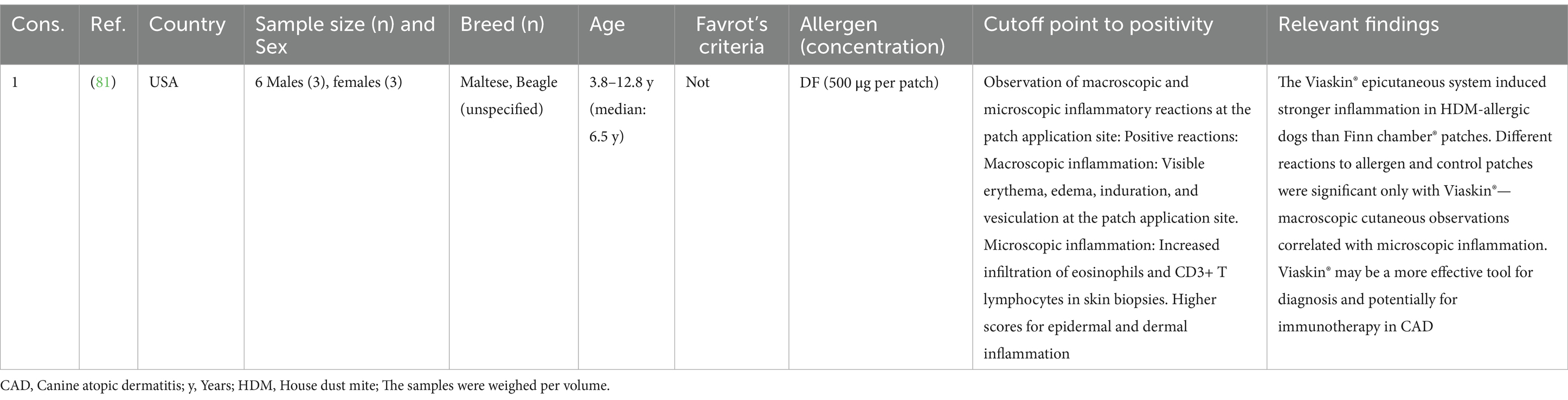
Table 4. The patch test was used for the diagnosis of canine atopic dermatitis (n = 1).
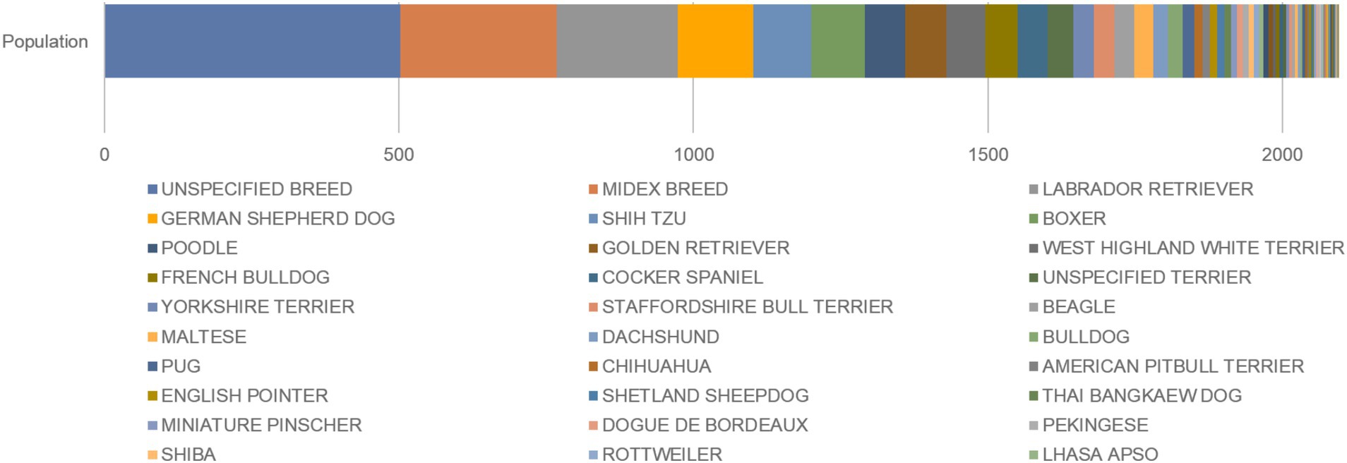
Figure 2. Distribution of dogs in the selected studies according to breed. Nomenclature according to the Fédération Cynologique Internationale (https://www.fci.be/en/nomenclature).
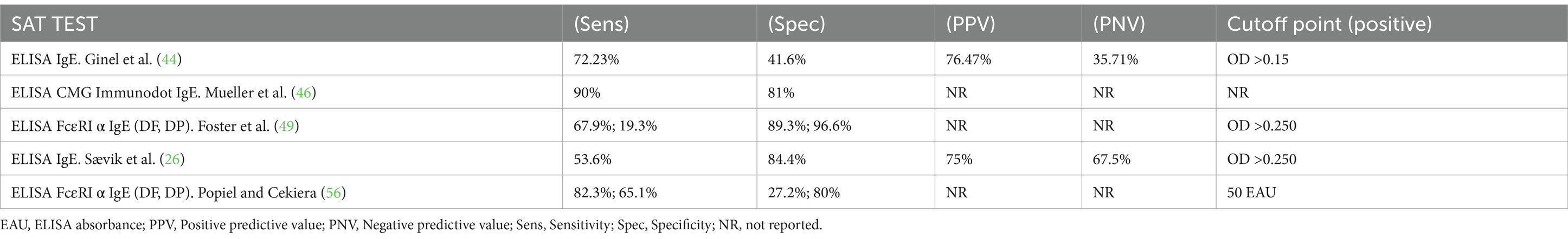
Table 5. SAT (specific allergen serological test) comparison table.
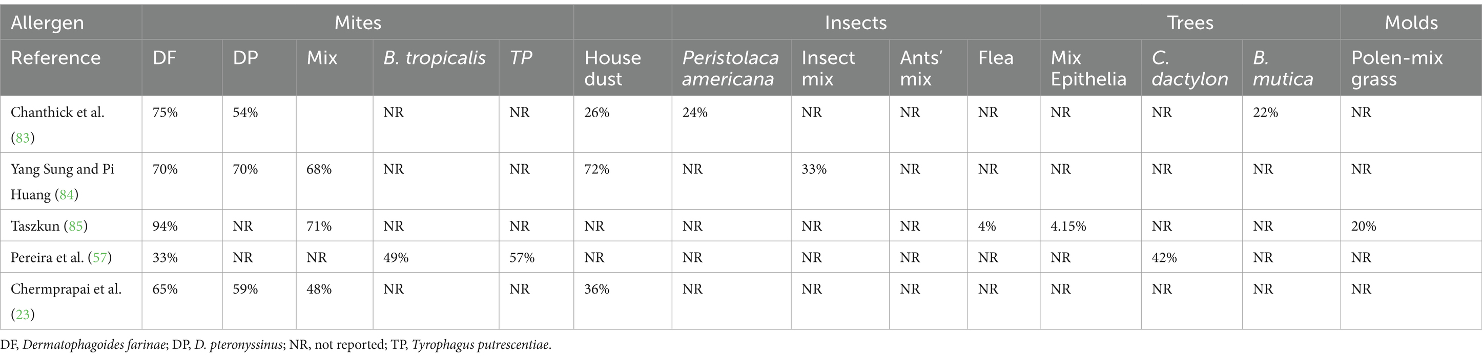
Table 6. Table of allergen prevalence according to allergen reactivity in canine atopic dermatitis.
Discussion
This systematic review describes the available literature on factors related to the diagnosis of allergens associated with CAD, a disease of complex etiology. Identifying specific clinical criteria is the cornerstone of CAD diagnosis. To identify allergens related to CAD provides the clinician with additional therapeutics to offering accurate therapeutic plans to their patients. Diagnostic tests with high reliability, specificity, repeatability, and sensitivity are needed as diagnostic alternatives for clinicians to achieve a more accurate diagnosis of the allergens involved. One of the most critical findings of this work was the failure to find a gold standard parameter. Another limitation in evaluating diagnostic performance is the heterogeneous concentrations of allergens used in dermabrasion tests, mode of preparation, and source of extraction.
For the SPT, which has been used regularly for several years in the diagnosis of allergens in human medicine, only two studies have evaluated the diagnostic performance of CAD in case-control studies. The findings of the two studies are contradictory, so SPT does not have enough references to judge its accuracy, making it necessary to conduct more studies on the subject since, owing to its easy design and application, it could be a useful tool in diagnosing allergens responsible for CAD crises. On the other hand, the measurement of serum IgE concentrations, which is often widely accepted in human AD allergen diagnosis, also presents a range of variations as complementary diagnoses of CAD. In addition, their results must be analyzed in the context of the patient’s clinical signs at a given time. The same doubts exist about IgE testing, as no evidence-based medical criteria were found. In this context, human dermatology has achieved an important consensus for diagnosing allergens associated with AD, but we still have a long way to go in CAD.
This systematic review identified 72 studies published between 1982 and 2023 in which microbial and nonmicrobial allergens were identified. In addition to IDT, SPT, the patch test, and ST, the study by Sævik et al., a combination of ELISA and intradermal injections of allergens to detect Ig-E-mediated reactivity in atopic dogs, revealed no concordance between serological and dermal tests and IgE positivity in the diagnosis of allergens related to CAD (26). Sasaki et al. used a crude allergen extract from Japanese cedar pollen to assess reactivity against it via IDT in dogs (27).
The results of this systematic review highlight the actual limitations in defining the utility of diagnostic tests to identify allergens in CAD patients. This discussion has taken place in the medical and scientific community, and the results of our systematic review did not answer the research question, which was intended to identify diagnostic tests that could be considered consistent, reliable, sensitive, and specific for CAD.
The role of immunoglobulins in the pathogenesis and diagnostic criteria for CAD has not been fully elucidated (28). Mueller et al. reported no statistically significant differences in the IgA concentration in skin washings between atopic and clinically healthy dogs (28). In contrast, Noli et al. reported a lack of IgE reactivity of serum from atopic dogs against Dermatophagoides pteronyssinus-derived allergens (Der p I, Der p II), and D. farinae antigens Der f I, and Der f II, which seem unlikely to be major allergens in dogs with CAD. However, the serum of atopic dogs reacts to a 90 kDa polypeptide of D. farinae, according to Western blot analysis (29). Hämmerling, evaluating IgE and the IgGa subclass in atopic dermatitis dogs, concluded that finding DP- and DF-related IgE renders this IG isotype more relevant than IgGa in detecting DA-related allergens (30).
Willense et al., and Moya et al. reported IgE and the IgG 1, IgG 2 subclasses against fractions of Der f 2 and Der f 1, Der f 18 in a lesser proportion; the IgG1 subclass seems to distinguish both IgE and House Dust Mites Allergens (HDMs) in CAD (31, 32). IgE reactivity against HDMs is more common than IgG reactivity in CAD (33). However, several factors must be considered when interpreting the results that affect their titer. Day and Corato reported heterogeneous IgG subclass reactivity against the most common allergens related to atopic dermatitis in dogs (34). Interestingly, helminth parasite infestation stimulates IgG production against IgE epitopes and decreases their levels in atopic dogs concomitantly infected by helminths and acari (35). Some patterns of IgE sensitization in dogs have been described in humans (e.g., Der f 18) (35), and the positive results of dermabrasion tests with concordant allergy-specific IgE levels in atopic dogs in Brazil are evidenced by the high prevalence of dust mites in Sao Paulo (24). For some authors, total IgE tests are unreliable and do not differentiate CAD (36). Jang et al. reported no significant differences in allergen reactivity between IDT and SAT (37).
Cross-reactivity has been reported in CAD dogs against plant allergens (38) and HDMs (DF and DP) (39), as well as with invertebrates and storage mites (Tyrophagus putrescentiae) with DF (40). Dogs sensitized to grass pollen often react to other allergens, particularly DF (24, 38, 39), and there is apparent sensitization between related allergen groups (house dust/storage mites, epidermis/fibers, trees, weeds, grass pollen, and molds) (41, 42).
The predominant sensitization against HDMs was common in several seasonal and nonseasonal countries reported from 1983 to 2023 (1–26, 28–31, 33–40, 43–76), indicating a consensus on a higher prevalence of HDMs than other allergenic groups, followed by plant pollen (39), animal allergens and molds, except for the study by Jung Kim et al., which described a higher prevalence of mold (77) (related to closed spaces, climatic changes that can influence findings).
Allergens have been described in HDMs, especially DFs, with high and low molecular weights classified as major and minor allergens according to their allergenic potential. Among the major allergens, the following have been reported: chitinase (Der f15) (48) and Der f 18, with greater sensitization in CAD (84.6%) than in human DA (50%) (48). Minor: In the Der 1 and 2 groups, approximately 50% of CAD dogs were sensitized to Der 1 and Der 2 in a study in Japan (40), and little importance in CAD has been reported (47), similar to the Der p I, Der p II, Der f 1, and Der f 2 (48) fractions. They were also described as irrelevant in the study by Noli et al. (29). Der f 1 is the major allergen in humans, and Der f 2 in SAT in CAD is poorly recognized by IgE (53). Other studies reported a high prevalence (86.7% in humans and 94.1% in dogs) (40). Khantavee et al. reported five major DF fractions (Der f Alt a 10, EF1-α, the gelsolin-like allergen Der f 16, Der f 28 and Der f 2) and Der f 3, Der f 10, Der f 20, and Der f 32 as minor allergens (33). Exposure to furniture and textiles has been associated with DF sensitization via the Zen 1 fraction, which strongly correlates with the crude DF extract in the SAT (62).
Geographical variations accounting for the prevalence and diversity of allergens (humidity and temperature in HDMs) should be taken into account (67), reflecting exposure factors (50). There are allergens specific to each region, including Japanese cedars (JCs) and pollen in Japan (38). Rumex acetosa (sorrel) in Italy (61), the pollen of some weeds in Thailand (23), and Cynodon dactylon in Brazil (57). In addition, environmental and surrounding conditions increase the prevalence of certain storage mites, as reported in Portugal (65), Brazil (T. putrescentiae, Lepidoglyphus destructor, and Blomia tropicalis) (53), and Thailand (23), where IgE titers increased during the rainy season (23). Air currents affect pollen distribution (39), although indoor aeroallergen deposition is independent of seasonality (42).
There are discrepancies between SATs (43) and various IgE measurement techniques lacking quality control and providing different results when the same sample is analyzed simultaneously with other equipment and methods (59). There is intra- and interlaboratory variability, so a quality assurance program (reliability) is needed (52).
Thirty-seven out of the 72 articles evaluated were related to IDT: only five studies reported sensitivity and specificity measurements (Table 5). Considering the positivity cutoff point as a determinant of these values is important. The positivity cutoff points for SAT do not have a consensus, ranging from OD >0.15 (44) to OD >0.250 (26, 49); other reported 50 EAU (56), or units were not reported (46).
Lowering cutoff point arbitrarily increases the probability of false positives (decreasing the test’s specificity). On the contrary, raising cutoff point affects the sensitivity and the possibility of false negatives. Standardizing this evaluation according to the techniques used and the allergens evaluated (allergenic potential) is essential (26). Cross-reactivity should be reduced with inhibitors of carbohydrate cross-reacting determinants (CCDs) (60). The study of allergenic fractions with allergenic potential detected by specific IgE would increase the effectiveness and greater reliability of the tests, mitigating false positives. IgG1, which reacts against DF and DP, was also reported as the predominant IgG subtype in a set of atopic dogs (33).
Threshold concentrations (positive nonirritating reactions) in IDT in dogs have been reported to be similar to those in humans (66). Standardized extracts for human medicine are often effective in CAD (78). Some studies define thresholds for certain allergens (values expressed in Protein Nitrogen Units, PNU): grasses, weeds, trees, molds and insects (1,750 PNU/mL), flea allergens (1:500 w/v), DPs (250 PNU/mL), DFs and T. putrescentiae (100 PNU/mL), epidermis (all at least 1,250 PNU/mL), human dander (300 PNU/mL), the optimal concentration of histamine (positive control) at 1:10,000 w/v. (76), Irritability at 1:10,000 w/v for DF and DP; 1,000 PNU for dust (36); and highly concentrated pollen extracts of 1,000–1,500 PNU (66). It has also been shown that mixed allergen extracts (26) in HDMs and house dust result in false negatives and low sensitivity and specificity. Individual DF and DP extracts, not house dust, are indicated (75).
IDT usually exhibits high sensitivity and specificity in detecting allergens in dogs with CAD (26). However, these values may vary depending on the allergen and concentration used. There is no clear consensus on the optimal allergen concentrations for IDT, which may affect the accuracy and reproducibility of the results. Some studies have evaluated different allergen concentrations and reported that higher concentrations may increase sensitivity and the risk of false positive reactions (36, 76). Interpretation of IDT results can be subjective and can vary among raters. Scales from 0 to 4 assess papule size, turgor, and erythema. Standardizing interpretation methods to reduce variability and improve comparability between studies and clinical settings is crucial.
Most of the works found in this systematic review reported prevalence values of mixtures or individual allergens without homogeneity in concentrations on the basis of the definition of a case or disease as CAD. The current clinical criteria were established in 2010 by Favrot (14), who issued eight clinical criteria, more than five of which had a sensitivity of 85% and a specificity of 80%. Assuming these criteria as a case definition, the previous prevalence would be reconsidered (because of the possibility to have been included nonatopic individuals), and therefore, false-positive results would have been obtained (Table 5).
Cross-reactivity between related allergens can complicate the interpretation of IDT results (38, 39, 42). Identifying and considering these factors when allergens are selected for testing are important. Co-sensitization to multiple allergens is common in dogs with AD (41), which may influence allergen choice (67). Allergen prevalence can vary significantly by geographic region, highlighting the importance of tailoring allergen panels to local conditions. Factors such as humidity and temperature can affect allergen sensitivity and should be considered when interpreting IDT results (61).
Standard protocols for performing and interpreting the IDT, including allergen concentration and interpretation criteria, are needed. Combining subjective and objective methods may yield more reliable IDT results (79). Longitudinal studies have been conducted to assess the long-term effectiveness of IDT and IDT-based immunotherapy. The inclusion of new relevant allergens in different geographic regions should be investigated to improve the accuracy and utility of IDT.
The study by Carmona et al. revealed a moderate sensitivity of 66% for SPT and a high specificity of 100% (80), in parallel with the study by Fleischman and Morris (25), where the intradermal test (IDT) and the SPT were compared, with a low sensitivity of 47% and a high specificity of 92.1% for the latter. SPT does not require sedation, is less invasive and comfortable for the animal, is less expensive than IDT, and has high specificity, reducing the probability of false positives. It appears to have moderate to low sensitivity, limiting its usefulness. However, the low reactivity to allergen mixtures (used in both studies) due to the dilution of individual components must be considered, which can result in false negatives. The interpretation of the results can be subjective and vary between evaluators, as with IDT. The agreement between the SPT and the IDT in the Fleischman and Morris (25) study was moderate (Cohen’s kappa value = 0.424), suggesting that both methods may complement each other. Studies should be conducted in larger and more diverse populations to validate the findings and improve the generalizability of the results with individual allergens instead of mixtures by increasing the reactivity and accuracy of the SPT; additionally, protocols for the performance and interpretation of the SPT should be developed and standardized to reduce variability and improve the comparability of results. Panels of new allergens relevant to different geographical regions should be included (25).
In the study by Olivry et al., an extract of D. farinae was used (500 μg per patch) (81). The presence of visible erythema, edema, induration, and vesiculation at the application site was evaluated. Macroscopic observations of skin inflammation were compared with microscopic observations. The study was conducted with a small, nonrepresentative sample. We wondered whether individual extracts should be used instead of mixtures with different standardized concentrations, considering irritant thresholds. Studies should be designed with more individuals and diverse races to validate the findings and compare the system evaluated with other diagnostic and treatment methods to evaluate its relative effectiveness.
In the study by Sævik et al., intradermal injections of sera from allergen-positive dogs were performed via ELISA. On the other hand, the challenge test involves the controlled administration of a specific allergen to the patient to observe whether an allergic reaction occurs (26), using a crude allergen extract from Japanese cedar pollen in dogs. The challenge test fell into disuse owing to the risk of adverse reactions and has been replaced by dermabrasion tests (27).
Given the gaps in knowledge, this manuscript proposes bringing together world experts in veterinary allergology to agree on critical points and issuing a general guide as a starting point for new research with internal and external validity, reproducibility, and comparative capacity. It is necessary to clearly and reliably define prevalence tables and statistical association measures to support the diagnosis of CAD. We urge the execution of studies with significant sample sizes, diverse racial groups, and sexual parity, considering the factors mentioned earlier, such as the environment, seasonality, humidity, temperature, and geographic variations in the prevalence of allergens (67).
Advantages and limitations of systematic review
Our systematic review has several strengths. We followed a structured approach anchored in a clearly defined research question previously documented and validated by systematic reviews from health field experts. Our extensive literature search spanned numerous sources, including general databases, search engines, journals, and conference proceedings, enabling us to capture data as far back as 1949. By excluding geographic and time-based limitations, we aimed to reduce potential biases. Additionally, the extracted information was rigorously organized; one author prepared a matrix of findings, which a second author subsequently reviewed to ensure accuracy across studies of varying quality and methodology. As a limitation, 21 relevant documents identified through abstract screening were not available as open access, so they could not be fully processed as complete texts to determine their definitive relevance, and we did not consider gray literature. To mitigate this, we employed snowballing techniques.
Finally, this manuscript presents a detailed, comprehensive, and transparent systematic review of allergen diagnostic methods for CAD, following PRISMA standards. The methodology used in the review, including exhaustive search and content analysis with Atlas TI software, ensures the robustness and reproducibility of the findings. This includes the evaluation of multiple databases and the application of rigorous inclusion and exclusion criteria. Different diagnostic methods are compared, providing a clear overview of their advantages, limitations, and relative effectiveness. We report sensitivity and specificity data from relevant studies, which helps the scientific community understand the gaps in knowledge and scientific rigor to improve the accuracy of these diagnostic methods. The most common allergens used in the studies were identified and reported, which could guide future research. We highlight the application of Favrot’s clinical criteria in 41.6% of the studies since 2010 (14), highlighting the importance of these criteria in defining CAD cases. We identified gaps in current knowledge that suggest the need for future research to standardize allergenic extracts, define cutoff points in serological tests, and consider environmental, geographic, and demographic variables. These findings are valuable to the scientific community, as they provide a solid basis for improving the diagnosis and management of CAD and guiding future research in this field.
Conclusion
Even though most scientist and practitioners consider IDT as the standard test to diagnose allergen sensitization in CAD patients, our results suggest caution must be kept when considering the available tests as a gold standard to identify allergen sensitization in dogs affected by CAD. Current methods have advantages and limitations. There is no consensus on antigen concentrations, no standardized cutoff points for SAT positivity have been defined, and there is no laboratory regulation or monitoring. Favrot’s clinical criteria have been applied in 41.6% of studies since 2010, highlighting their importance in defining CAD cases. A total of 136 different allergens used in the studies were identified, providing a basis for future research and clinical practice. The development and standardization of allergenic extracts for diagnostic testing are crucial, ensuring the consistency and comparability of results between different studies and laboratories. Studies that consider environmental, geographic, and demographic variables should be conducted to better understand how these factors affect the prevalence and severity of CAD. We urge further studies to evaluate the efficacy and applicability of the Favrot’s criteria in different populations and settings. Longitudinal studies should be conducted to better understand the progression of CAD and the long-term effectiveness of different diagnostic methods and treatments and to further explore co-sensitization and cross-reactivity between different allergens to improve the accuracy of diagnostic tests and the formulation of specific immunotherapies. Investigate the impact of CAD on the quality of life of dogs and their owners and how different diagnostic methods and treatments can improve these aspects. These proposals may help advance the knowledge and management of CAD, improving the diagnosis and treatment of this condition.
Data availability statement
The raw data supporting the conclusions of this article will be made available by the authors without undue reservation.
Author contributions
RM-R: Data curation, Formal analysis, Investigation, Methodology, Writing – original draft, Writing – review & editing, Conceptualization. MG-D: Investigation, Supervision, Validation, Writing – review & editing. JS: Supervision, Writing – review & editing, Formal analysis, Validation. NC-V: Conceptualization, Data curation, Formal analysis, Investigation, Writing – original draft, Writing – review & editing, Methodology. JM-E: Conceptualization, Data curation, Investigation, Writing – original draft, Writing – review & editing, Funding acquisition, Project administration, Supervision.
Funding
The author(s) declare that financial support was received for the research and/or publication of this article. This work was funded by the Universidad de Antioquia through the call for consolidation of research groups 2023-2024 (RM-R, NC-V, JS, JGM-E). MSG-D was funded by Universidad CES and Bivett.
Acknowledgments
Research Activities of World Class research group in the University of Antioquia (Grupos Minciencias Categoría A1 y A) is supported by “Estrategias de Sostenibilidad para grupos de Investigación” Calls 2021 and 2023. OHVRI Research Group, Group of Clinical and Experimental Allergy, and CENTAURO Research Group, are supported by these University of Antioquia internal Calls. INCA-CES Research group scientific activities are supported by Universidad CES.
Conflict of interest
The authors declare that the research was conducted in the absence of any commercial or financial relationships that could be construed as a potential conflict of interest.
Generative AI statement
The authors declare that Gen AI was used in the creation of this manuscript. During the preparation of this work, the authors used OpenAI ChatGPT (October 2023 version, https://chat.openai.com/) to improve the readability and language of the manuscript. After using this tool, the authors reviewed and edited the content as needed and took full responsibility for the content of the published article. Several Grammarly reviewing sessions were applied before submitting the final version of the manuscript.
Publisher’s note
All claims expressed in this article are solely those of the authors and do not necessarily represent those of their affiliated organizations, or those of the publisher, the editors and the reviewers. Any product that may be evaluated in this article, or claim that may be made by its manufacturer, is not guaranteed or endorsed by the publisher.
Supplementary material
The Supplementary material for this article can be found online at: https://www.frontiersin.org/articles/10.3389/fvets.2025.1551207/full#supplementary-material
Footnotes
References
1. Nielsen, TD, Dean, RS, Robinson, NJ, Massey, A, and Brennan, ML. Survey of the UK veterinary profession: common species and conditions nominated by veterinarians in practice. Vet Rec. (2014) 174:324–4. doi: 10.1136/vr.101745
2. Favrot, C, Linek, M, Mueller, R, and Zini, Efor the International Task Force on Canine Atopic Dermatitis. Development of a questionnaire to assess the impact of atopic dermatitis on health-related quality of life of affected dogs and their owners. Vet Dermatol. (2010) 21:64–70. doi: 10.1111/j.1365-3164.2009.00781.x
3. Noli, C . Assessing quality of life for pets with dermatologic disease and their owners. Vet Clin N Am Small Anim Pract. (2019) 49:83–93. doi: 10.1016/j.cvsm.2018.08.008
4. Harvey, N, Craigon, P, Shaw, S, Blott, S, and England, G. Behaviooural differences in dogs with atopic dermatitis suggest stress could be a significant problem associated with chronic pruritus. Animals. (2019) 9:813. doi: 10.3390/ani9100813
5. Chermprapai, S, Anukkul, PC, Kritsadasima, T, Kromkhun, P, and Thengchaisri, N. Comparing the results of intradermal skin tests for four dust mite allergens in dogs with atopic dermatitis in Thailand. Vet World. (2020) 13:2381–7. doi: 10.14202/vetworld.2020.2381-2387
6. Basagaña, M, Bartolomé, B, Pastor, C, Torres, F, Alonso, R, Vivanco, F, et al. Allergy to human seminal fluid: cross-reactivity with dog dander. J Allergy Clin Immunol. (2008) 121:233–9. doi: 10.1016/j.jaci.2007.10.008
7. Agler, CS, Friedenberg, S, Olivry, T, Meurs, KM, and Olby, NJ. Genome-wide association analysis in West Highland White terriers with atopic dermatitis. Vet Immunol Immunopathol. (2019) 209:1–6. doi: 10.1016/j.vetimm.2019.01.004
8. Brownstone, ND, and Koo, J. Is the pathology in atopic dermatitis limited only to the skin? J Allergy Clin Immunol Pract. (2020) 8:3507. doi: 10.1016/j.jaip.2020.07.045
9. Marsella, R, and Girolomoni, G. Canine models of atopic dermatitis: a useful tool with untapped potential. J Invest Dermatol. (2009) 129:2351–7. doi: 10.1038/jid.2009.98
10. Hamed, A, Todd, I, Tighe, PJ, Powell, RJ, Harrison, T, and Fairclough, LC. Array-based measurements of aero-allergen-specific IgE correlate with skin-prick test reactivity in asthma regardless of specific IgG4 or total IgE measurements. J Immunol Methods. (2021) 492:112999. doi: 10.1016/j.jim.2021.112999
11. Lockey, RF In: FL Richard and DK Ledford, editors. Allergens and allergen immunotherapy. Boca Raton, FL, USA: CRC Press (2008)
12. Mueller, RS, Chapman, PL, Rosychuk, REW, Bettenay, SV, and Fieseler, KV. Evaluation of cross-reactivity of allergens by use of intradermal testing in atopic dogs. Am J Vet Res. (2002) 63:874–9. doi: 10.2460/ajvr.2002.63.874
13. Plant, JD, Neradelik, MB, Polissar, NL, Fadok, VA, and Scott, BA. Agreement between allergen-specific IgE assays and ensuing immunotherapy recommendations from four commercial laboratories in the USA. Vet Dermatol. (2014) 25:15–e6. doi: 10.1111/vde.12104
14. Favrot, C, Steffan, J, Seewald, W, and Picco, F. A prospective study on the clinical features of chronic canine atopic dermatitis and its diagnosis. Vet Dermatol. (2010) 21:23–31. doi: 10.1111/j.1365-3164.2009.00758.x
15. Nuttall, TJ, and Halliwell, REW. Serum antibodies to Malassezia yeasts in canine atopic dermatitis. Vet Dermatol. (2001) 12:327–32. doi: 10.1046/j.0959-4493.2001.00261.x
16. Levy, RM, Gelfand, JM, and Yan, AC. The epidemiology of atopic dermatitis. Clin Dermatol. (2003) 21:109–15. doi: 10.1016/S0738-081X(02)00360-7
17. Hillier, A, and Griffin, CE. The ACVD task force on canine atopic dermatitis (I): incidence and prevalence. Vet Immunol Immunopathol. (2001) 81:147–51. doi: 10.1016/S0165-2427(01)00296-3
18. Wilhem, S, Kovalik, M, and Favrot, C. Breed-associated phenotypes in canine atopic dermatitis. Vet Dermatol. (2011) 22:143–9. doi: 10.1111/j.1365-3164.2010.00925.x
19. Cohn, L, and Coté, E. Atopic dermatitis In: Cote’s clinical veterinary advisor dogs and cats. St Luis, Missouri: Elsevier (2020). 91–3.
20. Olivry, T, Deangelo, KB, Dunston, SM, Clarke, KB, and McCall, CA. Patch testing of experimentally sensitized beagle dogs: development of a model for skin lesions of atopic dermatitis. Vet Dermatol. (2006) 17:95–102. doi: 10.1111/j.1365-3164.2006.00502.x
21. Olivry, T, and Mueller, RS. Critically appraised topic on adverse food reactions of companion animals (7): signalment and cutaneous manifestations of dogs and cats with adverse food reactions. BMC Vet Res. (2019) 15:140. doi: 10.1186/s12917-019-1880-2
22. Page, MJ, McKenzie, JE, Bossuyt, PM, Boutron, I, Hoffmann, TC, Mulrow, CD, et al. The PRISMA 2020 statement: an updated guideline for reporting systematic reviews. BMJ. (2021) 372:n71. doi: 10.1136/bmj.n71
23. Chermprapai, S, and Thengchaisri, N. A descriptive study of allergen-specific IgE serological tests for canine atopic dermatitis in Thailand. BMC Vet Res. (2020) 16:475. doi: 10.1186/s12917-020-02684-x
24. Frezza, APDM, Lourenço, MLG, Meira, J, Tsukui, T, Kageyama, M, and De Araújo Machado, LH. Seroprevalence of Dermatophagoides farinae-specific IgE in dogs with atopic dermatitis in São Paulo, Brazil. Res Vet Sci. (2023) 164:105002. doi: 10.1016/j.rvsc.2023.105002
25. Fleischman, DA, and Morris, DO. Pilot study to determine the concordance between skin prick and intradermal test (IDT) reactivity to environmental allergens in atopic dogs using IDT as the gold standard. Vet Dermatol. (2023) 34:505–13. doi: 10.1111/vde.13187
26. Sævik, BK, Ulstein, TL, and Larsen, HJS. Evaluation of a commercially available enzyme-linked immunosorbent assay for the detection of allergen-specific IgE antibodies in dogs. Res Vet Sci. (2003) 74:37–45. doi: 10.1016/S0034-5288(02)00151-0
27. Sasaki, Y, Kitagawa, H, Fujioka, T, Kitoh, K, Iwasaki, T, Sakaguchi, M, et al. Hypersensitivity to Japanese cedar (Cryptomeria japonica) pollen in dogs. J Vet Med Sci. (1995) 57:683–5. doi: 10.1292/jvms.57.683
28. Mueller, RS, Cannon, A, Reubl, GH, and Ihrke, PJ. Serum and skin IgA concentrations in normal and atopic dogs. Aust Vet J. (1997) 75:906–9. doi: 10.1111/j.1751-0813.1997.tb11264.x
29. Noli, C, Bernadina, WE, and Willemse, T. The significance of reactions to purified fractions of Dermatophagoides pteronyssinus and Dermatophagoides farinae in canine atopic dermatitis. Vet Immunol Immunopathol. (1996) 52:147–57. doi: 10.1016/0165-2427(96)05550-x
30. Hämmerling,, and De Weck,. Comparison of two diagnostic tests for canine atopy using monoclonal anti-IgE antibodies. Vet Dermatol. (1998) 9:191–9. doi: 10.1046/j.1365-3164.1998.00110.x
31. Moya, R, Carnés, J, Sinovas, N, Ramió, L, Brazis, P, and Puigdemont, A. Immunoproteomic characterization of a Dermatophagoides farinae extract used in the treatment of canine atopic dermatitis. Vet Immunol Immunopathol. (2016) 180:1–8. doi: 10.1016/j.vetimm.2016.08.004
32. Willemse, A, Noordzij, A, Van den Brom, WE, and Rutten, VP. Allergen specific IgGd antibodies in dogs with atopic dermatitis as determined by the enzyme linked immunosorbent assay (ELISA). Clin Exp Immunol. (1985) 59:359–63.
33. Khantavee, N, Chanthick, C, Tungtrongchitr, A, Techakriengkrai, N, Suradhat, S, Sookrung, N, et al. Immunoglobulin G1 subclass responses can be used to detect specific allergy to the house dust mites Dermatophagoides farinae and Dermatophagoides pteronyssinus in atopic dogs. BMC Vet Res. (2021) 17:71. doi: 10.1186/s12917-021-02768-2
34. Day, MJ, Corato, A, and Shaw, SE. Subclass profile of allergen-specific IgG antibodies in atopic dogs. Res Vet Sci. (1996) 61:136–42. doi: 10.1016/s0034-5288(96)90088-0
35. Hammerberg, B, Bevier, D, DeBoer, DJ, Olivry, T, Orton, SM, Gebhard, D, et al. Auto IgG anti-IgE and IgG × IgE immune complex presence and effects on ELISA-based quantitation of IgE in canine atopic dermatitis, demodectic acariasis and helminthiasis. Vet Immunol Immunopathol. (1997) 60:33–46. doi: 10.1016/S0165-2427(97)00119-0
36. Lian, TM, and Halliwell, RE. Allergen-specific IgE and IgGd antibodies in atopic and normal dogs. Vet Immunol Immunopathol. (1998) 66:203–23. doi: 10.1016/s0165-2427(98)00199-8
37. Jang, H-M, Yeo, G-S, Kim, J-H, Hwang, C-Y, Hyun, J-E, Kim, S-S, et al. Prevalence of serum allergen-specific immunoglobulin E for canine atopic dermatitis in Korea. J Biomed Res. (2014) 15:162–9. doi: 10.12729/jbr.2014.15.4.162
38. Masuda, K, Sakaguchi, M, Saito, S, Deboer, DJ, Fujiwara, S, Kurata, K, et al. In vivo and in vitro tests showing sensitization to Japanese cedar (Cryptomeria japonica) pollen allergen in atopic dogs. J Vet Med Sci. (2000) 62:995–1000. doi: 10.1292/jvms.62.995
39. Mueller, RS, Bettenay, SV, and Tideman, L. Aero-allergens in canine atopic dermatitis in southeastern Australia based on 1000 intradermal skin tests. Aust Vet J. (2000) 78:392–9. doi: 10.1111/j.1751-0813.2000.tb11824.x
40. Song, H, Lee, J, Jeong, KY, Cheon, D-S, and Park, J-W. Comparison of sensitization patterns to dust mite allergens between atopic dermatitis patients and dogs, and non-specific reactivity of canine IgE to the storage mite Tyrophagus putrescentiae. Exp Appl Acarol. (2022) 88:41–55. doi: 10.1007/s10493-022-00744-5
41. Buckley, L, Schmidt, V, McEwan, N, and Nuttall, T. Cross-reaction and co-sensitization among related and unrelated allergens in canine intradermal tests. Vet Dermatol. (2013) 24:422. doi: 10.1111/vde.12044
42. Roussel, AJJ, Bruet, V, and Bourdeau, PJ. Characterisation of dog sensitisation to grass pollen in western France from 1999 to 2010. Vet Rec. (2013) 172:686. doi: 10.1136/vr.100710
43. Willemse, A, and van den Brom, WE. Investigations of the symptomatology and the significance of immediate skin test reactivity in canine atopic dermatitis. Res Vet Sci. (1983) 34:261–5. doi: 10.1016/S0034-5288(18)32221-5
44. Ginel, PJ, Riaño, C, and Lucena, R. Evaluation of a commercial ELISA test for the detection of allergen-specific IgE antibodies in atopic dogs. Zentralbl Veterinarmed B. (1998) 45:421–5. doi: 10.1111/j.1439-0450.1998.tb00811.x
45. Zunic,. Comparison between IMMUNODOT tests and the intradermal skin test in atopic dogs. Vet Dermatol. (1998) 9:201–5. doi: 10.1046/j.1365-3164.1998.00125.x
46. Mueller, RS, Burrows, A, and Tsohalis, J. Comparison of intradermal testing and serum testing for allergen-specific IgE using monoclonal IgE antibodies in 84 atopic dogs. Aust Vet J. (1999) 77:290–4. doi: 10.1111/j.1751-0813.1999.tb10263.x
47. Masuda, K, Tsujimoto, H, Fujiwara, S, Kurata, K, Hasegawa, A, Yasueda, H, et al. IgE sensitivity and cross-reactivity to crude and purified mite allergens (Der f 1, Der f 2, Der p 1, Der p 2) in atopic dogs sensitive to Dermatophagoides mite allergens. Vet Immunol Immunopathol. (1999) 72:303–13. doi: 10.1016/s0165-2427(99)00142-7
48. Nuttall, TJ, Lamb, JR, and Hill, PB. Characterisation of major and minor Dermatophagoides allergens in canine atopic dermatitis. Res Vet Sci. (2001) 71:51–7. doi: 10.1053/rvsc.2001.0485
49. Foster, AP, Littlewood, JD, Webb, P, Wood, JLN, Rogers, K, and Shaw, SE. Comparison of intradermal and serum testing for allergen-specific IgE using a FcεRIα-based assay in atopic dogs in the UK. Vet Immunol Immunopathol. (2003) 93:51–60. doi: 10.1016/S0165-2427(03)00052-7
50. Arlian, LG, Schumann, RJ, Morgan, MS, and Glass, RL. Serum immunoglobulin E against storage mite allergens in dogs with atopic dermatitis. Am J Vet Res. (2003) 64:32–6. doi: 10.2460/ajvr.2003.64.32
51. Goicoa, A, Espino, L, Rodríguez, I, Puigdemont, A, Brazis, P, and Rejas, J. Importance of house dust and storage mites in canine atopic dermatitis in the geographic region of Galicia, Spain. Acta Veterinaria Hungarica. (2008) 56:163–71. doi: 10.1556/avet.56.2008.2.3
52. Thom, N, Favrot, C, Failing, K, Mueller, RS, Neiger, R, and Linek, M. Intra- and interlaboratory variability of allergen-specific IgE levels in atopic dogs in three different laboratories using the fc-ɛ receptor testing. Vet Immunol Immunopathol. (2010) 133:183–9. doi: 10.1016/j.vetimm.2009.07.019
53. Cunha, VES, Silva, MH, and Faccini, JLH. Serological identification of house dust mite allergens in dogs with atopic dermatitis. Pesq Vet Bras. (2012) 32:917–21. doi: 10.1590/S0100-736X2012000900016
54. Bjelland, AA, Dolva, FL, Nødtvedt, A, and Sævik, BK. Prevalence of and risk factors for increased serum levels of allergen-specific IgE in a population of Norwegian dogs. Acta Vet Scand. (2014) 56:81. doi: 10.1186/s13028-014-0081-z
55. Kang, M-H, Kim, H-J, Jang, H-J, and Park, H-M. Sensitization rates of causative allergens for dogs with atopic dermatitis: detection of canine allergen-specific IgE. J Vet Sci. (2014) 15:545–50. doi: 10.4142/jvs.2014.15.4.545
56. Popiel, J, and Cekiera, A. Comparison of IgE test results with intradermal skin tests for dust mites and storage mites in atopic dogs. Pol J Vet Sci. (2015) 18:351–6. doi: 10.1515/pjvs-2015-0045
57. Pereira, DT, Cunha, VES, Schmidt, C, Magnus, T, and Krause, A. Sensitization study of dogs with atopic dermatitis in the central region of Rio Grande do Sul. Arq Bras Med Vet Zootec. (2015) 67:1533–8. doi: 10.1590/1678-4162-8224
58. Patel, A, Curtis, CF, and Cerundolo, R. Incidence of anti-Der f 2 and anti-Zen 1-specific immunoglobulin E antibodies in atopic dogs from south-East England. Vet Rec. (2019) 184:317–7. doi: 10.1136/vr.104810
59. Zhou, Z, Pieper, JB, and Campbell, K. Intralaboratory reliability and variability for allergen-specific immunoglobulin type E serology testing. J Am Anim Hosp Assoc. (2019) 55:124–9. doi: 10.5326/JAAHA-MS-6761
60. Lee, KW, McKinney, BH, Blankenship, KD, and Morris, DO. Detection and inhibition of IgE for cross-reactive carbohydrate determinants evident in an enzyme-linked immunosorbent assay for detection of allergen-specific IgE in the sera of dogs and cats. Vet Dermatol. (2020) 31:439–e116. doi: 10.1111/vde.12904
61. Di Tommaso, M, Luciani, A, Crisi, PE, Beschi, M, Rosi, P, Rocconi, F, et al. Detection of serum allergen-specific IgE in atopic dogs tested in northern Italy: preliminary study. Animals. (2021) 11:358. doi: 10.3390/ani11020358
62. Ludwig, L, Tsukui, T, Kageyama, M, and Farias, M. Evaluation of sensitization to the crude extract of Dermatophagoides farinae and its derived allergens, Der f 2 and Zen 1, in dogs with atopic dermatitis in southern Brazil. Vet Immunol Immunopathol. (2021) 234:110199. doi: 10.1016/j.vetimm.2021.110199
63. Chan, WY, Selvarajah, GT, Ajat, M, Suzuki, R, and Tsukui, T. The detection of house dust mite Dermatophagoides farinae, Der f 2 and Zen-1 allergen-specific immunoglobulin E antibodies in dogs with atopic dermatitis in Malaysia. Vet Immunol Immunopathol. (2019) 212:43–9. doi: 10.1016/j.vetimm.2019.05.002
64. Adam, GO, Park, Y-G, Cho, J-H, Choi, J, and Oh, H-G. Detecting common allergens in dogs with atopic dermatitis in south Korean provinces using a serological immunoglobulin E-specific allergen test. Vet World. (2022) 15:1996–2003. doi: 10.14202/vetworld.2022.1996-2003
65. Martins, LML . Survey of sensitization to common Fungi in an allergic dog population: the need for further focus on sensitization and allergy to Fungi in veterinary medicine. JoF. (2023) 9:1075. doi: 10.3390/jof9111075
66. Willemse, A, and van den Brom, WE. Evaluation of the intradermal allergy test in normal dogs. Res Vet Sci. (1982) 32:57–61. doi: 10.1016/S0034-5288(18)32438-X
67. Sture, GH, Halliwell, REW, Thoday, KL, Van Den Broek, AHM, Henfrey, JI, Lloyd, DH, et al. Canine atopic disease: the prevalence of positive intradermal skin tests at two sites in the north and south of Great Britain. Vet Immunol Immunopathol. (1995) 44:293–308. doi: 10.1016/0165-2427(94)05306-D
68. Halliwell, REW, and Gorman, NT eds. Veterinary clinical immunology. Philadelphia: Saunders (1989). 548 p.
69. Yasueda, H, Saito, A, Akiyama, K, Maeda, Y, Shida, T, Sakaguchi, M, et al. Estimation of Der p and Der f I quantities in the reference preparations of Dermatophagoides mite extracts. Clin Exp Allergy. (1994) 24:1030–5. doi: 10.1111/j.1365-2222.1994.tb02739.x
70. Yasueda, H, Mita, H, Yui, Y, and Shida, T. Isolation and characterization of two allergens from Dermatophagoides farinae. Int Arch Allergy Appl Immunol. (1986) 81:214–23. doi: 10.1159/000234137
71. Yasueda, H, Mita, H, Yui, YS, and Shida, T. Measurement of allergens associated with dust mite allergy. I. Development of sensitive radioimmunoassays for the two groups of Dermatophagoides mite allergens, Der I and Der II. Int Arch Allergy Appl Immunol. (1989) 90:182–9. doi: 10.1159/000235021
72. Saridomichelakis, MN, Koutinas, AF, Gioulekas, D, and Leontidis, L. Canine atopic dermatitis in Greece: clinical observations and the prevalence of positive intradermal test reactions in 91 spontaneous cases. Vet Immunol Immunopathol. (1999) 69:61–73. doi: 10.1016/s0165-2427(99)00040-9
73. Reedy, LM, Miller, WH, and Willemse, T. Allergic skin diseases of dogs and cats. 2nd ed. London Philadelphia Toronto [etc.]: W. B. Saunders (1997).
74. Park, S, Ohya, F, Yamashita, K, Nishifuji, K, and Iwasaki, T. Comparison of response to immunotherapy by intradermal skin test and antigen-specific IgE in canine atopy. J Vet Med Sci. (2000) 62:983–8. doi: 10.1292/jvms.62.983
75. Hillier, A, Kwochka, KW, and Pinchbeck, LR. Reactivity to intradermal injection of extracts of Dermatophagoides farinae, Dermatophagoides pteronyssinus, house dust mite mix, and house dust in dogs suspected to have atopic dermatitis: 115 cases (1996-1998). J Am Vet Med Assoc. (2000) 217:536–40. doi: 10.2460/javma.2000.217.536
76. Hensel, P, Austel, M, Medleau, L, Zhao, Y, and Vidyashankar, A. Determination of threshold concentrations of allergens and evaluation of two different histamine concentrations in canine intradermal testing. Vet Dermatol. (2004) 15:304–8. doi: 10.1111/j.1365-3164.2004.00400x
77. Kim, H-J, Kang, M-H, and Park, H-M. Common allergens of atopic dermatitis in dogs: comparative findings based on intradermal tests. J Vet Sci. (2011) 12:287–90. doi: 10.4142/jvs.2011.12.3.287
78. Cunha, VES, Hahnstadt, RL, Soares, AMB, and Faccini, JLH. Evaluation of skin sensitivity in dogs bearing allergic dermatitis to standardized allergenic extract of house dust and storage mites. Pesq Vet Bras. (2007) 27:341–4. doi: 10.1590/S0100-736X2007000800004
79. Hubbard, TL, and White, PD. Comparison of subjective and objective intradermal allergy test scoring methods in dogs with atopic dermatitis. J Am Anim Hosp Assoc. (2011) 47:399–405. doi: 10.5326/JAAHA-MS-5638
80. Carmona-Gil, AM, Sánchez, J, and Maldonado-Estrada, J. Evaluation of skin prick-test reactions for allergic sensitization in dogs with clinical symptoms compatible with atopic dermatitis. A pilot study. Front Vet Sci. (2019) 6:448. doi: 10.3389/fvets.2019.00448
81. Olivry, T, Linder, KE, Paps, JS, Bizikova, P, Dunston, S, Donne, N, et al. Validation of a novel epicutaneous delivery system for patch testing of house dust mite-hypersensitive dogs. Vet Dermatol. (2012) 23:525–e106. doi: 10.1111/j.1365-3164.2012.01111.x
82. Mueller, RS, Fieseler, KV, Rosychuk, RAW, and Greenwalt, T. Intradermal testing with the storage mite Tyrophagus putrescentiae in normal dogs and dogs with atopic dermatitis in Colorado. Vet Dermatol. (2005) 16:27–31. doi: 10.1111/j.1365-3164.2005.00415.x
83. Chanthick, C, Anaman, S, and Buathet, K. The prevalence of positive intradermal allergy tests in 114 dogs with atopic dermatitis in the Bangkok metropolis, Thailand. Vet Immunol Immunopathol. (2008) 126:256–62. doi: 10.1016/j.vetimm.2008.07.010
84. Yang, S, and Huang, P. The incidence of positive intradermal skin test reactions in dogs with atopic dermatitis. JVCS. (2009) 2:30–6.
85. Taszkun, I . The results of intradermal skin tests (IDST) in dogs with atopic dermatitis from the Lublin voivodeship. Pol J Vet Sci. (2011) 14:95–101. doi: 10.2478/v10181-011-0014-y
86. Khantavee, N, Reamtong, O, Sookrung, N, Suradhat, S, and Prapasarakul, N. Allergen components of Dermatophagoides farinae recognised by serum immunoglobulin (Ig)E in Thai dogs with atopic dermatitis. Vet Dermatol. (2021) 32:338–e94. doi: 10.1111/vde.12967
Glossary
AD - Human Atopic Dermatitis
ASIS - Allergen-specific IgE serology
BU - Bethesda Unit
CAD - Canine Atopic Dermatitis
CADESI - Canine Atopic Dermatitis Extent and Severity Index
CCDs - Carbohydrate Cross-Reacting Determinants
CMG Immunodot - ELISA test using allergen-specific anti-canine IgE monoclonal antibodies.
Der f 15, Der f 18 - Dermatophagoides farinae-derived Major allergens. Each one are the major allergens of the American house dust mites.
Der f 1, Der f 2, Der f 18 - Dermatophagoides farinae-derived Minor allergens. Each one are the major allergens of the American house dust mites.
Der p I, Der p II, - Dermatophagoides pteronyssinus-derived allergens (major allergens of the American house dust mites).
DF - Dermatophagoides farinae
DP - Dermatophagoides pteronyssinus
EAU - ELISA Absorbance Units
ELISA - Enzyme-Linked Immunosorbent Assay
HDM - House Dust Mites Allergens
IDT - Intradermal Test
IgE - Immunoglobulin type E
IgG - Immunoglobulin type G
IgG1 - Immunoglobulin type G subclass 1
IgG2 - Immunoglobulin type G subclass 2
MWD - Mean Wheal Diameter
NE - Nitrogen Equivalents
OD - Optical Density
PNU - Protein Nitrogen Units
PNV - Predictive Negative Value
PPV - Predictive Positive Value
PVAS - Pruritus Visual Analog Scale
SAT - Serologic-specific Allergen Test
SD - Standard Deviation
Sens - Sensitivity
Spec - Specificity
SPT - Skin Prick Test
w/v - Weight-to-volume Ratio
Keywords: Allergens, canine atopic dermatitis, diagnostic methods, IDT, IgE, SAT, SPT, systematic review
Citation: Morales-Romero R, Gonzalez-Dominguez MS, Sánchez J, Correa-Valencia NM and Maldonado-Estrada JG (2025) Efficacy of diagnostic testing for allergen sensitization in canine atopic dermatitis: a systematic review. Front. Vet. Sci. 12:1551207. doi: 10.3389/fvets.2025.1551207
Edited by:
Carlos Eduardo Fonseca-Alves, Paulista University, BrazilReviewed by:
Fifa Argentina, Sriwijaya University, IndonesiaGrzegorz Kalisz, Medical University of Lublin, Poland
Copyright © 2025 Morales-Romero, Gonzalez-Dominguez, Sánchez, Correa-Valencia and Maldonado-Estrada. This is an open-access article distributed under the terms of the Creative Commons Attribution License (CC BY). The use, distribution or reproduction in other forums is permitted, provided the original author(s) and the copyright owner(s) are credited and that the original publication in this journal is cited, in accordance with accepted academic practice. No use, distribution or reproduction is permitted which does not comply with these terms.
*Correspondence: Juan G. Maldonado-Estrada, anVhbi5tYWxkb25hZG9AdWRlYS5lZHUuY28=