 Hinrich Tönjes Wolff1*
 Ana Cristina Piroth1
 Hilke Oltmanns2
 Jessica Meißner2
 Jutta Verspohl3
 Holger Andreas Volk1
 Claudia Busse1
- 1Small Animal Clinic, University of Veterinary Medicine Hannover, Foundation, Hannover, Germany
- 2Department of Pharmacology, Toxicology and Pharmacy, University of Veterinary Medicine Hannover, Foundation, Hannover, Germany
- 3Institute for Microbiology, University of Veterinary Medicine Hannover, Foundation, Hannover, Germany
Purpose: To determine the minimal bactericidal concentration (MBC) of polyhexanide (PHMB), povidone-iodine (PVP-I), N-acetylcysteine (NAC), and hypochlorous acid (HOCl) for bacterial species commonly found in canine and feline infectious keratitis.
Methods: MBCs for clinical isolates of Staphylococcus (S.) pseudintermedius (n = 11), including 3 methicillin-resistant strains, Pseudomonas (P.) aeruginosa (n = 8), and Streptococcus (Str.) canis (n = 11), including the corresponding control strains, were examined. All testing substances were serially diluted in phosphate-buffered saline (PBS) and cation-adjusted Mueller–Hinton Broth (CAMHB) and inoculated with the bacterial suspension for 10 min. Afterwards, a neutralisation with Dey–Engley neutralising broth was performed, followed by plating onto Columbia sheep–blood agar. After incubation, plates were visually examined for bacterial growth. Tests were carried out in triplicate.
Results: MBCs in PBS for polyhexanide ranged 0.8–1.6 mg/L for S. pseudintermedius and 1.6–3.2 mg/L for P. aeruginosa and Str. canis. For povidone-iodine, MBCs in PBS were observed at concentrations ranging 8–32 mg/L for S. pseudintermedius and P. aeruginosa and 8–16 mg/L for Str. canis. MBCs in PBS for NAC were recorded at a range of 6,400–12,800 mg/L for S. pseudintermedius, whereas those for P. aeruginosa and Str. canis ranged 3,200–6,400 mg/L. Results for HOCl in PBS ranged 0.4–1.6 mg/L for S. pseudintermedius and 0.4–0.8 mg/L for P. aeruginosa and Str. canis. MBCs in CAMHB for polyhexanide were found in the range between 3.2 and >12.8 mg/L, those for povidone-iodine between 6,400 and >12,800 mg/L, and for NAC between 6,400 and >12,800 mg/L, across the tested species. When dissolved in CAMHB, no antimicrobial effect could be observed for HOCl in concentrations up to 137.5 mg/L.
Conclusion: All tested substances had an in vitro bactericidal effect against all three bacterial species with MBCs below known tolerated ocular concentrations when dissolved in PBS. Povidone-iodine and hypochlorous acid showed a marked reduction in their in vitro efficacy in the presence of protein. Nevertheless, our results provide a promising outlook on alternatives or adjuvants to antibiotics in ophthalmology that align with the One Health approach.
1 Introduction
Bacterial ulcerative keratitis is a commonly occurring and potentially vision- and globe-threatening disease (1, 2). Suter et al. (3) reported that nearly 20% of all ophthalmic small animals presented to their referral centre during a 20-year period exhibited corneal defects. In dogs and cats, bacterial keratitis is most commonly associated with Staphylococcus (S.) species, Pseudomonas (P.) species, and Streptococcus (Str.) species, although specific geographical and interspecies differences exist (3–13).
The mainstay of therapy for infectious ulcerative keratitis is topical antimicrobial therapy (14, 15). However, there are several factors that provide challenges to the treatment and prophylaxis of infectious keratitis. Due to the different modes of action and spectra of activity of available antibiotics, there is a lack of an antimicrobial with an effective range against all associated bacterial species (3–10). This is further complicated by frequently occurring co-infections (11). Some authors have therefore recommended the use of different topical antibiotics in conjunction with each other (16). Although interference between the different antibiotics has been reported and it may lead to a reduction in efficacy (17). Additionally, epitheliotoxicity is described for many topical antibiotics, and, therefore, these antibiotics will hinder corneal wound healing (18).
Antibiotic resistance is frequently reported for bacterial strains commonly seen in bacterial keratitis in dogs and cats (3–8, 10, 19). According to the World Health Organisation (WHO), antibiotic resistance is a major concern for human health (20). Considering the One Health approach, it is essential to combat antibiotic resistances in veterinary medicine (20), given the transfer of resistant bacterial strains between companion animals and humans (21–23). Therefore, the need for alternatives or adjuvant therapies to topical antibiotics arises.
Possible alternatives are antiseptics, for example, polyhexanide (PHMB), also known as polyhexamethylene biguanide, which is a synthetic polymer with a broad spectrum of antimicrobial activity against bacteria, some fungi, and protozoa (24, 25). Povidone-iodine (PVP-I) is still regarded as the predominant antiseptic for presurgical antisepsis in ophthalmology (26). N-acetylcysteine (NAC) is widely used in the treatment of corneal ulcerations due to its collagenase-inhibiting properties (27–30). Antimicrobial properties have also been reported in recent years (31–37). Hypochlorous acid (HOCl), a product of the respiratory burst of neutrophils (38–41), has an antimicrobial effect on a wide variety of bacteria, fungi, and viruses (38, 42–49).
2 Purpose
The purpose of this study was to determine the in vitro antimicrobial efficacy of the four antiseptics—polyhexanide, povidone-iodine, N-acetylcysteine, and hypochlorous acid—against bacterial species, most commonly associated with bacterial keratitis in dogs and cats. To investigate their potential as an alternative or adjuvant to topical antibiotics.
3 Materials and methods
3.1 Bacterial isolates
A total of 27 clinical isolates originating from infected canine (n = 26) and feline (n = 1) corneal ulcerations were evaluated together with the corresponding control strains. Investigated bacterial strains included Staphylococcus (S.) pseudintermedius (n = 11), Streptococcus (Str.) canis (n = 11), and Pseudomonas (P.) aeruginosa (n = 8), including the control strains (S. pseudintermedius DSM 25714, Str. canis DSM 20716, P. aeruginosa DSM 19880). Three isolates of S. pseudintermedius were methicillin and multidrug-resistant (MRSP). The clinical isolates were the same as those tested by Walter et al. (31), who reported MICs for NAC for these isolates. Isolates were stored at −70°C in glycerol and lysogeny broth at the Department of Pharmacology, Toxicology and Pharmacy of the University of Veterinary Medicine Hannover, Foundation, but were originally collected by a diagnostic laboratory (LABOKLIN, Bad Kissingen, Germany) (31).
3.2 Minimal inhibitory concentration (MIC)
Minimal inhibitory concentrations were tested using the microdilution procedure described in the Clinical and Laboratory Standards Institute (CLSI) protocol (50). In short: Stock solutions of the tested substances were prepared in sterile cation-adjusted Mueller–Hinton Broth (CAMHB; Mueller–Hinton–Bouillion, Carl Roth GmbH + Co. KG, Karlsruhe, Germany) and serially diluted for a total of eight concentrations per substance. Polyhexanide (Polihexanid-Lösung 20%; Fagron GmbH & Co. KG, Glinde, Germany) was tested at doubling concentrations from 0.1 to 12.8 mg/L. For povidone-iodine (PVP1-100G, poly(vinylpyrrolidone)-Iodine complex, Sigma–Aldrich Chemie GmbH, Steinheim, Germany), the pure substance was diluted to final concentrations from 100 to 12,800 mg/L. Hypochlorous acid was tested using a commercially available veterinary product (Vetericyn®VF plus eye & ear solution, Ecuphar GmbH, Greifswald, Germany) in concentrations up to 137.5 mg/L (0.01375%). It showed no observable antimicrobial effect in CAMHB. Further testing of the MIC values for this agent was therefore discontinued.
Each stock solution was freshly prepared on each day of testing.
For Str. canis inactivated chicken serum (chicken serum, sterile filtered, Bio&Sell GmbH, Feucht, Germany) was added to the stock solution (115 μL serum in 11.5 mL CAMHB) as described by Walter et al. in 2023 (31).
The bacteria were cultured on 5% Columbia sheep–blood agar (Columbia Agar with Sheep Blood, Oxoid Deutschland GmbH, Wesel, Germany) and incubated overnight at 37°C. A bacterial solution with a density of 0.5 McFarland units (MFU) was prepared in sterile saline (NaCl 0.9%, B. Braun Medical AG, Sempach, Switzerland) and 20 μL of this bacterial solution was added to 180 μL of the diluted stock solution in a sterile U-bottom 96-well cell culture plate with a lid (CELLSTAR, Greiner Bio-One GmbH, Frickenhausen, Germany). A negative control for each concentration was prepared by adding 20 μL of sterile saline instead of the bacterial solution. Eight positive controls were prepared by adding 20 μL of the bacterial solution to 180 μL of CAMHB. Afterwards, the 96-well plates were incubated at 37°C for 16–20 h and visually assessed for bacterial growth in the form of a button or turbidity formation. All tests were carried out in triplicate. The MIC is defined as the lowest concentration of an antimicrobial substance that inhibits visual growth of a microorganism (50); hence, the lowest concentration at which no turbidity or button formation could be detected was considered the MIC. Results for each bacterial isolate were recorded, and 96-well plates were documented photographically.
3.3 Validation of Dey–Engley neutralising broth
Prior to the MBC testing, a validation of the Dey–Engley neutralising broth (Dey-Engley-Neutralisierungs-Bouillon; Millipore, Merck KGaA, Darmstadt, Germany) was performed for double the highest tested concentration used in MBC testing of each substance (polyhexanide and HOCl at 25.6 mg/L, povidone-iodine and NAC at 25,600 mg/L) for all three control strains, following the ASTM International standard E 1054 – 2 (51).
3.4 Minimal bactericidal concentration (MBC)
For MBCs, stock solutions of polyhexanide, povidone-iodine, N-acetylcysteine, and hypochlorous acid were prepared and serially diluted in sterile phosphate-buffered saline (PBS) and in CAMHB, except for HOCl, which was only tested in PBS, as MIC testing had already shown a lack of efficacy for HOCl if dissolved in CAMHB.
All tests were carried out in triplicate. All 30 isolates were tested in PBS, whereas the MBCs in CAMHB were tested for all three control strains and all clinical isolates of S. pseudintermedius to validate the effect of CAMHB on the efficacy of the antiseptics.
Tested concentrations for polyhexanide ranged 0.1–12.8 mg/L. For povidone-iodine, two different concentration ranges were investigated depending on the solvent. Concentrations in PBS were 100-fold lower than those used for MIC testing, at 1–128 mg/L, whereas those in CAMHB were identical to the ones used in MIC testing, ranging 100–12,800 mg/L. NAC was tested in concentrations ranging 100–12,800 mg/L, regardless of whether dissolved in CAMHB or PBS. HOCl was tested solely in PBS in concentrations ranging 0.1–12.8 mg/L.
After bacterial cultivation as described above, bacterial solutions with a density of 0.5 MFU were prepared in PBS or sterile saline, depending on the solvent of the antiseptics, with sterile saline being used for testing in CAMHB. In a preliminary study, 0.5 MFU of the bacterial suspension of S. pseudintermedius, P. aeruginosa, and Str. canis were found to contain ~1.3 × 10⁸ CFU/mL, 8 × 10⁷ CFU/mL, and 3 × 10⁷ CFU/mL, respectively.
The bacterial suspension was diluted 100-fold. Afterwards, the testing solutions were inoculated in a one-to-one ratio with the bacterial solution. After 10 min of incubation at room temperature, 100 μL of the inoculated solution was neutralised by adding it to 900 μL of Dey–Engley neutralising broth. After at least 5 min of neutralisation, 100 μL were plated on Columbia sheep–blood agar to plate ~6,500 CFU/agar (6.5 × 10⁴ CFU/mL), 4,000 CFU/agar (4 × 10⁴ CFU/mL), or 1,500 CFU/agar (1.5 × 10⁴ CFU/mL), respectively, and incubated at 37°C overnight.
As MBCs are defined as a 3-log reduction (99.9%) of the viable bacterial load (52), evaluation was performed by counting and photographically documenting colonies on each agar and the MBCs threshold was determined to be equal to or < 6 CFU/agar (60 CFU/mL), 4 CFU/agar (40 CFU/mL), or 1 CFU/agar (10 CFU/mL) for S. pseudintermedius, P. aeruginosa, and Str. canis, respectively (see Figure 1).
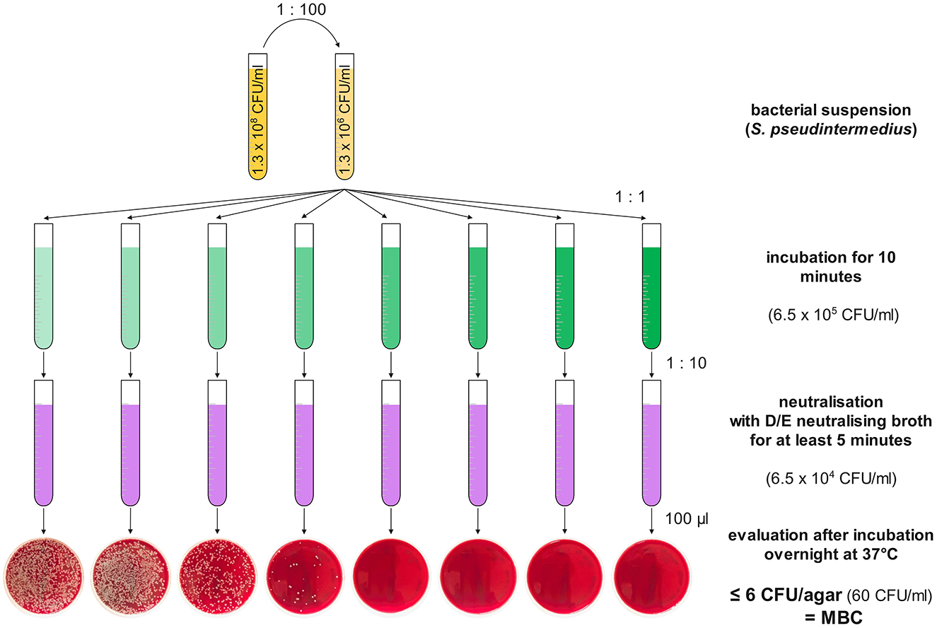
Figure 1. Schematic overview of the experimental design for testing MBCs (regardless of solvent) for Staphylococcus pseudintermedius. In yellow are the bacterial solutions starting with 0.5 MFU on the left, being 100-fold diluted to ~1.3 × 10⁶ CFU/mL. One of the antiseptics in rising (always doubling) concentrations from left to right is shown in green. The different concentrations of the antiseptics were inoculated in a one-to-one ratio with the bacterial solution. After 10 min of incubation, 100 μL of the antiseptic-bacterial mixture was neutralised with 900 μL of D/E-neutralising broth (in violet) for at least 5 min, before plating 100 μL on Columbia sheep–blood agar, incubation overnight, and visual evaluation of the CFUs/agar.
3.5 Statistical analysis
The statistical analysis comprised a descriptive analysis of the collected data for MICs and MBCs (Excel 365, Version 2409, Microsoft 365 Apps for Enterprise), whereas the analysis for the validation of the neutraliser was performed according to ASTM International standard E 1054–2 (51) using SAS Studio software (version 3.8 on SAS 9.4 of the SAS System for Windows. Copyright©2012–2020, SAS Institute, Inc. SAS and all other SAS Institute, Inc. product or service names are registered trademarks or trademarks of SAS Institute, Inc., Cary, NC, USA).
4 Results
4.1 MICs
For polyhexanide, MICs for S. pseudintermedius ranged 1.6–3.2 mg/L, with all three MRSP strains having their MICs at 1.6 mg/L, whereas the MICs for P. aeruginosa and Str. canis ranged 6.4−12.8 mg/L and 3.2–12.8 mg/L, respectively. Additionally, polyhexanide was found to precipitate in concentrations equal to or higher than 25.6 mg/L in CAMHB in a preliminary experiment.
For povidone-iodine MICs for S. pseudintermedius and Str. canis were consistent at 6,400 mg/L, whereas those for P. aeruginosa ranged 3,200–12,800 mg/L.
CAMHB inactivated the antimicrobial effect of HOCl as no MIC or bactericidal effect could be observed in concentrations up to 137.5 mg/L for any of the tested isolates.
4.2 Validation of the Dey–Engley neutralising broth
Dey–Engley neutralising broth was confirmed to be an effective agent for neutralising polyhexanide, povidone-iodine, N-acetylcysteine, and hypochlorous acid within < 5 s, at least up to the tested concentrations, as there was no significant decrease in the bacterial load between the neutralised antiseptics and the positive control. The neutralisation broth had no intrinsic effect on bacterial growth and could therefore be classified as non-toxic to the tested bacterial species.
4.3 MBCs in CAMHB
MBCs in CAMHB for polyhexanide for S. pseudintermedius ranged 3.2–12.8 mg/L, for P. aeruginosa DSM 19880 at 6.4 mg/L, and for Str. canis DSM 20716 at 12.8 mg/L and above.
For povidone-iodine, MBCs for S. pseudintermedius ranged from 6,400 mg/L to >12,800 mg/L, whereas those for P. aeruginosa DSM 19880 were at 6,400 mg/L, and for Str. canis DSM 20716 at 12,800 mg/L and above.
MBCs of NAC for S. pseudintermedius were at 12,800 mg/L, with six of the 33 approaches being slightly above the threshold and therefore must be considered to be above 12,800 mg/L. The MBCs for P. aeruginosa DSM 19880 and Str. canis DSM 20716 were at 6,400 and 12,800 mg/L, respectively.
4.4 MBCs in PBS
MBCs in PBS for polyhexanide were at 0.8–1.6 mg/L for S. pseudintermedius and at 1.6–3.2 mg/L for P. aeruginosa and Str. canis.
For povidone-iodine MBCs in PBS were at 8–32 mg/L for S. pseudintermedius and P. aeruginosa and at 8–16 mg/L for Str. canis.
NAC was effective against S. pseudintermedius in concentrations ranging 6,400–12,800 mg/L when dissolved in PBS and against P. aeruginosa and Str. canis in concentrations ranging 3,200–6,400 mg/L. Additionally, it was found that the pH of 12,800 mg/L NAC in PBS was highly acidic at a pH of ~2.45, rising to pH 4.12 at 1,600 mg/L, where a significant increase to pH 6.48 occurred if further diluted to 800 mg/L.
MBCs for hypochlorous acid for S. pseudintermedius were found at concentrations ranging 0.4–1.6 mg/L, with 1.6 mg/L being required for a total of four approaches of two isolates and for P. aeruginosa and Str. canis at 0.4–0.8 mg/L (see also Figure 2 and Table 1).
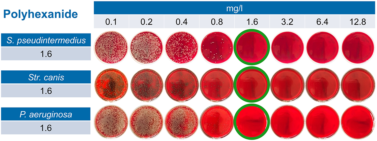
Figure 2. Exemplary results for polyhexanide in PBS across all three tested bacterial species. For Staphylococcus pseudintermedius, for example, one can observe a reduction in the CFU/agar starting at 0.4 mg/L of polyhexanide; nevertheless, a reduction by 99.9% (≤ 6 CFU/agar) is not observed until 1.6 mg/L. Therefore, 1.6 mg/L of polyhexanide dissolved in PBS is considered MBC for this isolate.
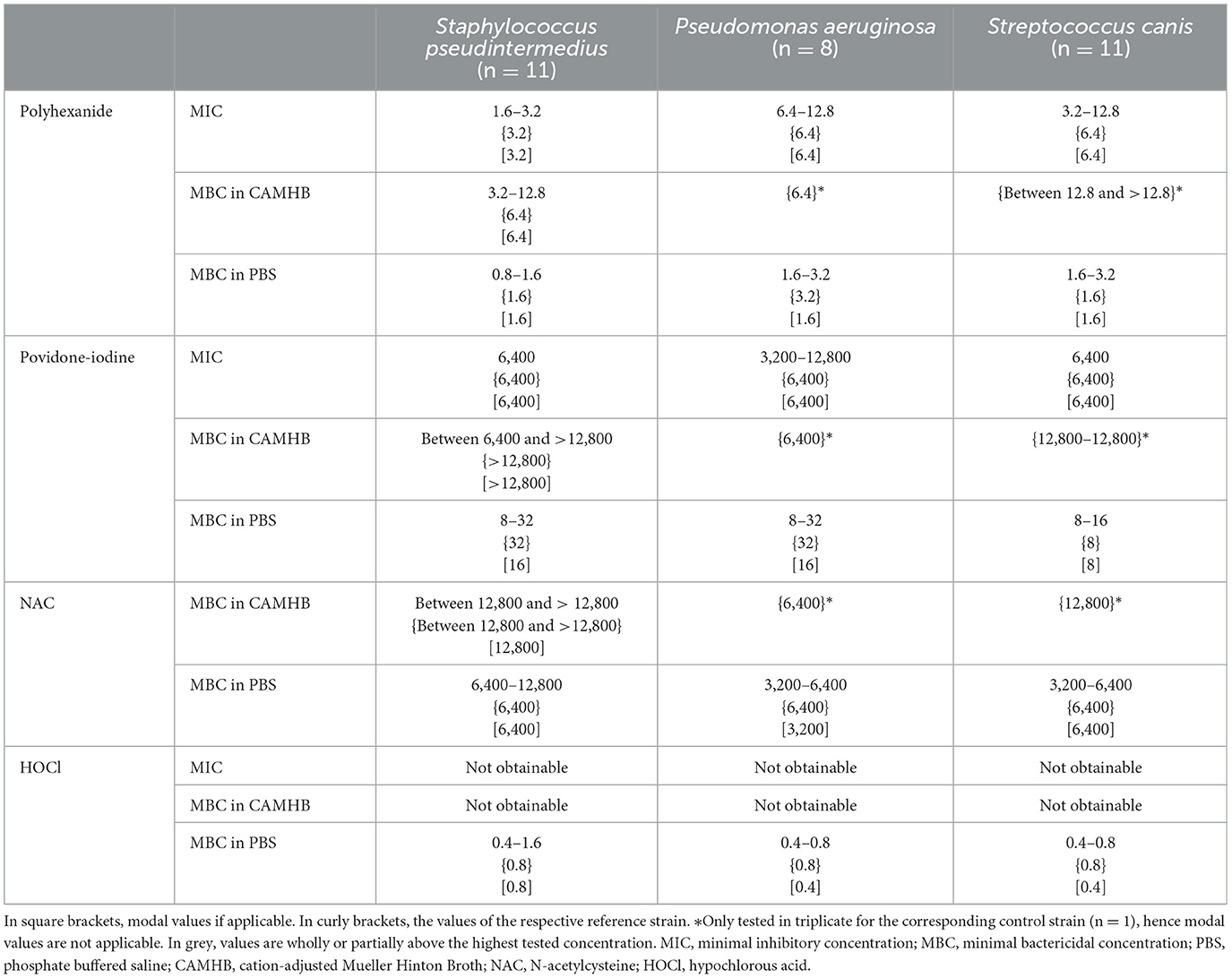
Table 1. Results for MICs and MBCs in CAMHB and PBS in mg/L.
5 Discussion
Our results show that polyhexanide, povidone-iodine, N-acetylcysteine, and hypochlorous acid have a strong in vitro antimicrobial effect against all tested bacterial species, most commonly associated with bacterial keratitis in dogs and cats, especially when tested in PBS. Notably, all tested methicillin-resistant strains of S. pseudintermedius were equally susceptible to all tested antiseptics. All four antiseptics—polyhexanide (53–55), povidone-iodine (33–37), NAC (38, 47, 56, 57), and HOCl (38, 47, 56, 57)—are reported to be effective against biofilms in various locations and settings, which is of particular interest as many of the bacterial species associated with infectious keratitis are known to form biofilms (58–61).
In this study, MBCs were defined as a 3 log reduction of the viable bacterial load as stated by the Clinical and Laboratory Standards Institute (CLSI) (52). Although other definitions of MBCs exist, for example, in DIN EN 1040:2005 (62), we chose the definition provided by the CLSI as it is recognised as a main standard for antimicrobial testing (63, 64) and to improve potential inter-study comparability.
5.1 Polyhexanide (PHMB)
Polyhexanide (PHMB) is used as an antiseptic and disinfectant in a wide array of medical and non-medical settings (24, 25, 65, 66).
It interacts with negatively charged bacterial membranes, leading to their disruption (67, 68), as well as translocating across the membrane and interacting with the genetic material (66, 69, 70), while having a relatively low activity on mammalian cell membranes (71, 72).
In human ophthalmology, it is used as the mainstay for the treatment of Acanthamoeba keratitis (67, 68) and as an alternative for presurgical antisepsis (66, 69, 70), where it is reported to have an extended duration of antisepsis on the ocular surface compared to povidone-iodine (69). Despite being used for years, no bacterial resistance has been reported until now (24) and there is no evidence of resistance development after repeated incubation with polyhexanide (73). One study has shown a protective effect of polyhexanide on human keratocytes being co-cultured with Staphylococcus aureus (74).
Polyhexanide was deemed to be safe for use on the ocular surface in concentrations up to 0.08% (800 mg/L) in human trials (71) and no cytotoxicity to the corneal epithelium was found in an in vitro and ex vivo study for 0.04% (400 mg/L) after an exposure time of 30 min (72), therefore giving a significant margin over our observed MICs and MBCs regardless of solvent.
Polyhexanide is not readily biodegradable, but is still considered to have a low potential for bioaccumulation (75). However, it is considered very toxic to aquatic life with long-lasting effects (75). Although the environmental risk of eyedrops might be less significant, care should be taken when disposing of leftovers.
5.2 Povidone-iodine (PVP-I)
Povidone-iodine (PVP-I) exerts its antimicrobial effect against bacteria, fungi, protozoa, and some viruses (76–79) by gradually releasing free iodine (77, 80), which rapidly penetrates the microorganisms and attacks key groups of proteins, nucleotides, and fatty acids, leading to cell death (77, 81). Despite being used for decades, no induced resistance development has been reported (73, 77, 82).
It is still regarded as the predominant antiseptic for presurgical antisepsis in both veterinary and human ophthalmology (26, 82–84). Additionally, it is also used in the treatment of human ophthalmia neonatorum, some cases of keratitis, and conjunctivitis (26).
Concentrations used and considered to be safe on the ocular surface are somewhat inconsistent. Still, the American Academy of Ophthalmology and European Society of Refractive Surgeons guidelines recommend a concentration of 5% (50,000 mg/L) for corneal antisepsis (83, 84). Foja et al. (72) did not find evidence that using 1 and 5% povidone-iodine for up to 2 min had a cytotoxic effect on porcine corneas, whereas 5% povidone-iodine caused damage to the ocular surface of rabbits in a time-dependent manner (for 3 and 10 min) in another study (85). Our MBCs in PBS are therefore ~1,500-fold lower than the currently recommended concentrations in human ophthalmology. In contrast, the MBCs measured if dissolved in CAMHB are closer to the maximum tolerated concentrations. This is most likely due to protein interference, as discussed later.
As iodine occurs naturally in the environment in relatively high concentrations, the actual risks to the environment arising from the use of iodine are considered acceptable according to the European Chemicals Agency (ECHA). Nevertheless, iodine is considered toxic to aquatic life with long-lasting effects (86) and therefore should not be released carelessly into the environment.
5.3 N-acetylcysteine (NAC)
N-Acetylcysteine (NAC) is an acetylated form of the amino acid L-cysteine and an active precursor of glutathione, which acts as an antioxidant (29, 31, 87, 88). NAC is used regularly in medicine for its mucolytic, antioxidative, and chelating properties (31, 87–92) and additionally has anti-inflammatory properties by the mediation of cytokine release (31, 87, 89, 93).
In ophthalmology, it is widely used in the treatment of corneal ulcerations due to its collagenase-inhibiting properties (27–30), which makes it a mainstay for the treatment of melting ulcers (94). It is also used in the context of dry eye disease and meibomian gland dysfunction (29). One study reported a significant acceleration of corneal wound healing in dogs (95). In recent years, several studies have described antimicrobial properties of NAC against a wide array of bacterial species, including species commonly associated with infectious keratitis in dogs and cats (31–37).
In our study, we could confirm a bactericidal effect of NAC, in concentrations of 3,200–12,800 mg/L (0.32–1.28%) when dissolved in PBS. pH values of the effective concentrations of NAC in PBS ranged from approximately pH 2.45–3.12. The significance of this finding is unclear, and further research is required to assess the effect of the pH values on the bacterial isolates and the efficiency of the NAC itself.
NAC is safe for use on the ocular surface in concentrations of at least 2.5% (96) (25,000 mg/L) and possibly even up to concentrations of 20%, although there are conflicting reports (95, 97). Our reported MBCs are lower than concentrations currently used in veterinary ophthalmology for their collagenase-inhibiting properties. Due to the low margin over the MBCs, NAC is more likely to be used as a prophylactic agent in corneal ulcers, to prevent secondary infection, rather than as a sole treatment option for infectious keratitis.
5.4 Hypochlorous acid (HOCl)
Hypochlorous acid (HOCl) belongs to the group of reactive species (40, 41). It is used as an antiseptic and disinfectant in many medical and non-medical applications (38, 56, 57, 98–101). Additionally, HOCl has been shown to have anti-inflammatory properties (45), as well as favourable effects on fibroblast and keratinocyte migration (38).
In human ophthalmology, it is currently mainly used in the treatment of blepharitis (49, 102–104) and fungal keratitis (44–46). In our study, HOCl was highly effective against all three tested bacterial species in concentrations as low as 0.4–1.6 mg/L when dissolved in PBS. However, when dissolved in CAMHB, no antibacterial effect could be observed in concentrations up to 137.5 mg/L. This effect is most likely due to the interference of organic matter and protein (105), as discussed later. No concerns have been raised about the ocular toxicity of HOCl (106). Wang et al. (39) found HOCl to be non-irritating and non-sensitising to the ocular surface in various animal models in all tested concentrations up to 0.013% (130 mg/L).
Production of hypochlorous acid is relatively inexpensive, though the solution needs to be stabilised (107) and degrades rapidly when coming into contact with organic matter, therefore, the ECHA assesses the potential environmental risk as acceptable (108, 109). Hence, it is a great candidate for rinsing eyes affected by corneal ulcers, as it first lowers the protein content of the tear film and exerts antimicrobial effects afterwards, therefore leading to a reduction of the bacterial load by two different pathways. It has also been reported to reduce the bacterial load without affecting the bacterial diversity (49) or the biodiversity in the meibomian gland secretions of patients with internal hordeolum (110), making it also interesting for preventing secondary infection on the ocular surface.
5.5 Protein interference
For all four antiseptics, differences between the MICs and MBCs in CAMHB and those in PBS were observed. Notably, for HOCl, no antimicrobial effect could be observed up to concentrations of 137.5 mg/L when dissolved in CAMHB, compared to a maximum observed MBC of 1.6 mg/L in PBS. For povidone-iodine, over 100-fold higher concentrations were required to achieve MBCs in CAMHB compared to PBS. This trend was also observable for polyhexanide and NAC, although differences were less marked.
This discrepancy in the effectiveness of the antiseptics depending on the solvent might be due to the protein content of ~2% in CAMHB compared to the protein-free environment of PBS. Since interference between proteins and the antiseptics might lead to a reduction in their efficacy (105, 111).
The protein content of the healthy canine eye ranges from 2.8 to 4.03 μg/μL (112) (0.28–0.403%) and is, therefore, approximately five times lower than the protein content of CAMHB. Hence, a reduction in the efficiency of the antiseptics, especially povidone-iodine and HOCl, is expected upon contact with the tear film. Nevertheless, the reported MBCs in CAMHB for polyhexanide are well, and for povidone-iodine and NAC, they were still below the documented highest tolerated respective concentrations on the ocular surface.
5.6 In vivo effects
In addition to possible interference with organic matter, one must consider the further dilution of the antiseptic upon application onto the ocular surface due to the tear film. In commercially available eyedroppers, the volume of one drop ranges from 26.4 μL up to 69.4 μL (113), with another study finding an average drop volume of 39.0 μL (114). The reported median volume of the tear film of dogs and cats is 65.3 and 32.1 μL, respectively (115), while the reported volumetric capacity of the canine palpebral fissure was 31.3 ± 8.9 μL (15–45 μL) in healthy beagles (116). Therefore, the further dilution of the antiseptics is estimated to be approximately at a one-to-one to one-to-two ratio. As our reported MBCs (especially in PBS) are significantly lower than the reported concentrations deemed to be safe on the ocular surface, there should be a sufficient margin to accommodate the dilution by the tear film.
A further aspect, which needs to be taken into account, is the limited contact time achievable in vivo due to the reflex tear film turnover time, which has been reported to be ~50.0%/min in dogs and cats (115). Therefore, further research is required to determine the time-kill kinetics of the antiseptics on the bacterial species associated with canine and feline infectious keratitis.
Furthermore, it is unclear what effect the antiseptics might have on the tear film quality and composition, as well as on the microbiome in the canine and feline eye. Therefore, further research should be performed in this regard. Additionally, other in vivo aspects, for example, the lipid component of the tear film, might affect the antiseptics; further research is required in this regard.
5.7 Study limitations
Limitations of this study are its in vitro nature, as the in vivo effects and required concentrations might differ, the relatively small sample size, and most of the clinical isolates being of canine origin. Although just one feline isolate was tested, its MIC and MBC were found to be similar to those of the canine isolates and the respective reference strain. Additionally, since MICs and MBCs for the reference strains for all tested bacterial strains were similar to the MICs and MBCs of the clinical isolates, and interspecies variability toward the bactericidal effect was low, we consider it likely for other clinical isolates of feline origin of the tested bacterial strains to behave likewise. Further research toward this aspect is required.
6 Conclusion
Our results show a potent in vitro antimicrobial effect of polyhexanide, povidone-iodine, N-acetylcysteine, and hypochlorous acid when dissolved in PBS against S. pseudintermedius, including methicillin-resistant strains, P. aeruginosa, and Str. canis, which are all commonly associated with canine and feline infectious keratitis. The recorded MBCs were well below known tolerated ocular concentrations. Therefore, the tested antiseptics might be an ideal alternative or addition to topical antibiotics in the treatment and prophylaxis of infectious keratitis, especially as many of the studied substances have been reported to have additional beneficial effects on corneal healing, inflammation, prevention of melting of corneal ulcers, and more. However, some antiseptics, notably povidone-iodine and hypochlorous acid, show a marked reduction in their in vitro efficacy when dissolved in a protein-containing broth, which might indicate a lower efficacy after contact with the tear film in vivo. Further research is required to assess several in vivo factors and time-kill kinetics. All in all, antiseptics might play an essential role in reducing the use of topical antibiotics in veterinary ophthalmology, therefore combating antimicrobial resistance and its development in line with the One Health approach.
Data availability statement
The raw data supporting the conclusions of this article will be made available by the authors, without undue reservation.
Author contributions
HW: Conceptualisation, Investigation, Methodology, Project administration, Visualisation, Writing – original draft, Writing – review & editing, Formal analysis. AP: Investigation, Methodology, Writing – review & editing. HO: Conceptualisation, Methodology, Supervision, Writing – review & editing, Resources. JM: Conceptualisation, Methodology, Resources, Supervision, Writing – review & editing, Funding acquisition. JV: Resources, Writing – review & editing, Conceptualisation. HV: Funding acquisition, Writing – review & editing, Resources. CB: Conceptualisation, Funding acquisition, Project administration, Supervision, Writing – review & editing, Resources, Methodology, Visualisation.
Funding
The author(s) declare that financial support was received for the research and/or publication of this article. We acknowledge financial support by the Open Access Publication Fund of the University of Veterinary Medicine Hannover, Foundation.
Conflict of interest
The authors declare that the research was conducted in the absence of any commercial or financial relationships that could be construed as a potential conflict of interest.
The author(s) declared that they were an editorial board member of Frontiers, at the time of submission. This had no impact on the peer review process and the final decision.
Generative AI statement
The author(s) declare that no Gen AI was used in the creation of this manuscript.
Publisher's note
All claims expressed in this article are solely those of the authors and do not necessarily represent those of their affiliated organizations, or those of the publisher, the editors and the reviewers. Any product that may be evaluated in this article, or claim that may be made by its manufacturer, is not guaranteed or endorsed by the publisher.
References
1. Belknap EB. Corneal emergencies. Top Companion Anim Med. (2015) 30:74–80. doi: 10.1053/j.tcam.2015.07.006
2. Jeng BH, Gritz DC, Kumar AB, Holsclaw DS, Porco TC, Smith SD, et al. Epidemiology of ulcerative keratitis in Northern California. Arch Ophthalmol. (2010) 128:1022–8. doi: 10.1001/archophthalmol.2010.144
3. Suter A, Voelter K, Hartnack S, Spiess BM, Pot SA. Septic keratitis in dogs, cats, and horses in Switzerland: associated bacteria and antibiotic susceptibility. Vet Ophthalmol. (2018) 21:66–75. doi: 10.1111/vop.12480
4. Hewitt JS, Allbaugh RA, Kenne DE, Sebbag L. Prevalence and antibiotic susceptibility of bacterial isolates from dogs with ulcerative keratitis in midwestern United States. Front Vet Sci. (2020) 7:583965. doi: 10.3389/fvets.2020.583965
5. Goldreich JE, Franklin-Guild RJ, Ledbetter EC. Feline bacterial keratitis: clinical features, bacterial isolates, and in vitro antimicrobial susceptibility patterns. Vet Ophthalmol. (2020) 23:90–6. doi: 10.1111/vop.12693
6. Ekapopphan D, Srisutthakarn A, Moonarmart W, Buddhirongawatr R, Bangphoomi N. Identification and antimicrobial susceptibility of microorganisms isolated from severe corneal ulcers of dogs in Thailand. J Vet Med Sci. (2018) 80:1259–65. doi: 10.1292/jvms.18-0045
7. Hindley KE, Groth AD, King M, Graham K, Billson FM. Bacterial isolates, antimicrobial susceptibility, and clinical characteristics of bacterial keratitis in dogs presenting to referral practice in Australia. Vet Ophthalmol. (2016) 19:418–26. doi: 10.1111/vop.12325
8. Lin CT, Petersen-Jones SM. Antibiotic susceptibility of bacterial isolates from corneal ulcers of dogs in Taiwan. J Small Anim Pract. (2007) 48:271–4. doi: 10.1111/j.1748-5827.2007.00348.x
9. Prado MR, Rocha MFG, Brito ÉHS, Girão MD, Monteiro AJ, Teixeira MFS, et al. Survey of bacterial microorganisms in the conjunctival sac of clinically normal dogs and dogs with ulcerative keratitis in Fortaleza, Ceará, Brazil. Vet Ophthalmol. (2005) 8:33–7. doi: 10.1111/j.1463-5224.2005.04061.x
10. Tolar EL, Hendrix DV, Rohrbach BW, Plummer CE, Brooks DE, Gelatt KN. Evaluation of clinical characteristics and bacterial isolates in dogs with bacterial keratitis: 97 cases (1993-2003). J Am Vet Med Assoc. (2006) 228:80–5. doi: 10.2460/javma.228.1.80
11. Tsvetanova A, Powell RM, Tsvetanov KA, Smith KM, Gould DJ. Melting corneal ulcers (keratomalacia) in dogs: a 5-year clinical and microbiological study (2014–2018). Vet Ophthalmol. (2021) 24:265–78. doi: 10.1111/vop.12885
12. Wang L, Pan Q, Zhang L, Xue Q, Cui J, Qi C. Investigation of bacterial microorganisms in the conjunctival sac of clinically normal dogs and dogs with ulcerative keratitis in Beijing, China. Vet Ophthalmol. (2008) 11:145–9. doi: 10.1111/j.1463-5224.2008.00579.x
13. Verdenius CY, Broens EM, Slenter IJM, Djajadiningrat-Laanen SC. Corneal stromal ulcerations in a referral population of dogs and cats in the Netherlands (2012-2019): bacterial isolates and antibiotic resistance. Vet Ophthalmol. (2024) 27:7–16. doi: 10.1111/vop.13080
14. Ollivier FJ. Bacterial corneal diseases in dogs and cats. Clin Tech Small Anim Pract. (2003) 18:193–8. doi: 10.1016/S1096-2867(03)90016-8
15. Schaefer F, Bruttin O, Zografos L, Guex-Crosier Y. Bacterial keratitis: a prospective clinical and microbiological study. Br J Ophthalmol. (2001) 85:842–7. doi: 10.1136/bjo.85.7.842
16. Joksimovic M, Ford BA, Lazic T, Soldatovic I, Luzetsky S, Grozdanic S. Antibiotic recommendations for treatment of canine stromal corneal ulcers. Vet Sci. (2023) 10:20066. doi: 10.3390/vetsci10020066
17. Yilancioglu K. Antimicrobial drug interactions: systematic evaluation of protein and nucleic acid synthesis inhibitors. Antibiotics. (2019) 8:30114. doi: 10.3390/antibiotics8030114
18. Stern GA, Schemmer GB, Farber RD, Gorovoy MS. Effect of topical antibiotic solutions on corneal epithelial wound healing. Arch Ophthalmol. (1983) 101:644–7. doi: 10.1001/archopht.1983.01040010644025
19. Kang MH, Chae MJ, Yoon JW, Kim SG, Lee SY, Yoo JH, et al. Antibiotic resistance and molecular characterization of ophthalmic Staphylococcus pseudintermedius isolates from dogs. J Vet Sci. (2014) 15:409–15. doi: 10.4142/jvs.2014.15.3.409
20. World Health Organisation Pabst S. Antimicrobial Resistance WHO—Newsroom: World Health Organisation. (2023). Available online at: https://www.who.int/news-room/fact-sheets/detail/antimicrobial-resistance (accessed November 21, 2023).
21. Somayaji R, Priyantha MA, Rubin JE, Church D. Human infections due to Staphylococcus pseudintermedius, an emerging zoonosis of canine origin: report of 24 cases. Diagn Microbiol Infect Dis. (2016) 85:471–6. doi: 10.1016/j.diagmicrobio.2016.05.008
22. Pomba C, Rantala M, Greko C, Baptiste KE, Catry B, van Duijkeren E, et al. Public health risk of antimicrobial resistance transfer from companion animals. J Antimicrob Chemother. (2017) 72:957–68. doi: 10.1093/jac/dkw481
23. van Duijkeren E, Kamphuis M, van der Mije IC, Laarhoven LM, Duim B, Wagenaar JA, et al. Transmission of methicillin-resistant Staphylococcus pseudintermedius between infected dogs and cats and contact pets, humans and the environment in households and veterinary clinics. Vet Microbiol. (2011) 150:338–43. doi: 10.1016/j.vetmic.2011.02.012
24. Wessels S, Ingmer H. Modes of action of three disinfectant active substances: a review. Regul Toxicol Pharmacol. (2013) 67:456. doi: 10.1016/j.yrtph.2013.09.006
25. Allen MJ, Morby AP, White GF. Cooperativity in the binding of the cationic biocide polyhexamethylene biguanide to nucleic acids. Biochem Biophys Res Commun. (2004) 318:397–404. doi: 10.1016/j.bbrc.2004.04.043
26. Grzybowski A, Kanclerz P, Myers WG. The use of povidone–iodine in ophthalmology. Curr Opin Ophthalmol. (2018) 29:19–32. doi: 10.1097/ICU.0000000000000437
27. Berman. Collagenase inhibitors: rationale for their use in treating corneal ulceration. Int Ophthalmol Clin. (1975) 15:49–66. doi: 10.1097/00004397-197501540-00006
28. Brooks DE, Ollivier FJ. Matrix metalloproteinase inhibition in corneal ulceration. Vet Clin North Am Small Anim Pract. (2004) 34:611–22. doi: 10.1016/j.cvsm.2003.12.005
29. Eghtedari Y, Oh LJ, Girolamo ND, Watson SL. The role of topical N-acetylcysteine in ocular therapeutics. Surv Ophthalmol. (2022) 67:608–22. doi: 10.1016/j.survophthal.2021.07.008
30. Haffner JC, Fecteau KA, Eiler H. Inhibition of collagenase breakdown of equine corneas by tetanus antitoxin, equine serum and acetylcysteine. Vet Ophthalmol. (2003) 6:67–72. doi: 10.1046/j.1463-5224.2003.00271.x
31. Walter H, Verspohl J, Meißner J, Oltmanns H, Geks AK, Busse C. In vitro antimicrobial activity of N-acetylcysteine against pathogens most commonly associated with infectious keratitis in dogs and cats. Antibiotics. (2023) 12:559. doi: 10.3390/antibiotics12030559
32. Chan WY, Khazandi M, Hickey EE, Page SW, Trott DJ, Hill PB. In vitro antimicrobial activity of seven adjuvants against common pathogens associated with canine otitis externa. Vet Dermatol. (2019) 30:133–e38. doi: 10.1111/vde.12712
33. Drago L, De Vecchi E, Mattina R, Romanò CL. Activity of N-acetyl-L-cysteine against biofilm of Staphylococcus aureus and Pseudomonas aeruginosa on orthopedic prosthetic materials. Int J Artif Organs. (2013) 36:39–46. doi: 10.5301/ijao.5000135
34. Eroshenko D, Polyudova T, Korobov V. N-acetylcysteine inhibits growth, adhesion and biofilm formation of Gram-positive skin pathogens. Microb Pathog. (2017) 105:145–52. doi: 10.1016/j.micpath.2017.02.030
35. Moon JH, Choi YS, Lee HW, Heo JS, Chang SW, Lee JY. Antibacterial effects of N-acetylcysteine against endodontic pathogens. J Microbiol. (2016) 54:322–9. doi: 10.1007/s12275-016-5534-9
36. Onger ME, Gocer H, Emir D, Kaplan S. N-acetylcysteine eradicates Pseudomonas aeruginosa biofilms in bone cement. Scanning. (2016) 38:766–70. doi: 10.1002/sca.21326
37. Quah SY, Wu S, Lui JN, Sum CP, Tan KS. N-acetylcysteine inhibits growth and eradicates biofilm of Enterococcus faecalis. J Endod. (2012) 38:81–5. doi: 10.1016/j.joen.2011.10.004
38. Sakarya S, Gunay N, Karakulak M, Ozturk B, Ertugrul B. Hypochlorous acid: an ideal wound care agent with powerful microbicidal, antibiofilm, and wound healing potency. Wounds. (2014) 26:342–50.
39. Wang L, Bassiri M, Najafi R, Najafi K, Yang J, Khosrovi B, et al. Hypochlorous acid as a potential wound care agent: part I. Stabilized hypochlorous acid: a component of the inorganic armamentarium of innate immunity. J Burns Wounds. (2007) 6:e5.
40. da Cruz Nizer WS, Inkovskiy V, Overhage J. Surviving reactive chlorine stress: responses of gram-negative bacteria to hypochlorous acid. Microorganisms. (2020) 8:1220. doi: 10.3390/microorganisms8081220
41. Andrés CMC, Pérez de la Lastra JM, Juan CA, Plou FJ, Pérez-Lebeña E. Hypochlorous acid chemistry in mammalian cells-influence on infection and role in various pathologies. Int J Mol Sci. (2022) 23:10735. doi: 10.3390/ijms231810735
42. Dianty R, Hirano J, Anzai I, Kanai Y, Hayashi T, Morimoto M, et al. Electrolyzed hypochlorous acid water exhibits potent disinfectant activity against various viruses through irreversible protein aggregation. Front Microbiol. (2023) 14:1284274. doi: 10.3389/fmicb.2023.1284274
43. Hatanaka N, Yasugi M, Sato T, Mukamoto M, Yamasaki S. Hypochlorous acid solution is a potent antiviral agent against SARS-CoV-2. J Appl Microbiol. (2022) 132:1496–502. doi: 10.1111/jam.15284
44. Odorcic S, Haas W, Gilmore MS, Dohlman CH. Fungal infections after boston type 1 keratoprosthesis implantation: literature review and in vitro antifungal activity of hypochlorous acid. Cornea. (2015) 34:1599–605. doi: 10.1097/ICO.0000000000000639
45. Zhao K, Hu F, Zhang Z, Yin X, Wang H, Li M. 001% hypochlorous acid treats aspergillus fumigatus keratitis in rats by reducing fungal load and inhibiting the inflammatory response. Transl Vis Sci Technol. (2023) 12:3. doi: 10.1167/tvst.12.8.3
46. Wang H, Yin X, Zhang Z, Wang Y, Zhang L, Guo J, et al. Evaluation of 001% hypochlorous acid eye drops combined with conventional treatment in the management of fungal corneal ulcers: randomized controlled trial. Curr Eye Res. (2023) 48:887–93. doi: 10.1080/02713683.2023.2226374
47. Romanowski EG, Stella NA, Yates KA, Brothers KM, Kowalski RP, Shanks RMQ. In vitro evaluation of a hypochlorous acid hygiene solution on established biofilms. Eye Contact Lens. (2018) 44(Suppl. 2):S187–s91. doi: 10.1097/ICL.0000000000000456
48. Anagnostopoulos AG, Rong A, Miller D, Tran AQ, Head T, Lee MC, et al. 001% Hypochlorous acid as an alternative skin antiseptic: an in vitro comparison. Dermatol Surg. (2018) 44:1489–93. doi: 10.1097/DSS.0000000000001594
49. Stroman DW, Mintun K, Epstein AB, Brimer CM, Patel CR, Branch JD, et al. Reduction in bacterial load using hypochlorous acid hygiene solution on ocular skin. Clin Ophthalmol. (2017) 11:707–14. doi: 10.2147/OPTH.S132851
50. CLSI. Methods for Dilution Antimicrobial Susceptibility Tests for Bacteria That Grow Aerobically Approved Standard—Ninth Edition. M07-A9 Vol 32 No 2. Wayne, PA: Clinical and Laboratory Standards Institute (2012).
51. ASTM. Standard Test Methods for Evaluation of Inactivators of Antimicrobial Agents. E (1054) – 02. West Conshohocken, PA: ASTM International (2022).
52. CLSI. Methods for Determining Bactericidal Activity of Antimicrobial Agents; Approved Guideline. M26-A Vol 19 No 18. Wayne, PA: Clinical and Laboratory Standards Institute (1999).
53. Hoekstra MJ, Westgate SJ, Mueller S. Povidone-iodine ointment demonstrates in vitro efficacy against biofilm formation. Int Wound J. (2017) 14:172–9. doi: 10.1111/iwj.12578
54. Oduwole KO, Glynn AA, Molony DC, Murray D, Rowe S, Holland LM, et al. Anti-biofilm activity of sub-inhibitory povidone-iodine concentrations against Staphylococcus epidermidis and Staphylococcus aureus. J Orthop Res. (2010) 28:1252–6. doi: 10.1002/jor.21110
55. Capriotti K, Pelletier J, Barone S, Capriotti J. Efficacy of dilute povidone-iodine against multi- drug resistant bacterial biofilms, fungal biofilms and fungal spores. J Clin Res Dermatol. (2018) 5:1–5. doi: 10.15226/2378-1726/5/1/00174
56. Chen CJ, Chen CC, Ding SJ. Effectiveness of hypochlorous acid to reduce the biofilms on titanium alloy surfaces in vitro. Int J Mol Sci. (2016) 17:71161. doi: 10.3390/ijms17071161
57. Aherne O, Ortiz R, Fazli MM, Davies JR. Effects of stabilized hypochlorous acid on oral biofilm bacteria. BMC Oral Health. (2022) 22:415. doi: 10.1186/s12903-022-02453-2
58. Wang Z, Guo L, Li J, Li J, Cui L, Dong J, et al. Antibiotic resistance, biofilm formation, and virulence factors of isolates of staphylococcus pseudintermedius from healthy dogs and dogs with keratitis. Front Vet Sci. (2022) 9:903633. doi: 10.3389/fvets.2022.903633
59. Juárez-Verdayes MA, Reyes-López MA, Cancino-Díaz ME, Muñoz-Salas S, Rodríguez-Martínez S, de la Serna FJ, et al. Isolation, vancomycin resistance and biofilm production of Staphylococcus epidermidis from patients with conjunctivitis, corneal ulcers, and endophthalmitis. Rev Latinoam Microbiol. (2006) 48:238–46.
60. Lorenzo D. Chloramphenicol resurrected: a journey from antibiotic resistance in eye infections to biofilm and ocular microbiota. Microorganisms. (2019) 7:7090278. doi: 10.3390/microorganisms7090278
61. Płoneczka-Janeczko K, Lis P, Bierowiec K, Rypuła K, Chorbiński P. Identification of bap and icaA genes involved in biofilm formation in coagulase negative staphylococci isolated from feline conjunctiva. Vet Res Commun. (2014) 38:337–46. doi: 10.1007/s11259-014-9615-0
62. DIN. DIN EN. 1040: Chemical Disinfectants and Antiseptics—Quantitative Suspension Test for the Evaluation of Basic Bactericidal Activity of Chemical Disinfectants and Antiseptics—Test Method and Requirements (Phase 1). Berlin: Deutsches Institut für Normung eV (2005).
63. Ishak A, Mazonakis N, Spernovasilis N, Akinosoglou K, Tsioutis C. Bactericidal vs. bacteriostatic antibacterials: clinical significance, differences and synergistic potential in clinical practice. J Antimicrob Chemother. (2024) 80:1–17. doi: 10.1093/jac/dkae380
64. Cushnie TPT, Cushnie B, Echeverría J, Fowsantear W, Thammawat S, Dodgson JLA, et al. Bioprospecting for antibacterial drugs: a multidisciplinary perspective on natural product source material, bioassay selection and avoidable pitfalls. Pharm Res. (2020) 37:125. doi: 10.1007/s11095-020-02849-1
65. Sowlati-Hashjin S, Carbone P, Karttunen M. Insights into the polyhexamethylene biguanide (PHMB) mechanism of action on bacterial membrane and DNA: a molecular dynamics study. J Phys Chem B. (2020) 124:4487–97. doi: 10.1021/acs.jpcb.0c02609
66. Hübner N-O, Kramer A. Review on the efficacy, safety and clinical applications of polihexanide, a modern wound antiseptic. Skin Pharmacol Physiol. (2010) 23(Suppl. 1):17–27. doi: 10.1159/000318264
67. Szentmáry N, Shi L, Daas L, Seitz B. Diagnostics and management approaches for Acanthamoeba keratitis. Expert Opin Orphan Drugs. (2020) 8:227–36. doi: 10.1080/21678707.2020.1791081
68. Dart JKG, Saw VPJ, Kilvington S. Acanthamoeba keratitis: diagnosis and treatment update 2009. Am J Ophthalmol. (2009) 148:487–99.e2. doi: 10.1016/j.ajo.2009.06.009
69. Hansmann F, Kramer A, Ohgke H, Strobel H, Müller M, Geerling G. Lavasept as an alternative to PVP-iodine as a preoperative antiseptic in ophthalmic surgery. Randomized, controlled, prospective double-blind trial. Ophthalmologe. (2005). 102:1043–6, 8–50. doi: 10.1007/s00347-004-1120-3
70. Rusiecka-Ziółkowska J, Hill-Bator A, Piatkowska E, Mimier-Janczak M, Bator K, Misiuk-Hojło M. The eye wipes with polyhexanide (HexaClean) in preoperative prophylaxis of cataract surgery. Ophthalmol J. (2023) 8:38–45. doi: 10.5603/OJ.2023.0009
71. Papa V, van Der Meulen IJE, Rottey S, Sallet G, Overweel I, op't Hof M, et al. Ocular safety of high doses Polyhexanide (PHMB) in healthy volunteers. Investig Ophthalmol Visual Sci. (2017) 58:5170.
72. Foja S, Heinzelmann J, Viestenz A, Rueger C, Hecht S, Viestenz A. Evaluation of the possible influence of povidone iodine (PVI) solution and polyhexanide (PHMB) on wound healing in corneal epithelial regeneration. J Clin Med. (2024) 13:20588. doi: 10.3390/jcm13020588
73. Wiegand C, Abel M, Ruth P, Hipler UC. Analysis of the adaptation capacity of Staphylococcus aureus to commonly used antiseptics by microplate laser nephelometry. Skin Pharmacol Physiol. (2012) 25:288–97. doi: 10.1159/000341222
74. Wiegand C, Eberlein T, Andriessen A. Antibacterial activity of polihexanide formulations in a co-culture of HaCaT keratinocytes and Staphylococcus aureus and at different pH levels. Wound Repair Regen. (2017) 25:423–31. doi: 10.1111/wrr.12528
75. European CHemicals Agency. Regulation (EU) No 528/2012 Concerning the Making Available on the Market and Use of Biocidal Products, Evaluation of Active Substances, Assessment Report, Polyhexamethylene biguanide (Mn = 1415. PDI =4.7) PHMB (1415; 4.7)Product type PT01 (Human hygiene). Helsinki: European Chemicals Agency (ECHA) (2017).
76. Kunisada T, Yamada K, Oda S, Hara O. Investigation on the efficacy of povidone-iodine against antiseptic-resistant species. Dermatology. (1997) 195(Suppl. 2):14–8. doi: 10.1159/000246025
77. Lepelletier D, Maillard JY, Pozzetto B, Simon A. Povidone iodine: properties, mechanisms of action, and role in infection control and Staphylococcus aureus decolonization. Antimicrob Agents Chemother. (2020) 64:e00682-20. doi: 10.1128/AAC.00682-20
78. Lachapelle J-M, Castel O, Casado AF, Leroy B, Micali G, Tennstedt D, et al. Antiseptics in the era of bacterial resistance: a focus on povidone iodine. Clin Pract. (2013) 10:579. doi: 10.2217/cpr.13.50
79. Wutzler P, Sauerbrei A, Klöcking R, Brögmann B, Reimer K. Virucidal activity and cytotoxicity of the liposomal formulation of povidone-iodine. Antiviral Res. (2002) 54:89–97. doi: 10.1016/S0166-3542(01)00213-3
80. Bigliardi PL, Alsagoff SAL, El-Kafrawi HY, Pyon J-K, Wa CTC, Villa MA. Povidone iodine in wound healing: a review of current concepts and practices. Int J Surg. (2017) 44:260–8. doi: 10.1016/j.ijsu.2017.06.073
81. McDonnell G, Russell AD. Antiseptics and disinfectants: activity, action, and resistance. Clin Microbiol Rev. (1999) 12:147–79. doi: 10.1128/CMR.12.1.147
82. Soleimani M, Haydar AA, Cheraqpour K, Zeidabadinejad H, Esfandiari A, Eshaghhosseiny N, et al. In praise of povidone-iodine application in ophthalmology. Surv Ophthalmol. (2024) 69:211–23. doi: 10.1016/j.survophthal.2023.11.002
83. Barry P, Cordovés L, Gardner S. ESCRS Guidelines for Prevention and Treatment of Endophthalmitis Following Cataract Surgery: Data, Dilemmas and Conclusions. (2013). Available online at: https://www.escrs.org/media/uljgvpn1/english_2018_updated.pdf (accessed September 9, 2024).
84. Musumeci R, Troiano P, Martinelli M, Piovella M, Carbonara C, Rossi S, et al. Effectiveness of 0.66% povidone-iodine eye drops on ocular surface flora before cataract surgery: a nationwide microbiological study. J Clin Med. (2021) 10:10102198. doi: 10.3390/jcm10102198
85. Kim S, Ahn Y, Lee Y, Kim H. Toxicity of Povidone-iodine to the ocular surface of rabbits. BMC Ophthalmol. (2020) 20:359. doi: 10.1186/s12886-020-01615-6
86. European CHemicals Agency. Regulation (EU) n°528/2012 Concerning the Making Available on the Market and Use of Biocidal Products, Evaluation of Active Substances, Assessment Report, Iodine (including PVP-iodine), Product types 1, 3, 4, 22. Helsinki: European Chemicals Agency (ECHA) (2013).
87. Tieu S, Charchoglyan A, Paulsen L, Wagter-Lesperance LC, Shandilya UK, Bridle BW, et al. N-Acetylcysteine and its immunomodulatory properties in humans and domesticated animals. Antioxidants. (2023) 12:12101867. doi: 10.3390/antiox12101867
88. Hou Y, Wang L, Yi D, Wu G. N-acetylcysteine and intestinal health: a focus on its mechanism of action. Front Biosci. (2015) 20:872–91. doi: 10.2741/4342
89. Tenório M, Graciliano NG, Moura FA, Oliveira ACM, Goulart MOF. N-Acetylcysteine (NAC): impacts on human health. Antioxidants. (2021) 10:10060967. doi: 10.3390/antiox10060967
90. Wang H, Li C, Peng M, Wang L, Zhao D, Wu T, et al. N-Acetylcysteine improves intestinal function and attenuates intestinal autophagy in piglets challenged with β-conglycinin. Sci Rep. (2021) 11:1261. doi: 10.1038/s41598-021-80994-2
91. Yi D, Hou Y, Xiao H, Wang L, Zhang Y, Chen H, et al. N-Acetylcysteine improves intestinal function in lipopolysaccharides-challenged piglets through multiple signaling pathways. Amino Acids. (2017) 49:1915–29. doi: 10.1007/s00726-017-2389-2
92. Boman G, Bäcker U, Larsson S, Melander B, Wåhlander L. Oral acetylcysteine reduces exacerbation rate in chronic bronchitis: report of a trial organized by the Swedish Society for Pulmonary Diseases. Eur J Respir Dis. (1983) 64:405–15.
93. Zafarullah M, Li WQ, Sylvester J, Ahmad M. Molecular mechanisms of N-acetylcysteine actions. Cell Mol Life Sci. (2003) 60:6–20. doi: 10.1007/s000180300001
94. Kimmitt BA, Moore GE, Stiles J. Comparison of the efficacy of various concentrations and combinations of serum, ethylenediaminetetraacetic acid, tetracycline, doxycycline, minocycline, and N-acetylcysteine for inhibition of collagenase activity in an in vitro corneal degradation model. Am J Vet Res. (2018) 79:555–61. doi: 10.2460/ajvr.79.5.555
95. Aldavood SJ, Behyar R, Sarchahi AA, Rad MA, Noroozian I, Ghamsari SM, et al. Effect of acetylcysteine on experimental corneal wounds in dogs. Ophthalmic Res. (2003) 35:319–23. doi: 10.1159/000074070
96. Thermes F, Molon-Noblot S, Grove J. Effects of acetylcysteine on rabbit conjunctival and corneal surfaces. A scanning electron microscopy study. Invest Ophthalmol Vis Sci. (1991) 32:2958–63.
97. Fischak C, Klaus R, Werkmeister RM, Hohenadl C, Prinz M, Schmetterer L, et al. Effect of topically administered chitosan-n-acetylcysteine on corneal wound healing in a rabbit model. J Ophthalmol. (2017) 2017:5192924. doi: 10.1155/2017/5192924
98. Fukuzaki S. Uses of gaseous hypochlorous acid for controlling microorganisms in indoor spaces. J Microorg Control. (2023) 28:165–75. doi: 10.4265/jmc.28.4_165
99. Feng KC, Ghai A, Liu H, Salerno A, Miller C, Liu J, et al. Efficacy of hypochlorous acid (HOCl) fog in sanitizing surfaces against Enterococcus faecalis. Am J Infect Control. (2022) 50:1311–5. doi: 10.1016/j.ajic.2022.03.009
100. Rutala WA, Weber DJ. Uses of inorganic hypochlorite (bleach) in health-care facilities. Clin Microbiol Rev. (1997) 10:597–610. doi: 10.1128/CMR.10.4.597
101. Mueller RS, Baumann KN, Boehm T, Dörfelt S, Kasper B, Udraite-Vovk L. Evaluation of hypochlorous acid as an ear flush in dogs with chronic otitis externa. Vet Dermatol. (2023) 34:134–41. doi: 10.1111/vde.13142
102. Mencucci R, Morelli A, Favuzza E, Galano A, Roszkowska AM, Cennamo M. Hypochlorous acid hygiene solution in patients affected by blepharitis: a prospective randomised study. BMJ Open Ophthalmol. (2023) 8:e001209. doi: 10.1136/bmjophth-2022-001209
103. Bertone C, Mollicone A, Russo S, Sasso P, Fasciani R, Riccardi C, et al. The role of hypochlorous acid in the management of eye infections: a case series. Drugs Context. (2022) 11:2022-3-10. doi: 10.7573/dic.2022-3-10
104. Zhang H, Wu Y, Wan X, Shen Y, Le Q, Yang P, et al. Effect of hypochlorous acid on blepharitis through ultrasonic atomization: a randomized clinical trial. J Clin Med. (2023) 12:12031164. doi: 10.3390/jcm12031164
105. Ishihara M, Murakami K, Fukuda K, Nakamura S, Kuwabara M, Hattori H, et al. Stability of weakly acidic hypochlorous acid solution with microbicidal activity. Biocontrol Sci. (2017) 22:223–7. doi: 10.4265/bio.22.223
106. Gold MH, Andriessen A, Bhatia AC, Bitter P, Jr., Chilukuri S, Cohen JL, et al. Topical stabilized hypochlorous acid: the future gold standard for wound care and scar management in dermatologic and plastic surgery procedures. J Cosmet Dermatol. (2020) 19:270–7. doi: 10.1111/jocd.13280
107. Block MS, Rowan BG. Hypochlorous acid: a review. J Oral Maxillofac Surg. (2020) 78:1461–6. doi: 10.1016/j.joms.2020.06.029
108. European CHemicals Agency. Regulation (EU) No 528/2012 Concerning the Making Available on the Market and Use of Biocidal Products, Evaluation of Active Substances, Assessment Report: Active Chlorine Released From Hypochlorous Acid, Product-Type 2, (Disinfectants and Algaecides not Intended for Direct Application to Humans or Animals). Helsinki: European Chemicals Agency (ECHA) (2020).
109. European CHemicals Agency. Biocidal Products Committee (BPC), Opinion on the Application for Approval of the Active Substance: Active Chlorine Released from Hypochlorous Acid, Product Type: 1, ECHA/BPC/255/2020. In: European CHemical Agency, editor. Helsinki: European Chemicals Agency (ECHA) (2020).
110. Yang S, Wu BC, Cheng Z, Li L, Zhang YP, Zhao H, et al. The microbiome of meibomian gland secretions from patients with internal hordeolum treated with hypochlorous acid eyelid wipes. Dis Markers. (2022) 2022:7550090. doi: 10.1155/2022/7550090
111. Kramer A, Dissemond J, Kim S, Willy C, Mayer D, Papke R, et al. Consensus on wound antisepsis: update 2018. Skin Pharmacol Physiol. (2018) 31:28–58. doi: 10.1159/000481545
112. Ritchoo S. Havanapan P-o, Phungthanom N, Rucksaken R, Muikaew R, Sussadee M. Analysis and comparison of tear protein profiles in dogs using different tear collection methods. BMC Vet Res. (2022) 18:442. doi: 10.1186/s12917-022-03543-7
113. Van Santvliet L, Ludwig A. Determinants of eye drop size. Surv Ophthalmol. (2004) 49:197–213. doi: 10.1016/j.survophthal.2003.12.009
114. Lederer CM, Jr., Harold RE. Drop size of commercial glaucoma medications. Am J Ophthalmol. (1986) 101:691–4. doi: 10.1016/0002-9394(86)90771-3
115. Sebbag L, Allbaugh RA, Wehrman RF, Uhl LK, Ben-Shlomo G, Chen T, et al. Fluorophotometric assessment of tear volume and turnover rate in healthy dogs and cats. J Ocul Pharmacol Ther. (2019) 35:497–502. doi: 10.1089/jop.2019.0038
Keywords: bacterial keratitis, antibiotic resistance, One Health, polyhexanide, povidone-iodine, N-acetylcysteine, hypochlorous acid
Citation: Wolff HT, Piroth AC, Oltmanns H, Meißner J, Verspohl J, Volk HA and Busse C (2025) Commercially available antiseptics show high in vitro efficacy against pathogens most commonly associated with canine and feline infectious keratitis. Front. Vet. Sci. 12:1552230. doi: 10.3389/fvets.2025.1552230
Received: 27 December 2024; Accepted: 29 April 2025;
Published: 21 May 2025.
Edited by:
Dirk Werling, Royal Veterinary College (RVC), United KingdomReviewed by:
Cristin Coman, “Cantacuzino”, National Institute of Medical-Military Research and Development, RomaniaPiera Anna Martino, University of Milan, Italy
Copyright © 2025 Wolff, Piroth, Oltmanns, Meißner, Verspohl, Volk and Busse. This is an open-access article distributed under the terms of the Creative Commons Attribution License (CC BY). The use, distribution or reproduction in other forums is permitted, provided the original author(s) and the copyright owner(s) are credited and that the original publication in this journal is cited, in accordance with accepted academic practice. No use, distribution or reproduction is permitted which does not comply with these terms.
*Correspondence: Hinrich Tönjes Wolff, aGlucmljaC53b2xmZkB0aWhvLWhhbm5vdmVyLmRl