 Ehab Kotb Elmahallawy1,2*
 Marwa F. Hassan3
 David Cano-Terriza1,4
 Nada Oudah Albalawi5
 Tomás Fajardo1
 Asmaa Aboelabbas Gouda6
 Ayman Atiba7Ahmed Hendawy7
 Isabelle Villena8
 Ashraf Mohamed Barakat9Hind Alzaylaee10
 Sonia Almería11
 Ignacio García-Bocanegra1,4*
- 1Departamento de Sanidad Animal, Grupo de Investigación en Sanidad Animal y Zoonosis (GISAZ), Universidad de Córdoba, Córdoba, Spain
- 2Department of Zoonoses, Faculty of Veterinary Medicine, Sohag University, Sohag, Egypt
- 3Department of Biochemistry, Toxicology and Feed Deficiency, Animal Health Research Institute (AHRI), Agriculture Research Center (ARC), Dokki, Giza, Egypt
- 4CIBERINFEC, ISCIII CIBER de Enfermedades Infecciosas, Instituto de Salud Carlos III, Madrid, Spain
- 5Department of Biology, Faculty of Science, Taibah University, Al-Madinah Province, Saudi Arabia
- 6Department of Parasitology, Faculty of Veterinary Medicine, Zagazig University, Zagazig, Egypt
- 7Department of Surgery, Anesthesiology and Radiology, Faculty of Veterinary Medicine, Kafrelsheikh University, Kafr El Sheikh, Egypt
- 8Laboratory of Parasitology, National Reference Centre for Toxoplasmosis, Reims Hospital, University of Reims Champagne-Ardenne, UR 7510, Reims, France
- 9Department of Zoonotic Diseases, National Research Centre, Giza, Egypt
- 10Department of Biology, College of Science, Princess Nourah bint Abdulrahman University, Riyadh, Saudi Arabia
- 11Virology and Parasitology Branch, Department of Health and Human Services, Division of Food and Environmental Safety, Office of Applied Microbiology and Technology (OAMT), Office of Laboratory Operations and Applied Sciences (OLOAS), Food and Drug Administration, Laurel, MD, United States
Introduction: Toxoplasmosis, caused by the intracellular protozoan Toxoplasma gondii (T. gondii), continues to be a widespread parasitic zoonotic disease globally. The seroepidemiology of T. gondii infection in Egyptian equids, particularly donkeys, remains insufficiently explored. The present study was designed to assess the seroprevalence of T. gondii in equines from Northern Egypt.
Methods: A total of 360 serum samples from two equine species (157 horses and 203 donkeys) were obtained during 2023. The Modified Agglutination Test (MAT, cut-off of 1:25) was used to screen for the anti-T. gondii antibodies. The study also analyzed potential risk factors that could contribute to the exposure of the animals to the parasite, including species, breed, sex, age, and the specific location of each animal.
Results: The overall seroprevalence of T. gondii among examined equines was 41.11% (95% Confidence Interval [CI]: 36.03–46.19). The relationships between seropositivity and explanatory variables were analyzed using a Generalized Estimating Equation (GEE) approach. The seroprevalence of T. gondii was significantly higher in donkeys (51.23%) than in horses (28.03%; p < 0.001; odds ratio [OR] = 2.99; 95% CI: 2.35–3.81).
Conclusions: Collectively, our findings revealed a high T. gondii exposure among equine species in Northern Egypt, with a notably higher seroprevalence in donkeys compared to horses. This study represents one of the most extensive serosurveys of T. gondii in equids conducted in Egypt, featuring the largest sample size of donkeys examined to date. It also examined previously unexplored risk factors related to parasite exposure in equids. The present findings highlight the critical importance of performing periodical surveillance, monitoring, and management of the parasite among equids, which might have a major impact on animal and public health.
1 Introduction
Toxoplasma gondii, the causative agent of toxoplasmosis, remains one of the most common intracellular protozoa in the world. Sexual reproduction of these protozoa occurs in felids, the definitive hosts, which release oocysts in their feces. Intermediate hosts, which encompass nearly all warm-blooded species, can also become infected (1, 2). Humans and animals most commonly contract T. gondii through several primary routes: consuming undercooked or raw meat containing tissue cysts of the parasite, ingesting food or water contaminated with sporulated T. gondii oocysts, and via blood transfusions or transplacental transmission involving tachyzoites. In relation to its clinical impact, T. gondii infections are typically sub-clinical in immunocompetent persons; nevertheless, in immunocompromised individuals, this opportunistic protozoon may induce fatal conditions and even death as well as cause abortion, congenital malformations, and stillbirth (1).
Horses and donkeys are essential for agricultural work, transportation, and economic support, especially in rural areas, and they hold cultural and historical significance, contributing to traditional practices and local economies. However, equid production faces major challenges from various pathogens, including parasites. Among others, T. gondii infection in equines occurs primarily through food or water contaminated with sporulated oocysts, nonetheless, tachyzoites may also be transferred from the mare to the fetus through the placenta (3, 4). Despite T. gondii infection in horses being usually subclinical, atypical clinical signs of toxoplasmosis such as ataxia, fever, encephalomyelitis, and retinal degeneration have been reported (5). Abortion, fever, stillbirth, and degeneration in retina were also reported in pregnant mares infected with T. gondii (4, 6). Additionally, some previous works reported the potential association between clinical equine protozoal myeloencephalitis in horses and T. gondii seropositivity (7, 8). In the USA, fatal toxoplasmosis was observed in a horse (9). Collectively, T. gondii infection in equines may have a substantial impact, posing animal health risks and contributing to significant economic and reproductive losses (10). Taking this into account, consumption of equine meat is still prevalent in several countries, particularly those of the European Union (EU) (11). Previous studies have established the epidemiological link between the consumption of horse meat and clinical toxoplasmosis in humans (12). Additionally, the rising popularity of raw donkey milk has raised concerns, suggesting that consuming such milk from seropositive donkeys may increase the risk of human toxoplasmosis (13). In Egypt, horses and donkeys play a crucial role in agriculture, transportation, and economic sustainability, particularly in rural areas. Beyond their practical use, these animals also hold cultural and historical significance, contributing to traditional practices and local economies. Furthermore, raw meat of these animals is used as feed for carnivorous zoo animals, which may be represent a potential source of a source of infection if the meat is contaminated (14). In this context, viable T. gondii was detected in tissues of 25 donkeys slaughtered at the Giza Zoo abattoir (15). Clearly, infection by T. gondii might have an important impact on public health (10).
Monitoring exposure to the parasite is pivotal for implementation of effective control measures. Serological tests are the primary methods for diagnosing T. gondii in farm animals, including equids (14–16). These techniques are helpful tools for conducting screening surveys since they allow us to identify Toxoplasma-positive animals and farms as well as to analyze the associated risk factors linked to parasite exposure (1).
Previous literature showed that numerous serological investigations have been assessing T. gondii exposure in equids worldwide (7, 17). In Egypt, seroprevalence rates of T. gondii in horses and donkeys have been reported to vary widely, ranging from 12.0% to 68.4% (Table 1) (15, 18–25). Most previous studies involved only a limited number of animals and were performed over short periods of time. In addition, existing data on the seroprevalence of T. gondii in equines from Northern Egypt, particularly donkeys, remain insufficient, leaving a gap in comprehensive data. Consequently, the current study aimed to evaluate the seroprevalence of T. gondii and identify potential risk factors associated with exposure to this zoonotic protozoan in equids from Northern Egypt.
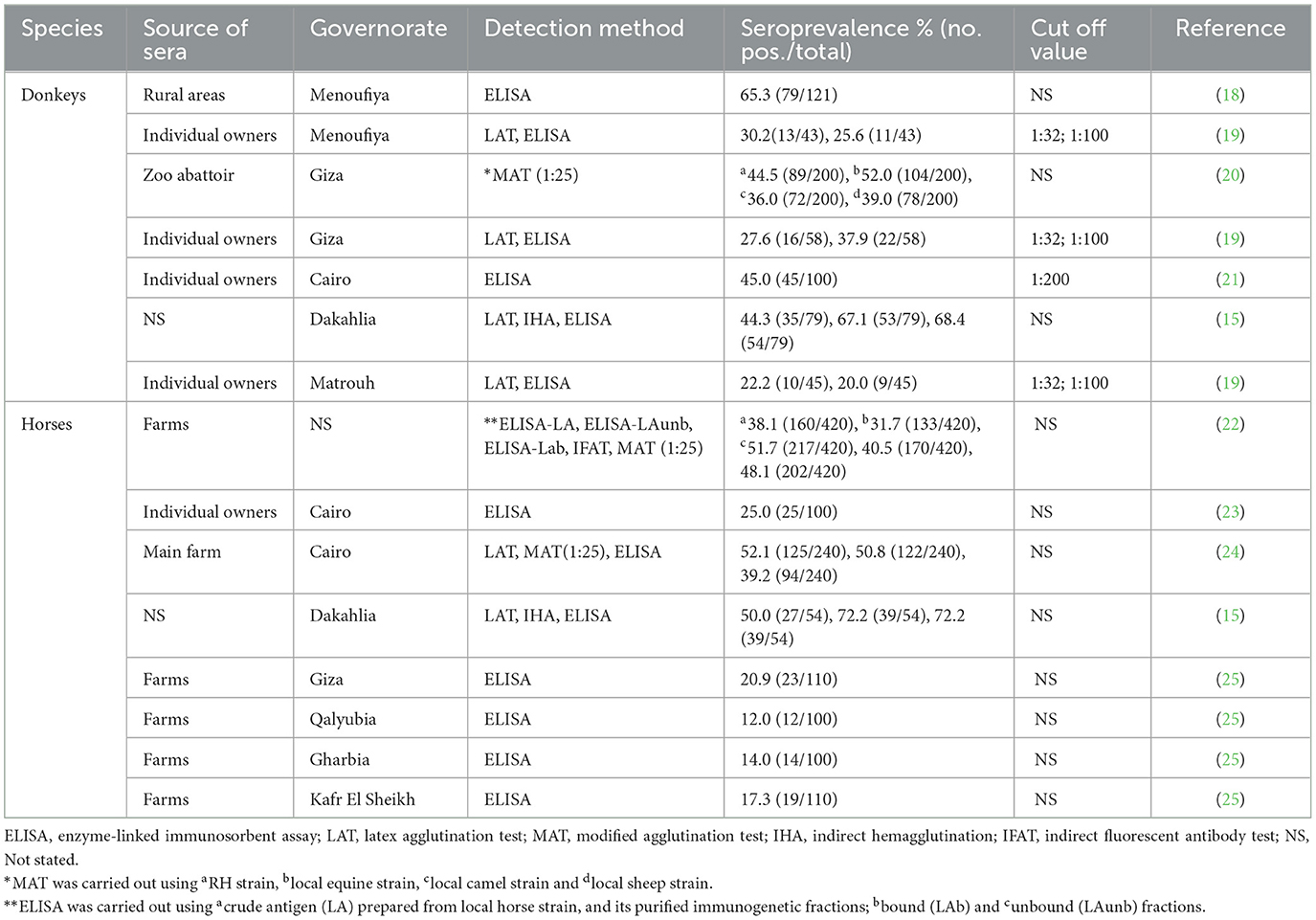
Table 1. Seroprevalence of Toxoplasma gondii reported in equines in Northern Egypt.
2 Materials and methods
2.1 Study area
In 2019, Egypt's equid population was reported to be 958.190, with donkeys making up 90.9% of this number, approximately 871.447 donkeys (26). The present study was conducted in Northern Egypt, primarily focusing on two regions: Cairo and Kafr El Sheikh, which are geographically at 30° 02' 30” N, 031° 14' 07” E and 31° 06' 42” N, 30° 56' 45” E respectively (Figure 1). Both regions experience mild to hot weather throughout most of the year, with average summer temperatures ranging from 31 to 34°C (88.5–92°F) and winter temperatures between 13.5 and 18°C (56.3–65°F). Northern Egypt receives an average annual rainfall of only 100–200 mm (4–8 inches), primarily occurring during the winter months.
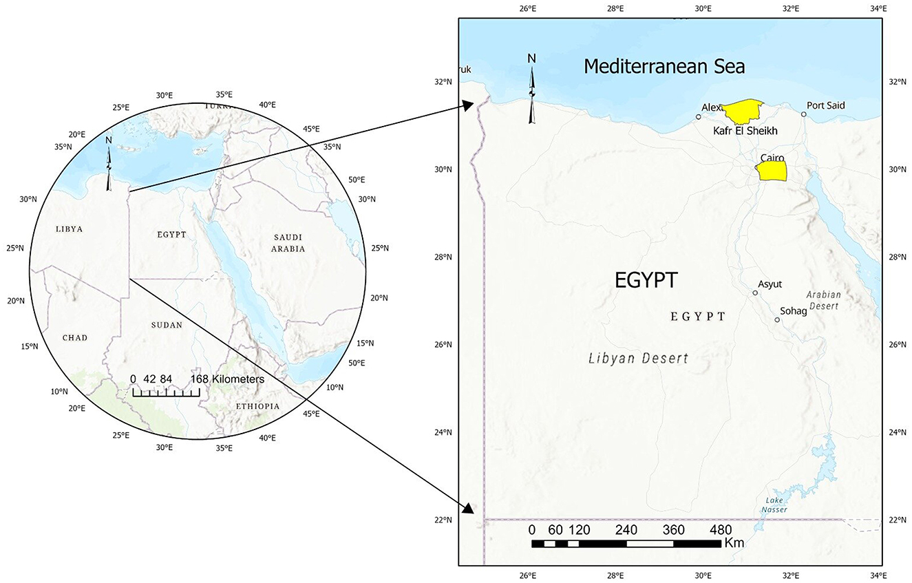
Figure 1. Map of Egypt showing the studied area and sampling sites.
2.2 Animals and samples
Between January and December 2023, blood samples were collected from 360 equines, across Northern Egypt (Figure 1). Samples were collected randomly from donkeys and horses owned by small stakeholders relying on them for transportation and agriculture, taking advantage of opportunities presented by local veterinary campaigns. Considering the number of equids in the study area (n > 10.000), an estimated prevalence of 36% (based on an estimated national seroprevalence threshold of 35.79% [Table 1]), an accepted error of 5% and a 95% confidence interval (95%CI) resulted in 354 animals to be sampled. A total of 360 equids, including 157 horses, 203 donkeys, were finally included in the study. A total of 10 ml blood samples were collected from each animal by puncturing the jugular vein. Sera were obtained by centrifugation at 3,000 rpm for 10 min and were kept at −20°C until serological assessment. The information on animals was thoroughly documented, whenever possible. Information collected included species, breed, sex, age, and the specific location of each animal. Age of examined animals were classified into four categories: foal animals (< 1 year), young (1–4 years), adult (4–15 years) and geriatric (>15 years) as outlined in prior studies (27, 28). The ages of the examined equines were based on the animal's dentition (29, 30).
2.3 Serological examination
The presence of T. gondii antibodies in the serum of equids was evaluated by the modified agglutination test (MAT). This method has been previously validated in equine and performed according to the protocols established by Dubey and Desmonts (31). Briefly, the test was utilized to detect T. gondii antibodies, employing whole-killed tachyzoites as antigens which is kindly provided by Laboratory of Parasitology, University of Reims Champagne-Ardenne, France. The protocol (31) incorporated 2-mercaptoethanol directly into the antigen rather than treating the serum. Equine serum samples were first diluted at a 1:20 ratio in phosphate-buffered saline (PBS, pH 7.2), and 0.05 mL of the diluted sample was added to U-bottom microtiter plate wells. Serial two- to fourfold dilutions were then performed using PBS. The antigen stock solution was diluted (1:10) in a freshly prepared alkaline buffer containing 2-mercaptoethanol, which was either made fresh or stored for no longer than 2 weeks. The antigen solution (0.05 mL) was then added to each well, and the plates were sealed with cellophane before being incubated overnight at 37°C in a humidified environment. The results of the assay were assessed using a microtiter plate under appropriate lighting conditions. A diffuse mat across the wellthe well indicated a positive reaction, whereas a compact button at the center signified a negative result. To ensure assay accuracy and reproducibility, each run included validated positive and negative serum controls. A titer of 1:25, was applied as a cut-off for T. gondii seropositivity as previously considered for these animal species (32). Additionally, serum that initially tested positive at ≥1:25 dilution was thereafter retested at 1:25 and 1:50.
2.4 Statistical analysis
To establish the seroprevalence of T. gondii, we computed the percentage of seropositive samples relative to the total number of examined equids, with a 95% confidence interval (95% CI). Relation between explanatory variables (species, breeds, sex, age, and region) and serological results was estimated using a Pearson's chi-square or Fisher exact tests to allocate the relevance of these variables in the risk of exposure of animals to T. gondii in a bivariate analysis. Variables with a p < 0.10 were selected for multivariate analysis, considering collinearity through Cramer's V coefficients. Generalized Estimating Equation (GEE) models were utilized to assess the influence of explanatory variables identified in the bivariate analysis (33), with municipality as a random effect. Statistical significance was set at p < 0.05, and analysis was conducted using SPSS 25.0 software.
3 Results
In this work, anti-T. gondii antibodies were detected in 148 of 360 equines (41.11%; 95% CI: 36.03–46.19). Seropositivity by species was 51.23% (104/203) in donkeys and 28.03% (44/157) in horses (Figure 2, Table 2). Among the positive samples, T. gondii titres in donkeys were 1:25 in 43.3% (45/104) and 1:50 in 56.3% (59/104). In horses, 54.5% (24/44) had titers of 1:25, while 45.4% (20/44) of positive animals showed titers of 1:50. Table 2 presents the distribution of T. gondii seropositivity, as determined by the bivariate analysis and the epidemiological questionnaire results. Two out of five explanatory variables associated (p < 0.10) with T. gondii seropositivity in equines were selected after data exploration and bivariate analyses and included in the multivariable analysis. The final GEE multivariable analysis identified species as risk factorpotentially linked with the exposure to T. gondii (Table 3). In this respect, significantly higher seropositivity was found in donkeys (p < 0.001, OR = 2.99; 95% CI: 2.35–3.81) compared to horses.
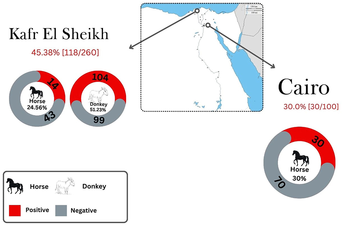
Figure 2. Seroprevalence of Toxoplasma gondii in horses and donkeys in Northern Egypt by location.
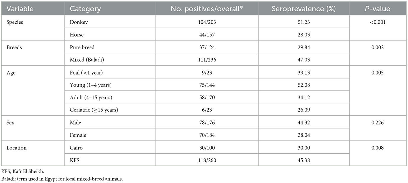
Table 2. Univariable analysis of risk factors associated with T. gondii exposure in equines.
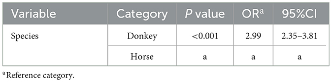
Table 3. Results of the generalized estimating equations model identified potential risk factors associated with T. gondii exposure in equines.
4 Discussion
Toxoplasma gondii remains a highly prevalent zoonotic pathogen worldwide, affecting a broad spectrum of intermediate hosts, including equines. This far-reaching impact emphasizes the global significance of T. gondii infection (15, 19, 34), as equine toxoplasmosis is increasingly recognized as a major potential source of human infection (34). Unfortunately, there is a paucity of current data on the epidemiology of T. gondii infection in equids in Egypt, especially among donkeys. Most existing research is outdated and based on small sample sizes (15, 18, 19). This present study is considered one of the largest investigations into the seroprevalence of T. gondii in equids in Egypt, with a particular focus on donkeys.
The individual seroprevalence noted in horses in the current study (28.03%) is consistent with a previous study conducted in Cairo which reported a similar seroprevalence rate of 25% (25/100) (23). However, the present results are lower than those reported in various previous investigations in Cairo Governorate which showed seroprevalence values of 48.1% (202/420) (22) and 50.8% (122/240) (24). Another previous investigation reported an even higher seroprevalence rate in this species (72.2%; 39/54) in Dakahlia Governorate, Northern Egypt (15). In contrast, previous investigations reported a lower seroprevalences of 20.9% (23/110), 12.0% (12/100), 14.0% (14/100) and 17.3% (19/110) in horse populations in Giza, Qalyubia, Gharbia, and Kafr El Sheikh Governorates, respectively (25).
In the present study, the individual seroprevalence reported in donkeys was 51.23%, which is within the range previously recorded in this species in Egypt (Table 1). A nearly similar seroprevalence rate against T. gondii was observed in donkeys in Cairo Governorate (45%; 45/100) (21). However, our findings were lower than those reported in Menoufiya Governorate, Northern Egypt, where the seroprevalence was 65.3% (79/121) (18). Another study conducted in Dakahlia Governorate, Northern Egypt reported a higher seroprevalence rate of T. gondii, at 68.4% (54/79), among donkey populations in that area (15). In contrast, the current study found higher seroprevalence rates compared to those reported in Giza, Menoufiya, and Matrouh Governorates, Northern Egypt, using ELISA, where the seroprevalence rates were 37.9% (22/58), 25.6% (11/43), and 20.0% (9/45), respectively (19). The variation in T. gondii seroprevalence rates between the current study and previous research on equines could likely be attributed to several factors, primarily differences in farming management and sanitation practices, the specific serological tests used and the cutoff titers used for interpretation, timing of sampling, sample size, and the density of infected definitive hosts and their interaction with the animals (10, 15, 25, 35, 36). Likewise, climate-related factors such as geographic distribution, population density, and the abundance of cats (more prevalent in rural areas compared to urban governorates) would play a crucial role in the maintenance, spread, survival, and transmission of T. gondii, which contribute to the parasite's dynamics (2, 10, 37).
As shown in the present work, the risk of T. gondii seropositivity was 2.9 times greater in donkeys than in horses, indicating that donkeys are more susceptible to T. gondii exposure. These findings are consistent with previously reported data (38, 39). The variations in T. gondii seroprevalence rates between donkeys and horse may be attributed to differences in feeding, management, and sanitation practices including regular stall cleaning, disinfection, pasture rotation, proper manure disposal, and clean water supply (34). Donkeys in Egypt, often kept in extensive management systems under improper management and sanitation practices, and used more frequently for work, are more exposed to the parasite through ingestion of oocysts contaminating their environment, compared to horses (38). A previous investigation Munhoz et al. (39) concluded that donkeys could maintain detectable T. gondii antibody titers for a longer duration than horses. Additionally, the animal species susceptibility can also influence this difference (38, 40–43).
The current study has several limitations that should be noted. Firstly, the sample size from Cairo Governorate was small, and no donkey samples were collected in that area, which may limit the results' applicability to the overall equine population in this region. Cairo is a densely populated area with relatively few donkeys, which further constrains the extrapolation of our findings. Additionally, the study faced some challenges due to the small number of foals and geriatric equids, resulting in sample sizes variations between age groups. Some epidemiological factors, including breeding systems, sanitation conditions, and the presence of cats and rodents, were not assessed in this study. To gain a more comprehensive understanding, future large-scale serological and molecular studies should integrate these variables.
5 Conclusions
The present study constitutes one of the most extensive serosurveys of T. gondii in equines conducted in Egypt. The high level of exposure detected in these species raises animal and public health concerns. Notably, the study identified higher seroprevalence rates in donkeys compared to horses and represents the largest survey of donkeys ever conducted in Egypt so far. The study also offers new insights into the influence of various risk factors associated with the rate of exposure to the parasite. Given its significance for animal and public health, our seroepidemiological findings provide valuable information for Egyptian authorities to enhance control and prevention strategies for toxoplasmosis in equids. The study also underscores the importance of implementing strict measures to prevent potential transmission of the parasite to humans through the consumption of raw or undercooked meat and milk from the investigated species. Further large-scale serosurveys and molecular studies are warranted to genotype circulating T. gondii strains in equids and clarify their role in transmission to humans and animals in Egypt.
Data availability statement
The original contributions presented in the study are included in the article/supplementary material, further inquiries can be directed to the corresponding authors.
Ethics statement
The study received approval from the Ethics Committee of the Faculty of Veterinary Medicine, Aswan University, Egypt (No. 11/2022/015). The studies were conducted in accordance with the local legislation and institutional requirements. Written informed consent was obtained from the owners for the participation of their animals in this study.
Author contributions
EE: Conceptualization, Data curation, Formal analysis, Funding acquisition, Investigation, Methodology, Project administration, Resources, Software, Supervision, Validation, Visualization, Writing – original draft, Writing – review & editing. MH: Data curation, Formal analysis, Software, Validation, Writing – review & editing. DC-T: Data curation, Formal analysis, Methodology, Software, Supervision, Validation, Writing – review & editing. NA: Formal analysis, Software, Validation, Writing – review & editing, Data curation, Visualization. TF: Formal analysis, Software, Validation, Writing – review & editing, Methodology. AG: Data curation, Formal analysis, Resources, Software, Validation, Visualization, Writing – review & editing. AA: Formal analysis, Software, Validation, Writing – review & editing. AH: Formal analysis, Software, Writing – review & editing. IV: Software, Validation, Writing – review & editing. AB: Data curation, Software, Validation, Writing – review & editing. HA: Data curation, Funding acquisition, Resources, Software, Validation, Visualization, Writing – review & editing. SA: Data curation, Formal analysis, Software, Validation, Writing – review & editing. IG-B: Conceptualization, Data curation, Formal analysis, Investigation, Resources, Software, Supervision, Validation, Visualization, Writing – review & editing.
Funding
The author(s) declare that financial support was received for the research and/or publication of this article. EE received support through a postdoctoral fellowship from the María Zambrano Program at the University of Córdoba, funded by the Program of Requalification of the Spanish University System, sponsored by the Spanish Ministry of Universities and financed by the European Union-NextGenerationEU.
Acknowledgments
This study was supported by Princess Nourah bint Abdulrahman University Researchers Supporting Project No. (PNURSP2025R401), Princess Nourah bint Abdulrahman University, Riyadh, Saudi Arabia.
Conflict of interest
The authors declare that the research was conducted in the absence of any commercial or financial relationships that could be construed as a potential conflict of interest.
The author(s) declared that they were an editorial board member of Frontiers, at the time of submission. This had no impact on the peer review process and the final decision.
Generative AI statement
The author(s) declare that no Gen AI was used in the creation of this manuscript.
Publisher's note
All claims expressed in this article are solely those of the authors and do not necessarily represent those of their affiliated organizations, or those of the publisher, the editors and the reviewers. Any product that may be evaluated in this article, or claim that may be made by its manufacturer, is not guaranteed or endorsed by the publisher.
References
2. Elmahallawy EK, Elbarbary NK, Cano-Terriza D, Fajardo T, Albalawi NO, Jiménez-Martín D, et al. Toxoplasma gondii in dromedary camels (Camelus dromedarius) in Egypt: a comparative seroepidemiological study in Upper and Lower Egypt. Front Vet Sci. (2025) 11:1508496. doi: 10.3389/fvets.2024.1508496
3. Hill D, Dubey J. Toxoplasma gondii: transmission, diagnosis and prevention. Clin Microbiol Infect. (2002) 8:634–40. doi: 10.1046/j.1469-0691.2002.00485.x
5. James KE, Smith WA, Packham AE, Conrad PA, Pusterla N. Toxoplasma gondii seroprevalence and association with equine protozoal myeloencephalitis: a case–control study of Californian horses. Vet J. (2017) 224:38–43. doi: 10.1016/j.tvjl.2017.05.008
6. Marques LC, Costa AD, Lopes CW, Moraes FD, Moraes JD. Experimental toxoplasmosis in pregnant mares: a study of fetuses and placentas. Braz J Vet Res Anim Sci. (1995) 32:246–50. doi: 10.11606/issn.1678-4456.bjvras.1994.52118
7. Dubey JP, Murata FHA, Cerqueira-Cézar CK, Kwok OCH. Toxoplasma gondii infections in horses, donkeys, and other equids: the last decade. Res Vet Sci. (2020) 132:492–99. doi: 10.1016/j.rvsc.2020.07.005
8. Schale S, Howe D, Yeargan M, Morrow JK, Graves A, Johnson AL. Protozoal coinfection in horses with equine protozoal myeloencephalitis in the eastern United States. J Vet Intern Med. (2018) 32:1210–14. doi: 10.1111/jvim.15127
9. Kimble KM, Gomez G, Szule JA, Dubey JP, Buchanan B, Porter BF. Systemic toxoplasmosis in a horse. J Comp Pathol. (2021) 182:27–31. doi: 10.1016/j.jcpa.2020.11.004
10. Stelzer S, Basso W, Silván JB, Ortega-Mora LM, Maksimov P, Gethmann J, et al. Toxoplasma gondii infection and toxoplasmosis in farm animals: risk factors and economic impact. Food Waterborne Parasitol. (2019) 15:e00037. doi: 10.1016/j.fawpar.2019.e00037
11. Stanciu S. Horse meat consumption - between scandal and reality. Econ Finance. (2015) 23:697–703. doi: 10.1016/S2212-5671(15)00392-5
12. Pomares C, Ajzenberg D, Bornard L, Bernardin G, Hasseine L, Dardé ML, et al. Toxoplasmosis and horse meat, France. Emerg Inf Dis. (2011) 17:1327–28. doi: 10.3201/eid1707.101642
13. Mancianti F, Nardoni S, Papini R, Mugnaini L, Martini M, Altomonte I, et al. Detection and genotyping of Toxoplasma gondii DNA in the blood and milk of naturally infected donkeys (Equus asinus). Parasit Vectors. (2014) 7:1–3. doi: 10.1186/1756-3305-7-165
14. Shaapan R, Ghazy A. Isolation of Toxoplasma gondii from horse meat in Egypt. Pak J Biol Sci. (2007) 10:174–77. doi: 10.3923/pjbs.2007.174.177
15. Younis EE, Abou-Zeid NZ, Zakaria M, Mahmoud MR. Epidemiological studies on toxoplasmosis in small ruminants and equine in Dakahlia Governorate, Egypt. Assiut Vet Med J. (2015) 61:22–31. doi: 10.21608/avmj.2015.169756
16. Almeria S, Dubey JP. Foodborne transmission of Toxoplasma gondii infection in the last decade. An overview. Res Vet Sci. (2021) 135:371–85. doi: 10.1016/j.rvsc.2020.10.019
17. Tenter AM, Heckeroth AR, Weiss LM. Toxoplasma gondii: from animals to humans. Int J Parasitol. (2000) 30:1217–58. doi: 10.1016/S0020-7519(00)00124-7
18. El-Ghaysh A. Seroprevalence of Toxoplasma gondii in Egyptian donkeys using ELISA. Vet Parasitol. (1998) 80:71–3. doi: 10.1016/S0304-4017(98)00177-0
19. Fereig RM, Mahmoud HYAH, Mohamed SGA, Mohamed AEA, Nishikawa Y. Seroprevalence and epidemiology of Toxoplasma gondii in farm animals in different regions of Egypt. Vet Parasitol. (2016) 3–4:1–6. doi: 10.1016/j.vprsr.2016.05.002
20. Shaapan RM, Khalil AMF. Evaluation of different Toxoplasma gondii isolates as antigens used in the modified agglutination test for the detection of toxoplasmosis in camels and donkeys. Am-Eurasian J Agric & Environ Sci. (2008) 3:837–41.
21. Haridy FM, Saleh NMK, Khalil HH, Morsy TA. Anti-Toxoplasma gondii antibodies in working donkeys and donkey's milk in greater Cairo, Egypt. J Egypt Soc Parasitol. (2010) 40:459–64.
22. Ghazy A, Shaapan R, Abdel-Rahman EH. Comparative serological diagnosis of toxoplasmosis in horses using locally isolated Toxoplasma gondii. Vet Parasitol. (2007) 145:31–6. doi: 10.1016/j.vetpar.2006.11.010
23. Haridy FM, Shoukry NM, Hassan AA, Morsy TA. ELISA-seroprevalence of Toxoplasma gondii in draught horses in Greater Cairo, Egypt. J Egypt Soc Parasitol. (2009) 39:821–26.
24. Shaapan RM Abo-ElMaaty AM Abd El-Razik KA Abd El-Hafez SM PCR and and serological assays for detection of Toxoplasma gondii infection in sport horses in Cairo Egypt. Asian J Anim Vet Adv. (2012) 7:158–65. doi: 10.3923/ajava.2012.158.165
25. Marzok M, AL-Jabr OA, Salem M, Alkashif K, Sayed-Ahmed M, Wakid MH, et al. Seroprevalence and risk factors for Toxoplasma gondii infection in horses. Vet Sci. (2023) 10:237. doi: 10.3390/vetsci10030237
26. Food and Agriculture Organization of the United Nations (FAO). FAOSTAT. (2021). Available online at: (http://www.fao.org/faostat/en/#data/) (accessed 05 September, 2024).
27. Chhabra MB, Gautam OP. Antibodies to Toxoplasma gondii in equids in north India. Equine Vet J. (1980) 12:146–48. doi: 10.1111/j.2042-3306.1980.tb03407.x
28. Lester HE, Morgan ER, Hodgkinson JE, Matthews JB. Analysis of strongyle egg shedding consistency in horses and factors that affect it. J Equine Vet Sci. (2018) 60:113–19. doi: 10.1016/j.jevs.2017.04.006
29. Muylle S, Simoens P, Lauwers H. Ageing horses by an examination of their incisor teeth: an (im) possible task? Vet Rec. (1996) 138:295–301. doi: 10.1136/vr.138.13.295
30. Martin MT. American association of equine practitioners, texas department of criminal justice, Texas A & M University College of Veterinary Medicine. In: Guide for Determining the Age of the Horse 7th, ed. Lexington, KY: American Association of Equine Practitioners. (2007).
31. Dubey JP, Desmonts G. Serological responses of equids fed Toxoplasma gondii oocysts. Equine Vet J. (1987) 19:337–39. doi: 10.1111/j.2042-3306.1987.tb01426.x
32. Dubey J, Murata F, Cerqueira-Cézar C, Kwok O, Su C. Economic and public health importance of Toxoplasma gondii infections in sheep: 2009–2020. Vet Parasitol. (2020) 286:109195. doi: 10.1016/j.vetpar.2020.109195
33. Hanley JA, Negassa A, Edwardes MDd, Forrester JE. Statistical analysis of Correlated Data using generalized estimating equations: an orientation. Am J Epidemiol. (2003) 157:364–75. doi: 10.1093/aje/kwf215
35. Dubey JP, Jones JL. Toxoplasma gondii infection in humans and animals in the United States. Int J Parasitol. (2008) 38:1257–78. doi: 10.1016/j.ijpara.2008.03.007
36. Liang W, Zhao S, Wang N, Tang Z, Zhao F, Liu M, et al. Molecular occurrence and risk factors for Toxoplasma gondii infection in equids in Jilin, China. Sci Rep. (2022) 12:13121. doi: 10.1038/s41598-022-16658-6
37. Prestrud KW, Åsbakk K, Fuglei E, Mørk T, Stien A, Ropstad E, et al. Serosurvey for Toxoplasma gondii in arctic foxes and possible sources of infection in the high Arctic of Svalbard. Vet Parasitol. (2007) 150:6–12. doi: 10.1016/j.vetpar.2007.09.006
38. García-Bocanegra I, Cabezón O, Arenas-Montes A, Carbonero A, Dubey J, Perea A. et al.Seroprevalence of Toxoplasma gondii in equids from Southern Spain. Parasitol Int. (2012) 61:421–24. doi: 10.1016/j.parint.2012.02.003
39. Munhoz AD, Souza MA, Costa SCL, Freitas JDS, Silva AND, Lacerda LC, et al. Factors associated with the distribution of natural Toxoplasma gondii infection among equids in northeastern Brazil. Rev Bras Parasitol Vet. (2019) 28:283–90. doi: 10.1590/s1984-29612019035
40. Zhang XX, Ren WX, Hou G, Liu Q, Yu TQ, Zhao Q, et al. Seroprevalence and risk factors of Toxoplasma gondii infection in horses in Jilin Province and Inner Mongolia Autonomous Region, Northern China. Acta Trop. (2018) 187:119–23. doi: 10.1016/j.actatropica.2018.07.030
41. Almeida JC, Vidotto O, Ferreira EP, Ribeiro LP, Mongruel AC, Vieira TS, et al. Serosurvey of anti-Toxoplasma gondii antibodies in sport horses from Paraiba state, Northeastern Brazil. Acta Parasitol. (2017) 62:225–27. doi: 10.1515/ap-2017-0028
42. Meng QF Li D, Yao GZ, Zou Y, Cong W, Shan XF. Seroprevalence of Toxoplasma gondii infection and variables associated with seropositivity in donkeys in eastern China. Parasite. (2018) 25:66. doi: 10.1051/parasite/2018066
43. Huertas-López A, Cantos-Barreda A, Sánchez-Sánchez R, Martínez-Carrasco C, Ibáñez-López FJ, Martínez-Subiela S, et al. A systematic review and meta-analysis of the validation of serological methods for detecting anti-Toxoplasma gondii antibodies in humans and animals. Vet Parasitol. (2024) 328:110173. doi: 10.1016/j.vetpar.2024.110173
Keywords: Toxoplasma gondii, Modified Agglutination Test (MAT), horse, donkeys, Egypt
Citation: Elmahallawy EK, Hassan MF, Cano-Terriza D, Albalawi NO, Fajardo T, Gouda AA, Atiba A, Hendawy A, Villena I, Barakat AM, Alzaylaee H, Almería S and García-Bocanegra I (2025) Seroepidemiological study of Toxoplasma gondii in equines in Northern Egypt. Front. Vet. Sci. 12:1561145. doi: 10.3389/fvets.2025.1561145
Received: 15 January 2025; Accepted: 14 April 2025;
Published: 19 May 2025.
Edited by:
Calin Mircea Gherman, University of Agricultural Sciences and Veterinary Medicine of Cluj-Napoca, RomaniaReviewed by:
Anamaria Pastiu, University of Agricultural Sciences and Veterinary Medicine of Cluj-Napoca, RomaniaIgor Falco Arruda, Oswaldo Cruz Foundation (Fiocruz), Brazil
Maryna Galat, National University of Life and Environmental Sciences of Ukraine, Ukraine
Copyright © 2025 Elmahallawy, Hassan, Cano-Terriza, Albalawi, Fajardo, Gouda, Atiba, Hendawy, Villena, Barakat, Alzaylaee, Almería and García-Bocanegra. This is an open-access article distributed under the terms of the Creative Commons Attribution License (CC BY). The use, distribution or reproduction in other forums is permitted, provided the original author(s) and the copyright owner(s) are credited and that the original publication in this journal is cited, in accordance with accepted academic practice. No use, distribution or reproduction is permitted which does not comply with these terms.
*Correspondence: Ehab Kotb Elmahallawy, c2EyZWxlbGVAdWNvLmVz; Ignacio García-Bocanegra, djYyZ2FyYm9AdWNvLmVz