 Andreia Valença
 Gonçalo Fernandes1
 Adriana Belas
- 1Faculty of Veterinary Medicine, Lusófona University - Lisbon University Centre, Lisbon, Portugal
- 2I-MVET-Research in Veterinary Medicine, Faculty of Veterinary Medicine, Lusófona University - Lisbon University Centre, Lisbon, Portugal
- 3Superior School of Health, Protection and Animal Welfare, Polytechnic Institute of Lusophony, Lisbon, Portugal
- 4CECAV - Animal and Veterinary Research Center, Faculty of Veterinary Medicine, Lusófona University - Lisbon University Centre, Lisbon, Portugal
Common leopard geckos (Eublepharis macularius) and central bearded dragon (Pogona vitticeps) are widely kept as pets but can harbor pathogenic bacteria, including antimicrobial-resistant (AMR) bacteria. This study aimed to research the frequency of β-lactamase-producing Enterobacterales in these two reptile species. A total of 132 samples were collected from the oral and cloacal cavities of healthy common leopard geckos and central bearded dragons in the Lisbon area, Portugal. Antimicrobial resistance was assessed for third-generation cephalosporin (3GC)-resistant Enterobacterales. The results revealed that 3GC-resistant Enterobacterales were observed in 17.9% (n = 14/78) of the reptiles. The most commonly identified species were: Citrobacter freundii and Klebsiella aerogenes. Furthermore, some isolates produced extended-spectrum β-lactamases (ESBLs) and AmpC β-lactamases (AmpC) encoding genes such as blaCMY-2, blaCTX-M-15, and blaTEM-1. These findings emphasize the potential role of these reptiles in the spread of AMR bacteria, particularly in urban settings where human- animal interactions are frequent. Given the zoonotic risks, this study emphasizes the importance of continued surveillance and responsible antimicrobial use in both veterinary and human medicine to mitigate the spread of AMR bacteria.
Introduction
Among exotic species, reptiles are popular pets that can harbor a variety of pathogens. Wild-caught reptiles, in particular, pose a risk not only to themselves, but also to humans and other animals due to the unpredictable pathogens they may carry (1–3). Nowadays, zoonotic diseases originating at the interface of wildlife, livestock/domestic animals and humans represent a significant global public health challenge (4). Reptiles can serve as reservoirs of pathogenic bacteria, transmitting them through direct contact, bites, and/or scratches (5, 6). While commensal bacteria typically do not cause disease in healthy individuals, they can cause severe illness in immunocompromised hosts. Therefore, strict hygiene and proper animal management are essential to minimize health risks.
Central bearded dragons (Pogona vitticeps) and common leopard geckos (Eublepharis macularius) are among the most popular reptile pets and are highly trafficked globally. Their popularity stems from their wide range of color variations sought by breeders and owners, docile nature, minimal space requirements, ease of care and potential longevity (7). Despite their popularity, there is limited research on the potential pathogenic microorganisms they may harbor and transmit to humans and other animals. As demand for these reptiles increases, it is crucial to consider the health risks associated with human-animal contact. The transmission of pathogenic bacteria and antimicrobial-resistant (AMR) bacteria between these reptiles and humans could contribute to the spread of resistance, complicating treatment options and posing a growing public health threat. A significant knowledge gap exists regarding AMR bacteria in these species, necessitating further research.
Global concern about antimicrobial misuse has grown in recent years, with international meetings supporting the One Health approach (8). This approach emphasizes responsible antimicrobial use in both human and veterinary medicine to preserve the effectiveness of existing antimicrobials (9). Moreover, non-prudent and indiscriminate antimicrobial use has led to increasing antimicrobial resistance and multidrug-resistant (MDR) bacteria in pets with different types of infection, as well as increased colonization with MDR bacteria following otherwise successful clinical treatment. In recent years, the emergence of extended-spectrum β-lactamases (ESBLs), cephalosporinases (encoded by ESBL and pAmpC genes, respectively), and carbapenemase–producing Gram-negative bacteria has become a growing concern. Several studies have demonstrated the potential transmission of some strains of bacteria and antimicrobial resistance genes between humans and pets (9–12). Altogether, further research on the role of these animals in the spread of pathogens, including MDR bacteria, is urgently needed for a One Health approach to combat antimicrobial resistance dissemination.
Antimicrobials are regularly used for infection prevention and control in pets, and many are the same as or closely related to those used in human medicine (13). β-lactams are among the most important antimicrobial classes. Over the past decades, the prevalence of β-lactam resistance, including resistance to carbapenems, has increased worldwide, becoming a major public health problem. ESBLs are enzymes that confer resistance to most β-lactams, including penicillins, cephalosporins, and aztreonam, except for cephamycins and carbapenems (14). In addition to ESBLs, Enterobacterales can acquire plasmid-mediated AmpC genes (pAmpC), another important resistance mechanism against β-lactams. AmpC β-lactamases hydrolyze several β-lactam antibiotics, including cephamycins, oxyimino-cephalosporins and aztreonam (15). Genes encoding ESBL/AmpC and carbapenemases (CP) are located on mobile genetic elements, many of which are plasmid-mediated and transferable between different bacteria species. The dissemination of ESBL/pAmpC- and CP-producing Enterobacterales is a complex issue, as their emergence in reptiles remains poorly understood. Therefore, this study aimed to identify ESBL/AmpC- and CP-producing Enterobacterales in healthy, domesticated common leopard geckos (Eublepharis macularius) and central bearded dragons (Pogona vitticeps).
Materials and methods
Sample collection
Between November 2022 and April 2023, a total of 132 samples were collected from healthy common leopard geckos (Eublepharis macularius, n = 58) and central bearded dragons (Pogona vitticeps, n = 20). Animals were sourced from 12 private breeders in the Lisbon area, Portugal. All animals were deemed healthy at the time of sampling based on a physical examination conducted by a veterinarian. Additionally, none of the animals had received antimicrobial treatment within 3 months prior to sampling. Common leopard geckos were primarily fed commercially available live insects, including mealworms, cockroaches (Blaptica dubia), Zophobas morio larvae, honey larvae, crickets, and locusts. Regarding central bearded dragons, their main diet was vegetables and insects.
Swab samples were collected from the oral cavity and cloaca using sterile swabs from common leopard geckos (n = 49 and n = 43, respectively) and central bearded dragons (n = 20 and n = 20, respectively). The samples were transported in Amies agar gel medium (Copan Diagnostics Inc., CA, USA) under refrigerated conditions and processed immediately upon arrival at the Laboratory of Microbiology, Faculty of Veterinary Medicine, Lusófona University, Lisbon.
Ethical approval for this study was granted by the Ethics and Animal Welfare Committee of the Faculty of Veterinary Medicine, Lusófona University - Lisbon University Centre (approval numbers 20/2022 and 03/2023).
Third-generation cephalosporin (3GC)-resistant Enterobacterales isolation, identification, and DNA extraction
All oral and cloacal swabs were cultured on MacConkey agar plates (Scharlau, Barcelona, Spain) supplemented with 1.0 μg/mL of cefotaxime (CTX; Sigma–Aldrich, St. Louis, MO, USA) and 1.5 μg/mL of meropenem (MEM; TCI GmbH, Eschborn, Germany) and incubated for 24 h at 35 ± 2°C. Positive samples were screened for different colony morphologies of CTX- and MEM-resistant Enterobacterales. One isolate of each unique morphology per positive sample was selected for further analysis.
Bacterial species were identified by matrix-assisted laser desorption ionization-time of flight mass spectrometry (MALDI-TOF MS, bioMérieux, Marcy-l’Étoile, France) and/or by species-specific PCR (16–18). DNA extraction was performed using the boiling method (19), and DNA collected from the supernatant was stored at −20°C for further analysis. Each PCR reaction contained Supreme NZYTaq II 2x Green Master Mix (NZYTech, Lisbon, Portugal), 0.5 μM of each primer (Table 1), and template DNA. PCR amplification was performed using a Biometra Uno II thermal cycler (Biometra Tone Series, Analytik Jena, Jena, Germany). PCR products were analyzed by 1.5% (w/v) agarose gel electrophoresis, stained with GreenSafe Premium (NZYTech), and visualized under UV light using a UView™ Mini Transilluminator (Bio-Rad, France).
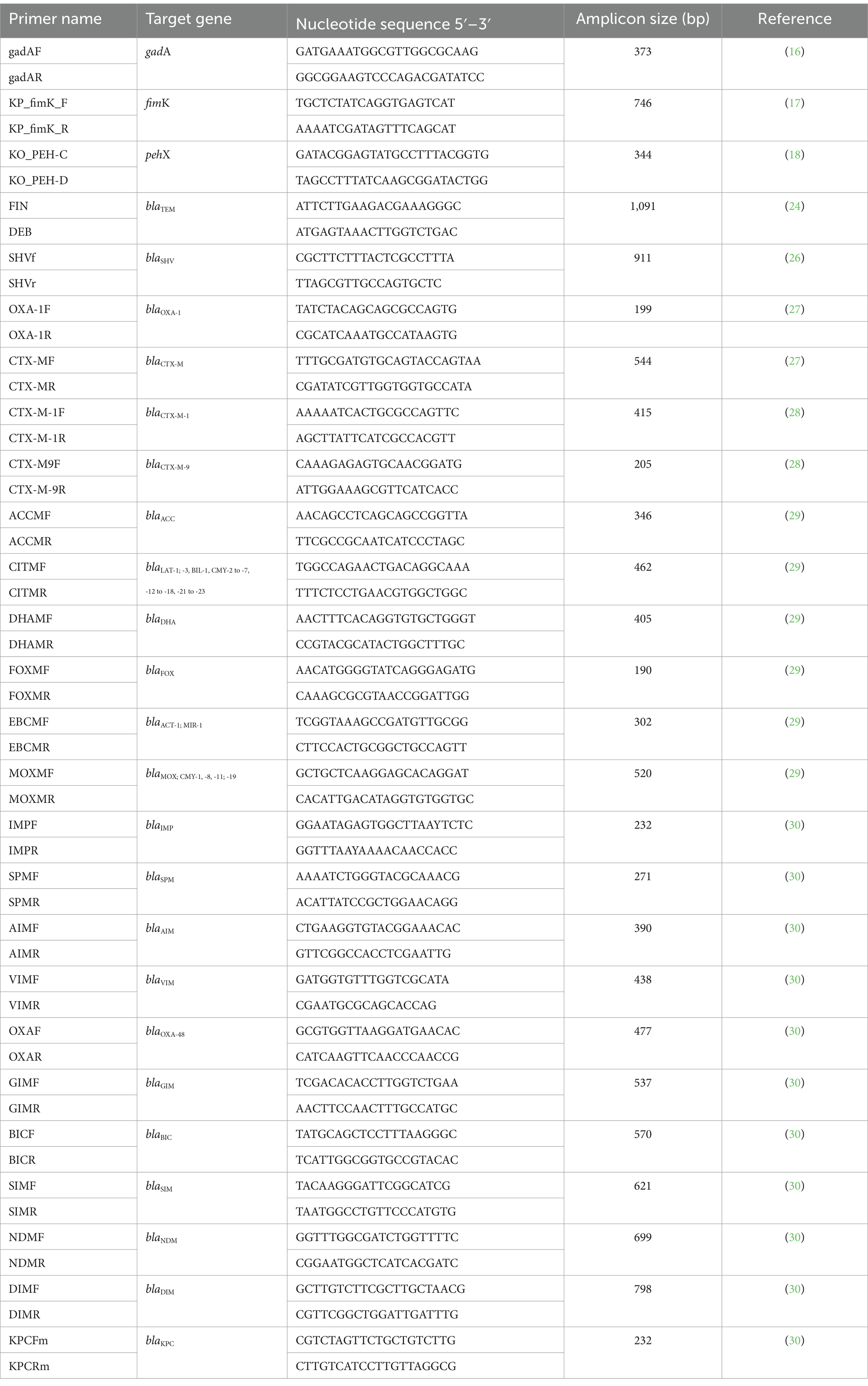
Table 1. Primers used in this study.
Antimicrobial susceptibility testing
Antimicrobial susceptibility testing and interpretation were performed using the disk diffusion method according to the guidelines of the European Committee on Antimicrobial susceptibility testing (EUCAST) (20) and the Clinical and Laboratory Standards Institute (CLSI) (21, 22).
The following antimicrobial disks (Oxoid, Basingstoke, Hampshire, UK) were tested: ampicillin (AMP, 10 μg), amoxicillin/clavulanic acid (AMC, 30 μg), amikacin (AK, 30 μg), nalidixic acid (NA, 30 μg), cephalothin (KF, 30 μg), cefotaxime (CTX, 5 μg), cefoxitin (FOX, 30 μg), cefepime (FEP, 30 μg), ceftazidime (CAZ, 10 μg), chloramphenicol (C, 30 μg), ciprofloxacin (CIP, 5 μg), ertapenem (ETP, 10 μg), gentamicin (CN, 10 μg), imipenem (IMP, 10 μg), levofloxacin (LEV, 5 μg), meropenem (MEM, 10 μg), piperacillin (PRL, 30 μg), piperacillin/tazobactam (TZP, 36 μg), trimethoprim/sulfamethoxazole (STX, 25 μg), and tetracycline (TE, 30 μg). The control strain used for disk diffusion was Escherichia coli ATCC® 25922™. Additionally, carbapenem activity in Enterobacterales was detected using the MAST® CAT-ID test (D71C, Mast Group, Germany) according to the manufacturer’s instructions.
Multidrug-resistant (MDR) bacteria were defined as bacteria not susceptible to at least one antimicrobial agent in three or more antimicrobial classes, following the classification proposed by Magiorakos et al. (23).
Molecular detection of β-lactamases genes
3GC-resistant Enterobacterales isolates were screened by PCR to detect the presence of various β-lactamases genes, including: blaSHV, blaOXA-1, blaTEM, blaCTX-M, blaCTX-M-1group and blaCTX-M-9group (24–28). Additionally, the following pAmpC-encoding genes were screened: blaDHA-1, blaDHA-2, blaMOX-1, blaMOX-2, blaCMY-1 to blaCMY-11, blaACC, blaBIL-1, blaLAT-1 to blaLAT-4, blaMIR-T, blaACT-1 and blaFOX-1 to FOX-5 (29). Furthermore, isolates were screened for 11 carbapenemase genes (blaIMP, blaOXA-48, blaVIM, blaNDM, blaKPC, blaSPM, blaGIM, blaSIM, blaBIC, blaAIM, and blaDIM) as previously described (30). The primers used are listed in Table 1. Each PCR reaction contained Supreme NZYTaq II 2x Green Master Mix (NZYTech), 0.5 μM of each primer, and template DNA. PCR amplification was performed using a Biometra Uno II thermal cycler (Analytik Jena). Negative controls and previously sequenced positive controls were included in each PCR run. PCR products were analyzed by 1.5% (w/v) agarose gel electrophoresis, stained with GreenSafe Premium (NZYTech) and visualized under UV light using a UView™ Mini Transilluminator (Bio-Rad).
PCR products were then purified using the NZYTech Gel Pure Kit (NZYTech) and sequencing was performed by StabVida (Caparica, Portugal). The obtained sequences were compared to published DNA sequences using BLAST.
Results
Most of the samples were collected from female common leopard geckos (65.5%, n = 38/58), with a median age of 9 months (range: 7–29 months), while 34.5% (n = 20/58) were from males, with a median age of 10 months (range: 7–29 months). For central bearded dragons, 45% (n = 9/20) of the samples were from females (median age: 9 months; range: 7–60 months) and 55% (n = 11/20) were from males (median age: 9 months; range: 5–60 months).
3GC-resistant Enterobacterales were detected in 17.9% (n = 14/78) of the reptiles. In leopard geckos, samples were collected from two anatomical sites: oral cavity (n = 49) and cloaca (n = 43). Of the oral cavity samples from the leopard geckos, 12.2% (n = 6/49) were colonized with 3GC-resistant Enterobacterales isolates, with the following bacterial species identified Enterobacter cloacae (n = 1), Klebsiella pneumoniae (n = 1), Klebsiella aerogenes (n = 1), Citrobacter freundii (n = 2), and Pseudocitrobacter faecalis (n = 1). Among the cloacal samples, 20.9% (n = 9/43) of the animals were colonized with 3GC-resistant Enterobacterales isolates, with the following species identified Klebsiella aerogenes (n = 4) and Citrobacter freundii (n = 7) (Table 2).
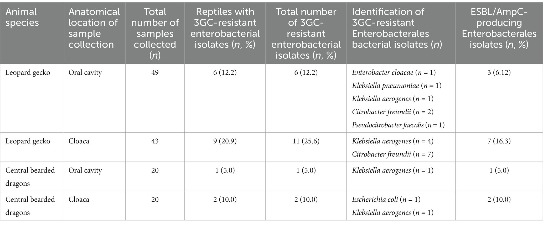
Table 2. 3GC-resistant Enterobacterales from leopard geckos (N = 56) and central bearded dragons (N = 20) by anatomical location.
Regarding central bearded dragons (n = 20) samples were collected from the same two anatomical locations: oral cavity (n = 20) and cloaca (n = 20). Among the oral cavity samples, only one animal (5.0%, n = 1/20) was colonized with 3GC-resistant Enterobacterales isolates (Klebsiella aerogenes). Among the cloacal samples, 10.0% (n = 2/20) of the animals were colonized with 3GC-resistant Enterobacterales bacteria. The following bacterial species were identified: Escherichia coli (n = 1) and Klebsiella aerogenes (n = 1) (Table 2).
Among the common leopard geckos, 58.8% (n = 10/17) of the 3GC-resistant Enterobacterales were ESBL/AmpC producers, with three isolates from the oral cavity and seven isolates from the cloaca being ESBL producers (Table 2).
For the C. freundii isolates obtained from the oral cavity (n = 2) and cloaca (n = 7), the most common antimicrobial resistance phenotype was AMPR-AMCR-KFR-CTXR-FOXR (n = 4/9). None of the isolates were MDR. All C. freundii isolates harbored the blaCMY-2 gene (Table 3).
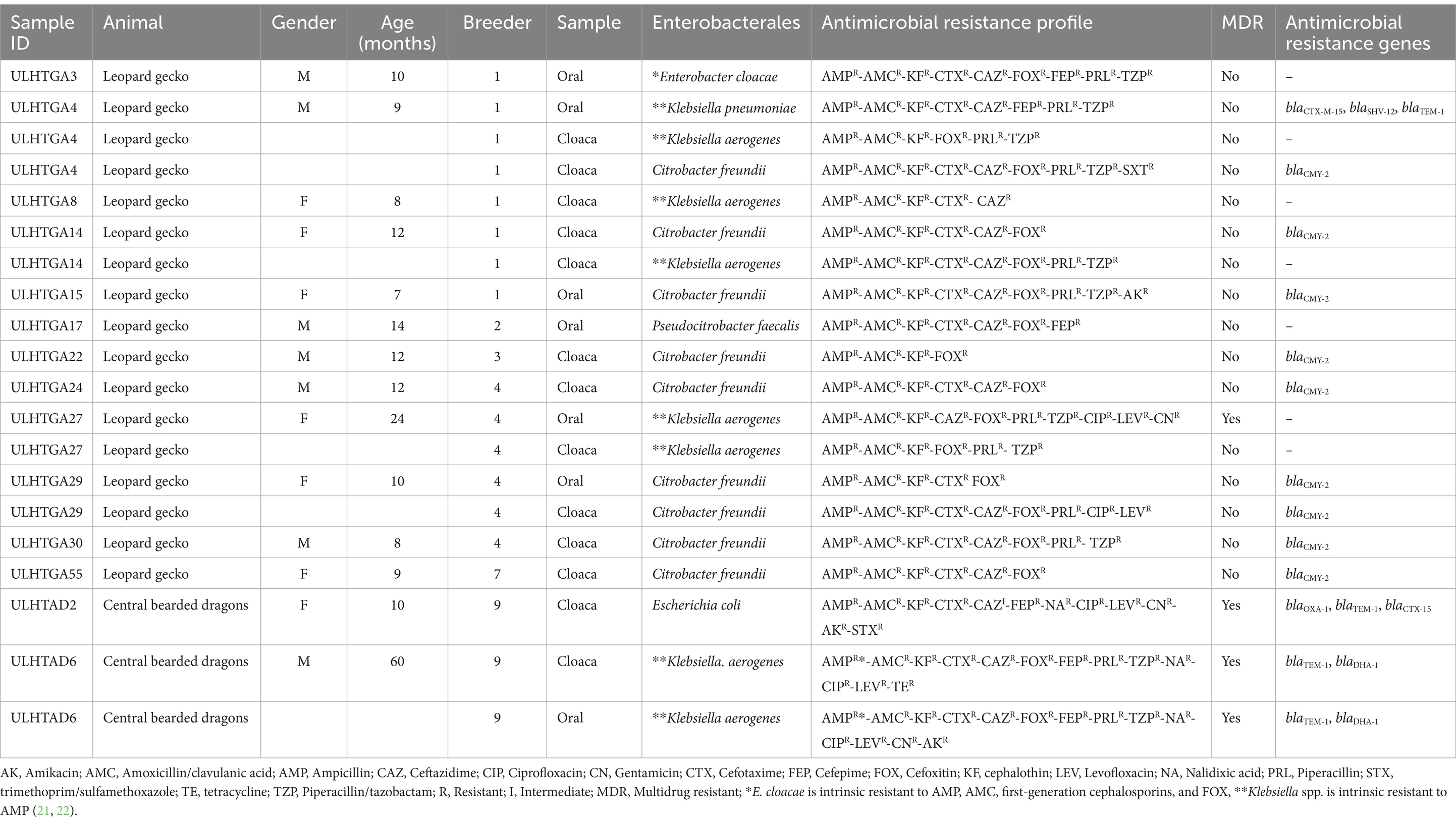
Table 3. 3GC-resistant Enterobacterales isolates from the oral and cloacal cavities of common leopard geckos and central bearded dragons.
The K. pneumoniae isolate exhibited the antimicrobial resistance phenotype AMPR-AMCR-KFR-CTXR-FOXR-CAZR-FEPR-PRLR-TZPR. This isolate was not MDR and harbored blaCTX-M-15, blaSHV-12, and blaTEM-1 genes (Table 3).
For the K. aerogenes isolates obtained from the oral cavity (n = 1) and cloaca (n = 4), the most common antimicrobial resistance phenotype was AMPR-AMCR-KFR-FOXR-PRLR-TZPR (n = 2/5). The oral isolate was MDR, showing resistance to β-lactams, fluoroquinolones, and aminoglycosides, whereas the remaining isolates were not MDR (Table 3). Notably, no β-lactamases genes were identified in these isolates.
Additionally, E. cloacae and P. faecalis were isolated from the oral cavity of common leopard geckos and exhibited the following resistance phenotypes: AMPR-AMCR-KFR-CTXR-CAZR-FOXR-FEPR-PRLR-TZPR and AMPR-AMCR-KFR-CTXR-CAZR-FOXR-FEPR, respectively (Table 3).
Among central bearded dragons, the 3GC-resistant Enterobacterales isolates were all ESBL/AmpC producers (Table 2). 3GC-resistant K. aerogenes were detected in both the oral cavity and cloaca, whereas 3GC-resistant E. coli was isolated from the cloaca (Tables 2, 3). Both K. aerogenes isolates exhibited resistance to multiple classes of antimicrobials (AMPR-AMCR-KFR-CTXR-CAZR-FOXR-FEPR-PRLR-TZPR-NAR-CIPR-LEVR-CNR-AKR), classifying as MDR. Genotypic characterization revealed the presence of blaTEM-1 and blaDHA-1 genes in both K. aerogenes isolates (Table 3). The E. coli isolate was MDR, showing the following resistance phenotype: AMPR-AMCR-KFR-CTXR-CAZI-FEPR-NAR-CIPR-LEVR-CNR-AKR-STXR, and harbored the blaOXA-1, blaTEM-1 and blaCTX-15 genes (Table 3).
No carbapenem resistance was observed in any isolate.
Discussion
This study provides valuable insight into the prevalence and antimicrobial resistance profiles of 3GC-resistant Enterobacterales in reptile species, specifically common leopard geckos (Eublepharis macularius) and central bearded dragons (Pogona vitticeps). The findings indicate that both species can act as reservoirs for AMR bacteria, highlighting the need for monitoring resistance patterns in exotic animal populations.
This study identified several 3CG-resistant Enterobacterales species, including P. faecalis, C. freundii, E. cloacae, K. aerogenes, E. coli and K. pneumoniae. These bacteria are recognized as opportunistic pathogens capable of infecting humans, especially immunocompromised individuals. They are commonly associated with various infections in human and companion animals, including urinary tract infections, pneumonia, and bloodstream infections (11, 31–33). The oral cavity and cloaca of reptiles are common sites for the colonization of resistant bacteria, facilitating transmission to humans through direct contact or contaminated surfaces. The presence of 3GC-resistant Enterobacterales in the oral cavities and cloacae of the studied reptiles suggests these animals may serve as reservoirs of antimicrobial resistance genes for humans.
Among the identified bacterial species, C. freundii was the most prevalent, followed by K. aerogenes. C. freundii is associated with several opportunistic infections, including severe diarrhea, urinary tract infections, pneumonia, neonatal meningitis, and brain abscesses, typically affecting infants and immunocompromised individuals (34, 35). However, C. freundii can acquire virulent factors, such as Shiga-like toxin genes, in addition to proteolysis, hemolysis, and biofilm formation (36). This bacterium is commonly found in reptiles, particularly in turtles. It is a recognized causative agent of septicemic cutaneous ulcerative disease, characterized by anorexia, lethargy, necrosis, and petechial hemorrhages on the skin (37). Hossain et al. reported a high frequency (87.5%) of C. freundii in cloacal samples from healthy turtles (Trachemys scripta), suggesting that even asymptomatic animals can act as carriers, shedding the bacteria in their feces and posing a transmission risk to other animals and humans (37). In addition to its pathogenic potential, C. freundii is a well-known producer of AmpC, conferring resistance to β-lactams. In this study, C. freundii isolates exhibited resistance to 3GC, consistent with previous findings (38, 39). Notably, the blaCMY-2 gene, associated with AmpC production was detected in all C. freundii isolates.
Regarding Klebsiella spp., K. aerogenes was the most frequently identified species in this study. However, there is little information on K. aerogenes in reptiles. In humans, it is primarily associated with respiratory tract, gastrointestinal tract, urinary tract and blood infections (40). Klebsiella spp. isolates from both reptile species exhibited resistance to multiple antimicrobial classes. Some isolates carried blaCTX-M-15, blaSHV-12 and blaTEM-1, common resistance determinants associated with β-lactams. These findings are consistent with previous reports of Klebsiella spp. acquiring and disseminating antimicrobial resistance through horizontal gene transfer. The detection of blaTEM-1 and blaDHA-1 in K. aerogenes isolates from central bearded dragons further emphasizes the role of genetic factors in antimicrobial resistance persistence and spread. Notably, in K. aerogenes isolates from common leopard geckos, resistance to FOX or AMC and CTX or CAZ could not be explained by ESBL/pAmpC genes, suggesting the involvement of other resistance mechanisms, such as inhibitor-resistant TEM (blaIRT) or other extended-spectrum AmpC β-lactamases (41, 42).
The identification of K. pneumoniae and E. coli, along with the presence of antimicrobial resistance genes (blaTEM-1, blaCTX-M-15, blaSHV-12 and blaCMY-2), aligns with previous studies on 3CG-resistant Enterobacterales isolates from humans and companion animals in Portugal (11, 12, 43–45). Furthermore, ESBL-producing strains often exhibit resistance to other antimicrobial classes, such as aminoglycosides, fluoroquinolones, and sulfonamides, further complicating treatment options (14).
This study also identified P. faecalis in the oral cavity of a common leopard gecko. P. faecalis, a Gram-negative bacterium belonging to the order Enterobacterales, was first described in 2010 from a human clinical setting in Pakistan, where it exhibited carbapenem resistance due to the production of blaNDM-1 (46). Subsequently, it was reported in a human bloodstream infection in China, associated with blaOXA-181 production (47). The emergence of hospital-acquired infections involving resistant Enterobacterales suggests possible transmission through environmental contamination or the food chain (47). To our knowledge, this is the first report of P. faecalis in healthy reptiles.
Enterobacter cloacae is a ubiquitous environmental species and a commensal of the intestinal tract in both humans and animals. It is known to contaminate medical devices, contributing to skin and soft tissue infections, urinary tract infections and intra-abdominal infections (31).
While no carbapenem resistance was observed in this study, ongoing surveillance is crucial given the growing concern about the rapid spread of carbapenem resistance in humans and animals, particularly dogs and cats (10, 43, 44, 48, 49).
The main limitations of this study include the relatively small sample size, the limited number of reptile species analyzed and the geographic scope, as it was conducted in a specific region and may not reflect the microbiota and antimicrobial resistance patterns in other locations. Antimicrobial resistance profiles can vary significantly depending on environmental factors, veterinary practices and antimicrobial stewardship programs. Furthermore, although our study focused on ESBL and pAmpC—producing Enterobacterales, the characterization of all Gram-negative microbiota in these animals could have provided a more comprehensive understanding of the potential zoonotic bacteria present within the oral cavity and cloaca of these reptiles and the prevalence of antimicrobial resistance.
Nonetheless, to the best of our knowledge, this is the first study on ESBL/AmpC-producing Enterobacterales in reptiles, specifically in common leopard geckos and central bearded dragons in Portugal.
To minimize the risk of transmission of ESBL/AmpC-producing Enterobacterales, exotic pet owners and breeders should adopt strict hygiene measures, including handwashing after handling animals, proper cleaning of enclosures, and avoiding direct contact with the animals’ mouths, cloaca, or contaminated surfaces. Public health guidelines for exotic pet management should be updated to include recommendations for antimicrobial stewardship in their care and raise awareness about the potential zoonotic transmission of resistant bacteria.
Conclusion
This study highlights the presence of ESBL/AmpC-producing Enterobacterales in common leopard geckos (Eublepharis macularius) and central bearded dragons (Pogona vitticeps), emphasizing the importance of ongoing antimicrobial resistance surveillance in pet reptiles. Implementing an epidemiological surveillance program is essential for developing future strategies to control the spread of AMR bacteria using a One Health approach.
Data availability statement
The original contributions presented in the study are included in the article/supplementary material, further inquiries can be directed to the corresponding author/s.
Ethics statement
The animal studies were approved by Ethics and Animal Welfare Committee of the Faculty of Veterinary Medicine, Lusófona University - University Center of Lisbon. The studies were conducted in accordance with the local legislation and institutional requirements. Written informed consent was obtained from the owners for the participation of their animals in this study.
Author contributions
AV: Conceptualization, Writing – original draft, Writing – review & editing. GF: Methodology, Writing – review & editing. JS: Methodology, Writing – review & editing. RP: Methodology, Supervision, Writing – review & editing. AB: Conceptualization, Data curation, Formal analysis, Funding acquisition, Investigation, Methodology, Project administration, Resources, Supervision, Visualization, Writing – original draft, Writing – review & editing.
Funding
The author(s) declare that financial support was received for the research and/or publication of this article. This research was funded and supported by FMV-ULusófona - PathoGeckos (2022–2023).
Acknowledgments
The authors would like to thank FMV-ULusófona for their support in carrying out this work.
Conflict of interest
The authors declare that the research was conducted in the absence of any commercial or financial relationships that could be construed as a potential conflict of interest.
Generative AI statement
The authors declare that no Gen AI was used in the creation of this manuscript.
Publisher’s note
All claims expressed in this article are solely those of the authors and do not necessarily represent those of their affiliated organizations, or those of the publisher, the editors and the reviewers. Any product that may be evaluated in this article, or claim that may be made by its manufacturer, is not guaranteed or endorsed by the publisher.
References
1. Rom, B, Kornaś, S, and Basiaga, M. Endoparasites of pet reptiles based on coprosopic methods. Ann Parasitol. (2018) 64:115–20. doi: 10.17420/ap6402.142
3. Ebani, VV. Domestic reptiles as source of zoonotic bacteria: a mini review. Asian Pac J Trop Med. (2017) 10:723–8. doi: 10.1016/j.apjtm.2017.0.7.020
4. González-Barrio, D. Zoonoses and wildlife: one health approach. Animals (Basel). (2022) 12:480. doi: 10.3390/ani12040480
5. Babalola, MO, and Balogun, JA. The ecology and potential health risk of the oral microflora of Python regius and Clelia scyntalina. Int J Microbiol Res. (2013) 5:349–56. doi: 10.9735/0975-5276.5.1.349-356
6. Zancolli, G, Mahsberg, D, Sickel, W, and Keller, A. Reptiles as reservoirs of bacterial infections: real threat or methodological bias? Microb Ecol. (2015) 70:579–84. doi: 10.1007/s00248-015-0618-3
7. Valdez, JW. Using Google trends to determine current, past, and future trends in the reptile pet trade. Animals (Basel). (2021) 11:676. doi: 10.3390/ani11030676
8. One Health Commission. What is one health? (2021). Available online at: https://www.onehealthcommission.org/en/why_one_health/what_is_one_health (Accessed December 8, 2024).
9. McEwen, SA, and Collignon, PJ. Antimicrobial resistance: a one health perspective. Microbiol Spectr. (2018) 6:6. doi: 10.1128/microbiolspec.ARBA-0009-2017
10. Nigg, A, Brilhante, M, Dazio, V, Clément, M, Collaud, A, Gobeli Brawand, S, et al. Shedding of OXA-181 carbapenemase-producing Escherichia coli from companion animals after hospitalisation in Switzerland: an outbreak in 2018. Euro Surveill. (2019) 24:1900071. doi: 10.2807/1560-7917.ES.2019.24.39.1900071
11. Belas, A, Marques, C, Menezes, J, Telo da Gama, L, Cavaco-Silva, P, and Pomba, C. ESBL/pAmpC-producing Escherichia coli causing urinary tract infections in non-related companion animals and humans. Antibiotics. (2022) 11:559. doi: 10.3390/antibiotics1150559
12. Belas, A, Menezes, J, Telo da Gama, L, and Pomba, C. Sharing of clinically important antimicrobial resistance genes by companion animals and their human household members. Microb Drug Resist. (2020) 26:1174–85. doi: 10.1089/mdr.2019.0380
13. Prescott, JF, and Boerlin, P. Antimicrobial use in companion animals and good stewardship practice. Vet Rec. (2016) 179:486–8. doi: 10.1136/vr.i5908
14. Paterson, DL, and Bonomo, RA. Extended-spectrum beta-lactamases: a clinical update. Clin Microbiol Rev. (2005) 18:657–86. doi: 10.1128/CMR.18.4.657-686.2005
15. Bush, K, and Jacoby, GA. Updated functional classification of beta-lactamases. Antimicrob Agents Chemother. (2010) 54:969–76. doi: 10.1128/AAC.01009-09
16. Doumith, M, Day, MJ, Hope, R, Wain, J, and Woodford, N. Improved multiplex PCR strategy for rapid assignment of the four major Escherichia coli phylogenetic groups. J Clin Microbiol. (2012) 50:3108–10. doi: 10.1128/JCM.01468-12
17. Padmavathy, B, Vinoth Kumar, R, Patel, A, Deepika Swarnam, S, Vaidehi, T, and Jaffar Ali, BM. Rapid and sensitive detection of major uropathogens in a single-pot multiplex PCR assay. Curr Microbiol. (2012) 65:44–53. doi: 10.1007/s00284-012-0126-3
18. Kovtunovych, G, Lytvynenko, T, Negrutska, V, Lar, O, Brisse, S, and Kozyrovska, N. Identification of Klebsiella oxytoca using a specific PCR assay targeting the polygalacturonase pehX gene. Res Microbiol. (2003) 154:587–92. doi: 10.1016/S0923-2508(03)00148-7
19. Rebelo, A, Bortolaia, V, Kjeldgaard, J, Pedersen, SK, Leekitcharoenphon, P, Hansen, IM, et al. Multiplex PCR for detection of plasmid-mediated colistin resistance determinants, mcr-1, mcr-2, mcr-3, mcr-4 and mcr-5 for surveillance purposes. Euro Surveill. (2018) 23:17-00672. doi: 10.2807/1560-7917.ES.2018.23.6.17-00672
20. The European Committee on Antimicrobial Susceptibility Testing (EUCAST). Breakpoint tables for interpretation of MICs and zone diameters. Version 15.0. (2025). Available online at: http://www.eucast.org (Accessed on January 16, 2025).
21. CLSI ed. Performance standards for antimicrobial disk and dilution susceptibility tests for bacteria isolated from animals. 5th ed CLSI supplement VET01S. Wayne, PA, USA: Clinical and Laboratory Standards Institute (2020).
22. CLSI ed. Performance standards for antimicrobial susceptibility testing. 34th ed CLSI supplement M100. Wayne, PA, USA: Clinical and Laboratory Standards Institute (2024).
23. Magiorakos, AP, Srinivasan, A, Carey, RB, Carmeli, Y, Falagas, ME, Giske, CG, et al. Multidrug-resistant, extensively drug-resistant and pandrug-resistant bacteria: an international expert proposal for interim standard definitions for acquired resistance. Clin Microbiol Infect. (2012) 18:268–81. doi: 10.1111/j.1469-0691.2011.03570.x
24. Caniça, MM, Lu, CY, Krishnamoorthy, R, and Paul, GC. Molecular diversity and evolution of blaTEM genes encoding β-lactamases resistant to clavulanic acid in clinical E. coli. J Mol Evol. (1997) 44:57–65. doi: 10.1007/pl00006121
25. Edelstein, M, Pimkin, M, Palagin, I, Edelstein, I, and Stratchounski, L. Prevalence and molecular epidemiology of CTX-M extended-spectrum β-lactamase-producing Escherichia coli and Klebsiella pneumoniae in Russian hospitals. Antimicrob Agents Chemother. (2003) 47:3724–32. doi: 10.1128/AAC.47.12.3724-3732.2003
26. Rasheed, JK, Jay, C, Metchock, B, Berkowitz, F, Weigel, L, Crellin, J, et al. Evolution of extended-spectrum beta-lactam resistance (SHV-8) in a strain of Escherichia coli during multiple episodes of bacteremia. Antimicrob Agents Chemother. (1997) 41:647–53. doi: 10.1128/AAC.41.3.647
27. Féria, C, Ferreira, E, Correia, JD, Gonçalves, J, and Caniça, M. Patterns and mechanisms of resistance to beta-lactams and beta-lactamase inhibitors in uropathogenic Escherichia coli isolated from dogs in Portugal. J Antimicrob Chemother. (2002) 49:77–85. doi: 10.1093/jac/49.1.77
28. Woodford, N, Fagan, EJ, and Ellington, MJ. Multiplex PCR for rapid detection of genes encoding CTX-M extended-spectrum β-lactamases. J Antimicrob Chemother. (2006) 57:154–5. doi: 10.1093/jac/dki412
29. Perez-Perez, FJ, and Hanson, ND. Detection of plasmid-mediated AmpC β-lactamase genes in clinical isolates by using multiplex PCR. J Clin Microbiol. (2002) 40:2153–62. doi: 10.1128/JCM.40.6.2153-2162.2002
30. Poirel, L, Walsh, TR, Cuvillier, V, and Nordmann, P. Multiplex PCR for detection of acquired carbapenemase genes. Diagn Microbiol Infect Dis. (2011) 70:119–23. doi: 10.1016/j.diagmicrobio.2010.12.002
31. Davin-Regli, A, and Pagès, JM. Enterobacter aerogenes and Enterobacter cloacae; versatile bacterial pathogens confronting antibiotic treatment. Front Microbiol. (2015) 6:392. doi: 10.3389/fmicb.2015.00392
32. Ndlovu, T, Kgosietsile, L, Motshwarakgole, P, and Ndlovu, SI. Evaluation of potential factors influencing the dissemination of multidrug-resistant Klebsiella pneumoniae and alternative treatment strategies. Trop Med Infect Dis. (2023) 8:381. doi: 10.3390/tropicalmed8080381
33. Elbehiry, A, Al Shoaibi, M, Alzahrani, H, Ibrahem, M, Moussa, I, Alzaben, F, et al. Enterobacter cloacae from urinary tract infections: frequency, protein analysis, and antimicrobial resistance. AMB Express. (2024) 14:17. doi: 10.1186/s13568-024-01675-7
34. Pardia, SN, Verma, IC, Deb, M, and Bhujwala, RA. An outbreak of diarrhea due to Citrobacter freundii in a neonatal special care nursery. Indian J Pediatr. (1980) 47:81–4. doi: 10.1007/BF02900180
35. Badger, JL, Stins, MF, and Kim, KS. Citrobacter freundii invades and replicates in human brain microvascular endothelial cells. Infect Immunity. (1999) 67:4208–15. doi: 10.1128/IAI.67.8.4208-4215.1999
36. Fakruddin, M, Rahaman, MM, Ahmed, MM, and Hoque, M. Antimicrobial resistance and virulence factors of enterobacteriaceae isolated from food samples of Bangladesh. Int J Microbiol Immunol Res. (2014) 3:12–8.
37. Hossain, S, Wimalasena, SHMP, and Heo, GJ. Virulence factors and antimicrobial resistance pattern of Citrobacter freundii isolated from healthy pet turtles and their environment. Asian J Anim Vet Adv. (2017) 12:10–6. doi: 10.3923/ajava.2017.10.16
38. Barlow, M, and Hall, BG. Origin and evolution of the AmpC beta-lactamases of Citrobacter freundii. Antimicrob Agents Chemother. (2002) 46:1190–8. doi: 10.1128/AAC.46.5.1190-1198.2002
39. Meini, S, Tascini, C, Cei, M, Sozio, E, and Rossolini, GM. AmpC β-lactamase-producing Enterobacterales: what a clinician should know. Infection. (2019) 47:363–75. doi: 10.1007/s15010-019-01291-9
40. Gu, H, Cai, Q, Dai, X, Wang, H, Xu, W, Cao, X, et al. A case report of Klebsiella aerogenes-caused lumbar spine infection identified by metagenome next-generation sequencing. BMC Infect Dis. (2022) 22:616. doi: 10.1186/s12879-022-07583-0
41. Carattoli, A, Lovari, S, Franco, A, Cordaro, G, Di Matteo, P, and Battisti, A. Extended-spectrum β-lactamases in Escherichia coli isolated from dogs and cats in Rome, Italy, from 2001 to 2003. Antimicrob Agents Chemother. (2005) 49:833–5. doi: 10.1128/AAC.49.2.833-835.2005
42. Mammeri, H, Poirel, L, Fortineau, N, and Nordmann, P. Naturally occurring extended-spectrum cephalosporinases in Escherichia coli. Antimicrob Agents Chemother. (2006) 50:2573–6. doi: 10.1128/AAC.01633-05
43. Moreira da Silva, J, Menezes, J, Fernandes, L, Santos Costa, S, Amaral, A, and Pomba, C. Carbapenemase-producing Enterobacterales strains causing infections in companion animals-Portugal. Microbiol Spectr. (2024) 12:e0341623. doi: 10.1128/spectrum.03416-23
44. Menezes, J, Frosini, SM, Weese, S, Perreten, V, Schwarz, S, Amaral, AJ, et al. Transmission dynamics of ESBL/AmpC and carbapenemase-producing Enterobacterales between companion animals and humans. Front Microbiol. (2024) 15:1432240. doi: 10.3389/fmicb.2024.1432240
45. Carvalho, I, Alonso, CA, Silva, V, Pimenta, P, Cunha, R, Martins, C, et al. Extended-spectrum beta-lactamase-producing Klebsiella pneumoniae isolated from healthy and sick dogs in Portugal. Microb Drug Resist. (2020) 26:709–15. doi: 10.1089/mdr.2019.0205
46. Kämpfer, P, Glaeser, SP, Raza, MW, Abbasi, SA, and Perry, JD. Pseudocitrobacter gen. nov., a novel genus of the Enterobacteriaceae with two new species Pseudocitrobacter faecalis sp. nov., and Pseudocitrobacter anthropi sp. nov, isolated from fecal samples from hospitalized patients in Pakistan. Syst Appl Microbiol. (2014) 37:17–22. doi: 10.1016/j.syapm.2013.08.003
47. Shi, Q, Guo, Y, Yang, Y, Wu, S, Han, R, Ding, L, et al. Characterization of the first carbapenem-resistant Pseudocitrobacter faecalis harboring blaOXA-181 in China. Antibiotics. (2022) 11:737. doi: 10.3390/antibiotics11060737
48. Köck, R, Daniels-Haardt, I, Becker, K, Mellmann, A, Friedrich, AW, Mevius, D, et al. Carbapenem-resistant Enterobacteriaceae in wildlife, food-producing, and companion animals: a systematic review. Clin Microbiol Infect. (2018) 24:1241–50. doi: 10.1016/j.cmi.2018.04.004
Keywords: reptiles, third-generation cephalosporins, antimicrobial resistance, Enterobacterales, extended-spectrum β-lactamases, AmpC β-lactamases
Citation: Valença A, Fernandes G, Smolders J, Patrício R and Belas A (2025) ESBL/pAmpC-producing Enterobacterales in common leopard geckos (Eublepharis macularius) and central bearded dragons (Pogona vitticeps) from Portugal. Front. Vet. Sci. 12:1579193. doi: 10.3389/fvets.2025.1579193
Edited by:
Saúl Jiménez-Ruiz, University of Cordoba, SpainReviewed by:
Rachel Amanda Hickman, Independent Researcher, Uppsala, SwedenAdrian Canizalez-Roman, Autonomous University of Sinaloa, Mexico
Copyright © 2025 Valença, Fernandes, Smolders, Patrício and Belas. This is an open-access article distributed under the terms of the Creative Commons Attribution License (CC BY). The use, distribution or reproduction in other forums is permitted, provided the original author(s) and the copyright owner(s) are credited and that the original publication in this journal is cited, in accordance with accepted academic practice. No use, distribution or reproduction is permitted which does not comply with these terms.
*Correspondence: Adriana Belas, YWRyaWFuYS5iZWxhc0B1bHVzb2ZvbmEucHQ=