 Atiyeh Peiravan1*
 Mazdak Salavati2
 Androniki Psifidi3
 Mellora Sharman4Andrew Kent5†
 Penny Watson5
 Karin Allenspach6
 Dirk Werling1*
- 1Centre for Vaccinology and Regenerative Medicine, Department of Pathology and Pathogen Biology, Royal Veterinary College, University of London, North Mymms, United Kingdom
- 2Dairy Research and Innovation Centre, Scotland’s Rural College, Dumfries, United Kingdom
- 3Department of Clinical Sciences and Services, Royal Veterinary College, University of London, North Mymms, United Kingdom
- 4VetCT, St John’s Innovation Park, Cambridge, United Kingdom
- 5Queen’s Veterinary School Hospital, University of Cambridge, Cambridge, United Kingdom
- 6College of Veterinary Medicine, University of Georgia, Athens, GA, United States
Canine Inflammatory bowel disease (IBD) is a chronic multifactorial disease, resulting from complex interactions between the intestinal immune system, microbiota and environmental factors in genetically predisposed dogs. Previously, we identified several single nucleotide polymorphisms (SNP) and regions on chromosomes (Chr) 7, 9, 11 and 13 associated with IBD in German shepherd dogs (GSD) using GWAS and FST association analyses. Here, building on our previous results, we performed a targeted next-generation sequencing (NGS) of a two Mb region on Chr 9 and 11 that included 14 of the newly identified candidate genes, to identify potential functional SNPs that could explain these association signals. Furthermore, correlations between genotype and treatment response were estimated. Results revealed several SNPs in the genes for canine EEF1A1, MDH2, IL3, IL4, IL13 and PDLIM. Based on the known function of their human orthologues, these results further our insight into their potential contribution to the pathogenesis of IBD in dogs. In addition, several pathways involved in innate and adaptive immunity and inflammatory responses (i.e., T helper cell differentiation, Th1 and Th2 activation pathway, communication between innate and adaptive immune cells and differential regulation of cytokine production in intestinal epithelial cells by IL-17A and IL-17F), were constructed involving the gene products in the candidate regions for IBD susceptibility. Interestingly, some of the identified SNPs were present in only one outcome group, suggesting that different genetic factors are involved in the pathogenesis of IBD in different treatment response groups. This also highlights potential genetic markers to predict the response in dogs treated for IBD.
Introduction
Inflammatory bowel disease (IBD) is a common cause of chronic gastrointestinal disease in humans and dogs. IBD is characterized by persistent or recurrent gastrointestinal signs (GI) including chronic diarrhoea, vomiting and weight loss and with histological evidence of inflammation in the lamina propria of the small intestine, large intestine or both (1). The diagnosis of IBD in both humans and dogs is by exclusion, as several diseases can cause chronic gastrointestinal inflammation secondarily (2, 3).
The pathogenesis of IBD is believed to be multifactorial in both humans and dogs, caused by a complex interaction between the intestinal innate and adaptive immune system, the intestinal microbiome, and the genetic make-up of an individual. Although IBD can affect multiple dog breeds, breed-specific disease phenotypes and associations have been reported (4, 5). In the United Kingdom (UK), German shepherd dogs (GSD) have been reported to be at increased risk of developing the disease (6).
IBD is not a curable disease, therefore the aim of current treatment approches is to minimize the severity and frequency of the clinical signs. In general, treatment protocols include dietary modifications, antibiotics (7, 8), and corticosteroid (9) treatment trials. As such, canine IBD is generally classified only retrospectively based on response to treatment into food responsive disease/diarrhoea (FRD), antibiotic responsive disease/diarrhoea (ARD), or steroid responsive disease/diarrhoea (SRD), which are usually used interchangeably with idiopathic IBD. Canine IBD patients are considered as FRD and ARD if their clinical signs improve or resolve following dietary modification and/or antibiotic treatment. Those that fail to respond to a change of diet and/or antibiotic therapy require immunosuppressive treatment (usually corticosteroids) to treat their clinical signs (SRD) (9). To date, treatment with anti-inflammatory/immunomodulatory medication such as corticosteroids is the mainstay treatment for both human and canine IBD patients (7). However, up to 50% of dogs with IBD that are initially managed with steroids will develop resistance and/or significant side effects, which ultimately leads to euthanasia for many of them (9, 10).
Recent advances in clinical genetics make it possible to use the patients’ genetic profile to predict response to treatment (11, 12). Similar to human IBD, it is hoped that identifying the genes involved in canine IBD will provide insights into disease pathogenesis in canine IBD. This could lead to the development of genetic screening panels useful for both diagnosis and identifying dogs that are more likely to fall into specific groups of treatments.
So far, studies of canine IBD using a candidate gene approach, have identified a number of single nucleotide polymorphisms (SNP) associated with the disease in genes encoding pattern recognition receptors (PRR) of the innate immunity (6, 13, 14) as well as associations between SNPs in Major histocompatibility class (MHC) II haplotypes and a potentially increased resistance to IBD in GSD (15). The release of the re-assembled dog genome and development of high-density canine DNA SNP arrays have enabled several successful GWAS studies aimed at investigating the genetic architecture of both monogenic and polygenic complex diseases of the dog (16–19). Previously, based on GWAS and FST association analyses of IBD cases and controls, we identified several SNPs and regions on chromosomes (Chr) 7,9,11 and 13 associated with IBD in GSD, including a total of 80 genes. Using a combination of pathways analysis and a list of genes that have been reported to be involved in human IBD, we identified 16 candidate genes potentially associated with IBD in GSD (20).
Genome-wide association studies rely on the principal of linkage disequilibrium (LD). While the extensive LD and long haplotype blocks (0.5–1.0 Mb) within breeds, resulted from genetic bottlenecks during domestication of dogs and breed formation, is an advantage in the initial GWAS it complicates the subsequent identification of the causative variant(s) (21, 22). The association signals identified through GWAS represent most likely markers that are not the causal variants themselves but are linked instead with nearby causative polymorphisms. Therefore, in order to generate a hypothesis about mechanisms underlying a specific phenotype, it is important to detect the causal variants themselves.
In the present study, we performed a targeted NGS of 2 Mb regions on Chr 9 and 11, which include 14 of the newly identified candidate genes, aiming to identify potential functional SNPs that could explain the GWAS association signal. We also investigated whether there was a correlation between the identified SNPs and response to treatment in the IBD cases, used in this study.
Materials and methods
Ethics and welfare statement
All blood samples used in this study were collected in ethylenediaminetetraacetic acid (EDTA) and represented residual material following completion of clinical diagnostic testing. Residual samples were utilized for research with informed owner consent. The use of these residual EDTA blood samples and buccal swab samples within the current study was approved by the RVC Animal Welfare Ethical Review Body (AWERB; approval number 2013 1,210).
Selection of cases and controls for targeted sequencing
All IBD cases and controls were identified based on inclusion criteria described previously (20). The case population was identified using stringent inclusion criteria: all cases have been affected by chronic gastrointestinal signs (i.e., >3 weeks duration), including diarrhoea and/or vomiting and/or anorexia and/or weight loss. Other causes of chronic gastrointestinal disease had been excluded, by performing routine hematology, serum biochemistry, fecal parasitology and bacteriology, abdominal ultrasonography, serum ACTH stimulation tests and measurement of serum canine trypsin-like immunoreactivity concentration (TLI) (20). All dogs had undergone gastrointestinal endoscopic examination for the collection of mucosal intestinal biopsies. Histological examination of the biopsies had revealed lymphoplasmacytic and/or eosinophilic infiltration of the lamina propria. None of the cases suffered from concurrent diseases of known or suspected immune-mediated origin, dermatological conditions or diseases for which GSD are known to be predisposed for (i.e., anal furunculosis, atopic dermatitis or superficial keratitis). A follow-up on all cases and controls was performed by contacting the referring veterinary surgeons and/or owners to gather information on treatment response of the dogs, including assessment of any changes to the course of treatment, and if so what the response to the new treatment was.
A total number of 48 GSDs with adequate followup information and available genomic (g)DNA samples were enrolled, including 28 cases (diagnosed with IBD) and 20 controls (non-inflammatory disease). Among the IBD cases, there were 9 food responsive disease/diarrhoea (FRD), 4 antibiotic responsive disease/diarrhoea (ARD), 11 steroid responsive disease/diarrhoea (SRD) and 4 NRS/PTS (No Response to Steroid, PTS: Put To Sleep) cases.
Control dogs were breed-matched that were presented with variety of non-inflammatory conditions or no-known diseases. Control dogs were retrospectively phenotyped, either by owner or veterinary surgeon telephone contact, to ensure they had neither current nor previous history of gastrointestinal signs for a duration of more than 5 consecutive days and did not suffer from conditions of known or suspected immune-mediated origin.
SureSelect XT library preparation and sequence capture
A custom-designed sequence capture array (SelectSure XT custom 0.5–2.9 Mb, Agilent), was designed and manufactured by Agilent, in order to isolate the targeted region identified on Chr 9 and 11, 1 Mb up- and downstream of the most significant SNPs identified by the previous GWAS (20), from total gDNA, using start and end coordinates of the associated regions. Properties of the final design of each array designed for the capture of the target regions on Chr 9 and 11 are shown in Table 1.
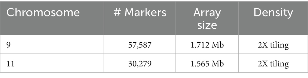
Table 1. Properties of the capture arrayes designed for isolation of the target regions on Chromosomes 9 and 11.
For each gDNA sample (A260/A280 ratio > 1.7) to be sequenced, an individual indexed library was prepared. DNA libraries were prepared using the SureSelect XT Library Prep Kit (Agilent) following the manufacturer instructions. In brief, 200 ng of the gDNA samples were fragmented by sonication and the paired-end adaptors were ligated to the blunt-ended fragments using the SureSelect XT Library Prep Kit ILM. Then, the adaptor-ligated fragments were PCR amplified, purified and hybridized to the capture array, using the SureSelectXT DNA kit following, the hybridized manufacturer instructions. Then, unbound fragments were washed away. Subsequently, the SureSelect- captured DNA libraries were eluted, purified and PCR amplified using an individual indexing primer for each sample. Following quality control, captured libraries were sequenced with a read length of 150 bp (paired end reads) on an Illumina NextSeq (500 Mid Output) platform. Sequencing was performed at the Genomic Centre of Queen Mary University of London (QMUL).
Analysis of sequencing data
The Genome Analysis Toolkit (GATK 3.8)1 best practice workflow, in house bash and R scripting was used for processing of raw sequencing data. BAMStats 2.1, which provides descriptive statistics for various metrics, was used to calculate average coverage.
Raw sequencing data were visualized and inspected using FastQC (v0.11.6).2 Thereafter, reads were trimmed and filtered using Trimmomatic (v0.36) (23). The trimmed reads were aligned and mapped to the canine genome (CanFam3.1), using Burrow-wheeler aligner (BWA, v0.7.15) with default parameters (bwa mem -M -t -R). BWA–MEM is designed for aligning relatively short sequence reads ranging from 70 bp to 1 Mb against a long reference and is generally recommended for high-quality queries (24). Once the reads were mapped to the canine genome and merged, they were sorted by the coordinates using the “samtools sort” command (24).
Potential PCR duplicates were flagged in the read’s SAM record using Picard tool (hosted by SAMtools), so that duplicates could be identified during downstream processing. Most GATK tools will then ignore the flagged reads by default, through the internal application of a read filter. Base quality score was adjusted according to a model of covariations bulit based on the data and a set of known variants.3
Genetic variants were called individually on each sample’s BAM file(s) using the HaplotypeCaller (GATK 3.8) in -ERC gVCF mode to produce an intermediate file format called gVCF file (genomic VCF). Following variant calling, the GVCFs of multiple samples were run through joint genotyping to produce a multi-sample VCF callset, using GenotypeGVCFs. Then, the raw SNPs extracted from the multi-sample VCF callset produced at genotyping stage, was subject to hard filtering. All the scripts that were used for variant calling, genotyping, and filtering annotations and values recommended by GATK best practice for hard filtering are shown in Supplementary Table 1.
Analysis of variants
The coordinate positions of the filtered SNPs were used to categorize them into different groups; (a) SNPs only present in cases, (b) SNPs only present in controls and (c) SNPs present in both cases and controls. The latest group was further divided into SNPs with the same alternate allele in both populations and those with different alternate in case or control population.
SNPs in each group were then annotated using variant effect predictor (VEP4). The effect of SNPs on genes, transcripts, and protein sequence, as well as regulatory regions were determined and SIFT predictions of the effects of SNPs (tolerated or deleterious) were also acquired through VEP. The focus was on detection of exonic SNPs with high importance such as non-sense (stop-gain) and missense (non-synonymous) exonic and splicing, since these can affect considerably the function of the encoded protein. In addition, genomic regions located within 1 Kb up- and downstream of the candidate genes were analyzed in order to detect SNPs with putative regulatory effect. For the case–control (overlapped) group with the same alternate allele in case and control group, high impact SNPs with deleterious SIFT score were detected and then the frequency of the identified SNPs compared between the two groups to identify those with statisticaly significant difference between cases and controls. The integrative genomic viewer (IGV 2.4.) was also used for manual visualization of SNPs.
Statistical analysis
Allele frequencies were calculated and compared using VassarStats (Web Site for Statistical Computation, Vassar College, Poughkeepsie, NY).5 Two-way contingency tables were used to calculate two-tailed Fisher’s exact probability statistic for association of each allele with disease status. Statistical significance was set at a p value of < 0.05. The calculation of Hardy–Weinberg equilibrium (HWE) for the identified variants was carried out performing chi-square test using the SNPSTATS program.6
Pathway and enrichment analysis
The candidate gene lists for IBD susceptibility were analyzed using the IPA program7 to identify canonical pathways and gene networks constructed by the products of the genes. IPA builds several possible pathways and networks serving as hypotheses for the biological mechanism underlying the data based on a large-scale causal network obtained from the Ingenuity Knowledge Base, which are subsequently summarized by the identification of the most suitable pathways and networks based on their statistical significance.
In addition to IPA analysis, a second approach was used to identify the best candidate genes associated with IBD in GSDs, using Enrichr, which is a web-based enrichment analysis tool.8 For this analysis, the default statistical tests and corrections for multiple testing to maintain an overall p-value of 0.05 were applied.
Association of variants with treatment response
To investigate whether there was a correlation between variants identified and response to treatment, the 28 IBD cases that were used in the current study were categorized into two groups (FRD + ADR = 13 and SRD + NRS/PTS = 15).
Results
Quality control of gDNA, captured DNA libraries and sequencing data
The results of Qubit system, used to quantify gDNA before library preparation, are shown in Supplementary Table 2. The quality and size of fragmented DNA was assessed with D1000 ScreenTape on Agilent 2,200 TapeStation. The electropherogram produced for each sample confirmed that the DNA samples had been fragmented to the required size with the shearing size range of 100 to 500 bp, with a peak at 150–200 bp. The concentration of the amplified DNA libraries after capture, the number of libraries and their size are shown in Supplementary Table 3. The electropherogram produced for each sample showed a distribution with a DNA fragment size peak of approximately 225 to 275 bp.
All samples were successfully sequenced and passed internal QC at the genomic center of QMUL. The distribution of coverage was similar between the two target regions. Average mapped-sequencing coverage was 256 × and 230 × for 97.5% of the regions on Chr 9 and 11, respectively (Supplementary Table 4). There were no significant differences in coverage rates between cases and controls on any of the two target regions. Average coverage of the target region on Chr 9 was 254 × and 258 × for cases and controls, respectively. For the target region on Chr 11, the average coverage was 228 × for cases and 233 × for controls.
Analysis of variants and variant annotation
The number of raw SNPs within the target regions on Chr 9 and 11 and the number of SNPs that passed hard filtering are shown in Table 2. Table 3 shows the number of SNPs in each group per chromosome. SNPS identified in each group, and their effect and properties are presented in the Supplementary Tables 5, 6.
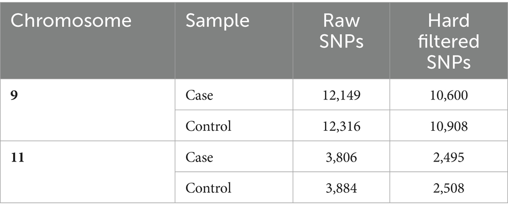
Table 2. Number of raw and hard filtered SNPs in the case and control groups in the target region on chromosomes 9 and 11.
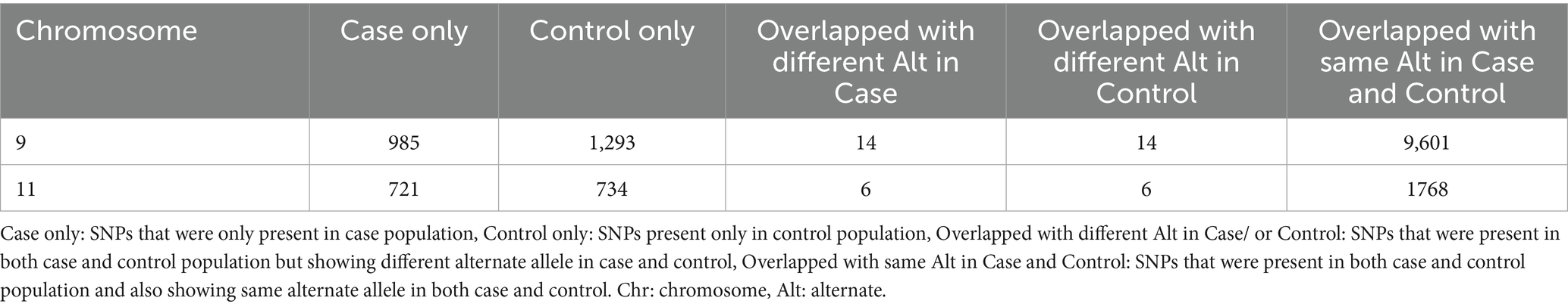
Table 3. Number of SNPs in each group on Chr 9 and 11.
Variants on chromosome 9
Twelve variants with high or moderate impact, which were deleterious based on their SIFT score, were detected only in cases (Table 4). Among these, there were two stop_ gained variants with high impact. The remaining ten variants were missense variants with moderate impact. In addition, 48 variants with modifier impact were identified within 1 kb up- and downstream of the candidate genes present only in cases (Supplementary Table 5).
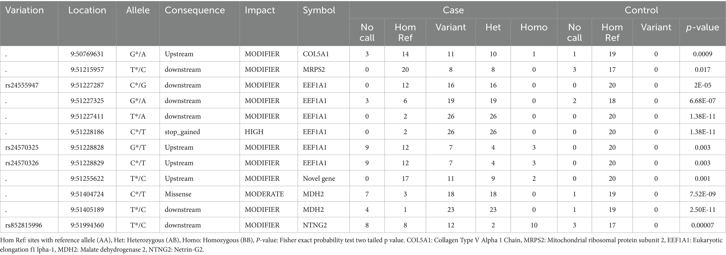
Table 4. Chr 9, variants with statistically significant differences in frequency between the case and control populations (present only in the case population).
Thirty missense variants (2 known and 28 new) with moderate impact that were deleterious according to their SIFT score were detected only in controls. In addition, 65 variants with modifier impact (13 known and 52 new) were observed within 1 kb up- and downstream of genes (Supplementary Table 5).
Twenty-six missense variants with moderate impact were identified (14 known and 12 new) in both cases and controls. However, no significant differences in the frequency of these variants were detected between cases and controls (Supplementary Table 5).
Variants on chromosome 11
One new missense SNP (deleterious based on SIFT score, SIFT: 0.03) was found only in cases, and this SNP seems to have a moderate impact in a novel protein coding gene that is orthologous to human ATP5MF (ATP synthase membrane subunit F; for more details, please see Supplementary Table 6). In addition, five SNPs (3 known and 2 new) were identified within 1 kb upstream of gene TSS coordinates. In addition, 12 SNPs (3 known and 8 new) were detected within 1 Kb downstream of the hypothesized candidate genes. Interestingly, associations between several of these candidate genes and human IBD have already been described. The details of the identified variants are shown in Supplementary Table 6.
Two of these SNPs with modifier impact were located within 1 kb upstream of IL3, a hemopoietic cytokine driving the development of myeloid stem cells that was previously identified to be associated with IBD (20). Two SNPs were found downstream of PDLIM (a protein with cysteine-rich double zinc fingers involved in protein–protein interaction and cytoskeletal organization) and IL13 (a Th2 cytokine involved in IgE synthesis, chitinase upregulation and hyperresponsiveness of mucosal surfaces) and one new SNP was found downstream of IL4 (a Th2 cytokine produced by mast cells, eosinophils and basophils that stimulates B cells into plasma cells and shares functions similar to IL-13). All these genes have already been reported to be associated with human IBD in previous studies. SNPs with a statistically significant difference (p-value <0.05) between cases and controls are shown in Table 5.
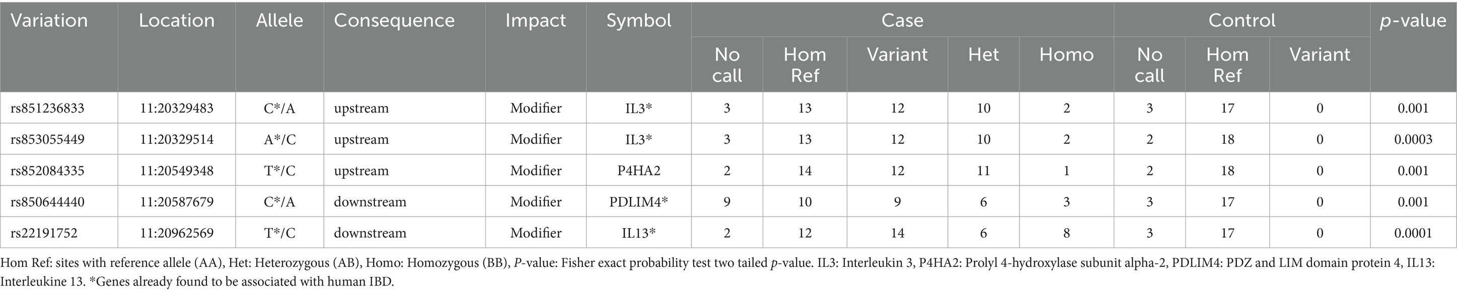
Table 5. Chr 11, variants with statistically significant differences in frequency between the case and control populations (present only in the case population).
Two new missense SNPs, deleterious based on SIFT score, were identified in a protein coding gene, orthologous to human HINT1 (histidine triad nucleotide binding protein 1) in the control population. HINT1 is a hydrolase and gene ontology annotations related to this gene include nucleotide binding and protein kinase C binding. Furthermore, five SNPs (1 known and 4 new) within 1 Kb upstream and ten SNPs (1 known and 9 new) within 1 Kb downstream of the candidate genes and Small nucleolar RNAs (snoRNA) on Chr 11 were identified in the control population. Two SNPs downstream of a snoRNA were observed to have significant p value (Supplementary Table 6).
Ten missense SNPs with high and moderate impact were detected in both cases and controls. However, only one SNP in IL13 was found to be deleterious based on the SFIT score and there was no statistically significant difference in the frequency of this SNP between case (Maf:0.054) and control group (Maf:0.074; Table 6).
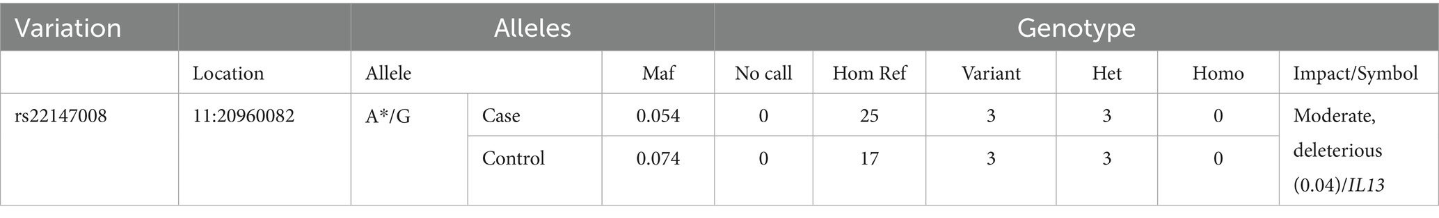
Table 6. The only variant overlapped between the case and control populations, with same ALT in the case and control, with deleterious SIFT score.
Pathway and network analysis reveal impact on genes involved in inflammatory response and metabolism
IPA analysis
Several pathways involved in innate and adaptive immune and inflammatory response (i.e., T helper cell differentiation, Th1 and Th2 activation pathway, communication between innate and adaptive immune cells and differential regulation of cytokine production in intestinal epithelial cells by IL-17A and IL-17F) and metabolism (i.e., TCA cycle II (Eukaryotic), gluconeogenesis I, aspartate degradation II and pyrimidine deoxyribonucleotides De Novo biosynthesis) were constructed by the gene products in the candidate regions for IBD susceptibility (Figure 1 and Supplementary Table 7). Moreover, four networks of molecular interactions related to cell cycle (IPA Score: 22) (Figure 2), hereditary disorder and metabolic disease (IPA Score: 17), cellular movement (IPA Score: 14) and small molecule biochemistry and metabolism (IPA Score: 12) were constructed, using the list of candidate genes, located in the targeted regions for IBD (Supplementary Table 7).
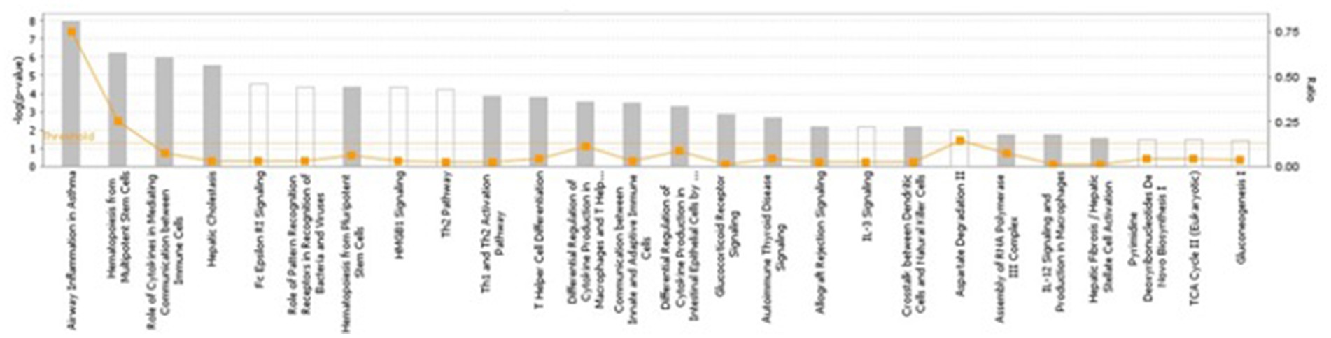
Figure 1. The most highly represented canonical pathways of genes located at the candidate regions. The solid yellow line indicates the significance threshold. The line with squares represents the ratio of the genes represented within each pathway to the total number of genes in the pathway.
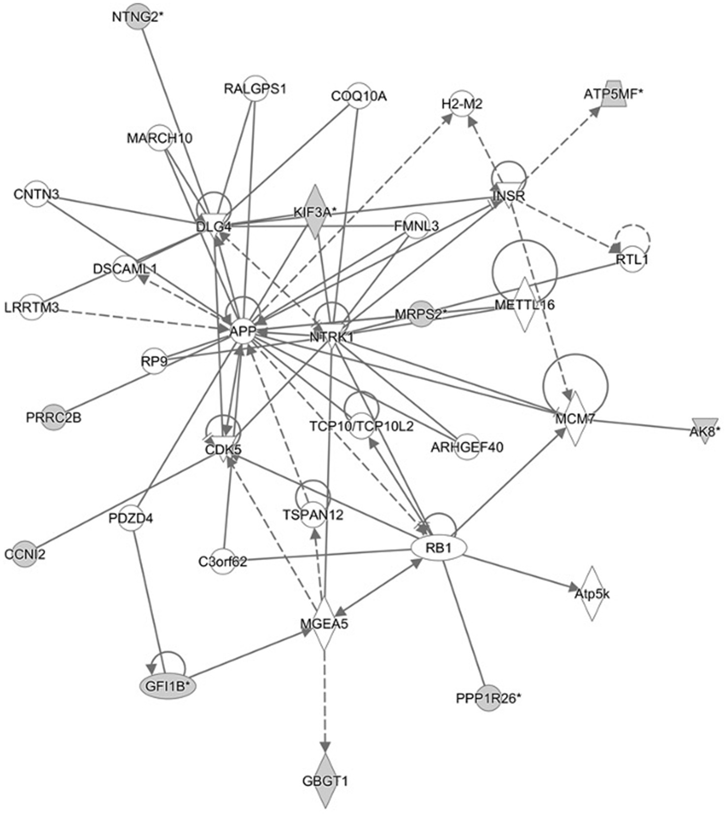
Figure 2. Cell cycle network and genes involved in the network. Gray filled shapes represent genes included in the list of candidate genes identified in the targeted regions. Solid and dotted lines represent direct and indirect interaction between genes, respectively.
Enrichment pathway analysis
The results of the Enrichr analysis showed that several genes within our list appear to be involved in biological processes and/or are molecular components that have been associated or directly/indirectly involved in human IBD. Results of the pathway association analysis using different databases and details of pathways and genes involved in each pathway are shown in Supplementary Tables 8 and 9.
Association of variants with treatment response
The majority of SNPs identified in the candidate regions for IBD were present in FRD, ARD and the steroid treated group, including SRD and those dogs that were euthanized (NRS/PTS). Several missense and modifier SNPs were present in either FRD and ARD (20 SNPs) or the steroid treated group (10 SNPs) including SRD and NRS/PTS dogs, however, these SNPs were present in only one or two IBD cases (more details are shown in Supplementary Tables 8 and 9).
Discussion
In the present study, we performed a targeted NGS experiment in previously identified candidate region for canine IBD susceptibility to detect good candidate genes and mutations. Most variants identified were novel variants. Some of the SNPs identified were not in HWE. Deviation from HWE in case–control genetic association studies is indicative of genetic association (25). However, intensive selective breeding during breed formation, and therefore loss of random mating that would normally enrich gene pool and maintain HWE, may also explain why some of the SNPs were not in HWE (25). In addition, due to the small sample size used in this study and missing calls in some of the SNPs that could affect the significance of each genotype, some SNPs may not be in HWE. It is worth mentioning that several SNPs identified in the case population were only seen in the heterozygote state, such as those in EEF1A1 (Elongation Factor 1 Alpha). One possible explanation could be that homozygote SNPs might have been present but have not been captured (low quality/filtered out etc.). Another possibility is that the heterozygotes were introduced recently by either random mutation or outbreeding of GSDs, as heterozygosity in each breed is very low (26). It may also be possible that a homozygote state could be lethal.
Two novel stop-gained SNPs in EEF1A1, were identified only in cases and were present in 26 out of 28 cases. EEF1A1 is an important protein that initiates protein translation elongation and triggers the initiation of protein translation elongation (27, 28). Apart from its canonical function in translation elongation by ribosomes, EEF1A1 plays an important role in a wide variety of cellular processes including signaling transduction, heat shock response, cytoskeleton regulation (29) and cellular apoptosis (30). It has been also documented that binding of EEF1A1 to STAT3 is crucial for STAT3 phosphorylation and for NF-κB/STAT3 activation, which enhances IL-6 expression (28). Elevated levels of this cytokine were reported to play a pivotal role in the initiation of inflammatory processes and progression of disease in many clinical conditions including rheumatoid arthritis, Alzheimer’s disease and Crohn’s disease (31–34). However, no previous association with IBD has been reported.
MDH2 (Mitochondrial malate dehydrogenase) is another good candidate gene located on Chr 9, since several missense mutations were detected in this gene, and were only present in IBD cases. In addition, IPA analysis showed that this gene was part of a gene network involved in hereditary disorders and metabolic diseases, as well as involvement in pathways associated with metabolism. The mitochondrial malate dehydrogenase, encoded by MDH2, is a mitochondrial protein, which plays an important role in energy production. Altered expression of MDH2 has been reported in studies investigating differentially expressed proteins in intestinal biopsies of IBD patients. Down-regulation of mitochondrial proteins involved in energy production including MDH2 in the colonic mucosal biopsies of ulcerative colitis (UC) patients was previously reported by Hsieh et al. (35). Results of this study suggested the implications of colonocyte mitochondrial dysfunction and perturbed mucosa immune regulation in the pathogenesis of UC.
In the control population, two known SNPs (rs850782880 and rs852254668) that were identified within less than 300 bp downstream of the MRPS2 gene, were of particular interest. As these two SNPs were present in all 20 control GSDs used in this study, they therefore could potentially be considered as important variants for IBD in GSDs. MRPS2 encodes mitochondrial ribosomal protein subunit 2, which is involved in protein synthesis within the mitochondrion. Most of the mitochondrial proteins including the ribosomal proteins and translation factors that are responsible for the expression of the mitochondrial genome, are synthesized on cytoplasmic ribosomes and imported into mitochondria post-translationally. The mitochondrial oxidative phosphorylation system, which produces the bulk of ATP for almost all eukaryotic cells to sustain cells’ normal functions, depends on the translation of 13 mitochondrial DNA (mtDNA)-encoded polypeptides by mitochondria-specific ribosomes in the mitochondrial matrix. All these peptides are members of the oxidative phosphorylation complexes (36). Several genetic mutations in nuclear genes coding for mitochondrial proteins or mitochondrial genes that can cause defects in mitochondrial transcripts or mitochondrial proteins leading to mitochondrial dysfunction and consequently impaired energy production, have been linked to many inherited diseases [reviewed in (37)]. Mutations affecting MRPS2 were observed to cause mitochondrial disorder, altered cellular metabolism, developmental delay, and multiple defects in the oxidative phosphorylation system in a study by Gardeitchik and colleagues (38).
Given that most cellular functions as well as tight junction maintenance and maintenance of the epithelial barrier are energy-dependent, it could be assumed that mitochondrial dysfunction may play a key role in both the onset and recurrence of IBD. The intestinal mucosa of IBD patients is in a state of energy deficiency, characterized by alterations in the oxidative metabolism of epithelial cells and reduced levels of ATP within the intestine (39–41). Several studies have provided evidence of mitochondrial stress and abnormalities within the intestinal epithelium of patients with IBD and mice with experimentally induced colitis (42–44). The hallmarks of mitochondrial dysfunction, including oxidative stress and impaired ATP production, have been observed in the intestines of patients with IBD (45–47) however, it is yet unclear whether these processes occur as a cause or consequence of the disease.
On Chr11, we identified several SNPs within 1 Kb up- and downstream of genes. Two SNPs with modifier impact within 1 kb upstream of IL3, 2 SNPs downstream of PDLIM and IL13 and one novel SNP downstream of IL4, were identified. Interestingly, all these genes have previously been shown to be associated with human IBD (48). According to our results, the same genes identified as potentially good candidates for IBD in GSDs, further supporting the usefulness of the domestic dog as a natural animal model for human diseases and especially for IBD.
Although disease-associated genetic variations are commonly thought to affect the coding regions of genes, it has been observed that some may alter normal gene expression (49). Thus, it might be possible that the identified SNPs may alter the expression of the genes. It is worth noting that altered expression of interleukins at mRNA and protein levels in human and canine IBD has been reported in several studies.
In addition, there is evidence that a conserved noncoding element (CNE) located between IL4 and IL13 controls expression of both genes, as well as IL5. A conserved element was identified by cross-species sequence comparison in the intergenic space between the IL4 and IL13 cytokine genes. Targeted deletion in mice revealed it to be a coordinate enhancer for IL4 and IL13, as well as for the more distant IL5 gene. This deletion was also affecting the gene expression in Th2 cells (50, 51).
We also assessed whether there is a correlation between variants identified in the case population and response to treatment. The majority of identified SNPs were present in all treatment response groups and therefore we were unable to detect statistically significant differences. Several missense and modifier SNPs were present in either FRD and ARD or SRD and NRS (PTS) group however, these SNPs were present in only one or two cases. Further studies using a bigger sample size are needed to confirm these results, since the lack of association between SNP markers and response to treatment in the present study maybe attributable to small sample size.
Canine IBD, similar to the condition in humans, is considered to be a complex multifactorial disorder that seems to occur in genetically susceptible individuals after exposure to one or more environmental triggers. In general, it is believed that a number of “susceptibility variants” may cause a general predisposition to IBD, and additional genetic variation or environmental factors may influence specific phenotypic characteristics of the individual such as disease site, disease behavior or response to treatment. In humans, it has been shown that some susceptibility loci are shared between CD and UC, the two major subtypes of IBD, but others are specific to either CD or UC, which perhaps are responsible for diverging disease courses (52–54).
The results of the present study suggest that the sample size of 28 IBD cases may not have enough power to detect associations of rare alleles with the disease. A larger genotyped population may be necessary due to the complex genetic architecture of the disease and environmental effects. In addition, studying environmental differences in dogs with different responses to treatment may help to identify environmental factors that could affect response to treatment. In humans, a number of associations have been reported between environmental factors such as infections in childhood, diet and smoking, and increased risk of developing IBD and their effect on the efficacy of treatments (55–59). Further investigation of potential environmental associations, such as deworming and dietary history, vaccination, as well as previous occurrences of infection in GSDs might therefore help identifying factors affecting treatment response.
By performing targeted NGS of the two associated regions identified by GWAS (20), an attempt was made to identify variants contributing to susceptibility or resistance to canine IBD and then evaluate their correlation with response to treatment. Here, a number of good candidate SNPs with strong association and a potential functional effect were identified. While one or several of these variants may be the causal variant(s), it is also possible that actual causal variants may have been missed in the targeted re-sequencing process or in the genotyping process for technical reasons. Considering the limited sample size of this study, missing calls in some of the SNPs could affect the significance of the results. Therefore, it may be useful to genotype the cases and controls that have not been called properly at these positions, before performing further investigations. The actual functional variant may also be an indel or CNV which has not been investigated in the current study.
A follow-up study in a larger population of IBD cases with different treatment responses is essential to validate results and confirm the variants and genes significantly associated with disease. The SNPs detected by NGS could be further genotyped using a custome made genotyping platform such as Sequenom MassARRAY iPLEX in a larger population of GSDs. In addition, targeted NGS of the other two associated regions on Chr 7 and 13, identified by GWAS will help to identify causal variants and subsequent functional analysis of the causal variants may reveal insights into mechanisms involved in the pathogenesis of canine IBD.
The results presented here represent a starting point for further studies of genetic factors associated with canine IBD. Further studies are necessary to conclusively define whether there is a correlation between certain sets of variants, including newly identified variants and previously known variants in Toll-like receptors (TLR)4, 5 and Nucleotide-binding oligomerization domain-containing protein 2 (NOD2) and response to treatment in GSDs with IBD. Identification of variants associated with the disease could potentially lead to the development of a genetic screening test to assist veterinarians with a diagnosis of IBD, and screening for SNPs that are predictive of response to a specific therapy could, potentially maximize treatment efficiency.
Given the heterogeneity of IBD, it is unlikely that any single marker or class of markers could successfully predict response to treatment. However, a combination of several classes of markers, including genetic, serological and inflammatory markers, may have valuable potential to predict the outcome of a treatment.
Data availability statement
The datasets presented in this study can be found in online repositories. The names of the repository/repositories and accession number(s) can be found in the article/Supplementary material.
Ethics statement
The animal studies were approved by All blood samples used in this study were collected in ethylenediaminetetraacetic acid (EDTA) and represented residual material following completion of clinical diagnostic testing. Residual samples were utilized for research with informed owner consent. The use of these residual EDTA blood samples and buccal swab samples within the current study was approved by the RVC Animal Welfare Ethical Review Body (AWERB; approval number 2013 1210). The studies were conducted in accordance with the local legislation and institutional requirements. Written informed consent was obtained from the owners for the participation of their animals in this study.
Author contributions
AtP: Formal analysis, Writing – original draft, Data curation, Methodology, Investigation, Validation, Writing – review & editing. MaS: Data curation, Writing – review & editing, Formal analysis, Validation, Resources. AnP: Data curation, Methodology, Writing – review & editing, Formal analysis. MeS: Investigation, Resources, Writing – review & editing. AK: Investigation, Data curation, Resources, Writing – review & editing. PW: Writing – review & editing, Investigation, Validation, Resources. KA: Investigation, Funding acquisition, Supervision, Writing – review & editing. DW: Writing – original draft, Writing – review & editing, Project administration, Funding acquisition, Supervision, Resources, Conceptualization.
Funding
The author(s) declare that financial support was received for the research and/or publication of this article. This work was funded through a BBSRC iCASE studentship (BB/J01236X/1) awarded to KA and DW with Laboklin GmBH (Bad Kissingen, Germany) as industrial partner as well as the American Kennel Club. We are grateful to owners of GSD who gave permission for their dogs to participate in this study. The authors declare that this study received funding from Laboklin GmBH (Bad Kissingen, Germany). The funder was not involved in the study design, collection, analysis, interpretation of data, the writing of this article, or the decision to submit it for publication.
Conflict of interest
The authors declare that the research was conducted in the absence of any commercial or financial relationships that could be construed as a potential conflict of interest.
The author(s) declared that they were an editorial board member of Frontiers, at the time of submission. This had no impact on the peer review process and the final decision.
Generative AI statement
The authors declare that no Gen AI was used in the creation of this manuscript.
Publisher’s note
All claims expressed in this article are solely those of the authors and do not necessarily represent those of their affiliated organizations, or those of the publisher, the editors and the reviewers. Any product that may be evaluated in this article, or claim that may be made by its manufacturer, is not guaranteed or endorsed by the publisher.
Supplementary material
The Supplementary material for this article can be found online at: https://www.frontiersin.org/articles/10.3389/fvets.2025.1648911/full#supplementary-material
Footnotes
1. ^https://software.broadinstitute.org/gatk
2. ^http://www.bioinformatics.babraham.ac.uk/projects/fastqc/
3. ^https://m.ensembl.org/info/genome/variation/species/sources_documentation.html#canis_lupus_familiaris
4. ^https://www.ensembl.org/Tools/VEP
5. ^http://www.vassarstats.net/odds2x2.html
References
1. Albert, EJ. Inflammatory bowel disease: current perspectives. Vet Clin N Am Small Anim Pract. (1999) 29:501–21. doi: 10.1016/S0195-5616(99)50032-6
2. Guilford, WG. Idiopathic inflammatory bowel diseases In: Strombecks small animal gastroenterology (1996). 451–86.
3. Hendrickson, BA, Gokhale, R, and Cho, JH. Clinical aspects and pathophysiology of inflammatory bowel disease. Clin Microbiol Rev. (2002) 15:79–94. doi: 10.1128/CMR.15.1.79-94.2002
4. Kimmel, SE, Waddell, LS, and Michel, KE. Hypomagnesemia and hypocalcemia associated with protein-losing enteropathy in Yorkshire terriers: five cases (1992-1998). J Am Vet Med Assoc. (2000) 217:703–6. doi: 10.2460/javma.2000.217.703
5. Simpson, KW, Dogan, B, Rishniw, M, Goldstein, RE, Klaessig, S, McDonough, PL, et al. Adherent and invasive Escherichia Coli is associated with granulomatous colitis in boxer dogs. Infect Immun. (2006) 74:4778–92. doi: 10.1128/IAI.00067-06
6. Kathrani, A, House, A, Catchpole, B, Murphy, A, Werling, D, and Allenspach, K. Breed-independent toll-like receptor 5 polymorphisms show association with canine inflammatory bowel disease. Tissue Antigens. (2011) 78:94–101. doi: 10.1111/j.1399-0039.2011.01707.x
7. German, AJ, Day, MJ, Ruaux, CG, Steiner, JM, Williams, DA, and Hall, EJ. Comparison of direct and indirect tests for small intestinal bacterial overgrowth and antibiotic-responsive diarrhea in dogs. J Vet Intern Med. (2003) 17:33–43.
8. Westermarck, E, Skrzypczak, T, Harmoinen, J, Steiner, JM, Ruaux, CG, Williams, DA, et al. Tylosin-responsive chronic diarrhea in dogs. J Vet Intern Med. (2005) 19:177–86. doi: 10.1111/j.1939-1676.2005.tb02679.x
9. Allenspach, K, Wieland, B, Gröne, A, and Gaschen, F. Chronic enteropathies in dogs: evaluation of risk factors for negative outcome. J Vet Intern Med. (2007) 21:700–8. doi: 10.1111/j.1939-1676.2007.tb03011.x
10. Allenspach, K, Rüfenacht, S, Sauter, S, Gröne, A, Steffan, J, Strehlau, G, et al. Pharmacokinetics and clinical efficacy of cyclosporine treatment of dogs with steroid-refractory inflammatory bowel disease. J Vet Intern Med. (2006) 20:239–44. doi: 10.1892/0891-6640(2006)20[239:paceoc]2.0.co;2
11. Roberts, RL, and Barclay, ML. Current relevance of pharmacogenetics in immunomodulation treatment for Crohn’s disease. J Gastroenterol Hepatol. (2012) 27:1546–54. doi: 10.1111/j.1440-1746.2012.07220.x
12. Sandborn, WJ. Pharmacogenomics and IBD. Inflamm Bowel Dis. (2004) 10:S35–7. doi: 10.1097/00054725-200402001-00008
13. Kathrani, A, Lee, H, White, C, Catchpole, B, Murphy, A, German, A, et al. Association between nucleotide oligomerisation domain two (Nod2) gene polymorphisms and canine inflammatory bowel disease. Vet Immunol Immunopathol. (2014) 161:32–41. doi: 10.1016/j.vetimm.2014.06.003
14. Kathrani, A, House, A, Catchpole, B, Murphy, A, German, A, Werling, D, et al. Polymorphisms in the Tlr4 and Tlr5 gene are significantly associated with inflammatory bowel disease in German shepherd dogs. PLoS One. (2010) 5:1–10. doi: 10.1371/journal.pone.0015740
15. Peiravan, A, Allenspach, K, Boag, AM, Soutter, F, Holder, A, Catchpole, B, et al. Single nucleotide polymorphisms in Major histocompatibility class II haplotypes are associated with potential resistance to inflammatory bowel disease in German shepherd dogs. Vet Immunol Immunopathol. (2016) 182:101–5. doi: 10.1016/j.vetimm.2016.10.012
16. Lequarré, A-S, Andersson, L, André, C, Fredholm, M, Hitte, C, Leeb, T, et al. LUPA: a European initiative taking advantage of the canine genome architecture for unravelling complex disorders in both human and dogs. Vet J. (2011) 189:155–9. doi: 10.1016/j.tvjl.2011.06.013
17. Tengvall, K, Kierczak, M, Bergvall, K, Olsson, M, Frankowiack, M, Farias, FHG, et al. Genome-wide analysis in German shepherd dogs reveals association of a locus on CFA 27 with atopic dermatitis. PLoS Genet. (2013) 9:1–12.
18. Wilbe, M, Jokinen, P, Truvé, K, Seppala, EH, Karlsson, EK, Biagi, T, et al. Genome-wide association mapping identifies multiple loci for a canine SLE-related disease complex. Nat Genet. (2010) 42:250–4. doi: 10.1038/ng.525
19. Wood, SH, Ke, X, Nuttall, T, McEwan, N, Ollier, WE, and Carter, SD. Genome-wide association analysis of canine atopic dermatitis and identification of disease related SNPs. Immunogenetics. (2009) 61:765–72. doi: 10.1007/s00251-009-0402-y
20. Peiravan, A, Bertolini, F, Rothschild, MF, Simpson, KW, Jergens, AE, Allenspach, K, et al. Genome-wide association studies of inflammatory bowel disease in German shepherd dogs John Green. PLoS One. (2018) 13:e0200685. doi: 10.1371/journal.pone.0200685
21. Karlsson, EK, Baranowska, I, Wade, CM, Salmon, NHC, Hillbertz, MC, Zody, NA, et al. Efficient mapping of Mendelian traits in dogs through genome-wide association. Nat Genet. (2007) 39:1321–8. doi: 10.1038/ng.2007.10
22. Sutter, NB. Extensive and breed-specific linkage disequilibrium in Canis Familiaris. Genome Res. (2004) 14:2388–96. doi: 10.1101/gr.3147604
23. Bolger, AM, Lohse, M, and Usadel, B. Trimmomatic: a flexible trimmer for Illumina sequence data. Bioinformatics. (2014) 30:2114–20. doi: 10.1093/bioinformatics/btu170
24. Li, H, and Durbin, R. Fast and accurate short read alignment with burrows-wheeler transform. Bioinformatics. (2009) 25:1754–60. doi: 10.1093/bioinformatics/btp324
25. Ziegler, A, Van Steen, K, and Wellek, S. Investigating hardy–weinberg equilibrium in case–control or cohort studies or meta-analysis. Breast Cancer Res Treat. (2011) 128:197–201. doi: 10.1007/s10549-010-1295-z
26. Lindblad-Toh, K, Wade, CM, Mikkelsen, TS, Karlsson, EK, Jaffe, DB, Kamal, M, et al. Genome sequence, comparative analysis and haplotype structure of the domestic dog. Nature. (2005) 438:803–19. doi: 10.1038/nature04338
27. Kapp, LD, and Lorsch, JR. The molecular mechanics of eukaryotic translation. Annu Rev Biochem. (2004) 73:657–704. doi: 10.1146/annurev.biochem.73.030403.080419
28. Schulz, I, Engel, C, Niestroj, AJ, Kehlen, A, Rahfeld, J-U, Kleinschmidt, M, et al. A non-canonical function of eukaryotic elongation factor 1A1: regulation of Interleukin-6 expression. Biochimica Biophysica Acta. (2014) 1843:965–75. doi: 10.1016/j.bbamcr.2014.01.022
29. Negrutskii, B, Vlasenko, D, and El’skaya, A. From global phosphoproteomics to individual proteins: the case of translation elongation factor EEF1A1. Expert Rev Proteomics. (2012) 9:71–83.
30. Kobayashi, Y, and & Yonehara, S. (2009). “Novel cell death by downregulation of EEF1A1 expression in tetraploids.” Cell Death Differ 16: 139–150, doi: 10.1038/cdd.2008.136
31. Cacquevel, M, Lebeurrier, N, Cheenne, S, and Vivien, D. Cytokines in Neuroinflammation and Alzheimers disease. Curr Drug Targets. (2004) 5:529–34. doi: 10.2174/1389450043345308
32. Ito, H. Novel therapy for Crohn’s disease targeting IL-6 Signalling. Expert Opin Ther Targets. (2004) 8:287–94. doi: 10.1517/14728222.8.4.287
33. Murphy, SF, Kwon, JH, and Boone, DL. Novel players in inflammatory bowel disease pathogenesis. Curr Gastroenterol Rep. (2012) 14:146–52. doi: 10.1007/s11894-012-0250-z
34. Nishimoto, N. Interleukin-6 in Rheumatoid Arthritis. Curr Opin Rheumatol. (2006) 18:277–81. doi: 10.1097/01.bor.0000218949.19860.d1
35. Hsieh, S-Y, Shih, T-C, Yeh, C-Y, Lin, C-J, Chou, Y-Y, and Lee, Y-S. Comparative proteomic studies on the pathogenesis of human ulcerative colitis. Proteomics. (2006) 6:5322–31. doi: 10.1002/pmic.200500541
36. Ojala, D, Montoya, J, and Attardi, G. TRNA punctuation model of RNA processing in human mitochondria. Nature. (1981) 290:470–4. doi: 10.1038/290470a0
37. Vafai, SB, and Mootha, VK. Mitochondrial disorders as windows into an ancient organelle. Nature. (2012) 491:374–83. doi: 10.1038/nature11707
38. Gardeitchik, T, Mohamed, M, Ruzzenente, B, Karall, D, Guerrero-Castillo, S, Dalloyaux, D, et al. Bi-allelic mutations in the mitochondrial ribosomal protein MRPS2 cause sensorineural hearing loss, hypoglycemia, and multiple OXPHOS complex deficiencies. Am J Hum Genet. (2018) 102:685–95. doi: 10.1016/j.ajhg.2018.02.012
39. Fukushima, K, and Fiocchi, C. Paradoxical decrease of mitochondrial DNA deletions in epithelial cells of active ulcerative colitis patients. Am J Physiol Gastrointestinal Liver Physiol. (2004) 286:G804–13.
40. Kameyama, JIN-ICHI, Narui, HIDEO, Inui, MASARU, and Sato, TOSHIO. Energy level in large intestinal mucosa in patients with ulcerative colitis. Tohoku J Exp Med. (1984) 143:253–4. doi: 10.1620/tjem.143.253
41. Roediger, WEW. The colonic epithelium in ulcerative colitis: an energy-deficiency disease? Lancet. (1980) 316:712–5. doi: 10.1016/S0140-6736(80)91934-0
42. Delpre, G, Avidor, I, Steinherz, R, Kadish, U, and Ben-Bassat, M. Ultrastructural abnormalities in endoscopically and histologically Normal and involved Colon in ulcerative colitis. Am J Gastroenterol. (1989) 84:1038–46.
43. Nazli, A, Yang, P-C, Jury, J, Howe, K, Watson, JL, Söderholm, JD, et al. Epithelia under metabolic stress perceive commensal Bacteria as a threat. Am J Pathol. (2004) 164:947–57. doi: 10.1016/S0002-9440(10)63182-3
44. Rodenburg, W, Keijer, J, Kramer, E, Vink, C, van der Meer, R, and Bovee-Oudenhoven, IMJ. Impaired barrier function by dietary Fructo-oligosaccharides (FOS) in rats is accompanied by increased colonic mitochondrial gene expression. BMC Genomics. (2008) 9:144. doi: 10.1186/1471-2164-9-144
45. Kruidenier, L, and Verspaget, HW. Review article: oxidative stress as a pathogenic factor in inflammatory bowel disease--radicals or ridiculous? Aliment Pharmacol Ther. (2002) 16:1997–2015. doi: 10.1046/j.1365-2036.2002.01378.x
46. Pravda, J. Radical induction theory of ulcerative colitis. World J Gastroenterol. (2005) 11:2371–84. doi: 10.3748/wjg.v11.i16.2371
47. Rezaie, A, Parker, RD, and Abdollahi, M. Oxidative stress and pathogenesis of inflammatory bowel disease: An epiphenomenon or the cause? Dig Dis Sci. (2007) 52:2015–21. doi: 10.1007/s10620-006-9622-2
48. Jostins, L, Ripke, S, Weersma, RK, Duerr, RH, McGovern, DP, Hui, KY, et al. Host-microbe interactions have shaped the genetic architecture of inflammatory bowel disease. Nature. (2012) 491:119–24. doi: 10.1038/nature11582
49. Kleinjan, DA, and van Heyningen, V. Long-range control of gene expression: emerging mechanisms and disruption in disease. Am J Hum Genet. (2005) 76:8–32. doi: 10.1086/426833
50. Loots, GG. Identification of a coordinate regulator of interleukins 4, 13, and 5 by cross-species sequence comparisons. Science. (2000) 288:136–40. doi: 10.1126/science.288.5463.136
51. Mohrs, M, Blankespoor, CM, Wang, ZE, Loots, GG, Afzal, V, Hadeiba, H, et al. Deletion of a coordinate regulator of type 2 cytokine expression in mice. Nat Immunol. (2001) 2:842–7. doi: 10.1038/ni0901-842
52. Abraham, C, and Cho, JH. Inflammatory bowel disease. N Engl J Med. (2009) 361:2066–78. doi: 10.1056/NEJMra0804647
53. Ahmad, T, Satsangi, J, McGovern, D, Bunce, M, and Jewell, DP. Review article: the genetics of inflammatory bowel disease. Aliment Pharmacol Ther. (2001) 15:731–48. doi: 10.1046/j.1365-2036.2001.00981.x
54. Satsangi, J, Jewell, DP, and Bell, JI. The genetics of inflammatory bowel disease. Gut. (1997) 40:572–4. doi: 10.1136/gut.40.5.572
55. Chapman-Kiddell, CA, Davies, PSW, Gillen, L, and Radford-Smith, GL. Role of diet in the development of inflammatory bowel disease. Inflamm Bowel Dis. (2010) 16:137–51. doi: 10.1002/ibd.20968
56. Kiss, LS, Szamosi, T, Molnar, T, Miheller, P, Lakatos, L, Vincze, A, et al. Early clinical remission and normalisation of CRP are the strongest predictors of efficacy, mucosal healing and dose escalation during the first year of adalimumab therapy in Crohn’s disease. Aliment Pharmacol Ther. (2011) 34:911–22. doi: 10.1111/j.1365-2036.2011.04827.x
57. Sandborn, WJ, Melmed, GY, McGovern, DPB, Loftus, EV, Choi, JM, Cho, JH, et al. Clinical and demographic characteristics predictive of treatment outcomes for certolizumab pegol in moderate to severe Crohn’s disease: analyses from the 7-year precise 3 study. Aliment Pharmacol Ther. (2015) 42:330–42. doi: 10.1111/apt.13251
58. Sandborn, WJ, Abreu, MT, D’Haens, GR, Colombel, J-F, Vermeire, S, Mitchev, K, et al. S1030 predictors of response and remission to certolizumab pegol in patients with Crohn’s disease: data from the WELCOME study. Gastroenterology. (2010) 138:S-164
Keywords: inflammatory bowel disease, chronic enteropathy, NGS, GSD, canine
Citation: Peiravan A, Salavati M, Psifidi A, Sharman M, Kent A, Watson P, Allenspach K and Werling D (2025) Targeted next-generation sequencing of candidate regions identified by GWAS revealed SNPs associated with IBD in GSDs. Front. Vet. Sci. 12:1648911. doi: 10.3389/fvets.2025.1648911
Edited by:
Josipa Kuleš, University of Zagreb, CroatiaReviewed by:
Ravikanthreddy Poonooru, University of Missouri, United StatesKeon Kim, Chonnam National University, Republic of Korea
Copyright © 2025 Peiravan, Salavati, Psifidi, Sharman, Kent, Watson, Allenspach and Werling. This is an open-access article distributed under the terms of the Creative Commons Attribution License (CC BY). The use, distribution or reproduction in other forums is permitted, provided the original author(s) and the copyright owner(s) are credited and that the original publication in this journal is cited, in accordance with accepted academic practice. No use, distribution or reproduction is permitted which does not comply with these terms.
*Correspondence: Atiyeh Peiravan, YXBlaXJhdmFuQHJ2Yy5hYy51aw==; Dirk Werling, ZHdlcmxpbmdAcnZjLmFjLnVr
†Present address: Andrew Kent,Blaise Veterinary Referral Hospital, Birmingham, United Kingdom