 María Villalba-Orero1,2Camila A. Gómez3Marta Valero-Gónzalez3Noelia Venegas3Guadalupe Criado4
 María Martín-Cuervo3*
- 1Department of Animal Medicine and Surgery, Faculty of Veterinary Medicine, Universidad Complutense de Madrid, Madrid, Spain
- 2Complutense Veterinary Teaching Hospital, Universidad Complutense de Madrid, Madrid, Spain
- 3Grupo de Investigación Medicina y Cirugía Animal (MECIAN), Animal Medicine Department, Faculty of Veterinary, University of Extremadura, Cáceres, Spain
- 4Private Equine Practitioner, Barcelona, Spain
Background: Rhodococcus equi is a facultative intracellular bacterium recognized to cause pneumonia in foals aged 1 to 6 months. Currently, it remains a challenge to identify foals at risk.
Hypothesis/Objectives: We hypothesize that a certain grade of immaturity may enhance the risk for future R. equi infection. This study aims to analyze blood parameters and passive immunity transfer within the first 24 hours of life as predictive markers for the development of R. equi pneumonia during the first 6 months of life.
Methods: A total of 207 Arabian or Arabian-crossed breed foals from the same breeding center, from birth to 6 months of age, were included. Blood samples were obtained from foals during the first 24 hours after birth. Parameters analyzed were hematocrit (Hto), total white blood cell count (WBC), total plasma proteins (PT), albumin (ALB), fibrinogen (FBG), urea (U), creatinine (CREA), gamma-glutamyl transferase (GGT), iron (Fe), and serum immunoglobulin G (IgG). In addition, colostrum Brix was measured. Foals were classified into the following groups: foals presenting R. equi (R group) and healthy foals (H group), which showed no signs of pneumonia during a surveillance period of six months.
Results: Comparisons were performed between the two groups, and univariable and multivariable logistic regression were used to assess possible predictors for R. equi development. Of the 207 foals, 25 became ill (12.08 %). Foals with R. equi showed lower U levels [H: 29 (23-37) Vs R: 24 (20-31); p = 0.04], as higher CREA [H: 1.3 (1.1-1.6) Vs R: 1.5 (1.3-1.7); p = 0.03], and GGT [H: 14 (10-24) Vs R: 21 (12-39); p = 0.004] than foals with a healthy status. Multivariable logistic regression highlighted that higher GGT at birth was associated with R. equi development.
Discussion: High values of GGT and CREA have been associated with fetal immaturity, which could be related to immaturity of the immune system, especially of alveolar macrophages, and may predispose to early infection by R. equi. Interestingly, neonatal GGT may serve as a possible risk factor for developing the infection.
1 Introduction
Rhodococcus equi (R. equi) is a Gram-positive bacterium recognized for causing pneumonia in foals, representing a common cause of economic losses in the equine industry (1). As a facultative intracellular bacterium, it relies on its ability to survive within macrophages (2). This phenomenon is associated with the presence of a plasmid encoding virulence genes, among which vapA is considered the most crucial (3–5). The expression of the virulence-associated protein A (VapA) is markedly enhanced under iron-restricted conditions (6). Foals commonly develop R. equi between 1 and 6 months of age. However, exposure to this pathogen has been reported during the first week of life (7, 8). Enhanced intracellular replication of R. equi in bronchoalveolar macrophages of young foals, accompanied by age- and macrophage-specific cytokine expression patterns, may contribute to the pathogen’s pulmonary tropism and the age-dependent susceptibility to infection (9).
The primary manifestation of R. equi infection is a respiratory disorder, characterized by the development of pyogranulomatous lesions in the lungs (10). The first clinical signs may include sporadic or intermittent cough, pyrexia, lethargy, respiratory distress, reduced appetite, and weight loss (11). However, extrapulmonary infections and immunomediated inflammatory disorders are also common (12–15). Diagnosis typically relies on clinical presentation, hematological abnormalities, as well as radiographic and ultrasonographic evidence of pulmonary lesions (16). Additionally, cytological evidence of intracellular Gram-positive or acid-fast coccobacilli and/or detection of virulent R. equi in tracheal aspirates can be pursued (17, 18).
Several reports agree that the primary risk factor for pneumonia is the concentration of virulent R. equi in the environment (19–21) and the foal’s exposure to this pathogen (22, 23). Differences in the host’s innate and adaptive immune responses (24) and genetic variations (25) are also considered significant factors contributing to the disease’s development. Despite considerable advances in understanding this pathogen, controlling and preventing R. equi pneumonia in equine farms remains a challenge. Consequently, many efforts are focused on elucidating predictors of the disease before its clinical onset (26–28). Ultrasound diagnosis has been used as a screening method to identify affected animals early on (29), although it has been found that this diagnostic method is more useful for assessing the severity of the disease than as a screening tool (30). Something similar happens with radiographic diagnosis, where its most evident utility is in locating deep abscesses or determining the prognosis in affected animals (31). Acute-phase inflammation proteins have been explored as predictive indicators of the disease, as well as the significance of leukocyte count (27, 32, 33). However, their relationship with other blood parameters as predisposing factors is still unknown.
This study aims to assess whether hematological and biochemical blood parameter levels, as well as the colostrum quality in newborn foals, may anticipate the subsequent development of R. equi pneumonia. These parameters could potentially serve as biomarkers for the early detection of the infection.
2 Materials and methods
2.1 Animals
This study included all foals born healthy in a breeding farm of Arabian and Anglo-Arabian equine barn in 2018, located in the Autonomous Community of Catalonia (Spain). Foals were considered healthy at birth when their vital signs and hematology were within the physiological ranges for their age, exhibited normal behavior, and had adequate total IgG concentrations. All foals were under surveillance from birth to 6 months of age, both by farm workers and the veterinarians employed in the center. The diagnosis of R. equi was based on clinical signs and confirmed by PCR.
2.2 Sample collection
Before foals were 24 h old, venous blood samples were collected in tubes with EDTA anticoagulant and lithium heparin from the jugular vein to perform hematological and biochemical analysis, respectively. Hematological analyses and colostrum quality assessments were performed at the time of sample collection. For the measurement of biochemical parameters, the plasma was separated and frozen. Samples were frozen at −80°C and transferred to the Clinical Laboratory of the Veterinary Hospital of the University of Extremadura, where they were subsequently processed.
2.3 Hematology and serum biochemistry
The hematological analysis was performed with Mindray BC-5300 equipment, parameters measured were hematocrit (Hto; %) and total white blood cell count (WBC; ×108/μL) specifying the count for lymphocytes, neutrophils, and monocytes. The biochemical analysis was performed with Crony instruments Saturno 100 equipment, and total plasma proteins (TP; g/dL), albumin (ALB, g/dL), fibrinogen (FBG, g/dL), urea (U, mg/dL) creatinine (mg/dL), gamma-glutamyl transferase (gamma GT; U/L); iron (Fe, ng/dL), and immunoglobulin G (IGG; mg/dL) was measured by quantitative determination, using MAI Animal Health’s DVM Rapid Test™ II.
2.4 Brix
Additionally, right after the delivery, the colostrum produced by the corresponding mothers was assessed using a Brix scale refractometer. The quality of the colostrum was measured within the first 12 h postpartum.
2.5 Diagnosis of Rhodococcus equi pneumonia
The foals included in this study were kept under supervision for their first 6 months of life and were checked every week from 3 weeks of age using an Easote MyLab™OneVET ultrasonography equipment equipped with the SV3513 VET rectal linear 10–5 Mhz-probe. The presumptive diagnosis of R. equi pneumonia was made based on the detection of clinical signs, such as cough, runny nose, pyrexia, lethargy, respiratory distress, decreased appetite, and weight loss, together with the detection of pulmonary abscesses compatible with pyogranulomatous pneumonia on the ultrasound examination. The diagnosis was confirmed by PCR. Accordingly, foals were classified into the following groups: foals presenting R. equi (R group) and healthy foals (H group), which showed no signs of pneumonia during this surveillance period.
2.6 Statistical analysis
Normality distribution was assessed using the Kolmogorov–Smirnov test. Data are presented as means ± standard deviations (SD) for variables with a normal distribution, and as the medians with interquartile ranges (IQT; 25th–75th) when normality could not be assumed. Firstly, data were grouped in foals that developed R. equi during the first 6 months of life (R group) or not (H group), and a Student T-test or Mann–Whitney U test (parametric or non-parametric, respectively) was applied to determine differences in measured parameters. The development of a subgroup in the R group with survivors and non-survivors was not performed, as only 2 foals died. In addition to establishing a relationship between the studied parameters at born and the outcome of development R. equi, all variables obtaining a p < 0.20 in the comparative analysis performed between R and H groups (neutrophils, albumin, urea, creatinine, gamma GT, and IGG) were selected for the logistic regression. Univariable logistic regression was performed to establish crude univariate odds ratios (OR) and 95% confidence intervals (CI) and a multivariate analysis was done to finally calculate the risk factor that better fix the model. The multivariate logistic regression model was built using automatic backward stepwise selection. Statistical differences were considered if p < 0.05. All statistical analyses were performed using SPSS statistical software (IBM SPSS Statistics 20; Chicago, IL, United States) and graphics were performed using GraphPad Prism version 9 (San Diego, CA, United States).
3 Results
3.1 Patient population
This retrospective study involved a total of 207 Arabian or Arabian-crossed breed foals. Of the 207 foals included, 25 foals (12.08%) developed pneumonia due to R. equi during the first 6 months of life (R group), and the remaining 178 foals maintained healthy conditions (H group).
3.2 General outcome
Twenty-two sick foals developed abscesses more than 1 cm in diameter and received rifampicin (10 mg/kg every 12 h) and azithromycin (10 mg/kg every 24 h) until the resolution of clinical signs. The other 3 foals showed abscesses less than 1 cm in diameter and did not receive antibiotics (additional information in Supplementary Table 1). The survival rate of the studied population was 92% (23/25) in the R group and 100% (178) in the H group.
3.3 Comparative analysis
Corresponding demographic data and blood work parameters in H and R groups are described in Table 1. Comparing H and R group, no statistical differences were observed for Hto (H: 39.7 ± 4.0 vs. R: 40 ± 3.2), total WBC (H: 7.663 ± 2.430 vs. R: 7.228 ± 2.470), and its components, total plasma proteins [H: 6.0 (5.3–6.7) vs. 6.0 (5.5–6.5)], albumin [H: 3.3 (3.1–3.5) vs. 3.4 (3.2–3.5)], fibrinogen [H: 378 (361–408) vs. 378 (327–463)], iron (H: 268.8 ± 89.6 vs. R: 255.2 ± 60.8), IGG levels (H: 1322.35 ± 539.187 vs. R: 1491.64 ± 657.577) at birth.
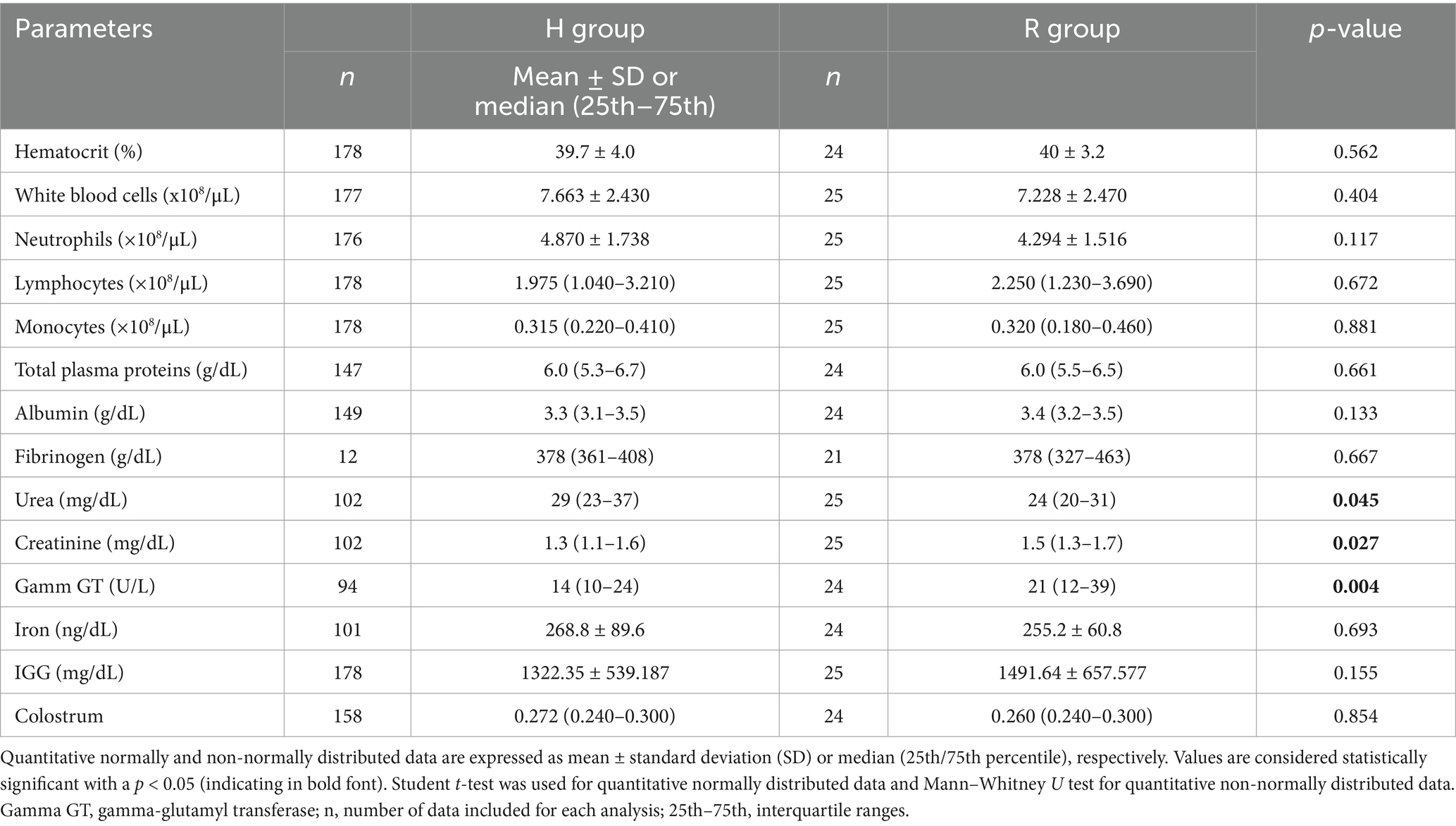
Table 1. Comparison of hematological data recruited at born, of 178 healthy foals (H group) and 25 foals that develop R. equi (N = 212 foals).
However, as represented in Figure 1, foals with R. equi showed lower urea [H: 29 (23–37) vs. R: 24 (20–31); p = 0.04], as well as higher creatinine [H: 1.3 (1.1–1.6) vs. R: 1.5 (1.3–1.7); p = 0.03], and gamma GT [H: 14 (10–24) vs. R: 21 (12–39); p = 0.004] than foals with a healthy status. The two foals that died presented an urea of 44 and 19 (mmol/L), a creatinine of 2, 5, and 1 (mg/dL) and a gamma GT of 46 and 20 (Ul/L).
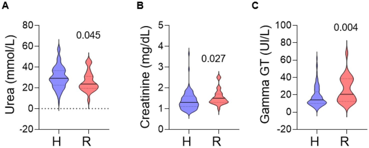
Figure 1. Violin plots representing a full distribution of raw data highlighting median (central line), and interquartile range (lower and upper lines). Comparisons are performed for blood urea (A), creatinine (B), and gamma GT (C) between foals that developed Rhodococcus equi in the first 6 months of life and those that remain healthy. Samples were collected during the first 24 h after birth. H, healthy group; R, Rhodococcus equi group.
3.4 Brix
As shown in Table 1, 189 colostrum samples were evaluated, of which 24 were produced by the mothers of foals that subsequently developed R. equi pneumonia. Comparison between H and R groups highlighted that there was no significant difference regarding the quality of colostrum suckled and colostrum [H: 0.272 (0.240–0.300) vs. 0.260 (0.240–0.300)]. There was also no significant difference in IgG concentration between these 2 groups.
3.5 Factors associated with the development of Rhodococcus equi
Association between measurements of variables in foals born with the future development of R. equi was studied, including neutrophils, albumin, urea, creatinine, gamma GT, and IGG (variables obtaining a p < 0.20 in the comparative analysis performed between R and H groups, Table 1). As shown in Table 2, the univariable logistic regression evidenced that neutrophil count, albumin, urea, creatinine, and IGG at birth were not independently associated with future R. equi. However, this analysis reveals that higher blood levels of gamma GT at born are a risk factor for the development of the disease (OR: 1.05; 95% IC: 1.01–1.08; p = 0.008) and that higher urea levels tend to increase the risk of R. equi (OR: 0.96; 95% IC: 0.91–1.00; p = 0.068). For the multivariable logistic regression, a total of 115 foals (55.3%; 92 and 23 foals in the H and R groups, respectively) were automatically included. Variables that finally were maintained in the regression were gamma GT (OR: 1.043; 95% IC: 1.01–1.01; p = 0.019) and urea (OR: 0.956; 95% IC: 0.91–1.01; p = 0.070). These, formally studied parameters, presenting higher gamma GT at birth, were associated with R. equi development.
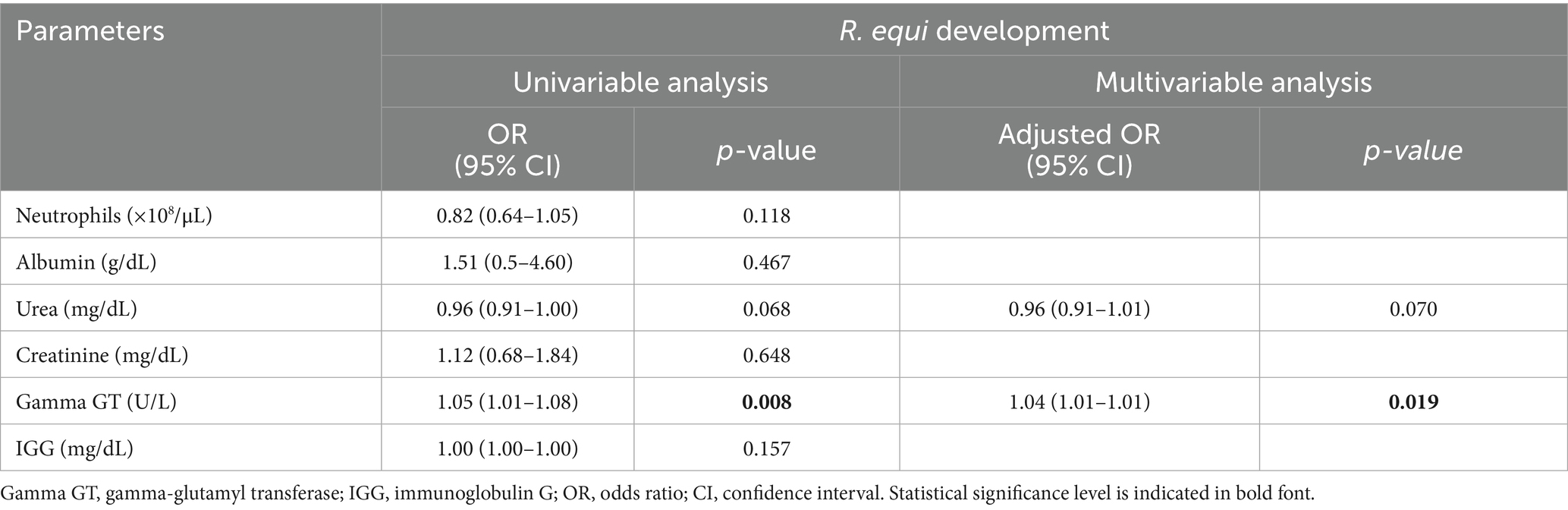
Table 2. Logistic regression showing data analyzed to establish risk factors associated with the development of R. equi of 178 healthy foals (H group) and 25 foals (N = 212 foals).
4 Discussion
This study highlights differences in urea, serum creatinine, and gamma GT at birth between foals that develop R. equi pneumonia and those that remain healthy. Interestingly, neonatal gamma GT has been identified as an early risk factor for the future development of the infection.
Our research was performed on foals less than 24 h of life. It has been proposed that lung infection is the result of inhalation of virulent R. equi by aerosols from contaminated soil (34), with intestinal colonization observed within the first week of life and R. equi being isolated in the feces of 3-day-old foals (35). Also, increased mRNA expression of different interleukins has been found in bronchoalveolar macrophages in foals 1 to 3 days old (9). This background justifies that the collection of samples was performed during the first day after the delivery, since it is suggested that the most likely time of infection would be soon after birth (22).
Urea is a product generated in the liver because of protein metabolism. The lower values observed in the foals that developed the disease in this study can also be explained by hepatic immaturity. Another possibility is that these foals had a diet lower in protein due to poorer milk quality from their mothers. However, this seems unlikely since there were no differences in colostrum quality, and the samples were taken within the first 24 h of life, which is not enough time for metabolic alterations attributable to diet to develop. In human neonates, changes in urea concentrations are due to factors other than dietary and renal ones (36). Interestingly, in human medicine, it has also been observed that premature neonates with functional immaturity are unable to increase plasma urea concentrations despite the administration of protein-rich diets, assuming that this is due to hepatic functional immaturity (37). This fact could explain why foals suffering from the disease had lower urea concentrations than healthy ones and could also be related to the immaturity of other systems, in this case, the respiratory immune system.
In this study, serum creatinine was higher in foals that developed R. equi. However, the mean value observed in those animals was within the range considered physiological in the neonatal foal (1.5–2.9 mg/dL) (38). Foals present a higher upper limit value than adult horses (39). Similarly, the serum concentration of gamma GT was higher in the sick group but also within the reference range. In neonatal foals, the reference ranges for hematological and biochemical parameters are highly variable and difficult to interpret. As both hypercreatinemia and pathological increased gamma GT levels have been linked to placental insufficiency, fetal stress, and immaturity (40, 41), is it likely that a certain level of immaturity might be present in foals that developed pneumonia in this study. Additionally, the importance of the adaptive immune system in human neonates has been highlighted in the development of respiratory diseases (42, 43). The association of gamma GT as a risk variable for R. equi pneumonia in this study could be considered a potential marker useful for identifying foals at risk, allowing for the necessary preventive measures to be taken to avoid the development of the infection.
Virulent R. equi replicates within macrophages, beginning its replication within 6–12 h, affecting macrophage viability within 48 h (3). Although macrophages do not prevent intracellular bacterial replication, R. equi infection induces an immune response in macrophages characterized by cytokine secretion (44). Studies in rats, pigs, and primates have shown that alveolar macrophages from young animals are less efficient, both in phagocytosis and in their ability to kill bacteria, particularly in the first weeks of life (45–47). Considering these findings and the research in horses, alveolar macrophages in newborn foals would be less bactericidal than in older foals (2). In our research, sick foals showed higher creatinine and gamma GT levels, indicating a certain degree of immaturity, which may induce a greater disease susceptibility. Indeed, several cases of pneumonia and other infections caused by R. equi have been detected in people (48–50), and this pathogen is always associated with states of immunosuppression (51–53).
Although in previous studies, WBC count and fibrinogen concentration have proven to be influential parameters in identifying foals affected with R. equi, no significant differences between groups were observed in this study evaluating foals less than 24 h of life. Low leukocyte and segmented neutrophil counts have been considered an early indicator of this disease when blood samples were obtained from foals aged 1 and 4 weeks (8), as well as a value of more than 13.000 leukocytes/μL in foals aged 3–5 weeks of age showed a sensitivity and specificity of 95.2 and 61.2%, respectively, to detect foals infected with R. equi pneumonia (33). Similarly, foals aged 3 to 19 weeks have also been analyzed hematologically with an interval of 4 weeks, where a total leukocyte count ≥13,000/μL and neutrophil count ≥10,000/μL were found with a sensitivity of 59 and 50% and a specificity of 37 and 55%, respectively (34).
All the studies mentioned in the previous paragraph were carried out in older foals and the samples were measured several times. This fact contrasts with our study, where blood samples were analyzed once within the first 24 h of life, which probably represents a more immature immune system. It has been observed that neutrophils have a reduced phagocytic capacity during the first 3 weeks and a decreased bactericidal capacity during the first 3 months of life (39). Both chemotaxis and neutrophil phagocytosis are low at birth, increasing significantly after colostrum ingestion (54). This suggests that newborn foal phagocytic cells are functionally mature but with limited chemotactic and phagocytic functions. According to this, age could be an important factor in the variability of the leukocyte count, especially the neutrophil count, so these values could not be considered reliable indicators to predict R. equi pneumonia within the first hours of life. Evidence suggests that foal neutrophils undergo numerical expansion and functional maturation in response to microbial exposure and other environmental stimuli (19, 55, 56), a phenomenon similarly observed in other species. The absence of significant differences in neutrophil counts may reflect insufficient postnatal time for activation or adaptation to environmental cues, as samples were obtained within the first 24 h of life. Furthermore, it seems evident that respiratory tract macrophages are involved in the initiation and early stages of this disease’s development, while neutrophils would be involved at a later stage, in the development of pulmonary abscesses (24, 57).
Similar to WBC count, serum fibrinogen concentration at birth was similar between healthy foals and those who became ill. While fibrinogen concentrations of 400 mg/dL are often considered the physiological upper limit in adult horses, it appears that in foals, the physiological values of fibrinogen vary most dramatically over the first weeks of life (27). Like our findings, previous studies have shown no differences in this parameter between foals aged 1 and 4 weeks (8). Another study observed an increase in plasma fibrinogen concentrations early in the development of R. equi pneumonia. Additionally, in foals older than 3 weeks of life, the sensitivity and specificity of fibrinogen concentration (≥600 mg/dL) as a predictive value were 59 and 33%, respectively (32), which means relatively low-reliability values. However, a more recent study analyzed weekly blood concentrations of fibrinogen in foals from the first 12 h of life to the seventh week, where it did observe statistically significant differences between concentrations of healthy foals versus preclinically/subclinically infected foals, suggesting fibrinogen is most likely to have a predictive value between the first and fourth week of infection, regardless of age (27). This background could explain our results, since considering that the incubation period of R. equi is 3 days to 4 weeks, depending on the dose of infection, and that the response time of equine fibrinogen occurs on average within 24–72 h, it is very unlikely that significant differences will be observed between the two groups studied within the first 24 h of life.
Serum concentration of total plasma proteins and albumin did not differ between the two groups studied, which was expected, since, to the author’s knowledge, they have not been directly related to the development of pneumonia by R. equi. However, we included these parameters as a measurement of total proteins and albumin are routinely assessed in the evaluation of neonatal foals to assess the degree of dehydration and/or failure of passive transfer of immunity, together with other markers such as urine density and IgGs. Additionally, these parameters were taken into account along with the parameters of the physical examination to determine if the foals were healthy at birth.
In this research, iron serum concentration did not change between healthy and sick foals, and values remained within the reference ranges (262–488 μg/dL) (57). This parameter was included in the study as iron is used by R. equi for the intracellular multiplication (6), which occurs around the first 6–12 h in culture, reaching a fivefold increase of bacteria after 48 h (3, 58). Accordingly, several studies propose the occurrence of an early infection of this pathogen after birth (7, 9). Additionally, in sick neonatal foals, plasma iron concentration does not seem to be an early indicator of systemic inflammation (57) unlike in adult horses, in which a decrease in serum iron levels is shown in acute inflammation (59). Based on this, we suggest monitoring iron concentrations to assess their relationship with the onset of clinical signs.
It has been hypothesized that failure of passive transfer immunity would increase the risks of infections and that foals with fewer colostrum antibodies are more susceptible to R. equi infection (60). Nevertheless, this study shows no difference in colostrum Brix values that suckled foals, nor in serum IgG concentration between healthy foals and those with pneumonia. These results are consistent with previous studies (16, 38). All these results indicate that the protection provided by colostrum and IgG levels are not strongly associated with the development of R. equi, a disease that is likely induced by a multifactorial cause (61). Indeed, a limitation of this study was that we only recruited one blood sample per foal, therefore, there was not a follow-up from birth to the onset of clinical signs of pneumonia. However, we aimed to identify changes at birth that may predict future development of the disease, as most studies that look for early indicators of R. equi pneumonia are carried out in foals older than 1 week. Additionally, although the sample size was high (207 foals), the number of sick foals was low (25 foals) and may limit the results obtained. In addition, due to the retrospective nature of this study, the sample size varied for each parameter analyzed. This was mainly caused by missing data and inconsistencies in the parameters originally recorded. Such heterogeneity in sample sizes may affect the statistical power of certain analyses, particularly those with fewer measurements, such as the fibrinogen.
To the author’s knowledge, this work is the first to assess predictive markers of R. equi pneumonia in neonatal foals in less than 24 h. Our results show differences in urea, serum concentrations of creatinine, and gamma GT between foals that became ill and those that remained healthy, and remarkably, these values remained in range. Since these findings could be related to the immaturity of macrophages and a more diminished innate immune response, it would be interesting to conduct studies that sought to relate neonatal immaturity to the risk of suffering pneumonia by R. equi in the future. In addition, findings in this report may indicate that special attention should be paid to neonatal foals presenting changes in these three parameters, mainly in endemic farms for R. equi. Interestingly, neonatal gamma GT may serve as a possible risk factor for developing the infection.
Data availability statement
The original contributions presented in the study are included in the article/Supplementary material, further inquiries can be directed to the corresponding author.
Ethics statement
Ethical approval was not required for the studies involving animals in accordance with the local legislation and institutional requirements because the samples were obtained as part of routine clinical procedures. At the farm in question, surplus material is routinely frozen, and this residual material was subsequently used retrospectively for the present study. A draft of the manuscript was submitted to the Bioethics Committee, which determined that formal evaluation was not required, as the samples had not been collected specifically for research purposes but rather as part of standard veterinary clinical practice at the facility. Written informed consent was obtained from the owners for the participation of their animals in this study.
Author contributions
MV-O: Data curation, Formal analysis, Supervision, Validation, Visualization, Writing – original draft, Writing – review & editing. CG: Conceptualization, Investigation, Validation, Visualization, Writing – original draft, Writing – review & editing. MV-G: Conceptualization, Supervision, Validation, Visualization, Writing – review & editing. NV: Supervision, Validation, Visualization, Writing – review & editing. GC: Conceptualization, Investigation, Resources, Supervision, Validation, Visualization, Writing – review & editing. MM-C: Conceptualization, Data curation, Formal analysis, Funding acquisition, Investigation, Methodology, Project administration, Resources, Supervision, Validation, Visualization, Writing – original draft, Writing – review & editing.
Funding
The author(s) declare that financial support was received for the research and/or publication of this article. This work has received 85% co-funding from the Unión Europea, Fondo Europeo de Desarrollo Regional (FEDER), and Junta de Extremadura. Autoridad de Gestión. Ministerio de Hacienda. Project Grant number: GR24079. Research Group: CTS041.
Acknowledgments
We would like to thank the veterinary and breeding team of Jumas Team for their collaboration with the samples and data. We also wish to thank the Sample Service and the Medical Pathology Laboratory of the HCV at the University of Extremadura (UEx), especially Rafael Barrera, for their assistance in processing the samples.
Conflict of interest
The authors declare that the research was conducted in the absence of any commercial or financial relationships that could be construed as a potential conflict of interest.
Generative AI statement
The authors declare that no Gen AI was used in the creation of this manuscript.
Any alternative text (alt text) provided alongside figures in this article has been generated by Frontiers with the support of artificial intelligence and reasonable efforts have been made to ensure accuracy, including review by the authors wherever possible. If you identify any issues, please contact us.
Publisher’s note
All claims expressed in this article are solely those of the authors and do not necessarily represent those of their affiliated organizations, or those of the publisher, the editors and the reviewers. Any product that may be evaluated in this article, or claim that may be made by its manufacturer, is not guaranteed or endorsed by the publisher.
Supplementary material
The Supplementary material for this article can be found online at: https://www.frontiersin.org/articles/10.3389/fvets.2025.1654052/full#supplementary-material
References
1. Vázquez-Boland, JA, Sanz, MG, and Prescott, JF. Rhodococcus equi In: Pathogenesis of bacterial infections in animals : John Wiley & Sons, Ltd. (2022). 715–35.
2. Hietala, SK, and Ardans, AA. Neutrophil phagocytic and serum opsonic response of the foal to Corynebacterium equi. Vet Immunol Immunopathol. (1987) 14:279–94. doi: 10.1016/0165-2427(87)90096-1
3. Hondalus, MK, and Mosser, DM. Survival and replication of Rhodococcus equi in macrophages. Infect Immun. (1994) 62:4167–75. doi: 10.1128/iai.62.10.4167-4175.1994
4. Wada, R, Kamada, M, Anzai, T, Nakanishi, A, Kanemaru, T, Takai, S, et al. Pathogenicity and virulence of Rhodococcus equi in foals following intratracheal challenge. Vet Microbiol. (1997) 56:301–12. doi: 10.1016/s0378-1135(97)00098-9
5. Coulson, GB, Agarwal, S, and Hondalus, MK. Characterization of the role of the pathogenicity island and vapG in the virulence of the intracellular actinomycete pathogen Rhodococcus equi. Infect Immun. (2010) 78:3323–34. doi: 10.1128/IAI.00081-10
6. Jordan, MC, Harrington, JR, Cohen, ND, Tsolis, RM, Dangott, LJ, Weinberg, ED, et al. Effects of iron modulation on growth and viability of Rhodococcus equi and expression of virulence-associated protein a. Am J Vet Res. (2003) 64:1337–46. doi: 10.2460/ajvr.2003.64.1337
7. Horowitz, ML, Cohen, ND, Takai, S, Becu, T, Chaffin, MK, Chu, KK, et al. Application of Sartwell’s model (lognormal distribution of incubation periods) to age at onset and age at death of foals with Rhodococcus equi pneumonia as evidence of perinatal infection. J Vet Intern Med. (2001) 15:171–5. doi: 10.1892/0891-6640(2001)015<>2.3.co;2
8. Chaffin, MK, Cohen, ND, Martens, RJ, Edwards, RF, and Nevill, M. Foal-related risk factors associated with development of Rhodococcus equi pneumonia on farms with endemic infection. J Am Vet Med Assoc. (2003) 223:1791–9. doi: 10.2460/javma.2003.223.1791
9. Berghaus, LJ, Giguère, S, and Sturgill, TL. Effects of age and macrophage lineage on intracellular survival and cytokine induction after infection with Rhodococcus equi. Vet Immunol Immunopathol. (2014) 160:41–50. doi: 10.1016/j.vetimm.2014.03.010
10. Lührmann, A, Mauder, N, Sydor, T, Fernandez-Mora, E, Schulze-Luehrmann, J, Takai, S, et al. Necrotic death of Rhodococcus equi-infected macrophages is regulated by virulence-associated plasmids. Infect Immun. (2004) 72:853–62. doi: 10.1128/IAI.72.2.853-862.2004
11. Prescott, JF, Nicholson, VM, Patterson, MC, Zandona Meleiro, MC, Caterino de Araujo, A, Yager, JA, et al. Use of Rhodococcus equi virulence-associated protein for immunization of foals against R equi pneumonia. Am J Vet Res. (1997) 58:356–9. doi: 10.2460/ajvr.1997.58.04.356
12. Reuss, SM, Keith Chaffin, M, and Cohen, ND. Extrapulmonary disorders associated with Rhodococcus equi infection in foals: 150 cases (1987–2007). J Am Vet Med Assoc. (2009) 235:855–63. doi: 10.2460/javma.235.7.855
13. Giguère, S, Cohen, ND, Chaffin, MK, Hines, SA, Hondalus, MK, Prescott, JF, et al. Rhodococcus equi: clinical manifestations, virulence, and immunity. J Vet Intern Med. (2011) 25:1221–30. doi: 10.1111/j.1939-1676.2011.00804.x
14. Labordère, AL, Rodgerson, DH, and Kennedy, LA. Osteomyelitis of the scapulohumeral joint associated with Rhodococcus equi and unresponsive to antibiotherapy in a foal. Equine Vet Educ. (2022) 34:e331–6. doi: 10.1111/eve.13561
15. Le Corre, S, Janes, J, and Slovis, NM. Multiple extra-pulmonary disorders associated with Rhodococcus equi infection in a 2-month-old foal. Equine Vet Educ. (2021) 33:e231–8. doi: 10.1111/eve.13279
16. Muscatello, G. Rhodococcus equi pneumonia in the foal – part 2: diagnostics, treatment and disease management. Vet J. (2012) 192:27–33. doi: 10.1016/j.tvjl.2011.08.009
17. Sellon, DC, Besser, TE, Vivrette, SL, and McConnico, RS. Comparison of nucleic acid amplification, serology, and microbiologic culture for diagnosis of Rhodococcus equi pneumonia in foals. J Clin Microbiol. (2001) 39:1289–93. doi: 10.1128/JCM.39.4.1289-1293.2001
18. Vitale, V, Sgorbini, M, Cuteri, V, Preziuso, S, Attili, AR, and Bonelli, F. Cytological findings in bronchoalveolar lavage fluid of foals with pneumonia caused by Rhodococcus equi and other bacteria. J Equine Vet Sci. (2019) 79:9–12. doi: 10.1016/j.jevs.2019.05.009
19. Cohen, ND, O’Conor, MS, Chaffin, MK, and Martens, RJ. Farm characteristics and management practices associated with development of Rhodococcus equi pneumonia in foals. J Am Vet Med Assoc. (2005) 226:404–13. doi: 10.2460/javma.2005.226.404
20. Kuskie, KR, Smith, JL, Wang, N, Carter, CN, Chaffin, MK, Slovis, NM, et al. Effects of location for collection of air samples on a farm and time of day of sample collection on airborne concentrations of virulent Rhodococcus equi at two horse breeding farms. Am J Vet Res. (2011) 72:73–9. doi: 10.2460/ajvr.72.1.73
21. Cohen, ND, Kuskie, KR, Smith, JL, Slovis, NM, Brown, SE 2nd, Stepusin, RS, et al. Association of airborne concentration of virulent Rhodococcus equi with location (stall versus paddock) and month (January through June) on 30 horse breeding farms in Central Kentucky. Am J Vet Res. (2012) 73:1603–9. doi: 10.2460/ajvr.73.10.1603
22. Cohen, ND, Carter, CN, Scott, HM, Chaffin, MK, Smith, JL, Grimm, MB, et al. Association of soil concentrations of Rhodococcus equi and incidence of pneumonia attributable to Rhodococcus equi in foals on farms in Central Kentucky. Am J Vet Res. (2008) 69:385–95. doi: 10.2460/ajvr.69.3.385
23. Kuskie, KR, Smith, JL, Sinha, S, Carter, CN, Chaffin, MK, Slovis, NM, et al. Associations between the exposure to airborne virulent Rhodococcus equi and the incidence of R equi pneumonia among individual foals. J Equine Vet Sci. (2011) 31:463–9. doi: 10.1016/j.jevs.2011.03.002
24. Dawson, TRMY, Horohov, DW, Meijer, WG, and Muscatello, G. Current understanding of the equine immune response to Rhodococcus equi. An immunological review of R. equi pneumonia. Vet Immunol Immunopathol. (2010) 135:1–11. doi: 10.1016/j.vetimm.2009.12.004
25. Mcqueen, CM, Dindot, SV, Foster, MJ, and Cohen, ND. Genetic susceptibility to Rhodococcus equi. J Vet Intern Med. (2015) 29:1648–59. doi: 10.1111/jvim.13616
26. Chicken, C, Muscatello, G, Freestone, J, Anderson, GA, Browning, GF, and Gilkerson, JR. Air sampling in the breathing zone of neonatal foals for prediction of subclinical Rhodococcus equi infection. Equine Vet J. (2012) 44:203–6. doi: 10.1111/j.2042-3306.2011.00450.x
27. Passamonti, F, Vardi, DM, Stefanetti, V, Marenzoni, ML, Prato, S, Cévese, P, et al. Rhodococcus equi pneumonia in foals: an assessment of the early diagnostic value of serum amyloid a and plasma fibrinogen concentrations in equine clinical practice. Vet J. (2015) 203:211–8. doi: 10.1016/j.tvjl.2014.08.033
28. Madrigal, RG, Shaw, SD, Witkowski, LA, Sisson, BE, Blodgett, GP, Chaffin, MK, et al. Use of serial quantitative PCR of the vapA gene of Rhodococcus equi in feces for early detection of R. equi pneumonia in foals. J Vet Intern Med. (2016) 30:664–70. doi: 10.1111/jvim.13828
29. Ramirez, S, Lester, GD, and Roberts, GR. Diagnostic contribution of thoracic ultrasonography in 17 foals with Rhodococcus equi pneumonia. Vet Radiol Ultrasound. (2004) 45:172–6. doi: 10.1111/j.1740-8261.2004.04028.x
30. Huber, L., Gressler, L. T., Sanz, M. G., Garbade, P., Vargas, Á., and Silveira, B. P. Monitoring foals by thoracic ultrasonography, bacterial culture, and PCR: diagnostic of Rhodococcus equi subclinical pneumonia in south of Brazil. J Equine Vet Sci 60, 104–108.e1 (2018).
31. Giguère, S, and Roberts, GD. Association between radiographic pattern and outcome in foals with pneumonia caused by Rhodococcus equi. Veterinary Radiology and Ultrasound. (2012) 53:601–4. doi: 10.1111/j.1740-8261.2012.01964.x
32. Giguère, S, Hernandez, J, Gaskin, J, Miller, C, and Bowman, JL. Evaluation of white blood cell concentration, plasma fibrinogen concentration, and an agar gel immunodiffusion test for early identification of foals with Rhodococcus equi pneumonia. J Am Vet Med Assoc. (2003) 222:775–81. doi: 10.2460/javma.2003.222.775
33. Chaffin, MK, Cohen, ND, Martens, RJ, Edwards, RF, Nevill, M, and Smith, R III. Hematologic and immunophenotypic factors associated with development of Rhodococcus equi pneumonia of foals at equine breeding farms with endemic infection. Vet Immunol Immunopathol. (2004) 100:33–48. doi: 10.1016/j.vetimm.2004.02.010
34. Muscatello, G, Gilkerson, JR, and Browning, GF. Detection of virulent Rhodococcus equi in exhaled air samples from naturally infected foals. J Clin Microbiol. (2009) 47:734–7. doi: 10.1128/JCM.01395-08
35. Takai, S, Ohkura, H, Watanabe, Y, and Tsubaki, S. Quantitative aspects of fecal Rhodococcus (Corynebacterium) equi in foals. J Clin Microbiol. (1986) 23:794–6. doi: 10.1128/jcm.23.4.794-796.1986
36. Roggero, P, Giannì, ML, Morlacchi, L, Piemontese, P, Liotto, N, Taroni, F, et al. Blood urea nitrogen concentrations in low-birth-weight preterm infants during parenteral and enteral nutrition. J Pediatr Gastroenterol Nutr. (2010) 51:213–5. doi: 10.1097/MPG.0b013e3181cd270f
37. Boehm, G, Müller, DM, Beyreiss, K, and Räihä, NC.Evidence of functional immaturity of the ornithine-urea cycle in very-low-birthweight infants. Biol Neonate. (1988) 54:121–5. doi: 10.1159/000242842
38. Gold, J. Clinical chemistry in the foal In: DM Wong and PA Wilkins. (Editors) Equine neonatal medicine. Hoboken, NJ, USA: John Wiley & Sons, Ltd (2024). 1060–72.
39. Barton, MH, and Hart, KA. Clinical pathology in the foal. Vet Clin N Am Equine Pract. (2020) 36:73–85. doi: 10.1016/j.cveq.2019.11.003
40. Bozorgmanesh, R, Thornton, J, Snyder, J, Fletcher, C, Mack, R, Coyne, M, et al. Symmetric dimethylarginine concentrations in healthy neonatal foals and mares. J Vet Intern Med. (2021) 35:2891–6. doi: 10.1111/jvim.16295
41. Feijó, LS, Curcio, BR, Pazinato, FM, Almeida, BA, Moraes, BSS, Borba, LA, et al. Hematological and biochemical indicators of maturity in foals and their relation to the placental features. Pesqui Vet Bras. (2018) 38:1232–8. doi: 10.1590/1678-5150-pvb-5503
42. Cabrera-Abreu, JC, and Green, A. γ-Glutamyltransferase: value of its measurement in paediatrics. Ann Clin Biochem. (2002) 39:22–5. doi: 10.1258/0004563021901685
43. Pieren, D. K. J., Boer, M. C., and Wit, J.de The adaptive immune system in early life: the shift makes it count Front Immunol. (2022) 13:1031924. doi: 10.3389/fimmu.2022.1031924MID:
44. Giguère, S, Giguère, G, and Prescott, JF. Cytokine induction in murine macrophages infected with virulent and avirulent Rhodococcus equi. Infect Immun. (1998) 66:1848–54. doi: 10.1128/iai.66.5.1848-1854.1998
45. Kurland, G, Cheung, AT, Miller, ME, Ayin, SA, Cho, MM, and Ford, EW. The ontogeny of pulmonary defenses: alveolar macrophage function in neonatal and juvenile rhesus monkeys. Pediatr Res. (1988) 23:293–7. doi: 10.1203/00006450-198803000-00013
46. Bakker, JM, Broug-Holub, E, Kroes, H, Van Rees, EP, Kraal, G, and Van Iwaarden, JF. Functional immaturity of rat alveolar macrophages during postnatal development. Immunology. (1998) 94:304–9. doi: 10.1046/j.1365-2567.1998.00518.x
47. Gröndahl, G, Sternberg, S, Jensen-Waern, M, and Johannisson, A. Opsonic capacity of foal serum for the two neonatal pathogens Escherichia coli and Actinobacillus equuli. Equine Vet J. (2001) 33:670–5. doi: 10.2746/042516401776249381
48. Hayes, D, Diaz-Guzman, E, and Hoopes, CW. Rhodococcus equi infection after lung transplantation. Respir Care. (2011) 56:1605–7. doi: 10.4187/respcare.01132
49. Guyssens, V, Vandekerckhove, L, Colle, I, De Rudder, P, Blot, S, and Vogelaers, D. Invasive infection with Rhodococcus equi – two case reports and review of literature. Acta Clin Belg. (2010) 65:271–5. doi: 10.1179/acb.2010.058
50. Perez, MGV, Vassilev, T, and Kemmerly, SA. Rhodococcus equi infection in transplant recipients: a case of mistaken identity and review of the literature. Transpl Infect Dis. (2002) 4:52–6. doi: 10.1034/j.1399-3062.2002.01001.x
51. Gundelly, P, Thornton, A, Greenberg, RN, Mccormick, M, and Myint, T. Rhodococcus equi pericarditis in a patient living with HIV/AIDS. J Int Assoc Provid AIDS Care. (2014) 13:309–12. doi: 10.1177/2325957414520979
52. Alkhalifa, W, Ali, BA, AlDandan, H, Aljehani, H, Alwazzeh, M, and Diab, A. Rhodococcus equi bacteremia with necrotizing pneumonia and brain abscess in a newly diagnosed HIV patient in Saudi Arabia: a case report and review of literature. Heliyon. (2023) 9:e21953. doi: 10.1016/j.heliyon.2023.e21953
53. Sladek, GG, and Frame, JN. Rhodococcus equi causing bacteremia in an adult with acute leukemia. Southern Med J. (1993) 86:244–6. doi: 10.1097/00007611-199302000-00024
54. Meijer, WG, and Prescott, JF. Rhodococcus equi. Vet Res. (2004) 35:383–96. doi: 10.1051/vetres:2004024
55. Takai, S, Fujimori, T, Katsuzaki, K, and Tsubaki, S. Ecology of Rhodococcus equi in horses and their environment on horse-breeding farms. Vet Microbiol. (1987) 14:233–9. doi: 10.1016/0378-1135(87)90110-6
56. Dindot, SV, Doan, RN, Kuskie, KR, Hillman, PR, Whitfield, CM, McQueen, CM, et al. Postnatal changes in epigenetic modifications of neutrophils of foals are associated with increased ROS function and regulation of neutrophil function. Dev Comparat Immunol. (2018) 87:182–7. doi: 10.1016/j.dci.2018.06.012
57. Sanmartí, J, Armengou, L, Viu, J, Alguacil, E, Civit, S, Ríos, J, et al. Plasma iron concentrations and systemic inflammatory response syndrome in neonatal foals. J Vet Intern Med. (2020) 34:1325–31. doi: 10.1111/jvim.15770
58. Hondalus, MK. Pathogenesis and virulence of Rhodococcus equi. Vet Microbiol. (1997) 56:257–68. doi: 10.1016/S0378-1135(97)00094-1
59. Borges, AS, Divers, TJ, Stokol, T, and Mohammed, OH. Serum iron and plasma fibrinogen concentrations as indicators of systemic inflammatory diseases in horses. J Vet Intern Med. (2007) 21:489–94. doi: 10.1111/j.1939-1676.2007.tb02995.x
60. Hietala, SK, and Ardans, AA. Interaction of Rhodococcus equi with phagocytic cells from R. equi-exposed and non-exposed foals. Vet Microbiol. (1987) 14:307–20. doi: 10.1016/0378-1135(87)90118-0
Keywords: Rhodococcus equi, foal, pneumonia, Brix, biochemistry, creatinine, gamma GT
Citation: Villalba-Orero M, Gómez CA, Valero-Gónzalez M, Venegas N, Criado G and Martín-Cuervo M (2025) Blood parameters in neonatal foal and colostrum quality as possible early markers for increased risk of developing Rhodococcus equi pneumonia. Front. Vet. Sci. 12:1654052. doi: 10.3389/fvets.2025.1654052
Edited by:
Francisco José Pallarés, University of Córdoba, SpainReviewed by:
Karla Georges, The University of the West Indies St. Augustine, Trinidad and TobagoBibiana Petri Da Silveira, Texas A&M Veterinary Medical Diagnostic Laboratory, United States
Copyright © 2025 Villalba-Orero, Gómez, Valero-Gónzalez, Venegas, Criado and Martín-Cuervo. This is an open-access article distributed under the terms of the Creative Commons Attribution License (CC BY). The use, distribution or reproduction in other forums is permitted, provided the original author(s) and the copyright owner(s) are credited and that the original publication in this journal is cited, in accordance with accepted academic practice. No use, distribution or reproduction is permitted which does not comply with these terms.
*Correspondence: María Martín-Cuervo, bWFyaWFtY0B1bmV4LmVz