 Ivan Camilo Sanchez-Rojas1†
 D. Katterine Bonilla-Aldana2†Catherin Lorena Solarte-Jimenez1Jorge Luis Bonilla-Aldana3Marixa Belisario-Tovar4Sidaly Ortega-Gómez5Vilma Marielis Zambrano-Quenan6Julian Camilo Perafan-Gomez7Carlos Hernan Gomez-Ocampo4Mayerly Delgado-Cajigas8
 Alfonso J. Rodriguez-Morales9,10*
- 1Grupo de Investigación en Recursos Naturales Amazónicos GRAM, Institución Universitaria del Putumayo, Mocoa, Colombia
- 2College of Medicine, Korea University, Seoul, Republic of Korea
- 3Grupo de Virologia, Universidad El Bosque, Bogotá, Colombia
- 4Veterinarian Div., CEA CORPOAMAZONIA, Mocoa, Colombia
- 5General Direction, Corporación para el Desarrollo Sostenible del Sur de la Amazonia (CORPOAMAZONIA), Mocoa, Colombia
- 6Subdirection of Environmental Administration, Corporación para el Desarrollo Sostenible del Sur de la Amazonia (CORPOAMAZONIA), Mocoa, Colombia
- 7Advisory Board Adjunct to General Direction, CORPOAMAZONIA, Mocoa, Colombia
- 8Biology Div., CEA CORPOAMAZONIA, Mocoa, Colombia
- 9Faculty of Health Sciences, Universidad Científica del Sur, Lima, Peru
- 10Grupo de Investigación Biomedicina, Faculty of Medicine, Fundación Universitaria Autónoma de las Américas-Institución Universitaria Visión de las Américas, Pereira, Colombia
Background: Yellow fever virus (YFV) remains a re-emerging zoonotic threat in South America. While epizootics in free-ranging Alouatta spp. are well-documented, little is known about YFV infection in other Neotropical non-human primates (NHPs), particularly in captive settings. Here, we report eight NHP fatalities associated with YFV occurring in early 2025, in the Colombian department of Putumayo, a known endemic area.
Cases description: Between February and May 2025, eight fatal YFV cases were confirmed via RT-PCR in four NHP genera—Cebus albifrons, Ateles fusciceps (IUCN-endangered), Lagothrix lagotricha (vulnerable), and Aotus spp.—housed at wildlife centers or found nearby. Clinical signs included jaundice, lethargy, dyspnea, and mucosal pallor. Gross pathology revealed multisystemic involvement, with frequent hepatic necrosis, myocarditis, pulmonary edema, and severe parasitism. Histopathological examination in three representative cases identified hallmark features of yellow fever hepatitis: midzonal to centrilobular necrosis, Councilman bodies, steatosis, and sinusoidal congestion. These findings confirm fulminant YFV infection in previously undocumented captive primate hosts.
Conclusion: This report presents the first evidence of natural YFV infection in C. albifrons, A. fusciceps, and L. lagotricha under managed care conditions. The presence of YFV in endangered and vulnerable NHPs has critical implications for conservation and public health. Epizootic surveillance protocols must expand beyond Alouatta spp. to include a broader range of species and captive populations. Reinforced vector control, biosafety measures, and One Health-based interventions are urgently needed to prevent spillover and enhance preparedness for future outbreaks.
Introduction
South America is experiencing a worrying resurgence of yellow fever (YF) in 2024/2025 (1, 2). Up to August 14, 2025, 326 confirmed human cases have been reported across Bolivia, Brazil, Colombia, Ecuador, Guyana, and Peru, significantly evolving to severe disease, with a case fatality rate approaching 42% (136 deaths)1 (3, 4). Notably, the yellow fever virus (YFV) is spreading beyond traditional Amazonian zones into more populated areas such as São Paulo, Brazil, and Tolima, Colombia, increasing the risk of urban or periurban transmission (5–8). These outbreaks are being driven by sylvatic spillover from infected non-human primates (NHP) and persistently low vaccination coverage (9, 10), which remains below the threshold required for herd immunity (6, 11–14). In response, public health authorities are intensifying surveillance, laboratory testing, and both routine and emergency vaccination campaigns to contain the outbreaks and prevent further transmission across the region (15, 16).
Colombia is one of the most a ffected countries in South America due to YF (1, 2, 5, 8). Up to August 14, 2025, 130 cases have been confirmed (0.25 cases per 100,000 pop) (2.47 cases per 1,000,000 pop), with 55 deaths (42.31%)2 (17). Ten out of 32 departments (and the Capital District) are currently affected by YF during the 2024/2025 YFV outbreak. After Tolima (110 cases, with 40 of them, with fatal outcomes) (36%), Putumayo is the department with the highest number of confirmed cases, eight in total, with six fatal outcomes (75%). Four of those cases were reported early in the outbreak in 2024 (all of them fatal) (8). This department, located along the borders with Ecuador and Peru, far from Tolima (>500 kms), is historically significant for YFV and other vector-borne infections, posing ongoing risks due to cross-border mobility, dense rainforest ecosystems, and sylvatic vector presence (18–21). Putumayo is located in the south of the Eastern foothills of the central mountain range, which has been described as an area previously classified as endemic for sylvatic YF in Colombia (22).
In this context, epizootic surveillance is a critical component in the early detection and prevention of YF outbreaks in South America, where NHPs serve as critical sentinel species (6). Monitoring NHP morbidity and mortality enables the timely identification of viral circulation, guiding public health interventions such as targeted vaccination and vector control (23–26). Continued research on affected NHPs, including clinical, pathological, and molecular studies, is essential to deepen our understanding of virus ecology, transmission dynamics, and species susceptibility. Strengthening these efforts supports a One Health approach, protecting both human and wildlife populations from future outbreaks (27, 28).
So far, in South America, more than 168 epizootics have been investigated, especially in Brazil and Colombia3 (6). In Colombia, more than 55 of them have been reported, most of them affecting Alouatta seniculus (75.0%) in Tolima and Huila4 (6).
Despite the public health efforts to study epizootics and to better understand the impact of YFV infection in different contexts, there is a lack of published studies among captive NHPs in South America (29–31). Additionally, there is a lack of descriptions in the literature of YFV infection among Cebus albifrons, Lagothrix lagotricha, and Ateles fusciceps (an endangered species according to the International Union for Conservation of Nature, IUCN) NHP species. Then, here we described eight cases of YFV infection, confirmed by RT-PCR, among Cebus albifrons, Lagothrix lagotricha, Ateles fusciceps, and Aotus sp. NHPs were primarily found at our facilities, including the Corpoamazonia’s Wildlife Care and Assessment Center (Centro de Atención y Valoración de Fauna) (6 cases) (Supplementary Figure S1), and two were found at nearby locations.
Cases description
Between February and May 2025, eight cases of fatal YFV infection were identified among captive and semi-captive NHPs in the department of Putumayo, southern Colombia. All cases were laboratory-confirmed via RT-PCR on tissue samples (Table 1). The affected individuals belonged to four different genera: Cebus albifrons (white-fronted capuchin), Ateles fusciceps (black-faced spider monkey), Lagothrix lagotricha (Humboldt’s wooly monkey), and Aotus spp. (night monkeys). Most individuals were housed in the Corpoamazonia’s Wildlife Care and Assessment Center (Centro de Atención y Valoración de Fauna) or the Suruma Amazonian Emblematic Fauna Park, with two Aotus spp. found deceased in the vicinity of Orito (Supplementary Figure S1).
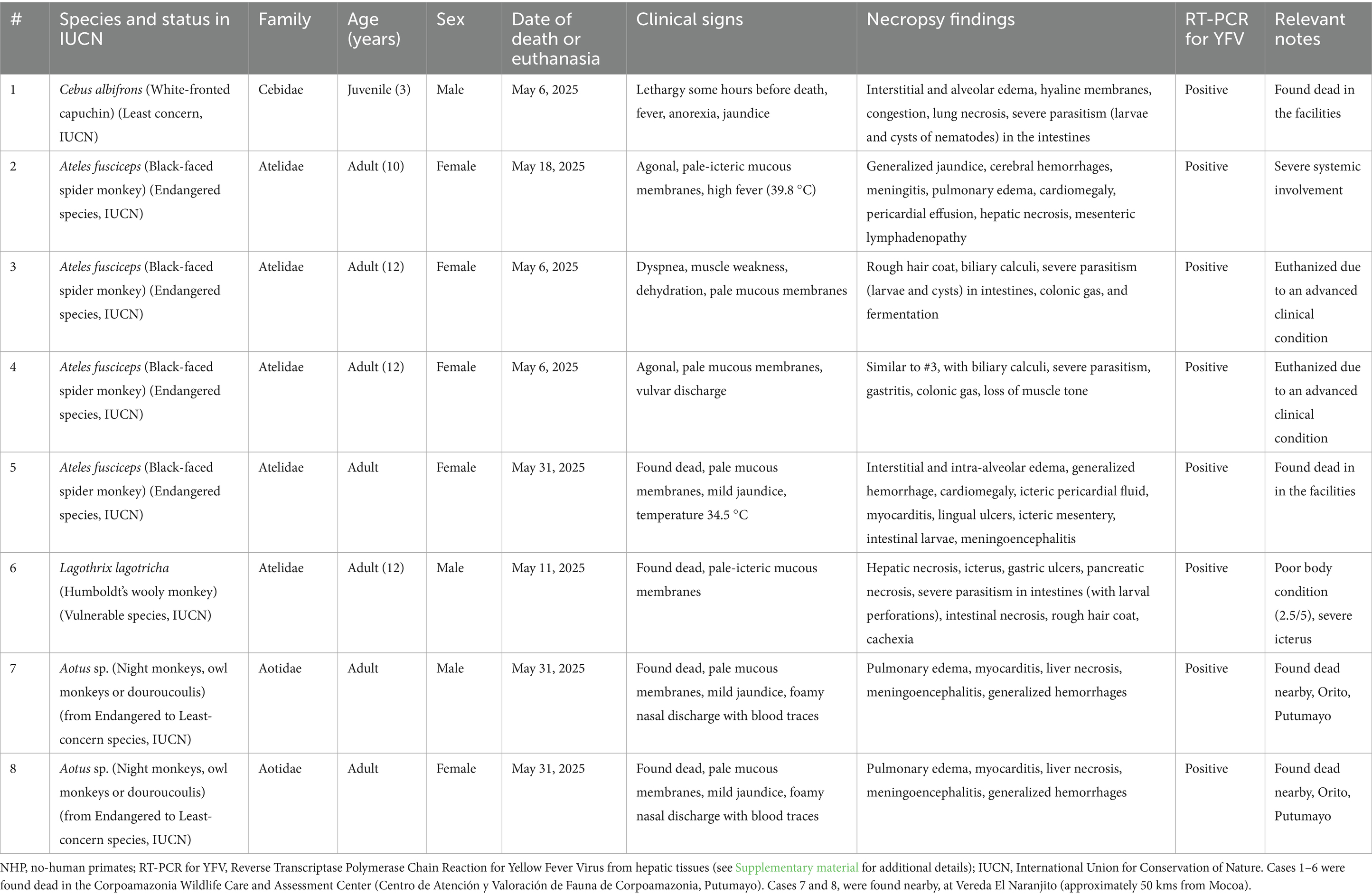
Table 1. Main clinical, pathological, and molecular findings in NHP fatalities associated with yellow fever in Putumayo, Colombia, 2025.
Case 1 involved a juvenile male Cebus albifrons presenting with lethargy, anorexia, jaundice, and fever before being found dead. Gross examination revealed pulmonary congestion, interstitial and alveolar edema, hyaline membrane formation, and necrotizing lung lesions. Severe intestinal parasitism, characterized by the presence of larvae and cysts, was also noted. Histological evaluation of the lung tissue (Figure 1B) confirmed edema and necrosis, with minimal epithelial damage, consistent with viral pneumonia (Table 1; Figure 1).
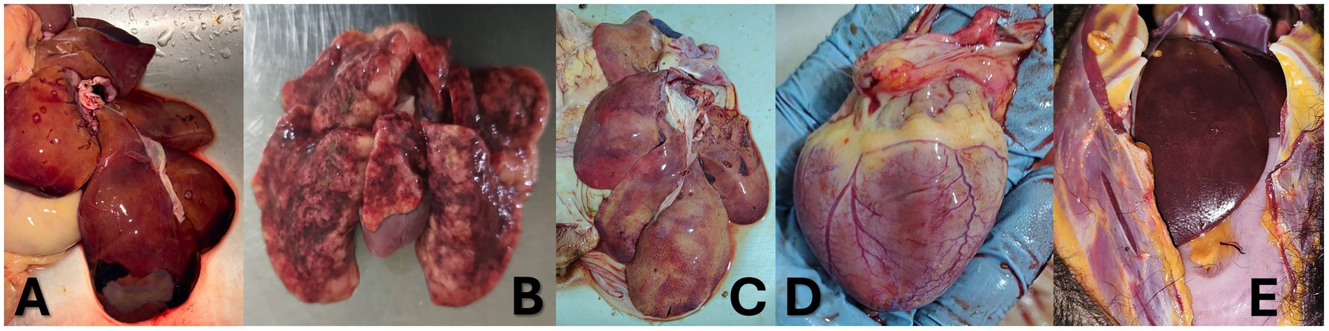
Figure 1. Appearance of organs of the dead NHPs with YFY infection. (A) Wooly monkey (Lagothrix lagotricha)—Liver. Hepatic necrosis is evident, predominantly affecting the centrilobular (zone 3) regions of the hepatic lobules. The tissue also exhibits discoloration consistent with jaundice. (B) White-faced capuchin monkey (Cebus albifrons), juvenile—Lung. There is prominent interstitial and intra-alveolar edema, with minimal epithelial damage. Occasional fibrinoid hyaline membranes are observed, along with generalized vascular congestion and scattered necrotic foci. (C) Black-headed spider monkey (Ateles fusciceps)—Liver. Marked discoloration and jaundice are present, accompanied by focal vascular congestion and architectural disruption, particularly in the right cranial hepatic lobe. (D) Black-headed spider monkey (Ateles fusciceps)—Heart. The myocardium shows yellowish discoloration (jaundice) with generalized vascular congestion, findings consistent with viral myocarditis. (E) Black-headed spider monkey (Ateles fusciceps)—Skin and subcutaneous tissue. Jaundice is evident in the subcutaneous fat and fascial connective tissue.
Cases 2 to 5 involved adult female Ateles fusciceps, a species classified as endangered by the IUCN. They exhibited overlapping clinical signs such as mucosal pallor, jaundice, dehydration, vulvar discharge, and dyspnea. Body temperatures ranged from hypothermic (34.5 °C) to febrile (39.8 °C), and two monkeys were euthanized due to severe clinical deterioration. One of the monkeys had a poor nutritional status, and post-mortem scoring (see Supplementary material) indicated a low body condition (3/5). Common pathological findings included cardiomegaly, icteric mesentery, myocarditis, pulmonary edema, hepatic necrosis, gastrointestinal parasitism, and neurologic involvement (meningitis or meningoencephalitis). One individual (Case 5) had significant lingual ulceration and evidence of generalized hemorrhage (Table 1; Figures 1, 2).
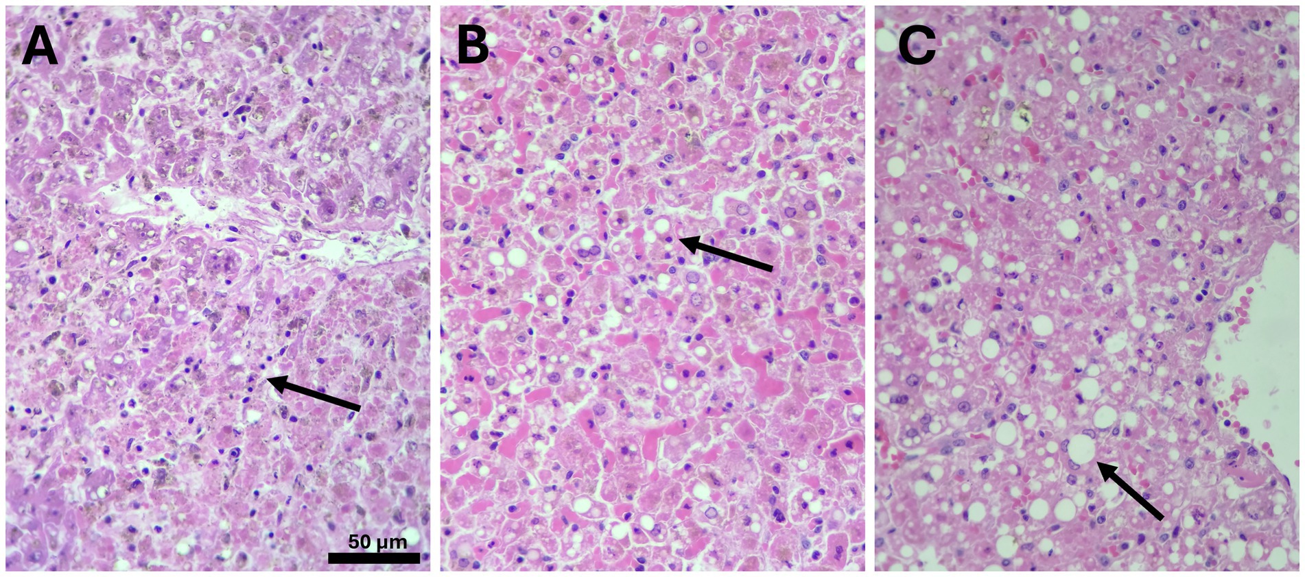
Figure 2. Histopathological findings at the liver of cases 5 (Ateles fusciceps) (A), 7 (B), and 8 (C) (Aotus sp.) [H&E-stained, 20X (A) and 40X (B,C)]. These histological images are strongly compatible with yellow fever virus-induced hepatitis, a condition that in non-human primates species typically presents with distinctive pathological features. Among these, midzonal (Zone 2) hepatocellular necrosis is a hallmark, often accompanied by numerous Councilman bodies, which represent apoptotic hepatocytes (arrows). Despite the extensive hepatocellular damage, the inflammatory response is characteristically minimal. Additionally, some specimens exhibit hepatocyte steatosis, which may reflect early injury or underlying metabolic stress associated with the YFV infection (Table 2).
Of these, Case 6 (Lagothrix lagotricha), an adult male, was found deceased with pale-icteric mucosa and marked cachexia. Necropsy revealed hepatic necrosis, gastric ulcers, pancreatic necrosis, and intestinal damage with evidence of larval perforation—an indication of concurrent severe parasitic burden. The animal had a poor nutritional status, and post-mortem scoring indicated a low body condition (2.5/5). Histologically, hepatic tissue showed centrilobular necrosis and marked discoloration consistent with jaundice (Table 1; Figure 1).
Cases 7 and 8, involving adult male and female Aotus spp., were discovered dead near the rural area of Vereda El Naranjito, Orito, near the Corpoamazonia facilities, in Mocoa (Supplementary Figure S1). Both presented with mild jaundice, foamy nasal discharge with blood traces, and generalized pallor. Both animals had a poor nutritional status, and post-mortem scoring indicated a low body condition (3/5). Pathological examination confirmed multi-organ involvement with prominent pulmonary edema, liver necrosis, myocarditis, and meningoencephalitis. These findings underscore the capacity of YFV to induce multisystemic and fulminant disease even in smaller-bodied nocturnal primates. Histopathological evaluation showed severe midzonal to centrilobular hepatocellular necrosis, Councilman bodies, steatosis, and sinusoidal congestion—hallmarks of YFV-induced hepatitis (Tables 1, 2; Figures 1, 2).
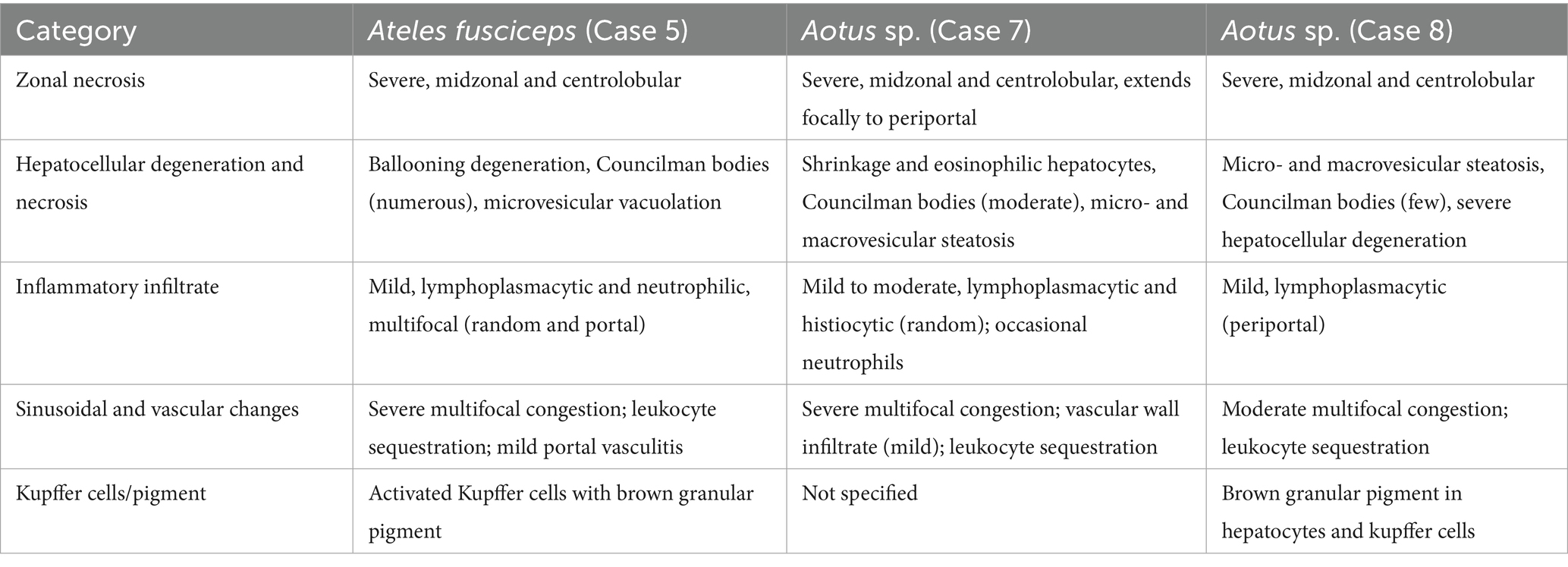
Table 2. Main histopathological findings in three of the cases.
Histopathological analysis, performed in three representative cases (Cases 5, 7, and 8), revealed consistent patterns of liver injury across species (Tables 1, 2; Figure 2). All showed midzonal necrosis with variable extension to centrilobular and periportal areas. The presence of Councilman bodies (apoptotic hepatocytes) ranged from a few to numerous. Hepatocellular steatosis, degeneration, and ballooning were also observed. Inflammatory infiltrates were mild and predominantly lymphoplasmacytic, with occasional histiocytes and neutrophils. Vascular changes included multifocal sinusoidal congestion and leukocyte sequestration, sometimes with mild vasculitis. Brown granular pigments, likely representing bile or iron, were detected in Kupffer cells and hepatocytes in two of the three animals (Table 2; Figure 2).
These findings align with classic YF pathology in primates and support the diagnosis of YFV infection as the primary cause of death. The involvement of multiple threatened and endangered species (Ateles fusciceps, Lagothrix lagotricha, and some Aotus spp.) emphasizes the ecological and conservation implications of the outbreak. Notably, most affected animals also had signs of severe parasitism, suggesting potential compounding effects of comorbidities on disease severity.
Discussion
This cluster represents a rare and valuable documentation of YFV infection in captive NHPs under managed care in South America (32, 33). It highlights the vulnerability of non-Alouatta genera to fatal outcomes and underscores the importance of active epizootic surveillance, even in controlled environments (34, 35). Given the location of Putumayo along international borders and within endemic YFV transmission zones, these findings serve as a critical early warning for both public health and wildlife conservation authorities (8, 18–21). These threatened animals are often victims of illegal wildlife trafficking, a key issue that deserves emphasis and further documentation. Corpoamazonia, like others affected by the outbreak, has implemented prevention strategies and worked to conserve NHPs, fulfilling our duty to protect wild species.
Regarding Cebus albifrons, Lagothrix lagotricha, Ateles fusciceps (an endangered species according to the International Union for Conservation of Nature, IUCN), NHP species, we were not able to find in the biomedical literature reports of natural infection with YFV to any of these species. In the case of Lagothrix lagotricha, reports of experimental infection have been published before. In PubMed, only 50 publications in general about L. lagotricha can be currently retrieved. In 1930, a study found that L. lagotricha is relatively resistant to YFY (36). Only three individuals (25%) showed mild fever, none developed typical disease lesions, and virus transfer back to rhesus monkeys was rarely successful. However, their sera conferred protective immunity, indicating subclinical infection (36). No other study has been found regarding YFV and L. lagotricha. This species is susceptible to other infectious agents, including hepatitis B virus (36, 37), filarial nematodes (38), and Toxoplasma gondii (37).
In the case of C. albifrons and A. fusciceps, no reports at all of YFV infection (not even experimental) were found in the literature. In PubMed, only 110 publications in general about C. albifrons can be currently retrieved. This species is susceptible to other infectious agents, including Leptospira (38), Trypanosoma (39), and herpesvirus (40, 41), among others. In PubMed, only 38 publications in general about A. fusciceps can currently be retrieved. This species is susceptible to other infectious agents, including Leptospira (42), Campylobacter hyointestinalis (43), and SARS-CoV-2 (44), among others.
The detection of fatal YFV infection among captive individuals of C. albifrons, L. lagotricha, and A. fusciceps represents an unprecedented and alarming development. While Alouatta sp. have been extensively documented as highly susceptible to YFV and have historically served as sentinel species in South America, the involvement of other genera—especially those previously thought to be resistant or undocumented in natural conditions—calls for an urgent re-evaluation of current surveillance strategies (45–48). This outbreak reveals that reliance solely on Alouatta sp. as indicators of sylvatic YFV circulation may be insufficient in specific ecological settings or under evolving epidemiological pressures (6, 33, 49, 50).
The clinical and pathological findings in this report reaffirm the capacity of YFV to induce systemic, fulminant, and multisystemic disease, even in species with no known history of natural infection (50–52). The presence of characteristic histopathological lesions, including midzonal hepatic necrosis, Councilman bodies, myocarditis, and meningoencephalitis, supports the conclusion of YFV as the primary cause of death in these animals (53–55). Notably, several individuals showed concurrent evidence of severe parasitism and malnutrition, which may have contributed to increased susceptibility and worse clinical outcomes. These findings align with prior observations in other NHP epizootics in Brazil, where co-infections and stress-related immunosuppression potentially influenced mortality (14, 33, 56–58).
This outbreak must be interpreted in the context of the broader resurgence of YF in Colombia and South America (1, 2, 5–8, 11). In Colombia alone, more than 130 human cases and more than 50 deaths have been confirmed during the current outbreak. The department of Putumayo is among the most severely affected regions (8). Its location along the borders with Ecuador and Peru, coupled with dense rainforest ecosystems, high vector density, and frequent human-wildlife interactions, renders it a hotspot for arboviral emergence and re-emergence (18–21). In Brazil, according to the Ministry of Health, as of August 14, 2025, 87 cases of YFV infection have been confirmed in NHP in 70 municipalities. Notably, there is geographical overlap with human cases, particularly in the southern states affected by the disease.5
The role of captive NHPs in the epidemiology of YF must be carefully considered (6, 36, 48, 50, 53, 58). These animals are not only vulnerable to infection. Still, they may also serve as early indicators of viral circulation, particularly in areas where free-ranging NHPs are difficult to observe or are absent. Wildlife rescue centers, zoos, sanctuaries, and conservation parks thus represent valuable but underutilized platforms for syndromic and laboratory-based epizootic surveillance. The systematic incorporation of these facilities into national YF monitoring programs is strongly recommended (35, 59, 60).
Furthermore, this outbreak raises concerns about potential breaches in biosecurity and vector control within captive settings. Although it is unclear how the virus reached these managed populations, sylvatic vectors (Haemagogus spp., Sabethes spp.) likely entered the enclosures. In the case of this outbreak, the staff were not completely vaccinated before. Additionally, more than 30 tourists arrive daily at the center, and the vaccine certificate was not mandatory. However, such a possibility underscores the necessity for stringent mosquito control measures, enclosure design modifications (e.g., insect-proof netting), staff and visiting people vaccination, and regular entomological surveillance within and around such facilities (31, 61, 62).
In line with the OneHealth approach, this outbreak highlights the interconnectedness of human, animal, and environmental health. The death of NHPs in managed care settings is not only a tragic conservation loss—especially considering that Ateles fusciceps is an IUCN-listed endangered species—but also a sentinel signal for possible human spillover. Several historical YF outbreaks in South America have been preceded by or concurrent with NHP die-offs. Timely detection and reporting of these events are therefore critical for guiding human vaccination campaigns and vector control interventions (57, 63, 64). YF poses a serious threat to neotropical primates, notably howler monkeys, due to high mortality rates (65, 66).
Recommendations arising from this outbreak investigation include enhanced surveillance, with a focus on routine monitoring of all NHP species—not only Alouatta spp.—within epizootic surveillance protocols. This should encompass both free-ranging and captive populations, particularly in protected areas, wildlife rescue centers, and zoos (33, 63). Biosafety reinforcement in captive facilities is also essential; institutions housing NHPs must implement rigorous vector control strategies, such as physical barriers to prevent mosquito entry, environmental management to eliminate breeding sites, and indoor residual spraying where suitable. Vaccination of personnel working in close contact with NHPs in endemic regions should be prioritized, according to WHO guidelines, alongside regular health monitoring (10). Wildlife centers must have the capacity for rapid sample collection, preservation, and shipment to national reference laboratories, and the use of point-of-care diagnostics or field-deployable RT-PCR systems should be considered for timely detection (67, 68). Cross-sectoral collaboration between national public health and wildlife authorities is crucial for establishing integrated frameworks to detect, report, and respond to YFV epizootics. Finally, outbreak preparedness drills, including simulation exercises and staff training in captive facilities, should be conducted to ensure readiness for future epizootics, covering quarantine implementation and safe necropsy procedures (69, 70).
NHPs serve as vital sentinels for detecting YF outbreaks, and vaccination and eco-friendly vector control are key to prevention. The 17DD YF vaccine is safe and immunogenic in several neotropical primate species, though immune responses varied by species. Given the expanding geographic spread of YF in South America and its impact on NHP populations, it is crucial to intensify efforts aimed at protecting these species (65, 66).
From a scientific standpoint, this report recommends further research into the pathogenesis of YFV in understudied NHP genera. The susceptibility of C. albifrons, A. fusciceps, and L. lagotricha, in particular, deserves closer examination through experimental and immunological studies. The findings of subclinical or mild disease in early experimental infections, as reported in historical studies from 1930, contrast sharply with the fulminant presentations observed in this outbreak (36). Whether these differences are due to genetic factors, variations in viral strain, host immune status, or ecological pressures remains an open question (14, 63).
Moreover, the zoonotic risk of YF remains a pressing concern, particularly in the context of global warming, deforestation, and the expansion of human encroachment into wildlife habitats (6, 71–73). These drivers are likely to increase the frequency and reach of arboviral spillover events (74, 75). Thus, strengthening sentinel surveillance among NHPs—including those under human care—should be seen not as an isolated veterinary endeavor but as an essential component of public health strategy in the Amazonian and Andean regions (35).
We suggest including the environmental impact of YF on the ecosystem, emphasizing how viral persistence in NHPs may affect their evolutionary trajectory, an aspect crucial from our perspective as an environmental authority.
It is essential to stress the conservation implications of these findings. Beyond their role in disease ecology, NHPs are vital to the biodiversity and functioning of Neotropical forest ecosystems. Mass die-offs of species such as A. fusciceps may have long-term ecological consequences, including disruption of seed dispersal and forest regeneration dynamics. Their loss to YFV—potentially preventable through improved surveillance and protection—represents not only a failure in disease prevention but also in biodiversity conservation (76, 77). These findings may also have implications in other countries, such as Ecuador, Peru, Bolivia, and Brazil, as these species (C. albifrons, A. fusciceps, and L. lagotricha) (Supplementary Figure S1) are also present in these countries, where YF epidemics are currently occurring.
Finally, it is essential to emphasize that, while fatal YFV infections were observed in endangered and vulnerable NHP species, such as Ateles fusciceps and Lagothrix lagotricha, our findings do not confirm their role in sustaining YFV transmission cycles. Given the presence of comorbidities—including malnutrition and parasitism—it remains plausible that these cases reflect incidental infections rather than evidence of their participation as reservoir hosts. Caution must be exercised to avoid misinterpreting these events as indicative of a broader epidemiological role, particularly considering the conservation status of these species and the need to avoid undue alarm that may hinder their protection.
Limitations
This investigation has several limitations. First, detailed histopathological studies could not be performed on all eight cases due to limitations in tissue availability and preservation, which restricted a more comprehensive pathological analysis across all species affected. Only three cases underwent histopathological examination, limiting the extrapolation of these findings to the broader group. Second, immunohistochemical analyses were not conducted, which would have provided more specific confirmation of YFV antigens within tissue lesions and further clarified the pathogenesis at the cellular level. Third, genomic studies, including the sequencing of viral strains, were not feasible due to resource and infrastructure constraints. As a result, we were unable to assess potential genetic variations in the circulating virus, which could influence virulence, host range, or transmission dynamics. These limitations underscore the need for strengthened diagnostic capabilities and comprehensive laboratory approaches in future outbreaks affecting NHPs in both wild and captive settings.
Conclusion
In conclusion, this report presents novel and urgent data on the impact of the YF virus in previously undocumented NHP hosts in captivity. It underscores the need to broaden the taxonomic and ecological scope of YFV surveillance and to integrate One Health principles into regional outbreak response. The lessons learned from this cluster in Putumayo should inform national and international strategies to prevent, detect, and mitigate future YF outbreaks in both humans and NHPs.
Data availability statement
The original contributions presented in the study are included in the article/Supplementary material, further inquiries can be directed to the corresponding author.
Author contributions
IS-R: Visualization, Data curation, Investigation, Conceptualization, Validation, Writing – original draft, Writing – review & editing, Resources, Formal analysis. DB-A: Resources, Conceptualization, Visualization, Validation, Funding acquisition, Investigation, Project administration, Supervision, Methodology, Formal analysis, Writing – original draft, Writing – review & editing, Data curation. CS-J: Investigation, Writing – original draft, Writing – review & editing, Methodology, Visualization, Data curation, Validation. JB-A: Writing – original draft, Visualization, Writing – review & editing, Data curation, Investigation, Validation. MB-T: Data curation, Project administration, Writing – original draft, Visualization, Resources, Conceptualization, Supervision, Writing – review & editing, Formal analysis, Investigation, Methodology. SO-G: Writing – review & editing, Validation, Methodology, Supervision, Data curation, Project administration, Writing – original draft, Investigation, Visualization, Resources. VZ-Q: Visualization, Methodology, Validation, Data curation, Writing – review & editing, Investigation, Resources, Writing – original draft, Project administration, Supervision. JP-G: Writing – review & editing, Writing – original draft, Resources, Validation, Supervision, Methodology. CG-O: Investigation, Writing – review & editing, Validation, Supervision, Resources, Methodology, Writing – original draft, Visualization. MD-C: Visualization, Writing – original draft, Investigation, Writing – review & editing, Resources, Methodology, Validation, Supervision. AR-M: Resources, Writing – original draft, Supervision, Formal analysis, Project administration, Visualization, Data curation, Writing – review & editing, Investigation, Conceptualization, Validation, Funding acquisition, Methodology.
Funding
The author(s) declare that financial support was received for the research and/or publication of this article. Partially supported by Instituto Tecnológico del Putumayo, Korea University, Seoul, Republic of Korea, Corporación para el Desarrollo Sostenible del Sur de la Amazonia, Universidad Científica del Sur, and Institución Universitaria Visión de las Américas. We thank the generous waiver of the Editorial Team of Frontiers of Veterinary Science for this publication.
Acknowledgments
The authors thank Dr. Sazi Alberto Camacho, Veterinary Pathologist, Laboratory of Veterinary Pathology, Department of Animal Health, Faculty of Veterinary Medicine and Zootechnics, Universidad Nacional de Colombia, Bogotá, Colombia, for his assistance with the histopathological studies presented in this report. This article has been registered in the Research Proposal Registration of the Coordination of Scientific Integrity and Surveillance of Universidad Cientifica del Sur, Lima, Peru.
Conflict of interest
The authors declare that the research was conducted in the absence of any commercial or financial relationships that could be construed as a potential conflict of interest.
The author(s) declared that they were an editorial board member of Frontiers, at the time of submission. This had no impact on the peer review process and the final decision.
Generative AI statement
The authors declare that no Gen AI was used in the creation of this manuscript.
Any alternative text (alt text) provided alongside figures in this article has been generated by Frontiers with the support of artificial intelligence and reasonable efforts have been made to ensure accuracy, including review by the authors wherever possible. If you identify any issues, please contact us.
Publisher’s note
All claims expressed in this article are solely those of the authors and do not necessarily represent those of their affiliated organizations, or those of the publisher, the editors and the reviewers. Any product that may be evaluated in this article, or claim that may be made by its manufacturer, is not guaranteed or endorsed by the publisher.
Supplementary material
The Supplementary material for this article can be found online at: https://www.frontiersin.org/articles/10.3389/fvets.2025.1655474/full#supplementary-material
Footnotes
1. ^https://shiny.paho-phe.org/yellowfever/
2. ^https://www.ins.gov.co/Noticias/Paginas/fiebre-amarilla-datos-ciencia-prevencion.aspx
3. ^https://shiny.paho-phe.org/yellowfever/
4. ^https://www.who.int/news-room/events/detail/2025/06/11/default-calendar/who-epi-win-webinar-yellow-fever-in-the-americas-what-we-know
5. ^https://www.gov.br/saude/pt-br/composicao/svsa/cnie/painel-febre-amarela
References
1. Angerami, RN, Socorro Souza Chaves, TD, and Rodríguez-Morales, AJ. Yellow fever outbreaks in South America: current epidemiology, legacies of the recent past and perspectives for the near future. New Microbes New Infect. (2025) 65:101580. doi: 10.1016/j.nmni.2025.101580
2. Perez, LJ, Perez-Restrepo, LS, Ciuoderis, K, Usuga, J, Moreno, I, Vargas, V, et al. Emergence, persistence, and positive selection of yellow fever virus in Colombia. Front Microbiol. (2025) 16:1548556. doi: 10.3389/fmicb.2025.1548556
3. Forero-Delgadillo, AJ, Morales-Olivera, JA, Celis-Guzmán, JF, Zapata-Díaz, OE, González-Varona, GA, Acevedo-Bedoya, CA, et al. Colombian consensus on the care of critically ill patients with suspected or confirmed severe yellow fever. Lancet Regional Health Americas. (2025) 48:101144. doi: 10.1016/j.lana.2025.101144
4. Rodriguez-Morales, AJ, Alhazmi, AH, Katime, A, Hameed, AA, Morales, A, Lepetic, AC, et al. Yellow fever in South America – a plea for action and call for prevention in travelers from SLAMVI, ESGITM, EVASG, ALEIMC, GEPI-SEIMC, SEMEVI, and CMTZMV-ACIN. Travel Med Infect Dis. (2025) 67:102871. doi: 10.1016/j.tmaid.2025.102871
5. Alvarez-Moreno, CA, and Rodriguez-Morales, AJ. Challenges of the current yellow fever outbreak in Colombia. Lancet. (2025) 405:2273. doi: 10.1016/S0140-6736(25)01175-4
6. Bonilla-Aldana, DK, Bonilla-Aldana, JL, Castellanos, JE, and Rodriguez-Morales, AJ. Importance of epizootic surveillance in the epidemiology of yellow fever in South America. Curr Trop Med Rep. (2025) 12:16. doi: 10.1007/s40475-025-00349-z
7. Cunha, MDP, Duarte-Neto, AN, Pour, SZ, Ortiz-Baez, AS, Černý, J, Pereira, BBS, et al. Origin of the São Paulo yellow fever epidemic of 2017-2018 revealed through molecular epidemiological analysis of fatal cases. Sci Rep. (2019) 9:20418. doi: 10.1038/s41598-019-56650-1
8. Sanchez-Rojas, IC, Solarte-Jimenez, CL, Chamorro-Velazco, EC, Diaz-Llerena, GE, Arevalo, CD, Cuasquer-Posos, OL, et al. Yellow fever in Putumayo, Colombia, 2024. New Microbes New Infect. (2025) 64:101572. doi: 10.1016/j.nmni.2025.101572
9. Rodríguez-Morales, AJ, Bonilla-Aldana, DK, Suárez, JA, Franco-Paredes, C, Forero-Peña, DA, Mattar, S, et al. Yellow fever reemergence in Venezuela - implications for international travelers and Latin American countries during the COVID-19 pandemic. Travel Med Infect Dis. (2021) 44:102192. doi: 10.1016/j.tmaid.2021.102192
10. Reno, E, Quan, NG, Franco-Paredes, C, Chastain, DB, Chauhan, L, Rodriguez-Morales, AJ, et al. Prevention of yellow fever in travellers: an update. Lancet Infect Dis. (2020) 20:e129–37. doi: 10.1016/S1473-3099(20)30170-5
11. Rodriguez-Morales, AJ, Sah, R, Silva-Ramos, CR, and Pava-Garzón, DM. Challenges in emerging and reemerging Arboviral diseases: the examples of Oropouche and yellow fever. Pathogens. (2025) 14:621. doi: 10.3390/pathogens14070621
12. Fraser, K, Hamlet, A, Jean, K, Ramos, DG, Romano, A, Horton, J, et al. Assessing yellow fever outbreak potential and implications for vaccine strategy. PLOS Glob Public Health. (2024) 4:e0003781. doi: 10.1371/journal.pgph.0003781
13. Li, SL, Acosta, AL, Hill, SC, Brady, OJ, de Almeida, MAB, Cardoso, JDC, et al. Mapping environmental suitability of Haemagogus and Sabethes spp. mosquitoes to understand sylvatic transmission risk of yellow fever virus in Brazil. PLoS Negl Trop Dis. (2022) 16:e0010019. doi: 10.1371/journal.pntd.0010019
14. Saad, LD, and Barata, RB. Yellow fever outbreaks in São Paulo state, Brazil, 2000-2010. Epidemiol Serv Saude. (2016) 25:531–40. doi: 10.5123/S1679-49742016000300009
15. Gianchecchi, E, Cianchi, V, Torelli, A, and Montomoli, E. Yellow fever: origin, epidemiology, preventive strategies and future prospects. Vaccine. (2022) 10:372. doi: 10.3390/vaccines10030372
16. Shearer, FM, Longbottom, J, Browne, AJ, Pigott, DM, Brady, OJ, Kraemer, MUG, et al. Existing and potential infection risk zones of yellow fever worldwide: a modelling analysis. Lancet Glob Health. (2018) 6:e270–8. doi: 10.1016/S2214-109X(18)30024-X
17. Rodriguez-Morales, AJ, and Villamil-Gómez, WE. Yellow Fever: still of concern for travelers of Colombia? Infectio. (2018) 22:171–2. doi: 10.22354/in.v22i4.733
18. Barreto, M, Burbano, ME, and Barreto, P. Lutzomyia sand flies (Diptera: Psychodidae) from middle and lower Putumayo department, Colombia, with new records to the country. Mem Inst Oswaldo Cruz. (2000) 95:633–9. doi: 10.1590/S0074-02762000000500009
19. Castro, M, Quintana, N, and Quiñones, PM. Evaluating two pyrethroids in dengue vector control in Putumayo, Colombia. Rev Salud Publica (Bogota). (2007) 9:106–16. doi: 10.1590/s0124-00642007000100011
20. Orjuela, LI, Herrera, M, Erazo, H, and Quiñones, ML. Anopheles species present in the department of Putumayo and their natural infectivity with plasmodium. Biomedica. (2013) 33:42–52. doi: 10.1590/S0120-41572013000100006
21. van der Ende, J, Nipaz, V, Carrazco-Montalvo, A, Trueba, G, Grobusch, MP, and Coloma, J. Cocirculation of 4 dengue virus serotypes, Putumayo Amazon Basin, 2023-2024. Emerg Infect Dis. (2025) 31:202–4. doi: 10.3201/eid3101.240888
22. Rodríguez, G, Velandia, M, and Boshell, J. Fiebre amarilla: La enfermedad y su control. Bogotá: Instituto Nacional de Salud (2003).
23. Srivastava, S, Dhoundiyal, S, Kumar, S, Kaur, A, Khatib, MN, Gaidhane, S, et al. Yellow fever: global impact, epidemiology, pathogenesis, and integrated prevention approaches. Infez Med. (2024) 32:434–50. doi: 10.53854/liim-3204-3
24. Oyono, MG, Kenmoe, S, Abanda, NN, Takuissu, GR, Ebogo-Belobo, JT, Kenfack-Momo, R, et al. Epidemiology of yellow fever virus in humans, arthropods, and non-human primates in sub-Saharan Africa: a systematic review and meta-analysis. PLoS Negl Trop Dis. (2022) 16:e0010610. doi: 10.1371/journal.pntd.0010610
25. Silva, NIO, Sacchetto, L, de Rezende, IM, Trindade, GS, LaBeaud, AD, de Thoisy, B, et al. Recent sylvatic yellow fever virus transmission in Brazil: the news from an old disease. Virol J. (2020) 17:9. doi: 10.1186/s12985-019-1277-7
26. de Oliveira, FP, Stoffella-Dutra, AG, Barbosa Costa, G, Silva de Oliveira, J, Dourado Amaral, C, Duarte Santos, J, et al. Re-emergence of yellow fever in Brazil during 2016-2019: challenges, lessons learned, and perspectives. Viruses. (2020) 12:1233. doi: 10.3390/v12111233
27. Giovanetti, M, de Mendonça, MCL, Fonseca, V, Mares-Guia, MA, Fabri, A, Xavier, J, et al. Yellow fever virus reemergence and spread in Southeast Brazil, 2016-2019. J Virol. (2019) 94:e01623-19. doi: 10.1128/JVI.01623-19
28. Silva, NIO, Albery, GF, Arruda, MS, Oliveira, GFG, Costa, TA, de Mello, ÉM, et al. Ecological drivers of sustained enzootic yellow fever virus transmission in Brazil, 2017-2021. PLoS Negl Trop Dis. (2023) 17:e0011407. doi: 10.1371/journal.pntd.0011407
29. Chaves, A, Piche-Ovares, M, Ibarra-Cerdena, CN, Corrales-Aguilar, E, Suzan, G, Moreira-Soto, A, et al. Serosurvey of nonhuman Primates in Costa Rica at the human-wildlife Interface reveals high exposure to Flaviviruses. Insects. (2021) 12:554. doi: 10.3390/insects12060554
30. de Miranda, RM, Ferreira-de-Brito, A, Silva, JDS, Xavier, ADS, Freitas Silva, SO, Alencar, J, et al. Mosquito fauna and spatial distribution in an Atlantic Forest area in Rio de Janeiro state, Brazil, reveal a high risk of transmission of yellow fever and other arboviruses. Trop Med Infect Dis. (2022) 7:410. doi: 10.3390/tropicalmed7120410
31. de Miranda, RM, Fernandes, RS, da Silva-Fernandes, AT, Ferreira-de-Brito, A, Moreira, SB, Pereira, RC, et al. Neotropical sylvatic mosquitoes and Aedes aegypti are not competent to transmit 17DD attenuated yellow fever virus from vaccinated Viremic New World non-human Primates. Viruses. (2022) 14:2231. doi: 10.3390/v14102231
32. de Oliveira Figueiredo, P, Stoffella-Dutra, AG, Costa, GB, de Oliveira, JS, Amaral, CD, Alves, PA, et al. Absence of yellow fever virus circulation in wildlife rodents from Brazil. Braz J Microbiol. (2022) 53:647–54. doi: 10.1007/s42770-022-00688-3
33. Mares-Guia, M, Horta, MA, Romano, A, Rodrigues, CDS, Mendonça, MCL, Dos Santos, CC, et al. Yellow fever epizootics in non-human primates, southeast and Northeast Brazil (2017 and 2018). Parasit Vectors. (2020) 13:90. doi: 10.1186/s13071-020-3966-x
34. Kersul, MG, Abreu, FVS, Pinter, A, Campos, FS, Andrade, MS, Teixeira, DS, et al. Exploring environmental and climate features associated with yellow fever across space and time in the Brazilian Atlantic Forest biome. PLoS One. (2024) 19:e0308560. doi: 10.1371/journal.pone.0308560
35. Hill, SC, de Souza, R, Thézé, J, Claro, I, Aguiar, RS, Abade, L, et al. Genomic surveillance of yellow fever virus epizootic in São Paulo, Brazil, 2016 - 2018. PLoS Pathog. (2020) 16:e1008699. doi: 10.1371/journal.ppat.1008699
36. Davis, NC. The transmission of yellow fever: experiments with the "woolly monkey" (LAGOTHRIX LAGO-TRICHA Humboldt), the "spider monkey" (ATELEUS ATER F. Cuvier), and the "squirrel monkey" (SAIMIRI SCIREUS LINNAEUS). J Exp Med. (1930) 51:703–20. doi: 10.1084/jem.51.5.703
37. Gyimesi, ZS, Lappin, MR, and Dubey, JP. Application of assays for the diagnosis of toxoplasmosis in a colony of woolly monkeys (Lagothrix lagotricha). J Zoo Wildl Med. (2006) 37:276–80. doi: 10.1638/05-018.1
38. Romero, MH, Astudillo, M, Sánchez, JA, González, LM, and Varela, N. Leptospiral antibodies in a Colombian zoo's neotropical primates and workers. Rev Salud Publica (Bogota). (2011) 13:814–23. doi: 10.1590/S0124-00642011000500010
39. Carrillo-Bilbao, G, Navarro, JC, Martin-Solano, S, Chávez-Larrea, MA, Cholota-Iza, C, and Saegerman, C. First molecular identification of trypanosomes and absence of Babesia sp. DNA in faeces of non-human primates in the Ecuadorian Amazon. Pathogens. (2022) 11:1490. doi: 10.3390/pathogens11121490
40. Lewis, MA, Frye, LD, Gibbs, CJ Jr, Chou, SM, Cutchins, EC, Gajdusek, DC, et al. Isolation and characterization of two new herpes-like viruses from capuchin monkeys. Infect Immun. (1976) 14:759–66. doi: 10.1128/iai.14.3.759-766.1976
41. Furusato, IN, Figueiredo, KB, de Carvalho, A, da Silva Ferreira, CS, Takahashi, JPF, Kimura, LM, et al. Detection of herpesviruses in neotropical primates from São Paulo, Brazil. Braz J Microbiol. (2023) 54:3201–9. doi: 10.1007/s42770-023-01105-z
42. Woolf, D, Sanchez, C, Gonzalez-Astudillo, V, Navarro, M, Tapia, CC, Franco, M, et al. Leptospira species status of captive nonhuman primates and free-ranging rodents at the Barranquilla zoo, Colombia, 2013. J Zoo Wildl Med. (2021) 51:780–8. doi: 10.1638/2019-0192
43. Meadows, SNA, Hung, CC, Chen, JW, Soukup, S, and Sander, SJ. Campylobacter hyointestinalis isolation from howler (Alouatta caraya) and spider monkeys (Ateles fusciceps robustus) at a zoologic facility in central illinois. J Zoo Wildl Med. (2024) 54:810–6. doi: 10.1638/2022-0166
44. Carvajal, M, Saenz, C, Fuentes, N, Guevara, R, Muñoz, E, Prado-Vivar, B, et al. SARS-CoV-2 infection in brown-headed spider monkeys (Ateles fusciceps) at a wildlife rescue center on the coast of Ecuador-South America. Microbiol Spectr. (2024) 12:e0274123. doi: 10.1128/spectrum.02741-23
45. Holzmann, I, Agostini, I, Areta, JI, Ferreyra, H, Beldomenico, P, and Di Bitetti, MS. Impact of yellow fever outbreaks on two howler monkey species (Alouatta Guariba clamitans and A. caraya) in Misiones, Argentina. Am J Primatol. (2010) 72:475–80. doi: 10.1002/ajp.20796
46. Guerra, JM, Ferreira, C, Díaz-Delgado, J, Takahashi, JPF, Kimura, LM, de Araújo, LJT, et al. Concurrent yellow fever and pulmonary aspergillosis due to Aspergillus fumigatus in a free-ranging howler monkey (Alouatta sp). J Med Primatol. (2021) 50:201–4. doi: 10.1111/jmp.12522
47. Sallis, ES, de Barros, VL, Garmatz, SL, Fighera, RA, and Graça, DL. A case of yellow fever in a brown howler (Alouatta fusca) in southern Brazil. J Vet Diagn Invest. (2003) 15:574–6. doi: 10.1177/104063870301500611
48. Siconelli, MJL, Jorge, DMM, and Castro-Jorge, LA. Lopes da fonseca BA. coding-complete genome sequence of a yellow fever virus isolated from a baby howler monkey (Alouatta caraya) from São Paulo State, Brazil, in 2016. Microbiol Resour Announc. (2021) 10:e01244-20. doi: 10.1128/MRA.01244-20
49. Abreu, FVS, de Andreazzi, CS, Neves, M, Meneguete, PS, Ribeiro, MS, Dias, CMG, et al. Ecological and environmental factors affecting transmission of sylvatic yellow fever in the 2017-2019 outbreak in the Atlantic Forest, Brazil. Parasit Vectors. (2022) 15:23. doi: 10.1186/s13071-021-05143-0
50. Leal, SG, Romano, AP, Monteiro, RV, Melo, CB, Vasconcelos, PF, and Castro, MB. Frequency of histopathological changes in howler monkeys (Alouatta sp.) naturally infected with yellow fever virus in Brazil. Rev Soc Bras Med Trop. (2016) 49:29–33. doi: 10.1590/0037-8682-0363-2015
51. Garcia-Oliveira, GF, Guimarães, A, Moreira, GD, Costa, TA, Arruda, MS, de Mello, ÉM, et al. Yellow alert: persistent yellow fever virus circulation among non-human primates in urban areas of Minas Gerais state, Brazil (2021-2023). Viruses. (2023) 16:31. doi: 10.3390/v16010031
52. Santos, DOD, de Oliveira, AR, de Lucena, FP, de Mattos, SA, Carvalho, TP, Costa, FB, et al. Histopathologic patterns and susceptibility of Neotropical primates naturally infected with yellow fever virus. Vet Pathol. (2020) 57:681–6. doi: 10.1177/0300985820941271
53. Ferreira, MS, Júnior, PSB, Cerqueira, VD, Rivero, GRC, Júnior, CAO, Castro, PHG, et al. Experimental yellow fever virus infection in the squirrel monkey (Saimiri spp.) I: gross anatomical and histopathological findings in organs at necropsy. Mem Inst Oswaldo Cruz. (2020) 115:e190501. doi: 10.1590/0074-02760190501
54. Guimarães, A, Oliveira, MC, Kierulff, MCM, Mendonça-Furtado, O, Baptista, MNM, Mendes, SL, et al. Epidemiologic profile and histopathological findings in Neotropical Primates during and after the yellow fever outbreak in Espírito Santo, Brazil. An Acad Bras Cienc. (2022) 94:e20211229. doi: 10.1590/0001-3765202220211229
55. Sacchetto, L, Silva, NIO, Rezende, IM, Arruda, MS, Costa, TA, de Mello, ÉM, et al. Neighbor danger: yellow fever virus epizootics in urban and urban-rural transition areas of Minas Gerais state, during 2017-2018 yellow fever outbreaks in Brazil. PLoS Negl Trop Dis. (2020) 14:e0008658. doi: 10.1371/journal.pntd.0008658
56. de Azevedo Fernandes, NCC, Guerra, JM, Díaz-Delgado, J, Cunha, MS, Saad, LD, Iglezias, SD, et al. Differential yellow fever susceptibility in New World nonhuman Primates, comparison with humans, and implications for surveillance. Emerg Infect Dis. (2021) 27:47–56. doi: 10.3201/eid2701.191220
57. Abreu, FVS, Ferreira-de-Brito, A, Azevedo, AS, Linhares, JHR, de Oliveira Santos, V, Hime Miranda, E, et al. Survey on non-human primates and mosquitoes does not provide evidences of spillover/spillback between the urban and sylvatic cycles of yellow fever and Zika viruses following severe outbreaks in Southeast Brazil. Viruses. (2020) 12:364. doi: 10.3390/v12040364
58. de Almeida, MAB, Dos Santos, E, Cardoso, JDC, da Silva, LG, Rabelo, RM, and Bicca-Marques, JC. Predicting yellow fever through species distribution modeling of virus, vector, and monkeys. EcoHealth. (2019) 16:95–108. doi: 10.1007/s10393-018-1388-4
59. Almeida, MAB, Santos, ED, Cardoso, JDC, Noll, CA, Lima, MM, Silva, FAE, et al. Detection of antibodies against Icoaraci, Ilhéus, and Saint Louis encephalitis arboviruses during yellow fever monitoring surveillance in non-human primates (Alouatta caraya) in southern Brazil. J Med Primatol. (2019) 48:211–7. doi: 10.1111/jmp.12417
60. Baranowski, LA, Dias, HG, Familiar-Macedo, D, Sabino-Santos, G, Herrera, HM, Slhessarenko, RD, et al. Investigation of yellow fever virus at the human-animal Interface after a Zika virus outbreak in Midwest Brazil. Microorganisms. (2024) 12:594. doi: 10.3390/microorganisms12030594
61. Cunha, MS, Tubaki, RM, de Menezes, RMT, Pereira, M, Caleiro, GS, Coelho, E, et al. Possible non-sylvatic transmission of yellow fever between non-human primates in São Paulo city, Brazil, 2017-2018. Sci Rep. (2020) 10:15751. doi: 10.1038/s41598-020-72794-x
62. Santos, M, Collado Mariscal, L, Henríquez, B, Garzón, J, González, P, Carrera, JP, et al. Implementation of bamboo and monkey-pot traps for the sampling cavity-breeding mosquitoes in Darién, Panama. Acta Trop. (2020) 205:105352. doi: 10.1016/j.actatropica.2020.105352
63. Moreno, ES, Agostini, I, Holzmann, I, Di Bitetti, MS, Oklander, LI, Kowalewski, MM, et al. Yellow fever impact on brown howler monkeys (Alouatta guariba clamitans) in Argentina: a metamodelling approach based on population viability analysis and epidemiological dynamics. Mem Inst Oswaldo Cruz. (2015) 110:865–76. doi: 10.1590/0074-02760150075
64. Andrade, MS, Campos, FS, Oliveira, CH, Oliveira, RS, Campos, AAS, Almeida, MAB, et al. Fast surveillance response reveals the introduction of a new yellow fever virus sub-lineage in 2021, in Minas Gerais, Brazil. Mem Inst Oswaldo Cruz. (2022) 117:e220127. doi: 10.1590/0074-02760220127
65. Paula, NF, Vieira, AD, Santos, DOD, Souza, LDR, Coelho, CM, Tinoco, HP, et al. Safety and immunogenicity of the attenuated yellow fever vaccine in several neotropical primate species. Vaccines (Basel). (2025) 13:487. doi: 10.3390/vaccines13050487
66. Nederlof, RA, Virgilio, T, Stemkens, HJJ, da Silva, L, Montagna, DR, Abdussamad, AM, et al. Yellow fever in non-human primates: a veterinary guide from a one health perspective. Vet Sci. (2025) 12:339. doi: 10.3390/vetsci12040339
67. da Costa Faria, NR, Monteiro-Maia, R, Nunes, PCG, da Rocha Queiroz Lima, M, and de Bruycker-Nogueira, F. Conventional RT-PCR for yellow fever virus. Methods Mol Biol. (2025) 2913:103–15. doi: 10.1007/978-1-0716-4458-4_10
68. Faggioni, G, De Santis, R, Moramarco, F, Di Donato, M, De Domenico, A, Molinari, F, et al. Pan-yellow fever virus detection and lineage assignment by real-time RT-PCR and amplicon sequencing. J Virol Methods. (2023) 316:114717. doi: 10.1016/j.jviromet.2023.114717
69. Cano-Terriza, D, Beato-Benítez, A, Fernández-Bastit, L, Segalés, J, Vergara-Alert, J, Martínez-Nevado, E, et al. SARS-CoV-2 in captive nonhuman Primates, Spain, 2020-2023. Emerg Infect Dis. (2024) 30:1253–7. doi: 10.3201/eid3006.231247
70. Shinde, DP, Plante, JA, Plante, KS, and Weaver, SC. Yellow fever: roles of animal models and arthropod vector studies in understanding epidemic emergence. Microorganisms. (2022) 10:1578. doi: 10.3390/microorganisms10081578
71. Rifakis, PM, Benitez, JA, De-la-Paz-Pineda, J, and Rodriguez-Morales, AJ. Epizootics of yellow fever in Venezuela (2004-2005): an emerging zoonotic disease. Ann N Y Acad Sci. (2006) 1081:57–60. doi: 10.1196/annals.1373.005
72. Kalbus, A, de Souza Sampaio, V, Boenecke, J, and Reintjes, R. Exploring the influence of deforestation on dengue fever incidence in the Brazilian Amazonas state. PLoS One. (2021) 16:e0242685. doi: 10.1371/journal.pone.0242685
73. Silva-Ramos, CR, Rodriguez-Morales, AJ, and Hidalgo, M. Repercussions of the end of the armed conflict in Colombia and its influence on the emergence of zoonotic pathogens related to acute undifferentiated febrile illness: future challenges to be addressed. Acta Trop. (2025) 267:107680. doi: 10.1016/j.actatropica.2025.107680
74. Tappan, J. Wandering epizootics and zones of emergence: constructing yellow fever endemicity in Africa. Health Place. (2022) 77:102770. doi: 10.1016/j.healthplace.2022.102770
75. Cano, ME, Marti, GA, Alencar, J, Silva, SOF, and Micieli, MV. Categorization by score of mosquito species (Diptera: Culicidae) related to yellow fever epizootics in Argentina. J Med Entomol. (2022) 59:1766–77. doi: 10.1093/jme/tjac079
76. Ilacqua, RC, Medeiros-Sousa, AR, Ramos, DG, Obara, MT, Ceretti-Junior, W, Mucci, LF, et al. Reemergence of yellow fever in Brazil: the role of distinct landscape fragmentation thresholds. J Environ Public Health. (2021) 2021:1–7. doi: 10.1155/2021/8230789
Keywords: Ateles, Cebus, Lagothrix, Aotus, flavivirus, yellow fever, non-human primates, Colombia
Citation: Sanchez-Rojas IC, Bonilla-Aldana DK, Solarte-Jimenez CL, Bonilla-Aldana JL, Belisario-Tovar M, Ortega-Gómez S, Zambrano-Quenan VM, Perafan-Gomez JC, Gomez-Ocampo CH, Delgado-Cajigas M and Rodriguez-Morales AJ (2025) Fatal yellow fever among captive non-human primates in southern Colombia, 2025. Front. Vet. Sci. 12:1655474. doi: 10.3389/fvets.2025.1655474
Edited by:
Francisco Javier Salguero, UK Health Security Agency (UKHSA), United KingdomReviewed by:
Marli Cupertino, Federal University of Viçosa, BrazilMonica Salas-Rojas, Instituto Mexicano del Seguro Social, Mexico
Copyright © 2025 Sanchez-Rojas, Bonilla-Aldana, Solarte-Jimenez, Bonilla-Aldana, Belisario-Tovar, Ortega-Gómez, Zambrano-Quenan, Perafan-Gomez, Gomez-Ocampo, Delgado-Cajigas and Rodriguez-Morales. This is an open-access article distributed under the terms of the Creative Commons Attribution License (CC BY). The use, distribution or reproduction in other forums is permitted, provided the original author(s) and the copyright owner(s) are credited and that the original publication in this journal is cited, in accordance with accepted academic practice. No use, distribution or reproduction is permitted which does not comply with these terms.
*Correspondence: Alfonso J. Rodriguez-Morales, YXJvZHJpZ3Vlem1vQGNpZW50aWZpY2EuZWR1LnBl
†These authors have contributed equally to this work