 Mauricio Xavier Salas-Rueda1Patricia Peralta-Ortiz2Jairo Guama-Tipas3Katherine Natalia Chávez Toledo4Mónica Espadero1Pedro Webster1Juan Masache-Masache5Karla Monica Illescas Sigcha1Fabiola Estefania Yungazaca Jaramillo1Angel Sebastian Rodríguez-Pazmiño6Fabricio Arcos Alcivar7Solon Alberto Orlando8,9
 Miguel Ángel Garcia-Bereguiain6*
- 1Universidad Politécnica Salesiana, Cuenca, Ecuador
- 2Clínica Veterinaria El Gran Pastor, Cuenca, Ecuador
- 3Clínica Veterinaria Wasi Vet, Ibarra, Ecuador
- 4Universidad Católica Santiago de Guayaquil, Guayaquil, Ecuador
- 5Gobierno Autónomo Descentralizado de Sayausi, Cuenca, Ecuador
- 6One Health Research Group, Universidad de Las Américas, Quito, Ecuador
- 7Universidad Ecotec, Guayaquil, Ecuador
- 8Instituto Nacional de Salud Pública e Investigación, Guayaquil, Ecuador
- 9Universidad Espíritu Santo, Guayaquil, Ecuador
The guinea pig (Cavia porcellus) is commonly used as a laboratory model or kept as a pet in many Western countries; however, in Andean countries like Ecuador, it is raised as livestock. Despite its importance to rural local economies, specific management guidelines for guinea pig farming have not been enforced by animal or public health authorities. Several reports indicate that guinea pigs raised as livestock serve as incidental host for respiratory and enteric pathogens, including Toxoplasma gondii. This study analysed the seroprevalence of antibodies against several pathogens relevant to public health and animal production in Ecuador: Influenza A, Brucella spp., Coxiella burnetii, Toxoplasma gondii, and Neospora caninum. Blood samples from 240 guinea pigs were collected in the cantons of Cuenca, Paute, and Gualaceo, in the Azuay province of Ecuador. Seropositive animals were detected for two pathogens—Influenza A and T. gondii—with prevalence rates of 1.67% (95% CI: 0.46–4.21) and 16.25% (95% CI: 11.82–21.54), respectively. There were not seropositive animals for Brucella spp., Coxiella burnetii and Neospora caninum. These results underscore the potential role of guinea pigs as incidental host for Influenza A and support their inclusion in surveillance programs for panzootic flu outbreaks. Additionally, guinea pigs may play a significant role in the epidemiology of toxoplasmosis in the Andean regions of Ecuador, Peru, and Colombia, where similar findings have been reported.
Introduction
Although the guinea pig (Cavia porcellus) is commonly used as a laboratory model or kept as a pet in many Western countries (1), it is raised as livestock in Andean countries of South America, including Colombia, Ecuador, Peru, and Bolivia (2). Its meat is valued for its low fat and high protein content (3), but guinea pigs are still bred using traditional methods, often raised inside rural homes in groups of up to 50 animals per household. Additionally, more industrialized farms housing thousands of animals also exist. In Ecuador alone, at least 700,000 families are involved in guinea pig farming, with an estimated annual production of 47 million animals (4, 5). However, specific management guidelines for guinea pig farming have not been implemented by animal or public health authorities to ensure food safety and quality (6).
Although there is extensive literature on the use of guinea pigs as models for infectious disease research and zoonotic transmission from pet guinea pigs, there is limited information on guinea pigs raised as livestock (6). Nevertheless, several studies have reported the presence of respiratory pathogens such as yeasts, Influenza virus, Methicillin-Resistant Staphylococcus aureus (MRSA), and Streptococcus pneumoniae in livestock guinea pigs (7–13). Guinea pigs also act as zoonotic reservoirs for enteric bacterial pathogens such as Campylobacter jejuni (14), and parasites including Blastocystis, Entamoeba, and Cryptosporidium (15). Furthermore, two recent studies from Colombia and Peru have described, for the first time, the role of guinea pigs as reservoirs for Toxoplasma gondii, the parasite responsible for toxoplasmosis (16, 17). T. gondii has a life cycle including a sexual cycle within a feline definitive host and an asexual cycle with a wide range of intermediate avian and mammal hosts (16, 17). Oocyst are produced within the intestines of felines and shed with their feces into the environment; when ingested by intermediate hosts, oocysts develop into infective tachyzoites that penetrate host tissue to form cysts of slow growing bradyzoites (16, 17). Infection of definitive or intermediate host may also happened through the ingestion of tissue cysts of infected animals (16, 17). These findings are particularly concerning for food safety, given the high prevalence rates (over 20%) detected in organs like muscle (16, 17). Other zoonotic diseases like bartonelosis (18), leishmaniases (19) and brucelosis (20) have also been associated to guinea pigs.
Other zoonotic diseases, such as brucellosis and Q-fever, have been reported in Ecuador in association with cattle, wildlife, and free-roaming dogs (21–26). Brucellosis, caused by bacteria of the genus Brucella, and Q-fever, caused by Coxiella burnetii, are both panzootic pathogens capable of infecting multiple mammalian species including wild fauna (26, 27). The use of guinea pigs as models to study these diseases underscores their susceptibility to infection (28, 29). Additionally, the parasite Neospora caninum is also highly prevalent in cattle, free-roaming dogs and wild mammals (21, 30, 31). Although not zoonotic, it causes reproductive problems in livestock (21). To the best of our knowledge, there is no existing information on the presence of the aforementioned pathogens in guinea pigs raised as livestock.
In this context, the aim of this study is to characterize the seroprevalence of several pathogens relevant to animal production and public health in guinea pigs raised as livestock in Ecuador. These diseases include Influenza A, brucellosis, Q-fever, toxoplasmosis and neosporosis, relevant pathogens to public and animal health that support the idea of improving the One Health perspective within guinea pig farming.
Materials and methods
Study design and setting
This was a cross-sectional study including guinea pigs from the Azuay province. This province, located in the Andean region of Ecuador at 2,500 meters above sea level, is one of the country’s main producers of guinea pig meat (32). Samples were collected from guinea pig slaughterhouses in three cantons of Azuay province based on accessibility: Paute (89 samples), Gualaceo (12 samples), and Cuenca (139 samples). Sample collection was conducted throughout 2022.
Animal selection
Blood samples were obtained from 240 healthy guinea pigs. Animals were selected based on convenience sampling at slaughterhouses, depending on the availability of these facilities to permit sample collection. There were no exclusion criteria, and any animal slaughtered while we were present was included in the study. Although convenience sampling method have the limitation of potential selection bias, it was the only possible approach to carry out this study as guinea pig breeders were not willing to allow blood sample collection out of the slaughterhouses.
Sample collection
Two milliliters of blood were collected from the jugular vein using red-top tubes containing serum clot activators. Veterinary staff carrying protective gear and sterile material was used for sample collection to guarantee an aseptic blood extraction. The samples were stored at 4 °C (ice bucket with termometer to control temperature) within 1 min after collection and transported to the laboratory within 2 h after collection. After clotting, 1 to 1.5 mL of serum was separated and transferred into 2 mL Eppendorf tubes. Serum samples were stored at −20 °C until analysis within 5 h after collection.
Laboratory analysis
Commercial indirect ELISA kits, “ID Screen® Brucellosis Serum Indirect” (lot number: BRUS-MS-5P J67; expiration date: 03/2024), “ID Screen® Q Fever Indirect Multi-species” (lot number: FQS-MS-5P K69; expiration date: 11/2024), “ID Screen® Influenza A Antibody Competition Multi-species” (lot number: INFS-MS-5P G44; expiration date: 02/2024), “ID Screen® Toxoplasmosis Indirect Multi-species” (lot number: TOXOS-MS-2P K35; expiration date: 06/2024) and” ID Screen ® Neospora caninum Competition Multi-species”(lot number: NEOS-MS-5P K47; expiration date: 10/2024) (IDVet, France), were used to detect antibodies against Brucella spp. (B. abortus, B. melitensis, or B. suis), Coxiella burnetii, Influenza A, Toxoplasma gondii, and Neospora caninum, respectively (See Supplementary Table 1). The procedures were carried out in accordance with the manufacturer’s instructions.
The optical density cut-off values for determining positive and negative results were based on the manufacturer’s instructions. The S/P ratio, calculated as the sample optical density relative to the positive control provided with the kit, was used to determine results based on the following cut-off values: (1) for Brucellosis: S/P % ≤ 110% negative, 110% < S/P % < 120% inconclusive, S/P % ≥ 120% positive; (2) for Q-fever: S/P % ≤ 40% negative; 40% < S/P % < 50% inconclusive; 50% < S/P positive; (3) for Influenza A: S/P % ≤ 45% negative, 45% < S/P % < 50% inconclusive, S/P % ≥ 50% positive; (4) for Toxoplasmosis: S/P % < 40% negative, 40% < S/P % < 50% inconclusive, S/P % > 50% positive; for Neospora caninum: S/P % ≤ 40% negative, 40% < S/P % < 50% inconclusive, S/P % ≥ 50% positive. All samples were tested in duplicate. In cases where both replicates were “inconclusive” (as per manufacturer’s manual definition), a third replicate was performed. If the result remained “inconclusive,” it was recorded as negative. As far as we could not confirm a sample as positive and without any other diagnosis tool available, we preferred to consider positive samples only those ones with conclusive results. The performance metrics of the ELISA kits were inferred from the validated species mentioned above. Moreover, to avoid samples cross-contamination, a reference negative serum (provided with the kit) was always processed within every set of guinea pig samples.
According to the manufacturer, the ELISA kit for Brucella spp. antibodies has a sensitivity of 100% (95% CI: 89.57–100%) and a specificity of 99.74% (95% CI: 99.24–99.91%); the ELISA kit for C. burnetii has a sensitivity of 100% (C.I. 95%: 89.28–100%) and a specificity of 100% (C. I. 95%: 97.75–100%); the ELISA kit for Influenza A has a sensitivity of 97.30% (C. I. 95%: 86.18–99.52%) and specificity of 100% (C. I. 95%: 99.36–100%); the ELISA kit for T. gondii has a sensitivity of 98.36% (C. I. 95%: 95.30–99.40%) and a specificity of 99.42% (C. I. 95%: 98.50–99.70%); the ELISA kit for N. caninum has a sensitivity of 100% (C. I. 95%: 98.10–100%) and a specificity of 100% (C. I. 95%: 97.70–100%) (See Supplementary Table 1).
All these IDVet kits have been validated in ruminants (cattle, sheep, goats), pigs and dogs. According to the supplier, the kits employ conjugates that detect anti-mammalian antibodies. So, we have used these kits in guinea pigs based on cross-reactivity of anti-mammalian conjugates.
Statistical analysis
The data were processed and analysed using EpiInfo version 7.2.5.0. Prevalence percentages along with 95% confidence intervals (Wilson score method as per software default settings) were calculated. Chi-square test was used to compare prevalence among cantons. A p-value of < 0.05 was considered statistically significant. For missing data in each ELISA kit test, the sample was eliminated from further analysis. As we have stated above, inconclusive results were considered negative for statistical analysis.
Ethical considerations
According to national regulations in Ecuador, no IRB approval is needed for surveillance and diagnosis of diseases in domestic animals. The animal handling was carried by certified veterinarians following standard procedures for animal welfare.
Results
A total of 240 guinea pigs were included in the study, distributed across three cantons in Azuay province: 139 from Cuenca, 89 from Paute, and 12 from Gualaceo. The seroprevalence results for antibodies against Influenza A, Brucella spp., Coxiella burnetii, Toxoplasma gondii, and Neospora caninum in each canton are presented in Table 1. The study’s flow diagram is shown in Figure 1.
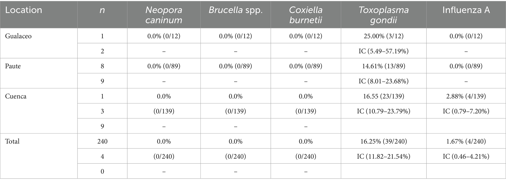
Table 1. Seroprevalence of antibodies against the 5 pathogens causing the diseases included in the study for guinea pigs.
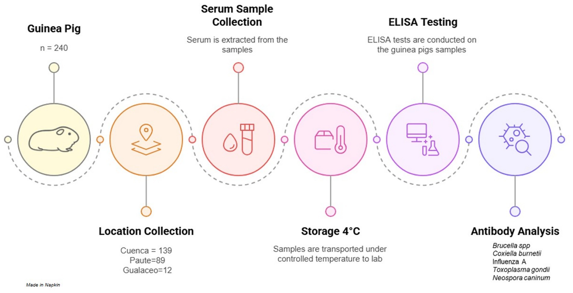
Figure 1. Flow diagram from sample collection and processing in our study.
No seropositive animals were detected for three of the pathogens: Brucella spp., Coxiella burnetii, and Neospora caninum. No inconclusive samples were found.
Four guinea pigs tested seropositive for antibodies against Influenza A. The overall seroprevalence was 1.67% (95% CI: 0.46–4.21). All four seropositive animals were from the Cuenca canton. The prevalence in Cuenca was 2.88% (4/139) (95% CI: 0.79–7.20). No inconclusive samples were found.
Thirty-nine guinea pigs tested seropositive for antibodies against T. gondii. The overall seroprevalence was 16.25% (95% CI: 11.82–21.54). The 39 seropositive animals were distributed among all three cantons: Cuenca (23/139, 16.55%; 95% CI: 10.79–23.79), Paute (13/89, 14.61%; 95% CI: 8.01–23.68), and Gualaceo (3/12, 25.00%; 95% CI: 5.49–57.19). The differences in T. gondii antibody prevalence among the three cantons were not statistically significant (p = 0.65). Two inconclusive samples were found, and as it was detailed in the method, these samples were considered negative.
Discussion
Guinea pigs have been shown to be incidental hosts for zoonotic transmission of respiratory pathogens of public health concern, including yeasts, Influenza virus, Methicillin-Resistant Staphylococcus aureus (MRSA), and Streptococcus pneumoniae (7–13). A pioneering study conducted in Ecuador in 2012 reported a high prevalence of antibodies against Influenza A and B in guinea pigs from markets in Manabí Province, Guayaquil, and Cuenca (8).
Our study confirms previous findings on the circulation of Influenza A in guinea pigs in Ecuador. This fact has implications for the ongoing panzootic of highly pathogenic avian Influenza A H5 in the Americas (33–35). The current panzootic has caused outbreaks not only in wild birds and poultry but also in several mammalian species, including dairy cows (36). By one hand, there is circulation of Influenza A and B in guinea pigs in Ecuador. On the other hand, intensive small mammal farming (minks) and explosive H5 influenza outbreaks have been reported in Europe (37). In this context, sentinel surveillance for avian and swine influenza should extend beyond poultry and pigs to include guinea pig.
Two recent studies from Colombia and Peru were the first to report the role of guinea pigs as reservoirs for Toxoplasma gondii (16, 17). These findings are particularly concerning due to the high T. gondii prevalence—23.3% in Cuzco, Peru, and 27.5% in Nariño, Colombia (16, 17). Moreover, these prevalence rates were based on PCR detection of T. gondii DNA, confirming active infection (16, 17). Additionally, T. gondii DNA was detected in multiple tissues, particularly in the brain, as well as the heart and muscle (16, 17). Our study demonstrated the circulation of T. gondii in Ecuadorian guinea pigs at a high prevalence (over 16%), corroborating previous findings from Peru and Colombia (16, 17). Guinea pigs are traditionally cooked and consumed whole, including the head, where the parasite was found at higher prevalence (16, 17). Our study, along with those from Peru and Colombia, underscores the need for public and animal health authorities to recognize guinea pigs as incidental host for zoonotic transmission of toxoplasmosis in the Andean region. From a food safety perspective, thorough cooking of guinea pigs should be recommended to prevent infection.
It is important to emphasize that Brucella spp., Coxiella burnetii, or Neospora caninum was not found in guinea pigs in our study. However, these three pathogens have been reported at high prevalence in cattle, wildlife, and free-roaming dogs in Ecuador and elsewhere (21–31). Further studies are needed to confirm the absence of this pathogens in guinea pigs considering their presence in livestock in Ecuador. Nevertheless, restricting access of all domestic animals, including pets, should be established as mandatory policy in guinea pig farms. Such measures would help prevent the spill over of zoonotic diseases like Q-fever and brucellosis to guinea pigs.
Our study has some limitations that we would like to acknowledge. Sampling was done at convenience and location was selected based on accessibility granted for blood collection. These facts may introduce sample bias. Also, the sample size for Gualaceo canton was only 12 animals, and that may also introduce sample bias. As sample collection was done at slaughterhouses, no risks factor analysis was possible as we could not identify the farm of origin of each animal. So far, further studies with larger population of guinea pigs are recommended. Moreover, the diagnosis was based on serology and molecular or microbiological diagnostics is recommended to confirm active infections in further studies.
In conclusion, guinea pigs raised as livestock in Ecuador serve as incidental host for the zoonotic transmission of Influenza A and T. gondii. The growing body of literature on guinea pig health and production highlights the need for increased awareness regarding their role as zoonotic reservoirs. However, the number of studies remains limited, and most are local and descriptive, lacking analysis of potential risk factors associated with pathogen presence. Future research should include larger sample sizes across multiple provinces in Ecuador and incorporate risk factor analyses related to guinea pig farming—such as proximity to other livestock, presence of domestic animals within farms, and farmers’ use of protective equipment. Such studies are essential to better understand the public health risks associated with guinea pig farming and to develop evidence-based guidelines for improving guinea pig health and production within a One Health framework.
Data availability statement
The original contributions presented in the study are included in the article/Supplementary material, further inquiries can be directed to the corresponding author.
Ethics statement
Ethical approval was not required for the studies involving animals in accordance with the local legislation and institutional requirements because According to local regulations in Ecuador, no IRB approval is needed for sample collection of livestock for diagnosis of infectious diseases. Written informed consent was obtained from the owners for the participation of their animals in this study.
Author contributions
MS-R: Conceptualization, Formal analysis, Writing – original draft, Methodology, Writing – review & editing, Data curation, Investigation, Project administration. PP-O: Formal analysis, Project administration, Investigation, Writing – review & editing, Methodology. JG-T: Formal analysis, Methodology, Data curation, Writing – review & editing, Investigation. KC: Writing – review & editing, Formal analysis, Methodology, Investigation. ME: Investigation, Writing – review & editing, Methodology, Formal analysis. PW: Data curation, Methodology, Investigation, Writing – review & editing. JM-M: Writing – review & editing, Investigation, Supervision, Methodology, Project administration, Formal analysis. KI: Writing – review & editing, Methodology, Investigation, Formal analysis, Data curation. FY: Methodology, Investigation, Writing – review & editing, Formal analysis. AR-P: Formal analysis, Methodology, Writing – review & editing, Investigation. FA: Investigation, Writing – review & editing, Formal analysis, Methodology. SO: Supervision, Project administration, Funding acquisition, Writing – review & editing, Methodology, Investigation, Conceptualization. MG-B: Writing – review & editing, Methodology, Supervision, Investigation, Writing – original draft, Funding acquisition, Data curation, Conceptualization, Formal analysis.
Funding
The author(s) declare that financial support was received for the research and/or publication of this article. This study was funded by Universidad Politécnica Salesiana and Universidad de Las Américas (MED. MGB.23.13.01).
Acknowledgments
We thank the workers of guinea pig slaughterhouses that allow us to collect samples for the study. We also thank Universidad Politécnica Salesiana and Universidad de Las Américas for partially funding this study.
Conflict of interest
The authors declare that the research was conducted in the absence of any commercial or financial relationships that could be construed as a potential conflict of interest.
The reviewer IT declared a shared affiliation with the author(s) AR-P, MG-B to the handling editor at the time of review.
The author(s) declared that they were an editorial board member of Frontiers, at the time of submission. This had no impact on the peer review process and the final decision.
Generative AI statement
The authors declare that no Gen AI was used in the creation of this manuscript.
Any alternative text (alt text) provided alongside figures in this article has been generated by Frontiers with the support of artificial intelligence and reasonable efforts have been made to ensure accuracy, including review by the authors wherever possible. If you identify any issues, please contact us.
Publisher’s note
All claims expressed in this article are solely those of the authors and do not necessarily represent those of their affiliated organizations, or those of the publisher, the editors and the reviewers. Any product that may be evaluated in this article, or claim that may be made by its manufacturer, is not guaranteed or endorsed by the publisher.
Supplementary material
The Supplementary material for this article can be found online at: https://www.frontiersin.org/articles/10.3389/fvets.2025.1657510/full#supplementary-material
References
2. Weir, BJ, and Weir, B. Notes on the origin of the domestic guinea-pig. Symposia Zool Soc Lond. (1975) 34:437–46.
3. Flores-Mancheno, CI, Duarte, C, and Salgado-Tello, IP. Caracterización de la carne de cuy (Cavia porcellus) para utilizarla en la elaboración de un embutido fermentado. Ciencia y Agricultura. (2017) 14:39–45. doi: 10.19053/01228420.v14.n1.2017.6086
4. El Telégrafo - Más de 710 mil familias se dedican a la crianza de cuyes en el país. Available online at:https://www.eltelegrafo.com.ec/noticias/2015/1/mas-de-710-mil-familias-se-dedican-a-la-crianza-de-cuyes-en-el-pais.
5. Crianza de cuyes ayuda a reconversión de actividades productivas – Ministerio de Agricultura y Ganadería. Available online at: https://www.agricultura.gob.ec/crianza-de-cuyes-ayuda-a-reconversion-deactividades-productivas.
6. Salas Rueda, M, Rodriguez Pazmino, AS, Orlando, SA, and Garcia-Bereguiain, MA. Livestock guinea pigs: a comprehensive review from a one health perspective. Trop Anim Health Prod. (2025)
7. Buela, L, Cuenca, M, Sarmiento, J, Peláez, D, Mendoza, AY, Cabrera, EJ, et al. Role of Guinea pigs (Cavia porcellus) raised as livestock in Ecuadorian Andes as reservoirs of zoonotic yeasts. Animals. (2022) 12:3449. doi: 10.3390/ani12243449
8. Leyva-Grado, VH, Mubareka, S, Krammer, F, Cárdenas, WB, and Palese, P. Influenza virus infection in Guinea pigs raised as livestock, Ecuador. Emer Infec Dis. (2012) 18:1135–8. doi: 10.3201/EID1807.111930
9. Rodriguez-Pazmiño, AS, Zambrano-Mila, M, Salas-Rueda, M, Cáceres-Orellana, MV, Buele-Chica, D, Barrera-Barroso, L, et al. Respiratory pathogens carriage in Guinea pigs raised as livestock in Ecuador: a proxy to study a neglected reservoir for zoonotic transmission in the Andean region. Acta Trop. (2025) 261:107505. doi: 10.1016/J.ACTATROPICA.2024.107505
10. Zambrano-Mila, M, Rodriguez, AS, Rivera-Olivero, IA, Salas-Rueda, M, Caceres-Orellana, MV, de Waard, JH, et al. Methicillin resistant Staphylococcus Aureus carriage among Guinea pigs raised as livestock in Ecuador. One Health. (2020) 9:100118. doi: 10.1016/J.ONEHLT.2019.100118
11. Moraes, MA, de Moraes, RS, de Moura, FC, Benevenuto, LGD, Onuma, TP, Leite, NC, et al. Cytological and histopathological lesions consistent with cutaneous nasal cryptococcosis in a guinea pig (Cavia porcellus). Revue Vétérinaire. Clinique. (2025) 60:136–9. doi: 10.1016/j.anicom.2025.05.001
12. Prazeres Júnior, FR, Moreira, AC, Medeiros, NO, Carmo, MCC, and Lima, MPS. Sporotrichosis in guinea pig (Cavia porcellus)-case report. Arquivo Brasileiro de Medicina Veterinária e Zootecnia. (2024) 76:e13132. doi: 10.1590/1678-4162-13132
13. Simmons, JH, Purdy, GA, Franklin, CL, Trottier, P, Churchill, AE, Russell, RJ, et al. Characterization of a novel parainfluenza virus, caviid parainfluenza virus 3, from laboratory guinea pigs (Cavia porcellus). Comp Med. (2002) 52:548–54.
14. Graham, JP, Vasco, K, and Trueba, G. Hyperendemic Campylobacter jejuni in guinea pigs (Cavia porcellus) raised for food in a semi-rural community of Quito. Ecuador Environ Microbiol Rep. (2016) 8:382–7. doi: 10.1111/1758-2229.12396
15. González-Ramírez, L, Joao Vázquez, C, Chimbaina, MB, Djabayan-Djibeyan, P, Prato-Moreno, JG, Trelis, M, et al. Ocurrence of enteroparasites with zoonotic potential in animals of the rural area of San Andres, Chimborazo, Ecuador. Vet Parasitol: Reg Stud Reports. (2021) 26:100630. doi: 10.1016/j.vprsr.2021.100630
16. Roller, S, Angulo-Tisoc, JM, Pacheco, JI, Jimenez, J, Vargas-Calla, A, Morales-Cauti, SM, et al. Molecular detection of Toxoplasma gondii in domestic and wild guinea pigs (Cavia spp) from the Marangani district in Cuzco, Peru. Vet Parasitol: Reg Stud Reports. (2024) 52:101038. doi: 10.1016/j.vprsr.2024.101038
17. Cañón-Franco, WA, Lopez-Orozco, N, Quiroz-Bucheli, A, Kwok, OCH, Dubey, JP, and Sepúlveda-Arias, JC. First serological and molecular detection of Toxoplasma gondii in guinea pigs (Cavia porcellus) used for human consumption in Nariño, Colombia, South America. Vet Parasitol Reg Stud Reports. (2022) 36:100801. doi: 10.1016/j.vprsr.2022.100801
18. Rizzo, MF, Osikowicz, L, Cáceres, AG, Luna-Caipo, VD, Suarez-Puyen, SM, Bai, Y, et al. Identification of Bartonella rochalimae in Guinea pigs (Cavia porcellus) and fleas collected from rural Peruvian households. The American Journal of Tropical Medicine and Hygiene. (2019) 101:1276–81. doi: 10.4269/ajtmh.19-0517
19. Rosa, LD, Soares, AG, Marcili, A, Diaz, JDS, Wolkmer, P, Bassuino, DM, et al. Cutaneous leishmaniasis in Cavia porcellus (guinea pig): case report. Arquivo Brasileiro de Medicina Veterinária e Zootecnia. (2020) 72:744–8. doi: 10.1590/1678-4162-11459
20. Verma, S, Katoch, RC, and Gupta, VK. Infection of Brucella melitensis in guinea pigs (Cavia porcellus). Indian J Anim Sci. (2000) 70
21. Changoluisa, D, Rivera-Olivero, IA, Echeverria, G, Garcia-Bereguiain, MA, and de Waard, JH. Serology for Neosporosis, Q fever and brucellosis to assess the cause of abortion in two dairy cattle herds in Ecuador. BMC Vet Res. (2019) 15:1–5. doi: 10.1186/s12917-019-1924-7
22. Echeverría, G, Reyna-Bello, A, Minda-Aluisa, E, Celi-Erazo, M, Olmedo, L, García, HA, et al. Serological evidence of Coxiella burnetii infection in cattle and farm workers: is Q fever an underreported zoonotic disease in Ecuador? Infect Drug Resist. (2019) 12:701–6. doi: 10.2147/IDR.S195940
23. Carbonero, A, Guzmán, LT, Montaño, K, Torralbo, A, Arenas-Montes, A, and Saa, LR. Coxiella burnetii seroprevalence and associated risk factors in dairy and mixed cattle farms from Ecuador. Prev Vet Med. (2015) 118:427–35. doi: 10.1016/j.prevetmed.2015.01.007
24. Carbonero, A, Guzmán, LT, García-Bocanegra, I, Borge, C, Adaszek, L, Arenas, A, et al. Seroprevalence and risk factors associated with Brucella Seropositivity in dairy and mixed cattle herds from Ecuador. Trop Anim Health Prod. (2018) 50:197–203. doi: 10.1007/s11250-017-1421-6
25. Rodriguez-Pazmiño, AS, Brito, CM, Salas-Rueda, M, Orlando, SA, and Garcia-Bereguiain, MA. A first insight into seropositivity and risk factors for Brucella spp. and Coxiella burnetii in free-roaming dogs in Ecuador. One Health. (2024) 19. doi: 10.1016/j.onehlt.2024.100909
26. Rivera, A, Zambrano-Mila, MS, Orlando, SA, Jiménez Valenzuela, F, Sanchez, E, Calderon, J, et al. A first insight into the occurrence of Leptospira, Brucella and Coxiella burnetii infections in wild mammals rescued from illegal trade in Ecuador: a proxy for one health conservation policies. One Health. (2025) 20:101045. doi: 10.1016/j.onehlt.2025.101045
27. Celina, SS, and Cerný, J. Coxiella burnetii in ticks, livestock, pets, and wildlife: a mini-review. Front Vet Sci. (2022) 9:1068–129. doi: 10.3389/fvets.2022.1068129
28. Hensel, ME, and Arenas-Gamboa, AM. A neglected animal model for a neglected disease: Guinea pigs and the search for an improved animal model for human brucellosis. Front Microbiol. (2018) 9:2593. doi: 10.3389/fmicb.2018.02593
29. Hirschmann, JV. The discovery of Q fever and its cause. Am J Med Sci. (2019) 358:3–10. doi: 10.1016/j.amjms.2019.04.006
30. Rodriguez-Pazmiño, AS, Brito, CM, Salas-Rueda, M, Orlando, SA, and Garcia-Bereguiain, MA. A first insight into seropositivity of Neospora caninum and associated risk factors in free-roaming dogs from Ecuador. Acta Trop. (2024) 256:107245. doi: 10.1016/j.actatropica.2024.107245
31. Runco, M, Gos, ML, Guichón, ML, Campero, LM, and Venturini, MC. Wild hosts in Argentina of the genera Sarcocystis, toxoplasma, Neospora, and Cryptosporidium. Analecta veterinaria. (2024) 44:6.
32. Reyes-Silva, F, Aguiar-Novillo, S, Enriquez-Estrella, M, and Uvidia-Cabadiana, H. Análisis Del Manejo, Producción y Comercialización Del Cuy (Cavia Porcellus L.) En Ecuador. Técnicas Aplicadas. (2021) 7:1004–18. doi: 10.23857/dc.v7i6.2377
33. Bruno, A, Alfaro-Núñez, A, de, D, Armas, R, Olmedo, M, Garcés, J, et al. First case of human infection with highly pathogenic H5 avian influenza a virus in South America: a new zoonotic pandemic threat for 2023? J Travel Med. (2023) 30. doi: 10.1093/jtm/taad03230
34. Bruno, A, Alfaro-Núñez, A, de Mora, D, Armas, R, Olmedo, M, Garcés, J, et al. Phylogenetic analysis reveals that the H5N1 avian influenza a outbreak in poultry in Ecuador in November 2022 is associated with the highly pathogenic clade 2.3.4.4b. Int J Infect Dis. (2023) 133:27. doi: 10.1016/j.ijid.2023.04.403
35. Bruno, A, de Mora, D, Olmedo, M, Garcés, J, Vélez, A, Alfaro‐Núñez, A, et al. Highly pathogenic avian influenza a (H5N1) virus outbreak in Ecuador in 2022–2024. Curr Infect Dis Rep. (2024) 26:245–53. doi: 10.1007/s11908-024-00849-5
36. Bruno, A, de Mora, D, Olmedo, M, Garcés, J, Marzal, A, Bereguiain, MA, et al. Highly pathogenic avian influenza a (H5N1) virus outbreaks in South America in 2022–2024: a comprehensive review of an ongoing panzootic. Virology. (2025).
Keywords: zoonosis, guinea pigs, Toxoplasma gondii , Influenza A, Ecuador
Citation: Salas-Rueda MX, Peralta-Ortiz P, Guama-Tipas J, Chávez Toledo KN, Espadero M, Webster P, Masache-Masache J, Illescas Sigcha KM, Yungazaca Jaramillo FE, Rodríguez-Pazmiño AS, Alcivar FA, Orlando SA and Garcia-Bereguiain MÁ (2025) Guinea pigs raised as livestock are incidental host of Toxoplasma gondii and Influenza A in Ecuador. Front. Vet. Sci. 12:1657510. doi: 10.3389/fvets.2025.1657510
Edited by:
Jose L. Gonzales, Wageningen University and Research, NetherlandsReviewed by:
José Manuel Verdes, Universidad de la República, UruguayIgnacio Troncoso, Universidad de las Américas, Chile
Copyright © 2025 Salas-Rueda, Peralta-Ortiz, Guama-Tipas, Chávez Toledo, Espadero, Webster, Masache-Masache, Illescas Sigcha, Yungazaca Jaramillo, Rodríguez-Pazmiño, Alcivar, Orlando and Garcia-Bereguiain. This is an open-access article distributed under the terms of the Creative Commons Attribution License (CC BY). The use, distribution or reproduction in other forums is permitted, provided the original author(s) and the copyright owner(s) are credited and that the original publication in this journal is cited, in accordance with accepted academic practice. No use, distribution or reproduction is permitted which does not comply with these terms.
*Correspondence: Miguel Ángel Garcia-Bereguiain, bWFnYmVyZWd1aWFpbkBnbWFpbC5jb20=