 Daniel Mota-Rojas1*
 Alexandra L. Whittaker2
 Lydia Lanzoni3
 Cécile Bienboire-Frosini4
 Adriana Domínguez-Oliva1
 Alfonso Chay-Canul5
 Vivian Fischer6
 Ismael Hernández-Avalos7
 Andrea Bragaglio8,9
 Eleonora Nannoni10
 Adriana Olmos-Hernández11
 Arthur Fernandes Bettencourt12
 Patricia Mora-Medina7Julio Martínez-Burnes13
 Alejandro Casas-Alvarado1
 Temple Grandin14*
- 1Neurophysiology, Behavior and Animal Welfare Assessment, DPAA, Universidad Autónoma Metropolitana (UAM), Mexico City, Mexico
- 2School of Animal and Veterinary Sciences, Roseworthy Campus, University of Adelaide, Roseworthy, SA, Australia
- 3Animal Production and Health Division, Food and Agriculture Organization (FAO), Rome, Italy
- 4EPLFPA-Avignon, Avignon, France
- 5División Académica de Ciencias Agropecuarias, Universidad Juárez Autónoma de Tabasco, Villahermosa, Mexico
- 6Department of Animal Science, Federal University of Rio Grande do Sul, Porto Alegre, Brazil
- 7Facultad de Estudios Superiores Cuautitlán, FESC, Universidad Nacional Autónoma de México (UNAM), Cuautitlán Izcalli, Mexico
- 8CREA Research Centre for Engineering and Agro-Food Processing, Consiglio per la Ricerca in Agricoltura el’Analisi dell’Economia Agraria, Treviglio, Italy
- 9Exo Research Organization, Potenza, Italy
- 10Department of Veterinary Medical Sciences, DIMEVET, University of Bologna, Bologna, Italy
- 11Division of Biotechnology-Bioterio and Experimental Surgery, Instituto Nacional de Rehabilitación Luis Guillermo Ibarra Ibarra (INR-LGII), Mexico City, Mexico
- 12Department of Animal Science, Federal University of Santa Maria, Santa Maria, Brazil
- 13Instituto de Ecología Aplicada, Facultad de Medicina Veterinaria y Zootecnia, Universidad Autónoma de Tamaulipas, Victoria, Mexico
- 14Department of Animal Science, Colorado State University, Fort Collins, CO, United States
Nonhuman animals use nonverbal cues to communicate their mental state about positive and negative events, including pain. Pain is a multidimensional process that elicits behavioral changes aimed at preventing further damage and promoting healing. These changes include restrictions on movement and/or activity, as well as adopting body postures to relieve pain. Additionally, changes in the ear and tail position have been associated with pain perception and are considered a sign of pain in several domestic species. Thus, this review aims to critically analyze and discuss the behavioral modifications and body language expressions associated with pain in domestic animals, with a particular emphasis on changes in tail position, ear posture, and overall postural dynamics. This review also aims to highlight the essential role of veterinarians and animal scientists in recognizing these subtle non-verbal indicators during clinical evaluation, thereby fostering early detection and effective pain management through more precise observational assessment.
1 Introduction
Pain assessment in veterinary medicine requires a multimodal approach that considers parameters beyond physiological and endocrine biomarkers due to its subjective and multidimensional nature (1–4). Some animals, such as horses and rodents, conceal signs of pain due to their prey nature, which forces them not to appear vulnerable to other individuals (5–9). Moreover, non-human animals cannot self-report the presence or intensity of pain (1). Thus, considering the animal’s nonverbal communication cues are essential to accurately evaluate pain (10). Nonverbal communication includes behavioral changes and modifications in body language (8, 11). Behavior refers to the movements and actions performed to respond to stimuli (e.g., withdrawal response, guarding the affected area, or vocalizing) (12). On the other hand, body language refers to changes in the animal’s body posture, as well as limb movements, gestures, and facial expressions (13, 14). Changes in behavior and body language are species-specific and have been recorded in animals exposed to noxious stimuli (15–17).
According to the neurobiology of pain, the activation of peripheral nociceptors (nerve fibers specialized in detecting noxious stimuli) and their projection to the brain results in the conscious perception of pain by the somatosensory cortex (Figure 1), also known as the affective component of pain (18–21). Pain demands attentiveness from animals. In consequence, this triggers several active or passive, defensive or reactive behavioral and body posture changes to prevent further damage and promote recovery (6, 22, 23). Due to the neurobiological association between pain processing and both behavioral and body posture changes, these aspects have been integrated into pain assessment scales for domestic mammals, which categorize pain by its intensity and duration (24–26). Additionally, characterization of pain requires consideration of the medical condition (e.g., surgical, traumatic, pathological, physiological) and the anatomical region (e.g., lumbar, abdominal, limbs) to objectively associate certain behaviors with pain (27).
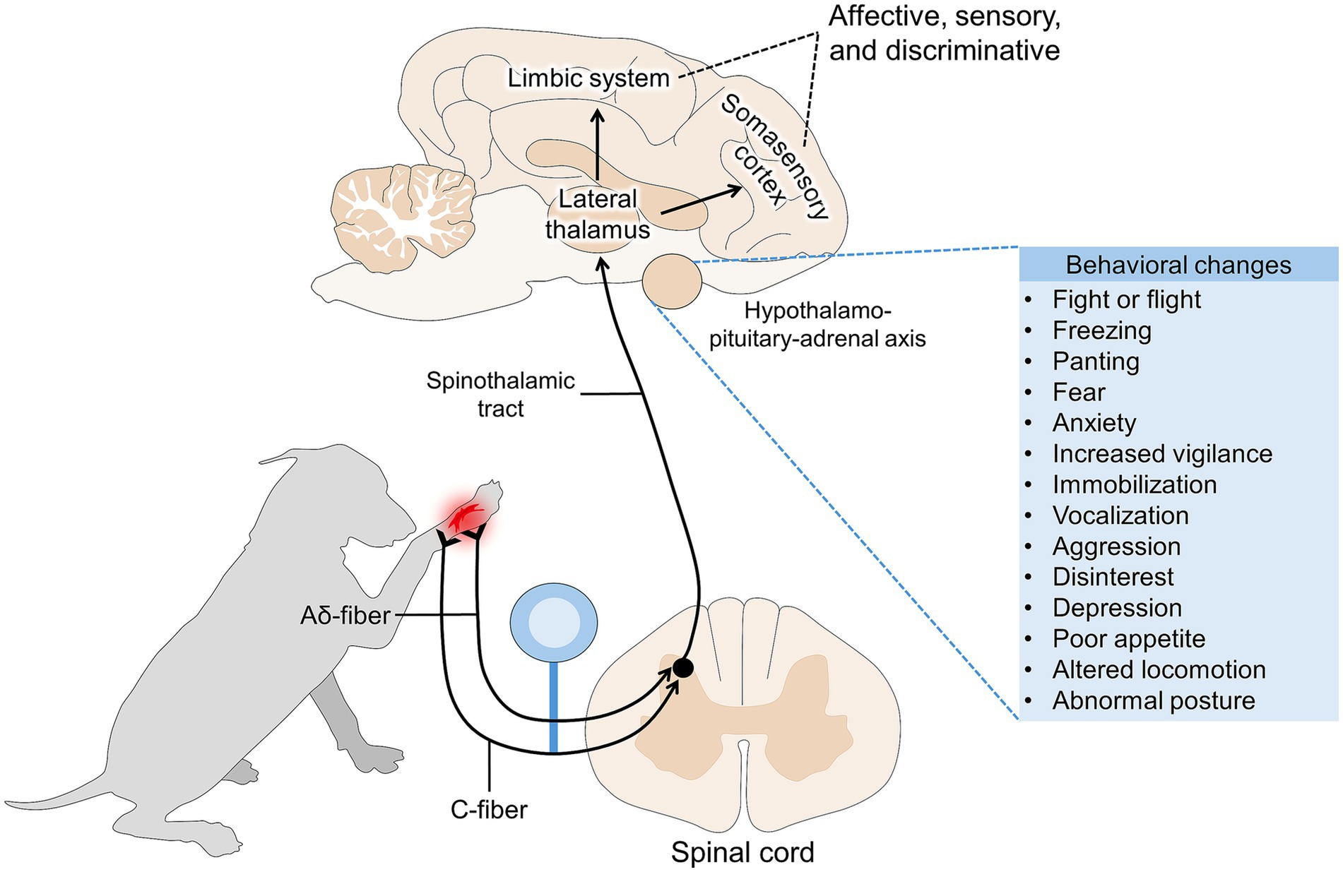
Figure 1. Pain pathway and its association with behavioral responses.
Regardless of the differences between species, in animals such as dogs, cats, horses, pigs, cattle, sheep, and goats, the modification of the position of the ears or tail is considered one of the main changes in body language related to the perception of pain (28–30). However, due to the variability in the expression of pain-associated responses in domestic mammals, assessment using pain scales requires training in the specific behavioral repertoire to detect alterations (31, 32). The complexity of recognizing behaviors and postures associated with pain in animals highlights the role that veterinarians have in promptly detecting pain and educating owners to detect it at home (33).
Through the recognition of the anatomical regions involved in pain processing and how pain manifests as changes in posture and behavior, a clinical and non-invasive evaluation of pain can be obtained. Thus, this review aims to critically analyze and discuss the behavioral modifications and body language expressions associated with pain in domestic animals, with particular emphasis on changes in tail position, ear posture, and overall postural dynamics. This review also aims to underscore the essential role of the veterinarian in recognizing these subtle non-verbal indicators during clinical evaluation, thereby fostering early detection and effective pain management through more precise observational assessment. Moreover, this review also provides practical information that veterinary practitioners can use to assess pain in different domestic mammals. Figure 2 schematizes the overall structure of the review.
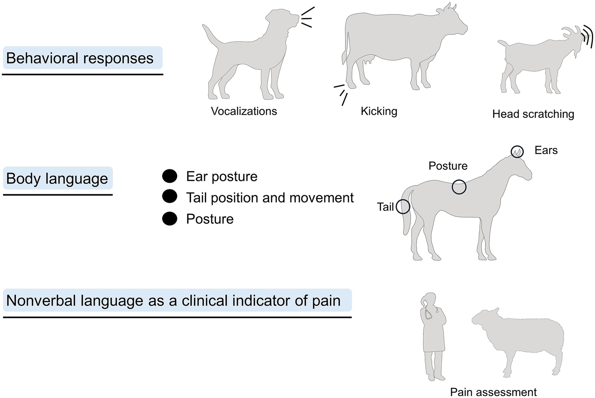
Figure 2. Overall structure of the review, where behavioral responses and body language will be discussed as methods to assess pain.
2 Behavioral responses associated with pain
Evaluating the behavioral responses of animals when experiencing pain is considered one of the main noninvasive methods to assess its affective component (34). As previously mentioned, pain-related behaviors in domestic mammals comprise a wide range of activities to reduce the discomfort caused by pain, such as protecting the injured area (35). As mentioned by Camps et al. (36), both losing the presentation of normal behaviors and developing abnormal ones are considered signs of pain. Among the main reported signs in animals experiencing pain are reluctance to move, depression, sleep disturbances, loss of appetite, restlessness, frequent vocalization, licking, biting, scratching, self-mutilation, anxiety, irritability, and aggressiveness (Figure 3) (25, 26, 37–43).
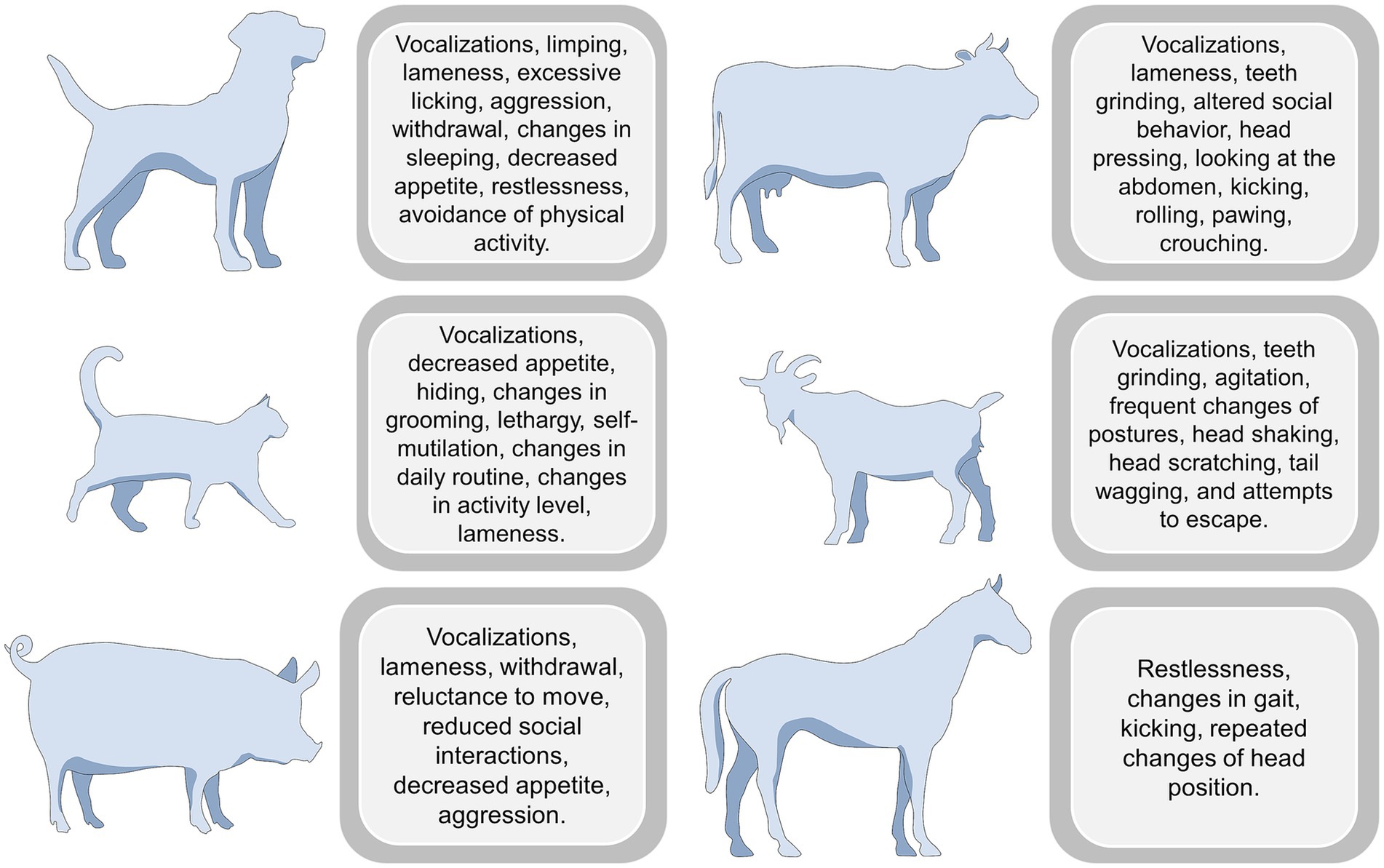
Figure 3. Behavioral responses to pain in different species.
The behavioral response and the change intensity depend on the species and the painful condition. For example, behavioral changes in domestic species such as dogs and cats are the basis for evaluating acute pain (as observed in their respective pain assessment scales) (25, 44). Firth and Haldane (45) were among the first researchers to highlight the importance of pain-related behaviors in dogs by developing a behavior-based scale to assess pain. In this scale, restlessness, vocalization, and reluctance to rise or sit are present in animals with severe surgical pain after ovariohysterectomy (OVH) or castration. Similarly, in Reid et al.’s (44) study, groaning or screaming, growling, and snapping in response to touch, as well as anxiety, fearfulness, or non-responsiveness to stimulation, are considered signs of severe pain during the postsurgical period. These results align with studies reporting that, after OVH and castrations, the response to palpation, reduced movement, and increased frequency of vocalizations are signs of pain regardless of the analgesic treatment (46).
When dogs perceive musculoskeletal pain, particularly in the joints (hip, stifle) or fore/hindlimbs, which represent very common (29–71%) sources of pain (47), main behavioral modifications are reduced general activity and resistance/stiffness to walking (48, 49). These changes may be accompanied by pain-related aggression, as observed in dogs (66.7%) with hip dysplasia (36). Stevens et al. (50) mention that scales scoring appendicular joint pain (in mani, carpi, elbows, shoulders, pes, tarsi, stifles, hips) consider aggression or intention to bite when trying to manipulate the injured area a sign of severe pain.
Additionally, this type of pain is also related to unwillingness to learn or participate in training sessions, house-soiling issues, and clinginess to the owner (47). An example is Dodd et al.’s (51) study focusing on military working dogs with lumbosacral stenosis. Twenty-one dogs (32.8%) presented behavioral alterations such as unwillingness or reluctance to jump (38%), self-mutilation in the affected area (25%), anxiety (25%), anorexia (25%), and reluctance to sit (25%). Figure 4 illustrates some examples of behavioral changes in companion dogs and how these can change according to the etiology of pain (e.g., pancreatitis, nasal transmissible venereal tumor, gastroenteritis, and postsurgical pain) (47, 52–56).
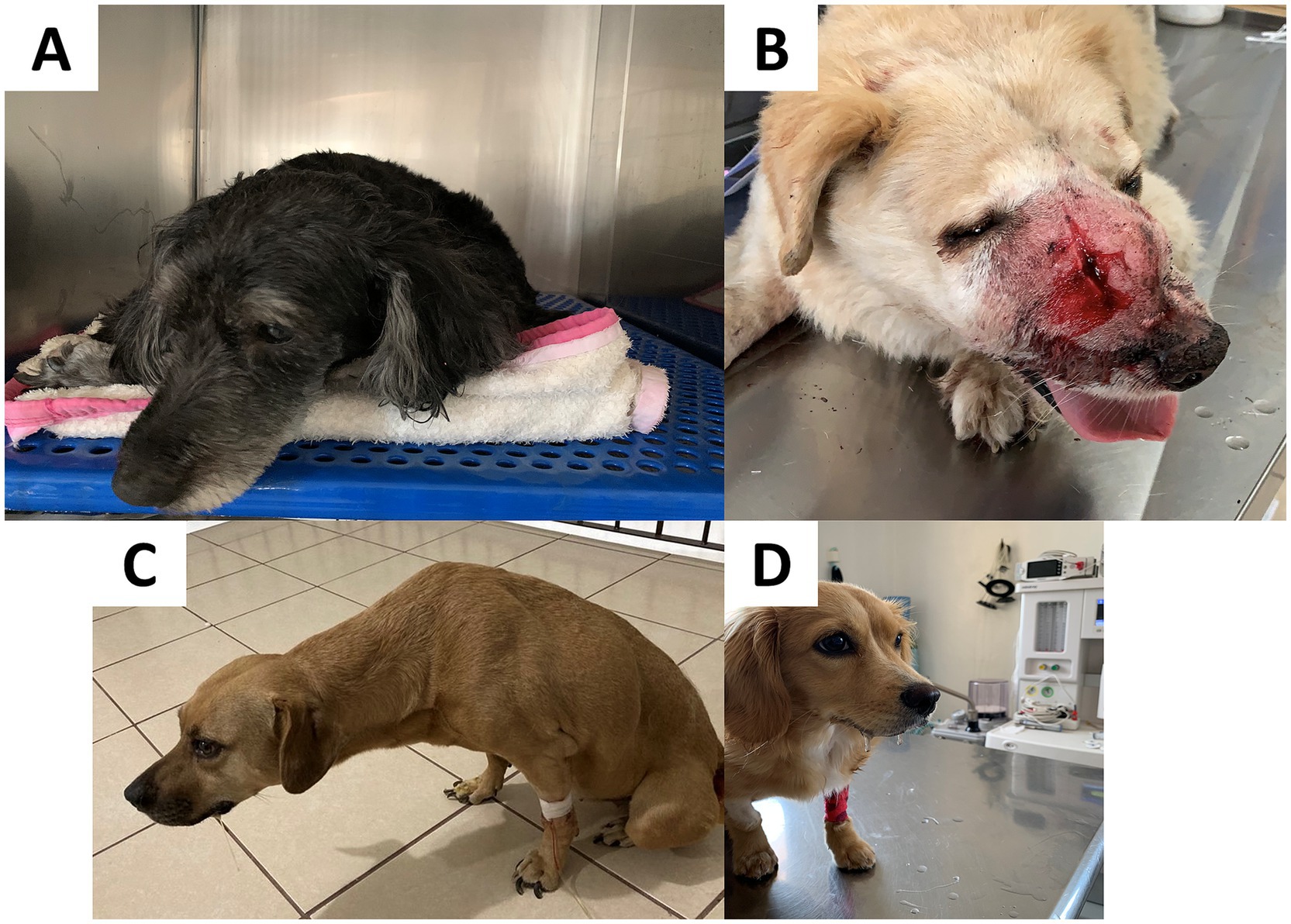
Figure 4. Behavioral changes observed in dogs during hospitalization. (A) A patient recovering from pancreatitis. This pathology is associated with restlessness and increased difficulty in adopting a comfortable position to rest. Slower reflexes, body stiffness, changes in appetite, and vocalization can also be observed if the pain is severe. (B) A dog diagnosed with a nasal transmissible venereal tumor. This clinical presentation is associated with nasal discharge, sneezing, nosebleeds, respiratory difficulty, and nasal deformity, which can lead to postural changes in patients due to perceived pain. (C) A patient with gastroenteritis. Among the behavioral alterations, lethargy, apathy, difficulty in standing, and walking are frequently observed. In addition, dogs may refuse abdominal palpation. Postural changes may include back arching and an orthopneic neck position. (D) A patient with excessive salivation is observed after elective OVH. Dogs experiencing postoperative pain may show rapid or abdominal breathing, reluctance to move, abnormal postures when sitting or lying down (e.g., hunched posture with a tense abdomen), and decreased appetite. Photos taken by the authors in a private clinic.
In dogs, gastrointestinal pain is associated with compulsive-type behaviors such as star gazing, excessive licking of surfaces, and pica (47). Bécuwe-Bonnet et al. (57) observed copious licking of surfaces (floors, walls, carpets, and furniture) in 59% of dogs diagnosed with eosinophilic and/or lymphoplasmacytic infiltration in the gastrointestinal tract, reduced gastric emptying, irritable bowel syndrome, pancreatitis, and giardiasis. Excessive licking might also progress to self-mutilation in cases of acral dermatitis (58). The overall reduction in activity and mobility observed in animals experiencing all types of pain is related to the protective nature of pain, i.e., its function to prevent further damage, avoid activities that might delay healing, and decrease the inflammatory response that frequently escalates to hyperactivation of peripheral receptors, sensitization, and chronic pain (23).
In the case of cats, contrary to dogs, pain evaluation and recognition of pain-related behaviors are challenging due to their tendency to hide any sign of discomfort unless severe (59). Due to this aspect, the behavioral modifications observed in dogs might not always be present in cats (or be less evident). For example, Monteiro and Steagall (60) mention that mobility changes are less common in domestic felines due to their species-specific behavioral repertoire and inclination to withdraw and hide when threatened. However, among the main behavioral changes related to abdominal pain are a reaction to palpation, decreased appetite, growling, groaning, and decreased grooming (61).
According to Brondani et al. (62), a cat with severe surgical pain licks/bites the surgical wound, reacts aggressively when touching the wound, vocalizes (growls, howls, hisses), shows restlessness and reluctance to move. Similarly, Marangoni et al. (27) mention descriptors such as the level of exploratory behavior, restlessness, grooming, stretching, attention to the wound, growling/hissing, and no interest in food. In the case of chronic pain, a reduction in the animal’s activity is observed, as well as loss of appetite, a tendency to hide or avoid social interaction, and excessive licking of the affected area, decreasing normal grooming (43, 63).
Likewise, some authors refer to key behaviors that help distinguish between painful and nonpainful cats, as reported in kittens subjected to OVH (64). When comparing kittens receiving opioid-free multimodal analgesia with those that did not receive analgesic drugs, animals in pain showed less interest in their surroundings (5 vs. 0%) and played less (7 vs. 35%). Temperament changes are also often reported in cats (91%), as mentioned by Bennett and Morton (65) in adult animals diagnosed with musculoskeletal pain, with reported avoidance of conspecifics and owners. In a case study, aggression due to fearfulness due to arthritic pain in the thoracolumbar spine was reported in a cat presenting house-soiling issues, posturing, and vocalization (47).
In companion animals, these behavioral changes help veterinarians rate the degree of pain. However, studies have shown that dog owners can identify pain through behavioral alterations. For example, 52.6% of owners reported that behavioral signs were very useful to assess pain, and 48.8% of owners reported that identifying these changes was very useful in determining whether they should consult a veterinarian (66). Similarly, in cats, 90% of owners consider it helpful to resort to behavioral evaluations to determine the animal’s degree of pain, and 86% find it helpful to seek veterinary care (67).
In the case of farm species, several instances might cause pain (30, 68). For example, pathological pain due to mastitis or laminitis in ruminants and horses or surgical pain due to castration in piglets and dehorning or disbudding in ruminants, respectively, are accompanied by behavioral modifications that, as seen with dogs, aim to decrease pain perception and promote recovery (69–71). In the first instance, several studies have reported behavioral alterations due to pathological conditions such as mastitis in cattle (72, 73). In this sense, Medrano-Galarza et al. (74) evaluated lying behavior and reactivity during milking (stepping, lifting, and kicking) and its relation to the inflammatory process of the mammary gland due to the presence of bacteria. The authors reported that animals in pain spent statistically significantly less time lying (707.5 min/24 h) than their healthy counterpart (742.5 min/24 h) and that the frequency of lifts and kicks was higher in cows with mastitis (0.70 and 0.10 per minute, respectively). Moreover, Peters et al. (73) evidenced that cows affected by subclinical and clinical mastitis had a lower thermal threshold (higher sensitivity to thermal stimulus) compared with healthy cows, which is observed as a fast foot-lift response at lower temperatures (e.g., 50.9 °C).
Fogsgaard et al. (75) reported that cows suffering from mastitis spent less time lying during the initial phase of the inflammatory disease (720 min/day), and had a higher frequency of kicking (more than 0.70 kicks/min/milking). In addition, Siivonen et al. (76) found that cows spend less time lying on the side with the inflamed udder (control quarter: 40.94 ± 4.60 min; affected quarter: 33.76 ± 2.32 min) and stepped more after an animal model of induced mastitis (up to 1,413 8.6 steps). Another routine procedure on dairy farms that can cause pain and discomfort is drying off, as the accumulation of milk within the mammary gland increases intramammary pressure. Rajala-Schultz et al. (77) observed that cows subjected to gradual drying-off spent more time lying down compared to those undergoing abrupt cessation. Similarly, Maynou et al. (78) reported that the use of acidogenic boluses reduced milk production, which in turn decreased intramammary pressure (55.0 vs. 61.9 kg/m/s2) and consequently increased lying time. In particular, lying behavior responses in cows are relevant due to their high motivation to lie down (79). In this sense, veterinarians and stockpeople could use lying time in cattle as a potential behavioral marker of pain, as lower lying times are primarily due to the pain and the inflammation of the udder (79, 80), which could help to promptly identify the painful condition and administer pharmacological treatment when necessary.
Tail docking also induces behavioral alterations, as reported in 21- to 42-day-old dairy heifers, tail banding without epidural anesthesia increased restlessness (up to three changes of posture/15 min) (81). Similarly, in crossbred beef heifers, Kroll et al. (82) compared the behavioral response of docked and undocked animals immediately after the procedure. The authors found significantly more steps (up to 200 counts/h), more rear foot stomping (87.2%), and less lying time (approximately 15 min/h) immediately after tail docking than in the subsequent days. A decreased appetite was reported by Eicher et al. (83) in cows after tail docking, reducing the time spent feeding from 17.8 to 13.3% and increasing the frequency of kicking the ground (4%), due to the adoption of alternative behaviors to scare away flies in docked animals. Thus, caudectomy in cattle could have behavioral repercussions because the tail is part of the body language of the species. Additionally, tail docking of dairy cows is declining and is banned in some countries (84). In other species of ruminants, such as sheep, the method of tail docking highly influences the degree of pain perceived by the animals. In this sense, Grant et al. (85) compared tail docking in lambs by rubber ring and hot iron for 90 min after the procedure. The authors found that tail docking by rubber rings significantly increased the frequency of pain-related behaviors such as vocalization (9.9 ± 3.0 animals), number of times the animal changed their lying posture (62.1 ± 5.2%), tail wagging (14.6 ± 2.6 times), kicking/stomping (8.3 ± 1.3 times), and lick/bite the affected area (4.7 ± 0.6 times).
In farm animals, vocalization and its acoustic characteristics during tail docking or castration are considered indicators of pain (86–88), as mentioned by Cordeiro et al. (89), who evaluated the maximum amplitude, pitch frequency, and intensity of vocalization in piglets undergoing castration and tail docking. After the procedure, the maximum amplitude, pitch frequency, and intensity increased by 0.78 Pa, 159 Hz, and 16.9 dB, respectively (89). Similarly, in piglets after hot tail docking, an increase in the frequency and duration of vocalizations was found along with increases in cortisol and β-endorphin levels (90). Hansson et al. (91) reported that the administration of local anesthesia decreases the number and intensity of vocalizations in castrated piglets.
Another common practice on livestock farms is castration. Among the castration methods commonly applied to farm animals are Burdizzo (B), rubber ring (RR), and surgical castration (S), which are frequently compared to a control group subjected only to scrotal handling (H) (92, 93). According to Melches et al. (93), lambs castrated using B and S exhibited more frequent pain-related behaviors during the procedure compared to those in RR and H groups. Moreover, lambs in the S group showed higher cortisol concentrations and a greater occurrence of abnormal postures on the day of castration, along with reduced feed intake and rumination during the first 6 days post-castration relative to the other groups. Similarly, Molony et al. (92) observed in calves that castration using RR was associated with more severe acute and chronic pain, with behavioral indicators of discomfort persisting for up to 42 days. In contrast, castration using S, B, or the combination of B + RR elicited comparatively lower behavioral and physiological stress responses, particularly during the chronic phase.
A study by Yun et al. (94) found that piglet castration without analgesics increased the observation of standing or sitting inactively (102 ± 25.3 counts) and lower frequencies of tail wagging (0.3 ± 0.1) compared to non-castrated animals. Some other behavioral changes were reported in lambs after castration, and similar to tail docking, the method influences the behavioral response. For example, when comparing castration in lambs by cutting with a knife and rubber rings, Lester et al. (95) concluded that behavioral alterations such as abnormal standing/walking and restlessness were predominantly observed in knife-treated lambs within the first four hours after the procedure. In contrast, Maslowska et al. (96) reported that rubber ring castration increased the frequency of active pain behaviors (observable actions when animals experience pain) (a frequency of 110.5) and 44.8% of lambs were more restless and painful than animals that were only handled. Thus, the variability of pain-related behaviors is closely related to the pain source or the method (i.e., castration method), as mentioned by Canozzi et al. (97).
In the case of goats undergoing elective surgical castration, behavioral modifications such as lying down motionless, standing still, and looking at the affected area were considered by Fonseca et al. (98) to develop the Unesp-Botucatu acute pain scale for goats. Vocalizing and teeth grinding have also been reported in adult goats and goat kids during husbandry practices, including castration, disbudding, and dehorning, and during pathological conditions such as lameness or mastitis (99). During disbudding, Kongara et al. (100) summarized that the main behavioral changes observed in kids were head and body shaking, head scratching, and tail shaking. Similar to these findings, Hempstead et al. (101) compared the frequency of pain-related behaviors in disbudded goat kids with cautery iron with a sham group. The results showed an increased frequency of head shaking (31.2 ± 3.11 vs. 17.5 ± 1.79), head scratching (15.8 ± 5.90 vs. 2.2 ± 1.11), head rubbing (4.2 ± 0.77 vs. 0.8 ± 0.27), and body shaking (6.1 ± 0.36 vs. 8.8 ± 0.49) in disbudded animals, which can be used as signs associated with pain. Recognizing these signs is essential to adopt adequate analgesic protocols. For example, Alvarez et al. (102) evaluated the effect of cornual nerve blocks on goat kids undergoing disbudding. The authors evaluated the total behavioral response (including struggle/attempts to escape, vocalizations, and tail movements). It was found that lidocaine administration did not decrease the mean number of said behaviors (control: 59.6 ± 6.8; lidocaine: 52 ± 6.8), suggesting that pain after disbudding should be complemented with other analgesics, such as non-steroidal drugs or general sedation.
Dehorning in cattle has also been associated with pain-related behaviors such as head-shaking, ear flicking, and increased inactivity (103, 104). This has been reported in Holstein calves (4–8 weeks old) after iron-hot dehorning (104). When compared to a control group without receiving analgesic drugs (ketoprofen), treated calves had a lower frequency of head shaking (0.74 ± 0.25 vs. 6.27 ± 2.57) and ear flicking (0.56 ± 0.17 vs. 11.43 ± 3.07) after dehorning. Similarly, the application of lidocaine reduced the frequency of head moving (2.9 ± 0.6 vs. 5.3 ± 1.5), head shaking (1.3 ± 0.6 vs. 27.4 ± 5.9), tail wagging (1.5 ± 0.5 vs. 3.5 ± 0.5), and rearing (0.4 ± 0.2 vs. 1.9 ± 0.5) when compared to a control group of calves dehorned without analgesic (105). Head shaking, ear flicking, and head scratching were also reported in calves dehorned with two methods: cream and hot iron (103). Additionally, in the same animals, a decrease in lying time was observed in comparison with the pre-dehorning period (from approximately 110 min to 75 min), together with decreased playing behavior (from approximately 180 min to 60 min).
Although the discussed pain-related behavioral responses in domestic mammals can differ according to the species and, particularly, to the pain source, these reactions arise to avoid further injury, increase survival chance, and promote healing (32). They are rapid responses of passive or active defense against pain. Therefore, veterinarians and animal handlers should receive training to recognize subtle changes in behavior, as behavioral modifications are one method of communicating pain in animals and may serve as an indicator of their welfare.
3 Body language as a tool to assess pain: anatomical structures related to pain perception
3.1 Ear posture
Ear position has been used as an indicator of an animal’s affective state, including pain (106–108). The changes in ear posture are related to the neurobiological processing of emotions eliciting different facial expressions according to the context (40, 109). Ear posture depends on motor control from the primary motor cortex and the subnuclei of the facial nerve (VII) (110–113). Although each nervous region has specific functional delimitations, the excitability of the motor cortex leads to positive feedback from the facial nerve, resulting in the contraction or relaxation of the muscles that control ear movement (20, 40). These changes are controlled by the ventral auricular, dorsal auricular, rostral auricular, and caudal auricular muscles (114, 115). However, changes in ear posture alone should not be considered an indicator of pain and must be considered among the several signs that animals show when perceiving pain.
The importance of ear posture as an indicator of pain in animals is reflected in the current scales that consider it as one of the most noticeable changes when animals are exposed to a noxious stimulus. These scales have been adapted to cats (116), mice (117), rats (114, 118), rabbits (119), pigs (120, 121), sheep (122, 123), goats (124), horses (9), donkeys (125), and cows (126) (Table 1). Although the change and position depend on the species and distinct anatomy, several similarities have been found (125, 127–129). When animals experience pain, stimulation of the auricular muscles causes flattening or retraction of the ears in all species (129–131). For example, a cat with severe pain shows ears that are markedly rotated outwards (132). Rats and mice in pain show ears that are curled, pointed, and/or angled forward or outward (118, 133). For rabbits, ears tightly folded against the neck, pulled back, and flattened are present when the animal is perceiving severe pain (134). Similarly, in farm species such as piglets and goats, severe pain is characterized by the ears drawn back from the forward position and hanging (124, 135, 136), which has also been reported in horses, donkeys, and cows (9) (Figure 5) (109, 118, 132–134, 137, 138).
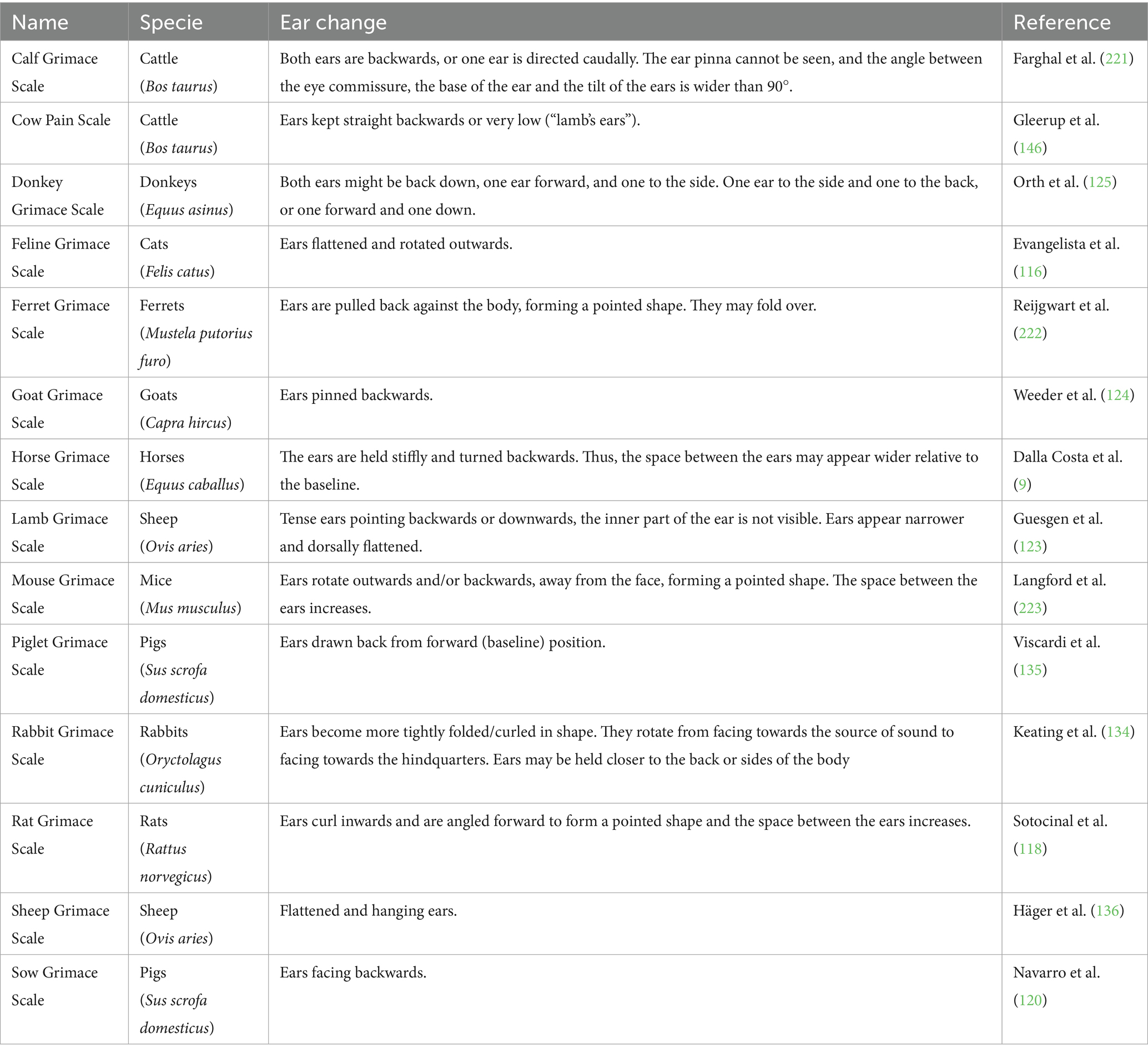
Table 1. Description of ear changes in the currently available Grimace Scales in domestic mammals.
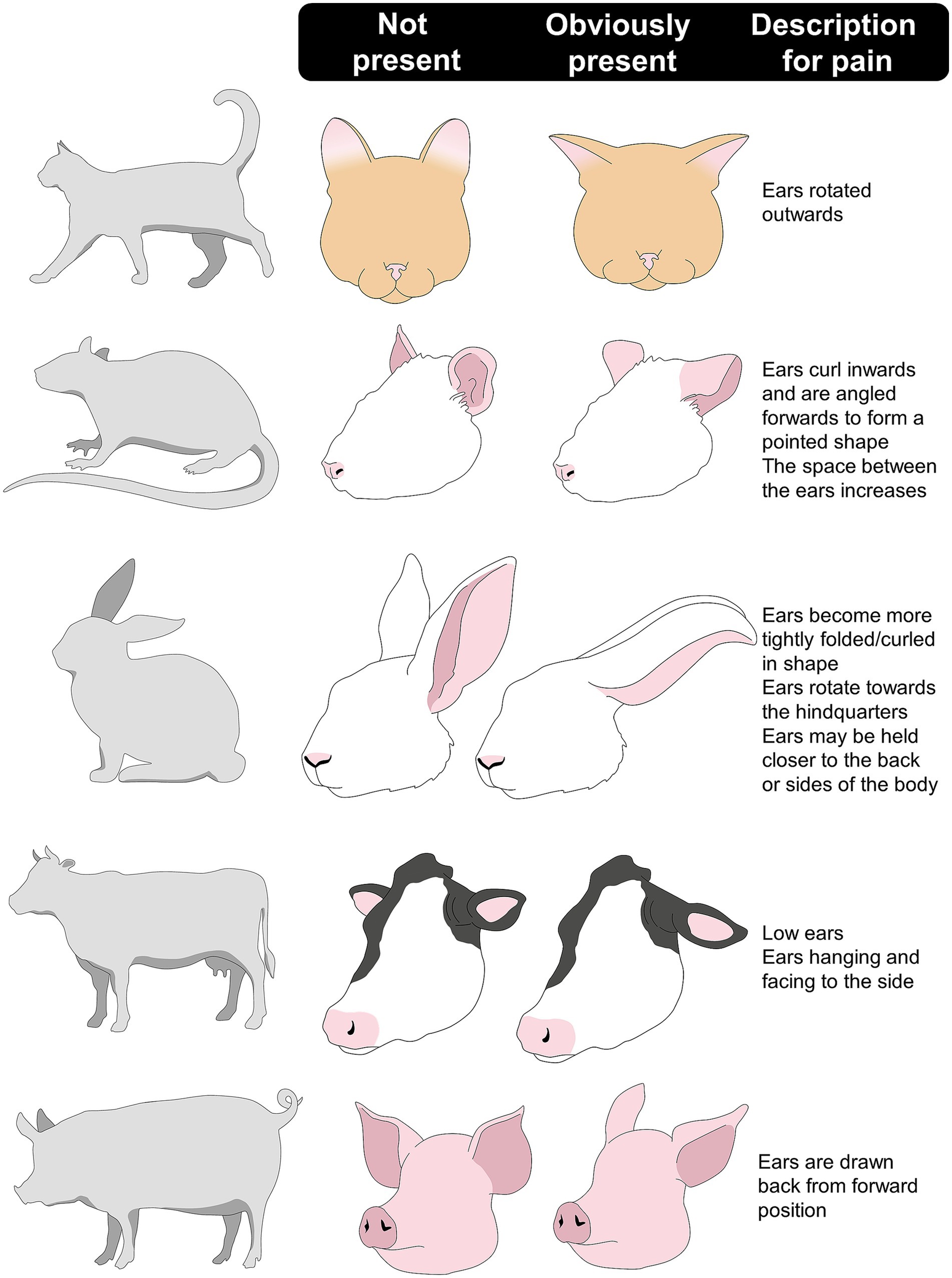
Figure 5. Description of the ear changes in some domestic mammals when perceiving pain.
Clinical examples of pain identification through changes in ear posture and other facial indicators have shown an accuracy of 87% in cats (139). Particularly, as Watanabe et al. (140) mention, ear posture has a good inter-rater reliability score (0.55–0.78) as it is one of the changes caregivers easily observe in cats that underwent procedures such as dental extractions. Holden et al. (141) found that the distance from the midpoint of the two ears is an indicator of pain, where a greater distance between the tips of the ears is considered indicative of pain and correctly classifies between pain-free and painful cats in 95% of cases. Additionally, Merola and Mills (142) mention that in cases of pain due to orthopedic conditions, cancer, urinary tract diseases, or dental issues, flattened ears are frequently observed when perceiving high levels of pain, although it may also be a sign of fear.
In the case of laboratory rodents, Mittal et al. (143) determined the association between pain in sickle mice and changes in the ear position (and other facial indicators). The authors found that exposure to 4 °C caused the ears to move parallel to the neckline, suggesting cold hypersensitivity and pain (scores of up to 1.5). In the same species, evaluations post-vasectomy with and without analgesics found that animals in pain frequently showed ears rotated outwards, and their assessment had an excellent (0.75) reliability score (144). For rabbits, Benato et al. (138) reported that the position and movement of the ears are accurate descriptors of pain. In this sense, flattened ears and lack/diminished ear movement were observed in rabbits after OVH and orchiectomy.
In farm animals, Tallet et al. (145) determined the effect that tail docking has on piglets’ behavior and body posture. In particular, the authors found that immediately after cautery iron docking, piglets held their ears perpendicular to the head-tail axis (70% of animals) and showed more ear posture changes (70%) than non-docked piglets (30 and 20%, respectively). This is similar to what was observed in Danish Holstein dairy cattle during castration, mastitis, or laminitis. In these animals, acute pain was observed as caudal rotation of the ears, along with other changes such as keeping the head below the horizontal axis of the animal, piloerection, arching of the back, and an increased reactivity (146). Additionally, ear posture can also suggest the emotional state of animals, as mentioned by Lambert and Carder (109), who evaluated the ear position of Holstein dairy cows under two different contexts (frustration and excitement). The authors found that cow ears had more changes in ear position during the frustration event (from 14.15 to 16.59 changes/15 min).
In small ruminants such as goats, Weeder et al. (124) analyzed the facial response of goats to induced lameness. Results showed that animals with obvious changes due to pain were characterized by both ears pulled backwards, along with behavioral modifications (e.g., increased lying time). Ear posture changes were also reported by Hussein and Hidayet (147) in goat kids (10–14-day-old) undergoing ear tagging. After the routine procedure, a significant increase in ears backward (from 0.7 ± 0.2 to 11.6 ± 1.7 s), number of posture changes (from 3.3 ± 0.4 to 9.8 ± 0.6), and a decrease in ears plane (e.g., perpendicular to the head-rump axis) (from 25.3 ± 1.5 to 11.8 ± 2.1 s) was observed.
Gleerup et al. (148) characterized the changes in ear position of adult horses exposed to experimental acute pain (a tourniquet to the forearm and the topical application of capsaicin). Both stimuli increased the time the horses maintained asymmetrical ears and in a low position (between 54 ± 0.5 and 51 ± 23%), which coincided with the significant increase in the pain assessment scale score. Similarly, Ask et al. (149) evaluated changes in ear position in horses that were administered lipopolysaccharide in the tarsal-crural joint to generate acute pain, highlighting ear flattening and lateral rotation. Additionally, in horses undergoing routine castration under general anesthesia, Dalla Costa et al. (144) found that stiffly backwards ears are associated with pain, with an excellent reliability coefficient of 0.96. Figure 6 shows the ear changes that can be observed in an equine patient with colic syndrome due to pain (150). This figure also shows the changes in the ear position of a feline patient with idiopathic cystitis (151).
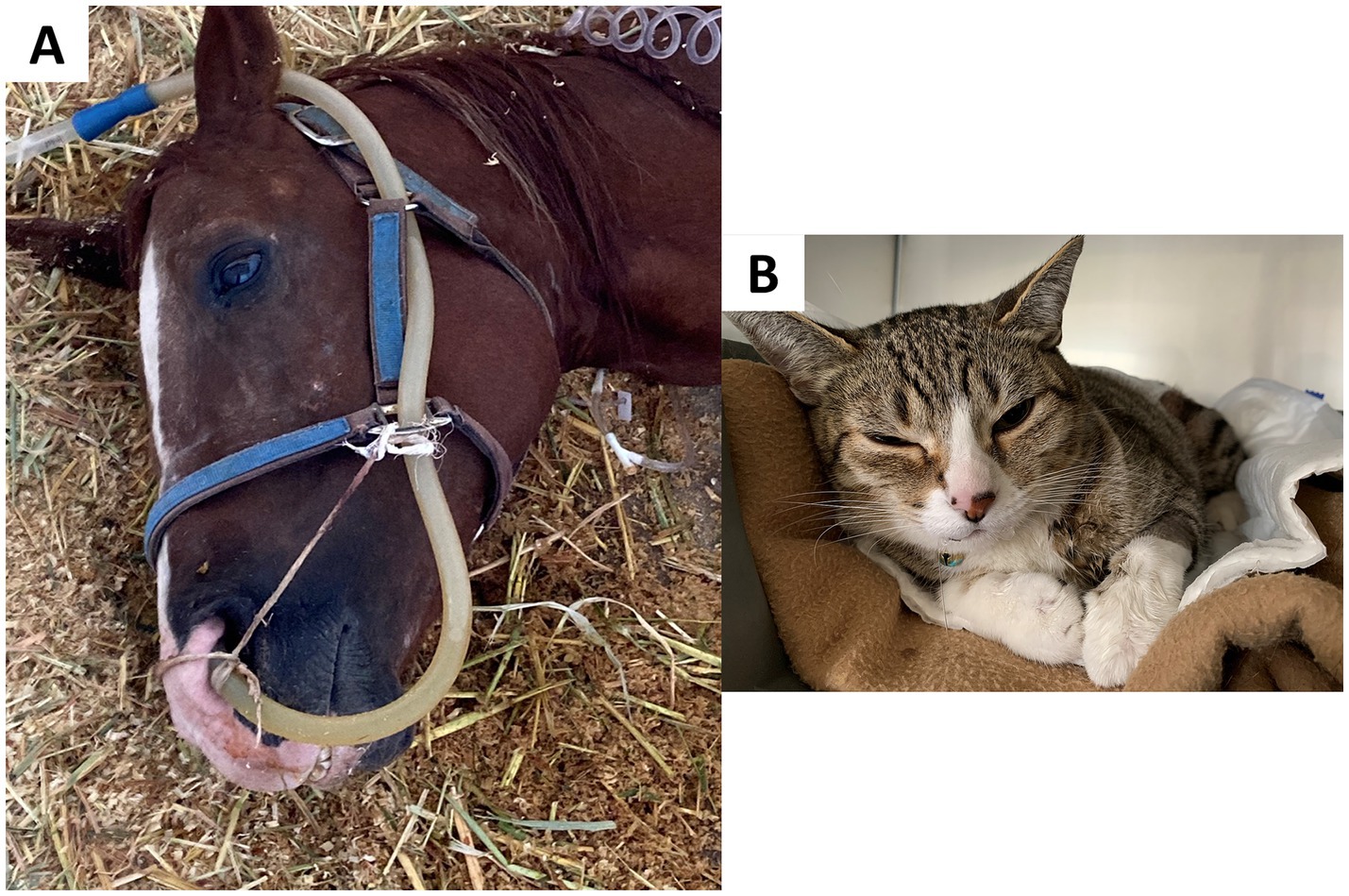
Figure 6. Ear changes in domestic animals during the perception of acute pain. (A) A prostrated male Quarter Horse with equine colic due to acute and severe abdominal pain. Facial changes, such as ear flattening, can be observed. (B) Feline with idiopathic cystitis. Note the changes in ear position, such as flattening, outward rotation, and being slightly pulled apart. Photos taken by the authors.
3.2 Tail position and movement
Tail position and movement have also been considered indicators of pain in animals (152). They have been particularly studied during routine procedures in farm animals such as surgical castration or tail docking (84, 153). During these events, animals in pain maintain their tail stiff, hide it, or swing it abruptly (153).
Changes in tail position or movement respond to adjacent nociceptors that send information through the pudendal and perineal nerves (154). These nerves reach the dorsal root ganglia of the spinal cord and deploy a neuronal and molecular communication circuit at the brain level. Within the brain, the reception and refinement of this information translates into an immediate pain response, coordinated by the amygdala, hypothalamus, and periaqueductal gray matter. The connections of these structures with the motor cortex cause the motor or reflex responses to noxious stimuli (20, 155). The tension of the tail and hiding it between the hindlimbs is due to the contraction of the coccygeus, sacrocaudalis ventralis, dorsalis, and caudae muscles, which stabilize the spine. As a compensatory response to exceeding the nociceptive threshold, the animal modifies the posture of the spine, including the tail, to achieve postural balance and provide greater support and protection (115).
Changes in tail position have been reported in several species (Figure 7) (48, 154, 156, 157). For example, in cats, Pereira et al. (158) mention that tail flicking (along with other behaviors and body postures) indicates pain. For example, in domestic dogs with diseases that generate chronic pain (such as osteoarthritis, cruciate ligament rupture, patellar luxation, pancreatitis, and neuropathic pain) 20% of the owners observed changes in tail posture, keeping it hidden between the pelvic limbs or directed downwards with tension (48). In addition, these changes were accompanied by behaviors associated with pain, such as directed aggression and vocalizations (48). Similarly, the position and laterality of tail movement change depending on the context and can be associated with emotional states (159) such as fear, pain (160), or pleasurable situations (161, 162). In pigs, as reviewed by Camerlink and Ursinus (163), victims of tail biting suffering from pain and (chronic) fear of being targeted keep their tail low and often tucked between their legs.
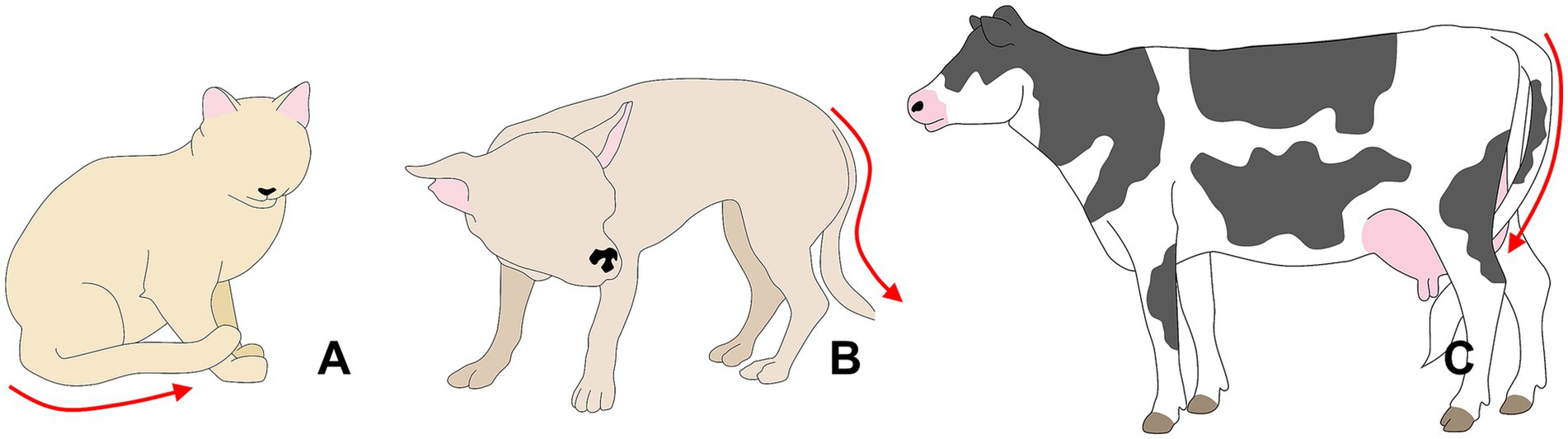
Figure 7. Tail posture as a pain sign. (A) In cats, a tail kept or tucked between the hindlimbs, close to the body, is a sign of pain. (B) Dogs experiencing pain might exhibit a tucked tail, similar to what is observed in cows (C). In the case of dogs, a tucked tail might also indicate fear. Thus, posture changes need to be interpreted together with other ethological evaluations.
Miller et al. (164) evaluated tail position in piglets after surgical castration with or without administration of local analgesics. It was found that piglets castrated without local analgesics had a higher frequency of changes in tail position, tail wagging, and maintained a straight (i.e., not curled) tail; in contrast, the non-castrated piglets kept their tail curled and hanging. Similarly, after cautery iron tail docking, piglets maintained an immobile tail in a horizontal position for longer (up to 20 s) than sham-docked piglets (approximately 16 s) (145).
In cattle, vigorous tail swinging vertically or horizontally is suggested as a key indicator for pain recognition; however, a static position or complete immobility of the tail is also associated with pain. In this regard, Tom et al. (165) assessed pain indicators in adult cows undergoing caudectomy with a rubber ring. Cows subjected to this procedure, regardless of analgesic treatment, reduced the frequency of tail shaking up to 6 days after tail-docking (0.8 ± 0.2), in addition to maintaining a straight, ventral position, which results in pressure against the hindquarters (between the anus and vulva) (24 animals). The authors suggested that pressing the tail towards the hindquarters counteracts the painful stimulation caused by the rubber ring and might reduce pain and inflammation (154, 156). Therefore, maintaining the tail static reduces the perception of pain in sensitized tissue.
The importance of tail movements is the reason why production units have focused on the tail to develop sophisticated technologies such as birth sensors (166). These sensors collect the number of times the tail is raised before calving, as this change in body posture is considered an imminent sign of the onset of calving due to the pain promoted by uterine contractions (167). The same posture has been observed in species such as the pig (168) and mice (169). However, tail movements in the peripartum might not always be entirely due to pain.
Therefore, tail position is a key indicator to recognize acute pain, providing valuable information during clinical assessment. However, variability in position and activity is wide across species and animal conditions or affective states. Like ear position, it should be considered an event that manifests together with multiple pain-related signs. Thus, an integrated, multimodal assessment incorporating multiple behavioral and physiological indicators is recommended to increase diagnostic sensitivity and efficacy.
4 Assessment of pain through postural changes
One of the most significant changes in animals experiencing pain is postural alterations to minimize pain perception (30, 170). Back arching, lateral or ventral tilt of the torso, and contraction of the abdominal muscles can indicate the presence of pain (Figure 8). Postural changes are triggered by modifications in the length of muscles, soft tissues, and the musculoskeletal system, influencing spinal alignment to reduce energy expenditure and change body weight distribution to facilitate balance (171, 172). Interception within the musculoskeletal system is carried out by sensory nerves located within the periosteum, spinal cord, and cortical bone (173, 174).
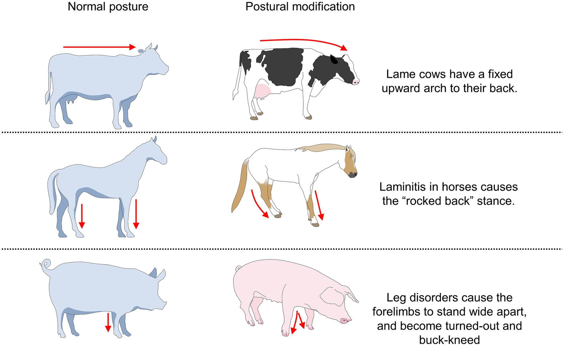
Figure 8. Some examples of body postures associated with pain in domestic animals.
The expression of pain through changes in the position of the trunk or back could be explained by the fact that nerve impulses are projected from peripheral endings processing pain, to the dorsal horn of the spinal cord to the higher structures of the Central Nervous System, specifically the somatosensory cortex (20, 175). Activation of the somatosensory cortex excites relevant regions such as the primary motor cortex, which is involved in the formation of motor actions, such as the withdrawal reflex or postural changes in response to tissue injury (40, 176, 177).
An example of change in posture is during parturition, as observed by Ison et al. (178) periparturient sows show a distinctive posture of back arching, accompanied by hindlimbs pointing forward due to uterine contractions and the expulsion of the piglets. Furthermore, the complete lateral tilt of the torso (animal lying down) was observed for 90% of the time between the onset of uterine contractions and 6 h after the expulsion of the first piglet. The authors state that these behavioral adjustments are indicators of pain and not simply assistive postures in the expulsion of the fetus through the birth canal. This was also observed in periparturient rats by Catheline et al. (169), where the administration of oxytocin, which is a potent intensifier of uterine contractions, increased torso stretching accompanied by abdominal tension (<6 times/min) during parturition. The visceral pain experienced during natural parturition is promoted by several mechanical factors such as uterine contractions, distension, elongation, and tearing of tissue, and pressure applied to adjacent anatomical structures (pelvis and perineum) (179, 180).
The adoption of these postures has been observed in other disorders where visceral pain is intense. For example, in horses with colic syndrome, abdominal pain comes mainly from visceral smooth muscles, which, when undergoing sudden changes such as stretching, tearing, perforation, or strangulation, exceed their nociceptive threshold (181). Fereig (182) associated excessive abdominal stretching with cranial and caudal extension of the fore and hindlimbs, respectively, to relieve mesenteric pressure caused by the accumulation of gas and fluid in the gastrointestinal tract. Laleye et al. (183) mention that early detection of abdominal pain in foals is based primarily on the identification of postural changes or behavioral modifications, among which abnormal body posture of complete lateral tilt and abdominal contraction were frequently reported by owners (47%) and veterinarians (78%). Figure 9 shows an example of postural changes in donkeys suffering from laminitis.
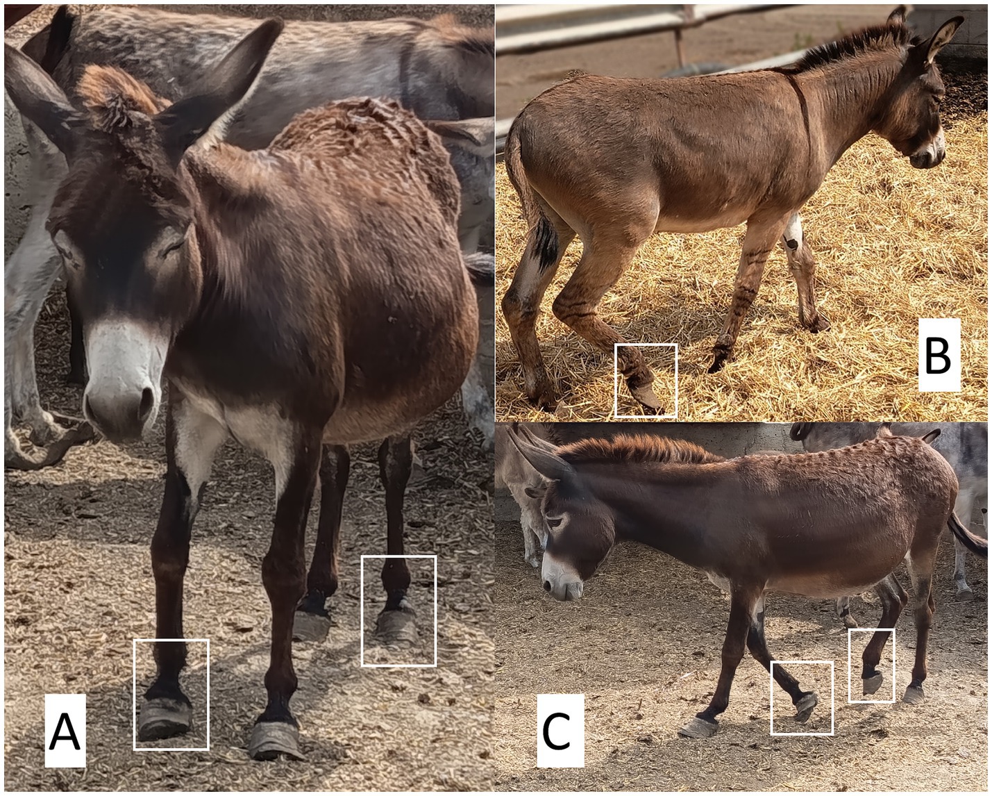
Figure 9. Postural alterations linked to nociception in the metacarpal and metatarsal regions, with clinical signs of laminitis in donkeys. (A) The animal displays a posterior shift of body weight, with the forelimbs slightly extended cranially and overextension of the right metacarpal and left metatarsal regions. This abnormal posture results from excessive hoof wall overgrowth. Such a stance is typical of animals experiencing hoof or joint pain, as they attempt to unload the affected areas. A tense facial expression indicative of discomfort is also evident. (B) Marked overgrowth of the hoof wall is observed in the right pelvic limb, with clear deformation of the hoof’s natural conformation. This alteration predisposes the animal to chronic pain due to abnormal pressure distribution, joint inflammation, and increased tendinous load. Lack of routine trimming compromises equine biomechanics, significantly altering weight distribution (C) The donkey adopts a non-weight-bearing stance, with overextension of the left thoracic limb and complete withdrawal of the right pelvic limb. This postural pattern is consistent with chronic pain, likely associated with laminitis or long-standing podal discomfort. Photos taken by the authors.
This can also be observed in domestic dogs with the so-called antalgic “prayer posture,” where animals stretch cranially the forelimbs and maintain a convex curvature of the back. By extending the thoracic region, this posture releases the abdominal pressure by the declination of the organs toward the cranial region. This posture is frequently observed when the origin of the pain is visceral and is used as an indicator of postsurgical pain (155). Other postures related to pain in dogs are rigid, hunched or tense, or guarding the affected area (45). Figure 10 shows frequently observed pain-related postures in companion animals due to visceral pain, kidney disease, and spinal and thoracic injury (25, 155, 184–186).
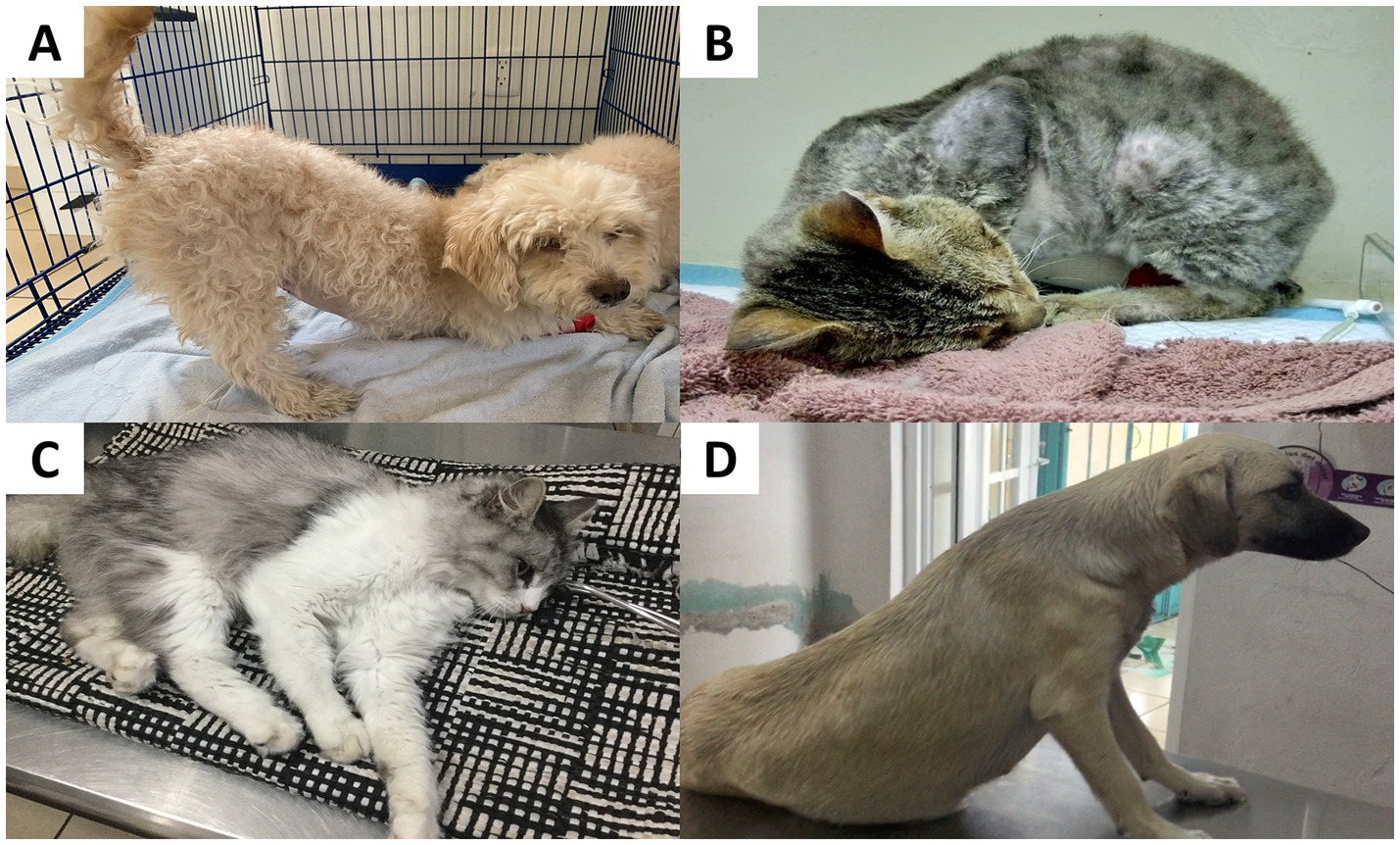
Figure 10. Pain-related postural changes in companion animals. (A) Prayer posture in a 2-year-old mixed-breed dog, showing extended pelvic limbs and a lowered head, allowing the pelvis to be raised towards the back. This relieves abdominal pressure when perceiving severe visceral pain. (B) Severe acute abdominal pain in a cat. A 6-year-old male cat with severe acute abdominal pain due to chronic kidney disease. The patient maintains a posture with the pelvic and thoracic limbs flexed towards the belly and a lowered head. (C) A male cat with the Schiff-Sherrington posture, characterized by extended thoracic limbs and an arched spine (kyphosis), and associated with a spinal injury. (D) A 4-year-old dog with a thoracic injury. The dog has an extended neck, with elbows abducted laterally and flexed pelvic limbs. This posture is known as orthopneic stance and occurs in cases of thoracic pain. It is important not to confuse the prayer posture position with the play bow. Play bows occur when a dog is inviting play, whereas the prayer posture is typically associated with discomfort or pain. The dog’s head is usually down when it performs the prayer posture and it is usually up during a play bow. A dog in a playful state is active and energetic, whereas a dog in pain tends to show reduced activity. Photos taken by the authors.
Pain-related postures are not exclusively manifested during visceral pain. For example, mouse models suffering from sickle cell disease adopt an arched back posture in response to a decrease in ambient temperature, a back posture that has also been correlated with other behavioral indicators of pain, such as facial expressions (143). Following abdominal surgical procedures such as OVH in companion animals, marked abdominal contraction is observed, along with exacerbated kyphosis (187), consistent with observations in surgically castrated piglets (164). In particular, the kyphosis manifested during pain in cats caused by musculoskeletal diseases progressively decreases after the administration of analgesic treatment, which suggests that the presentation of antalgic postures is associated with the intensity of pain (65).
Similarly, abnormal postures occur following routine handling procedures in farm animals. Castration in piglets causes back arching and abdominal tension (164). In small ruminants, Zebaria et al. (188) reported an increase in abnormal standing (standing unsteadily with tail wagging, 6.83%) in kid goats undergoing ear tagging. This is similar to what Fonseca et al. (98) reported in goats subjected to orchiectomy, where the occurrence of an unstable posture increased after the surgery (33.5%). In dairy cows with hoof trauma, Flower and Weary (189) reported marked dorsal arching in animals with plantar hemorrhages and ulcers. These changes were also accompanied by sudden head movements, decreased mobility, and reduced balance in a static state. In another study by Stojkov et al. (190) in cows, dorsal arching was associated with pain caused by inflammation of the uterine wall (metritis), while Rialland et al. (191) associated back arching with gastric problems such as traumatic reticulopericarditis. Figure 11 shows the pain-related postural changes observed in cattle and other species due to mastitis and fractures (192, 193).

Figure 11. Postural changes in cattle and other species. (A) Postural changes in a cow with clinical mastitis. A Holstein dairy cow maintains a low head posture with abducted or clubbed pelvic limbs. This posture helps to reduce contact of the limbs with the udder and diminishes local pain. (B) Posture of a rabbit with a fracture in the pelvic limb. A prostration posture and laterally lying on the affected limb apply pressure to the limb to reduce the pain. Photos taken by the authors.
Back arching is often accompanied by other body adjustments in the pelvic and thoracic limbs, tail position, neck tension, and head position (30). For example, after surgical castration of bulls, Esteves-Trindade et al. (194) found that the main changes associated with pain were extension of the head and neck, position of the head below the animal’s shoulders, and extended limbs. Recognizing these changes is important for veterinarians and also owners, as reported by Demirtas et al. (48), who evaluated the ability of dog owners to recognize postural changes. These authors observed that, limited joint movement of the caudal vertebrae, arching of the back, and reduced overall activity was present were the most frequently recognized postural changes. Similarly, Laleye et al. (183) evaluated early recognition of colic pain through 66 clinical histories of 40 horses (over 5 years old) and 26 foals (under 4 weeks old). The results indicated that more than 50% of physicians and caregivers use postural modifications as an early sign for colic pain recognition.
5 The importance of animals’ nonverbal language as a clinical indicator of pain for veterinarians and animal scientists
Pain recognition and assessment are essential to promote animals’ health and welfare (8, 195, 196). Failure to recognize pain in animals represents a welfare problem due to the physical and mental alterations, including activation of the sympathetic nervous system, immunosuppression, metabolism, and healing processes, as well as increased morbidity, disease progression, and prolonged recovery periods in surgical patients (2, 197). In human medicine, pain assessment is performed through verbal or written communication with the patient (198). In contrast, in veterinary medicine, pain is identified through nonverbal communication, such as changes in physiological and endocrine parameters, body language, and behavior (56).
Pain assessment and management require the veterinarian’s knowledge and objectivity. Therefore, physicians need to incorporate behavioral and postural indicators associated with pain into their daily practice (8, 30). The changes observed in animals are an integrated response aimed at reducing the painful stimulus (199). Moreover, owners need knowledge and awareness of pain behaviors as they are key for the early recognition, assessment, and management of pain (48, 200–202). Therefore, veterinarians need to identify and familiarize themselves with animal behaviors to detect and categorize pain, although factors such as environment, species, age, body condition, and type of disease must be considered (16, 203). Although behavioral scales exist to assess pain, surveys indicate that 73% of veterinarians consider these methods inadequate and have difficulty recognizing behavioral changes (204, 205), which has a direct impact on patients’ quality of life and welfare (206).
Although the study of these behavioral and body posture indicators has been explored in several domestic species, the anatomical differences must be considered to accurately evaluate pain. These changes should not be considered in isolation but as a part of a complementary evaluation considering physiological parameters. Therefore, it would be appropriate to investigate whether including these changes in assessment scales improves the sensitivity of these tools, as has been observed with facial expression (207). Similarly, standardizing changes in ear/tail position and postures for each species and each pain-inducing event is necessary, which could help increase the specificity and sensitivity and obtain an objective pain assessment. The development of multidimensional scales that consider both physiological and behavioral/body posture/facial expression parameters could be the best option to comprehensively evaluate pain in domestic mammals. For example, the Colorado State University Canine and Feline Pain Scale or the University of Melbourne Pain Scale consider physiological, behavioral, and postural responses to acute pain (45, 208, 209).
Although behavioral, postural, and facial recognition of pain can be performed manually by clinicians or stockpeople, automated techniques have been explored for multiple domestic species to increase the accuracy of the evaluation and prevent subjectivity. For example, in companion animals, the Facial Action Coding System for cats (catFACS) was used as an anatomical basis for a machine learning model to recognize pain in cats undergoing ovariohysterectomy (210). The accuracy of the technique was above 72%, indicating its usefulness for automating pain detection. Similar accuracy was reported by Martvel et al. (211), who used artificial intelligence to detect pain in cats by establishing 48 facial landmarks in videos. The authors reported an accuracy of over 70% in recognizing feline acute postsurgical pain. Furthermore, AI and machine learning techniques can help to differentiate breeds and cephalic types in addition to pain (212). Breed-specific morphology highly influences pain recognition in companion animals (213). This is particularly relevant for domestic dogs, as breed-specific face anatomy makes it challenging to recognize pain through facial cues (214). However, Zhu et al. (215) have recently proposed the application of machine learning to automatically identify pain in dogs.
Similarly, the adoption of techniques known as “precision livestock farming” or instruments that use artificial intelligence techniques can help objectively and automatically recognize changes in ear or tail position in farm species. An example is Feighelstein et al. (216), who used deep learning to detect pain from lateral images of horses undergoing routine castration. Using the Horse Grimace Scale (HGS) to embed the Facial Action Units (previously described in Table 1), authors reported an accuracy between 73 and 79% to detect equine pain. This might improve animal management and welfare in routine procedures that are still considered “not as painful.” Similarly, Lencioni et al. (217) developed a machine vision algorithm to detect acute pain in horses after surgical castration. Through facial expression, the authors found an overall accuracy of 75.8% when classifying pain into three categories (not present, moderately present, and obviously present, according to the HGS), or 88.3% when representing absent/present pain. Recent studies have suggested that the use of “regions of interest” instead of “facial landmarks” when using automatic detection of pain could increase the feasibility of adopting artificial intelligence in animal pain detection (218). Moreover, although most of the research is focused on identifying facial changes associated with pain –in horses–, Kil et al. reported a sensitivity of above 80% to detect behavioral changes in horses using machine learning (e.g., analysis of wither, tail, and nose changes).
In ruminants, Salzer et al. (219) developed an automatic warning system to identify mild pain (capsaicin application) in cows. Through a machine-learning algorithm, the authors were able to identify that decreased rumination and restlessness are present in animals experiencing pain with an accuracy of 82%. Additionally, micro expressions have also been adapted to computer vision methods to detect painful conditions such as lameness, metritis, mastitis, and pre-calving pain with an average precision of 83%. In other species, such as goats, Chiavaccini et al. (220) detected acute pain (due to conditions such as castration, mastectomy, dental cleaning, among others) through the analysis of raw facial video footage with machine learning. “Painful” and “non-painful” goats were differentiated with an accuracy of 60%. When automatically analyzing facial expressions of pain in sheep, studies have reported that artificial intelligence outperforms human experts, which has significant applications in farms.
Deep learning-based models and artificial intelligence are still under development for several species (224). However, these methods reduce human bias and the need to manually extract information, which is time-consuming. Therefore, these methods are current alternatives to improve pain assessment in domestic mammals for improving animal welfare, while preserving the importance of training veterinarians and animal caregivers to correctly interpret animal behavior and body language.
6 Conclusion
Animal body language serves as a means of understanding the emotional state of animals in response to positive and negative stimuli, such as pain. In domestic animals, variations in behavioral responses such as vocalizations, grooming, scratching, avoidance, escape, tonic immobility, as well as aggression, among other behaviors, are associated with the perception of pain. Additionally, about farm animals, changes in ear and tail position and in the overall posture have been reported to be indicative of pain in animals suffering from pain arising from, e.g., laminitis, visceral involvement, or routine painful procedures. Understanding these signals as a nonverbal communication of pain allows the efficient identification of pain for timely intervention and optimized management.
Author contributions
DM-R: Conceptualization, Project administration, Supervision, Validation, Visualization, Writing – original draft, Writing – review & editing. AW: Writing – original draft, Writing – review & editing. LL: Writing – original draft, Writing – review & editing. CB-F: Writing – original draft, Writing – review & editing. AD-O: Conceptualization, Supervision, Writing – original draft, Writing – review & editing. AC-C: Writing – original draft, Writing – review & editing. VF: Writing – original draft, Writing – review & editing. IH-A: Writing – original draft, Writing – review & editing. AB: Writing – original draft, Writing – review & editing. EN: Writing – original draft, Writing – review & editing. AO-H: Writing – original draft, Writing – review & editing. AF: Writing – original draft, Writing – review & editing. PM-M: Writing – original draft, Writing – review & editing. JM-B: Supervision, Writing – original draft, Writing – review & editing. AC-A: Writing – original draft, Writing – review & editing. TG: Conceptualization, Supervision, Validation, Visualization, Writing – original draft, Writing – review & editing.
Funding
The author(s) declare that no financial support was received for the research and/or publication of this article.
Conflict of interest
The authors declare that the research was conducted in the absence of any commercial or financial relationships that could be construed as a potential conflict of interest.
The author(s) declared that they were an editorial board member of Frontiers, at the time of submission. This had no impact on the peer review process and the final decision.
Generative AI statement
The authors declare that no Gen AI was used in the creation of this manuscript.
Any alternative text (alt text) provided alongside figures in this article has been generated by Frontiers with the support of artificial intelligence and reasonable efforts have been made to ensure accuracy, including review by the authors wherever possible. If you identify any issues, please contact us.
Publisher’s note
All claims expressed in this article are solely those of the authors and do not necessarily represent those of their affiliated organizations, or those of the publisher, the editors and the reviewers. Any product that may be evaluated in this article, or claim that may be made by its manufacturer, is not guaranteed or endorsed by the publisher.
References
1. Reid, J, Scott, M, Nolan, A, and Wiseman-Orr, L. PAin assessment in animals (2013) 34:2–6. doi: 10.1136/inp
2. Monteiro, BP, Lascelles, BDX, Murrell, J, Robertson, S, Steagall, PVM, and Wright, B. 2022 WSAVA guidelines for the recognition, assessment and treatment of pain. J Small Anim Pract. (2023) 64:177–254. doi: 10.1111/JSAP.13566
3. Mota-Rojas, D, Whittaker, AL, Bienboire-Frosini, C, Buenhombre, J, Mora-Medina, P, Domínguez-Oliva, A, et al. The neurobiological basis of emotions and their connection to facial expressions in non-human mammals: insights in nonverbal communication. Front Vet Sci. (2025) 12:1541615. doi: 10.3389/fvets.2025.1541615
4. Walsh, EA, Meers, LL, Samuels, WE, Boonen, D, Claus, A, Duarte-Gan, C, et al. Human-dog communication: how body language and non-verbal cues are key to clarity in dog directed play, petting and hugging behaviour by humans. Appl Anim Behav Sci. (2024) 272:106206. doi: 10.1016/j.applanim.2024.106206
5. Bateson, P. Assessment of pain in animals. Anim Behav. (1991) 42:827–39. doi: 10.1016/S0003-3472(05)80127-7
6. Mathews, KA. Pain assessment and general approach to management. Vet Clin North Am Small Anim Pract. (2000) 30:729–55. doi: 10.1016/S0195-5616(08)70004-4
7. Neindre, P, Le, GR, Guemene, D, Guichet, J-L, Latouche, K, Leterrier, C, et al. Douleurs animales. Les identifier, les comprendre, les limiter chez les animaux d’élevage. (2009):101. doi: 10.15454/XN0H-XM19
8. Anil, SS, Anil, L, and Deen, J. Challenges of pain assessment in domestic animals. J Am Vet Med Assoc. (2002) 220:313–9. doi: 10.2460/javma.2002.220.313
9. Dalla Costa, E, Minero, M, Lebelt, D, Stucke, D, Canali, E, and Leach, MC. Development of the horse grimace scale (HGS) as a pain assessment tool in horses undergoing routine castration. PLoS One. (2014) 9:e92281. doi: 10.1371/journal.pone.0092281
10. Keefe, FJ, Fillingim, RB, and Williams, DA. Behavioral assessment of pain: nonverbal measures in animals and humans. ILAR J. (1991) 33:3–13. doi: 10.1093/ilar.33.1-2.3
11. Olivier, IS, and Olivier, A In: M Glover and L Mithcer, editors. Pain in context: Indicators and expressions of animal pain. Cham, Switzerland: Palgrave Macmillan (2024). 61–96.
12. Breed, MD, and Moore, J. Animal behavior. 3rd editio. San Diego, USA: Academic Press. (2022). 1–2 p.
13. Ponzio, A. Body language In: K Brown, editor. Encyclopedia of Language & Linguistics. Amsterdam, Netherlands: Elsevier (2006) 78–85.
14. Abdulghafor, R, Turaev, S, and Ali, MAH. Body language analysis in healthcare: an overview. Healthcare. (2022) 10:1251. doi: 10.3390/healthcare10071251
15. Muir, WW. Physiology and pathophysiology of pain In: MSL M, editor. Handbook of veterinary pain management. Elsevier, USA: Mosby. (2009). 13–49.
16. Andrighetto Canozzi, ME, Rossi Borges, JA, and Jardim Barcellos, JO. Attitudes of cattle veterinarians and animal scientists to pain and painful procedures in Brazil. Prev Vet Med. (2020) 177:104909. doi: 10.1016/J.PREVETMED.2020.104909
17. de Grauw, JJC, and van Loon, JP. Systematic pain assessment in horses. Vet J. (2016) 209:14–22. doi: 10.1016/j.tvjl.2015.07.030
18. Domínguez-Oliva, A, Mota-Rojas, D, Hernández-Avalos, I, Mora-Medina, P, Olmos-Hernández, A, Verduzco-Mendoza, A, et al. The neurobiology of pain and facial movements in rodents: clinical applications and current research. Front Vet Sci. (2022) 9:1016720. doi: 10.3389/fvets.2022.1016720
19. Sneddon, LU. Comparative physiology of nociception and pain. Physiology. (2018) 33:63–73. doi: 10.1152/physiol.00022.2017
20. Bell, A. The neurobiology of acute pain. Vet J. (2018) 237:55–62. doi: 10.1016/j.tvjl.2018.05.004
21. Ziegler, K, Folkard, R, Gonzalez, AJ, Burghardt, J, Antharvedi-Goda, S, Martin-Cortecero, J, et al. Primary somatosensory cortex bidirectionally modulates sensory gain and nociceptive behavior in a layer-specific manner. Nat Commun. (2023) 14:2999. doi: 10.1038/s41467-023-38798-7
23. Williams, AC d C. Persistence of pain in humans and other mammals. Philos Trans R Soc Lond Ser B Biol Sci. (2019) 374:20190276. doi: 10.1098/rstb.2019.0276
24. Saberi Afshar, F, Shekarian, M, Baniadam, A, Avizeh, R, Hossein, N, and Pourmehdi, M. Comparison of different tools for pain assessment following ovariohysterectomy in bitches. Iran. J Vet Med. (2017) 11:255–65. doi: 10.22059/ijvm.2017.138815.1004701
25. Hernandez-Avalos, I, Mota-Rojas, D, Mora-Medina, P, Martínez-Burnes, J, Casas Alvarado, A, Verduzco-Mendoza, A, et al. Review of different methods used for clinical recognition and assessment of pain in dogs and cats. Int J Vet Sci Med. (2019) 7:43–54. doi: 10.1080/23144599.2019.1680044
26. Wiese, A, and Yaks, T. Nociception and pain mechanisms In: JS Gaynor and WW Muir editors. Handbook of veterinary pain management. Louis, Missouri, USA: Mosby-Elsevier (2015). 10–41.
27. Marangoni, S, Beatty, J, and Steagall, PV. An ethogram of acute pain behaviors in cats based on expert consensus. PLoS One. (2023) 18:e0292224. doi: 10.1371/journal.pone.0292224
28. Goldberg, ME, and Shaffran, N. Oxford: John Wiley & Sons. (2014). 15–29 p. Pain management for veterinary technicians and nurses.
29. Epstein, ME, Rodan, I, Griffenhagen, G, Kadrlik, J, Petty, MC, Robertson, SA, et al. AAHA/AAFP Pain management guidelines for dogs and cats. J Feline Med Surg. (2015) 17:251–72. doi: 10.1177/1098612X15572062
30. Prunier, A, Mounier, L, Le Neindre, P, Leterrier, C, Mormède, P, Paulmier, V, et al. Identifying and monitoring pain in farm animals: a review. Animal. (2013) 7:998–1010. doi: 10.1017/S1751731112002406
31. Hugonnard, M, Leblond, A, Keroack, S, Cadoré, J, and Troncy, E. Attitudes and concerns of French veterinarians towards pain and analgesia in dogs and cats. Vet Anaesth Analg. (2004) 31:154–63. doi: 10.1111/j.1467-2987.2004.00175.x
32. Sneddon, LU, Elwood, RW, Adamo, SA, and Leach, MC. Defining and assessing animal pain. Anim Behav. (2014) 97:201–12. doi: 10.1016/j.anbehav.2014.09.007
33. Mota-Rojas, D, Orihuela, A, Strappini-Asteggiano, A, Nelly Cajiao-Pachón, M, Agüera-Buendía, E, Mora-Medina, P, et al. Teaching animal welfare in veterinary schools in Latin America. Int J Vet Sci Med. (2018) 6:131–40. doi: 10.1016/j.ijvsm.2018.07.003
34. Broomé, S, Feighelstein, M, Zamansky, A, Carreira Lencioni, G, Haubro Andersen, P, Pessanha, F, et al. Going deeper than tracking: a survey of computer-vision based recognition of animal pain and emotions. Int J Comput Vis. (2023) 131:572–90. doi: 10.1007/s11263-022-01716-3
35. National Research Council. Recognition and alleviation of pain in laboratory animals. Washington, USA: The National Academies Press. (2009) 198. doi: 10.17226/12526
36. Camps, T, Amat, M, Mariotti, VM, Le Brech, S, and Manteca, X. Pain-related aggression in dogs: 12 clinical cases. J Vet Behav. (2012) 7:99–102. doi: 10.1016/J.JVEB.2011.08.002
37. Bonagura, J. Kirk terapéutica veterinaria de pequeños animales. 13th ed. Madrid, España: McGraw Hill/Interamericana. (2001). 91–97 p.
38. Wiseman-Orr, ML, Scott, M, Reid, J, and Nolan, AM. Validation of a structured questionnaire as an instrument to measure chronic pain in dogs on the basis of effects on health-related quality of life. Am J Vet Res. (2006) 67:1826–36. doi: 10.2460/AJVR.67.11.1826
39. Coria-Avila, GA, Herrera-Covarrubias, D, García, LI, Toledo, R, Hernández, ME, Paredes-Ramos, P, et al. Neurobiology of maternal behavior in nonhuman mammals: acceptance, recognition, motivation, and rejection. Animals. (2022) 12:3589. doi: 10.3390/ani12243589
40. Coria-Avila, GA, Pfaus, JG, Orihuela, A, Domínguez-Oliva, A, José-Pérez, N, Hernández, LA, et al. The neurobiology of behavior and its applicability for animal welfare: a review. Animals. (2022) 12:928. doi: 10.3390/ani12070928
41. Hellyer, P, Rodan, I, Brunt, J, Downing, R, Hagedorn, JE, and Robertson, SA. AAHA/AAFP pain management guidelines for dogs & cats. J Am Anim Hosp Assoc. (2007) 43:235–48. doi: 10.5326/0430235
42. Gaynor, JS, and Muir, WW. Handbook of veterinary pain management. 3rd edition. St. Louis, Missouri, USA: Elsevier mosby. (2014) 1–620. doi: 10.1016/C2010-0-67083-0
43. Essner, A, Zetterberg, L, Hellström, K, Gustås, P, Högberg, H, and Sjöström, R. Psychometric evaluation of the canine brief pain inventory in a Swedish sample of dogs with pain related to osteoarthritis. Acta Vet Scand. (2017) 59:44. doi: 10.1186/s13028-017-0311-2
44. Reid, J, Nolan, A, Hughes, J, Lascelles, D, Pawson, P, and Scott, E. Development of the short-form Glasgow composite measure pain scale (CMPS-SF) and derivation of an analgesic intervention score. Anim Welf. (2007) 16:97–104. doi: 10.1017/S096272860003178X
45. Firth, AM, and Haldane, SL. Development of a scale to evaluate postoperative pain in dogs. J Am Vet Med Assoc. (1999) 214:651–9. doi: 10.2460/javma.1999.214.05.651
46. Wagner, AE, Worland, GA, Glawe, JC, and Hellyer, PW. Multicenter, randomized controlled trial of pain-related behaviors following routine neutering in dogs. J Am Vet Med Assoc. (2008) 233:109–15. doi: 10.2460/javma.233.1.109
47. Mills, DS, Demontigny-Bédard, I, Gruen, M, Klinck, MP, McPeake, KJ, Barcelos, AM, et al. Pain and problem behavior in cats and dogs. Animals. (2020) 10:318. doi: 10.3390/ani10020318
48. Demirtas, A, Atilgan, D, Saral, B, Isparta, S, Ozturk, H, Ozvardar, T, et al. Dog owners’ recognition of pain-related behavioral changes in their dogs. J Vet Behav. (2023) 62:39–46. doi: 10.1016/j.jveb.2023.02.006
49. Zanusso, F, Contiero, B, Normando, S, Gottardo, F, and De Benedictis, GM. Qualitative behavioral assessment of dogs with acute pain. PLoS One. (2024) 19:e0305925. doi: 10.1371/journal.pone.0305925
50. Stevens, C, Kawecki-Wright, E, de Ortiz, AR, Thomson, A, Aker, S, Perry, E, et al. Factors influencing, and associated with, physical activity patterns in dogs with osteoarthritis-associated pain. Front Vet Sci. (2025) 12:1503009. doi: 10.3389/fvets.2025.1503009
51. Dodd, T, Jones, J, Holásková, I, and Mukherjee, M. Behavioral problems may be associated with multilevel lumbosacral stenosis in military working dogs. J Vet Behav. (2020) 35:8–13. doi: 10.1016/j.jveb.2019.07.010
52. Mansfield, C. Pancreatitis in the dog In: DS Bruyette, N Bexfield, S Kube, MA Oyama, LV Reiter, and C Ruaux, editors. Clinical Small Animal Internal Medicine. Hoboken, USA: Wiley Blackwell (2020). 591–600.
53. Lim, SY, Cridge, H, Twedt, DC, Ohta, H, Nuruki, T, and Steiner, JM. Management of acute-onset pancreatitis in dogs: a narrative review. J Am Vet Med Assoc. (2024) 262:1231–40. doi: 10.2460/javma.24.02.0107
54. Ignatenko, N, Abramenko, I, Soto, S, Mueller, R, Boehm, TMSA, Troedson, K, et al. Nasal transmissible venereal tumours in 12 dogs – a retrospective study. Tierärztliche Praxis Ausgabe K: Kleintiere / Heimtiere. (2020) 48:164–70. doi: 10.1055/a-1152-3772
55. Nepomuceno de Oliveira, M, de Oliveira, SL, Nóbrega Barbosa, J, da Silva, M, Santos, J, Noronha de Toledo, G, et al. Clinical and therapeutic aspects of dogs with transmissible venereal tumor in nasal cavity: report of two cases. Acta Vet Bras. (2024) 18:11–5. doi: 10.21708/avb.2024.18.1.11921
56. Holton, L, Pawson, P, Nolan, A, Reid, J, and Scott, EM. Development of a behaviour-based scale to measure acute pain in dogs. Vet Rec. (2001) 148:525–31. doi: 10.1136/vr.148.17.525
57. Bécuwe-Bonnet, V, Bélanger, M-C, Frank, D, Parent, J, and Hélie, P. Gastrointestinal disorders in dogs with excessive licking of surfaces. J Vet Behav. (2012) 7:194–204. doi: 10.1016/j.jveb.2011.07.003
58. Tynes, VV, and Sinn, L. Abnormal repetitive behaviors in dogs and cats. A guide for practitioners. Vet Clin North Am - Small Anim Pract. (2014) 44:543–64. doi: 10.1016/j.cvsm.2014.01.011
59. Brondani, JT, Luna, SPL, and Padovani, CR. Refinement and initial validation of a multidimensional composite scale for use in assessing acute postoperative pain in cats. Am J Vet Res. (2011) 72:174–83. doi: 10.2460/ajvr.72.2.174
60. Monteiro, BP, and Steagall, PV. Chronic pain in cats: recent advances in clinical assessment. J Feline Med Surg. (2019) 21:601–14. doi: 10.1177/1098612X19856179
61. Steagall, PV, and Monteiro, BP. Acute pain in cats: recent advances in clinical assessment. J Feline Med Surg. (2019) 21:25–34. doi: 10.1177/1098612X18808103
62. Brondani, JT, Mama, KR, Luna, SPL, Wright, BD, Niyom, S, Ambrosio, J, et al. Validation of the English version of the UNESP-Botucatu multidimensional composite pain scale for assessing postoperative pain in cats. BMC Vet Res. (2013) 9:143. doi: 10.1186/1746-6148-9-143
63. Reid, J, Nolan, AM, and Scott, EM. Measuring pain in dogs and cats using structured behavioural observation. Vet J. (2018) 236:72–9. doi: 10.1016/j.tvjl.2018.04.013
64. Marangoni, S, Frank, D, and Steagall, PV. Characterization of pain behaviors in kittens following ovariohysterectomy using video assessment. Sci Rep. (2025) 15:14886. doi: 10.1038/s41598-025-96453-1
65. Bennett, D, and Morton, C. A study of owner observed behavioural and lifestyle changes in cats with musculoskeletal disease before and after analgesic therapy. J Feline Med Surg. (2009) 11:997–1004. doi: 10.1016/J.JFMS.2009.09.016
66. Kogan, LR, Currin-McCulloch, J, Brown, E, and Hellyer, P. Dog owners’ perceptions and veterinary-related decisions pertaining to changes in their dog’s behavior that could indicate pain. J Am Vet Med Assoc. (2024) 262:1370–8. doi: 10.2460/javma.24.02.0120
67. Kogan, LR, Currin-McCulloch, J, and Hellyer, P. Behaviors suggestive of pain: the perceptions and veterinary-related decisions of cat guardians in the USA. J Feline Med Surg. (2024) 26:1098612X241272885. doi: 10.1177/1098612X241272885
68. Steagall, PV, Bustamante, H, Johnson, CB, and Turner, PV. Pain management in farm animals: focus on cattle, sheep and pigs. Animals. (2021) 11:1483. doi: 10.3390/ani11061483
69. Mertens, A. Agresividad canina In: Manual de comportamiento en pequeños animales. España: Ediciones S (2012). 313–46.
70. Lezama-García, K, Orihuela, A, Olmos-Hernández, A, Reyes-Long, S, and Mota-Rojas, D. Facial expressions and emotions in domestic animals. CAB Rev Perspect Agric Vet Sci Nutr Nat Resour. (2019) 14:1–12. doi: 10.1079/PAVSNNR201914028
71. Kustritz, MVR. Reproductive behavior of small animals. Theriogenology. (2005) 64:734–46. doi: 10.1016/j.theriogenology.2005.05.022
72. Mainau, E, Llonch, P, Temple, D, Goby, L, and Manteca, X. Alteration in activity patterns of cows as a result of pain due to health conditions. Animals. (2022) 12:176. doi: 10.3390/ani12020176
73. Peters, MDP, Silveira, IDB, and Fischer, V. Impact of subclinical and clinical mastitis on sensitivity to pain of dairy cows. Animal. (2015) 9:2024–8. doi: 10.1017/S1751731115001391
74. Medrano-Galarza, C, Gibbons, J, Wagner, S, de Passillé, AM, and Rushen, J. Behavioral changes in dairy cows with mastitis. J Dairy Sci. (2012) 95:6994–7002. doi: 10.3168/jds.2011-5247
75. Fogsgaard, KK, Bennedsgaard, TW, and Herskin, MS. Behavioral changes in freestall-housed dairy cows with naturally occurring clinical mastitis. J Dairy Sci. (2015) 98:1730–8. doi: 10.3168/jds.2014-8347
76. Siivonen, J, Taponen, S, Hovinen, M, Pastell, M, Lensink, BJ, Pyörälä, S, et al. Impact of acute clinical mastitis on cow behaviour. Appl Anim Behav Sci. (2011) 132:101–6. doi: 10.1016/j.applanim.2011.04.005
77. Rajala-Schultz, PJ, Gott, PN, Proudfoot, KL, and Schuenemann, GM. Effect of milk cessation method at dry-off on behavioral activity of dairy cows. J Dairy Sci. (2018) 101:3261–70. doi: 10.3168/jds.2017-13588
78. Maynou, G, Elcoso, G, Bubeck, J, and Bach, A. Effects of oral administration of acidogenic boluses at dry-off on performance and behavior of dairy cattle. J Dairy Sci. (2018) 101:11342–53. doi: 10.3168/jds.2018-15058
79. Tucker, CB, Jensen, MB, de Passillé, AM, Hänninen, L, and Rushen, J. Invited review: lying time and the welfare of dairy cows. J Dairy Sci. (2021) 104:20–46. doi: 10.3168/jds.2019-18074
80. Rial, C, Laplacette, A, Caixeta, L, Florentino, C, Peña-Mosca, F, and Giordano, JO. Metritis and clinical mastitis events in lactating dairy cows were associated with altered patterns of rumination, physical activity, and lying behavior monitored by an ear-attached sensor. J Dairy Sci. (2023) 106:9345–65. doi: 10.3168/jds.2022-23157
81. Schreiner, DA, and Ruegg, PL. Responses to tail docking in calves and heifers. J Dairy Sci. (2002) 85:3287–96. doi: 10.3168/jds.S0022-0302(02)74417-2
82. Kroll, LK, Grooms, DL, Siegford, JM, Schweihofer, JP, Daigle, CL, Metz, K, et al. Effects of tail docking on behavior of confined feedlot cattle1. J Anim Sci. (2014) 92:4701–10. doi: 10.2527/jas.2014-7583
83. Eicher, SD, Morrow-Tesch, JL, Albright, JL, Dailey, JW, Young, CR, and Stanker, LH. Tail-docking influences on behavioral, immunological, and endocrine responses in dairy heifers. J Dairy Sci. (2000) 83:1456–62. doi: 10.3168/jds.S0022-0302(00)75017-X
84. Sutherland, MA, and Tucker, CB. The long and short of it: a review of tail docking in farm animals. Appl Anim Behav Sci. (2011) 135:179–91. doi: 10.1016/j.applanim.2011.10.015
85. Grant, C. Behavioural responses of lambs to common painful husbandry procedures. Appl Anim Behav Sci. (2004) 87:255–73. doi: 10.1016/j.applanim.2004.01.011
86. Grandin, T. The feasibility of using vocalization scoring as an indicator of poor welfare during cattle slaughter. Appl Anim Behav Sci. (1998) 56:121–8. doi: 10.1016/S0168-1591(97)00102-0
87. Watts, JM, and Stookey, JM. Vocal behaviour in cattle: the animal’s commentary on its biological processes and welfare. Appl Anim Behav Sci. (2000) 67:15–33. doi: 10.1016/S0168-1591(99)00108-2
88. Schnaider, MA, Heidemann, MS, Silva, AHP, Taconeli, CA, and Molento, CFM. Vocalization and other behaviors indicating pain in beef calves during the ear tagging procedure. J Vet Behav. (2022) 47:93–8. doi: 10.1016/J.JVEB.2021.10.005
89. Da S, CAF, Nääs, DA I, Dos S, BM, Jacob, FG, and De, MDJ. The use of vocalization signals to estimate the level of pain in piglets. Eng Agrícola. (2018) 38:486–90. doi: 10.1590/1809-4430-eng.agric.v38n4p486-490/2018
90. Marchant-Forde, JN, Lay, DC, McMunn, KA, Cheng, HW, Pajor, EA, and Marchant-Forde, RM. Postnatal piglet husbandry practices and well-being: the effects of alternative techniques delivered in combination12. J Anim Sci. (2014) 92:1150–60. doi: 10.2527/jas.2013-6929
91. Hansson, M, Lundeheim, N, Nyman, G, and Johansson, G. Effect of local anaesthesia and/or analgesia on pain responses induced by piglet castration. Acta Vet Scand. (2011) 53:34. doi: 10.1186/1751-0147-53-34
92. Molony, V, Kent, JE, and Robertson, IS. Assessment of acute and chronic pain after different methods of castration of calves. Appl Anim Behav Sci. (1995) 46:33–48. doi: 10.1016/0168-1591(95)00635-4
93. Melches, S, Mellema, SC, Doherr, MG, Wechsler, B, and Steiner, A. Castration of lambs: a welfare comparison of different castration techniques in lambs over 10 weeks of age. Vet J. (2007) 173:554–63. doi: 10.1016/j.tvjl.2006.01.006
94. Yun, J, Ollila, A, Valros, A, Larenza-Menzies, P, Heinonen, M, Oliviero, C, et al. Behavioural alterations in piglets after surgical castration: effects of analgesia and anaesthesia. Res Vet Sci. (2019) 125:36–42. doi: 10.1016/j.rvsc.2019.05.009
95. Lester, S, Mellor, D, Holmes, R, Ward, RN, and Stafford, KJ. Behavioural and cortisol responses of lambs to castration and tailing using different methods. N Z Vet J. (1996) 44:45–54. doi: 10.1080/00480169.1996.35933
96. Masłowska, K, Mizzoni, F, Dwyer, CM, and Wemelsfelder, F. Qualitative behavioural assessment of pain in castrated lambs. Appl Anim Behav Sci. (2020) 233:105143. doi: 10.1016/j.applanim.2020.105143
97. Canozzi, MEA, Mederos, A, Manteca, X, Turner, S, McManus, C, Zago, D, et al. A meta-analysis of cortisol concentration, vocalization, and average daily gain associated with castration in beef cattle. Res Vet Sci. (2017) 114:430–43. doi: 10.1016/j.rvsc.2017.07.014
98. Fonseca, MW, Trindade, PHE, Pinho, RH, Justo, AA, and Tomacheuski, RMSilva, NE de OF da, et al. Development and validation of the UNESP-Botucatu goat acute pain scale. Animals. (2023) 13:2136. doi: 10.3390/ani13132136
99. Hempstead, MN, and Plummer, PJ. Behaviors associated with pain in goats In: G Kannan, editor. Small ruminant welfare, production and sustainability. London, UK: Academic Press (2025). 215–28.
100. Kongara, K, Singh, P, Venkatachalam, D, and Chambers, JP. Pain assessment in goat kids: focus on disbudding. Animals. (2023) 13:3814. doi: 10.3390/ani13243814
101. Hempstead, MN, Waas, JR, Stewart, M, Cave, VM, and Sutherland, MA. Behavioural response of dairy goat kids to cautery disbudding. Appl Anim Behav Sci. (2017) 194:42–7. doi: 10.1016/j.applanim.2017.04.001
102. Alvarez, L, De Luna, JB, Gamboa, D, Reyes, M, Sánchez, A, Terrazas, A, et al. Cortisol and pain-related behavior in disbudded goat kids with and without cornual nerve block. Physiol Behav. (2015) 138:58–61. doi: 10.1016/j.physbeh.2014.10.026
103. Cui, W, Liu, M, Gu, T, Zhao, S, and Yin, G. Multi-dimensional evaluation of pain response in low day-age calves to two types of dehorning. Front Vet Sci. (2024) 11:1406576. doi: 10.3389/fvets.2024.1406576
104. Faulkner, P, and Weary, D. Reducing pain after dehorning in dairy calves. J Dairy Sci. (2000) 83:2037–41. doi: 10.3168/jds.S0022-0302(00)75084-3
105. Graf, B, and Senn, M. Behavioural and physiological responses of calves to dehorning by heat cauterization with or without local anaesthesia. Appl Anim Behav Sci. (1999) 62:153–71. doi: 10.1016/S0168-1591(98)00218-4
106. Mota-Rojas, D, Martínez-Burnes, J, Napolitano, F, Domínguez-Muñoz, M, Guerrero-Legarreta, I, Mora-Medina, P, et al. Dystocia: factors affecting parturition in domestic animals. CABI Rev. (2020) 2020:1–16. doi: 10.1079/PAVSNNR202015013
107. Mota-Rojas, D, Lezama-García, K, Domínguez-Oliva, A, Olmos-Hernández, A, Verduzco-Mendoza, A, Casas-Alvarado, A, et al. Neurobiology of emotions in animal relationships: facial expressions and their biological functions in mammals. J Anim Behav Biometeorol. (2023) 11:e2023ss01. doi: 10.31893/jabb.23ss01
108. Cohen, S, and Beths, T. Grimace scores: tools to support the identification of pain in mammals used in research. Animals. (2020) 10:1726. doi: 10.3390/ani10101726
109. Lambert, H, and Carder, G. Positive and negative emotions in dairy cows: Can ear postures be used as a measure? Behav Process. (2019) 158:172–80. doi: 10.1016/j.beproc.2018.12.007
110. Morecraft, RJ, Louie, JL, Herrick, JL, and Stilwell-Morecraft, KS. Cortical innervation of the facial nucleus in the non-human primate: a new interpretation of the effects of stroke and related subtotal brain trauma on the muscles of facial expression. Brain. (2001) 124:176–208. doi: 10.1093/brain/124.1.176
111. Erickson, K, and Schulkin, J. Facial expressions of emotion: a cognitive neuroscience perspective. Brain Cogn. (2003) 52:52–60. doi: 10.1016/S0278-2626(03)00008-3
112. Morecraft, RJ, Stilwell-Morecraft, KS, and Rossing, WR. The motor cortex and facial expression: new insights from neuroscience. Neurologist. (2004) 10:235–49. doi: 10.1097/01.NRL.0000138734.45742.8D
113. Gothard, KM. The amygdalo-motor pathways and the control of facial expressions. Front Neurosci. (2014) 8:43. doi: 10.3389/fnins.2014.00043
114. Domínguez-Oliva, A, Olmos-Hernández, A, Hernández-Ávalos, I, Lecona-Butrón, H, Mora-Medina, P, and Mota-Rojas, D. Rat grimace scale as a method to evaluate animal welfare, nociception, and quality of the euthanasia method of Wistar rats. Animals. (2023) 13:3161. doi: 10.3390/ani13203161
115. International committee on veterinary gross anatomical nomenclature. Nomina Anatomica Veterinaria. Hanover, Germany: World Association of Veterinary Anatomist (2017) 1–178.
116. Evangelista, MC, Watanabe, R, Leung, VSY, Monteiro, BP, O’Toole, E, Pang, DSJ, et al. Facial expressions of pain in cats: the development and validation of a feline grimace scale. Sci Rep. (2019) 9:19128. doi: 10.1038/s41598-019-55693-8
117. Whittaker, AL, Liu, Y, and Barker, TH. Methods used and application of the mouse grimace scale in biomedical research 10 years on: a scoping review. Animals. (2021) 11:673. doi: 10.3390/ani11030673
118. Sotocinal, SG, Sorge, RE, Zaloum, A, Tuttle, AH, Martin, LJ, Wieskopf, JS, et al. The rat grimace dcale: a partially automated method for quantifying pain in the laboratory rat via facial expressions. Mol Pain. (2011) 7:55. doi: 10.1186/1744-8069-7-55
119. Shaw, D, Parkes, J, and Reynolds, H. Assessing pain in rabbits: how well does the rabbit grimace scale work in the veterinary practice? Vet Nurse. (2020) 11:282–7. doi: 10.12968/vetn.2020.11.6.282
120. Navarro, E, Mainau, E, and Manteca, X. Development of a facial expression scale using farrowing as a model of pain in sows. Animals. (2020) 10:2113. doi: 10.3390/ani10112113
121. Vullo, C, Barbieri, S, Catone, G, Graïc, J-M, Magaletti, M, Di Rosa, A, et al. Is the piglet grimace scale (PGS) a useful welfare indicator to assess pain after cryptorchidectomy in growing pigs? Animals. (2020) 10:412. doi: 10.3390/ani10030412
122. Zentrich, E, Wassermann, L, Struve, B, Selke, K, Buettner, M, Keubler, LM, et al. Postoperative severity assessment in sheep. Eur Surg Res. (2023) 64:27–36. doi: 10.1159/000526058
123. Guesgen, M, Beausoleil, N, Leach, M, Minot, EO, Stewart, M, and Stafford, KJ. Coding and quantification of a facial expression for pain in lambs. Behav Process. (2016) 132:49–56. doi: 10.1016/j.beproc.2016.09.010
124. Weeder, MM, Kleinhenz, MD, Reppert, EJ, Fritz, BR, Viscardi, AV, Montgomery, SR, et al. Optimal lameness induction model development using amphotericin B in meat goats. Transl Anim Sci. (2023) 7:txad105. doi: 10.1093/tas/txad105
125. Orth, EK, Navas González, FJ, Iglesias Pastrana, C, Berger, JM, Jeune, SSle, Davis, EW, et al., Development of a donkey grimace scale to recognize pain in donkeys (Equus asinus) post castration Animals (2020) 10:1411 doi: 10.3390/ani10081411
126. Gleerup, KB. Identifying pain behaviors in dairy cattle functions and effects of pain. WCDS Adv Dairy Tech. (2017) 29:231–9.
127. Tschoner, T. Methods for pain assessment in calves and their use for the evaluation of pain during different procedures—a review. Animals. (2021) 11:1235. doi: 10.3390/ani11051235
128. McLennan, K. Why pain is still a welfare issue for farm animals, and how facial expression could be the answer. Agriculture. (2018) 8:127. doi: 10.3390/agriculture8080127
129. Mogil, JS, Pang, DSJ, Silva Dutra, GG, and Chambers, CT. The development and use of facial grimace scales for pain measurement in animals. Neurosci Biobehav Rev. (2020) 116:480–93. doi: 10.1016/j.neubiorev.2020.07.013
130. Wathan, J, Burrows, AAM, Waller, BBM, and McComb, K. EquiFACS: the equine facial action coding system. PLoS One. (2015) 10:e0137818. doi: 10.1371/journal.pone.0137818
131. Lundblad, J, Rashid, M, Rhodin, M, and Andersen, PH. Facial expressions of emotional stress in horses. Anim Behav Cogn. (2020) 19:345231. doi: 10.1101/2020.10.19.345231
132. Monteiro, BP, Lee, NH, and Steagall, PV. Can cat caregivers reliably assess acute pain in cats using the feline grimace scale? A large bilingual global survey. J Feline Med Surg. (2023) 25:1098612X221145499. doi: 10.1177/1098612X221145499
133. Miller, A, and Leach, M. Using the mouse grimace scale to assess pain associated with routine ear notching and the effect of analgesia in laboratory mice. Lab Anim. (2015) 49:117–20. doi: 10.1177/0023677214559084
134. Keating, SCJ, Thomas, AA, Flecknell, PA, and Leach, MC. Evaluation of EMLA cream for preventing pain during tattooing of rabbits: changes in physiological, behavioural and facial expression responses. PLoS One. (2012) 7:e44437. doi: 10.1371/journal.pone.0044437
135. Viscardi, AV, Hunniford, M, Lawlis, P, Leach, M, and Turner, PV. Development of a piglet grimace scale to evaluate piglet pain using facial expressions following castration and tail docking: a pilot study. Front Vet Sci. (2017) 4:51. doi: 10.3389/fvets.2017.00051
136. Häger, C, Biernot, S, Buettner, M, Glage, S, Keubler, LM, Held, N, et al. The sheep grimace scale as an indicator of post-operative distress and pain in laboratory sheep. PLoS One. (2017) 12:e0175839. doi: 10.1371/journal.pone.0175839
137. Leslie, E, Hernández-Jover, M, Newman, R, and Holyoake, P. Assessment of acute pain experienced by piglets from ear tagging, ear notching and intraperitoneal injectable transponders. Appl Anim Behav Sci. (2010) 127:86–95. doi: 10.1016/j.applanim.2010.09.006
138. Benato, L, Murrell, J, Knowles, TG, and Rooney, NJ. Development of the Bristol rabbit pain scale (BRPS): a multidimensional composite pain scale specific to rabbits (Oryctolagus cuniculus). PLoS One. (2021) 16:e0252417. doi: 10.1371/journal.pone.0252417
139. Pertpraiwong, S, and Kangkachit, T. Assessment of pain level in cats using facial keypoint detection and the feline grimace scale. 2025 Joint International Conference on Digital Arts, Media and Technology with ECTI Northern Section Conference on Electrical, Electronics, Computer and Telecommunications Engineering (ECTI DAMT & NCON). IEEE (2025). p. 135–140
140. Watanabe, R, Doodnaught, GM, Evangelista, MC, Monteiro, BP, Ruel, HLM, and Steagall, PV. Inter-rater reliability of the feline grimace scale in cats undergoing dental extractions. Front Vet Sci. (2020) 7:302. doi: 10.3389/fvets.2020.00302
141. Holden, E, Calvo, G, and Collins, M. Evaluation of facial expression in acute pain in cats. J Small Anim Pract. (2014) 55:615–21. doi: 10.1111/jsap.12283
142. Merola, I, and Mills, DS. Behavioural signs of pain in cats: an expert consensus. PLoS One. (2016) 11:e0150040. doi: 10.1371/journal.pone.0150040
143. Mittal, A, Gupta, M, Lamarre, Y, Jahagirdar, B, and Gupta, K. Quantification of pain in sickle mice using facial expressions and body measurements. Blood Cells Mol Dis. (2016) 57:58–66. doi: 10.1016/J.BCMD.2015.12.006
144. Dalla Costa, E, Pascuzzo, R, Leach, MC, Dai, F, Lebelt, D, Vantini, S, et al. Can grimace scales estimate the pain status in horses and mice? A statistical approach to identify a classifier. PLoS One. (2018) 13:e0200339. doi: 10.1371/journal.pone.0200339
145. Tallet, C, Rakotomahandry, M, Herlemont, S, and Prunier, A. Evidence of pain, stress, and fear of humans during tail docking and the next four weeks in piglets (Sus scrofa domesticus). Front Vet Sci. (2019) 6:462. doi: 10.3389/fvets.2019.00462
146. Gleerup, KB, Andersen, PH, Munksgaard, L, and Forkman, B. Pain evaluation in dairy cattle. Appl Anim Behav Sci. (2015) 171:25–32. doi: 10.1016/j.applanim.2015.08.023
147. Hussein, NJ, and Hidayet, HM. Changes in ear postures of kid goats in response to ear tagging. Basrah J Agric Sci. (2019) 32:25–33. doi: 10.37077/25200860.2019.123
148. Gleerup, KBKKB, Forkman, B, Lindegaard, C, and Andersen, PH. An equine pain face. Vet Anaesth Analg. (2015) 42:103–14. doi: 10.1111/vaa.12212
149. Ask, K, Rhodin, M, Rashid-Engström, M, Hernlund, E, and Andersen, PH. Changes in the equine facial repertoire during different orthopedic pain intensities. Sci Rep. (2024) 14:129. doi: 10.1038/S41598-023-50383-Y
150. Sutton, GA, Dahan, R, Turner, D, and Paltiel, O. A behaviour-based pain scale for horses with acute colic: scale construction. Vet J. (2013) 196:394–401. doi: 10.1016/j.tvjl.2012.10.008
151. Jones, E, Palmieri, C, Thompson, M, Jackson, K, and Allavena, R. Feline idiopathic cystitis: pathogenesis, histopathology and comparative potential. J Comp Pathol. (2021) 185:18–29. doi: 10.1016/j.jcpa.2021.03.006
152. Mooring, MS, Blumstein, DT, Reisig, DD, Osborne, ER, and Niemeyer, JM. Insect-repelling behaviour in bovids: role of mass, tail length, and group size. Biol J Linn Soc. (2007) 91:383–92. doi: 10.1111/J.1095-8312.2007.00803.X
153. Orihuela, A, and Ungerfeld, R. Prácticas zootécnicas dolorosas. Evaluación y alternativas para el bienestar animal. Morelos, México: bba. (2019) 1–207.
154. Ashdown, RRH, Done, S, Barnett, SW, and Baines, EA. Atlas en color de anatomia veterianria rumiantes. Segunda. Barcelona: Elsevier (2011). 16–22 p.
155. Hernández- Avalos, I, Mota Rojas, D, Mendoza- Flores, JE, Casas- Alvarado, A, Flores- Padilla, K, Miranda- Cortes, AE, et al. Nociceptive pain and axiety in equines: physiological and behavioral alterations. Vet World. (2021) 14:2984–95. doi: 10.14202/vetworld.2021.2984-2995
156. Demidova-Rice, TN, Hamblin, MR, and Herman, IM. Acute and impaired wound healing: pathophysiology and current methods for drug delivery, part 1: Normal and chronic wounds: biology, causes, and approaches to care. Adv Skin Wound Care. (2012) 25:304–14. doi: 10.1097/01.ASW.0000416006.55218.D0
157. Seksel, K. The recognition and assessment of pain in cats In: CM Egger, L Love, and T Doherty, editors. Pain Management in Veterinary Practice. Iowa, USA: Wiley Blackwell (2013). 269–73. doi: 10.1002/9781118999196.ch24
158. Pereira, MAA, Campos, KD, Evangelista, MC, Gonçalves, LA, Thurler, RS, Nagashima, JK, et al. Recognition and behavioral assessment of acute pain in cats: literature review. Braz J Vet Res Anim Sci. (2017) 54:298. doi: 10.11606/issn.1678-4456.bjvras.2017.128900
159. Kiley-Worthington, M. The tail movements of ungulates, canids and felids with particular reference to their causation and function as displays. Behaviour. (2008) 56:69–114. doi: 10.1163/156853976x000307
160. Rizzuto, S, Evans, D, Wilson, B, and McGreevy, P. Exploring the use of a qualitative behavioural assessment approach to assess emotional state of calves in rodeos. Animals. (2020) 10:113. doi: 10.3390/ani10010113
161. Keeling, LJ, De Oliveira, D, Rustas, B-O, Keeling, LJ, De Oliveira, D, and Rustas, B-O. Use of mechanical rotating brushes in dairy cows—a potential proxy for performance and welfare In: C Kamphuis and W Steeneveld, editors. Precision dairy farming. Wageningen, Netherlands: Wageningen Academic Publishers (2016). 343–7.
162. Keeling, LJ, Winckler, C, Hintze, S, and Forkman, B. Towards a positive welfare protocol for cattle: a critical review of indicators and suggestion of how we might proceed. Front Anim Sci. (2021) 2:753080. doi: 10.3389/fanim.2021.753080
163. Camerlink, I, and Ursinus, WW. Tail postures and tail motion in pigs: a review. Appl Anim Behav Sci. (2020) 230:105079. doi: 10.1016/j.applanim.2020.105079
164. Miller, R, Grott, A, Patzkéwitsch, D, Döring, D, Abendschön, N, Deffner, P, et al. Behavior of piglets in an observation arena before and after surgical castration with local anesthesia. Anim an open access J from MDPI. (2023) 13:529. doi: 10.3390/ANI13030529
165. Tom, EM, Duncan, IJH, Widowski, TM, Bateman, KG, and Leslie, KE. Effects of tail docking using a rubber ring with or without anesthetic on behavior and production of lactating cows. J Dairy Sci. (2002) 85:2257–65. doi: 10.3168/jds.S0022-0302(02)74305-1
166. Khin, MP, Tin, P, Horii, Y, and Zin, TT. Predictive modeling of cattle calving time emphasizing abnormal and normal cases by using posture analysis. Sci Rep. (2024) 14:31871. doi: 10.1038/s41598-024-83279-6
167. Voß, AL, Fischer-Tenhagen, C, Bartel, A, and Heuwieser, W. Sensitivity and specificity of a tail-activity measuring device for calving prediction in dairy cattle. J Dairy Sci. (2021) 104:3353–63. doi: 10.3168/jds.2020-19277
168. Ison, SH, Clutton, RE, Di Giminiani, P, and Rutherford, KMD. A review of pain assessment in pigs. Front Vet Sci. (2016) 3:108. doi: 10.3389/fvets.2016.00108
169. Catheline, G, Touquet, B, Besson, JM, and Lombard, MC. Parturition in the rat. A physiological pain model. Anesthesiology. (2006) 104:1257–65. doi: 10.1097/00000542-200606000-00022
170. Hoffman, AC, Moore, DA, Vanegas, J, and Wenz, JR. Association of abnormal hind-limb postures and back arch with gait abnormality in dairy cattle. J Dairy Sci. (2014) 97:2178–85. doi: 10.3168/jds.2013-7528
171. Carini, F, Mazzola, M, Fici, C, Palmeri, S, Messina, M, Damiani, P, et al. Posture and posturology, anatomical and physiological profiles: overview and current state of art. Acta Biomed. (2017) 88:11–6. doi: 10.23750/ABM.V88I1.5309
172. Rodriguez, O, Regueiro-Purriños, M, Figueirinhas, P, Gonzalo-Orden, JM, Prada, I, Vilar, JM, et al. Dynamic and postural changes in forelimb amputee dogs: a pilot study. Animals. (2024) 14:1960. doi: 10.3390/ANI14131960/S1
173. Röijezon, U, Clark, NC, and Treleaven, J. Proprioception in musculoskeletal rehabilitation. Part 1: basic science and principles of assessment and clinical interventions. Man Ther. (2015) 20:368–77. doi: 10.1016/J.MATH.2015.01.008
174. Prochazka, A. Proprioception: clinical relevance and neurophysiology. Curr Opin Physio. (2021) 23:100440. doi: 10.1016/J.COPHYS.2021.05.003
175. Ellison, DL. Physiology of pain. Crit Care Nurs Clin North Am. (2017) 29:397–406. doi: 10.1016/j.cnc.2017.08.001
176. Mota-Rojas, D, Velarde, A, Marcet-Rius, M, Orihuela, A, Bragaglio, A, Hernández-Ávalos, I, et al. Analgesia during parturition in domestic animals: perspectives and controversies on its use. Animals. (2022) 12:2686. doi: 10.3390/ani12192686
177. Mota-Rojas, D, Bragaglio, A, Braghieri, A, Napolitano, F, Domínguez-Oliva, A, Mora-Medina, P, et al. Dairy buffalo behavior: calving, imprinting and allosuckling. Animals. (2022) 12:2899. doi: 10.3390/ani12212899
178. Ison, SH, Jarvis, S, and Rutherford, KMD. The identification of potential behavioural indicators of pain in periparturient sows. Res Vet Sci. (2016) 109:114–20. doi: 10.1016/j.rvsc.2016.10.002
179. Mota-Rojas, D, Fierro, R, Roldan-Santiago, P, Orozco-Gregorio, H, González-Lozano, M, Bonilla, H, et al. Outcomes of gestation length in relation to farrowing performance in sows and daily weight gain and metabolic profiles in piglets. Anim Prod Sci. (2015) 55:93. doi: 10.1071/AN13175
180. González-Lozano, M, Mota-Rojas, D, Orihuela, A, Martínez-Burnes, J, Di Francia, A, Braghieri, A, et al. Behavioral, physiological, and reproductive performance of buffalo cows during eutocic and dystocic parturitions. Appl Anim Sci. (2020) 36:407–22. doi: 10.15232/aas.2019-01946
181. Rhodes, DM, and Madrigal, R. Management of colic in the field. Vet Clin North Am Equine Pract. (2021) 37:421–39. doi: 10.1016/J.CVEQ.2021.04.010
182. Fereig, RM. A review on equine colic: etiology, differential diagnosis, therapy, and prevention. Ger J Vet Res. (2023) 3:1–12. doi: 10.51585/GJVR.2023.4.0063
183. Laleye, BOF-X V., Seye, M, and Chiavaccini, L. Early recognition of pain: improving colic outcomes in horses in Senegal. Front Pain Res (2024) 5:1429849. doi: doi: 10.3389/FPAIN.2024.1429849/BIBTEX
184. Cherry, H. Assessing pain and emotional wellbeing in feline patients with chronic kidney disease. Vet Nurse. (2014) 5:390–6. doi: 10.12968/vetn.2014.5.7.390
185. Olby, NJ, Risio, LDe, Muñana, KR, Wosar, MA, Skeen, TM, Sharp, NJH, et al., Development of a functional scoring system in dogs with acute spinal cord injuries. Am J Vet Res (2001) 62:1624–1628. doi: doi: 10.2460/ajvr.2001.62.1624
186. Mathews, K, Grubb, T, and Steele, A. Torso, thorax and thoracic cavity: illness and injury In: AM Steele and T Grubb, editors. Analgesia and anesthesia for the ill or injured dog and cat. Hoboken, USA: Wiley Blackwell (2018). 357–74.
187. Steagall, PV, Robertson, S, Simon, B, Warne, LN, Shilo-Benjamini, Y, and Taylor, S. 2022 ISFM consensus guidelines on the management of acute pain in cats. J Feline Med Surg. (2022) 24:4. doi: 10.1177/1098612X211066268
188. Zebaria, HMH, Hidayet, HM, Al-Naqshabendy, AAI, Hussein, NJ, and Kakarash, NA. Pain caused by ear tagging in kids of native black goats. Science Journal of University of Zakho. (2021) 9:15–9. doi: 10.25271/sjuoz.2021.9.1.781
189. Flower, FC, and Weary, DM. Effect of hoof pathologies on subjective assessments of dairy cow gait. J Dairy Sci. (2006) 89:139–46. doi: 10.3168/JDS.S0022-0302(06)72077-X
190. Stojkov, J, von Keyserlingk, MAG, Marchant, JN, and Weary, DM. Assessment of visceral pain associated with metritis in dairy cows. J Dairy Sci. (2015) 98:5352–61. doi: 10.3168/jds.2014-9296
191. Rialland, P, Otis, C, de Courval, ML, Mulon, PY, Harvey, D, Bichot, S, et al. Assessing experimental visceral pain in dairy cattle: a pilot, prospective, blinded, randomized, and controlled study focusing on spinal pain proteomics. J Dairy Sci. (2014) 97:2118–34. doi: 10.3168/JDS.2013-7142
192. de Boyer des Roches, A, Faure, M, Lussert, A, Herry, V, Rainard, P, Durand, D, et al. Behavioral and patho-physiological response as possible signs of pain in dairy cows during Escherichia coli mastitis: a pilot study. J Dairy Sci. (2017) 100:8385–97. doi: 10.3168/jds.2017-12796
193. Hetterich, J, Joos, D, Beyerbach, M, Cigler, P, Hackenbroich, C, Hatt, J, et al. Treatment options, complications and long-term outcomes for limb fractures in pet rabbits. Vet Rec. (2023) 192:e2344. doi: 10.1002/vetr.2344
194. Trindade, PHE, da Silva, GV, de Oliveira, FA, and Luna, SPL. Ranking bovine pain-related behaviors using a logistic regression algorithm. Appl Anim Behav Sci. (2024) 271:106163. doi: 10.1016/J.APPLANIM.2024.106163
195. Rae, L, MacNab, N, Bidner, S, Davidson, C, and McDonagh, P. Attitudes and practices of veterinarians in Australia to acute pain management in cats. J Feline Med Surg. (2022) 24:715–25. doi: 10.1177/1098612X211043086
196. Breivik, H, Borchgrevink, PC, Allen, SM, Rosseland, LA, Romundstad, L, Breivik Hals, EK, et al. Assessment of pain. Br J Anaesth. (2008) 101:17–24. doi: 10.1093/bja/aen103
197. Barletta, M, Young, CN, Quandt, JE, and Hofmeister, EH. Agreement between veterinary students and anesthesiologists regarding postoperative pain assessment in dogs. Vet Anaesth Analg. (2016) 43:91–8. doi: 10.1111/vaa.12269
198. Herr, K, Bjoro, K, and Decker, S. Tools for assessment of pain in nonverbal older adults with dementia: a state-of-the-science review. J Pain Symptom Manag. (2006) 31:170–92. doi: 10.1016/J.JPAINSYMMAN.2005.07.001
199. Hay, M, Vulin, A, Génin, S, Sales, P, and Prunier, A. Assessment of pain induced by castration in piglets: behavioral and physiological responses over the subsequent 5 days. Appl Anim Behav Sci. (2003) 82:201–18. doi: 10.1016/S0168-1591(03)00059-5
200. Reimert, I, Bolhuis, JE, Kemp, B, and Rodenburg, TB. Indicators of positive and negative emotions and emotional contagion in pigs. Physiol Behav. (2013) 109:42–50. doi: 10.1016/j.physbeh.2012.11.002
201. D’Eath, R, Arnott, G, Turner, S, Jensen, T, Lahrmann, HP, Busch, ME, et al. Injurious tail biting in pigs: how can it be controlled in existing systems without tail docking? Animal. (2014) 8:1479–97. doi: 10.1017/S1751731114001359
202. D’Eath, RB, Jack, M, Futro, A, Talbot, D, Zhu, Q, Barclay, D, et al. Automatic early warning of tail biting in pigs: 3D cameras can detect lowered tail posture before an outbreak. PLoS One. (2018) 13:e0194524. doi: 10.1371/JOURNAL.PONE.0194524
204. Raekallio, M, Heinonen, K, Kuussaari, J, and Vainio, O. Pain alleviation in animals: attitudes and practices of Finnish veterinarians. Vet J. (2003) 165:131–5. doi: 10.1016/S1090-0233(02)00186-7
205. Tomsič, K, Rakinić, K, Sokolov, C, and Seliškar, A. A survey study on the recognition and treatment of pain in dogs and cats by Slovenian veterinarians. Vet Anaesth Analg. (2021) 48:334–43. doi: 10.1016/j.vaa.2020.11.007
206. Arhant, C, Hörschläger, N, and Troxler, J. Attitudes of veterinarians and veterinary students to recommendations on how to improve dog and cat welfare in veterinary practice. J Vet Behav. (2019) 31:10–6. doi: 10.1016/J.JVEB.2019.01.004
207. Animal FACS. An anatomical tool to study facial movements in animals. Available online at: https://animalfacs.com/ [Accessed June 28, 2025]
208. Shipley, H, Guedes, A, Graham, L, Goudie-DeAngelis, E, and Wendt-Hornickle, E. Preliminary appraisal of the reliability and validity of the Colorado State University feline acute pain scale. J Feline Med Surg. (2019) 21:335–9. doi: 10.1177/1098612X18777506
209. Mich, PM, Hellyer, PW, Kogan, L, and Schoenfeld-Tacher, R. Effects of a pilot training program on veterinary students’ pain knowledge, attitude, and assessment skills. J Vet Med Educ. (2010) 37:358–68. doi: 10.3138/jvme.37.4.358
210. Feighelstein, M, Shimshoni, I, Finka, LR, Luna, SPL, Mills, DS, and Zamansky, A. Automated recognition of pain in cats. Sci Rep. (2022) 12:9575. doi: 10.1038/s41598-022-13348-1
211. Martvel, G, Lazebnik, T, Feighelstein, M, Henze, L, Meller, S, Shimshoni, I, et al. Automated video-based pain recognition in cats using facial landmarks. Sci Rep. (2024) 14:28006. doi: 10.1038/s41598-024-78406-2
212. Martvel, G, Lazebnik, T, Feighelstein, M, Meller, S, Shimshoni, I, Finka, L, et al. Automated landmark-based cat facial analysis and its applications. Front Vet Sci. (2024) 11:1442634. doi: 10.3389/fvets.2024.1442634
213. Bloom, T, Trevathan-Minnis, M, Atlas, N, MacDonald, DA, and Friedman, HL. Identifying facial expressions in dogs: a replication and extension study. Behav Process. (2021) 186:104371. doi: 10.1016/j.beproc.2021.104371
214. Casas-Alvarado, A, Martínez-Burnes, J, Mora-Medina, P, Hernández-Avalos, I, Miranda-Cortes, A, Domínguez-Oliva, A, et al. Facial action units as biomarkers of postoperative pain in ovariohysterectomized bitches treated with cannabidiol and meloxicam. Res Vet Sci. (2025) 193:105734. doi: 10.1016/j.rvsc.2025.105734
215. Zhu, H, Salgırlı, Y, Can, P, Atılgan, D, and Salah, AA. Video-based estimation of pain indicators in dogs In: 2023 11th international conference on affective computing and intelligent interaction (ACII). Cambridge, MA, USA: IEEE (2023). 1.
216. Feighelstein, M, Dalla Costa, E, Spadavecchia, C, Comin, M, and Zamansky, A. Automated pain recognition in horses from facial images. In European Conference on Precision Livestock Farming (2024) 11:598–604.
217. Lencioni, GC, de Sousa, RV, de Souza Sardinha, EJ, Corrêa, RR, and Zanella, AJ. Pain assessment in horses using automatic facial expression recognition through deep learning-based modeling. PLoS One. (2021) 16:e0258672. doi: 10.1371/journal.pone.0258672
218. Ruhof, JJ, Salah, AA, and Van Loon, TJPAM. Automatic pain estimation in equine faces: more effective uses for regions of interest. 2024 12th international conference on affective computing and intelligent interaction workshops and demos (ACIIW). Glasgow, UK: IEEE (2024). p. 47–54
219. Salzer, Y, Honig, HH, Shaked, R, Abeles, E, Kleinjan-Elazary, A, Berger, K, et al. Towards on-site automatic detection of noxious events in dairy cows. Appl Anim Behav Sci. (2021) 236:105260. doi: 10.1016/j.applanim.2021.105260
220. Chiavaccini, L, Gupta, A, Anclade, N, Chiavaccini, G, De Gennaro, C, Johnson, AN, et al. Automated acute pain prediction in domestic goats using deep learning-based models on video-recordings. Sci Rep. (2024) 14:27104. doi: 10.1038/s41598-024-78494-0
221. Farghal, M, Pajor, E, Luna, SPL, Pang, D, Windeyer, MC, and Ceballos, MC. Development of the calf grimace scale for pain and stress assessment in castrated Angus beef calves. Sci Rep. (2024) 14:25620. doi: 10.1038/s41598-024-77147-6
222. Reijgwart, MLMML, Schoemaker, NJN, Pascuzzo, R, Leach, MC, Stodel, M, De Nies, L, et al. The composition and initial evaluation of a grimace scale in ferrets after surgical implantation of a telemetry probe. PLoS One. (2017) 12:e0187986. doi: 10.1371/journal.pone.0187986
223. Langford, DDJ, Bailey, ALAL, Chanda, MLM, Clarke, SE, Drummond, TE, Echols, S, et al. Coding of facial expressions of pain in the laboratory mouse. Nat Methods. (2010) 7:447–9. doi: 10.1038/nmeth.1455
Keywords: back arching, ear flattening, tucked tail, companion animals, farm animals
Citation: Mota-Rojas D, Whittaker AL, Lanzoni L, Bienboire-Frosini C, Domínguez-Oliva A, Chay-Canul A, Fischer V, Hernández-Avalos I, Bragaglio A, Nannoni E, Olmos-Hernández A, Fernandes Bettencourt A, Mora-Medina P, Martínez-Burnes J, Casas-Alvarado A and Grandin T (2025) Clinical interpretation of body language and behavioral modifications to recognize pain in domestic mammals. Front. Vet. Sci. 12:1679966. doi: 10.3389/fvets.2025.1679966
Edited by:
Melissa Bain, University of California, Davis, United StatesReviewed by:
Teddy Lazebnik, University of Haifa, IsraelLorenzo Alvarez, National Autonomous University of Mexico, Mexico
Copyright © 2025 Mota-Rojas, Whittaker, Lanzoni, Bienboire-Frosini, Domínguez-Oliva, Chay-Canul, Fischer, Hernández-Avalos, Bragaglio, Nannoni, Olmos-Hernández, Fernandes Bettencourt, Mora-Medina, Martínez-Burnes, Casas-Alvarado and Grandin. This is an open-access article distributed under the terms of the Creative Commons Attribution License (CC BY). The use, distribution or reproduction in other forums is permitted, provided the original author(s) and the copyright owner(s) are credited and that the original publication in this journal is cited, in accordance with accepted academic practice. No use, distribution or reproduction is permitted which does not comply with these terms.
*Correspondence: Temple Grandin, Y2hlcnlsLm1pbGxlckBjb2xvc3RhdGUuZWR1; Daniel Mota-Rojas, ZG1vdGExMDBAeWFob28uY29tLm14