- 1Department of Breast and Thyroid Surgery, Shaoxing People’s Hospital, Shaoxing, Zhejiang, China
- 2Department of Cadre Healthcare, Shaoxing People’s Hospital, Shaoxing, Zhejiang, China
- 3Department of Burn and Plastic Surgery-Hand Surgery, Changshu Hospital Affiliated to Soochow University, Changshu No. 1 People’s Hospital, Changshu, Jiangsu, China
Extracellular vesicles (EVs) are vital mediators of intercellular communication, helping to transfer bioactive molecules to target cells and demonstrating significant potential in antitumor therapy. Currently, EVs are primarily utilized in clinical applications such as biomarker discovery, cell-free therapeutic agents, drug delivery systems, pharmacokinetic studies, and cancer vaccines. Plant-derived EVs (P-EVs) contain a range of lipids, proteins, nucleic acids, and other metabolite cargos, and it is possible to extract them from various plant tissues, including juice, flesh, and roots. These vesicles perform multiple biological functions, including modulating cellular restructuring, enhancing plant immunity, and defending against pathogens. P-EVs have also been investigated in various clinical trials due to their promising therapeutic properties. In the context of precision medicine, selectively inhibiting solid tumor growth while preserving the viability of normal human cells remains a primary objective of cancer therapy. However, the tumor microenvironment (TME) supports tumor progression through the facilitation of immune evasion, supplying nutrients, and promoting invasive growth, metastatic processes, and treatment resistance. Consequently, the development of novel antitumor agents is essential. Owing to their inherent therapeutic properties and potential as treatment vectors, natural P-EVs represent a promising biocompatible platform for targeted solid tumor therapy. These vesicles may contribute to remodeling the TME and enhancing antitumor immunity, offering innovative avenues for cancer treatment and improved human health.
Background
Cancer remains one of the most prominent global causes of mortality. The International Agency for Research on Cancer estimates there were 20 million new cancer cases and 10 million cancer-related deaths recorded in 2024. Lung cancer (12.4%) has surpassed breast cancer (11.6%) as the most frequent cancer diagnosis, in addition to being the largest driver of cancer-associated mortality (18.7%), followed by colorectal cancer (9.3%) (Siegel et al., 2024; Qian et al., 2025). Therefore, the need for effective antitumor therapies remains a critical challenge. Conventional therapies for cancer, including surgery, chemotherapy, radiotherapy, and targeted therapy, are widely used (Anand et al., 2023; Zhan et al., 2023). Although these strategies effectively eliminate cancer cells, they are linked to significant adverse effects, such as delayed recovery and loss of physiological function following surgery, as well as organ toxicity and myelosuppression induced by chemotherapy, all of which adversely impact patients’ quality of life (Liu Y. Q. et al., 2021; Ghanbar and Suresh, 2024).
The emergence of personalized medicine has marked a turning point in cancer treatment, offering a more tailored approach to patient care. However, tumor heterogeneity and the complex compensatory mechanisms of the TME contribute to drug resistance and reduced treatment efficacy, leading to unsatisfactory outcomes in many clinical trials of precision medicine (Joo et al., 2020). Consequently, researchers continue to seek alternative therapeutic strategies that selectively target tumor cells while minimizing harm to healthy tissues, ultimately aiming to reduce treatment-related toxicity, provide patients with a better quality of life, and extend survival.
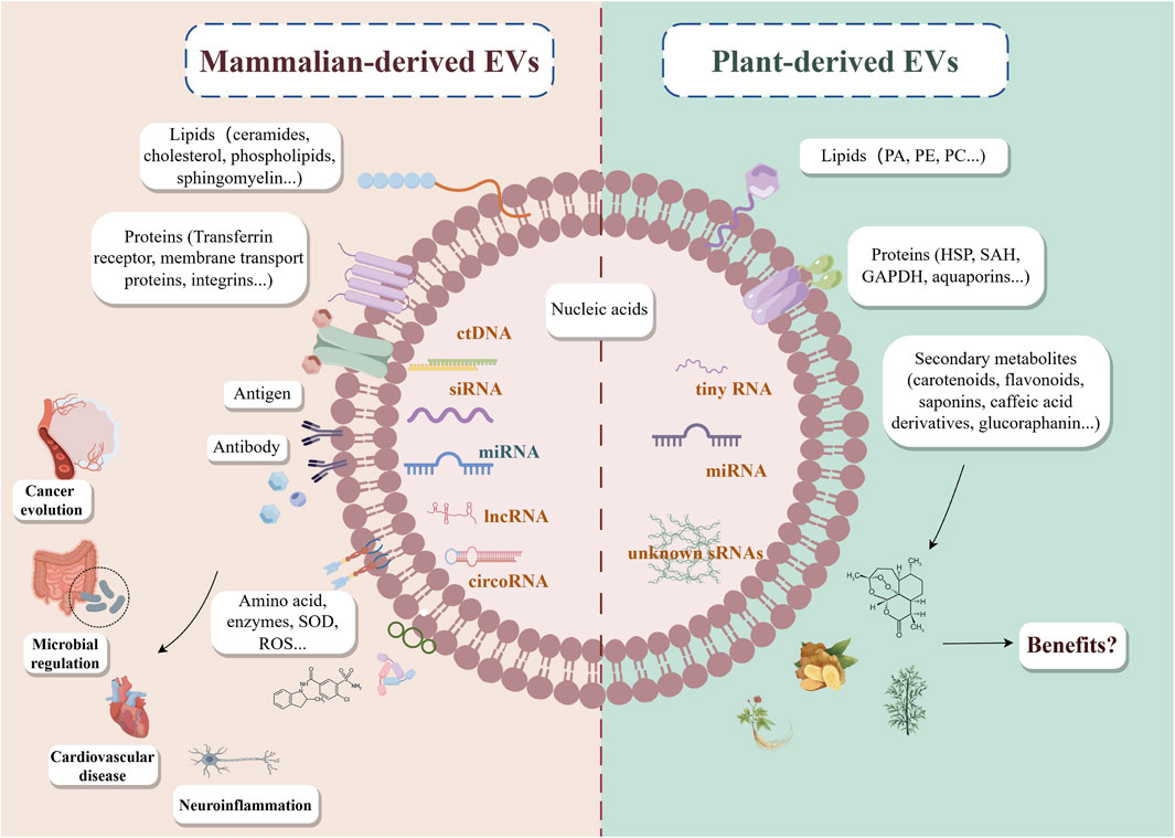
Exosomes, with an average diameter of approximately 100 nm, are nanoscale vesicles secreted by nearly almost all cells under both pathological and physiological conditions (Kalluri and LeBleu, 2020). These vesicles facilitate the transfer of diverse biomolecules, including mRNAs, miRNAs, proteins, lipids, and metabolites, from donor to recipient cells, influencing immunity, viral pathogenicity, pregnancy, cardiovascular diseases, neurological disorders, and cancer progression (He et al., 2021; Qiu et al., 2024) (Figure 1). Recently, research on EVs has predominantly focused on mammalian cell-derived exosomes. For instance, EVs from mesenchymal stem cells (MSC-EVs) have been explored for their potential in drug delivery, cancer immunotherapy, and regenerative medicine (Weng et al., 2021; Keshtkar et al., 2018). Additionally, EVs derived from cancer-associated fibroblasts (CAF-EVs) play central roles in inducing metabolic reprogramming and mediating immune suppression within the TME (Li et al., 2021). Despite their promising therapeutic potential, mammalian-derived exosomes pose certain risks, including immune rejection and the potential transmission of harmful substances such as tumor-associated molecules and infectious agents, coupled with the complexity of their own production and their scarcity, which present major challenges for cell-free therapy (Hanayama, 2021). In recent years, growing interest has emerged in the utilization of EVs derived from plants and traditional Chinese medicine for cancer treatment.

Figure 1. Current status of mammalian-derived EVs and plant-derived EVs. EVs, extracellular vehicles; PA, phosphatidic acid; PE, phosphatidylethanolamine; PC, phosphatidylcholine; SOD, superoxide dismutase; ROS, reactive oxygen; HSP, heat shock protein 70; SAH, S-adenosyl-homocysteinase; GAPDH, glyceraldehyde 3-phosphate dehydrogenase; ctDNA, circulating tumor DNA; siRNA, small interfering RNA; miRNA, microRNA; lncRNA, long non-coding RNA; circRNA, circular RNA; sRNAs, this refers to other undiscovered small RNAs.
Plant-derived EVs (P-EVs) possess multiple bioactive cargos akin to those found in mammalian-derived EVs. Increasing evidence suggests that P-EVs contain bioactive molecules with diverse functions. For example, exosome-like nanoparticles (ELNs) from ginger rhizomes can readily suppress the activation of the NLRP3 inflammasome, suggesting their potential as novel agents to suppress inflammasome assembly and activation (Chen et al., 2019). Similarly, the plant-derived compound curcumin modulates Nrf2, MAPK, and NF-κB signaling, exhibiting antioxidant properties, mitigating chemotherapy and radiotherapy-related side effects, and scavenging free radicals (Liu Y. Q. et al., 2021). Furthermore, plant-based compounds such as flavonoids, quercetin, and coumarins have demonstrated anti-inflammatory, antioxidant, immunomodulatory, and anticancer properties (Naksuriya et al., 2022; Zhao et al., 2019). While both P-EVs and plant extracts can provide therapeutic potential, P-EVs possess distinct advantages, including their encapsulation and delivery of bioactive molecules to recipient cells, enhancing bioavailability and therapeutic efficacy. As a promising candidate for solid tumor treatment, P-EVs may improve therapeutic outcomes by influencing tumor cell proliferation, apoptosis, metastasis, and TME homeostasis (Rome, 2019; Xu et al., 2023). Although P-EV-based therapies remain in the research phase, encouraging preclinical results have been achieved, and clinical trials are being conducted for many cancers, signaling a potential shift toward natural medicine-based cancer treatment.
This review offers a comprehensive overview of the plant sources of P-EVs, summarizes preclinical studies investigating their role in oncology, explores their impact on the TME. The objective is to highlight the promising future of P-EVs as novel therapeutic agents in oncology.
Composition and isolation of P-EVs
Over the past decade, EVs derived from plant cells have been established as biogenically and morphologically similar to their mammalian counterparts (An et al., 2007). Various terms, including P-EVs, edible plant-derived nanovesicles, plant-derived nanovesicles, and plant exosome-like nanovesicles, have been used to describe these nano-to micro-sized vesicles (50–1,000 nm) (Cui et al., 2020). P-EVs encapsulate a variety of bioactive molecules and function as mediators of communication between cells. For instance, studies indicate that P-EVs are rich in enzymes essential for the remodeling of the cell wall and proteins with antibacterial properties (Woith et al., 2019; Liu G. et al., 2021). Additionally, P-EVs can transport small non-coding RNAs among cells, influencing gene regulation, proliferation, and differentiation (Urzì et al., 2021). Evidence suggests that P-EVs exhibit stable intrinsic therapeutic activity, can be internalized by human cells, and influence cellular processes. Owing to their natural origin and minimal toxicity, P-EVs have emerged as attractive tools for cancer treatment.
To date, four principal compounds have been detected in P-EVs: proteins, nucleic acids, lipids, and secondary metabolites, with all of these playing crucial roles in the stability and function of these vesicles (Cai et al., 2021; Martínez-Ballesta et al., 2018).
Lipids
Previous studies have identified phosphatidic acid (PA), phosphatidylethanolamine (PE), and phosphatidylcholine (PC) as the major lipid constituents of P-EVs (Karamanidou and Tsouknidas, 2021). In contrast, mammalian-derived EVs predominantly consist of ceramides, cholesterol, phospholipids, and sphingomyelin (Donoso-Quezada et al., 2021). Differences in lipid composition influence the uptake of these vesicles by gut microbiota. For example, ginger-derived EVs enriched in PA exhibit preferential absorption by Lactobacillus rhamnosus, whereas grapefruit-derived EVs containing higher levels of PC are taken up predominantly by Ruminococcaceae (Teng et al., 2018). This suggests that specific lipid components may serve as molecular cues guiding preferential uptake by distinct intestinal microbiota, highlighting the potential of lipid-based targeting strategies for gut microbiota modulation in anti-tumor therapy. However, the precise mechanisms governing the absorption and internalization of P-EVs by target cells remain poorly understood, necessitating extensive lipidomic studies to elucidate the roles of these lipid components.
Proteins
Proteins within EVs facilitate host cell uptake and act as carriers of critical genetic information. While proteins in P-EVs play essential roles in plant physiology, their overall content is lower than that of mammalian-derived EVs. Pinedo et al. identified three conserved protein families in P-EVs: heat shock protein 70 (HSP-70), S-adenosyl-homocysteinase, and GAPDH in apoplast washing fluid (Pinedo et al., 2021). Notably, HSP-70 is linked to tumor invasion and treatment resistance in human cancers (Shevtsov et al., 2018) suggesting a potential but unexplored connection between P-EV proteins and their therapeutic effects. Membrane proteins in P-EVs also enhance their uptake by mammalian cells. For example, aquaporins on the surface of broccoli-derived P-EVs contribute to membrane stability and facilitate cellular absorption (Martínez-Ballesta et al., 2018). Additionally, cytoplasmic proteins play roles in cell wall remodeling and pathogen defense (De Palma et al., 2020). Although various studies have characterized P-EV protein compositions, many of their therapeutic properties remain unknown due to challenges in protein extraction. Further proteomic analyses are required to fully uncover their biological functions.
Nucleic acids
P-EVs contain nucleic acids, such as microRNAs (miRNAs), which regulate gene expression and receptor cell function (López de Las Hazas et al., 2023). Plant-derived miRNAs have demonstrated potential in preventing and treating various human diseases, including cancer (Yi et al., 2025), cardiovascular disorders (Hou et al., 2018), neurodegenerative diseases (Saiyed et al., 2022), and diabetes (Bajaj et al., 2025). For instance, Chin et al. reported that plant miRNA miR159 undergoes natural 2′-O-methylation at its 3′-end, which protects it from degradation and enhances its stability. A synthetic miR159 mimic was found to suppress breast cancer cell proliferation in mice via targeting TCF7, a WNT signaling transcription factor, resulting in reduced MYC protein levels while sparing normal breast epithelial cells (Chin et al., 2016). This study offers evidence that plant-derived miRNAs can inhibit tumor growth in mammalian models. Additionally, Teng et al. identified small RNAs and miRNAs in P-EVs that regulate gut microbiota composition and its metabolites, suppress inflammation, and mediate microbiota-host immune system crosstalk (Teng et al., 2018). Furthermore, Baldrich et al. discovered highly enriched “tiny RNAs” (10–17 nucleotides) in Arabidopsis thaliana-derived EVs, although their functions remain unclear (Baldrich et al., 2019). Advances in bioinformatics and transcriptomics are expected to reveal novel pharmacological and therapeutic roles of nucleic acids in P-EVs. Increasingly, research suggests that nucleic acids within P-EVs may target mammalian genes associated with inflammation and cancer, underscoring their potential as therapeutic agents.
Metabolites
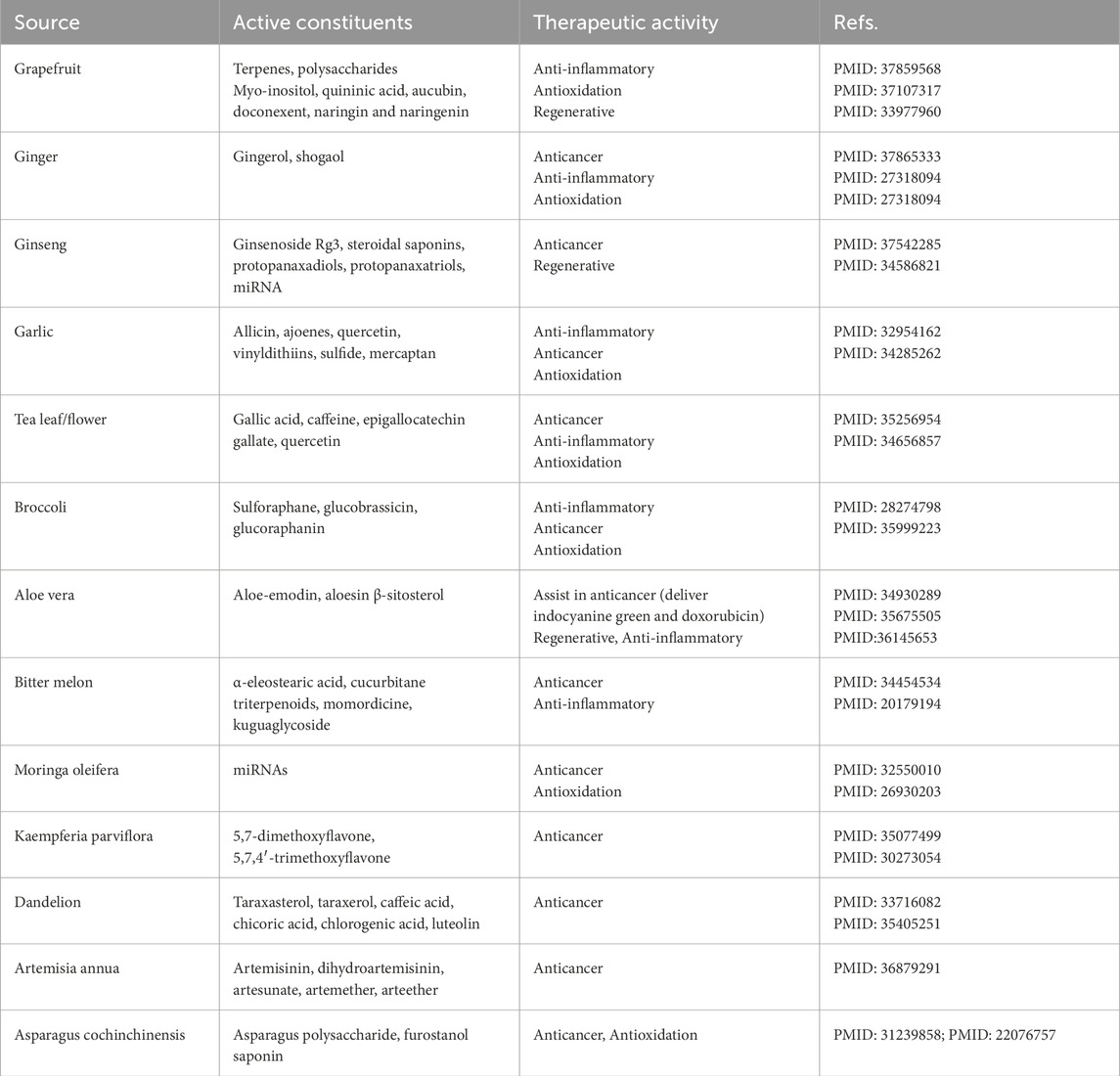
Plant-derived secondary metabolites, including carotenoids, flavonoids, saponins, curcuminoids, and caffeic acid derivatives, possess significant biochemical activity and therapeutic potential (Woith et al., 2021; Bailly and Vergoten, 2020). These metabolites may be encapsulated within P-EVs, contributing to their intrinsic therapeutic properties. For instance, the active compounds 6-gingerol and 6-shogaol, found in ginger, can be loaded into P-EVs and exhibit anti-inflammatory and anti-cancer activities (Bischoff-Kont and Fürst, 2021; Sharma et al., 2023; Karatay et al., 2020). Zeng et al. demonstrated that naringenin, a flavonoid present in grapefruit-derived EVs, has therapeutic potential in treating inflammation-related conditions, including sepsis, fulminant hepatitis, fibrosis, and cancer (Zeng et al., 2018). Table 1 provides a brief overview of the sources of some secondary metabolites or active constituents and their current therapeutic effects. However, not all plant-derived metabolites are incorporated into P-EVs. This variability may stem from differences in the lipid and membrane protein compositions of P-EVs across plant species, as well as physicochemical properties such as lipid or water solubility and molecular density. The mechanisms governing the selective packaging of metabolites into P-EVs remain unclear, highlighting the need for further research. Despite the limited number of metabolomic studies on P-EVs, advancing our understanding of their biochemical contents could pave the way for novel nanomedicines with significant therapeutic potential.

Table 1. Secondary metabolites or active ingredients of plants and their different therapeutic purposes.
Pharmacological activity of P-EVs in anticancer therapy
In recent years, the exploration of exosome-based therapeutic strategies has gained significant attention in clinical cancer research. One of the primary goals in modern oncology is to develop targeted tumor therapies that selectively eliminate cancer cells without harming normal tissues. P-EVs have been shown to regulate diverse biological processes and offer potential advantages in cancer treatment (Raimondo et al., 2019). Some studies have investigated the direct encapsulation of pharmacological agents within P-EVs via phagocytosis or endocytosis, facilitating targeted uptake by recipient cells (Wang et al., 2014). Additionally, exosomes derived from traditional Chinese medicine have emerged as promising candidates for disease treatment. For example, Forsythiaside A-loaded, hyaluronic acid (HA)-modified milk-derived exosomes (mExo) have been devised for the inhibition of NLRP3-mediated pyroptosis, offering a novel therapeutic approach for liver fibrosis (Gong et al., 2023). However, a growing area of research focuses on extracting exosome-like nanotherapeutics from plants and integrating them into conventional treatment strategies, opening new avenues for cancer therapy.
Among the various sources of P-EVs, those derived from ginger have demonstrated remarkable anticancer properties. Engineered ginger-derived EVs (GEVs) can be efficiently internalized by gastric adenocarcinoma cells, leading to dose-dependent inhibition of cell viability (Nemidkanam and Chaichanawongsaroj, 2022). Moreover, GEVs loaded with doxorubicin selectively target colon cancer cells, inducing apoptosis and promoting intestinal repair, thereby serving as a potential therapeutic strategy for inflammatory bowel disease and colitis-related cancers (Zhang et al., 2016). Chen et al. have isolated natural nanocarriers from tea flowers containing high levels of polyphenols, flavonoids, functional proteins, and lipids, which induce mitochondrial damage, amplify reactive oxygen species (ROS), arrest the tumor cell cycle, and promote breast tumor apoptosis while inhibiting lung metastasis (Chen et al., 2022). Similarly, Zu et al. developed tea leaf-derived EVs that suppress inflammatory cytokine expression, enhance macrophage-specific endocytosis, and restore gut microbiota diversity, improving treatment outcomes in colitis-related colon cancer (Zu et al., 2021). Additionally, Stanly et al. determined that grapefruit-derived EVs (GFEVs) downregulate the AKT-ERK signaling axis in various cancer cell lines, leading to cell cycle arrest and apoptosis (Stanly et al., 2020). Further advancements include the development of folic acid (FA)-conjugated GFEVs (GFEV-FAs) for targeted delivery of paclitaxel to colon tumors, enhancing therapeutic efficacy (Wang et al., 2013). Collectively, these findings underscore the significant potential of P-EVs in anticancer therapy.
P-EVs are also being explored as adjuncts to conventional chemotherapy and targeted therapy. For instance, GFEVs have been utilized not only to encapsulate paclitaxel (Wang et al., 2013), but also to deliver doxorubicin via heparin-based nanoparticles, enabling drug penetration across the blood-brain barrier and exerting inhibitory effects on glioma (Niu et al., 2021). Yang et al. found bitter melon-derived EVs (BMEVs) to downregulate NLRP3 expression and clear ROS in oral squamous cell carcinoma (OSCC), reducing OSCC resistance to 5-fluorouracil (5-FU) (Yang et al., 2021). Beyond their direct anticancer effects, P-EVs also mitigate side effects associated with conventional therapies. For example, Cui et al. found that BMEVs protect against myocardial cell damage and fibrosis induced by chest radiotherapy by reducing ROS levels and preserving mitochondrial homeostasis, offering potential protection against radiation-induced heart disease (Cui et al., 2022). Additionally, EVs from dandelion and rhodiola significantly alleviate bleomycin-induced pulmonary fibrosis in mice (Du et al., 2019). Notably, GEVs facilitate controlled doxorubicin release in the acidic tumor microenvironment (TME), limiting systemic toxicity (Luan et al., 2017). These findings highlight the promising role of P-EVs in enhancing therapeutic efficacy while minimizing adverse effects.
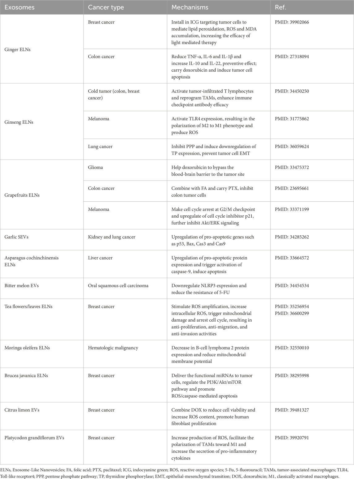
With growing research interest, P-EVs are being investigated for their potential applications beyond anticancer therapy, including antifibrotic, antiviral, gut microbiota-regulating, and TME-modulating activities. Table 2 provides an overview of the therapeutic applications of P-EVs from various plant sources in cancer treatment.
Indirect roles of P-EVs in reshaping the TME
The TME comprises a heterogeneous network of cells, such as infiltrating cancer cells, endothelial cells, epithelial cells, fibroblasts, and mesenchymal macrophages (de Visser and Joyce, 2023). These cells, together with their surrounding extracellular matrix, contribute to the formation of resistant and dynamic “cold tumors,” which evade immune detection and facilitate tumor progression. Excessive secretion of cytokines and chemokines within the TME induces an acidic milieu that sustains proliferative signaling, promotes angiogenesis, enhances invasion and metastasis, triggers pro-tumor inflammation, and enables immune evasion (Xiao and Yu, 2021). Consequently, a major research focus has been the modulation of the TME to transform it from a tumor-supportive to a tumor-suppressive environment. P-EVs have demonstrated significant potential in exerting anti-inflammatory effects and enhancing immune responses within the TME.
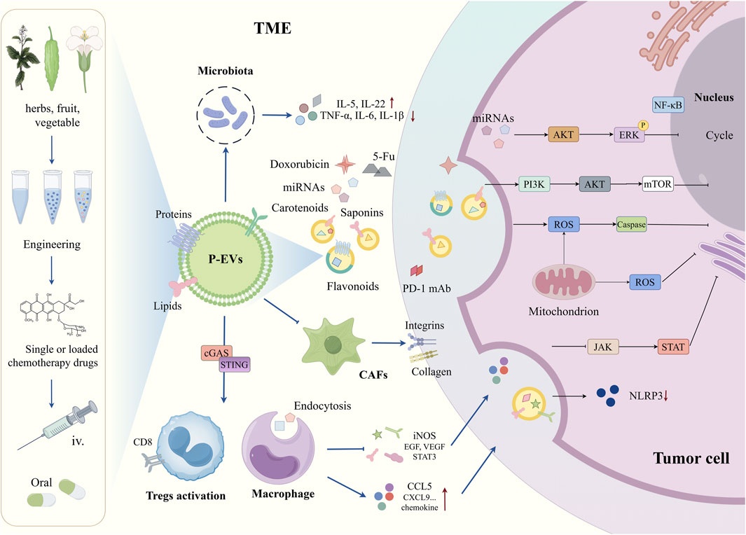
Recent studies highlight the potential of ginseng-derived EVs to penetrate the blood-brain barrier via multiple endocytic pathways, where they suppress regulatory T cell (Treg) activation and M2 macrophage polarization within the TME. These EVs downregulate key oncogenic factors, including iNOS, VEGF, EGF, and STAT3, leading to enhanced immune responses and glioma suppression (Kim et al., 2023). In another study, ginseng-derived EVs reprogram tumor-associated macrophages (TAMs), increasing the secretion of CCL5 and CXCL9 to recruit CD8+ T cells into the tumor core, thereby enhancing the efficacy of immune checkpoint blockade therapy with PD-1 monoclonal antibodies (Han et al., 2022). These findings further validate the immunomodulatory potential of ginseng, reinforcing its designation as the “king of herbs.”
Within the TME, the cGAS-STING pathway plays a pivotal role in modulating immune and inflammatory responses. Liu et al. successfully isolated and purified nanoscale vesicles from Artemisia annua and demonstrated that these vesicles can reprogram tumor-associated macrophages (TAMs) from a pro-tumor phenotype to a pro-inflammatory state. This transformation activates the cGAS-STING pathway, further reprogramming macrophages and enhancing cytotoxic T-cell responses, ultimately contributing to tumor regression and improving the efficacy of αPD-L1-mediated immunotherapy in lung cancer (Liu et al., 2023). Additionally, Dendropanax morbiferus-derived P-EVs have been reported to inhibit cancer-associated fibroblasts (CAFs) by modulating gene expression related to growth factors or extracellular matrix components including integrins and collagen. These findings highlight the potential of P-EVs as future anti-CAF agents (Kim et al., 2020). Autophagy, a self-protective process responsible for preserving cellular homeostasis, enables cancer cells to survive under nutrient-deficient and hypoxic conditions within the TME (Vessoni et al., 2013). Exosome-like nanovesicles from Brucea javanica have been found to suppress autophagy through the promotion of PI3K/Akt/mTOR phosphorylation while inducing apoptosis via ROS/Caspase activation and inhibiting the JAK/STAT signaling pathway, thereby exerting potent anti-cancer effects (Yan et al., 2024; Chen et al., 2020; Wang et al., 2019).
Inflammation is a fundamental and dynamic component of the TME, closely linked to metastasis, drug resistance, and poor prognosis (Jiang et al., 2025). As previously mentioned, GEVs can repair intestinal mucosal damage and prevent colitis-associated tumor formation. The underlying mechanism involves the upregulation of anti-inflammatory factors such as IL-10 and IL-22 and the downregulation of pro-inflammatory cytokines, including TNF-α, IL-6, and IL-1β, thereby modulating the inflammatory cytokine profile within the TME (Zhang et al., 2016). Numerous studies have emphasized the roles of miRNA-146a and miRNA-125a in negatively regulating the NF-κB pathway (Meisgen et al., 2014; Curtale et al., 2019), which is closely associated with macrophage stimulation, pro-inflammatory cytokine production, and chemokine release (Dorrington and Fraser, 2019). Notably, exosome-like vesicles derived from apples have been shown to induce the transcriptional activation of miRNA-146a and miRNA-125a, leading to a reduction in IL-8 and IL-1β expression, suppression of JNK and NF-κB pathway activation, and overall modulation of the inflammatory TME (Trentini et al., 2022). Collectively, these findings suggest that P-EVs possess the potential to transform “cold tumors,” which are typically resistant to conventional therapies, into “hot tumors” that exhibit enhanced responsiveness to treatment, thereby significantly improving therapeutic outcomes (Figure 2).

Figure 2. The existing pathways cascade of P-EVs in the TME (partial). TME, tumor microenvironment; CAFs, cancer-associated fibroblasts; Tregs, regulatory T cells; iv, intravenous; 5-Fu, 5-Fluorouracil; cGAS, cyclic GMP-AMP synthase; STING, stimulator of interferon genes; iNOS, inducible nitric oxide synthase; EGF, epidermal growth factor; VEGF, vascular endothelial growth factor; STAT, signal transducer and activator of transcription; CCL5, C-C motif chemokine ligand5; CXCL9, C-X-C motif chemokine ligand9; PD-1 mAb, programmed cell death protein1 monoclonal antibody; NF-kB, nuclear factor kappa-light-chain-enhancer of activated B cells; AKT, protein kinase B; ERK, extracellular signal-regulated kinase; PI3K, phosphatidylinositol 3-kinase; mTOR, mechanistic target of rapamycin; ROS, reactive oxygen; JAK, janus kinase; NLRP3, NOD-like receptor family, pyrin domain containing3.
Overall, recent studies underscore the promising anti-cancer potential of P-EVs in targeting the TME. However, the TME comprises a vast and intricate network of cellular interactions, making it far more complex than initially perceived. In addition to cancer cells, other cell types secrete extracellular vesicles, and factors such as hypoxia, low pH, and nutrient scarcity pose additional challenges for P-EV research. Nevertheless, the emergence of P-EVs offers a promising avenue for the development of low-toxicity cancer therapies.
Discussion
Targeted cancer therapy is expected to remain a sustainable and advancing field of research. Engineered extracellular vesicles derived from autologous cells or mammalian sources face challenges such as immune rejection, toxicity, and limited clearance rates. In contrast, P-EVs represent a relatively novel alternative with advantages including natural origin, low toxicity, reduced immunogenicity, and excellent biocompatibility (Den et al., 2017). Over the past decade, various anti-tumor mechanisms of P-EVs have been explored, with most studies focusing on their role in inducing cancer cell apoptosis (Stanly et al., 2020; Yang et al., 2021; Yan et al., 2024). However, research on the effects of P-EVs on the TME and its associated immune cells remains in its infancy. Compared with P-EVs, engineered mammalian cell-derived EVs are more flexible in cancer therapy. For example, endothelial cells derived from bone marrow derived mesenchymal stem cells (BMSCs) have the advantages of biocompatibility and low immunogenicity. Previous studies have expressed streptavidin in the cellular membrane and EVs derived from MSCs, which was engineered into plasma membrane-located proteins its fusion with the coding sequence of signal peptides and transmembrane regions, to achieve the functionalization of BMSC-EVs (Meng et al., 2023). It is worth mentioning that immune cells, such as natural killer cells, can also secrete EVs (NKEVs) with tumor-targeting capabilities. It has been reported that it can encapsulate paclitaxel through electroporation and target breast cancer cells to exert inhibitory effects (Han et al., 2020). However, NKEVs contain many molecules related to immune responses, including IFN-γ, TNF-α, IL-10, MHC-I and MHC-2, and other chemokines, etc. This means that for engineered NKEVs, rejection reactions must be taken into account (Wu et al., 2021). In contrast, P-EVs do not include the above-mentioned cytokines or chemokines, low human homology and its composition is simpler than that of mammalian cell-derived EVs (Wang et al., 2014). Furthermore, Zhou et al. have engineered BMSCs derived from autologous stem cells, loaded the obtained EVs with miR-138-5p and the anti-fibrotic agent pirfenidone (PFD) and subjected to surface modification with integrin α5-targeting peptides to reprogram cancer-associated fibroblasts (CAFs) and enhance its tumor-targeting ability, remodeling the TME of pancreatic cancer, improving tumor hypoxia and enhancing gemcitabine sensitivity (Zhou et al., 2024). Mammalian derived EVs can rely on surface proteins (such as integrins) to achieve natural targeting ability, while P-EVs has not been fully developed the natural targeting property and lacked protein markers with biochemical characteristics. Currently, commonly measured markers include CD63, CD81and CD9, etc. (Hong et al., 2023; Jokhio et al., 2024), the specific biomarkers of P-EVs are still lacking, which required more engineering modification. Overall, compared to mammalian cell-derived extracellular vesicles, additional studies are required to better understand the biogenesis, protein surface markers, and targeting mechanisms of P-EVs.
In addition to low immunogenicity, high biocompatibility and low toxicity are also advantages of P-EVs. For example, sodium thiosulfate (STS) is a clinically approved drug for the treatment of vascular calcification, but its efficacy is limited by poor bioavailability and severe adverse effects. Feng et al. have created a bionic GFEVs that can be loaded with STS and targeted delivery without inducing hemolysis or causing any damage to other organs (Feng et al., 2023). Some edible fruit plants are non-toxic by themselves and can prevent the toxic and side effects brought about by traditional treatments. For instance, grapes derived EVs can effectively prevent oral mucositis in patients with head and neck tumors caused by radiotherapy and chemotherapy. A Phase I clinical trial has now been initiated (NCT01668849). Fang et al. have isolated and purified kiwifruit derived EVs (KEVs) and delivered the lipophilic multi-targeted kinase inhibitor sorafenib (KEV-SFB) in a targeted manner, evidence indicates that the KEV-SFB reduces the leakage of SFB in the gastrointestinal environment, which was able to achieve liver accumulation and was predominantly taken up by HepG2 cells in mice without toxic effects on normal hepatocytes (Fang et al., 2023). Previous researchers have successfully loaded doxorubicin in GEVs (DOX-GEVs) and released DOX into the acidic TME in a pH-dependent manner, which has been proven to be effectively absorbed by colon-26 and HT-29 through endocytosis and has non-toxic in vivo (Zhang et al., 2016). Despite P-EVs have general low toxicity, they are not entirely free from adverse effects. For instance, intravenous administration of exosome-like vesicles derived from tea flowers has been shown to induce tumor cell cycle arrest and apoptosis. However, mice receiving intravenous treatment exhibited significant weight loss and signs of liver toxicity compared to those treated via oral administration (Chen et al., 2022). This finding suggests that oral administration may be a safer and more viable route for P-EV delivery. Nevertheless, enzymatic degradation in the digestive tract, interactions with gut microbiota, and external factors such as diet and concomitant drug administration can influence orally administered engineered P-EV bioavailability and therapeutic efficacy. Thus, there is an urgent need to develop novel P-EV-based treatment strategies that are non-toxic, stable, and readily absorbable while maintaining compatibility with other therapeutic agents. Ensuring the efficient release and targeted delivery of P-EVs following systemic administration remains a critical challenge.
Addressing the complexities of the ever-evolving TME has been a formidable challenge in recent years. Tumor-derived exosomes (TDEs) carry various bioactive molecules, including TGF-β, caveolin-1, HIF-1α, and β-catenin, which collectively enhance the invasive and migratory capabilities of recipient cells, promote immune evasion, and contribute to drug resistance (Syn et al., 2016). Although engineered P-EVs can cross biological the blood-brain barrier and skin barrier, their precise route of delivery to tumor target cells within the TME remains highly complex and challenging. At present, most studies focus on developing drug delivery platforms for engineered EVs, while very few studies concentrate on how to enhance the cancer cell and tumor-targeting specificity. Chemical modification of the surface of P-EVs may improve targeting ability and reduce the off-targeted side effects. There is a study has modified GEVs using the tumor-targeting ligand iRGD, iRGD is a cyclic peptide widely used in tumor-targeted drug delivery and has a high binding affinity for integrins and NRP-1 receptors that are overexpressed in tumors. Compared with unmodified GEVs, GEV-iRGD significantly enhances the ability to be absorbed by tumor cells (Wang et al., 2025). Kang et al. have enhanced the targeting of red cabbage-derived EVs (RabEVs) to intestinal epithelial cells and immune cells by coupling hyaluronic acid (t-Rabex) with the surface of RabEVs, thereby highlighted their anti-inflammatory, antioxidative, and tight-junction maintenance properties in inflammatory bowel disease (IBD) (Kang et al., 2024). In a recent study, Yang et al. have integrated the cell membrane fragments of breast cancer cell line 4T1 into lemon-derived nanovesicles (LEVs), and then loaded the anti-cancer drug DOX to establish the composite nanomedicine delivery vector LEVBD, which significantly improved the targeting of homologous tumors and promoted the transcellular transport of drugs, suppressed tumor growth in mice after intravenous injection, and had no observable toxic and side effects (Yang et al., 2025). It can be seen from these studies that not only the development of P-EVs has aroused the interest of researchers, but also how to better transform these natural EVs to make them more targeted and penetrating is the direction that more and more scholars are curious about and focus on. However, the stability of P-EVs is uncertain. For example, Temperature and pH can affect its integrity and activity. Zhang et al. once used a simulated gastrointestinal fluid system to evaluate the particle size and zeta potential changes of GEVs, the results showed that compared with the buffer PBS, the size of GEVs increased in the stomach-like solution and further enlarged in a small intestine like solution (Zhang et al., 2016). This is also the risk and challenge faced by oral administration in exerting its efficacy. It further underscores the necessity of enhancing P-EV stability. Future research on P-EVs should prioritize improving their purity and yield, optimizing cost-effectiveness during isolation and storage, and ensuring their safety and efficacy for clinical applications. Efforts should be directed toward accelerating early-stage clinical investigations to bring P-EVs closer to therapeutic implementation. With continued advancements, P-EVs hold significant promise for revolutionizing cancer treatment by providing a natural, effective, and low-toxicity therapeutic approach.
Author contributions
HW: Writing – original draft, Writing – review and editing. MS: Writing – original draft. LM: Supervision, Writing – review and editing. JQ: Writing – original draft. YJ: Writing – review and editing. DQ: Funding acquisition, Writing – review and editing. FS: Funding acquisition, Writing – review and editing.
Funding
The author(s) declare that financial support was received for the research and/or publication of this article. The research was supported by Zhejiang Province Medical and Health Science and Technology Plan Project (No. 2025KY1660) and the National Natural Science Foundation of China (Grant No. 82404685), Research Project of Jiangsu Association of Chinese Medicine (CYTF 2024050), Suzhou Science and Technology Development Program (SKYD2023087) and Science and Technology Project of Changshu Health Commission (CSWS 202208 and CSWS 202211).
Conflict of interest
The authors declare that the research was conducted in the absence of any commercial or financial relationships that could be construed as a potential conflict of interest.
Generative AI statement
The author(s) declare that no Generative AI was used in the creation of this manuscript.
Publisher’s note
All claims expressed in this article are solely those of the authors and do not necessarily represent those of their affiliated organizations, or those of the publisher, the editors and the reviewers. Any product that may be evaluated in this article, or claim that may be made by its manufacturer, is not guaranteed or endorsed by the publisher.
References
An, Q., van Bel, A. J., and Hückelhoven, R. (2007). Do plant cells secrete exosomes derived from multivesicular bodies? Plant Signal Behav. 2, 4–7. doi:10.4161/psb.2.1.3596
Anand, U., Dey, A., Chandel, A. K. S., Sanyal, R., Mishra, A., Pandey, D. K., et al. (2023). Cancer chemotherapy and beyond: current status, drug candidates, associated risks and progress in targeted therapeutics. Genes Dis. 10, 1367–1401. doi:10.1016/j.gendis.2022.02.007
Bailly, C., and Vergoten, G. (2020). Proposed mechanisms for the extracellular release of PD-L1 by the anticancer saponin platycodin D. Int. Immunopharmacol. 85, 106675. doi:10.1016/j.intimp.2020.106675
Bajaj, G., Choudhary, D., Singh, V., Priyadarshi, N., Garg, P., Mantri, S. S., et al. (2025). MicroRNAs dependent G-ELNs based intervention improves glucose and fatty acid metabolism while protecting pancreatic β-Cells in type 2 diabetic mice. Small 21, e2409501. doi:10.1002/smll.202409501
Baldrich, P., Rutter, B. D., Karimi, H. Z., Podicheti, R., Meyers, B. C., and Innes, R. W. (2019). Plant extracellular vesicles contain diverse small RNA species and are enriched in 10- to 17-Nucleotide “tiny” RNAs. Plant Cell 31, 315–324. doi:10.1105/tpc.18.00872
Bischoff-Kont, I., and Fürst, R. (2021). Benefits of ginger and its constituent 6-Shogaol in inhibiting inflammatory processes. Pharmaceuticals (Basel) 14, 571. doi:10.3390/ph14060571
Cai, Q., He, B., Wang, S., Fletcher, S., Niu, D., Mitter, N., et al. (2021). Message in a bubble: shuttling small RNAs and proteins between cells and interacting organisms using extracellular vesicles. Annu. Rev. Plant Biol. 72, 497–524. doi:10.1146/annurev-arplant-081720-010616
Chen, Q., Li, Q., Liang, Y., Zu, M., Chen, N., Canup, B. S. B., et al. (2022). Natural exosome-like nanovesicles from edible tea flowers suppress metastatic breast cancer via ROS generation and microbiota modulation. Acta Pharm. Sin. B 12, 907–923. doi:10.1016/j.apsb.2021.08.016
Chen, X., Li, S., Li, D., Li, M., Su, Z., Lai, X., et al. (2020). Ethanol extract of Brucea javanica seed inhibit triple-negative breast cancer by restraining autophagy via PI3K/Akt/mTOR pathway. Front. Pharmacol. 11, 606. doi:10.3389/fphar.2020.00606
Chen, X., Zhou, Y., and Yu, J. (2019). Exosome-like nanoparticles from ginger rhizomes inhibited NLRP3 inflammasome activation. Mol. Pharm. 16, 2690–2699. doi:10.1021/acs.molpharmaceut.9b00246
Chin, A. R., Fong, M. Y., Somlo, G., Wu, J., Swiderski, P., Wu, X., et al. (2016). Cross-kingdom inhibition of breast cancer growth by plant miR159. Cell Res. 26, 217–228. doi:10.1038/cr.2016.13
Cui, W. W., Ye, C., Wang, K. X., Yang, X., Zhu, P. Y., Hu, K., et al. (2022). Momordica charantia-derived extracellular vesicles-like nanovesicles protect cardiomyocytes against radiation injury via attenuating DNA damage and mitochondria dysfunction. Front. Cardiovasc Med. 9, 864188. doi:10.3389/fcvm.2022.864188
Cui, Y., Gao, J., He, Y., and Jiang, L. (2020). Plant extracellular vesicles. Protoplasma 257, 3–12. doi:10.1007/s00709-019-01435-6
Curtale, G., Rubino, M., and Locati, M. (2019). MicroRNAs as molecular switches in macrophage activation. Front. Immunol. 10, 799. doi:10.3389/fimmu.2019.00799
Deng, Z., Rong, Y., Teng, Y., Mu, J., Zhuang, X., Tseng, M., et al. (2017). Broccoli-derived nanoparticle inhibits mouse colitis by activating dendritic cell AMP-activated protein kinase. Mol. Ther. 25, 1641–1654. doi:10.1016/j.ymthe.2017.01.025
De Palma, M., Ambrosone, A., Leone, A., Del Gaudio, P., Ruocco, M., Turiák, L., et al. (2020). Plant roots release small extracellular vesicles with antifungal activity. Plants (Basel) 9, 1777. doi:10.3390/plants9121777
de Visser, K. E., and Joyce, J. A. (2023). The evolving tumor microenvironment: from cancer initiation to metastatic outgrowth. Cancer Cell 41, 374–403. doi:10.1016/j.ccell.2023.02.016
Donoso-Quezada, J., Ayala-Mar, S., and González-Valdez, J. (2021). The role of lipids in exosome biology and intercellular communication: function, analytics and applications. Traffic 22, 204–220. doi:10.1111/tra.12803
Dorrington, M. G., and Fraser, I. D. C. (2019). NF-κB signaling in macrophages: dynamics, crosstalk, and signal integration. Front. Immunol. 10, 705. doi:10.3389/fimmu.2019.00705
Du, J., Liang, Z., Xu, J., Zhao, Y., Li, X., Zhang, Y., et al. (2019). Plant-derived phosphocholine facilitates cellular uptake of anti-pulmonary fibrotic HJT-sRNA-m7. Sci. China Life Sci. 62, 309–320. doi:10.1007/s11427-017-9026-7
Fang, Z., Song, M., Lai, K., Cui, M., Yin, M., and Liu, K. (2023). Kiwi-derived extracellular vesicles for oral delivery of sorafenib. Eur. J. Pharm. Sci. 191, 106604. doi:10.1016/j.ejps.2023.106604
Feng, W., Teng, Y., Zhong, Q., Zhang, Y., Zhang, J., Zhao, P., et al. (2023). Biomimetic grapefruit-derived extracellular vesicles for safe and targeted delivery of sodium thiosulfate against vascular calcification. ACS Nano 17, 24773–24789. doi:10.1021/acsnano.3c05261
Ghanbar, M. I., and Suresh, K. (2024). Pulmonary toxicity of immune checkpoint immunotherapy. J. Clin. Invest 134, e170503. doi:10.1172/JCI170503
Gong, L., Zhou, H., Zhang, S., Wang, C., Fu, K., Ma, C., et al. (2023). CD44-Targeting drug delivery system of exosomes loading forsythiaside A combats liver fibrosis via regulating NLRP3-Mediated pyroptosis. Adv. Healthc. Mater 12, e2202228. doi:10.1002/adhm.202202228
Han, D., Wang, K., Zhang, T., Gao, G. C., and Xu, H. (2020). Natural killer cell-derived exosome-entrapped paclitaxel can enhance its anti-tumor effect. Eur. Rev. Med. Pharmacol. Sci. 24, 5703–5713. doi:10.26355/eurrev_202005_21362
Han, X., Wei, Q., Lv, Y., Weng, L., Huang, H., Wei, Q., et al. (2022). Ginseng-derived nanoparticles potentiate immune checkpoint antibody efficacy by reprogramming the cold tumor microenvironment. Mol. Ther. 30, 327–340. doi:10.1016/j.ymthe.2021.08.028
Hanayama, R. (2021). Emerging roles of extracellular vesicles in physiology and disease. J. Biochem. 169, 135–138. doi:10.1093/jb/mvaa138
He, J., Chen, N. N., Li, Z. M., Wang, Y. Y., Weng, S. P., Guo, C. J., et al. (2021). Evidence for a novel antiviral mechanism of teleost fish: serum-derived exosomes inhibit virus replication through incorporating Mx1 protein. Int. J. Mol. Sci. 22, 10346. doi:10.3390/ijms221910346
Hong, R., Luo, L., Wang, L., Hu, Z. L., Yin, Q. R., Li, M., et al. (2023). Lepidium meyenii walp (Maca)-derived extracellular vesicles ameliorate depression by promoting 5-HT synthesis via the modulation of gut-brain axis. Imeta 2, e116. doi:10.1002/imt2.116
Hou, D., He, F., Ma, L., Cao, M., Zhou, Z., Wei, Z., et al. (2018). The potential atheroprotective role of plant MIR156a as a repressor of monocyte recruitment on inflamed human endothelial cells. J. Nutr. Biochem. 57, 197–205. doi:10.1016/j.jnutbio.2018.03.026
Jiang, Y., Qiu, J., Ye, N., and Xu, Y. (2025). Current status of cytokine-induced killer cells and combination regimens in breast cancer. Front. Immunol. 16, 1476644. doi:10.3389/fimmu.2025.1476644
Jokhio, S., Peng, I., and Peng, C. A. (2024). Extracellular vesicles isolated from Arabidopsis thaliana leaves reveal characteristics of Mammalian exosomes. Protoplasma 261, 1025–1033. doi:10.1007/s00709-024-01954-x
Joo, J. I., Choi, M., Jang, S. H., Choi, S., Park, S. M., Shin, D., et al. (2020). Realizing cancer precision medicine by integrating systems biology and nanomaterial engineering. Adv. Mater 32, e1906783. doi:10.1002/adma.201906783
Kalluri, R., and LeBleu, V. S. (2020). The biology, function, and biomedical applications of exosomes. Science 367, eaau6977. doi:10.1126/science.aau6977
Kang, S. J., Lee, J. H., and Rhee, W. J. (2024). Engineered plant-derived extracellular vesicles for targeted regulation and treatment of colitis-associated inflammation. Theranostics 14, 5643–5661. doi:10.7150/thno.97139
Karamanidou, T., and Tsouknidas, A. (2021). Plant-derived extracellular vesicles as therapeutic nanocarriers. Int. J. Mol. Sci. 23, 191. doi:10.3390/ijms23010191
Karatay, K. B., Kılçar, A. Y., Derviş, E., and Müftüler, F. Z. B. (2020). Radioiodinated ginger compounds (6-gingerol and 6-shogaol) and incorporation assays on breast cancer cells. Anticancer Agents Med. Chem. 20, 1129–1139. doi:10.2174/1871520620666200128114215
Keshtkar, S., Azarpira, N., and Ghahremani, M. H. (2018). Mesenchymal stem cell-derived extracellular vesicles: novel frontiers in regenerative medicine. Stem Cell Res. Ther. 9, 63. doi:10.1186/s13287-018-0791-7
Kim, J., Zhu, Y., Chen, S., Wang, D., Zhang, S., Xia, J., et al. (2023). Anti-glioma effect of ginseng-derived exosomes-like nanoparticles by active blood-brain-barrier penetration and tumor microenvironment modulation. J. Nanobiotechnology 21, 253. doi:10.1186/s12951-023-02006-x
Kim, K., Jung, J. H., Yoo, H. J., Hyun, J. K., Park, J. H., Na, D., et al. (2020). Anti-metastatic effects of plant sap-derived extracellular vesicles in a 3D microfluidic cancer metastasis model. J. Funct. Biomater. 11, 49. doi:10.3390/jfb11030049
Li, C., Teixeira, A. F., Zhu, H. J., and Ten Dijke, P. (2021). Cancer associated-fibroblast-derived exosomes in cancer progression. Mol. Cancer 20, 154. doi:10.1186/s12943-021-01463-y
Liu, G., Kang, G., Wang, S., Huang, Y., and Cai, Q. (2021b). Extracellular vesicles: emerging players in plant defense against pathogens. Front. Plant Sci. 12, 757925. doi:10.3389/fpls.2021.757925
Liu, J., Xiang, J., Jin, C., Ye, L., Wang, L., Gao, Y., et al. (2023). Medicinal plant-derived mtDNA via nanovesicles induces the cGAS-STING pathway to remold tumor-associated macrophages for tumor regression. J. Nanobiotechnology 21, 78. doi:10.1186/s12951-023-01835-0
Liu, Y. Q., Wang, X. L., He, D. H., and Cheng, Y. X. (2021a). Protection against chemotherapy- and radiotherapy-induced side effects: a review based on the mechanisms and therapeutic opportunities of phytochemicals. Phytomedicine 80, 153402. doi:10.1016/j.phymed.2020.153402
López de Las Hazas, M. C., Tomé-Carneiro, J., Del Pozo-Acebo, L., Del Saz-Lara, A., Chapado, L. A., Balaguer, L., et al. (2023). Therapeutic potential of plant-derived extracellular vesicles as nanocarriers for exogenous miRNAs. Pharmacol. Res. 198, 106999. doi:10.1016/j.phrs.2023.106999
Luan, X., Sansanaphongpricha, K., Myers, I., Chen, H., Yuan, H., and Sun, D. (2017). Engineering exosomes as refined biological nanoplatforms for drug delivery. Acta Pharmacol. Sin. 38, 754–763. doi:10.1038/aps.2017.12
Martínez-Ballesta, M. D. C., García-Gomez, P., Yepes-Molina, L., Guarnizo, A. L., Teruel, J. A., and Carvajal, M. (2018). Plasma membrane aquaporins mediates vesicle stability in broccoli. PLoS One 13, e0192422. doi:10.1371/journal.pone.0192422
Meisgen, F., Xu Landén, N., Wang, A., Réthi, B., Bouez, C., Zuccolo, M., et al. (2014). MiR-146a negatively regulates TLR2-induced inflammatory responses in keratinocytes. J. Invest Dermatol 134, 1931–1940. doi:10.1038/jid.2014.89
Meng, W., Wang, L., Du, X., Xie, M., Yang, F., Li, F., et al. (2023). Engineered mesenchymal stem cell-derived extracellular vesicles constitute a versatile platform for targeted drug delivery. J. Control Release 363, 235–252. doi:10.1016/j.jconrel.2023.09.037
Naksuriya, O., Daowtak, K., Tima, S., Okonogi, S., Mueller, M., Toegel, S., et al. (2022). Hydrolyzed flavonoids from Cyrtosperma johnstonii with superior antioxidant, antiproliferative, and anti-inflammatory potential for cancer prevention. Molecules 27, 3226. doi:10.3390/molecules27103226
Nemidkanam, V., and Chaichanawongsaroj, N. (2022). Characterizing Kaempferia parviflora extracellular vesicles, a nanomedicine candidate. PLoS One 17, e0262884. doi:10.1371/journal.pone.0262884
Niu, W., Xiao, Q., Wang, X., Zhu, J., Li, J., Liang, X., et al. (2021). A biomimetic drug delivery system by integrating grapefruit extracellular vesicles and doxorubicin-loaded heparin-based nanoparticles for glioma therapy. Nano Lett. 21, 1484–1492. doi:10.1021/acs.nanolett.0c04753
Pinedo, M., de la Canal, L., and de Marcos Lousa, C. (2021). A call for rigor and standardization in plant extracellular vesicle research. J. Extracell. Vesicles 10, e12048. doi:10.1002/jev2.12048
Qian, D., Hong, W., Li, S., Liu, H., He, C., Liu, X., et al. (2025). Trends in the global, national, and regional burden of breast cancer among adolescents and young adults from 1990 to 2021: analyses of the 2021 global burden of disease study. Breast 82, 104486. doi:10.1016/j.breast.2025.104486
Qiu, J., Jiang, Y., Ye, N., Jin, G., Shi, H., and Qian, D. (2024). Leveraging the intratumoral microbiota to treat human cancer: are engineered exosomes an effective strategy? J. Transl. Med. 22, 728. doi:10.1186/s12967-024-05531-x
Raimondo, S., Giavaresi, G., Lorico, A., and Alessandro, R. (2019). Extracellular vesicles as biological shuttles for targeted therapies. Int. J. Mol. Sci. 20, 1848. doi:10.3390/ijms20081848
Rome, S. (2019). Biological properties of plant-derived extracellular vesicles. Food Funct. 10, 529–538. doi:10.1039/c8fo02295j
Saiyed, A. N., Vasavada, A. R., and Johar, S. R. K. (2022). Recent trends in miRNA therapeutics and the application of plant miRNA for prevention and treatment of human diseases. Futur J. Pharm. Sci. 8, 24. doi:10.1186/s43094-022-00413-9
Sharma, S., Shukla, M. K., Sharma, K. C., Kumar, L., Anal, J. M. H., Upadhyay, S. K., et al. (2023). Revisiting the therapeutic potential of gingerols against different pharmacological activities. Naunyn Schmiedeb. Arch. Pharmacol. 396, 633–647. doi:10.1007/s00210-022-02372-7
Shevtsov, M., Huile, G., and Multhoff, G. (2018). Membrane heat shock protein 70: a theranostic target for cancer therapy. Philos. Trans. R. Soc. Lond B Biol. Sci. 373, 20160526. doi:10.1098/rstb.2016.0526
Siegel, R. L., Giaquinto, A. N., and Jemal, A. (2024). Cancer statistics, 2024. CA Cancer J. Clin. 74, 12–49. doi:10.3322/caac.21820
Stanly, C., Alfieri, M., Ambrosone, A., Leone, A., Fiume, I., and Pocsfalvi, G. (2020). Grapefruit-derived micro and nanovesicles show distinct metabolome profiles and anticancer activities in the A375 human melanoma cell line. Cells 9, 2722. doi:10.3390/cells9122722
Syn, N., Wang, L., Sethi, G., Thiery, J. P., and Goh, B. C. (2016). Exosome-mediated metastasis: from epithelial-mesenchymal transition to escape from immunosurveillance. Trends Pharmacol. Sci. 37, 606–617. doi:10.1016/j.tips.2016.04.006
Teng, Y., Ren, Y., Sayed, M., Hu, X., Lei, C., Kumar, A., et al. (2018). Plant-derived exosomal MicroRNAs shape the gut microbiota. Cell Host Microbe 24, 637–652.e8. doi:10.1016/j.chom.2018.10.001
Trentini, M., Zanotti, F., Tiengo, E., Camponogara, F., Degasperi, M., Licastro, D., et al. (2022). An apple a day keeps the doctor away: potential role of miRNA 146 on macrophages treated with exosomes derived from apples. Biomedicines 10, 415. doi:10.3390/biomedicines10020415
Urzì, O., Raimondo, S., and Alessandro, R. (2021). Extracellular vesicles from plants: current knowledge and open questions. Int. J. Mol. Sci. 22, 5366. doi:10.3390/ijms22105366
Vessoni, A. T., Filippi-Chiela, E. C., Menck, C. F., and Lenz, G. (2013). Autophagy and genomic integrity. Cell Death Differ. 20, 1444–1454. doi:10.1038/cdd.2013.103
Wang, B., Zhuang, X., Deng, Z. B., Jiang, H., Mu, J., Wang, Q., et al. (2014). Targeted drug delivery to intestinal macrophages by bioactive nanovesicles released from grapefruit. Mol. Ther. 22, 522–534. doi:10.1038/mt.2013.190
Wang, F., Li, L., Deng, J., Ai, J., Mo, S., Ding, D., et al. (2025). Lipidomic analysis of plant-derived extracellular vesicles for guidance of potential anti-cancer therapy. Bioact. Mater 46, 82–96. doi:10.1016/j.bioactmat.2024.12.001
Wang, Q., Zhuang, X., Mu, J., Deng, Z. B., Jiang, H., Zhang, L., et al. (2013). Delivery of therapeutic agents by nanoparticles made of grapefruit-derived lipids. Nat. Commun. 4, 1867. doi:10.1038/ncomms2886
Wang, S., Hu, H., Zhong, B., Shi, D., Qing, X., Cheng, C., et al. (2019). Bruceine D inhibits tumor growth and stem cell-like traits of osteosarcoma through inhibition of STAT3 signaling pathway. Cancer Med. 8, 7345–7358. doi:10.1002/cam4.2612
Weng, Z., Zhang, B., Wu, C., Yu, F., Han, B., Li, B., et al. (2021). Therapeutic roles of mesenchymal stem cell-derived extracellular vesicles in cancer. J. Hematol. Oncol. 14, 136. doi:10.1186/s13045-021-01141-y
Woith, E., Fuhrmann, G., and Melzig, M. F. (2019). Extracellular vesicles-connecting kingdoms. Int. J. Mol. Sci. 20, 5695. doi:10.3390/ijms20225695
Woith, E., Guerriero, G., Hausman, J. F., Renaut, J., Leclercq, C. C., Weise, C., et al. (2021). Plant extracellular vesicles and nanovesicles: focus on secondary metabolites, proteins and lipids with perspectives on their potential and sources. Int. J. Mol. Sci. 22, 3719. doi:10.3390/ijms22073719
Wu, F., Xie, M., Hun, M., She, Z., Li, C., Luo, S., et al. (2021). Natural killer cell-derived extracellular vesicles: novel players in cancer immunotherapy. Front. Immunol. 12, 658698. doi:10.3389/fimmu.2021.658698
Xiao, Y., and Yu, D. (2021). Tumor microenvironment as a therapeutic target in cancer. Pharmacol. Ther. 221, 107753. doi:10.1016/j.pharmthera.2020.107753
Xu, Z., Xu, Y., Zhang, K., Liu, Y., Liang, Q., Thakur, A., et al. (2023). Plant-derived extracellular vesicles (PDEVs) in nanomedicine for human disease and therapeutic modalities. J. Nanobiotechnology 21, 114. doi:10.1186/s12951-023-01858-7
Yan, G., Xiao, Q., Zhao, J., Chen, H., Xu, Y., Tan, M., et al. (2024). Brucea javanica derived exosome-like nanovesicles deliver miRNAs for cancer therapy. J. Control Release 367, 425–440. doi:10.1016/j.jconrel.2024.01.060
Yang, L. Y., Liang, G. W., Cai, B. J., Xu, K. M., Zhao, Y. J., Xie, X. T., et al. (2025). Enhanced tumor self-targeting of lemon-derived extracellular vesicles by embedding homotypic cancer cell membranes for efficient drug delivery. J. Nanobiotechnology 23, 74. doi:10.1186/s12951-025-03161-z
Yang, M., Luo, Q., Chen, X., and Chen, F. (2021). Bitter melon derived extracellular vesicles enhance the therapeutic effects and reduce the drug resistance of 5-fluorouracil on oral squamous cell carcinoma. J. Nanobiotechnology 19, 259. doi:10.1186/s12951-021-00995-1
Yi, C., Lu, L., Li, Z., Guo, Q., Ou, L., Wang, R., et al. (2025). Plant-derived exosome-like nanoparticles for microRNA delivery in cancer treatment. Drug Deliv. Transl. Res. 15, 84–101. doi:10.1007/s13346-024-01621-x
Zeng, W., Jin, L., Zhang, F., Zhang, C., and Liang, W. (2018). Naringenin as a potential immunomodulator in therapeutics. Pharmacol. Res. 135, 122–126. doi:10.1016/j.phrs.2018.08.002
Zhan, C., Jin, Y., Xu, X., Shao, J., and Jin, C. (2023). Antitumor therapy for breast cancer: focus on tumor-associated macrophages and nanosized drug delivery systems. Cancer Med. 12, 11049–11072. doi:10.1002/cam4.5489
Zhang, M., Viennois, E., Prasad, M., Zhang, Y., Wang, L., Zhang, Z., et al. (2016). Edible ginger-derived nanoparticles: a novel therapeutic approach for the prevention and treatment of inflammatory bowel disease and colitis-associated cancer. Biomaterials 101, 321–340. doi:10.1016/j.biomaterials.2016.06.018
Zhao, Y. L., Yang, X. W., Wu, B. F., Shang, J. H., Liu, Y. P., Zhi, D., et al. (2019). Anti-inflammatory effect of pomelo peel and its bioactive coumarins. J. Agric. Food Chem. 67, 8810–8818. doi:10.1021/acs.jafc.9b02511
Zhou, P., Du, X., Jia, W., Feng, K., and Zhang, Y. (2024). Engineered extracellular vesicles for targeted reprogramming of cancer-associated fibroblasts to potentiate therapy of pancreatic cancer. Signal Transduct. Target Ther. 9, 151. doi:10.1038/s41392-024-01872-7
Keywords: plant-derived extracellular vesicles, solid tumors, tumor microenvironment, antitumor therapy, precision medicine
Citation: Wu H, Shi M, Meng L, Qiu J, Jiang Y, Qian D and Shen F (2025) Plant-derived extracellular vesicles as a novel tumor-targeting delivery system for cancer treatment. Front. Cell Dev. Biol. 13:1589550. doi: 10.3389/fcell.2025.1589550
Received: 07 March 2025; Accepted: 16 June 2025;
Published: 24 June 2025.
Edited by:
Masahide Takahashi, Fujita Health University, JapanCopyright © 2025 Wu, Shi, Meng, Qiu, Jiang, Qian and Shen. This is an open-access article distributed under the terms of the Creative Commons Attribution License (CC BY). The use, distribution or reproduction in other forums is permitted, provided the original author(s) and the copyright owner(s) are credited and that the original publication in this journal is cited, in accordance with accepted academic practice. No use, distribution or reproduction is permitted which does not comply with these terms.
*Correspondence: Da Qian, ZHJxaWFuZGFAaG90bWFpbC5jb20=; Fengqing Shen, c2hlbmZlbmdxaW5nQGhvdG1haWwuY29t
 Hanjin Wu1
 Jie Qiu
 Yuancong Jiang
 Da Qian