- 1State Key Laboratory for Animal Disease Control and Prevention, Harbin Veterinary Research Institute, Chinese Academy of Agricultural Sciences, Harbin, China
- 2School of Advanced Agricultural Sciences, Yibin Vocational and Technical College, Yibin, China
Background: Feline panleukopenia, feline calicivirus infection, feline viral rhinotracheitis, and feline infectious peritonitis are significant diseases that threaten feline health. The trend of mixed infections is increasing, and current diagnostic methods are limited in scope and unable to provide rapid, simultaneous detection of these diseases.
Methods: Four groups of primers and probes targeting the VP2 gene of Feline Panleukopenia virus (FPV), the TK gene of Feline Herpesvirus (FHV-1), the ORF2 gene of Feline Calicivirus (FCV), and the N gene of Feline Infectious Peritonitis Virus (FIPV) were designed. After optimizing the concentrations of primers and probes and annealing temperature, a quadruplex TaqMan MGB fluorescent quantitative PCR method was established to concurrently detect these four pathogens. Recombinant plasmid standards were constructed to establish standard curves, and the sensitivity, specificity, reproducibility, and clinical application of the assay were evaluated.
Results: The optimal final concentrations of primers for FPV, FHV-1, FCV, and FIPV were 0.08, 0.04, 0.06, and 0.12 μM, respectively, and the optimal final concentrations of probes were 0.08, 0.08, 0.12, and 0.12 μM, respectively. The best annealing temperature was 59°C. No cross-reaction was observed with common pathogens in infected cats. The minimal detection limits for recombinant plasmids of T-VP2, T-TK, T-ORF2, and T-N were 50.79, 53.21, 47.91 and 41.25 copies/μL, respectively. The R² values of standard curves are 0.994, 1.0, 0.998 and 0.999, respectively, and high amplification efficiencies of 105.05%, 96.28%, 98.82%, and 96.45%, respectively. The coefficient of variation for inter-batch and intra-batch tests ranged from 0.14 to 1.37%. Among 381 fecal samples from cats, the detection rates for FPV, FHV-1, FCV, and FIPV were 13.65% (52/381), 18.37% (70/381), 26.77% (102/381), and 9.71% (37/381), respectively, with a 100% agreement with previously reported methods and commercial kits.
Conclusion: The sensitive, specific, high-throughput, quadruplex TaqMan MGB quantitative fluorescent quantitative PCR method was successfully established for the simultaneous detection of FPV, FHV-1, FCV, and FIPV.
1 Introduction
Cats are known for their gentle and independent nature, their ability to provide therapeutic companionship, and their strong emotional bond with humans, making them one of the most important companion animals. According to the China Pet Industry White Paper, the number of pet cats in 2024 is projected to reach 71.93 million, marking a 2.5% increase from 2023 and continuing an upward trend (Source Network). However, a large body of clinical data indicates that feline panleukopenia, feline viral rhinotracheitis, feline infectious peritonitis, and feline calicivirus infections have relatively high incidence rates among felines and are often associated with mixed infections, posing a serious threat to the health of global feline populations (Tasker et al., 2023; Longobardi et al., 2024; Liu et al., 2025; Nishisaka et al., 2025).
Feline panleukopenia, also known as feline distemper, feline parvovirus, or feline infectious enteritis, is a disease caused by Feline Panleukopenia virus (FPV), a member of the Parvoviridae family (Yang et al., 2024a). It primarily affects kittens under one year of age (Ye et al., 2022). FPV is a single-stranded DNA virus with a genome of approximately 5,200 base pairs, which includes two open reading frames that encode the non-structural proteins (NS1 and NS2) and structural proteins (VP1 and VP2) (Li et al., 2022). FPV can infect various animals, including felids (such as tigers, leopards, and lions) and mustelids (such as raccoons and ferrets) (Huang et al., 2022; Wang et al., 2022). The virus is primarily transmitted through feces, saliva, and other secretions, with infected cats serving as the main source of transmission (Yu et al., 2024). The disease is most common during the spring and summer months and can affect cats of all ages, with kittens particularly vulnerable (Pacini et al., 2023). The infection rate in kittens can be as high as 70%, and the fatality rate ranges from 45% to 60% (Xue et al., 2023a; Xue et al., 2023b). A study conducted in Yanji, China, in 2021–2022 reported a 33.75% FPV positivity rate (27/80) (Xue et al., 2023a).
Feline viral rhinotracheitis is a viral infectious disease caused by Feline Herpesvirus 1 (FHV-1), which primarily affects the respiratory and ocular systems of felids (Synowiec et al., 2023). FHV-1 is a member of the Herpesviridae family, subfamily Alphaherpesvirinae, and genus Varicellovirus (Yang et al., 2024b). The FPV genome is a double stranded DNA composed of a unique region (UL) approximately 99 kb in length and short fragments of approximately 27 kb (Cavalheiro et al., 2023). Maeda et al. classified FHV-1 into three genotypes: F2, C7301, and C7805 (Maeda et al., 1995). FHV-1 mainly infects felids, particularly kittens, and is widely distributed globally (Jiao et al., 2024). The virus is primarily transmitted through direct contact and aerosols and often co-infects with FCV, leading to symptoms such as respiratory distress and oral ulcers (Gaskell and Povey, 1982). Its incidence can reach 100%, with a mortality rate between 20% and 50% (Liu et al., 2022). A study conducted in Kunshan, China, in 2022–2023 reported a 21.5% FHV-1 positivity rate (43/200) in conjunctival and nasal swabs from cats exhibiting respiratory distress, conjunctivitis, and corneal ulcers (Kim et al., 2024).
Feline calicivirus (FCV) infection primarily manifests as oral ulcers, pneumonia, chronic gastritis, arthritis, and lameness (Wei et al., 2024). FCV, a member of the Caliciviridae family, is one of the most common viral pathogens in domestic cats worldwide (Duclos et al., 2024). FCV genome is a single stranded RNA about 7700 bp in length, contains three open reading frames (ORFs): ORF1, ORF2, and ORF3 (Wei et al., 2024). ORF2 encodes the capsid protein VP1, which contains six variable regions (A-F) (Zhou et al., 2021). Based on genetic evolution, FCV strains are categorized into Type I and Type II. FCV is distributed globally and can infect not only domestic cats but also other felid species (Mao et al., 2022). The virus primarily spreads through respiratory secretions and saliva and mainly affects kittens aged 7–84 days, with the highest susceptibility seen in those aged 56–84 days (Wei et al., 2024). FCV has a high incidence rate, but its fatality rate is usually low (Wei et al., 2024). A study conducted in Guangdong Province, China, from 2018 to 2022 reported a 28.9% FCV positivity rate in throat and nasal swabs from doubtful FCV-infected cats (152 samples) (Mao et al., 2022).
Feline infectious peritonitis is caused by Feline Coronavirus (FCoV) (Thayer et al., 2022). FCoV is a single-stranded RNA virus with a genome about 30000 bp in length, containing 11 ORFs (Gao et al., 2023). The virus particles have an envelope with a diameter ranging from 80 to 120 nm (Kennedy, 2020). Based on differences in the spike protein and serotype, FCoV can be classified into two types: Feline infectious peritonitis virus (FIPV) and Feline Enteric Coronavirus (FECV) (Bubenikova et al., 2020; Chen et al., 2023). FECV infection often causes mild or asymptomatic diarrhea, but 5%–12% of infected cats will become Feline infectious peritonitis due to mutations of FECV into FIPV, with a fatality rate approaching 100% in cats with Feline infectious peritonitis (Lewis et al., 2015). Studies suggest that 0.3% to 1.4% of feline deaths in veterinary clinics are attributed to Feline infectious peritonitis (Thayer et al., 2022). Currently, there is no effective vaccine available to prevent this disease.
Common laboratory detection methods for FPV, FHV-1, FIPV, and FCV include viral isolation, serological tests, and molecular biological techniques (Liu et al., 2022; He et al., 2024). Virus isolation is complex, time-consuming, and difficult to implement clinically (Wang et al., 2024c). Serological tests may lead to false-negative owing to the stage of viral infection and the host’s immune status (Wang et al., 2024b). PCR technology, especially fluorescence quantitative PCR (qPCR), offers high specificity, sensitivity, and stability, and it can also quantify target genes (Wang et al., 2024a). However, most qPCR methods for detecting these four viruses are singleplex, limiting the ability to detect multiple pathogens simultaneously (Zhang et al., 2019; Liu et al., 2024). Therefore, the construction of a quadruplex TaqMan MGB qPCR method capable of simultaneously detecting FPV, FHV, FIPV, and FCV can not only improve diagnostic efficiency but also reduce testing costs.
In summary, the above four viruses have high prevalence rates among felines, causing significant harm, and are easy to mixed or secondary infections, become clinical diagnosis difficult. Additionally, vaccines have been developed for some of these viruses, such as the trivalent inactivated vaccine for feline viral rhinotracheitis, calicivirus disease, and panleukopenia (RPVF0304, RPVF0207, and RPVF0110 strains), as well as vaccines for feline rhinotracheitis, calicivirus, and panleukopenia (CP2, CC3, and VP2 proteins) (Source Network). However, there is an urgent need for rapid, efficient, sensitive, and specific diagnostic methods to screen antigen-negative cats, as these four viruses must be detected in the breeding of specific-pathogen-free (SPF) cats. In this study, four sets of specific primers and probes the VP2 gene of FPV, the TK gene of FHV-1, the N gene of FIPV, and the ORF2 gene of FCV were designed to establish a quadruplex TaqMan MGB fluorescent quantitative PCR method. The goal of this method is to provide rapid, sensitive, and specific detection of these four viruses, offering technical support for early diagnosis, timely treatment, vaccine evaluation, and SPF cats breeding, ultimately contributing to the improvement of feline health and reducing the risk of disease transmission.
2 Materials and methods
2.1 Nucleic acids of virus and bacterium
The nucleic acids of Feline Parvovirus (FPV), Feline Herpesvirus 1 (FHV-1), Feline Infectious Peritonitis Virus (FIPV), Feline Calicivirus (FCV), Salmonella, Pasteurella multocida, Staphylococcus aureus, Feline Leukemia Virus (FeLV), and Feline Immunodeficiency Virus (FIV) were stored in our laboratory. The clinical samples (nasal, oral swabs, and feces) of 381 cats were collected from some cat farms in Hebei Province, which were from healthy cats, cats with respiratory symptoms, or cats with diarrhea. It is imperative to underscore that no additional harm or intervention was imposed on the animals involved in this study. Given the nature of our research, the Institutional Review Board of the Harbin Veterinary Research Institute has determined that this study is exempt from the requirement for ethical review or approval.
2.2 Synthesis of plasmid standards and design of primers and probes
Based on the gene sequences registered in NCBI, four groups of specific primers and probes were designed targeting the VP2 gene of FPV (Accession No: X55115.1), the TK gene of FHV-1 (Accession No: MT813102.1), the ORF2 gene of FCV (Accession No: PP928983.1), and the N gene of FIPV (Accession No: KY566183.1) using SnapGene software and PrimerSelect software. The primers and probes were synthesized by Genscript (Table 1). The conserved sequences of the target genes were downloaded from NCBI, and then sent to Genscript for plasmid construction. The plasmids were cloned into the pMD-18T vector and named T-VP2, T-TK, T-ORF2, and T-N. The plasmid concentrations were determined, and the copy numbers were calculated using the standard formula.

Table 1. Sequences of primers and probes for the quadruplex TaqMan MGB fluorescent quantitative PCR method.
The recombinant plasmid standards copies/µL = (6.02×10²³) × (X* ng/µL × 10−9)/constructed plasmid length (bp) × 660.
* X: Standard plasmid concentration
2.3 Optimization of annealing temperature, primer, and probe concentrations
The four recombinant plasmid standards, T-VP2, T-TK, T-ORF2, and T-N, were mixed in equal proportions and used as templates. Four groups of primers and probes were employed for fluorescent quantitative TaqMan MGB PCR amplification in the same system. The annealing temperature (58, 59, 60, 61, 62, 63°C), four primer concentrations (final concentrations of 0.01-0.5 μM), and four probe concentrations (final concentrations of 0.01-0.5 μM) were optimized using the matrix assay. In addition, compare the effects of different cycle numbers (35, 40, 45 and 50) on amplification. The annealing temperature, sprimer, and probe concentrations were selected according to the smallest Ct values and maximum fluorescence signals to establish the optimal conditions for quadruplex TaqMan MGB fluorescent quantitative PCR method. The above experiments were repeated three times.
2.4 Construction of standard curves for quadruplex TaqMan MGB qPCR method
The four recombinant plasmid standards, T-VP2, T-TK, T-ORF2, and T-N, were each adjusted to 4 × 10¹⁰ copies/μL. After 10-fold serial dilution, they were mixed in equal proportions. Each concentration was repeated three times. Using the established quadruplex fluorescence quantitative TaqMan MGB PCR method for amplification, and standard curve was constructed with Ct values on the y-axis and the logarithm of template copy numbers on the x-axis.
2.5 Sensitivity test
The four recombinant plasmid standards, T-VP2, T-TK, T-ORF2 and T-N, were mixed in equal proportions after 10-fold serial dilution. The plasmid mixture with final concentrations ranging from 1 × 10⁷ to 1 × 100 copies/μL was used as the template (Repeat each gradient three times). The Quadruplex TaqMan MGB qPCR assay amplification was performed using the established method. Sensitivity of the method was evaluated by probit regression analysis (Ma et al., 2024).
2.6 Specificity test
DNA/RNA (approximately 50 ng/μL) from FPV, FHV-1, FIPV, FCV, Salmonella, Pasteurella multocida, Staphylococcus aureus, FeLV, and FIV were used as templates. The four recombinant plasmid standard mixtures were used as positive controls, and sterilized double-distilled water was used as the negative control. The established quadruplex TaqMan MGB qPCR assay was employed to assess the specificity of the method.
2.7 Repeatability test
The four recombinant plasmid standards, T-VP2, T-TK, T-ORF2, and T-N, were mixed in equal proportions after 10-fold serial dilution. The plasmid mixtures at final concentrations of 1 × 10⁷ copies/μL, 1 × 10⁵ copies/μL, and 1 × 10³ copies/μL were used as templates. Triple amplification was performed for each sample in an intra-assay repeatability test. The plasmid mixtures from different time points at the three concentrations were used as templates to conduct an inter-assay repeatability test, with each sample repeated three times. The coefficient of variation (CV) for intra-assay and inter-assay repeatability was calculated to assess the repeatability of the method. In addition, we used different experimenters and fluorescence quantitative PCR machines to test the stability of the method.
2.8 Detection of clinical samples
From August to December 2024, 381 clinical samples (swabs, feces, etc.) were collected from a cat farm in Hebei Province for testing. Resuspend the sample in sterile PBS buffer, centrifuge at 5000rpm for 3 minutes, and collect the supernatant. Extract DNA/RNA using VAMNE Magnetic Pathogen DNA/RNA Kit (Vazyme, China). The extracted nucleic acid is stored at -20°C or -80°C for future use. Then, the samples were simultaneously tested using the quadruplex TaqMan TaqMan MGB qPCR, reported methods, and commercial kits to verify the accuracy of this method (Cao et al., 2021; Zou et al., 2022; Liu et al., 2024).
3 Results and analysis
3.1 Determination of the optimal conditions for quadruplex TaqMan MGB qPCR method
The optimization results from the matrix method showed that the optimal reaction system for quadruplex TaqMan MGB fluorescent quantitative PCR was 25 μL: 12.5 μL of 2×Fast One Step Probe RT-qPCR Mix (Takara, China), 0.2 μL of forward and reverse primers for FPV, 0.2 μL of probe, 0.1 μL of forward and reverse primers for FHV-1, 0.2 μL of probe, 0.15 μL of forward and reverse primers for FCV, 0.3 μL of probe, 0.3 μL of forward and reverse primers for FIPV, 0.25 μL of probe (The working concentrations of primers and probes were 10 μ M), 2 μL of nucleic acids, and 7.6 μL of sterilized double-distilled water. Using different cycle numbers for amplification, the results showed that when the cycle number was 35, the amplification was insufficient; When the number of cycles is 45 and 50, there is non-specific amplification. So we chose the optimal number of cycles as 40. The amplification program was as follows: 52°C for 5 minutes, 95°C for 10 seconds, 95°C for 5 seconds, 59°C for 10 seconds, for 40 cycles, with fluorescence signal collection during each cycle.
3.2 Construction of standard curves for quadruplex TaqMan MGB fluorescent quantitative PCR method
The recombinant plasmid standards, T-VP2, T-TK, T-ORF2, and T-N, were mixed in equal proportions after 10-fold serial dilution. Amplification was performed using the method established in this study. A standard curve was constructed with Ct values on the y-axis and the logarithm of template copy numbers on the x-axis (Figure 1). For FPV, the standard curve was constructed with concentrations ranging from 1 × 10¹⁰ to 1 × 10⁵ copies/μL; for FHV-1, FCV, and FIPV, the standard curves were constructed with concentrations ranging from 1 × 10⁹ to 1 × 10⁴ copies/μL. The equations for the standard curves were as follows: FPV: Y = -3.207lg(X) + 39.607, R² = 0.994, EFF% = 105.053; FHV-1: Y = -3.414lg(X) + 40.823, R² = 1.0, EFF% = 96.284; FCV: Y = -3.351lg(X) + 40.105, R² = 0.998, EFF% = 98.821; FIPV: Y = -3.41lg(X) + 41.67, R² = 0.999, EFF% = 96.451%.
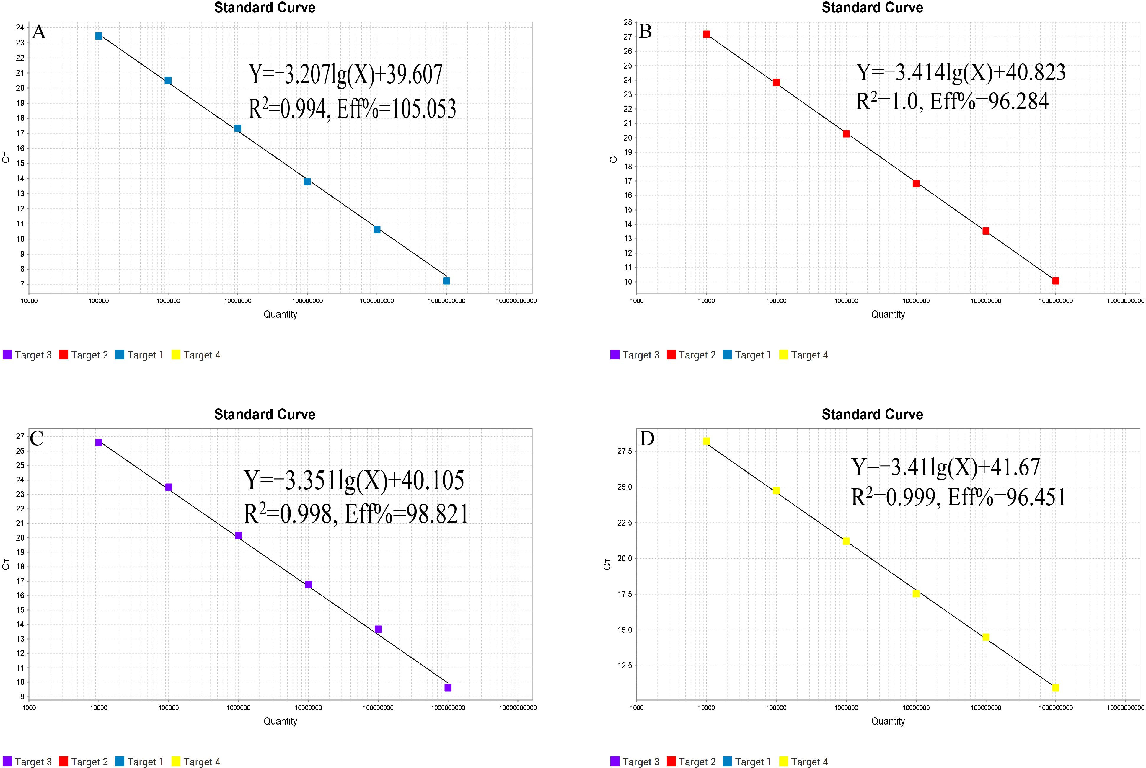
Figure 1. Standard curves of quadruplex TaqMan MGB qPCR method. (A) FPV; (B) FHV-1; (C) FCV; (D) FIPV.
3.3 Sensitivity results
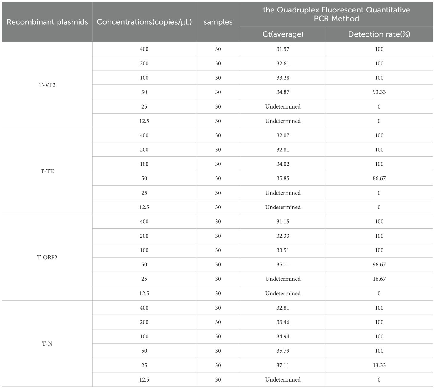
Plasmid standards T-VP2, T-TK, T-ORF2, and T-N were diluted in a 10-fold series and mixed in equal proportions (final concentrations ranging from 1 × 10¹⁰ copies/μL to 1 × 100 copies/μL) to serve as templates for the sensitivity test of the quadruplex TaqMan MGB qPCR method. The results showed that the lower detection limit of the quadruplex TaqMan MGB qPCR system reached 100 copies/μL, demonstrating that the method retains a certain level of sensitivity even in the competitive inhibitory quadruplex reaction system (Figure 2). Additionally, we used probability regression analysis to determine the minimum detection limit of this method. The quadruplex TaqMan MGB qPCR method was used to detect T-VP2, T-TK, T-ORF2, and T-N plasmid mixtures at concentrations of 400 copies/μL, 200 copies/μL, 100 copies/μL, 50 copies/μL, 25 copies/μL, and 12.5 copies/μL. The results, including average Ct values and hit rates, are shown in Table 2. The minimum detection limits for FPV, FHV-1, FCV, and FIPV were 50.79 (95% confidence interval: 46.69–58.68), 53.21 (95% confidence interval: 49.62–65.14), 47.91 (95% confidence interval: 41.02–57.62), and 41.25 (95% confidence interval: 32.62–100.59), respectively (Figure 3). When the Ct values of FPV, FHV-1, and FCV are less than 34 and the amplification curves are smooth, the above pathogens are considered positive; When the Ct value of FIPV is less than 36 and the amplification curve is smooth, FIPV is considered positive. When the Ct value of the four pathogens is 40 or no value, it is judged as negative. When the amplified Ct value is greater than the critical value and less than 40, it is considered suspicious and double retesting is recommended.
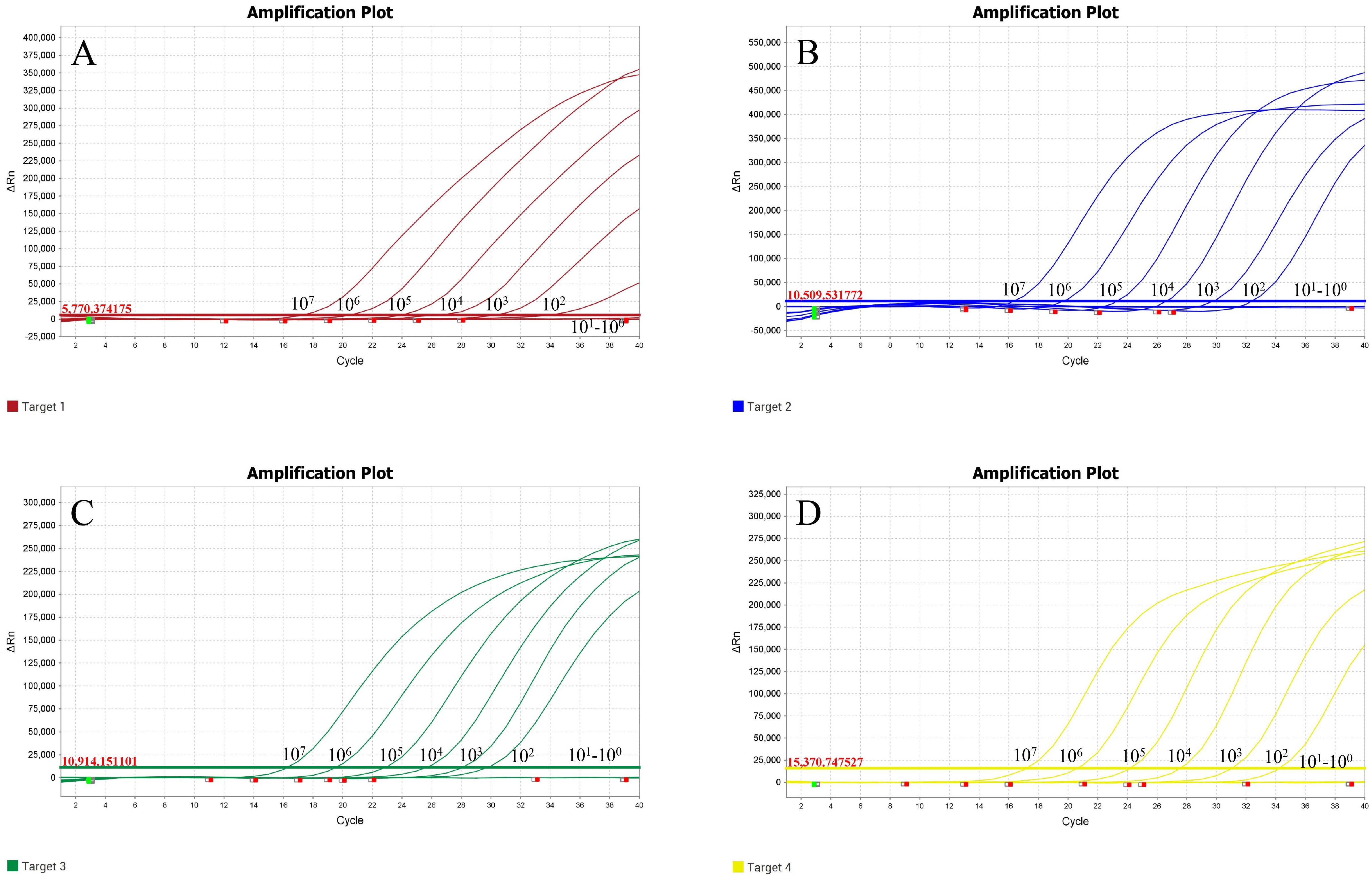
Figure 2. Sensitivity test results of the quadruplex TaqMan MGB qPCR method. (A) FPV; (B) FHV-1; (C) FCV; (D) FIPV.
3.4 Specificity results
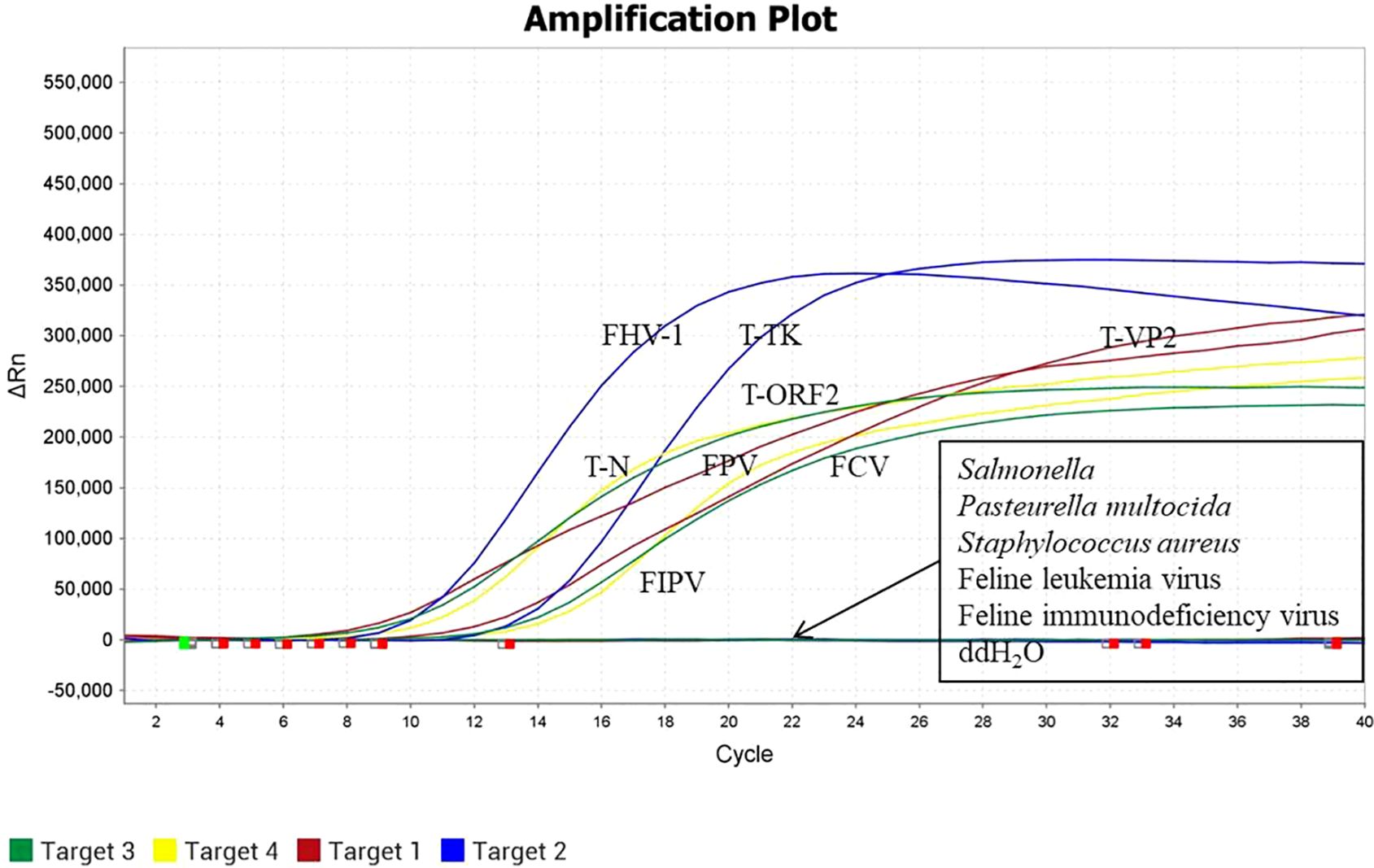
The quadruplex TaqMan MGB qPCR method was used to detect the DNA/RNA (approximately 50 ng/μL) of FPV, FHV-1, FIPV, FCV, Salmonella, Pasteurella multocida, Staphylococcus aureus, FeLV, FIV. The results showed that the nucleic acids of FPV, FHV-1, FIPV and FCV were specifically amplified, while no amplification was observed for the nucleic acids of other viruses or bacteria that infect cats (Figure 4). These findings indicate that the quadruplex TaqMan MGB qPCR method has high specificity.
3.5 Reproducibility results
Reproducibility tests, both intra-group and inter-group, were performed using the quadruplex TaqMan MGB qPCR method on plasmid standard mixtures at four concentrations (1×10⁷, 1×10⁵, and 1×10³ copies/μL). The results showed that the coefficient of variation (CV) of Ct values for both intra-group and inter-group tests was less than 1.5% (Table 3), indicating that the method has good reproducibility. This method has good stability among different experimenters and fluorescence quantitative PCR machines.
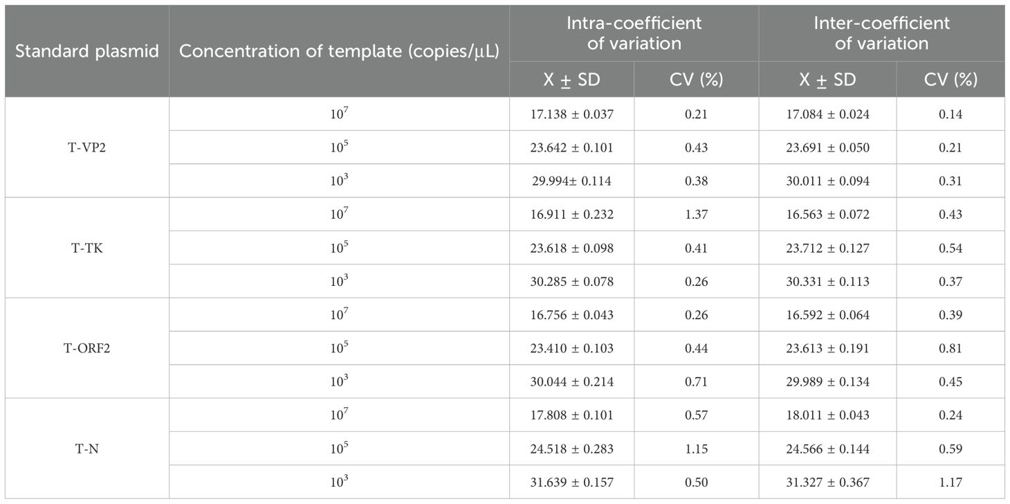
Table 3. Reproducibility test results of the quadruplex real time quantitative TaqMan MGB qPCR method.
3.6 Detection results of clinical samples
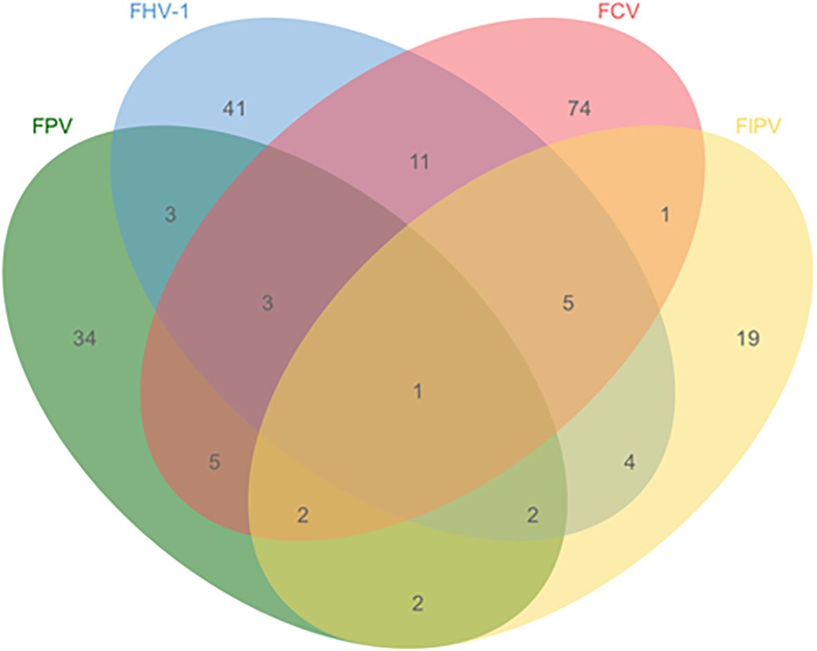
The quadruplex TaqMan MGB qPCR method was used to test 381 clinical cat clinical specimens. The results showed that the detection rates were 13.65% (52/381) for FPV, 18.37% (70/381) for FHV-1, 26.77% (102/381) for FCV, and 9.71% (37/381) for FIPV, with an overall positive rate of 54.59% (208/381) and a negative rate of 45.41% (173/381). The mixed infection situation is shown in Figure 5. Additionally, these samples were tested using previously reported methods and commercial kits. The results indicated that the detection outcomes from both assays were consistent with those obtained using the method established, with a 100% concordance rate between the three methods. This demonstrates that the assay can be applied to the detection of clinical samples.
4 Discussion
Cats are one of the most important companion animals to humans, yet diseases significantly impact their health. In some prosperous countries, the incidence of feline infectious diseases is prevented through vaccination and sanitation measures, whereas in developing countries, due to limited prevention and control technologies, the incidence of these diseases remains high (Cao et al., 2021). Among many diseases, FPV, FHV, FCV, and FIPV are the most common infectious diseases in felines (Xue et al., 2023b; Liu et al., 2025). These pathogens are highly contagious and cause severe gastrointestinal and respiratory symptoms, with a high mortality rate in kittens. Clinically, many newly adopted cats or those with suspected symptoms require screening for multiple infectious diseases to avoid misdiagnosis. The most commonly used detection method is the colloidal gold strip test, but this method has low sensitivity and requires subjective judgment by veterinary staff (Zhang et al., 2019; Ye et al., 2022). For cats with low viral loads, faint test lines on the strip may lead to inconclusive results, resulting in potential misdiagnosis and doubts from clients. Therefore, establishing a sensitive, specific, and efficient method to simultaneously differentiate FPV, FHV, FCV, and FIPV is crucial for developing targeted prevention and treatment strategies.
We targeted the VP2 gene of FPV, the TK gene of FHV-1, the ORF2 gene of FCV, and the N gene of FIPV, and established a specific, sensitive, accurate, and high-throughput quadruplex TaqMan MGB qPCR method that can tandem distinguish these four viruses. Optimization of qPCR conditions is a critical step in establishing this method. The quadruplex TaqMan MGB fluorescent quantitative PCR is favored because it can detect multiple target sequences in a single experiment. However, as the number of primers and probes increases, there is greater potential for interference, which could lead to unsmooth curves or inhibition of amplification. The selection of fluorescent groups is also critical; these need to have non-overlapping wavelengths that can be distinctly separated. Therefore, probes with different wavelength reporter groups were designed at the 5’ end, ensuring that each probe’s fluorescence signal could be independently identified using specific filters or detection channels, which avoids spectral overlap and reduces non-specific amplification or background noise, thereby enhancing specificity and the reliability of results. We optimized the reaction conditions using both matrix method. The optimal final concentrations of primers for FPV, FHV-1, FCV, and FIPV were 0.08, 0.04, 0.06, and 0.12 μM, respectively. The optimal final concentrations of probes were 0.08, 0.08, 0.12, and 0.12 μM, and through system optimization, we minimized interference between primers and probes.
This study used TaqMan MGB qPCR, which employs shorter probes than conventional TaqMan probes, with a binding moiety at the 3’ end that inserts into the minor groove of DNA. This increases the annealing temperature of the probes and fixities the probe-target complex, thereby improving sensitivity and specificity. The minimum detection limits for the quadruplex TaqMan MGB qPCR method were 50.79, 53.21, 47.91, and 41.25 copies for FPV, FHV-1, FCV, and FIPV, respectively. The sensitivity for detecting FPV, FHV-1, and FCV is slightly higher or comparable to that of the FPV (VP2), FHV-1 (TK), and FCV (ORF2) multiplex fluorescent quantitative PCR method established by Cao N et al. (50 copies/reaction) (Cao et al., 2021). The FPV (VP2) nanoPCR method established by Xue H et al. has a sensitivity of 7.97 × 10² copies/μL, while the sensitivity of our method is 100 times greater (Xue et al., 2023b). The dual SYBR Green I quantitative PCR method for FCoV (5’UTR) and FPV (VP2) established by Sun L et al. has detection limits of 47.4 and 77.7 copies/μL for FPV and FCoV, respectively, which are lower than our method’s sensitivity (Sun et al., 2021). Moreover, the SYBR Green I quantitative PCR method is prone to non-specific amplification, leading to false positives. The triplex fluorescent quantitative PCR method established by Liu Y et al. for detecting SARS-CoV-2 (N), CCoV (S), and FIP (N) had sensitivities of 21.83, 17.25, and 9.25 copies/μL, respectively (Liu et al., 2024). While our method’s sensitivity is slightly lower, it targets more pathogens, and the sensitivity of fluorescent quantitative PCR tends to decrease as the number of targets increases. Additionally, the quadruplex TaqMan MGB qPCR method established here can specifically detect FPV, FHV-1, FCV, and FIPV without cross-reactivity with other bacterial or viral infections in cats, indicating strong specificity.
Stability in fluorescent quantitative PCR is a key factor in ensuring accurate results. The reproducibility of quantitative PCR experiments is influenced by DNA polymerase activity, template DNA quality, and operational techniques. To ensure high reproducibility, it is important to use high-quality, consistently active DNA polymerase and to store template DNA at -20 or -80°C to avoid repeated freeze-thaw. During operation, sterile protocols should be followed, and DNA-free reagents should be used. It is also recommended to premix enzymes, primers, probes, and deionized water before adding template DNA and mixing thoroughly to minimize contamination and improve consistency. Using high-precision pipettes or increasing the sample volume can further improve accuracy and ensure stable and reliable results. The quadruplex TaqMan MGB qPCR method demonstrated good reproducibility, with both inter- and intra-batch variation coefficients below 2%. Clinical testing of 381 samples showed positive detection rates of 13.65% (52/381) for FPV, 18.37% (70/381) for FHV-1, 26.77% (102/381) for FCV, and 9.71% (37/381) for FIPV. These results were consistent with current reported prevalences of these pathogens, and mixed infections are becoming more common, indicating the need for prevention and control of feline diseases.
5 Conclusion
In this study, Specially sensitive primers and probes were designed based on the conserved and specific sequences of FPV, FHV-1, FCV, and FIPV. After systematic optimization of reaction conditions and system parameters, a quadruplex TaqMan MGB qPCR assay was established. This assay revealed good linearity of standard curves, high amplification efficiency, high sensitivity, strong specificity, good reproducibility, and high accuracy. It can supply technical support for the diagnosis and prevention of FPV, FHV-1, FCV and FIPV.
Data availability statement
The original contributions presented in the study are included in the article/supplementary material. Further inquiries can be directed to the corresponding authors.
Author contributions
HW: Conceptualization, Data curation, Formal Analysis, Methodology, Project administration, Writing – original draft. LX: Conceptualization, Data curation, Writing – original draft. LW: Conceptualization, Methodology, Writing – original draft. YL: Conceptualization, Data curation, Methodology, Project administration, Writing – original draft. JC: Conceptualization, Methodology, Supervision, Writing – review & editing. YS: Data curation, Software, Writing – original draft. TA: Conceptualization, Supervision, Writing – original draft. HC: Supervision, Writing – original draft. CY: Conceptualization, Data curation, Formal Analysis, Investigation, Methodology, Project administration, Software, Writing – original draft. CX: Conceptualization, Data curation, Formal Analysis, Funding acquisition, Investigation, Writing – original draft. HZ: Conceptualization, Data curation, Formal Analysis, Funding acquisition, Investigation, Methodology, Project administration, Resources, Software, Supervision, Validation, Visualization, Writing – original draft, Writing – review & editing.
Funding
The author(s) declare that financial support was received for the research and/or publication of this article. The research was supported by grants from the National Key R&D Program of China (2023YFF0724604); Heilongjiang Province natural fund joint guidance project (LH2024C059); National Key R&D Program of China (2023YFF0724603); Central Public-interest Scientific Institution Basal Research Fund (No.1610302023003); Central Guidance for Local Science and Technology Development Project (ZY04JD03).
Conflict of interest
The authors declare that the research was conducted in the absence of any commercial or financial relationships that could be construed as a potential conflict of interest.
Generative AI statement
The author(s) declare that no Generative AI was used in the creation of this manuscript.
Publisher’s note
All claims expressed in this article are solely those of the authors and do not necessarily represent those of their affiliated organizations, or those of the publisher, the editors and the reviewers. Any product that may be evaluated in this article, or claim that may be made by its manufacturer, is not guaranteed or endorsed by the publisher.
References
Bubenikova, J., Vrabelova, J., Stejskalova, K., Futas, J., Plasil, M., Cerna, P., et al. (2020). Candidate gene markers associated with fecal shedding of the feline enteric coronavirus (FECV). Pathogens 9. doi: 10.3390/pathogens9110958
Cao, N., Tang, Z., Zhang, X., Li, W., Li, B., Tian, Y., et al. (2021). Development and application of a triplex taqMan quantitative real-time PCR assay for simultaneous detection of feline calicivirus, feline parvovirus, and feline herpesvirus 1. Front. Vet Sci. 8. doi: 10.3389/fvets.2021.792322
Cavalheiro, J. B., Echeverria, J. T., Ramos, C., and Babo-Terra, V. J. (2023). Frequency of feline herpesvirus 1 (FHV-1) in domestic cats from Campo Grande, MS, Brazil. Acad. Bras Cienc 95, e20221010. doi: 10.1590/0001-3765202320221010
Chen, D., Lopez-Perez, A. M., Vernau, K. M., Maggs, D. J., Kim, S., and Foley, J. (2023). Prevalence of severe acute respiratory syndrome coronavirus 2 (SARS-CoV-2) and feline enteric coronavirus (FECV) in shelter-housed cats in the Central Valley of California, USA. Vet Rec Open 10, e73. doi: 10.1002/vro2.73
Duclos, A. A., Guzman, R. P., and Mooney, C. T. (2024). Virulent systemic feline calicivirus infection: a case report and first description in Ireland. Ir Vet J. 77, 1. doi: 10.1186/s13620-024-00262-3
Gao, Y. Y., Wang, Q., Liang, X. Y., Zhang, S., Bao, D., Zhao, H., et al. (2023). An updated review of feline coronavirus: mind the two biotypes. Virus Res. 326, 199059. doi: 10.1016/j.virusres.2023.199059
Gaskell, R. M. and Povey, R. C. (1982). Transmission of feline viral rhinotracheitis. Vet Rec 111, 359–362. doi: 10.1136/vr.111.16.359
He, M., Feng, S., Shi, K., Shi, Y., Long, F., Yin, Y., et al. (2024). One-step triplex TaqMan quantitative reverse transcription polymerase chain reaction for the detection of feline coronavirus, feline panleukopenia virus, and feline leukemia virus. Vet World 17, 946–955. doi: 10.14202/vetworld.2024.946-955
Huang, S., Li, X., Xie, W., Guo, L., You, D., Xu, H., et al. (2022). Molecular detection of parvovirus in captive siberian tigers and lions in northeastern China from 2019 to 2021. Front. Microbiol 13. doi: 10.3389/fmicb.2022.898184
Jiao, C., Liu, D., Jin, H., Huang, P., Zhang, H., Li, Y., et al. (2024). Immunogenicity evaluation of a bivalent vaccine based on a recombinant rabies virus expressing gB protein of FHV-1 in mice and cats. Vet J. 304, 106096. doi: 10.1016/j.tvjl.2024.106096
Kennedy, M. A. (2020). Feline infectious peritonitis: update on pathogenesis, diagnostics, and treatment. Vet Clin. North Am. Small Anim Pract. 50, 1001–1011. doi: 10.1016/j.cvsm.2020.05.002
Kim, S., Cheng, Y., Fang, Z., Zhongqi, Q., Weidong, Y., Yilmaz, A., et al. (2024). First report of molecular epidemiology and phylogenetic characteristics of feline herpesvirus (FHV-1) from naturally infected cats in Kunshan, China. Virol J. 21, 115. doi: 10.1186/s12985-024-02391-1
Lewis, C. S., Porter, E., Matthews, D., Kipar, A., Tasker, S., Helps, C. R., et al. (2015). Genotyping coronaviruses associated with feline infectious peritonitis. J. Gen. Virol 96, 1358–1368. doi: 10.1099/vir.0.000084
Li, S., Chen, X., Hao, Y., Zhang, G., Lyu, Y., Wang, J., et al. (2022). Characterization of the VP2 and NS1 genes from canine parvovirus type 2 (CPV-2) and feline panleukopenia virus (FPV) in Northern China. Front. Vet Sci. 9. doi: 10.3389/fvets.2022.934849
Liu, D., Zheng, Y., Yang, Y., Xu, X., Kang, H., Jiang, Q., et al. (2022). Establishment and application of ERA-LFD method for rapid detection of feline calicivirus. Appl. Microbiol Biotechnol. 106, 1651–1661. doi: 10.1007/s00253-022-11785-6
Liu, Y., Zhu, Z., Du, J., Zhu, X., Pan, C., Yin, C., et al. (2024). Development of multiplex real-time PCR for simultaneous detection of SARS-CoV-2, CCoV, and FIPV. Front. Vet Sci. 11. doi: 10.3389/fvets.2024.1337690
Liu, Z., Jiang, Q., Yang, Y., Qi, R., Gu, H., Chen, M., et al. (2025). Establishment of one-step duplex TaqMan real-time PCR for detection of feline coronavirus and panleukopenia virus. Appl. Microbiol Biotechnol. 109, 45. doi: 10.1007/s00253-024-13394-x
Longobardi, C., Damiano, S., Ferrara, G., Esposito, R., Montagnaro, S., Florio, S., et al. (2024). Green tea extract reduces viral proliferation and ROS production during Feline Herpesvirus type-1 (FHV-1) infection. BMC Vet Res. 20, 374. doi: 10.1186/s12917-024-04227-0
Ma, Y., Shi, K., Chen, Z., Shi, Y., Zhou, Q., Mo, S., et al. (2024). Simultaneous detection of porcine respiratory coronavirus, porcine reproductive and respiratory syndrome virus, swine influenza virus, and pseudorabies virus via quadruplex one-step RT-qPCR. Pathogens 13. doi: 10.3390/pathogens13040341
Maeda, K., Kawaguchi, Y., Ono, M., Tajima, T., and Mikami, T. (1995). Comparisons among feline herpesvirus type 1 isolates by immunoblot analysis. J. Vet Med. Sci. 57, 147–150. doi: 10.1292/jvms.57.147
Mao, J., Ye, S., Li, Q., Bai, Y., Wu, J., Xu, L., et al. (2022). Molecular characterization and phylogenetic analysis of feline calicivirus isolated in guangdong province, China from 2018 to 2022. Viruses 14. doi: 10.3390/v14112421
Nishisaka, Y., Fujii, H., Ono, F., Kadekaru, S., Kogiku, H., Une, Y., et al. (2025). Molecular characterization of feline caliciviruses isolated from several adult cats with atypical infection showing severe flu-like symptoms on a remote island in Ehime, Japan. Virus Res. 353, 199535. doi: 10.1016/j.virusres.2025.199535
Pacini, M. I., Forzan, M., Franzo, G., Tucciarone, C. M., Fornai, M., Bertelloni, F., et al. (2023). Feline parvovirus lethal outbreak in a group of adult cohabiting domestic cats. Pathogens 12. doi: 10.3390/pathogens12060822
Sun, L., Xu, Z., Wu, J., Cui, Y., Guo, X., Xu, F., et al. (2021). A duplex SYBR green I-based real-time polymerase chain reaction assay for concurrent detection of feline parvovirus and feline coronavirus. J. Virol Methods 298, 114294. doi: 10.1016/j.jviromet.2021.114294
Synowiec, A., Dabrowska, A., Pachota, M., Baouche, M., Owczarek, K., Nizanski, W., et al. (2023). Feline herpesvirus 1 (FHV-1) enters the cell by receptor-mediated endocytosis. J. Virol 97, e0068123. doi: 10.1128/jvi.00681-23
Tasker, S., Addie, D. D., Egberink, H., Hofmann-Lehmann, R., Hosie, M. J., Truyen, U., et al. (2023). Feline infectious peritonitis: european advisory board on cat diseases guidelines. Viruses 15. doi: 10.3390/v15091847
Thayer, V., Gogolski, S., Felten, S., Hartmann, K., Kennedy, M., and Olah, G. A. (2022). 2022 AAFP/everyCat feline infectious peritonitis diagnosis guidelines. J. Feline Med. Surg. 24(9), 905–933. doi: 10.1177/1098612X221118761. Erratum in: J Feline Med Surg. 24 (12), e676. doi: 10.1177/1098612X221126448
Wang, H., Chen, J., An, T., Chen, H., Wang, Y., Zhu, L., et al. (2024a). Development and application of quadruplex real time quantitative PCR method for differentiation of Muscovy duck parvovirus, Goose parvovirus, Duck circovirus, and Duck adenovirus 3. Front. Cell Infect. Microbiol 14. doi: 10.3389/fcimb.2024.1448480
Wang, H., Chen, J., Sun, Y., An, T., Wang, Y., Chen, H., et al. (2024b). Development and application of a quadruplex TaqMan fluorescence quantitative PCR typing method for Streptococcus suis generalis, type 2, type 7 and type 9. Front. Cell Infect. Microbiol 14. doi: 10.3389/fcimb.2024.1475878
Wang, J., Chen, X., Zhou, Y., Yue, H., Zhou, N., Gong, H., et al. (2022). Prevalence and characteristics of a feline parvovirus-like virus in dogs in China. Vet Microbiol 270, 109473. doi: 10.1016/j.vetmic.2022.109473
Wang, H., Sun, Y., Chen, J., Wang, W., Yu, H., Gao, C., et al. (2024c). Development and application of a quadruplex TaqMan real-time fluorescence quantitative PCR assay for four porcine digestive pathogens. Front. Cell Infect. Microbiol 14. doi: 10.3389/fcimb.2024.1468783
Wei, Y., Zeng, Q., Gou, H., and Bao, S. (2024). Update on feline calicivirus: viral evolution, pathogenesis, epidemiology, prevention and control. Front. Microbiol 15. doi: 10.3389/fmicb.2024.1388420
Xue, H., Hu, C., Ma, H., Song, Y., Zhu, K., Fu, J., et al. (2023a). Isolation of feline panleukopenia virus from Yanji of China and molecular epidemiology from 2021 to 2022. J. Vet Sci. 24, e29. doi: 10.4142/jvs.22197
Xue, H., Liang, Y., Gao, X., Song, Y., Zhu, K., Yang, M., et al. (2023b). Development and application of nanoPCR method for detection of feline panleukopenia virus. Vet Sci. 10. doi: 10.3390/vetsci10070440
Yang, M., Jiao, Y., Li, L., Yan, Y., Fu, Z., Liu, Z., et al. (2024a). A potential dual protection vaccine: Recombinant feline herpesvirus-1 expressing feline parvovirus VP2 antigen. Vet Microbiol 290, 109978. doi: 10.1016/j.vetmic.2023.109978
Yang, M., Mu, B., Ma, H., Xue, H., Song, Y., Zhu, K., et al. (2024b). The latest prevalence, isolation, and molecular characteristics of feline herpesvirus type 1 in yanji city, China. Vet Sci. 11. doi: 10.3390/vetsci11090417
Ye, J., Li, Z., Sun, F. Y., Guo, L., Feng, E., Bai, X., et al. (2022). Development of a triple NanoPCR method for feline calicivirus, feline panleukopenia syndrome virus, and feline herpesvirus type I virus. BMC Vet Res. 18, 379. doi: 10.1186/s12917-022-03460-9
Yu, Z., Wang, W., Yu, C., He, L., Ding, K., Shang, K., et al. (2024). Molecular characterization of feline parvovirus from domestic cats in henan province, China from 2020 to 2022. Vet Sci. 11. doi: 10.3390/vetsci11070292
Zhang, Q., Niu, J., Yi, S., Dong, G., Yu, D., Guo, Y., et al. (2019). Development and application of a multiplex PCR method for the simultaneous detection and differentiation of feline panleukopenia virus, feline bocavirus, and feline astrovirus. Arch. Virol 164, 2761–2768. doi: 10.1007/s00705-019-04394-8
Zhou, L., Fu, N., Ding, L., Li, Y., Huang, J., Sha, X., et al. (2021). Molecular characterization and cross-reactivity of feline calicivirus circulating in southwestern China. Viruses 13. doi: 10.3390/v13091812
Keywords: FPV, FHV-1, FCV, FIPV, quadruplex, TaqMan MGB fluorescent
Citation: Wang H, Xue L, Wang L, Liu Y, Chen J, Sun Y, An T, Chen H, Yu C, Xia C and Zhang H (2025) The quadruplex TaqMan MGB fluorescent quantitative PCR method for simultaneous detection of feline panleukopenia virus, feline herpesvirus 1, feline calicivirus and feline infectious peritonitis virus. Front. Cell. Infect. Microbiol. 15:1581946. doi: 10.3389/fcimb.2025.1581946
Received: 23 February 2025; Accepted: 06 May 2025;
Published: 30 May 2025.
Edited by:
Yanrong Zhou, Huazhong Agricultural University, ChinaReviewed by:
Jianzhong Wang, Shanxi Agricultural University, ChinaQinghe Zhu, Heilongjiang Bayi Agricultural University, China
Muhammad Muntazir Mehdi, Chinese Academy of Agricultural Sciences, China
Copyright © 2025 Wang, Xue, Wang, Liu, Chen, Sun, An, Chen, Yu, Xia and Zhang. This is an open-access article distributed under the terms of the Creative Commons Attribution License (CC BY). The use, distribution or reproduction in other forums is permitted, provided the original author(s) and the copyright owner(s) are credited and that the original publication in this journal is cited, in accordance with accepted academic practice. No use, distribution or reproduction is permitted which does not comply with these terms.
*Correspondence: He Zhang, emhhbmdoZTAxQGNhYXMuY24=; Changyou Xia, eGlhY2hhbmd5b3VAY2Fhcy5jbg==; Changqing Yu, eWNxXzE5MjZAMTI2LmNvbQ==
†These authors have contributed equally to this work
 Haojie Wang
 Lihong Xue1†
 Changqing Yu
 He Zhang