Abstract
Background:
The inflammasome NLRP3 (NOD-like receptor family pyrin domain containing 3) is critical for epithelial barrier integrity. Allergic asthma is characterized by airway inflammation, airway hyperresponsiveness (AHR), and mucus hypersecretion. To date, the key players and underlying mechanisms in the interaction between NLRP3 and epithelial barrier integrity in type 2-mediated allergic asthma are poorly understood.
Objective:
Our study aims to evaluate the protective mechanisms of NLRP3 on airway epithelium and its structural and functional components during type 2-mediated allergic asthma inflammation.
Methods:
Using an experimental model of allergic airway disease, NLRP3-deficient (NLRP3-/-) and wild-type (WT) mice were analyzed for AHR, mucus hyperplasia, airway inflammation and the alterations in the airway epithelium transcriptome.
Results:
In comparison to WT mice, NLRP3-/- mice exhibited significantly enhanced AHR and mucus production, while eosinophilic airway inflammation was comparable. Analysis of epithelial cell markers revealed decreased gene expression of the tight junction proteins Cld-18 and Tjp-1, and decreased expression of the epithelial transmembrane protein E-cadherin in the lungs of naïve NLRP3-/- mice compared to WT mice. Moreover, intranasal treatment with FITC-labelled OVA resulted in significantly higher allergen uptake by lung conventional dendritic cells (cDCs) in NLRP3-/- compared to WT mice indicating increased epithelial leakiness. In vitro, inhibition of NLRP3 in the human bronchial epithelial cell line 16HBE14o- with MCC950 resulted in the downregulation of Tjp-1 and CDH1 (E-cadherin).
Conclusion:
NLRP3 is essential for epithelial barrier integrity in the lung and protects from the development of allergic asthma in a murine model.
Introduction
Allergic asthma belongs to the most prevalent chronic respiratory diseases and is characterized by eosinophilic airway inflammation, mucus hypersecretion and airway hyperreactivity (1–5). In recent decades, advancements in understanding the diverse phenotypic and endotypic variations of this heterogeneous disease have facilitated the development of personalized therapeutic strategies, with a primary focus on targeting type 2 cytokines in severe eosinophilic asthma (6, 7). More knowledge is needed to improve individualized therapeutic approaches in this very common obstructive lung disease.
The inflammasome is a cytoplasmic multi‐protein complex that can be activated by various pathogen‐associated molecular patterns (PAMPs) such as lipopolysaccharide (LPS), bacterial and viral RNA or damage‐associated molecular patterns (DAMPs). The inflammasome NLRP3 (NOD‐like receptor pyrin domain‐related protein 3) is found in the cytosol of several cell types including monocytes, macrophages, dendritic cells, T and B cells, and epithelial cells (8–12). Upon activation, the inflammasome triggers the formation, production, and secretion of bioactive members of the IL-1 family, namely IL-1β and IL-18, which can initiate the inflammatory cell death pathway known as pyroptosis (13).
Several studies have shown that the NLRP3 inflammasome is involved in the development of chronic airway inflammation in asthma and COPD (14–16). However, its exact role in asthma and COPD remains controversial. Studies in mice have demonstrated that NLRP3 deficiency significantly reduces airway inflammation, immunoglobulin production, and cytokine release in response to various allergens (17–22). In contrast, other studies in different mouse models have suggested that NLRP3 may not play a significant role in acute or chronic asthma (23, 24). More recently, emerging evidence suggests that NLRP3 deficiency or deficiency of critical components of the NLRP3 inflammasome may promote the development of allergic asthma. These studies have shown increased airway inflammation, mucus secretion, and type 2 immune responses in the absence of NLRP3 or its key components (25–27).
Despite extensive research on NLRP3 in allergic airway disease, most studies have focused on its canonical inflammasome activity in immune cells and its role in promoting pro-inflammatory cytokines (28). In contrast, its function within the airway epithelium and its contribution to maintaining barrier integrity during allergic inflammation remain poorly understood. Addressing this gap is essential for understanding the broader regulatory roles of NLRP3 in airway homeostasis and for reconciling its seemingly dual function in inflammation and tissue protection.
In the healthy respiratory system, the airway epithelium acts as the first line of defense against viruses, microbes and particles as pollen by providing physical, functional, and innate immune defense protection (29, 30). It consists of ciliated and non-ciliated secretory epithelial cells, a dense mucus layer, and junctional complexes. It regulates mucus production and intercellular permeability, but also contributes to structural stability and plasticity (31–36). In allergic asthma, the epithelial barrier is compromised, resulting in increased epithelial permeability, impaired muco-ciliary clearance, and excessive mucus production (37–39).
Recent research has highlighted the critical regulatory role of NLRP3 in maintaining stability and permeability of the airway epithelium. In a mouse model of acute lung injury, NLRP3 increased adherence junctions in epithelial cells, a critical factor in maintaining airway epithelium integrity (40). Similar regulatory roles have been observed in the renal and intestinal epithelium, further highlighting the importance of NLRP3 in maintaining epithelial barrier function in other organs (41–43).
To investigate the role of NLRP3 regarding airway epithelial barrier integrity in the context of allergic disease, we compared C57BL/6 (WT) and NLRP3 knock-out (NLRP3-/-) mice in a well-established experimental model of allergic airway disease (44).
We found that loss of NLRP3 leads to significantly increased AHR, mucus production, and systemic IL-5 and IL-13 expression, while airway inflammation is not affected. Analysis of the epithelium of naïve unstimulated NLRP3-/- mice revealed significantly reduced gene expression of the tight junction proteins Cld-18 and Tjp-1, as well as reduced protein levels of the epithelial transmembrane protein E-cadherin in the lung. Allergen uptake by conventional dendritic cells (cDCs) was significantly higher in NLRP3-/- mice compared to WT mice, indicating increased epithelial permeability. Furthermore, we showed in vitro that NLRP3 has a regulatory effect on the transcriptional expression of Tjp-1 and E-cadherin (CDH1) in the human bronchial epithelial cell line 16HBE14o- after inhibition of the inflammasome with MCC950.
In summary, our findings highlight a protective role of NLRP3 in maintaining the local airway epithelial barrier and controlling airway hyperreactivity and mucus hypersecretion in a Th2-mediated allergic asthma model.
Materials and methods
Mice
Wild-type (WT) C57BL/6J and B6.129S6-Nlrp3tm1Bhk/J (NLRP3-/-; C57BL/6 background) were initially purchased from Jackson Laboratory and bred and housed at the SPF animal facility of the Hannover Medical School. All experiments were approved by the animal welfare committee (Niedersächsisches Landesamt für Verbraucherschutz und Lebensmittelsicherheit; Lower Saxony State Office for Consumer Protection and Food Safety (LAVES) Permit: 17/2548). Female mice aged 8 to 12 weeks were used. Mice were sacrificed by isoflurane overdose and terminal bleeding.
Induction of allergic airway inflammation
WT and NLRP3-/- mice were sensitized on days 0 and 7 intraperitoneally (i.p.) with Polymyxin-treated Grade V OVA (20μg in 0.9% NaCl; Sigma‐Aldrich, Munich, Germany; LPS content reduced <20 Endotoxin Units/mg protein; LAL Test Limulus Amebocyte Lysate, Lonza, Basel, Switzerland) adsorbed to 2 mg of Alum (Inject alum adjuvant, Thermo Fisher Scientific), followed by repeated (days 7, 8, 9, and 14, 15, 16) intranasal (i.n.) challenges (20μg OVA in 40 μl 0.9% NaCl). Control mice received Alum without OVA i.p. and 0.9% NaCl i.n. instead of OVA. Lung function was measured one day after the last OVA challenge, afterwards mice were sacrificed (Figure 1). In some experiments, mice were sacrificed without prior lung function and/or BALF flushing.
Figure 1
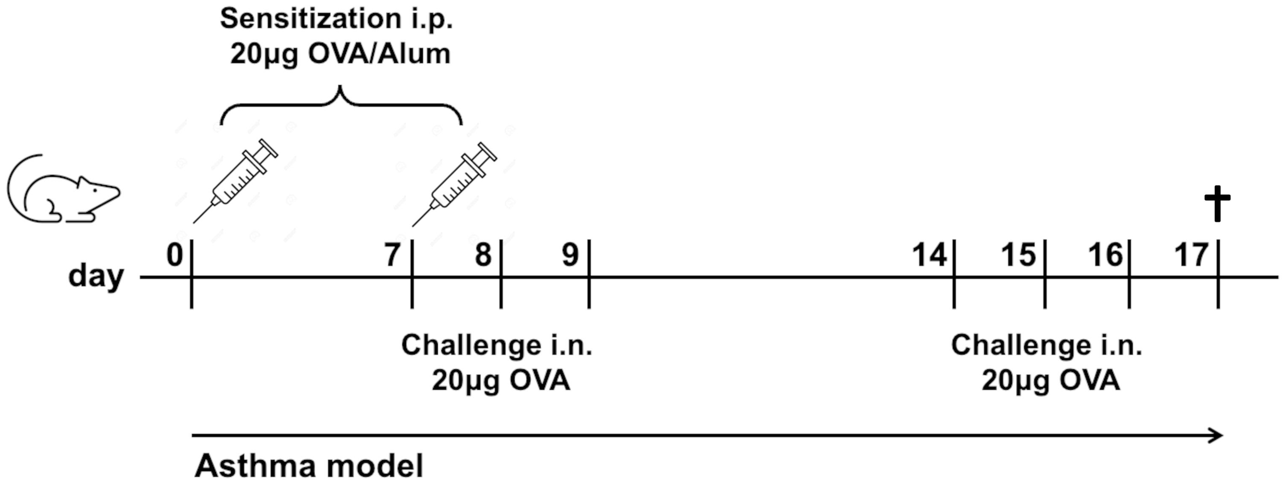
Experimental mouse model of allergic asthma. Mice were sensitized twice i.p. with OVA in Alum/NaCl 0.9% and challenged six times i.n. with OVA/NaCl 0.9%. Control mice received Alum/NaCl 0.9%, resp. NaCl 0.9% only. Mice were sacrificed one day after the last OVA challenge.
Measurement of lung function
Invasive lung function was assessed, as described previously (44, 45). In brief, AHR was defined as an increase in dynamic lung resistance (R) in response to increasing concentrations of MeCh (Sigma-Aldrich; 10, 20, and 30 mg/ml). One day after the last allergen challenge, mice were anesthetized with ketamine (Albrecht GmbH, Aulendorf, Germany) and xylazine (Rompun, Bayer Vital GmbH, Leverkusen, Germany), intubated, and mechanically ventilated via the flexiVent system (Scireq, Montreal, QC, Canada) using a tidal volume of 12 ml/kg at a frequency of 150 breaths/min. Baseline resistance was measured after exposure to 0.9% (w/v) NaCl, aerosolized in the Aeroneb nebulizer (Inspiration Medical, Bochum, Germany). Subsequently, increasing concentrations of methacholine (MeCh; 10, 20, and 30 mg/ml) were used to induce AHR. After deep inflation of the lungs, single frequency forced oscillations were performed; four peak values of resistance were analyzed for each MeCh concentration.
Isolation of mouse primary lung cells
Lungs were digested and cells isolated as described elsewhere (46). Briefly, lungs were digested according to the respective investigation, dispase-based digestion (DispaseII; Sigma-Aldrich, Munich, Germany) for epithelial and collagenase-based digestion (Merck, Darmstadt, Germany) for all other analysis. Erythrocytes were lysed, and cells filtered through a 70 μm strainer (BD, Franklin Lakes, NJ, USA), counted, and resuspended in complete medium (RPMI1640, 10% FCS, 100 U/ml penicillin, and 100 µg/ml streptomycin (RPMI: Bio&Sell, Feucht, Germany; Pen/Strep: Sigma-Aldrich, Munich, Germany; FCS: Biochrom, Cambridge, UK).
Analysis of bronchoalveolar lavage fluid
BALF was collected by flushing the right lung twice with 0.8 ml 2 mM EDTA in PBS. Total cells were counted using the Cedex HiRes automated analyzer (Roche, Basel, Switzerland). Differential cell counts were determined by staining cytospin preparations with May-Grünwald/Giemsa (Merck, Darmstadt, Germany) using standard microscopic criteria and counted in a blinded manner at 100× magnification (Axiovert 40 CFL, Zeiss, Oberkochen, Germany).
Lung histopathological examination
Immediately after collection of the BALF samples, lung tissues were fixed in 4% paraformaldehyde and then embedded in paraffin before sagittal cutting into 5 μm sections. Sections were stained with HE or PAS to analyze the inflammatory cells infiltration and global cell hyperplasia, respectively. Quantification was performed as described elsewhere (47). In short, PAS-stained bronchi were scored on a scale of 0 to 4, with 0 being negative and 1–4 being positive. The quantity of mucus production in positive bronchi was scored as follows: 1, 5– 25%; 2, 25– 50%; 3, 50 – 75%; and 4, >75%. Peribronchiolar and perivascular inflammation stained with HE was scored as follows: 0, normal; 1, few cells; 2, a ring of inflammatory cells one– cell layer deep; 3, a ring of inflammatory cells two- to four-cells deep; and 4, a ring of inflammatory cells of more than four-cells deep.
Flow cytometry analysis
Immunophenotyping of cells isolated from lung was performed using multicolor flow cytometry. Briefly, cells were blocked prior antibody staining for 15 min with a Fcγ receptor antibody (unlabeled CD16/32, clone 2.4G2; BD Biosciences, Franklin Lakes, New Jersey, USA). Exclusion of dead cells was ensured by using the LIVE/DEAD™ Fixable Aqua Dead Cell Stain Kit (ThermoFisher). Cells were stained with specific antibody panels on ice for 30 minutes (Supplementary Table S1) washed, resuspended in 0.1% BSA/PBS (Lonza, Basel, Switzerland), and acquired on a FACS Canto II (BD Biosciences) or CytoflexS (Beckman Coulter) flow cytometer. Data were analyzed using FlowJo software V10 (FlowJo LLC, Ashland, Oregon, USA).
Cytokine production
Lung cells and splenocytes were re-stimulated in vitro (1 x 10⁶ cells/ml) with OVA (200 µg/ml) in RPMI (supplemented with 10%FCS, 100 U/ml penicillin, and 100 µg/ml streptomycin) for 72h. Cytokines were measured in supernatants using LEGENDplex™ MU Th Cytokine Panel (12-plex) (BioLegend, San Diego, USA), according to the manufacturer’s instructions.
Measurement of OVA uptake
WT and NLRP3-/- mice received a single i.n. dose of OVA-FITC (20 μg in 40 μl 0.9% NaCl; nanocs, NY, USA). Control mice received 0.9% NaCl instead of OVA. After 2h mice were sacrificed, lung cells were isolated and measured by flow cytometry. The amount of OVA internalized and processed by cDCs was quantified as FITC-positive cell frequency as well as mean fluorescence intensity (MFI).
Gene expression analysis
Total RNA was extracted from lung cells using RNeasy Plus Mini or Micro Kit (Qiagen, Venlo, Netherlands) and reverse-transcribed with the High-Capacity cDNA Reverse Transcription Kit (ThermoFisher, Waltham, MA, USA). Quantitative PCR was performed in a 7500 Fast Real-Time PCR System (Applied Biosystems) using SYBR Green PCR Master Mix (ThermoFisher) and QuantiTect Primer Assays (Qiagen, Supplementary Table S2). Expression of genes was normalized to the house-keeping gene GAPDH using 2−ΔCt algorithm.
Cell culture and in vitro inhibition of NLRP3
Cells of the virus-immortalized human respiratory epithelial cell line 16HBE14o- (kindly provided by D. Gruenert (48)) were cultured on PureCol-coated plates (1:100; Cellsystems, Troisdorf, Germany) in MEM with 10% FCS, 1% Pen/Strep, and 1% L-Glutamine (all Thermo Fisher Scientific, Waltham, Massachusetts, USA). In vitro inhibition of NLRP3 was performed using the inhibitor MCC950 (Thermo Fisher, Waltham, Massachusetts, USA) at a concentration of 10µM and DMSO as mock control. In brief, cells were grown to 80% confluency, split, and seeded in petri dishes. Medium and inhibitor were exchanged every two days and finally harvested after 7 days. For RNA isolation, cells were snap-frozen in the gaseous phase of liquid N2 and stored at − 80 °C. RNA was extracted using Qiagen RNA easy Mini Kit and RNase-free DNase Set (both Qiagen, Hilden, Germany).
Statistical analysis
Statistical significance was assessed using an unpaired two-tailed Student’s t-test, two-way ANOVA or one-way ANOVA test with Bonferroni’s multiple comparison post-test (95% confidence interval; GraphPad Prism V9.5). Results are shown as mean +/- SEM. P values less than 0.05 were considered significant [(P < 0.05 (∗), P < 0.01 (∗∗), P < 0.001 (∗∗∗), and P < 0.0001 (∗∗∗∗)].
Results
AHR and mucus production are significantly increased in NLRP3-/- compared to WT mice in a model of allergic asthma
To analyze the impact of NLRP3 on the development of type 2-driven experimental allergic asthma, we compared NLRP3-/- and WT mice in a well-established murine asthma model (Figure 1).
Following our OVA sensitization and challenge protocol (see Figure 1), both NLRP3-/- and WT mice developed significantly increased AHR compared to their naïve control counterparts. Notably, resistance measured by invasive lung function was higher in NLRP3-/- compared to WT animals (Figures 2A, B). Similarly, PAS staining of lung sections from immunized versus naïve NLRP3-/- and WT mice revealed an increase in mucus production after OVA-immunization compared to naïve controls (Figure 2C). Similar to the observation of AHR, the increase in mucus production was significantly stronger in immunized NLRP3-/- mice compared to WT mice. Correspondingly, measurement of gene expression of MUC5AC, one of the major components of airway mucus (32), which was significantly increased in both immunized WT and NLRP3-/- mice, whereas it was again significantly higher in immunized NLRP3-/- compared to WT animals (Figure 2D). Re-stimulation of immunized NLRP3-/- splenocytes and lung cells with OVA revealed significantly increased systemic IL-5 and IL-13 levels in splenocyte cultures compared to immunized WT mice and a trend toward higher IL-5 and IL-13 levels in lung cell supernatants from NLRP3-/- compared to WT mice (Supplementary Figures S1D, S2). Cytokine levels in BALF remained below the detection limit (data not shown). Our data suggest an important role for NLRP3 in lung function and mucus production regulation.
Figure 2
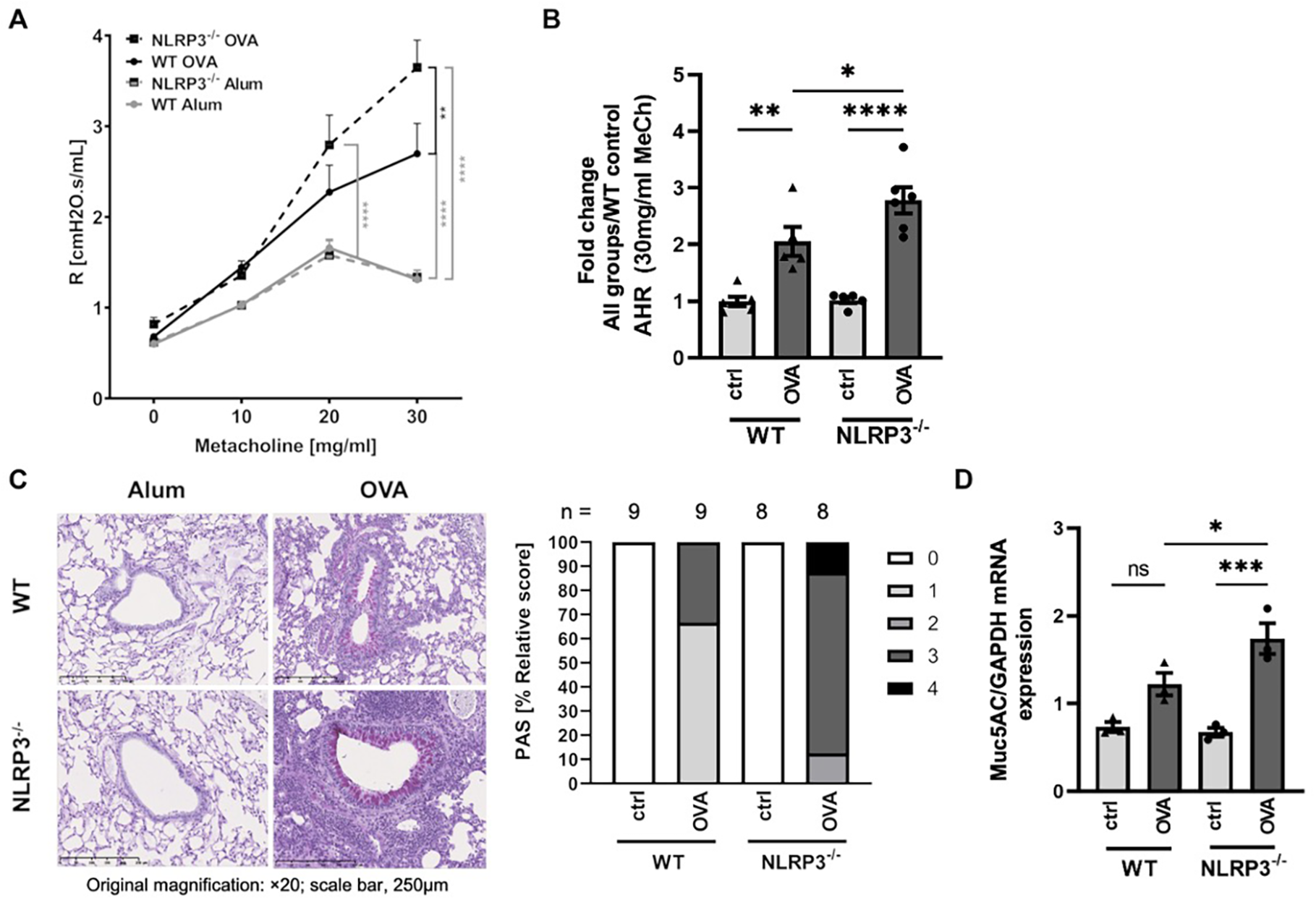
Increased AHR and mucus production in OVA-immunized NLRP3-/- compared to WT mice in an experimental model of allergic asthma. (A) Airway resistance (R) in OVA-immunized (OVA) and control (Alum) WT and NLRP3-/- mice, measured after challenge with metacholine (MeCh, n≥6/group). (B) Airway resistance values for 30 mg/ml MeCh as seen in A; Values are shown as fold change calculated relative to mean of WT control mice. (C) Levels of mucus production as determined by PAS staining of lung sections. The degree of mucus production in positive bronchi was graded as follows: 1: 5-25%; 2: 25-50%; 3: 50-75%, and 4: 75-100%. Original magnification, ×20; scale bar, 250μm; representatives of n≥8/group. (D) Gene expression of MUC5AC (n≥3/group, relative to GAPDH) in whole lung tissue, evaluated by RT-PCR. (A) Two-way ANOVA; (B&D) One-way ANOVA; mean ± SEM. ns not significant, *P < 0.05, **P < 0.01, ***P < 0.001 und ****P < 0.0001.
Eosinophilic airway inflammation is comparable in immunized NLRP3-/- and WT mice
In addition to increased AHR and mucus hypersecretion, allergic airway disease is characterized by eosinophilic lung inflammation. Analysis of the bronchoalveolar lavage fluid (BALF) from immunized and naïve NLRP3-/- and WT mice revealed a significant influx of cells, especially eosinophils, in both immunized NLRP3-/- and WT mice compared to naïve controls. In contrast to the findings for AHR and mucus hypersecretion, the eosinophilic inflammation was comparable in in NLRP3-/- and WT animals after OVA immunization (Supplementary Figure S1A). We observed an increase in lymphocyte counts in OVA-sensitized NLRP3-/- mice compared to controls. However, this difference was not statistically significant (WT OVA vs NLRP3-/- OVA, p = 0.2420). The Th2 cytokines IL-4, IL-5 and IL-13 in the BALF of mice were below the detection limit. We further assessed lung inflammation in H&E-stained lung sections, which confirmed a comparable eosinophilic inflammation in OVA-immunized WT and NLRP3-/- mice (Supplementary Figures S1B, C). IL-5 secretion, which plays an important role in eosinophil recruitment, was similar in OVA-stimulated lung cell cultures from immunized NLRP3-/- and WT mice (Supplementary Figure S1D). Our data show that eosinophilic airway inflammation in a model of experimental allergic asthma is not mediated by NLRP3.
NLRP3 regulates the expression of cell contact-related proteins in the airway epithelial barrier
Disruption of the epithelial layer, accompanied with enhanced epithelial permeability, is a frequently observed feature of allergic airway disease (37–39, 49). Previous studies have demonstrated the importance of intercellular cell-cell adhesion proteins for a functional epithelial barrier (50–54). Since our findings of strongly increased AHR and mucus hypersecretion in NLRP3-/- compared to WT mice primarily involve changes in the epithelial barrier function, we aimed to investigate structurally relevant components, such as tight junction and transmembrane proteins, along with the efficiency of barrier integrity. Gene expression analysis of the intercellular transmembrane proteins Claudin-18 (Cld-18) and tight junction protein-1 (Tjp-1) in total lung cells showed that their expression was significantly decreased in naïve NLRP3-/- mice compared to naïve WT mice (Figures 3A, B). Similarly, the expression of the cell-cell adhesion protein E-cadherin on CD45-CD31-CD326+ epithelial cells was significantly reduced in naïve NLRP3-/- compared to naïve WT mice (Figures 3C, D). However, gene expression of Cld-18 and Tjp-1 was comparable between OVA-treated NLRP3-/- and WT animals (Figures 3A, B).
Figure 3
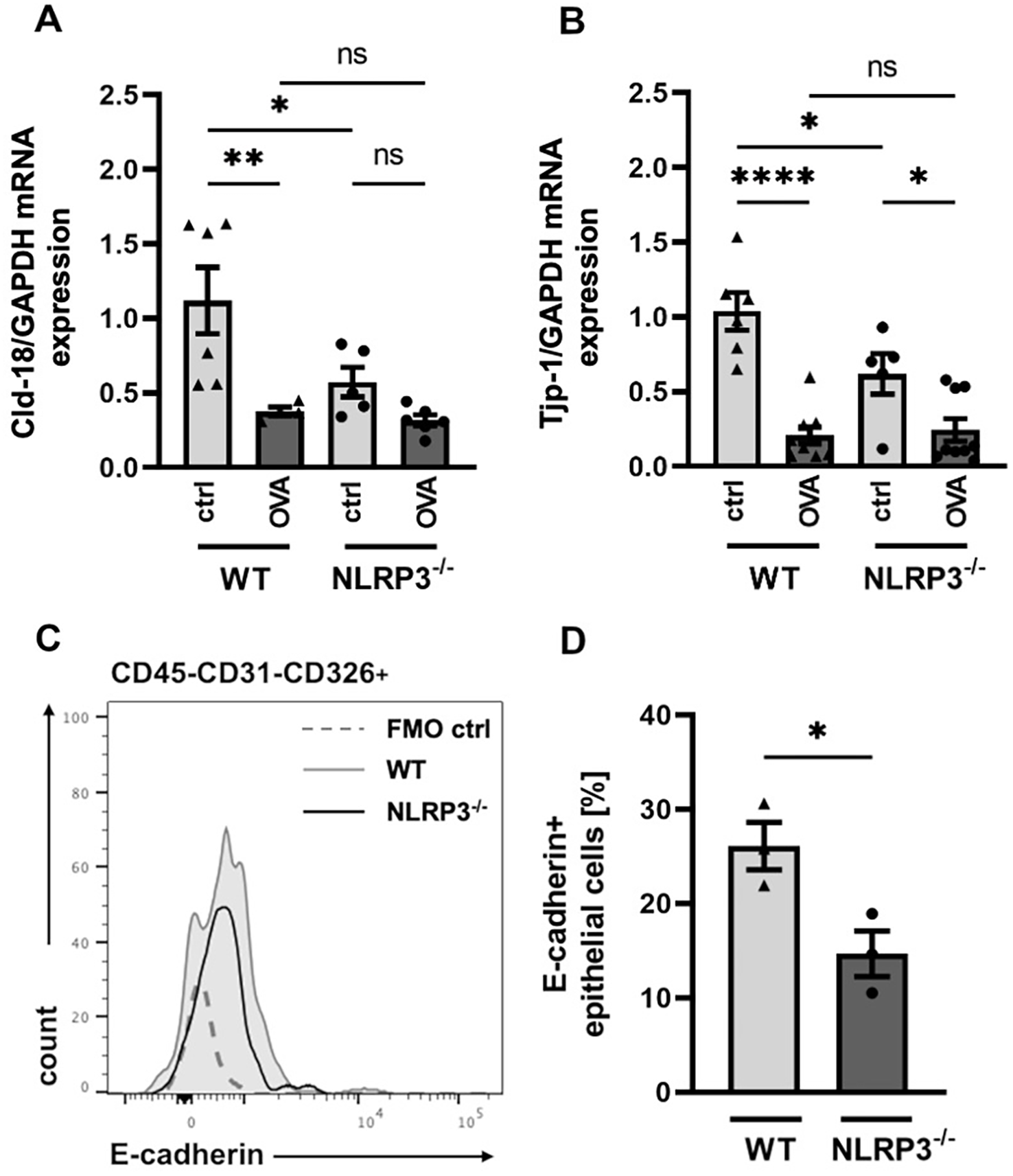
Significant differences in the expression of cell contact relevant proteins in the airway epithelial barrier of NLRP3-/- and WT mice. (A, B) Relative gene expression of Cld-18 (Claudin-18) and the tight junction protein Tjp-1 in WT and NLRP3-/- mice evaluated by RT-PCR in relation to GAPDH (n≥5/group). (C) Expression of E-cadherin on CD45-CD31-CD326+ epithelial cells isolated from lungs of WT (black line) and NLRP3-/- mice (grey shaded line); (FMO: dotted line). One representative of n=3/group. (D) Frequency of E-cadherin+ epithelial cells in whole lung tissue of WT and NLRP3-/- mice (n=3/group); unpaired two-tailed Student’s t-test; mean ± SEM. *P < 0.05, **P < 0.01, ****P < 0.0001.
Conventional dendritic cells from NLRP3-/- mice take up more allergen than WT mice following treatment with intra-nasal OVA
As outlined before (see Figure 3), we found that the gene expression of Cld-18, and Tjp-1, as well as E-cadherin protein expression was lower in NLRP3-/- compared to WT mice. These genes encode proteins that are essential for an intact cell-cell contact and a functioning epithelial barrier. To test whether this dysregulation of the epithelial barrier integrity results in increased epithelial permeability for allergens, we assessed the allergen uptake capacity of the airway epithelium in WT and NLRP3-/- mice. Intranasal application of FITC-labelled OVA resulted in significantly higher allergen uptake by lung resident cDCs in NLRP3-/- mice compared to WT mice (Figures 4A–C), while the frequency of cDCs was comparable in both mouse strains (Figure 4D). Our results demonstrate that NLRP3 plays a critical role for the integrity of the epithelial barrier protecting against allergen uptake and possibly allergic sensitization.
Figure 4
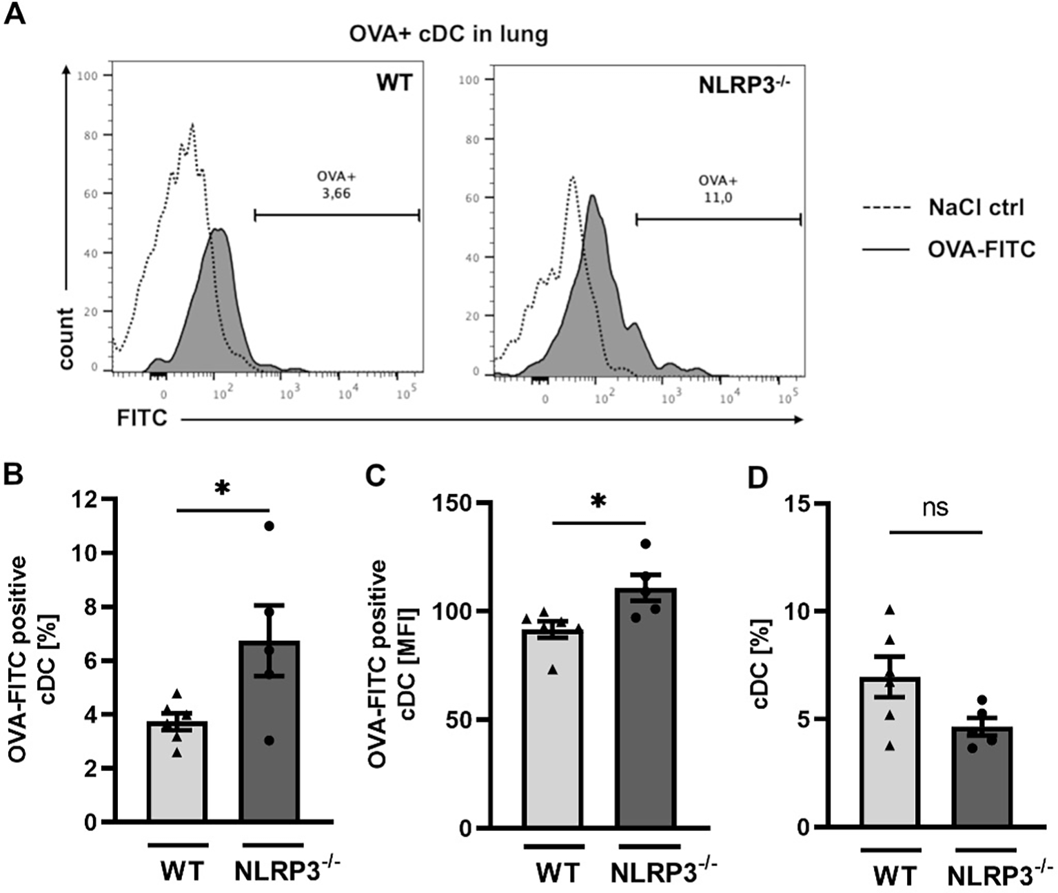
Increased OVA-uptake by conventional dendritic cells in NLRP3-/- compared to WT mice. (A-C) OVA uptake by WT and NLRP3-/- lung cDCs two hours after i.n. challenge with FITC-labeled OVA respectively. NaCl 0.9% was used as control. (A) Expression of FITC-OVA+ cDCs (black tinted) or control cDCs (dashed) determined by flow cytometry. Representatives of n≥5/group; (B) Frequencies and C mean fluorescence intensity (MFI) of FITC-OVA+ WT and NLRP3-/- cDCs (n≥5/group); (D) Frequencies of lung cDCs in the same mice analyzed in (B) and (C); unpaired two-tailed Student’s t-test; Mean ± SEM. ns not significant, *P < 0.05.
NLRP3 regulates tight junction protein and E-cadherin gene expression in human lung epithelial cell line 16HBE14o-
Having observed altered gene expression of proteins important for cell-cell contact in the lungs of NLRP3-/- mice, we tested our observation in a human lung epithelial cell line to see whether we could find evidence for a similar effect in humans. We added the NLRP3 inhibitor MCC950 to 16HBE14o- epithelial cells and analyzed gene expression of Tjp-1 and E-cadherin (CDH1). One week after continuous application of MCC950, both genes were downregulated in MCC950-treated human epithelial cells compared to mock-treated cells (Figures 5A, B). This finding suggests that NLRP3 has a regulatory and stabilizing effect on the expression of cell-cell contact-related proteins in mice and humans.
Figure 5
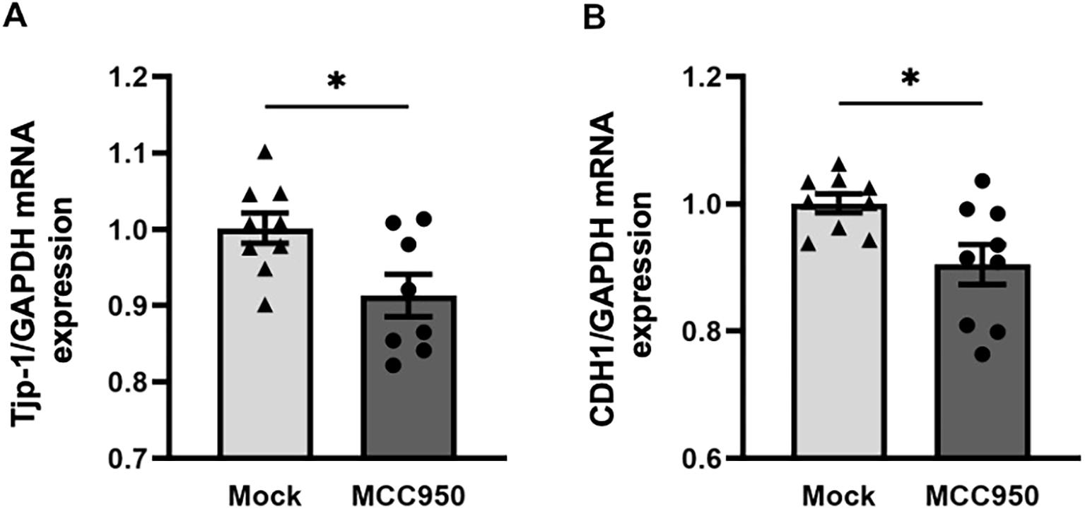
Application of the NLRP3 inhibitor MCC950 reduces the gene expression of tight junction protein 1 and E-cadherin in the human bronchial cell line 16HBE14o-. (A) Relative gene expression of the tight junction protein Tjp-1 and (B) the transmembrane protein CDH1 (E-cadherin) in cell lysates of the human bronchial cell line 16HBE14o- before and 7 days after treatment with the NLRP3 inhibitor MCC950 or DMSO. Expression was evaluated by RT-PCR in relation to GAPDH (n=9/group); Mock: DMSO, NLRP3 inhibitor: MCC950; unpaired two-tailed Student’s t-test; mean ± SEM. ns not significant, *P < 0.05.
Discussion
This study examined the role of the inflammasome NLRP3 in type 2 airway inflammation using OVA-sensitized NLRP3-/- and WT mice. Compared to WT mice, NLRP3-/- mice displayed significantly increased airway hyperresponsiveness (AHR), mucus production, and elevated systemic levels of IL-5 and IL-13. Naïve NLRP3-/- mice showed reduced expression of tight junction-associated genes and E-cadherin protein, indicating inherent epithelial barrier dysfunction. Functional assays revealed increased allergen uptake in NLRP3-/- mice, linking barrier disruption to the observed phenotypic changes. However, eosinophilic airway inflammation remained similar across genotypes, suggesting that this aspect of asthma pathophysiology is independent of NLRP3 expression. These observations contrast with prior studies, underscoring the context-dependent effects of NLRP3 in type 2 airway inflammation.
Previous studies investigating the specific roles of the NLRP3 inflammasome in asthma pathogenesis have utilized various murine asthma models, yielding inconsistent results. NLRP3 has been shown to have detrimental, protective, or negligible effects on AHR, airway remodeling, and type 2 immune responses (14–25). In models using OVA, NLRP3-/- mice mostly demonstrated significantly reduced inflammation and airway hyperresponsiveness compared to WT mice. Ritter et al. highlighted that NLRP3-deficient mice showed diminished levels of IL-1β, a crucial cytokine produced via the NLRP3 pathway (20). This indicates the pathway’s involvement in mediating the inflammatory response during acute allergic airway inflammation. Liu et al. also found reduced eosinophilic inflammation in NLRP3-deficient mice, primarily affecting the Th2 cytokine profile within the airways. This finding reinforces the idea that NLRP3 plays a pivotal role in promoting airway inflammation (22). However, Allen et al. showed that NLRP3 had no significant effect on airway hyperresponsiveness in allergic mice demonstrating that even in similar OVA challenge models results with NLRP3-/- mice have been inconsistent (23). This variability can be attributed to differences in mouse strains, host microbiota composition, and experimental methodologies, including allergen administration protocols, preparation techniques, and timing (23, 24, 55). Contrasting findings emerged in the context of neutrophilic asthma models induced by house dust mite (HDM). Studies have shown that inflammation can still occur through different pathways even in the absence of NLRP3, and that the innate immune response remains intact. Specifically, Tan et al. reported that, although NLRP3 inflammasome inhibition effectively mitigated HDM-induced inflammation, this did not result in the complete abrogation of asthma-like symptoms. These differential responses emphasize the redundant mechanisms that can compensate for NLRP3 deficiency, particularly in models that do not produce a classic Th2 response but instead exhibit a neutrophilic phenotype (56). The results obtained from different experimental setups partly illustrate the differences in experimental design previously outlined and underscore the need for standardized approaches to clarify the role of NLRP3 in airway inflammation. Furthermore, they imply that NLRP3 plays a pivotal role in asthma development, with its effects being partially reflective of the various underlying immune mechanisms of this heterogeneous disease, and that targeting NLRP3 could lead to different therapeutic outcomes depending on the asthma subtype. Further research is needed to decipher the multiple NLRP3-related pathways with the aim of tailoring inflammasome-targeting therapeutic strategies to individual asthma phenotypes.
Our data reveal an unexpected phenotype in naïve NLRP3-/- mice, suggesting a baseline defect in epithelial barrier integrity independent of inflammatory stimuli. This was not the primary focus of our study but emerged as a robust and reproducible observation. In the context of findings from other models and tissues, our data underscore a broader, homeostatic role for NLRP3 in maintaining epithelial stability. Notably, the epithelial barrier defect observed in NLRP3-/- mice may reflect a combination of canonical inflammasome-dependent and inflammasome-independent functions of NLRP3. While the activation of the NLRP3 inflammasome and subsequent release of IL-1β and IL-18 release have been linked to epithelial repair and barrier maintenance, accumulating evidence suggests that NLRP3 also operates beyond its classical inflammasome role. For instance, it can directly modulate TGF-β/Smad signaling or act as a nuclear regulator of IL-33 expression in epithelial cells, thereby affecting cell adhesion, tight-junction organization, and overall epithelial stability independently of caspase-1 or IL-1β/IL-18 (57–59). Distinguishing these pathways will be crucial when considering NLRP3-targeted strategies in asthma, since barrier-stabilizing effects may not solely reflect inflammasome inhibition but also loss of homeostatic epithelial functions of NLRP3. To further support this concept, evidence from other organ systems underscores the importance of NLRP3 in maintaining epithelial barrier integrity beyond the lung.
Beyond the variability in experimental factors, studies on gastrointestinal diseases highlight a role for NLRP3 in maintaining epithelial barrier integrity, which may also be relevant to the airways. In conditions such as Crohn’s disease, ulcerative colitis, renal disease, as well as infections with pathogens NLRP3 and its downstream cytokine IL-1β are essential for preserving intestinal epithelial structure, promoting barrier repair, and ensuring tissue homeostasis (41–43, 60, 61). Similarly, research by Kostadinova et al. demonstrated that NLRP3 expression in myeloid cells enhances alveolar epithelial integrity by promoting cellular adhesion in a Streptococcus pneumoniae-induced pneumonia model (40). These findings suggest that NLRP3 may play a broader role in epithelial barrier maintenance across organ systems, including the respiratory tract.
Epithelial barrier dysfunction is closely associated with dysregulated airway resistance and excessive mucus production, with epithelial damage correlating with AHR severity (37–39). The airway epithelium, composed of tightly interconnected monolayers, acts as a first-line defense against pathogens while maintaining innate immunity and tissue homeostasis in the lung and intestinal tract (62–67). Increased epithelial permeability has been linked to the downregulation of E-cadherin, a key cell-cell adhesion molecule, and the disruption of tight junction complexes, which are anchored to the actin cytoskeleton, such as Tjp-1 and claudin-18 (49, 54, 68). Their integrity is closely tied to the expression and functional activity of E-cadherin and is essential for regulating molecular diffusion across tissues and maintaining epithelial barrier function (51, 53, 68, 69). These fundamental mechanisms of epithelial barrier regulation provide the context to interpret the intrinsic defects observed in the naïve NLRP3-/- mice in our study.
Our findings of significantly reduced expression of tight junction-related genes and E-cadherin protein, coupled with increased allergen uptake in naïve NLRP3-/- mice, suggest an intrinsic compromise in epithelial barrier integrity in these mice. The overall low expression of tight junction (TJ) genes observed in OVA-sensitized and challenged WT and NLRP3-/- mice likely reflects allergen-induced epithelial barrier disruption, exacerbated by inflammatory cytokines and cellular stress responses (56, 70, 71). While Besnard et al. reported impaired DC recruitment to the draining lymph nodes in NLRP3-deficient mice, our OVA-FITC analysis in the lung did not reveal a major reduction of cDCs, making it more likely that the enhanced antigen signal we observed reflects increased epithelial leakiness rather than differences in DC antigen uptake capacity (19). Furthermore, IL-13 has been shown to impair TJ integrity in airway epithelial cells, as demonstrated by Wise et al., supporting our finding of significantly elevated IL-13 levels in both WT and NLRP3-/- mice following OVA sensitization (72).
Previous research has shown that TJs are critical for maintaining epithelial barrier integrity by regulating ion and solute flux while preventing uncontrolled mucin and protein efflux, particularly from goblet cells. In our NLRP3-/- mice, TJ disruption likely increases mucosal permeability, compromising selective barrier function and leading to excessive mucus secretion (52, 73–75). Therefore, our findings align with previous studies and suggest an important role of NLRP3 in the expression of TJs and, therefore, the maintenance of epithelial barrier function and regulating mucus production in response to inflammatory stimuli.
To investigate the cellular and molecular mechanisms governing airway tight junctions, epithelial barrier properties, and mucus production, immortalized human bronchial epithelial cell lines such as 16HBE14o- have been extensively utilized (76, 77). These cells, which exhibit characteristics of superficial epithelial cells, were originally derived through viral transformation of cells from human bronchial epithelium. We observed that treatment of 16HBE14o- cell monolayers with the highly specific NLRP3 inhibitor MCC950 (78–80) led to a significant reduction in the expression of Tjp-1 and CHD1 genes, further emphasizing the critical role of NLRP3 in maintaining cell-cell adhesion within the epithelial layer.
These findings likely reflect context- and model-dependent roles of epithelial NLRP3. In inflamed, primed airways, Toll-like receptor (TLR) ligands, allergens, and cytokines have been shown to upregulate NLRP3 and pro-IL-1β, thereby facilitating inflammasome activation (81, 82). In such settings, the inhibition of NLRP3 has been observed to offer protective effects (79). Conversely, in minimally primed monolayers, basal NLRP3 may contribute to junctional homeostasis, suggesting that acute blockade can transiently reduce Tjp-1 and CDH1. Varying results can also arise from differences between immortalized lines and well-differentiated primary human bronchial epithelial cells (HBECs), species and asthma endotypes, and from pharmacologic versus genetic disruptions (83). Furthermore, evidence suggests that epithelial-to-mesenchymal transition (EMT) and oxidative stress, driven by epidermal growth factor receptor (EGFR) and transforming growth factor beta (TGF−β), respectively, independently regulate tight junctions, providing an alternative explanation for the reduced expression of Tjp-1 and CDH1 following NLRP3 inhibition (84).
Combined with earlier studies that link NLRP3 expression to the integrity of the epithelial barrier, our findings indicate that NLRP3 is a crucial regulator of epithelial stability and barrier function. Importantly, we demonstrate here that naïve NLRP3-/- mice exhibit intrinsic defects in tight junction expression and E-cadherin levels, leading to increased allergen uptake, a baseline phenotype not previously reported in asthma models. This role highlights NLRP3 as an essential factor in epithelial homeostasis and underscores its potential as a therapeutic target for interventions to improve barrier integrity in respiratory diseases, such as asthma. Therapeutic strategies targeting NLRP3 in asthma will need to consider its dual role in both inflammation and epithelial barrier homeostasis. Selective modulation of inflammasome-dependent versus homeostatic functions may allow preservation of epithelial integrity while limiting type 2 airway inflammation. Studies in human airway models will be crucial for translating these insights into effective clinical interventions. In comparison to previous studies that focused on the impact of NLRP3 on epithelial permeability and mucus production during inflammation, our results support the hypothesis that NLRP3 is integral to maintaining both the structural and functional properties of epithelial barriers across various tissues.
Statements
Data availability statement
The original contributions presented in the study are included in the article/Supplementary Material. Further inquiries can be directed to the corresponding author.
Ethics statement
Ethical approval was not required for the studies on humans in accordance with the local legislation and institutional requirements because only commercially available established cell lines were used. The animal study was approved by Lower Saxony State Office for Consumer Protection and Food Safety (LAVES) Permit: 17/2548. The study was conducted in accordance with the local legislation and institutional requirements.
Author contributions
SD: Writing – review & editing, Formal analysis, Supervision, Methodology, Writing – original draft, Data curation, Visualization, Investigation, Conceptualization, Validation. RG: Investigation, Writing – review & editing. DF: Writing – review & editing. AH: Writing – review & editing. ST: Writing – review & editing, Methodology, Validation, Investigation. FS: Writing – review & editing, Methodology, Conceptualization. WG: Writing – review & editing. KB: Writing – review & editing. EH: Writing – review & editing. MK: Writing – review & editing. EV: Writing – review & editing. BS: Writing – review & editing. AJ: Writing – review & editing, Conceptualization, Methodology, Validation. GH: Conceptualization, Supervision, Writing – review & editing, Resources, Funding acquisition, Project administration.
Funding
The author(s) declare financial support was received for the research and/or publication of this article. This work was supported by the BMBF grant CHAMP (CHildhood Allergy and tolerance: Biomarkers and Predictors, 01GL174, 01GL1742A-F), the DZL (German Center for Lung Research, FKZ 82DZL002B1 and FKZ 82DZL002C1) and the Excellence Cluster RESIST (Cluster Resolving Infection Susceptibility, Germany’s Excellence Strategy-EXC 2155-project number 390874280).
Acknowledgments
The authors thank Jana Bergmann and Anika Dreier for their excellent technical assistance, Dr. João Tereno Monteiro for his invaluable advice and feedback, and Priv.-Doz. Dr. Susanne Rittinghausen (Fraunhofer ITEM, Hannover) for permission to document our immunohistochemical analysis. We also gratefully acknowledge the financial support from the BMBF grant CHAMP (CHildhood Allergy and tolerance: Biomarkers and Predictors, 01GL174, 01GL1742A-F), the DZL/BREATH (German Center for Lung Research/Biomedical Research in Endstage & Obstructive Lung Disease, FKZ 82DZL002B1 and FKZ 82DZL002C1), and the Excellence Cluster RESIST (Cluster Resolving Infection Susceptibility, Germany’s Excellence Strategy-EXC 2155-project number 390874280), which made this work possible.
Conflict of interest
The authors declare that the research was conducted in the absence of any commercial or financial relationships that could be construed as a potential conflict of interest.
Generative AI statement
The author(s) declare that no Generative AI was used in the creation of this manuscript.
Any alternative text (alt text) provided alongside figures in this article has been generated by Frontiers with the support of artificial intelligence and reasonable efforts have been made to ensure accuracy, including review by the authors wherever possible. If you identify any issues, please contact us.
Publisher’s note
All claims expressed in this article are solely those of the authors and do not necessarily represent those of their affiliated organizations, or those of the publisher, the editors and the reviewers. Any product that may be evaluated in this article, or claim that may be made by its manufacturer, is not guaranteed or endorsed by the publisher.
Supplementary material
The Supplementary Material for this article can be found online at: https://www.frontiersin.org/articles/10.3389/fimmu.2025.1655205/full#supplementary-material
Abbreviations
ALUM, aluminum hydroxide and magnesium hydroxide solution; AHR, airway hyperresponsiveness; BALF, bronchoalveolar lavage fluid; cDC, conventional dendritic cell; cDNA, complementary deoxyribonucleic acid; Cld-18, Claudin-18; DC, dendritic cell; FACS, Fluorescence Activated Cell Sorting; HE, hematoxylin and eosin; HDM, house dust mite; i.n., intranasally; i.p., intraperitoneally; MeCh, methacholine; MUC5AC, Mucin 5AC; NaCI, sodium chloride; NLRP3, NOD-like receptor family pyrin domain containing 3; NLRP3-/-, NLRP3 knock-out; ns, not significant; PAS, periodic acid–Schiff; PBS, phosphate buffered saline; PCR, polymerase chain reaction; R, resistance; RNA, ribonucleic acid; SEM, standard error of mean; SPF, Specific-pathogen free; Th2, type 2 T helper; Tjp-1, tight junction protein 1; WT, wild-type.
Group member of CHAMP study group
Gesine Hansen, Stephanie DeStefano, Bianca Schaub, Erika von Mutius, Andreas Böck, Jana Kristin Eckert, Kathrin Urner, Kathrin Kalkbrenner, Franziska Sattler, Sabine Schnadt, Julia Kahle, Wolfgang Greiner, Simone Kreimeier, Ole Marten, Kirsten Beyer, Young-Ae Lee, Josefine Dobbertin-Welsch, Eckard Hamelmann, Christina Schorlemer, Michael Kabesch, Parastoo Kheiroddin, Martin Depner, Daniel Walter.
References
1
Hammad H Lambrecht BN . The basic immunology of asthma. Cell. (2021) 184:1469–85. doi: 10.1016/j.cell.2021.02.016
2
Habener A Grychtol R Gaedcke S DeLuca D Dittrich AM Happle C et al . IgA + memory B-cells are significantly increased in patients with asthma and small airway dysfunction. Eur Respir J. (2022) 60:2102130. doi: 10.1183/13993003.02130-2021
3
Oppenheimer J Hoyte FCL Phipatanakul W Silver J Howarth P Lugogo NL . Allergic and eosinophilic asthma in the era of biomarkers and biologics: similarities, differences and misconceptions. Ann Allergy Asthma Immunol. (2022) 129:169–80. doi: 10.1016/j.anai.2022.02.021
4
GINA Report . Global Strategy for Asthma Management and Prevention (2023). Available online at: https://ginasthma.org/2023-gina-main-report/ (Accessed June 1, 2025).
5
Bradding P Porsbjerg C Côté A Dahlén SE Hallstrand TS Brightling CE . Airway hyperresponsiveness in asthma: The role of the epithelium. J Allergy Clin Immunol. (2024) 153:1181–93. doi: 10.1016/j.jaci.2024.02.011
6
Holguin F Cardet JC Chung KF Diver S Ferreira DS Fitzpatrick A et al . Management of severe asthma: a European Respiratory Society/American Thoracic Society guideline. Eur Respir J. (2020) 55:1900588. doi: 10.1183/13993003.00588-2019
7
Pasha MA Hopp RJ Habib N Tang DD . Biomarkers in asthma, potential for therapeutic intervention. J Asthma. (2024) 61:1376–91. doi: 10.1080/02770903.2024.2361783
8
Liston A Masters SL . Homeostasis-altering molecular processes as mechanisms of inflammasome activation. Nat Rev Immunol. (2017) 17:208–14. doi: 10.1038/nri.2016.151
9
Ali MF Dasari H Van Keulen VP Carmona EM . Canonical stimulation of the NLRP3 inflammasome by fungal antigens links innate and adaptive B-lymphocyte responses by modulating IL-1β and igM production. Front Immunol. (2017) 8:1504. doi: 10.3389/fimmu.2017.01504
10
Yashiro T Yamamoto M Araumi S Hara M Yogo K Uchida K et al . PU.1 and IRF8 modulate activation of NLRP3 inflammasome via regulating its expression in human macrophages. Front Immunol. (2021) 12:649572. doi: 10.3389/fimmu.2021.649572
11
Lebreton F Berishvili E Parnaud G Rouget C Bosco D Berney T et al . NLRP3 inflammasome is expressed and regulated in human islets. Cell Death Dis. (2018) 9:726. doi: 10.1038/s41419-018-0764-x
12
Zhang H Gao J Tang Y Jin T Tao J . Inflammasomes cross-talk with lymphocytes to connect the innate and adaptive immune response. J Adv Res. (2023) 54:181–93. doi: 10.1016/j.jare.2023.01.012
13
Hu X Liu S Jing Z He Y Qin G Jiang L . Immunomodulation in allergic rhinitis: Insights from Th2 cells and NLRP3/IL-18 pathway. Cell Biochem Funct. (2024) 42:e3997. doi: 10.1002/cbf.3997
14
Lachowicz-Scroggins ME Dunican EM Charbit AR Raymond W Looney MR Peters MC et al . Neutrophil extracellular traps, and inflammasome activation in severe asthma. Am J Respir Crit Care Med. (2019) 199:1076–85. doi: 10.1164/rccm.201810-1869OC
15
Kim RY Pinkerton JW Essilfie AT Robertson AAB Baines KJ Brown AC et al . Role for NLRP3 inflammasome–mediated, IL-1β–dependent responses in severe, steroid-resistant asthma. Am J Respir Crit Care Med. (2017) 196:283–97. doi: 10.1164/rccm.201609-1830OC
16
Wu Y Di X Zhao M Li H Bai L Wang K . The role of the NLRP3 inflammasome in chronic inflammation in asthma and chronic obstructive pulmonary disease. Immun Inflammation Dis. (2022) 10:e750. doi: 10.1002/iid3.750
17
Eisenbarth SC Colegio OR O’Connor W Sutterwala FS Flavell RA . Crucial role for the Nalp3 inflammasome in the immunostimulatory properties of aluminium adjuvants. Nature. (2008) 453:1122–6. doi: 10.1038/nature06939
18
Bruchard M Rebé C Derangère V Togbé D Ryffel B Boidot R et al . The receptor NLRP3 is a transcriptional regulator of TH2 differentiation. Nat Immunol. (2015) 16:859–70. doi: 10.1038/ni.3202
19
Besnard AG Guillou N Tschopp J Erard F Couillin I Iwakura Y et al . NLRP3 inflammasome is required in murine asthma in the absence of aluminum adjuvant: Role of NLRP3 inflammasome in murine asthma. Allergy. (2011) 66:1047–57. doi: 10.1111/j.1398-9995.2011.02586.x
20
Ritter M Straubinger K Schmidt S Busch DH Hagner S Garn H et al . Functional relevance of NLRP3 inflammasome-mediated interleukin (IL)-1β during acute allergic airway inflammation. Clin Exp Immunol. (2014) 178:212–23. doi: 10.1111/cei.12400
21
Ma M Li G Qi M Jiang W Zhou R . Inhibition of the inflammasome activity of NLRP3 attenuates HDM-induced allergic asthma. Front Immunol. (2021) 12:718779. doi: 10.3389/fimmu.2021.718779
22
Liu X Shen J Fan D Qiu X Guo Q Zheng K et al . Yupingfeng san inhibits NLRP3 inflammasome to attenuate the inflammatory response in asthma mice. Front Pharmacol. (2017) 8:944. doi: 10.3389/fphar.2017.00944
23
Allen IC Jania CM Wilson JE Tekeppe EM Hua X Brickey WJ et al . Analysis of NLRP3 in the development of allergic airway disease in mice. J Immunol. (2012) 188:2884–93. doi: 10.4049/jimmunol.1102488
24
Kool M Pétrilli V De Smedt T Rolaz A Hammad H Van Nimwegen M et al . Cutting edge: alum adjuvant stimulates inflammatory dendritic cells through activation of the NALP3 inflammasome. J Immunol. (2008) 181:3755–9. doi: 10.4049/jimmunol.181.6.3755
25
Madouri F Guillou N Fauconnier L Marchiol T Rouxel N Chenuet P et al . Caspase-1 activation by NLRP3 inflammasome dampens IL-33-dependent house dust mite-induced allergic lung inflammation. J Mol Cell Biol. (2015) 7:351–65. doi: 10.1093/jmcb/mjv012
26
Han M Ishikawa T Bermick JR Rajput C Lei J Goldsmith AM et al . IL-1β prevents ILC2 expansion, type 2 cytokine secretion, and mucus metaplasia in response to early-life rhinovirus infection in mice. Allergy. (2020) 75:2005–19. doi: 10.1111/all.14241
27
Han M Ishikawa T Stroupe CC Breckenridge HA Bentley JK Hershenson MB . Deficient inflammasome activation permits an exaggerated asthma phenotype in rhinovirus C-infected immature mice. Mucosal Immunol. (2021) 14:1369–80. doi: 10.1038/s41385-021-00436-0
28
Leszczyńska K Jakubczyk D Górska S . The NLRP3 inflammasome as a new target in respiratory disorders treatment. Front Immunol. (2022), 13:1006654. doi: 10.3389/fimmu.2022.1006654
29
Frey A Lunding LP Wegmann M . The dual role of the airway epithelium in asthma: active barrier and regulator of inflammation. Cells. (2023) 12:2208. doi: 10.3390/cells12182208
30
Kageyama T Ito T Tanaka S Nakajima H . Physiological and immunological barriers in the lung. Semin Immunopathol. (2024) 45:533–47. doi: 10.1007/s00281-024-01003-y
31
Button BM Button B . Structure and function of the mucus clearance system of the lung. Cold Spring Harb Perspect Med. (2013) 3:a009720–a009720. doi: 10.1101/cshperspect.a009720
32
Bonser L Erle D . Airway mucus and asthma: the role of MUC5AC and MUC5B. J Clin Med. (2017) 6:112. doi: 10.3390/jcm6120112
33
Rusu AD Georgiou M . The multifarious regulation of the apical junctional complex. Open Biol. (2020) 10:190278. doi: 10.1098/rsob.190278
34
Fu R Jiang X Li G Zhu Y Zhang H . Junctional complexes in epithelial cells: sentinels for extracellular insults and intracellular homeostasis. FEBS J. (2022) 289:7314–33. doi: 10.1111/febs.16174
35
Esteban Enjuto L Taty Poaty V Bouveret M Song H Constant S Patarin J . Rheological comparison of sputum and reconstituted airway epithelium mucus. Sci Rep. (2024) 14:31660. doi: 10.1038/s41598-024-80932-y
36
Cerutti A Filipska M Fa XM Tachó-Piñot R . Impact of the mucosal milieu on antibody responses to allergens. J Allergy Clin Immunol. (2022) 150:503–12. doi: 10.1016/j.jaci.2022.07.007
37
Carlier FM De Fays C Pilette C . Epithelial barrier dysfunction in chronic respiratory diseases. Front Physiol. (2021) 12:691227. doi: 10.3389/fphys.2021.691227
38
Gon Y Hashimoto S . Role of airway epithelial barrier dysfunction in pathogenesis of asthma. Allergol Int. (2018) 67:12–7. doi: 10.1016/j.alit.2017.08.011
39
Hellings PW Steelant B . Epithelial barriers in allergy and asthma. J Allergy Clin Immunol. (2020) 145:1499–509. doi: 10.1016/j.jaci.2020.04.010
40
Kostadinova E Chaput C Gutbier B Lippmann J Sander LE Mitchell TJ et al . NLRP3 protects alveolar barrier integrity by an inflammasome-independent increase of epithelial cell adherence. Sci Rep. (2016) 6:30943. doi: 10.1038/srep30943
41
Pulskens WP Butter LM Teske GJ Claessen N Dessing MC Flavell RA et al . Nlrp3 prevents early renal interstitial edema and vascular permeability in unilateral ureteral obstruction. Dussaule JC Editor PloS One. (2014) 9:e85775. doi: 10.1371/journal.pone.0085775
42
Song-Zhao GX Srinivasan N Pott J Baban D Frankel G Maloy KJ . Nlrp3 activation in the intestinal epithelium protects against a mucosal pathogen. Mucosal Immunol. (2014) 7:763–74. doi: 10.1038/mi.2013.94
43
Alipour M Lou Y Zimmerman D Bording-Jorgensen MW Sergi C Liu JJ et al . A Balanced IL-1β Activity Is Required for Host Response to Citrobacter rodentium Infection. Coers J, editor. PloS One. (2013) 8:e80656. doi: 10.1371/journal.pone.0080656
44
Happle C Jirmo AC Meyer-Bahlburg A Habener A Hoymann HG Hennig C et al . B cells control maternofetal priming of allergy and tolerance in a murine model of allergic airway inflammation. J Allergy Clin Immunol. (2018) 141:685–96. doi: 10.1016/j.jaci.2017.03.051
45
Glaab T Mitzner W Braun A Ernst H Korolewitz R Hohlfeld JM et al . Repetitive measurements of pulmonary mechanics to inhaled cholinergic challenge in spontaneously breathing mice. J Appl Physiol. (2004) 97:1104–11. doi: 10.1152/japplphysiol.01182.2003
46
Happle C Meyer-Decking L Dreier A Wetzke M Gläsener S Grychtol R et al . Improved protocol for simultaneous analysis of leukocyte subsets and epithelial cells from murine and human lung. Exp Lung Res. (2018) 44:127–36. doi: 10.1080/01902148.2018.1432721
47
Bandukwala HS Clay BS Tong J Mody PD Cannon JL Shilling RA et al . Signaling through FcγRIII is required for optimal T helper type (Th)2 responses and Th2-mediated airway inflammation. J Exp Med. (2007) 204:1875–89. doi: 10.1084/jem.20061134
48
Illek B Maurisse R Wahler L Kunzelmann K Fischer H Gruenert D . Cl transport in complemented CF bronchial epithelial cells correlates with CFTR mRNA expression levels. Cell Physiol Biochem. (2008) 22:57–68. doi: 10.1159/000149783
49
Xiao C Puddicombe SM Field S Haywood J Broughton-Head V Puxeddu I et al . Defective epithelial barrier function in asthma. J Allergy Clin Immunol. (2011) 128:549–56:e12. doi: 10.1164/ajrccm-conference.2011.A065
50
Wittekindt OH . Tight junctions in pulmonary epithelia during lung inflammation. Pflüg Arch Eur J Physiol. (2017) 469:135–47. doi: 10.1007/s00424-016-1917-3
51
West MR Ferguson DJP Hart VJ Sanjar S Man Y . Maintenance of the epithelial barrier in a bronchial epithelial cell line is dependent on functional E-cadherin local to the tight junctions. Cell Commun Adhes. (2002) 9:29–44. doi: 10.1080/15419060212185
52
Capaldo CT Powell DN Kalman D . Layered defense: how mucus and tight junctions seal the intestinal barrier. J Mol Med. (2017) 95:927–34. doi: 10.1007/s00109-017-1557-x
53
Goto Y Uchida Y Nomura A Sakamoto T Ishii Y Morishima Y et al . Dislocation of E-cadherin in the airway epithelium during an antigen-induced asthmatic response. Am J Respir Cell Mol Biol. (2000) 23:712–8. doi: 10.1165/ajrcmb.23.6.4031
54
Sweerus K Lachowicz-Scroggins M Gordon E LaFemina M Huang X Parikh M et al . Claudin-18 deficiency is associated with airway epithelial barrier dysfunction and asthma. J Allergy Clin Immunol. (2017) 139:72–81. doi: 10.1016/j.jaci.2016.02.035
55
Theofani E Semitekolou M Morianos I Samitas K Xanthou G . Targeting NLRP3 inflammasome activation in severe asthma. J Clin Med. (2019) 8:1615. doi: 10.3390/jcm8101615
56
Tan HT Hagner S Ruchti F Radzikowska U Tan G Altunbulakli C et al . Tight junction, mucin, and inflammasome-related molecules are differentially expressed in eosinophilic, mixed, and neutrophilic experimental asthma in mice. Allergy. (2019) 74:294–307. doi: 10.1111/all.13619
57
Zaki MH Boyd KL Vogel P Kastan MB Lamkanfi M Kanneganti TD . The NLRP3 Inflammasome Protects against Loss of Epithelial Integrity and Mortality during Experimental Colitis. Immunity. (2010) 32:379–91. doi: 10.1016/j.immuni.2010.03.003
58
Wang W Wang X Chun J Vilaysane A Clark S French G et al . Inflammasome-independent NLRP3 augments TGF-β Signaling in kidney epithelium. J Immunol. (2013) 190:1239–49. doi: 10.4049/jimmunol.1201959
59
Zheng J Yao L Zhou Y Gu X Wang C Bao K et al . A novel function of NLRP3 independent of inflammasome as a key transcription factor of IL-33 in epithelial cells of atopic dermatitis. Cell Death Dis. (2021) 12:871. doi: 10.1038/s41419-021-04159-9
60
Bording-Jorgensen M Armstrong H Wickenberg M LaPointe P Wine E . Macrophages and epithelial cells mutually interact through NLRP3 to clear infection and enhance the gastrointestinal barrier. Immuno. (2021) 2:13–25. doi: 10.3390/immuno2010002
61
Pellegrini C Antonioli L Lopez-Castejon G Blandizzi C Fornai M . Canonical and non-canonical activation of NLRP3 inflammasome at the crossroad between immune tolerance and intestinal inflammation. Front Immunol. (2017) 8:36/full. doi: 10.3389/fimmu.2017.00036/full
62
Draijer C Peters-Golden M . Alveolar macrophages in allergic asthma: the forgotten cell awakes. Curr Allergy Asthma Rep. (2017) 17:12. doi: 10.1007/s11882-017-0681-6
63
Bissonnette EY Lauzon-Joset JF Debley JS Ziegler SF . Cross-talk between alveolar macrophages and lung epithelial cells is essential to maintain lung homeostasis. Front Immunol. (2020) 11:583042. doi: 10.3389/fimmu.2020.583042
64
Yuksel H Turkeli A . Airway epithelial barrier dysfunction in the pathogenesis and prognosis of respiratory tract diseases in childhood and adulthood. Tissue Barriers. (2017) 5:e1367458. doi: 10.1080/21688370.2017.1367458
65
Peterson LW Artis D . Intestinal epithelial cells: regulators of barrier function and immune homeostasis. Nat Rev Immunol. (2014) 14:141–53. doi: 10.1038/nri3608
66
Johnston SL Goldblatt DL Evans SE Tuvim MJ Dickey BF . Airway epithelial innate immunity. Front Physiol. (2021) 12:749077. doi: 10.3389/fphys.2021.749077
67
Barclay AM Ninaber DK Van Veen S Hiemstra PS Ottenhoff THM van der Does AM et al . Airway epithelial cells mount an early response to mycobacterial infection. Front Cell Infect Microbiol. (2023), 13:1253037. doi: 10.3389/fcimb.2023.1253037
68
Post S Heijink IH Hesse L Koo HK Shaheen F Fouadi M et al . Characterization of a lung epithelium specific E-cadherin knock-out model: Implications for obstructive lung pathology. Sci Rep. (2018) 8:13275. doi: 10.1038/s41598-018-31500-8
69
Ghosh B Loube J Thapa S Ryan H Capodanno E Chen D et al . Loss of E-cadherin is causal to pathologic changes in chronic lung disease. Commun Biol. (2022) 5:1149. doi: 10.1038/s42003-022-04150-w
70
Nur Husna SM Md Shukri N Tuan Sharif SE Tan HTT Mohd Ashari NS Wong KK . IL-4/IL-13 axis in allergic rhinitis: elevated serum cytokines levels and inverse association with tight junction molecules expression. Front Mol Biosci. (2022) 9:819772. doi: 10.3389/fmolb.2022.819772
71
Alharris E Mohammed A Alghetaa H Zhou J Nagarkatti M Nagarkatti P . The ability of resveratrol to attenuate ovalbumin-mediated allergic asthma is associated with changes in microbiota involving the gut-lung axis, enhanced barrier function and decreased inflammation in the lungs. Front Immunol. (2022) 13:805770. doi: 10.3389/fimmu.2022.805770
72
Wise SK Laury AM Katz EH Den Beste KA Parkos CA Nusrat A . Interleukin-4 and interleukin-13 compromise the sinonasal epithelial barrier and perturb intercellular junction protein expression. Int Forum Allergy Rhinol. (2014) 4:361–70. doi: 10.1002/alr.21298
73
Hogg J . Bronchial mucosal permeability and its relationship to airways hyperreactivity. J Allergy Clin Immunol. (1981) 67:421–5. doi: 10.1016/0091-6749(81)90094-4
74
Li J Qi Z Li D Huang X Qi B Feng J et al . Alveolar epithelial glycocalyx shedding aggravates the epithelial barrier and disrupts epithelial tight junctions in acute respiratory distress syndrome. BioMed Pharmacother. (2021) 133:111026. doi: 10.1016/j.biopha.2020.111026
75
Everett RS Vanhook MK Barozzi N Toth I Johnson LG . Specific modulation of airway epithelial tight junctions by apical application of an occludin peptide. Mol Pharmacol. (2006) 69:492–500. doi: 10.1124/mol.105.017251
76
Wan H Winton HL Soeller C Stewart GA Thompson PJ Gruenert DC et al . Tight junction properties of the immortalized human bronchial epithelial cell lines Calu-3 and 16HBE14o-. Eur Respir J. (2000) 15:1058. doi: 10.1034/j.1399-3003.2000.01514.x
77
Van Den Bossche S Ostyn L Vandendriessche V Rigauts C De Keersmaecker H Nickerson CA et al . The development and characterization of in vivo-like three-dimensional models of bronchial epithelial cell lines. Eur J Pharm Sci. (2023) 190:106567. doi: 10.1016/j.ejps.2023.106567
78
Coates BM Staricha KL Ravindran N Koch CM Cheng Y Davis JM et al . Inhibition of the NOD-like receptor protein 3 inflammasome is protective in juvenile influenza A virus infection. Front Immunol. (2017) 8:782. doi: 10.3389/fimmu.2017.00782
79
Coll RC Robertson AAB Chae JJ Higgins SC Muñoz-Planillo R Inserra MC et al . A small-molecule inhibitor of the NLRP3 inflammasome for the treatment of inflammatory diseases. Nat Med. (2015) 21:248–55. doi: 10.1038/nm.3806
80
Gordon R Albornoz EA Christie DC Langley MR Kumar V Mantovani S et al . Inflammasome inhibition prevents α-synuclein pathology and dopaminergic neurodegeneration in mice. Sci Transl Med. (2018) 10:eaah4066. doi: 10.1126/scitranslmed.aah4066
81
Bauernfeind FG Horvath G Stutz A Alnemri ES MacDonald K Speert D et al . Cutting edge: NF-κB activating pattern recognition and cytokine receptors license NLRP3 inflammasome activation by regulating NLRP3 expression. J Immunol. (2009) 183:787–91. doi: 10.4049/jimmunol.0901363
82
Simpson JL Phipps S Baines KJ Oreo KM Gunawardhana L Gibson PG . Elevated expression of the NLRP3 inflammasome in neutrophilic asthma. Eur Respir J. (2014) 43:1067–76. doi: 10.1183/09031936.00105013
83
Fulcher ML Randell SH . Human nasal and tracheo-bronchial respiratory epithelial cell culture. In: RandellSHFulcherML, editors. Epithelial Cell Culture Protocols, vol. 945 . Humana Press, Totowa, NJ (2012). p. 109–21. Available online at: https://link.springer.com/10.1007/978-1-62703-125-7_8. Methods in Molecular Biology (Accessed November 5, 2025).
84
Rezaee F Georas SN . Breaking barriers. New insights into airway epithelial barrier function in health and disease. Am J Respir Cell Mol Biol. (2014) 50:857–69. doi: 10.1165/rcmb.2013-0541RT
Summary
Keywords
NLRP3, Th2-inflammation, asthma, epithelial barrier, tight junction proteins
Citation
DeStefano S, Grychtol R, Funken D, Habener A, Tamm S, Stanke F, Greiner W, Beyer K, Hamelmann E, Kabesch M, von Mutius E, Schaub B, Jirmo AC, Hansen G and CHAMP study group (2025) NLRP3 regulates epithelial barrier integrity and protects from airway hyperresponsiveness in experimental allergic asthma. Front. Immunol. 16:1655205. doi: 10.3389/fimmu.2025.1655205
Received
27 June 2025
Revised
07 November 2025
Accepted
10 November 2025
Published
27 November 2025
Volume
16 - 2025
Edited by
Rinku Sharma, Brigham and Women’s Hospital, United States
Reviewed by
Anshul Tiwari, Vanderbilt University, United States
Daniel Alvarez-Simon, INSERM Biologie cellulaire, développement et évolution, France
Updates
Copyright
© 2025 DeStefano, Grychtol, Funken, Habener, Tamm, Stanke, Greiner, Beyer, Hamelmann, Kabesch, von Mutius, Schaub, Jirmo, Hansen and CHAMP study group.
This is an open-access article distributed under the terms of the Creative Commons Attribution License (CC BY). The use, distribution or reproduction in other forums is permitted, provided the original author(s) and the copyright owner(s) are credited and that the original publication in this journal is cited, in accordance with accepted academic practice. No use, distribution or reproduction is permitted which does not comply with these terms.
*Correspondence: Gesine Hansen, Hansen.Gesine@mh-hannover.de
ORCID: Frauke Stanke, orcid.org/0000-0002-6186-0149; Wolfgang Greiner, orcid.org/0000-0001-9552-6969; Kirsten Beyer, orcid.org/0000-0003-1859-0419; Eckard Hamelmann, orcid.org/0000-0002-2996-8248; Erika von Mutius, orcid.org/0000-0002-8893-4515
Disclaimer
All claims expressed in this article are solely those of the authors and do not necessarily represent those of their affiliated organizations, or those of the publisher, the editors and the reviewers. Any product that may be evaluated in this article or claim that may be made by its manufacturer is not guaranteed or endorsed by the publisher.