 Qiya Yang1,2†Yulin Li2†Maurice T. Apaliya1
 Xiangfeng Zheng1Boateng N. A. Serwah1
 Xiaoyun Zhang1
 Hongyin Zhang1*
- 1School of Food and Biological Engineering, Jiangsu University, Zhenjiang, China
- 2Hubei Key Laboratory of Edible Wild Plants Conservation and Utilization, Hubei Normal University, Huangshi, China
Patulin (PAT) is a mycotoxin produced by some Penicillium, Aspergillus, and Byssochlamys species. Rhodotorula mucilaginosa is able to degrade PAT in vivo as well as in vitro, up till date, the process and molecular mechanism(s) involved patulin degradation still remains unknown. Protein lysine crotonylation (Kcr) plays an important role in regulating chromatin dynamics, gene expression, and metabolic pathways in mammals and eukaryotes. Investigation of the Kcr changes accompanying degradation of patulin in R. mucilaginosa were observed to investigate the mechanisms of patulin inhibition. Tandem mass tag (TMT) labeling and Kcro affinity enrichment, followed by high-resolution LC-MS/MS analysis, were used to perform quantitative lysine crotonylome analysis on R. mucilaginosa. Consequently, 1691 lysine crotonylation sites in 629 protein groups were identified, among which we quantified 1457 sites in 562 proteins. Among the quantified proteins, 79 and 46 crotonylated proteins were up-regulated and down-regulated, respectively. The differentially up expressed modified proteins were mainly involved in tricarboxylic acid cycle and gluconeogenic pathway. The differentially down expressed Kcr proteins were mainly classified to ribosome and carbohydrate transport and metabolism. Bioinformatic analyses were performed to annotate the quantifiable lysine crotonylated targets. Moreover, interaction networks and high confidence domain architectures of crotonylated proteins were investigated with the aid of bioinformatic tools, and these results showed that there was an increase in the number of yeasts with crotonylated proteins. The results also provided information on the various roles of crotonylation, which are involved in PAT degradation.
Introduction
Patulin (PAT) is a mycotoxin produced by some Penicillium, Aspergillus, and Byssochlamys species (Sommer et al., 1974; Paster et al., 1995). It is dangerous and lethal to human beings owing to its toxicity. The primary target organs of PAT are the kidney, liver, immune system, and gastrointestinal tract. This mycotoxin is placed in group 3 by the International Agency for Research on Cancer (Patulin, 1986). PAT contamination poses a major risk to mammals; therefore, regulations have been established worldwide, with specific tolerable limits for PAT contamination in fruit-based products and juices at 50 μg kg-1 for adults and at 10 μg kg-1 for children by the European Union (Commission Regulation [EC], 2006).
A number of studies have reported that the antagonist yeasts play an important role in the degradation and/or detoxification of PAT in vivo and in vitro (Castoria et al., 2011; Dong et al., 2015; Yang et al., 2015; Mahunu et al., 2016; Zheng et al., 2016). Pichia ohmeri 158 produced extracellular compounds during metabolism (Coelho et al., 2007). Zheng et al. (2016) indicated that intracellular as well as extracellular enzymes produced by Pichia caribbica through PAT induction have the ability to degrade PAT. Furthermore, yeast cells can degrade PAT into other compounds. For instance, Rhodosporidium kratochvilovae strain LS11, a basidiomycete yeast, degraded PAT into desoxypatulinic acid (DPA) as the final product, while (E)- ascladiol and (Z)-ascladiol were transient products (Castoria et al., 2011).
Patulin can cause an unbalanced redox state in cells through the accumulation of reactive oxygen species (ROS), which plays a crucial role in the adaptation processes (Papp et al., 2012, 2016). Research demonstrated that treatment with 500 μM PAT significantly increased the specific activities of Cu/Zn superoxide dismutase, catalase, and glutathione S-transferase in order to protect the cells against the ROS-induced unbalanced redox state (Papp et al., 2012). Although several physiological and biochemical studies have attempted to explain the underlying mechanism by which yeast cause PAT degradation, the molecular mechanisms remain unclear. Thus, there is a need to extend these investigations to a greater number of proteins or genes, as well as to determine the specific processes of PAT degradation. Zheng et al. (2016) performed proteomic analyses and reported that P. caribbica treated with PAT led to an up-regulation of proteins involved in metabolism and stress response processes. Moreover, some genes involved in protein synthesis and modification were identified including transcription, RNA processing, translation, protein phosphorylation, and biosynthesis of amino acids were identified (Ianiri et al., 2016). Recently, there is a lack of qualitative information about the effects of postharvest biocontrol yeasts on actual PAT degradation. For example, inhibition of protein, RNA, and DNA syntheses occurs immediately after incubating PAT with yeast.
Histone post-translational modifications (PTMs) play an important role in diverse processes, such as organismal development, cell differentiation, cell death, and also contribute to cancer development due to epigenetic deregulation (Berdasco and Esteller, 2010; Tan et al., 2011). There are many types of histone PTMs including methylation, acetylation, phosphorylation, and histone ubiquitination (Zhao et al., 2010; Tan et al., 2011; Zhang et al., 2011; Olsen and Mann, 2013; Weinert et al., 2013; Liu and Gao, 2016; Mohibi et al., 2016; Nishiyama et al., 2016; Penard-Lacronique and Bernard, 2016). In recent years, histone lysine crotonylation (Kcr), a new PTM has been discovered. Tan et al. (2011) investigated histone Kcr, which is different from histone lysine acetylation (Kac) in structure and function and found that histone Kcr marks potential enhancers and activates genes expressions in human somatic and mouse male germ cell genomes. Moreover, they found histone Kcr on sex chromosomes and specific genes in post-meiotic male germ cells. Histone Kcr marks post-meiotically expressed X-linked genes and likely affects the chromatin structure, thereby facilitating histone replacement. Montellier et al. (2012) studied the molecular basis of the epigenetic shaping of haploid male germ cells and found that histone Kcr contributes to the maintenance of male haploid cell genes in the active state and is associated with post-meiotic phases of spermatogenesis. Bao et al. (2014) found that Sirt1, Sirt2, and Sirt3 can catalyze the hydrolysis of lysine crotonylated histone peptides and proteins. More importantly, Sirt3 functions as a decrotonylase to regulate histone Kcr dynamics and gene transcription in living cells. Xiong et al. (2016) demonstrated a new regulatory mechanism of histone crotonylation recognized by double plant homeodomain finger (DPF) family members, which accommodated a wide range of histone Kcr. Crystal structures of the DPF domain of monocytic leukaemia zinc-finger protein (MOZ) in complex with H3K14 crotonylation (H3K14cr), H3K14 butyrylation (H3K14bu), and H3K14 propionylation (H3K14pr) peptides revealed that these non-acetyl acylations are anchored in a hydrophobic ‘dead-end’ pocket with selectivity for crotonylation that arises from intimate encapsulation and an amide-sensing hydrogen bonding network. Additionally, AF9 YEATS, which co-localizes with crotonylated histone H3, positively regulates gene expression in yeast in a domain-dependent manner. By using a cell-based model, they defined the evolutionarily conserved yeast domains as a family of crotonyl lysine readers and specifically demonstrated that the YEATS domain of AF9 directly links histone crotonylation to active transcription (Li et al., 2016).
Rhodotorula mucilaginosa was reported to degrade PAT (Yang et al., 2015); however, the cellular enzymes and proteomes that regulate the degradation of PAT remain unknown. Zheng et al. (2017) had researched patulin exposure prompt R. mucilaginosa to produce a series of actions to resist or degrade patulin, including Kac, they found the Kac level of pyruvate carboxylase and citrate synthase which were the key enzyme of TCA cycle were up-regulated proteins by the stress caused by pLINatulin. Wei et al. (2017) had found that histone crotonylation is not redundant to histone acetylation but is broadly essential for transcription and also supports potential roles of histone crotonylation in other chromatin-associated events. Investigation of the Kcr changes accompanying degradation of patulin in R. mucilaginosa were observed to investigate the mechanisms of patulin inhibition. The aim of this study was to use an integrated approach that involved TMT labeling, HPLC fractionation, Kcr antibody affinity enrichment, and LC-MS/MS to quantify the dynamic changes in the complete crotonylome in R. mucilaginosa. In this study, we evaluated the proteins that play a crucial role in the response to PAT treatment, analyzed the crosstalk and protein-protein interaction (PPI) network among the various Kcrs, and predicted the Kcr pathway that may be involved in PAT degradation.
Materials and Methods
Yeast
Rhodotorula mucilaginosa was isolated from the surface of peach orchard (Zhenjiang, Jiangsu Province). R. mucilaginosa isolates were prepared immediately prior to their use, as described by Yang et al. (2015) with slight modifications. Yeast cell pellets were suspended in sterile distilled water and adjusted with a hemocytometer to 1 × 10⁸ cells/mL. Suspensions of R. mucilaginosa were added to 250 mL Erlenmeyer flasks containing 50 mL of nutrient yeast dextrose broth (NYDB) medium and either the cell suspension of R. mucilaginosa (1 × 10⁸ cells/mL) alone or the cell suspension of R. mucilaginosa (1 × 10⁸ cells/mL) supplemented with 5 μg/mL patulin. The flasks were then incubated at 28°C for 15 h in a rotary shaker at 180 rpm. Following incubation, cells were centrifuged at 7000 × g for 10 min and the cells were harvested and freeze-dried for subsequent experiments.
Protein Extraction and Trypsin Digestion
In this experiment, the protein extraction and trypsin digestion were performed according to Hao et al. (2017) and Wang et al. (2017) with slight modifications. The protein was redissolved in buffer [8 M urea, 100 mM tetraethylammonium bromide (TEAB), pH 8.0] and the concentration was determined with a 2-D Quant kit (GE Healthcare, Buckinghamshire, United Kingdom) according to the manufacturer’s instructions.
For digestion, the protein solution was reduced with 10 mM DTT for 1 h at 37°C and alkylated with 20 mM iodoacetamide (IAA) for 45 min at room temperature in the dark. For the trypsin digestion, the protein sample was diluted by adding 100 mM TEAB to urea at a concentration less than 2 M. Finally, trypsin was added to the protein in a 1:50 ratio for the first digestion overnight and a 1:100 mass ratio for a second 4 h digestion. Approximately 100 μg of protein for each sample was digested with trypsin for the following experiments.
TMT Labeling
In this experiment, the method of protein extraction and trypsin digestion was carried out according to the method described by Wang et al. (2017) with some modifications. Briefly, one unit of TMT reagent (defined as the amount of reagent required to label 1 mg of protein) was thawed and reconstituted in 24 μL acetonitrile (ACN). The peptide mixtures were then incubated for 2 h at room temperature and pooled, desalted and dried by vacuum centrifugation.
Affinity Enrichment
In this experiment, the method of affinity enrichment was carried out according to the method described by Zheng et al. (2017) with some modifications. The bound peptides were eluted from the beads with 0.1% trifluoroacetic acid. The eluted fractions were combined and vacuum- dried. The resultant peptides were cleaned with C18 ZipTips (Millipore) according to the manufacturer’s instructions and were used in the LC-MS/MS analysis.
LC-MS/MS Analysis
The methods of protein extraction and trypsin digestion were performed according to the protocol reported in Sun et al. (2017) with slight modifications. The gradient was comprised of an increase from 6 to 22% of solvent B (0.1% FA in 98% ACN) for 40 min, from 22 to 35% over 12 min, climbing to 80% over 4 min, and then holding at 80% for the last 4 min. A constant flow rate of 300 nL/min was maintained on an EASY-nLC 1000 UPLC system, and the resulting peptides were analyzed by a Q ExactiveTM plus hybrid quadrupole-Orbitrap mass spectrometer (Thermo Fisher Scientific).
The peptides were subjected to Nanospray ionization (NSI) source followed by tandem mass spectrometry (MS/MS) in Q ExactiveTM plus (Thermo) coupled with an online Ultra Performance Liquid Chromatography (UPLC). For MS scans, the m/z scan range was 350 to 1800. Fixed first mass was set as 100 m/z.
Database Search
The resultant MS/MS data were processed using MaxQuant with the integrated Andromeda search engine (v.1.5.2.8). Tandem mass spectra were searched against the 6010.fasta database concatenated with a reverse decoy database. False discovery rate thresholds for proteins, peptides and modification sites were specified at 1%. The minimum peptide length was set at seven amino acids. For quantification, TMT-6-plex was selected. All the other parameters in MaxQuant were set to the default values. The site localization probability was set as >0.75.
Motif Analysis
Motif-x software was used to analyze the model of sequences that constituted amino acids at specific positions of modify-21-mers (10 amino acids upstream and downstream of the site) in all protein sequences. All the database protein sequences were used as background database parameters, while other parameters were set to their defaults.
Bioinformatic Methods
GO Annotation
The GO annotation proteome was obtained from the UniProt-GOA database1. Identified the protein IDs were converted to UniProt IDs and then mapped to GO IDs.
Enrichment of Protein Domain Analysis
Correction for multiple hypothesis testing was carried out using standard false discovery rate control methods and domains with a corrected p-value < 0.05 were considered significant.
PPI Analysis
To generate the PPI network, the identified proteins were queried against the STRING database (v10.0) for PPIs. Only the interactions between the proteins that belong to the searched data set were selected, thereby excluding external proteins. Interactions that have a STRING high confidence score were fetched (score ≥ 0.9 without ‘text-mining evidence,’ see STRING instructions for details). CytoScape (v3.3.0) was applied to visualize the interaction network generated from STRING.
Results
Overview of Quantitative Analysis
Altogether, 1691 crotonylation sites in 629 proteins were identified, among which 1457 crotonylation sites in 562 proteins were quantified (Table 1). In this study, the levels of proteins that showed quantitative ratios greater than 1.2 or less than 0.83 were considered up-regulated or down-regulated, respectively. The differentially quantified sites and proteins are summarized in Table 2. Up-regulation was observed for 102 crotonylation sites in 79 proteins, while 67 crotonylation sites in 46 proteins were down-regulated.

TABLE 1. Summary of identified and quantified sites and proteins.
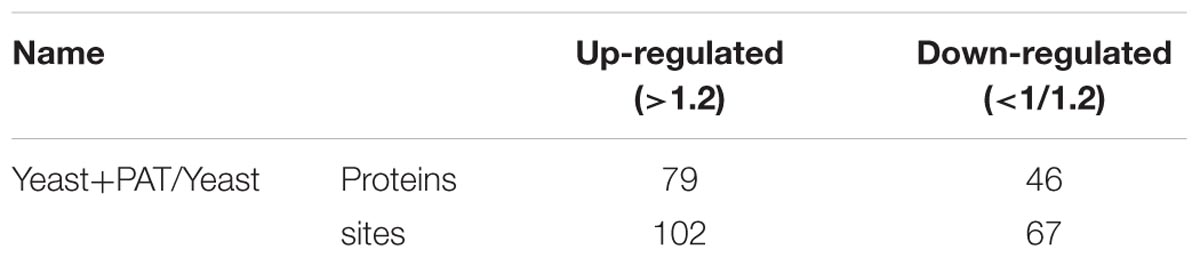
TABLE 2. Summary of differentially quantified sites and proteins.
Functional Classification of Differentially Expressed Proteins
Gene Ontology Classification
To understand the possible roles of the Kcr, functional classification of the identified proteins was performed. To further understand the distribution of identified proteins, information on GO annotation is summed up in accordance with each quantifiable level 2 protein GO term in Tables 3, 4. These tables present a functional view of the up-regulated and down-regulated proteins according to their categories such as cellular component, molecular function, and biological process. Among the assigned processes, the top three categories were metabolic process, cellular process, and single-organism process with up-regulated or down-regulated proteins. The three functions (catalytic activity, binding, and structural molecular activity) were the most important molecular functions for either up-regulated or down-regulated proteins. The GO analysis suggested that the crotonylated proteins had a wide range of biological processes and molecular functions in R. mucilaginosa.
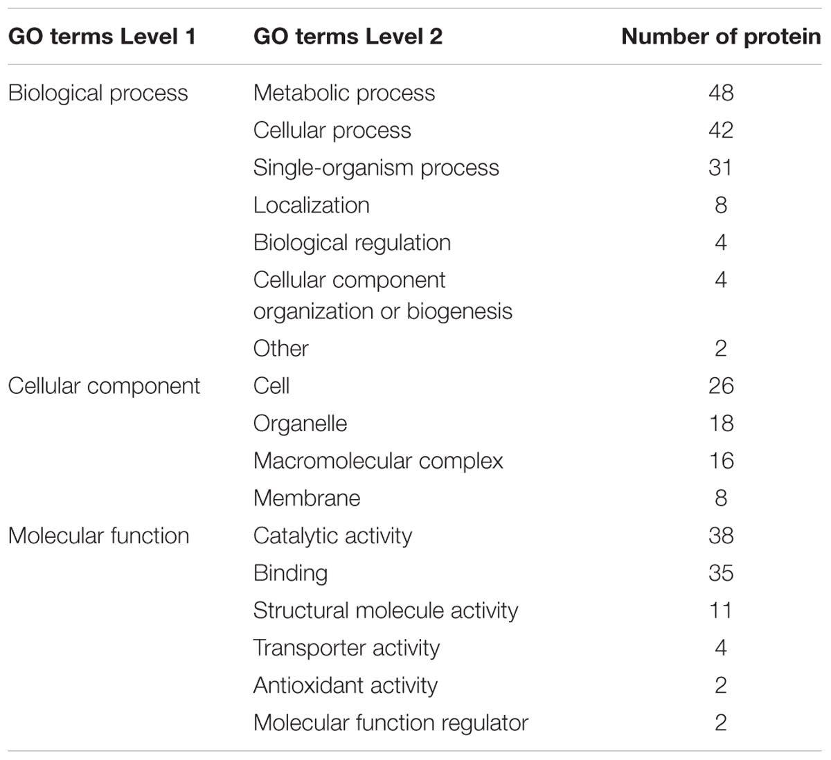
TABLE 3. The distribution of up-regulated proteins in GO terms of level 2 (Y+PAT/Y).
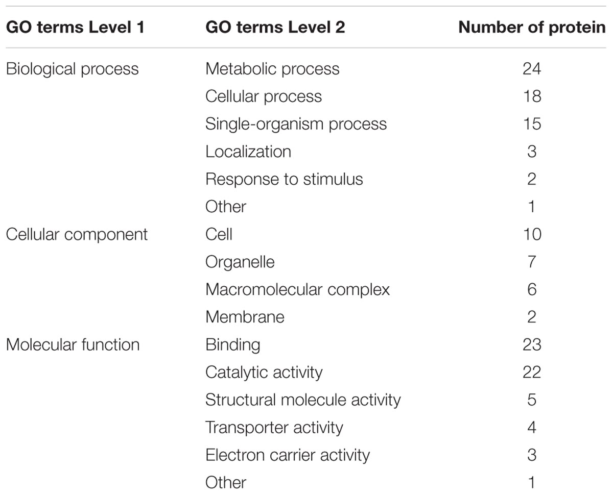
TABLE 4. The distribution of down-regulated proteins in GO terms of level 2 (Y+PAT/Y).
Classification of Subcellular Location
Based on the results of the subcellular location annotation of the identified proteins, we classified the quantifiable proteins into each subcellular location (Figure 1). The results indicated that the largest group of up-regulated proteins was associated with the mitochondria (39%), and the other two subcellular components were associated with the cytosol (32%) and the nucleus (14%) (Figure 1A). The overall trends in subcellular localization were similar in down-regulated proteins with the cytosol (37%), mitochondria (37%), nucleus (15%) among the subcellular components (Figure 1B).

FIGURE 1. The subcellular up-regulated and down-regulated proteins (Y+PAT/Y). (A) Subcellular localization of the up regulated proteins by patulin. (B) Subcellular localization of the down regulated proteins by patulin.
Motif Analysis
The motif-x program was used to extract enriched motifs from the set of sequences. The peptides were discovered to contain 20 amino acid residues that surround the crotonylation lysine (from positions -10 to +10), and these motifs were illustrated as EXXKcrKcr, EKcr, KcrKcr, KcrE, KcrD, FKcr, YKcr, AKcr, DKcr, NKcr, and KcrXXXXXXKcr. Kcr is the crotonylation at lysine, and X represents a random amino acid residue (Figure 2A). A survey of these motifs suggested that several kinds of amino acid residues are found within these motifs. These conserved amino acid residues include various kinds of amino acid including glutamate (E), aspartate (D), phenylalanine (F), tyrosine (Y), alanine (A), and asparagine (N) (Figure 2B). As illustrated in Figure 2B, these motifs can be sorted into four categories: (i) position +1 is an aspartate residue, (ii) position -1 is a tyrosine or a phenylalanine residue, (iii) position +3, +1, or -1 is a glutamate residue, (iv) position +10, +9, +8, +7, +5, +1, -6, -7, -9, or -10 is a lysine residue. These results suggest that the crotonylated system prefers to modify the lysine residue in order to be surrounded with negatively charged residues from -10 to +10. All the motifs varied in their abundance in the crotonylated peptides, and motif 2 was the most extensively distributed motif (Figure 2C). Enrichment analysis indicated that seven motifs exhibited different distributions over the KEGG pathway (Figure 2D). Motif 2, motif 3, motif 5, and motif 7 were significantly over-represented in the mRNA surveillance pathway, in the regulation of mitophagy, in valine, leucine, and isoleucine biosynthesis and in galactose metabolism, respectively. However, motif 11 was enriched in three pathways (glyoxylate and dicarboxylate metabolism, ribosome degradation, and valine, leucine, and isoleucine degradation).
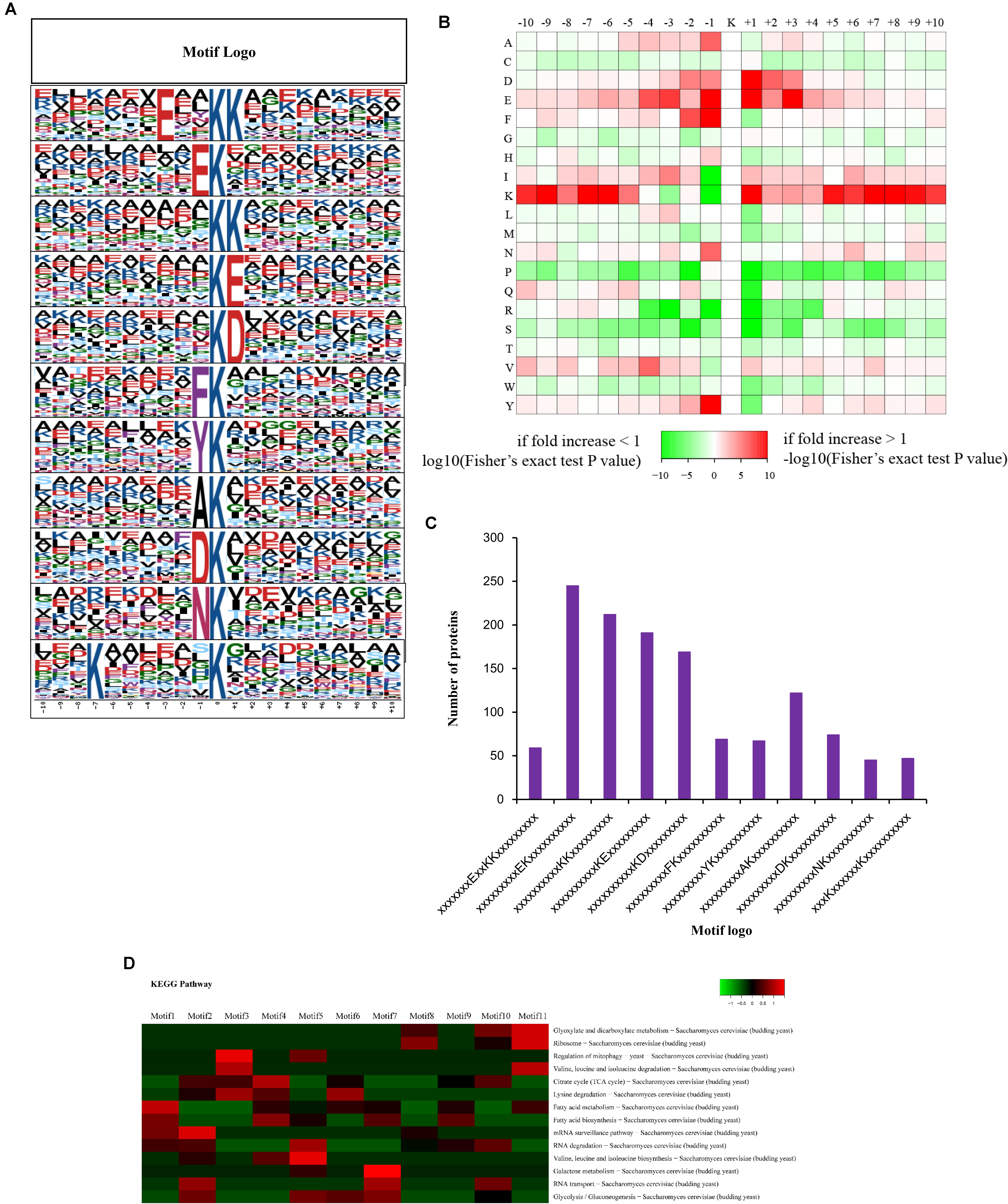
FIGURE 2. Analysis the characters of the identified peptides. (A) Crotonylation motifs were constructed with Motif-X software. The central K (at position 0) indicates the crotonylation lysine. All the surrounding amino acid residues are indicated with the letters in different heights which is consistent with their frequencies in respective positions. (B) Heat map showing the frequency of the amino acid residues around the crotonylated site based on analysis of all the identified peptides. (C) Number of the crotonylated peptides in each motif. (D) Heat map showing the enriched motifs in the representative KEGG pathways.
Functional Enrichment of Differentially Quantified Proteins
GO Enrichment
To determine the preferred protein types for Kcr, GO enrichment analysis was performed on the crotonylation data (Figure 3). Among the identified proteins, 465 crotonylated proteins were annotated for their biological process, and 366 for their corresponding molecular functions. Based on the biological process, the enrichment analysis of the identified proteins showed that proteins related to single-organism carbohydrate metabolism were high for up-regulated proteins, while the oxidation-reduction process was significant for down-regulated proteins.
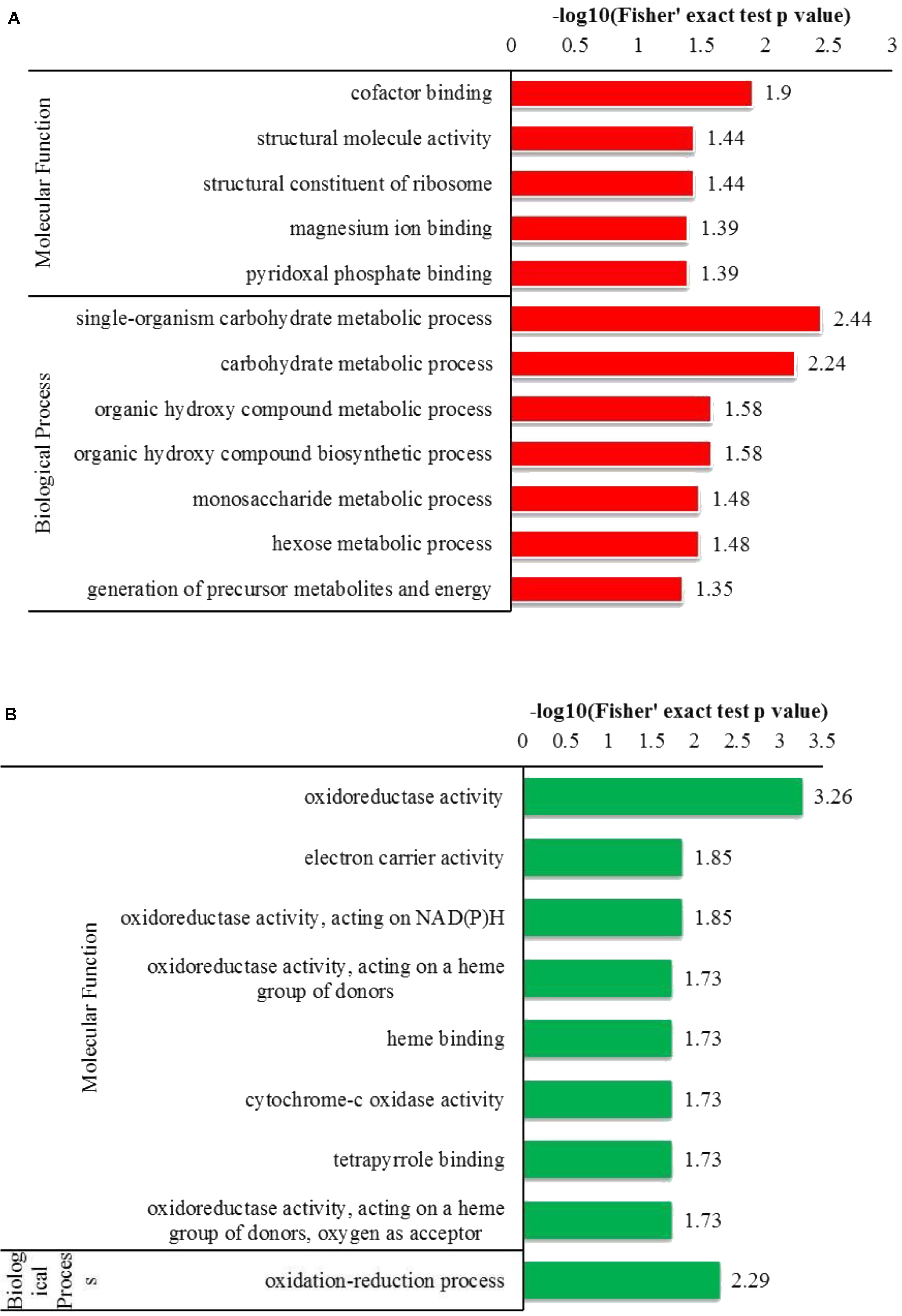
FIGURE 3. GO-based enrichment analysis of up-regulated and down-regulated proteins (Y+PAT/Y). (A) Classification of the up-regulated proteins with molecular function and biological process. (B) Classification of the down-regulated proteins with molecular function and biological process.
Domain Enrichment
Regarding the GO enrichment of domains, the analysis revealed that Kcr substrates were abundant in pyridoxal phosphate-dependent transferase. The other substrates that were abundant included subdomain 1 in up-regulated proteins and the NADP-dependent oxido-reductase domain and glutathione S-transferase (N-terminal) in down-regulated proteins (Figure 4). Overall, our data suggest that mitochondrial and cytosolic processes may be strictly regulated by crotonylation.
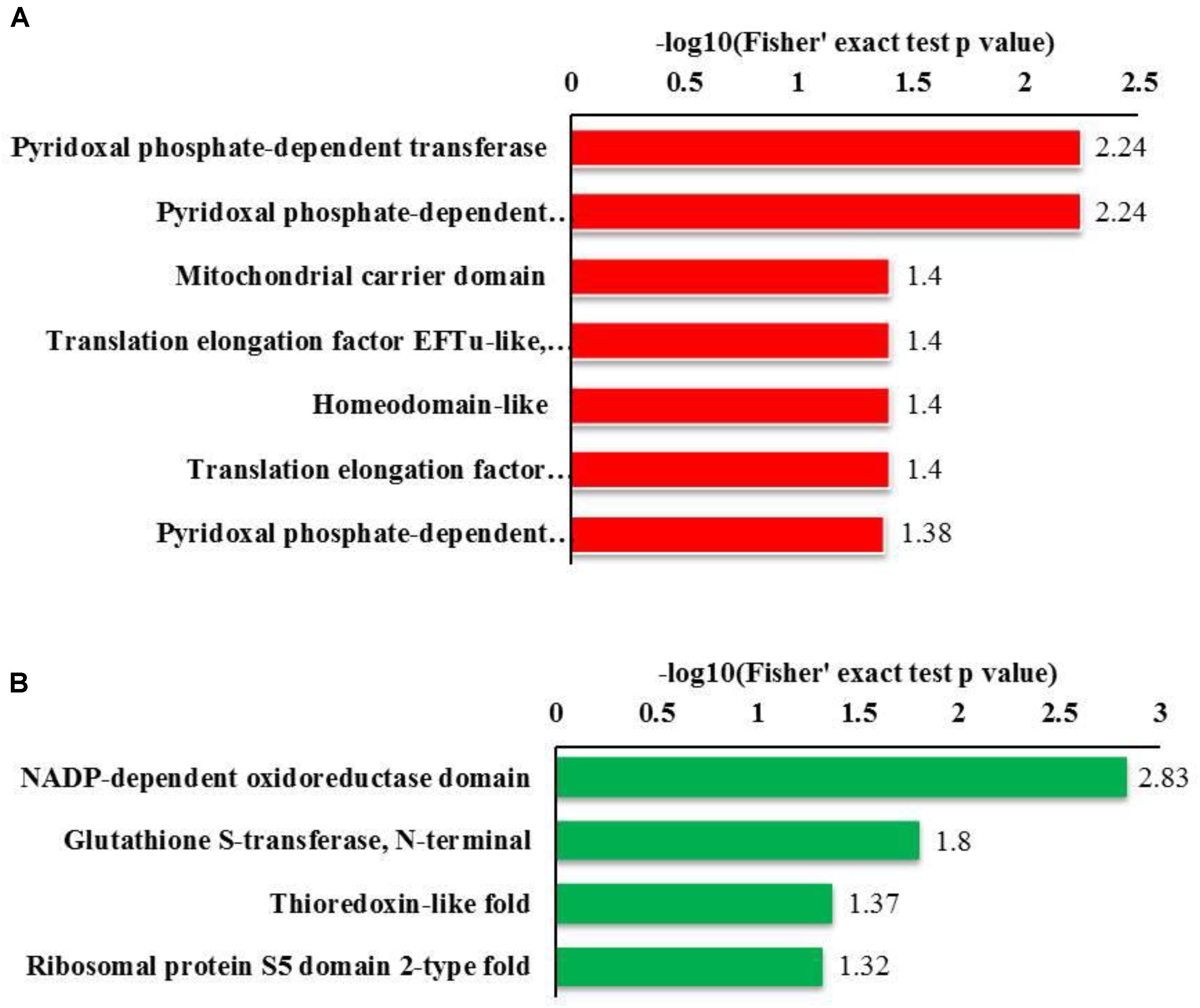
FIGURE 4. Bioinformatics analysis of the lysine crotonylated proteins of R. mucilaginosa (Y+PAT/Y). (A) Classification of the down regulated lysine crotonylated proteins with molecular function and biological process. (B) Classification of the up regulated lysine crotonylated proteins with molecular function and biological process.
Interaction Network of Crotonylated Proteins
The pathway analysis revealed that proteins with lysine crotonylated ions were involved in multiple biological processes, including the TCA cycle, glycolysis, and amino acids synthesis. There were several Kcr sites involved in TCA metabolic processes, such as pyruvate carboxylase, citrate synthase, homoisocitrate dehydrogenase, malate dehydrogenase, and fumarate hydratase (class II). These sites were up-regulated due to the stress caused by PAT (Figure 5A). The interaction networks of crotonylated proteins involved in the TCA cycle are shown in Figure 5B. Three up-regulated Kcr sites (pyruvate kinase, glyceraldehyde-3-phosphate dehydrogenase, and enolase) were involved in glycolysis. The PPIs for the identified crotonylated proteins revealed an interaction network that consisted of 408 direct physical interactions. This suggested that the crotonylated proteins were associated with a wide range of protein interactions. The number of crotonylation sites was represented for each interaction network (Figure 5C). The degree of the node was an important parameter to evaluate the correlation of a protein network. Crotonylated protein UBI4 showed the highest degree among the proteins in the network, followed by RPS3, RPL3, RPS5, and UBI4 proteins.
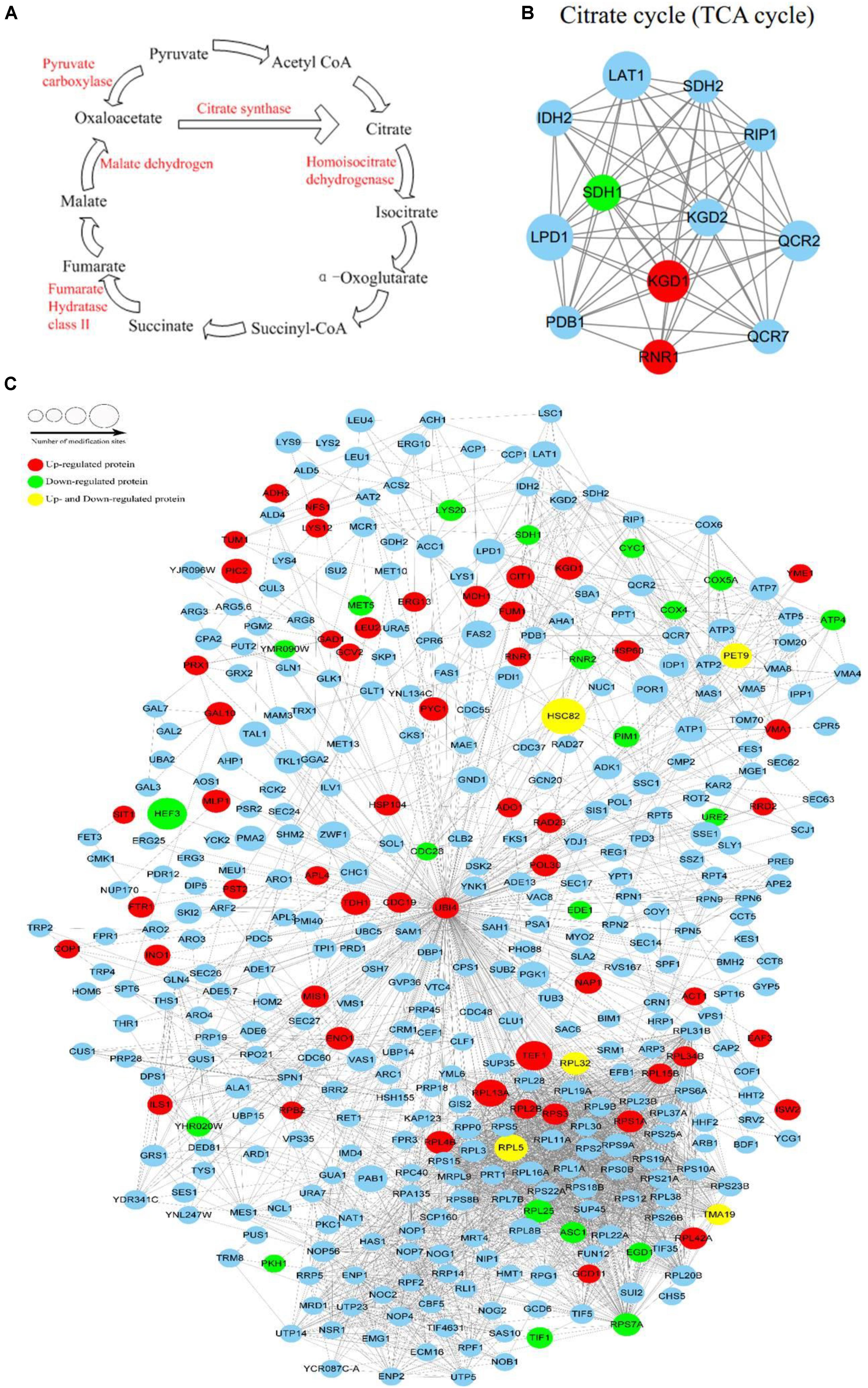
FIGURE 5. The metabolic pathway influenced by patulin and Protein interaction network of Kcr proteins. (A) Lysine crotonylation of metabolic enzymes identified by MS based proteomics in TCA cycle. (B) Protein interaction network of TCA cycle. (C) Protein interaction network of Kcr proteins. Size of the node represent different number of modification sites. Different colors represent differential regulated type: up-regulated protein (red); down-regulated protein (green); up-regulated and down-regulated protein (yellow); no differential expressed protein (light blue).
Discussion
Prior molecular studies clearly explain the complex mechanisms involved in PAT degradation and/or detoxification. In a previous study, unique genes were identified to degrade PAT in the model basidiomycete yeast Sporobolomyces (Ianiri et al., 2016). Proteomic profiling of protein Kcr was identified in human somatic and mouse male germ cell genomes (Tan et al., 2011). Wu et al. (2017) found that lysine crotonylation participates in a wide range of biological functions and processes, both the crotonylation and acetylation levels of most core histones sites and a number of non-histone proteins as well as some known substrates of class IIa and IIb HDACs were up-regulated after SAHA treatment. These results suggest that SAHA may have decrotonylation inhibitory activities on both histones and non-histone proteins by inhibiting HDACs. Although much is known about the currently identified histone Kcr and its functions in humans and mice, information about the functions of Kcr in yeast cells and its potential role in signaling during stress caused by PAT in yeast cells remains unclear. Bioinformatic tools were used to analyze the association of protein Kcr with cellular processes, metabolic pathways, and protein interactions.
The classification of the biological processes showed that the largest protein group is composed of enzymatic proteins that are associated with metabolic processes, which included both up-regulated and down-regulated proteins. Binding and catalytic activity were the dominant molecular functions of the up- and down-regulated proteins.
Riley and Showker (1991) found that the basic toxic effects of PAT were on plasma membranes and non-protein sulfhydryls (glutathione) in LLC-PK1 cells. Their results revealed that the endoplasmic reticulum calcium transport ATPase is a sensitive target for cyclopiazonic acid as well as the antioxidant activity of indole tetramic acids, which are serially connected but occur parallel to the depletion of sulfhydryls, an increase in 86Rb+ efflux, dome collapse, and eventually the loss of cell viability. Kim et al. (2010) used natural phenolic chemo-sensitizing agents to overcome fludioxonil resistance in Penicillium expansum, and found that fungal mitochondrial superoxide dismutase (Mn-SOD) plays a role in protecting cells from oxidative damage against alkyl gallates. Our results demonstrated that Mn-SOD was up-regulated in the lysine crotonylation levels, which suggested that the antioxidant response was activated in R. mucilaginosa cells against PAT. Enolase (Eno) plays an important role in transcription, apoptotic regulation, and cell differentiation. Eno is a phosphopyruvate hydratase that is considered to be a metallo-enzyme responsible for the catalysis or inter-conversion of 2-phosphoglycerate to phosphoenolpyruvate. Eno was down-regulated when P. expansum was exposed to nitric oxide; this disrupted glycolysis and subsequently influenced the Krebs cycle (Subramanian and Miller, 2000; Lai et al., 2014). In our results, we found up-regulation of Eno among the lysine crotonylation levels, indicating that PAT may have stimulated glyco-metabolism of R. mucilaginosa. Additionally, glycolysis of yeast cells was obstructed, thereby improving Eno activity. Ismaiel and Papenbrock (2014) found that maize root tip cells treated with 25 μg/mL PAT showed rumpled morphological changes in the cytoplasm and cytoplasmic organelles by using transmission electron microscopy. Cytoplasmic dissolution could be caused by the biochemical basis of PAT toxicity that involves respiratory inhibition. The present results demonstrated higher levels of cytoplasmic proteins with Kcr and confirmed that PAT could cause deterioration in cytoplasmic organelles. The changes in the cyto-morphological modification showed that R. mucilaginosa cells were stimulated by PAT.
Zhang et al. (2015) indicated that PAT caused an increase in ROS and a loss of SOD and glutathione (GSH) activities, which resulted in oxidative stress. These findings suggested that PAT might induce apoptosis in HEK293 cells through oxidative stress and damaged kidney cells. Moreover, PAT was found to cause rat hepatocyte cell damage by the formation of adducts with thiol-containing proteins in cellular components, such as GSH and cysteine-containing proteins. Simultaneous suppression of gap junction-mediated intercellular communication (GJIC) and GSH depletion initiates ROS generation, followed by mitochondrial membrane depolarization that causes a simultaneous increase in [Ca2+]i and cytoplasmic acidification, which leads to depolarization of the plasma membrane (Harwig et al., 1973; Barhoumi and Burghardt, 1996). There are many enzymes with a sulfhydryl group in their active sites that are sensitive to PAT (Moake et al., 2005), including Na+-K+- dependent ATPase (Phillips and Hayes, 1977, 1978; Riley and Showker, 1991), muscle aldolase (Ashoor and Chu, 1973), aminoacyl-tRNA synthetase (Arafat et al., 1985), and RNA polymerase (Moulé and François, 1977). Most heat shock proteins (HSPs) belong to a well-conserved family of cellular proteins that are responsible for maintaining the stability of other proteins within the cells, and for protecting proteins from damage due to environmental stress. Mitochondrial matrix HSPs, are typically responsible for pro-survival and pro-apoptotic functions, as well as the transportation and refolding of proteins from the cytoplasm into the mitochondrial matrix. HSPs have evolved as intracellular protein-folding molecules to protect newly synthesized polypeptides from misfolding, prevent improper interactions with other molecules, or mediate conformational changes of individual proteins and formation of multimeric complexes (Gullo and Teoh, 2004; Chandra et al., 2007; Kim et al., 2009). Based on 2-DE and MS analyses, Lai et al. (2014) identified that HSP70 was up-regulated and HSP60 was down-regulated in response to exogenous NO in P. expansum. These results suggested that NO might impair the synthesis and transportation of some mitochondrial proteins that are essential for cell growth. HSP60 and HSP70 are responsive to environmental stresses, including NO stress. In our results, HSPs were found to be up-regulated following PAT treatment, suggesting PAT treatment led to oxidative stress in R. mucilaginosa cells. Defense response by HSP activity can stimulate resistance to PAT toxicity and expel PAT from the yeast cells.
Moreover, PAT induced intra- and inter-molecular protein crosslinking in vitro. It was found that cysteine thiol group was preferred for PAT-mediated crosslinking reactions, but lysine and histidine side chains and α-amino groups also exhibited reactivity. PAT can act as a homobifunctional as well as a heterobifunctional crosslinking agent (Fliege and Metzler, 1999). Zhang et al. (2016) reported that the YEATS domain of AF9 preferably binds croton-lysine over acetyl-lysine in histone H3. Croton-lysine of histone H3 (lysine 18) was deeply engulfed in an aromatic cage of the YEATS domain where the carbonyl oxygen of croton-lysine forms a hydrogen bond with the amide backbone of the protein residue Tyr78. Their findings presented a new structural mechanism of protein–protein interactions mediated by histone Kcr, and showed the mechanism by which cells interpret acyl-lysine marks in different biological contexts.
Boussabbeh et al. (2015) indicated that PAT caused ER stress and activated the unfolded protein response. ER stress is related to the induction of the mitochondrial apoptotic pathway that occurs with ROS induction, a drop in mitochondrial membrane potential, and caspase activation. In our results, the largest group of up-regulated proteins were related to ROS activity and the TCA cycle and were associated with the mitochondria. In this study, we identified five crotonylated enzymes that were found to be associated with the TCA cycle. They include pyruvate carboxylase, citrate synthase, homoisocitrate dehydrogenase, fumarate hydratase (class II), and malate dehydrogenase. These five enzymes play important roles in the central metabolic pathway.
Wei et al. (2017) had found that histone crotonylation is not redundant to histone acetylation but is broadly essential for transcription and also supports potential roles of histone crotonylation in other chromatin-associated events. Zheng et al. (2017) had researched the Kac level of pyruvate carboxylase and citrate synthase which were the key enzyme of TCA cycle were up-regulated proteins by the stress caused by patulin. They also found that ROS accumulation increase the expression levels of oxidoreductase to maintain the balance of redox state and to reduce the patulin toxicity in R. mucilaginosa. Huang et al. (2018) found that the most downregulated crotonylome alterations under p300 deficiency concern components of the nonsense-mediated decay, infectious disease, and viral/eukaryotic translation pathways. Moreover, some p300-targeted Kcr substrates are potentially linked to diseases such as cancer. In our results, the largest group of up-regulated protein were related to ROS activity and the TCA cycle and were associated with the mitochondria. The results suggested that the Kac and Kcr regulated the TCA metabolic process and defense to ROS in response to the stress induced by the patulin. It was interesting to find the nice correlation between promoter Kac and Kcr and the mutant might repress transcription through deacylation other than decrotonylation.
Conclusion
Bioinformatic analysis to understand the regulation of differentially expressed proteins, transcription factors functions, and the enzymes that regulate the PAT degradation processes is extremely important for further research. However, in order to accomplish this goal using yeast, we must not only select the appropriate protein targets with specific functions but also develop antibodies specifically against those selected targets. This is important for examining the phenotypic and functional changes and to address the unanswered questions in the degradative process. Future work should be devoted to elucidate whether and how histone crotonylation readers mediate the essential function of histone crotonylation in transcription in R. mucilaginosa treated with patulin.
Author Contributions
HZ designed the experiments and revised the manuscript. QY and YL performed the experiments and analyzed results. MA and XfZ provided direction in experimental methods and revised the manuscript. BS and XyZ revised the manuscript.
Funding
This work was supported by National Natural Science Foundation of China (31772037 and 31772369), Jiangsu “Blue Project” of Colleges and Universities, and the Open Foundation of Hubei Key Laboratory of Edible Wild Plants Conservation and Utilization (EWPL201704).
Conflict of Interest Statement
The authors declare that the research was conducted in the absence of any commercial or financial relationships that could be construed as a potential conflict of interest.
The reviewer GP and handling Editor declared their shared affiliation.
Footnotes
References
Arafat, W., Kern, D., and Dirheimer, G. (1985). Inhibition of aminoacyl-tRNA synthetases by the mycotoxin patulin. Chem. Biol. Interact. 56, 333–349. doi: 10.1016/0009-2797(85)90015-8
Ashoor, S. H., and Chu, F. S. (1973). Inhibition of muscle aldolase by penicillic acid and patulin in vitro. Food Cosmet. Toxicol. 11, 995–1000. doi: 10.1016/S0015-6264(73)80429-8
Bao, X. C., Wang, Y., Li, X., Li, X. M., Liu, Z., Yang, T. P., et al. (2014). Identification of ‘erasers’ for lysine crotonylated histone marks using a chemical proteomics approach. eLife 3:e02999. doi: 10.7554/eLife.02999.
Barhoumi, R., and Burghardt, R.C. (1996). Kinetic analysis of the chronology of patulin- and gossypol-induced cytotoxicity in vitro. Fundam. Appl. Toxicol. 30, 290–297. doi: 10.1006/faat.1996.0067
Berdasco, M., and Esteller, M. (2010). Aberrant epigenetic landscape in cancer: how cellular identity goes awry. Dev. Cell 19, 698–711. doi: 10.1016/j.devcel.2010.10.005
Boussabbeh, M., Ben, S. I., Prola, A., Guilbert, A., Bacha, H., Abidessefi, S., et al. (2015). Patulin induces apoptosis through ROS-mediated endoplasmic reticulum stress pathway. Toxicol. Sci. 144, 328–337. doi: 10.1093/toxsci/kfu319
Castoria, R., Mannina, L., Durán-Patrón, R., Maffei, F., Sobolev, A. P., De Felice, D. V., et al. (2011). Conversion of the mycotoxin patulin to the less toxic desoxypatulinic acid by the biocontrol yeast Rhodosporidium kratochvilovae strain LS11. J. Agr. Food Chem. 59, 11571–11578. doi: 10.1021/jf203098v
Chandra, D., Choy, G., and Tang, D. G. (2007). Cytosolic accumulation of HSP60 during apoptosis with or without apparent mitochondrial release - Evidence that its pro-apoptotic or pro-survival functions involve differential interactions with caspase. J. Biol. Chem. 282, 31289–31301. doi: 10.1074/jbc.M702777200
Coelho, A. R., Celli, M. G., Ono, E. Y. S., Wosiacki, G., Hoffmann, F. L., Pagnocca, F. C., et al. (2007). Penicillium expansum versus antagonist yeasts and patulin degradation in vitro. Braz. Arch. Biol. Tech. 50, 725–733. doi: 10.1590/s1516-89132007000400019
Commission Regulation [EC] (2006). Commission Regulation (EC) No 1881/2006 of 19 december 2006 setting maximum levels for certain contaminants in foodstuffs. Off. J. Eur. l364, 5–24.
Dong, X. Y., Jiang, W., Li, C. S., Ma, N., Xu, Y., and Meng, X. H. (2015). Patulin biodegradation by marine yeast Kodameae ohmeri. Food Addit. Contam. A Chem. Anal. Control Expo. Risk Assess. 32, 352–360. doi: 10.1080/19440049.2015.1007090
Fliege, R., and Metzler, M. (1999). The mycotoxin patulin induces intra- and intermolecular protein crosslinks in vitro involving cysteine, lysine, and histidine side chains, and α-amino groups. Chem. Biol. Interact. 123, 85–103. doi: 10.1016/S0009-2797(99)00123-4
Gullo, C. A., and Teoh, G. (2004). Heat shock proteins: to present or not, that is the question. Immunol. Lett. 94, 1–10. doi: 10.1016/j.imlet.2004.04.002
Hao, J., Guo, H., Shi, X., Wang, Y., Wan, Q., Song, Y. B., et al. (2017). Comparative proteomic analyses of two Taxus species (Taxus × media and Taxus mairei) reveals variations in the metabolisms associated with paclitaxel and other metabolites. Plant Cell Physiol. 58, 1878–1890. doi: 10.1093/pcp/pcx128
Harwig, J., Scott, P. M., Kennedy, B. P. C., and Chen, Y. K. (1973). Disappearance of patulin from apple juice fermented by Saccharomyces spp. Can. Inst. Food Sci. Technol. J. 6, 45–46. doi: 10.1016/S0315-5463(73)73965-1
Huang, H., Wang, D. L., and Zhao, Y. M. (2018). Quantitative crotonylome analysis expands the roles of p300 in the regulation of lysine crotonylation pathway. Proteomics 22:e1700230. doi: 10.1002/pmic.201700230.
Ianiri, G., Idnurm, A., and Castoria, R. (2016). Transcriptomic responses of the basidiomycete yeast Sporobolomyces sp to the mycotoxin patulin. BMC Genomics 17:210. doi: 10.1186/s12864-016-2550-4
Ismaiel, A. A., and Papenbrock, J. (2014). The effects of patulin from Penicillium vulpinum on seedling growth, root tip ultrastructure and glutathione content of maize. Eur. J. Plant Pathol. 139, 497–509. doi: 10.1007/s10658-014-0406-9
Kim, J. H., Campbell, B. C., Mahoney, N., Chan, K. L., Molyneux, R. J., and Xiao, C. L. (2010). Use of chemosensitization to overcome fludioxonil resistance in Penicillium expansum. Lett. Appl. Microbiol. 51, 177–183. doi: 10.1111/j.1472-765X.2010.02875.x
Kim, Y. S., Koh, J. M., Lee, Y. S., Kim, B. J., Lee, S. H., Lee, K. U., et al. (2009). Increased circulating heat shock protein 60 induced by menopause, stimulates apoptosis of osteoblast-lineage cells via up-regulation of toll-like receptors. Bone 45, 68–76. doi: 10.1016/j.bone.2009.03.658
Lai, T., Chen, Y., Li, B., Qin, G., and Tian, S. (2014). Mechanism of Penicillium expansum in response to exogenous nitric oxide based on proteomics analysis. J. Proteomics 103, 47–56. doi: 10.1016/j.jprot.2014.03.012
Li, Y., Sabari, B. R., Panchenko, T., Wen, H., Zhao, D., Guan, H., et al. (2016). Molecular coupling of histone crotonylation and active transcription by AF9 YEATS domain. Mol. Cell 62, 181–193. doi: 10.1016/j.molcel.2016.03.028
Liu, X. Y., and Gao, C. (2016). Molecular Mechanisms of histone phosphorylation in regulating learning and memory. Prog. Biochem. Biophys. 43, 831–838. doi: 10.16476/j.pibb.2016.0106
Mahunu, G. K., Zhang, H. Y., Yang, Q. Y., Li, C. L., and Zheng, X. F. (2016). Biological control of patulin by antagonistic yeast: a case study and possible model. Crit. Rev. Microbiol. 42, 643–655. doi: 10.3109/1040841x.2015.1009823
Moake, M. M., Padilla-Zakour, O. I., and Worobo, R. W. (2005). Comprehensive review of patulin control methods in foods. Compr. Rev. Food Sci. Food Saf. 4, 8–21. doi: 10.1111/j.1541-4337.2005.tb00068.x
Mohibi, S., Srivastava, S., Bele, A., Mirza, S., Band, H., and Band, V. (2016). Acetylation of mammalian ADA3 is required for its functional roles in histone acetylation and cell proliferation. Mol. Cell. Biol. 36, 2487–2502. doi: 10.1128/mcb.00342-16
Montellier, E., Rousseaux, S., Zhao, Y., and Khochbin, S. (2012). Histone crotonylation specifically marks the haploid male germ cell gene expression program. Bioessays 34, 187–193. doi: 10.1002/bies.201100141
Moulé, Y., and François, H. (1977). Mechanism of the in vitro inhibition of transcription by patulin, a mycotoxin from Byssochlamys nivea. FEBS Lett. 74, 121–125. doi: 10.1016/0014-5793(77)80767-9
Nishiyama, A., Yamaguchi, L., and Nakanishi, M. (2016). Regulation of maintenance DNA methylation via histone ubiquitylation. J. Biochem. 159, 9–15. doi: 10.1093/jb/mvv113
Olsen, J. V., and Mann, M. (2013). Status of large-scale analysis of post-translational modifications by mass spectrometry. Mol. Cell. Proteomics 12, 3444–3452. doi: 10.1074/mcp.O113.034181
Papp, G., Horváth, E., Mike, N., Gazdag, Z., Belágyi, J., Gyöngyi, Z., et al. (2012). Regulation of patulin-induced oxidative stress processes in the fission yeast Schizosaccharomyces pombe. Food Chem. Toxicol. 50, 3792–3798. doi: 10.1016/j.fct.2012.07.001
Papp, G., Mate, G., Mike, N., Gazdag, Z., and Pesti, M. (2016). Regulation of the antioxidant system in cells of the fission yeast Schizosaccharomyces pombe after combined treatment with patulin and citrinin. Toxicon 111, 100–107. doi: 10.1016/j.toxicon.2015.12.021
Paster, N., Huppert, D., and Barkai-Golan, R. (1995). Production of patulin by different strains of Penicillium expansum in pear and apple cultivars stored at different temperatures and modified atmospheres. Food Addit. Contam. 12, 51–58. doi: 10.1080/02652039509374278
Penard-Lacronique, V., and Bernard, O. A. (2016). IDH1, histone methylation, and so forth. Cancer Cell 30, 501-501. doi: 10.1016/j.ccell.2016.08.010
Phillips, T. D., and Hayes, A. W. (1977). Effects of patulin on adenosine triphosphatase activities in the mouse. Toxicol. Appl. Pharmacol. 39, 71–79. doi: 10.1016/0041-008X(77)90178-8
Phillips, T. D., and Hayes, A. W. (1978). Effects of patulin on the kinetics of substrate and cationic ligand activation of adenosine triphosphatase in mouse brain. J. Pharmacol. Exp. Ther. 205, 606–616.
Riley, R. T., and Showker, J. L. (1991). The mechanism of patulin’s cytotoxicity and the antioxidant activity of indole tetramic acids. Toxicol. Appl. Pharm. 109, 108–126. doi: 10.1016/0041-008X(91)90195-K
Sommer, N. F., Buchanan, J. R., and Fortlage, R. J. (1974). Production of patulin by Penicillium expansum. Appl. Microbiol. 28, 589–593.
Subramanian, A., and Miller, D. M. (2000). Structural analysis of alpha-enolase. Mapping the functional domains involved in down-regulation of the c-myc protooncogene. J. Biol. Chem. 275, 5958–5965. doi: 10.1074/jbc.275.8.5958
Sun, H., Liu, X., Li, F., Li, W., Zhang, J., Xiao, Z., et al. (2017). First comprehensive proteome analysis of lysine crotonylation in seedling leaves of Nicotiana tabacum. Sci. Rep. 7:3013. doi: 10.1038/s41598-017-03369-6
Tan, M. J., Luo, H., Lee, S., Jin, F. L., Yang, J. S., Montellier, E., et al. (2011). Identification of 67 histone marks and histone lysine crotonylation as a new type of histone modification. Cell 146, 1015–1027. doi: 10.1016/j.cell.2011.08.008
Wang, Q., Tang, Y., Xu, Y., Xu, S., Jiang, Y., Dong, Q., et al. (2017). The X-linked deubiquitinase USP9X is an integral component of centrosome. J. Biol. Chem. 292, 12874-12884. doi: 10.1074/jbc.M116.769943
Wei, W., Liu, X. G., Chen, J. C, Gao, S. N., Lu, L., Zhang, H. F., et al. (2017). Class I histone deacetylases are major histone decrotonylases: evidence for critical and broad function of histone crotonylation in transcription. Cell Res. 27, 898–915. doi: 10.1038/cr.2017.68
Weinert, B. T., Schölz, C., Wagner, S. A., Iesmantavicius, V., Su, D., Daniel, J. A., et al. (2013). Lysine succinylation is a frequently occurring modification in prokaryotes and eukaryotes and extensively overlaps with acetylation. Cell Rep. 4, 842–851. doi: 10.1016/j.celrep.2013.07.024
Wu, Q., Li, W. T., Wang, C., Fan, P. S., Cao, L. J., Wu, Z. W., et al. (2017). Ultradeep lysine crotonylome reveals the crotonylation enhancement on both histones and non-histone proteins by SAHA treatment. J. Proteome Res. 16, 3664–3671. doi: 10.1021/acs.jproteome.7b00380
Xiong, X., Panchenko, T., Yang, S., Zhao, S., Yan, P., Zhang, W., et al. (2016). Selective recognition of histone crotonylation by double PHD fingers of MOZ and DPF2. Nat. Chem. Biol. 12, 1111–1118. doi: 10.1038/nchembio.2218
Yang, Q. Y., Zhang, H. Y., Zhang, X. Y., Zheng, X. F., and Qian, J. Y. (2015). Phytic acid enhances biocontrol activity of Rhodotorula mucilaginosa against Penicillium expansum contamination and patulin production in apples. Front. Microbiol. 6:1296. doi: 10.3389/fmicb.2015.01296
Zhang, B., Peng, X., Li, G., Xu, Y., Xia, X., and Wang, Q. (2015). Oxidative stress is involved in patulin induced apoptosis in HEK293 cells. Toxicon 94, 1–7. doi: 10.1016/j.toxicon.2014.12.002
Zhang, Q., Zeng, L., Zhao, C., Ju, Y., Konuma, T., and Zhou, M. M. (2016). Structural insights into histone crotonyl-lysine recognition by the AF9 YEATS domain. Structure 24, 1606–1612. doi: 10.1016/j.str.2016.05.023
Zhang, Z., Tan, M., Xie, Z., Dai, L., Chen, Y., and Zhao, T. (2011). Identification of lysine succinylation as a new post-translational modification. Nat. Chem. Biol. 7, 58–63. doi: 10.1038/nchembio.495
Zhao, S., Xu, W., Jiang, W., Yu, W., Lin, Y., Zhang, T., et al. (2010). Regulation of cellular metabolism by protein lysine acetylation. Science 327, 1000–1004. doi: 10.1126/science.1179689
Zheng, X. F., Yang, Q. Y., Zhang, H. Y., Cao, J., Zhang, X. Y., and Apaliya, M. T. (2016). The possible mechanisms involved in degradation of patulin by Pichia caribbica. Toxins 8:E289. doi: 10.3390/toxins8100289
Keywords: Rhodotorula mucilaginosa, patulin, post-translational modification, histone lysine crotonylation, degradation
Citation: Yang Q, Li Y, Apaliya MT, Zheng X, Serwah BNA, Zhang X and Zhang H (2018) The Response of Rhodotorula mucilaginosa to Patulin Based on Lysine Crotonylation. Front. Microbiol. 9:2025. doi: 10.3389/fmicb.2018.02025
Received: 13 February 2018; Accepted: 09 August 2018;
Published: 03 September 2018.
Edited by:
Rosanna Tofalo, Università degli Studi di Teramo, ItalyReviewed by:
Shihua Wang, Fujian Agriculture and Forestry University, ChinaGiorgia Perpetuini, Università degli Studi di Teramo, Italy
Copyright © 2018 Yang, Li, Apaliya, Zheng, Serwah, Zhang and Zhang. This is an open-access article distributed under the terms of the Creative Commons Attribution License (CC BY). The use, distribution or reproduction in other forums is permitted, provided the original author(s) and the copyright owner(s) are credited and that the original publication in this journal is cited, in accordance with accepted academic practice. No use, distribution or reproduction is permitted which does not comply with these terms.
*Correspondence: Hongyin Zhang, emhhbmdob25neWluMTI2QDEyNi5jb20=
†These authors have contributed equally to this work as co-first authors