- 1College of Food Science, South China Agricultural University, Guangzhou, China
- 2Guangdong Institute of Microbiology, Guangdong Academic of Science, State Key Laboratory of Applied Microbiology Southern China, Guangdong Provincial Key Laboratory of Microbial Culture Collection and Application, Guangdong Open Laboratory of Applied Microbiology, Guangzhou, China
- 3Infinitus (China) Company, Ltd., Guangzhou, China
Listeria monocytogenes is a foodborne pathogen with a high mortality rate in humans. This study aimed to identify the pathogenic potential of L. monocytogenes isolated from ready-to-eat (RTE) foods and pasteurized milk in China on the basis of its phenotypic and genotypic characteristics. Approximately 7.7% (44/570) samples tested positive for L. monocytogenes among 10.8% (39/360) RTE and 2.4% (5/210) pasteurized milk samples, of which 77.3% (34/44) had < 10 MPN/g, 18.2% (8/44) had 10–110 MPN/g, and 4.5% (2/44) had > 110 MPN/g. A total of 48 strains (43 from RTE foods and five from milk samples) of L. monocytogenes were isolated from 44 positive samples. PCR-serogroup analysis revealed that the most prevalent serogroup was II.2 (1/2b-3b-7), accounting for 52.1% (25/48) of the total, followed by serogroup I.1 (1/2a-3a) accounting for 33.3% (16/48), serogroup I.2 (1/2c-3c) accounting for 12.5% (6/48), and serogroup II.1 (4b-4d-4e) accounting for 2.1%. All isolates were grouped into 11 sequence types (STs) belonging to 10 clonal complexes (CCs) and one singleton (ST619) via multi-locus sequence typing. The most prevalent ST was ST87 (29.2%), followed by ST8 (22.9%), and ST9 (12.5%). Virulence genes determination showed that all isolates harbored eight virulence genes belonging to Listeria pathogenicity islands 1 (LIPI-1) (prfA, actA, hly, mpl, plcA, plcB, and iap) and inlB. Approximately 85.4% isolates carried full-length inlA, whereas seven isolates had premature stop codons in inlA, six of which belonged to ST9 and one to ST5. Furthermore, LLS (encoded by llsX gene, representing LIPI-3) displays bactericidal activity and modifies the host microbiota during infection. LIPI-4 enhances neural and placental tropisms of L. monocytogenes. Results showed that six (12.5%) isolates harbored the llsX gene, and they belonged to ST1/CC1, ST3/CC3, and ST619. Approximately 31.3% (15/48) isolates (belonging to ST87/CC87 and ST619) harbored ptsA (representing LIPI-4), indicating the potential risk of this pathogen. Antimicrobial susceptibility tests revealed that > 95% isolates were susceptible to 16 antimicrobials; however, 60.4 and 22.9% isolates were intermediately resistant to streptomycin and ciprofloxacin, respectively. The results show that several isolates harbor LIPI-3 and LIPI-4 genes, which may be a possible transmission route for Listeria infections in consumers.
Introduction
Listeria monocytogenes is a foodborne pathogen with a high mortality rate in humans. The vulnerable groups, such as the elderly, pregnant women, fetuses and immunocompromised individuals are the main population infected by L. monocytogenes (Cartwright et al., 2013). This pathogen has high tolerance against stressors associated with food processing, including refrigerating temperatures, low moisture content, high salinity, and a wide pH range (Hain et al., 2007). Furthermore, sequence type (ST) 8, ST5, ST87, ST2, ST121, ST14, and ST9 subtypes of L. monocytogenes are present and grow in some specific niches, resulting in long-term contamination during food production and processing (Fagerlund et al., 2016; Chen et al., 2017; Chau et al., 2017; Pasquali et al., 2018; Melero et al., 2019).
Listeria infections potentially result from ingestion of L. monocytogenes- contaminated foods. In 2017–2018, the largest listeriosis outbreak worldwide occurred in South Africa, resulting from ingestion of ready-to-eat (RTE) meat contaminated with L. monocytogenes ST6 (Smith et al., 2019). Previous studies reported an incidence of 0.46 cases per 100,000 individuals in Europe in 2015 (European Food Safety Authority and European Centre for Disease Prevention and Control, 2016). According to previous study, 147 clinical cases, 479 Listeria isolates and 82 outbreak-related cases were reported from 28 provinces between 1964 and 2010 in China, respectively (Feng et al., 2013). Fan et al. (2019) conducted a systematic review for the listeriosis in mainland China, wherein 136 records were identified, and reporting 562 patients with listeriosis from 2011 to 2017, indicating a drastic increase in the number of patients over the past decade. Listeriosis patients were primarily reported in developed cities (Beijing city and the coastal cities), probably owing to dietary habits and the high population density in these areas. Risk identification of L. monocytogenes in RTE foods is important because they provide critical information regarding food sources of listeriosis. Therefore, comprehensive surveillance of L. monocytogenes in RTE foods throughout China is of utmost importance.
Thirteen serotypes of L. monocytogenes have been assigned based on the somatic (O) and flagellar (H) antigens. Noticeably, serotypes 4b, 1/2a, 1/2b, and 1/2c account for > 95% of isolates recovered from foods and clinical cases (Pontello et al., 2012). At present, multi-locus sequence typing (MLST) (Ragon et al., 2008), and core genome MLST (cgMLST) (Chen et al., 2016) are used for molecular typing of L. monocytogenes. MLST is a reliable, high-resolution, and acceptable method for typing L. monocytogenes. Furthermore, the molecular virulence of L. monocytogenes isolates is closely related to the distribution patterns of virulence genes of Listeria pathogenicity island-1 (LIPI-1) and the inlAB operon in the genome (Vazquez-Boland et al., 2001). LIPI-1 includes prfA, actA, hly, mpl, iap, plcA, plcB genes (Chen et al., 2018a) and the inlAB operon encodes two internalins (InlA and InlB), which are critical for entry into hepatocytes (Gaillard et al., 1996). PCR analysis indicated that majority of L. monocytogenes isolates contained most of virulence genes of LIPI-1 and inlAB operon (Chen et al., 2018a, 2019a; Basha et al., 2019; Zhang et al., 2019). Additional lineage and STs specific pathogenicity islands, for example, llsX gene in LIPI-3 encodes the bacteriocin Listeriolysin S (LLS), is associated with hemolytic and cytotoxic activity, displays bactericidal activity and modifies the host microbiota during infection (Cotter et al., 2008; Quereda et al., 2017; Radoshevich and Cossart, 2018; Yin et al., 2019); LIPI-4, a putative cellobiose-family phosphotransferase system, is responsible for the neural and placental tropisms infection of L. monocytogenes, respectively (Maury et al., 2016). Previous studies reported llsX gene from LIPI-3 present in ST1, ST3, ST4, ST6, ST77, ST79, ST213, ST217, ST224, ST288, ST308, ST323, ST330, ST363, ST380, ST382, ST389, ST489, ST554, ST581, ST619, ST778, ST999, ST1000 and ST1001, and pstA gene from LIPI-4 present in ST4, ST87, ST213, ST217, ST310, ST363, ST382, ST619, ST663, ST1002, and ST1166 have been reported (Chen et al., 2018a, b, 2019b; Kim et al., 2018; Wang et al., 2018). Therefore, the virulence of different ST strains of L. monocytogenes may vary.
This study aimed to detect and enumerate L. monocytogenes in RTE foods and pasteurized milk products and to determine their heterogeneity, characteristics, and public health implications in Chinese retail outlets.
Materials and Methods
Samples
Between March 2014 and June 2016, 360 RTE foods and 210 pasteurized milk samples were collected from 21 cities in China, including cold vegetable dish in sauce (n = 60 samples), duck (n = 77), fried rice (n = 15), chicken (n = 139), pork (n = 61), goat meat (n = 8), and pasteurized milk (n = 210). All of RTE foods were loose-packed in different markets. All samples were immediately placed in sterile bags, kept in an insulated shipping cooler with frozen gel packs placed on the sides, middle, and above the samples to maintain below 4°C. All the samples were transferred back to the laboratory immediately and analyzed within 4 h of receiving the samples.
Qualitative and Quantitative Analysis
Qualitative detection of L. monocytogenes was performed on the basis of the guidelines of the National Food Safety Standard of China (4789.30-2010) (Anonymous, 2010), with minor modifications. Briefly, 25 g (mL) of homogenized samples were added to 225 mL Listeria enrichment broth 1 (LB1) (Guangdong Huankai, Co. Ltd., Guangzhou, China). The cultures in LB1 media were incubated at 30°C for 24 h. After incubation, 100 μL of the LB1 enrichment culture was transferred to 10 mL Listeria enrichment broth 2 (LB2) (Guangdong Huankai, Co. Ltd.) and incubated at 30°C for 24 h. A loopful (about 10 μL) of the LB2 enrichment culture was streaked onto Listeria CHROMagar plates (CHROM-agar, Paris, France) and incubated at 37°C for 48 h. At least three (when possible) presumptive colonies were selected for the identification of L. monocytogenes, using the Microgen ID Listeria identification system (Microgen, Camberley, United Kingdom) in accordance with the manufacturer’s instructions.
For quantitative detection, the most probable number (MPN) method using a nine-tube was followed, as reported previously (Gombas et al., 2003). Briefly, nine tubes were divided into three sets of three tubes each. Homogenized samples (25 g) were added to 225 mL half Fraser Broth (Guangdong Huankai, Co. Ltd.). The first set of tubes contained 10 mL of the sample homogenate in 225 mL half Fraser Broth, while the second and third sets contained 10 mL of half Fraser Broth inoculated with 1 and 0.1 mL of the homogenate, respectively. Different volumes, i.e., 10, 1, and 0.1 mL, of the sample homogenate represented 1.0, 0.1, and 0.01 g of the original sample, respectively. The nine tubes were incubated at 30 ± 2°C for 24 ± 2 h. The darkened Fraser tubes were streaked onto Listeria CHROMagar plates. If a Fraser Broth tube did not display darkening, it was reexamined after an additional 26 ± 2 h of incubation. The presumptive colonies were purified again on the Tryptic Soy Agar (TSA) plates (Guangdong Huankai, Co. Ltd.) and then identified using the Microgen ID Listeria identification system. The MPN value was calculated based on the number of positive tube(s) in each sample and the MPN table (U.S. Department of Agriculture, 1998).
PCR-Serogroup Analysis and Virulence Genes Determination
Genomic DNA was extracted from L. monocytogenes isolates using a HiPure Bacterial DNA Kit (Guangzhou Magen Biotechnology, Co. Ltd., Guangzhou, China) according to the manufacturer’s instructions. Serogroups of the L. monocytogenes isolates were differentiated via a multiplex PCR method reported previously by Doumith et al. (2004) (Supplementary Table S1). PCR was carried out using a thermal cycler (Biometra, Göttingen, Germany) at the following conditions: initial denaturation at 94°C for 3 min; followed by 35 cycles at 94°C for 35 s, 53°C for 50 s, and 72°C for 60 s, and final extension at 72°C for 5 min. The isolates were determined as serogroup I.1 (1/2a-3a), I.2 (1/2c-3c), II.1 (4b-4d-4e), II.2 (1/2b-3b-7), and III (4a-4c).
Virulence genes including LIPI-1 (prfA, actA, hly, mpl, plcA, plcB, iap) and inlA, inlB were detected via PCR. Furthermore, two additional PCRs were carried out to detect the llsX and ptsA genes (representing LIPI-3 and LIPI-4, respectively) in the L. monocytogenes isolates (Clayton et al., 2011; Maury et al., 2016). The PCR primers used herein are enlisted in Supplementary Table S1. The amplicons were separated on 1.5% agarose gels in TAE buffer (Biosharp, Co., Ltd., Hefei, China) and visualized via Goldview® (Beijing Solarbio Science & Technology, Co., Ltd., China) staining (0.005%, v/v). To determine the premature stop codon (PMSC) in the inlA gene, full-length inlA of each isolate was amplified and sequenced using external and internal primers (Wu et al., 2016). Compared to previous reported PMSCs types (Chen et al., 2018a), the PMSCs in the inlA gene were analyzed and determined using MEGA X software (Kumar et al., 2018).
Antimicrobial Susceptibility Test
The antimicrobial susceptibility of the L. monocytogenes isolates was assessed using the disk diffusion method based on breakpoints for Staphylococci spp. in accordance with the guidelines of the Clinical Laboratory Standards Institute (Clinical and Laboratory Standard Institute, 2017). The breakpoints of ampicillin and penicillin G for specific Listeria spp. have been defined (M45-A2 Vol. 30 No. 18). The following 17 common antimicrobial agents (disk load), including those used to treat human listeriosis, were assessed herein: kanamycin (30 μg), gentamicin (10 μg), ciprofloxacin (5 μg), levofloxacin (5 μg), ofloxacin (5 μg), sulfamethoxazole with trimethoprim (23.75/1.25 μg), streptomycin (10 μg), rifampin (5 μg), doxycycline (30 μg), chloramphenicol (30 μg), erythromycin (15 μg), tetracycline (30 μg), meropenem (10 μg), vancomycin (30 μg), linezolid (30 μg), amoxycillin/clavulanic acid (10 μg), and sulbactam/ampicillin (10/10 μg) (Oxoid, Basingstoke, United Kingdom). Briefly, L. monocytogenes isolates were seeded in tryptone soya broth supplemented with 0.6% yeast extract (TSB-YE) (Guangdong Huankai, Co. Ltd.) and incubated at 37°C overnight. The cell density of the suspension was adjusted to 1.0 McFarland standard, which is approximately 3 × 10⁸ CFU/mL, and then diluted to ∼105 CFU/mL with 0.85% NaCl (w/v). The suspension was spread onto the surface of Mueller-Hinton agar (Guangdong Huankai, Co. Ltd.). The diameters of the inhibition zones were measured using precision calipers after 24 h of incubation. Staphylococcus aureus ATCC 25923 and Escherichia coli ATCC 25922 were used as quality control strains. Multidrug-resistant isolates were defined as isolates displaying resistance to at least three classes of antimicrobial agents assessed herein (Magiorakos et al., 2012).
MLST Analysis
Using the method of Ragon et al. (2008), MLST analysis of L. monocytogenes was performed on the basis of the following seven housekeeping genes: acbZ (ABC transporter), bglA (beta-glucosidase), cat (catalase), dapE (Succinyl diaminopimelate desuccinylase), dat (D-amino acid aminotransferase), ldh (lactate dehydrogenase), and lhkA (histidine kinase) (Supplementary Table S2). A detailed protocol for the present MLST analysis, including primers, PCR conditions, was in accordance with the guidelines of the Pasteur Institute website1, and STs and clonal complexes (CCs) of each isolate were assigned on the basis of each variant locus of each housekeeping gene. A phylogenetic tree was generated to analyze relationships among the isolates, using MEGA X (Kumar et al., 2018).
Results
Occurrence and Contamination Levels of L. monocytogenes
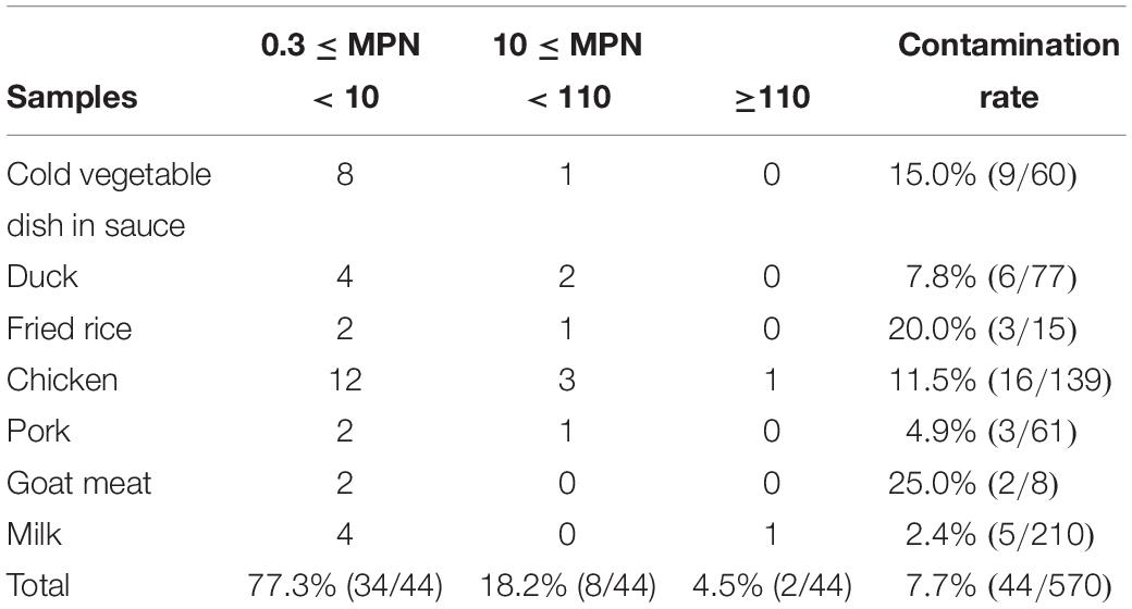
As shown in Table 1, 7.7% samples (39 RTE foods and 5 pasteurized milk samples) tested positive for L. monocytogenes from among 570 food samples. The contamination rate of goat meat (25.0%, 2/8) was the highest among the 7 types of RTE foods and milk, followed by fried rice (20.0%, 3/15) and a cold vegetable dish in sauce (15.0%, 9/60), chicken (11.5%, 16/139), duck (7.8%, 6/77), pork (4.9%, 3/61), and pasteurized milk (2.4%, 5/210). For quantitative analysis, 77.3% (34/44) samples contaminated with L. monocytogenes were below 10 MPN/g, 18.2% (8/44) were between 10 and 110 MPN/g, and only two (4.5%) samples were over 110 MPN/g, constituting sesame oil chicken and pasteurized milk, respectively.
PCR-Serogroup Analysis
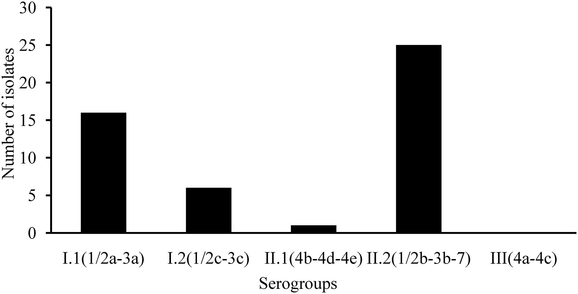
The ERIC-PCR fingerprinting was used to screen isolates from the same sample in order to remove the duplicate isolates (data not shown). L. monocytogenes isolates from the same sample with > 90% similarity was considered as clonal. Clonal isolates from individual sample were excluded. At least one L. monocytogenes isolate from each known source sample was submitted to further analysis. A total of 48 strains of L. monocytogenes were isolated from 44 L. monocytogenes-positive samples, including four samples (Cold vegetable dish in sauce, Pickled pig ear, Roasted chicken wing, and Salt baked chicken), each containing two different isolates. L. monocytogenes isolates recovered from RTE foods (43 isolates) and pasteurized milk samples (five isolates) were subjected to serogroup assessment via multiplex PCR analysis. As shown in Figure 1, 33.3% (16/48) isolates belonged to serogroup I.1 (1/2a-3a); 12.5% (6/48), serogroup I.2 (1/2c-3c); 2.1% (1/48), serogroup II.1 (4b-4d-4e); 52.1% (25/48), serogroup II.2 (1/2b-3b-7). None of the isolates belonged to serogroup III (4a-4c).

Figure 1. Serogroup identification of Listeria monocytogenes isolated from ready-to-eat foods and pasteurized milk.
Antimicrobial Susceptibility Test
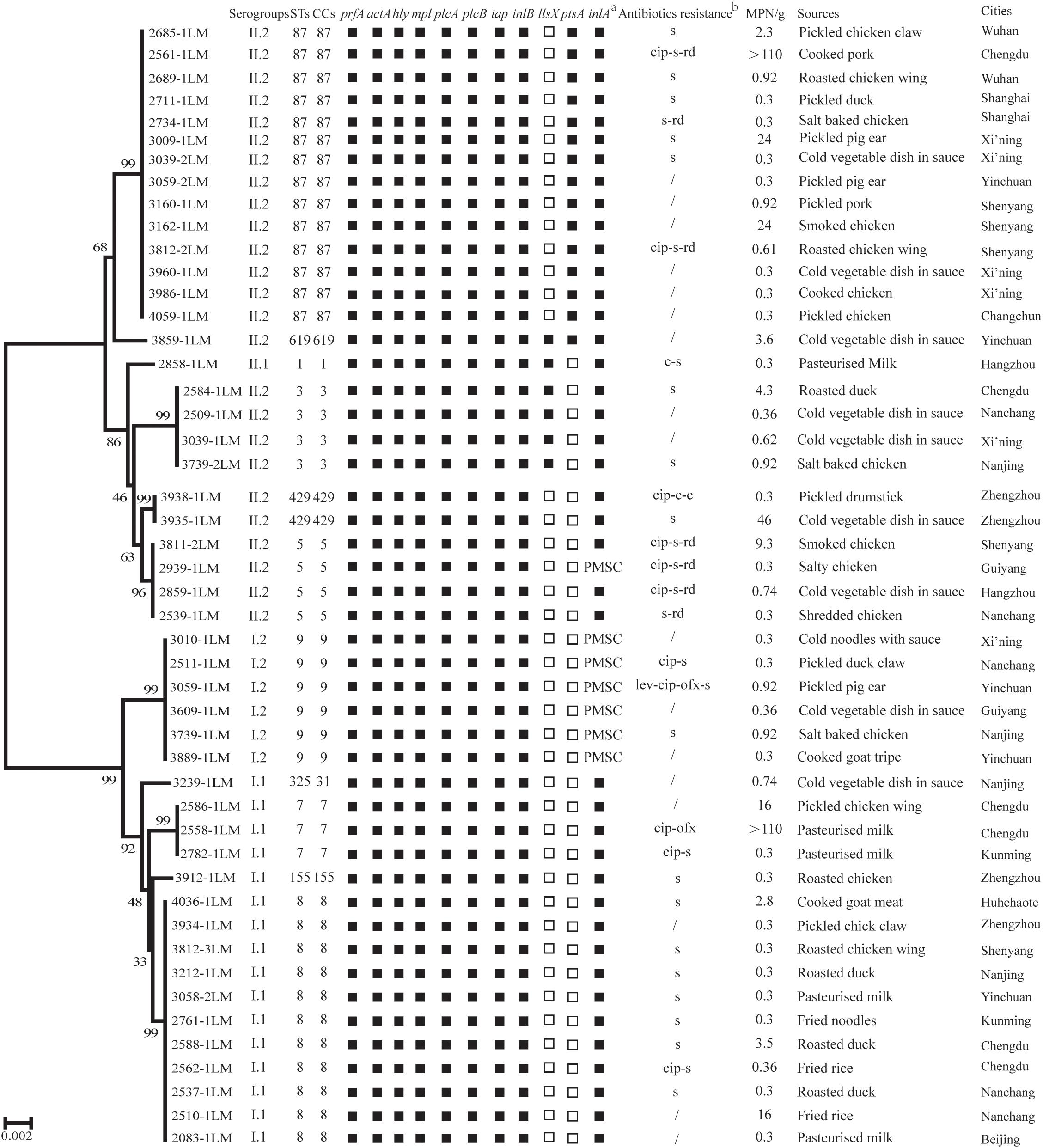
Susceptibility toward 19 antimicrobials was assessed among the 48 L. monocytogenes isolates. As shown in Supplementary Table S3, all isolates were susceptible to 12 antimicrobials, including kanamycin, gentamicin, sulfamethoxazole with trimethoprim, doxycycline, tetracycline, meropenem, vancomycin, linezolid, amoxicillin/clavulanic acid, sulbactam/ampicillin, ampicillin, and penicillin. More than 95.0% of isolates were susceptible to four antimicrobials (levofloxacin, ofloxacin, chloramphenicol, and erythromycin). However, 60.4% (29/48), 22.9% (11/48), and 14.6% (7/48) isolates were intermediately resistant to streptomycin, ciprofloxacin and rifampin, respectively. Seven resistance profiles were obtained for 48 L. monocytogenes isolates (Figure 2).

Figure 2. The phenotypic and genotypic characteristics of Listeria monocytogenes isolates. Of the 10 virulence genes (prfA, actA, hly, mpl, plcA, plcB, iap, inlB, llsX, and ptsA), black squares indicate the presence of the corresponding gene; white squares represent lack of corresponding gene. (a) PMSC, premature stop codons in inlA; black squares indicate the presence of full-length inlA. (b) c, chloramphenicol; cip, ciprofloxacin; e, erythromycin; lev, levofloxacin; ofx, ofloxacin; rd, rifampin; s, streptomycin;/indicates no resistance. Antibiotic abbreviations in lowercase indicate intermediate resistance. A neighbor-joining tree of L. monocytogenes based on the MLST of seven housekeeping genes was established using MEGA X with 1000 bootstrap replications. Bootstrap values are shown at the nodes.
MLST Analysis and Virulent Genes Profiles
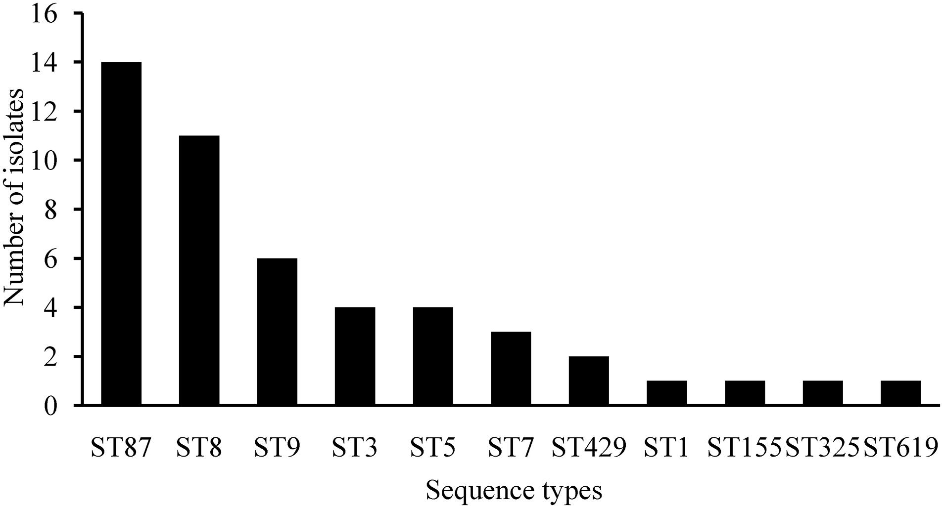
The genetic diversity of 48 isolates obtained from RTE foods and pasteurized milk were subjected to MLST analysis. As shown in Figure 3, 48 isolates belonged to 11 STs. ST87/CC87 (29.2%, 14/48) displayed the highest prevalence, followed by ST8/CC8 (22.9%, 11/48) and ST9/CC9 (12.5%, 6/48). The remaining eight CCs displayed a scattered distribution: ST3/CC3 (four isolates), ST5/CC5 (four isolates), ST7/CC7 (three isolates) ST429/CC429 (two isolates), ST1/CC1 (one isolates), ST155/CC155 (one isolates), ST325/CC31 (one isolates), and ST619 (one isolates). Virulence genes including LIPI-1, inlA, and inlB were present in all 11 STs/CCs isolates, while llsX (representing LIPI-3) was present exclusively in ST619 (one isolate), ST1/CC1 (one isolate), and ST3/CC3 (four isolates); ST619 (one isolate) and ST87/CC87 (14 isolates) isolates harbored the ptsA gene (representing LIPI-4). As shown in Figure 2, six isolates belonged to ST9/CC9 (6/6, 100%) and one isolate belonged to ST5/CC5 (1/4, 25%) and harbored PMSCs in the inlA gene. An adenylic acid deletion occurred at position 12 in the inlA gene in three isolates (2739-1LM, 3010-1LM, and 2511-1LM). A nonsense mutation at position 978 (GAA→TAA) in isolate 3609-1LM, position 1380 (TGG→TGA) in isolate 3883-1LM, and position 1605 (TGG→TGA) in isolate 2939-1LM were identified. In isolate 3059-1LM, a base of adenylic acid deletion occurred at position 1637, resulting in PMSCs in the inlA gene.

Figure 3. The sequence type distributions of Listeria monocytogenes isolated from ready-to-eat foods and pasteurized milk.
Discussion
Listeria monocytogenes is a prominent facultative foodborne pathogen with a worldwide prevalence. Numerous listeriosis outbreaks and sporadic cases have resulted from ingestion of L. monocytogenes-contaminated foods. In particular, RTE foods are considered vehicles of Listeria spp. owing to the absence of further heat treatment before consumption. The occurrence of listeriosis varies among different countries and usually occurs at 0.1–11.3 cases per million individuals (FAO/WHO, 2004). In this study, we assessed 570 food samples in China from 2014 to 2016, 39 RTE foods and 5 pasteurized milk samples (7.7%) tested positive for L. monocytogenes, concurrent with contamination of L. monocytogenes in RTE foods reported in Beijing (Wang W. et al., 2015) but significantly greater than that of RTE foods in Estonia (Koskar et al., 2019) and Tokyo, Japan (Shimojima et al., 2016). However, Wang et al. (2018) reported that 16.2% of samples were contaminated with L. monocytogenes in retail outlets selling RTE foods and restaurants serving mutton in Zigong, Sichuan Province. Among the pasteurized milk samples, only 2.4% (5/210) samples were positive for L. monocytogenes. Nonetheless, the contamination rate in other countries is markedly greater than that reported herein in China, amounting to 20% among pasteurized milk in Ethiopia (Seyoum et al., 2015) and 4.9% in milk and milk products in Tamil Nadu, India (Karthikeyan et al., 2015). This discrepancy is potentially attributed to differences in the sample size, sample constitution, hygiene conditions, or geographical locations.
Risk assessment of a L. monocytogenes infection through ingestion of RTE foods depended not only on the contamination rate of RTE foods, but also on their level of contamination. To limit the occurrence of listeriosis, several countries have formulated a series of food microbiological standards for L. monocytogenes for different foods. The United States Department of Agriculture (USDA) has adopted a zero-tolerance policy (absence of the organism in 25 g food sample) for L. monocytogenes in RTE foods (Orsi et al., 2011). The EU food safety regulations limit L. monocytogenes to < 100 CFU/g at the end of the shelf-life of a food product (Ziegler et al., 2019). Furthermore, China has adopted a zero-tolerance policy for RTE meat products (GB 29921-2013) (Anonymous, 2013); however, such regulations have not been adopted for pasteurized milk. In many European countries, L. monocytogenes counts must do not exceed 100 CFU/g during the shelf-life of RTE foods (Castro et al., 2017). Herein, 77.3% (34/44) samples were contaminated with L. monocytogenes below 10 MPN/g, 18.2% (8/44) were between 10 and 110 MPN/g, and only two (4.5%) samples were over 110 MPN/g. Concurrently, Koskar et al. (2019) reported that 0.3% of positive samples exceeded the food safety criterion of 100 CFU/g. The food matrix, storage time, and temperature were significant factors influencing the growth of L. monocytogenes (Ziegler et al., 2019). Results showed that the most common STs were ST8 strains in fried rice/noodles (100%, 3/3) and duck (50%, 3/6) isolates, while the ST87 strains (carried ptsA gene) were most found in chicken (44.4%, 8/18) and pork (75%, 3/4) isolates. The contamination rate of positive samples collected from Nanchang and Chengdu city were the highest among 43 cities, both being 50% (5/10), followed by Xi’ning (25%, 5/20). The count of L. monocytogenes in these positive samples may have drastically increased owing to the presence of numerous nutrients and an ambient storage temperature. The present results indicate that RTE foods are potentially an important transmission route for L. monocytogenes infections. Further surveillance of L. monocytogenes in RTE foods is warranted for risk assessment.
Serotyping is a classical typing method for L. monocytogenes. Serotypes 1/2a, 1/2b, 4b, and 1/2c account for 95% of clinical isolates (Pontello et al., 2012; Silk et al., 2012). In this study, 33.3% (16/48) isolates belonged to serogroup I.1 (1/2a-3a); 12.5% (6/48), serogroup I.2 (1/2c-3c); 2.1% (1/48), serogroup II.1 (4b-4d-4e); 52.1% (25/48), serogroup II.2 (1/2b-3b-7); this was concurrent with previous reports from China (Chen et al., 2009, 2015). For MLST analysis, 48 isolates belonging to 11 STs, ST87/CC87 (29.2%, 14/48), displayed the highest prevalence, followed by ST8/CC8 (22.9%, 11/48) and ST9/CC9 (12.5%, 6/48). Interestingly, ST87/CC87 was dominant in the listeriosis cases in China (Wang Y. et al., 2015). Huang et al. (2015) reported that ST87/CC87 were predominant among the 132 isolates obtained from listeriosis cases during 2000–2013 in Taiwan Province, China. Wang et al. (2018) reported that 24.2% (8/33) of listeriosis cases resulted from CC87 in Zigong, Sichuan Province; similarly, clinical cases resulted from RTE foods contaminated with ST87/CC87 strains (Zhang et al., 2018). These data suggest that ST87 poses potential health risks to consumers. To date, ST87/CC87-associated outbreaks and sporadic cases were seldom reported in other countries. However, 27 human listeriosis outbreaks primarily resulted from ST87/CC87 strains (One isolated from the foie gras that the patient ate) in Spain in 2013–2014 (Perez-Trallero et al., 2014), indicating that ST87/CC87 may potentially emerge as a hypervirulent strain worldwide. Furthermore, Chau et al. (2017) reported that ST87 was present in a particular brand of pre-packed salmon products over a 4-year period, implying a potential persistent contamination issue at the level of production. The predominance of ST87/CC87 of L. monocytogenes in foods is potentially associated with listeriosis, and further analysis of genotypic characteristics of foodborne and clinical isolates is required.
Virulence genes (LIPI-1, inlA, and inlB) were present in all L. monocytogenes isolates. Internalin A (InlA, encoded by inlA) is a major virulence factor associated with the invasiveness of L. monocytogenes and binds E-cadherin on host cells and facilitates the penetration of L. monocytogenes into intestinal epithelial cells (Vazquez-Boland et al., 2001). Therefore, PMSCs in the inlA gene may attenuate the virulence of L. monocytogenes strains with a lesser invasive phenotype among human intestinal epithelial cells (Nightingale et al., 2005; Van Stelten and Nightingale, 2008; Van Stelten et al., 2010). Interestingly, six (100%) ST9/CC9 and one (25%) ST5/CC5 isolates harbored PMSCs in the inlA gene, concurrent with previous studies reported in China (Chen et al., 2018a, 2019a). Similarly, the PMSCs in the inlA gene were frequent in ST121/CC121 strains, which were predominant in foods and associated environments (Ciolacu et al., 2015; Palma et al., 2017; Rychli et al., 2017, 2018). These data indicate that the ST9 and ST121 strains predominant in foods may acquire PMSCs in the inlA gene. However, the ecological significance of specific InlA variants is not yet understood (Ciolacu et al., 2015). Further research should be conducted to elucidate whether the PMSCs of inlA is associated with food-related stress. The bacteriocin LLS (encoded by the llsX and other genes) contributes to intestinal survival and virulence of L. monocytogenes in a murine oral infection model, alters the gut microbiota and increases L. monocytogenes persistence (Quereda et al., 2016). ST1, ST3, and ST619 strains harbored LIPI-3 (representing llsX), consistent with these STs also causing listeriosis both in China and other countries (Lv et al., 2014; Maury et al., 2016; Wang et al., 2018). ST87/CC87 (and ST619) strains harbored LIPI-4 (representing ptsA), responsible for neural and placental infections (Maury et al., 2016). It is noteworthy that one ST619 isolate had all virulence genes tested (especially llsX and ptsA), indicating a potential hypervirulent ST. Multiple reports also shown that ST619 strain was isolated in food and carried many virulence genes, including llsX and ptsA genes (Chen et al., 2018a, b, 2019b; Wang et al., 2018). In addition, to the best our knowledge, ST619 was only reported in clinic cases in China (Wang et al., 2018). However, to date, little information is available on the pathogenicity of ST619 strains, it is necessary to explore the virulence of ST619 strains in the future. Our data suggest that RTE foods and pasteurized milk contaminated with the isolates recovered herein may increase the risk of L. monocytogenes infection for consumers in China.
Listeriosis may be more difficult to control in future owing to the emergence of antimicrobial resistance among L. monocytogenes strains isolated from food products. β-lactam (penicillin and ampicillin) alone or in combination with an aminoglycoside (gentamycin) has been the first-line treatment alternative for listeriosis. However, patients presenting an allergic reaction to penicillin, a second-line treatment alternative, usually involve a combination of trimethoprim with a sulfonamide including sulfamethoxazole in co-trimoxazole (Alonso-Hernando et al., 2012). Fortunately, all 48 L. monocytogenes isolates were susceptible to β-lactam and aminoglycoside antimicrobials herein, indicating that these antimicrobials are still effective to treat listeriosis. Furthermore, rifampicin, tetracycline, chloramphenicol, and fluoroquinolones have been used to treat listeriosis (Allerberger and Wagner, 2010). Herein, L. monocytogenes isolates were intermediately resistant, to a certain extent, to fluoroquinolones, although no completely resistant isolate was observed. The mechanisms underlying fluoroquinolones resistance have been previously reported. Fluoroquinolone resistance may be mediated by mutations in quinolone resistance-determining regions, including GyrA, GyrB, ParC, and ParE (Bertrand et al., 2016). In addition, the expression of efflux pump FepR, MdeL, and Lde contributed to resistance toward fluoroquinolones (Lismond et al., 2008; Guerin et al., 2014; Lim et al., 2016; Wilson et al., 2018). Vancomycin and novel β-lactams constitute last-line therapy for Listeria infections, and no resistant isolate was observed herein. Therefore, future studies are required to focus on the mechanism underlying the acquisition of fluoroquinolone resistance in L. monocytogenes.
Conclusion
In summary, 7.7% (44/570) of RTE foods (39/360) and pasteurized milk (5/210) samples collected from 21 cities in China were positive for L. monocytogenes and two samples had a MPN > 110/g. Serogroup I.1 (ST8, 22.9%) and II.2 (ST87, 29.2%) were dominant among the 48 L. monocytogenes isolates, indicating that some specific serogroups and STs of L. monocytogenes may have distinct ecological niches. The nine classical virulence genes and additional llsX or/and ptsA potential hypervirulent genes were present in some specific L. monocytogenes isolates, indicating that the L. monocytogenes contaminated RTE foods and pasteurized milk may be a possible transmission route for Listeria infection in consumers. Although most isolates were susceptible to antimicrobials, except for streptomycin and ciprofloxacin (efflux pump mediated resistance), potential microbial safety issues in RTE foods and pasteurized milk requires close attention.
Data Availability Statement
The raw data supporting the conclusions of this manuscript will be made available by the authors, without undue reservation, to any qualified researcher.
Author Contributions
QW, JZ, and MC conceived and designed the experiments. MC, YC, JC, and QS performed the experiments. LX, HZ, SW, RP, and HW conducted the bioinformatics analyses. MC, QW, SZ, and TL drafted the manuscript. QW, QY, JW, WL, and XK reviewed the manuscript. All authors read and approved the final manuscript.
Funding
We would like to acknowledge the financial support from the National Natural Science Foundation of China (31701718), Pearl River S&T Nova Program of Guangzhou (201710010018), Natural Science Foundation of Guangdong Province, China (2017A030313173), GDAS’ Special Project of Science and Technology Development (2019GDASYL-0104010), Science and Technology Program of Guangdong Province (2017B090904004).
Conflict of Interest
WL and XK were employed by Infinitus (China) Company, Ltd.
The remaining authors declare that the research was conducted in the absence of any commercial or financial relationships that could be construed as a potential conflict of interest.
Acknowledgments
The authors would like to thank the team of curators of the Institute Pasteur MLST databases for curating the data and making them publicly available at http://bigsdb.pasteur.fr/.
Supplementary Material
The Supplementary Material for this article can be found online at: https://www.frontiersin.org/articles/10.3389/fmicb.2020.00642/full#supplementary-material
Footnotes
References
Allerberger, F., and Wagner, M. (2010). Listeriosis: a resurgent foodborne infection. Clin. Microbiol. Infect. 16, 16–23. doi: 10.1111/j.1469-0691.2009.03109.x
Alonso-Hernando, A., Prieto, M., Garcia-Fernandez, C., Alonso-Calleja, C., and Capita, R. (2012). Increase over time in the prevalence of multiple antibiotic resistance among isolates of Listeria monocytogenes from poultry in Spain. Food Control 23, 37–41. doi: 10.1016/j.foodcont.2011.06.006
Anonymous (2010). National Food Safety Standard Food Microbiological Examination: Listeria monocytogenes (GB4789.30-2010). Beijing: Ministry of Health of People’s Republic of China.
Anonymous (2013). National Food Safety Standard, Pathogen Limits for Food, GB29921-2013. Beijing: Standards Press of China.
Basha, K. A., Kumar, N. R., Das, V., Reshmi, K., Rao, B. M., Lalitha, K. V., et al. (2019). Prevalence, molecular characterization, genetic heterogeneity and antimicrobial resistance of Listeria monocytogenes associated with fish and fishery environment in Kerala, India. Lett. Appl. Microbiol. 69, 286–293. doi: 10.1111/lam.13205
Bertrand, S., Ceyssens, P. J., Yde, M., Dierick, K., Boyen, F., Vanderpas, J., et al. (2016). Diversity of Listeria monocytogenes strains of clinical and food chain origins in belgium between 1985 and 2014. PLoS One 11:e0164283. doi: 10.1371/journal.pone.0164283
Cartwright, E. J., Jackson, K. A., Johnson, S. D., Graves, L. M., Silk, B. J., and Mahon, B. E. (2013). Listeriosis outbreaks and associated food vehicles, United States, 1998-2008. Emerg. Infect. Dis. 19, 1–9. doi: 10.3201/eid1901.120393
Castro, H., Ruusunen, M., and Lindstrom, M. (2017). Occurrence and growth of Listeria monocytogenes in packaged raw milk. Int. J. Food Microbiol. 261, 1–10. doi: 10.1016/j.ijfoodmicro.2017.08.017
Chau, M. L., Aung, K. T., Hapuarachchi, H. C., Lee, P. S., Lim, P. Y., Kang, J. S., et al. (2017). Microbial survey of ready-to-eat salad ingredients sold at retail reveals the occurrence and the persistence of Listeria monocytogenes Sequence Types 2 and 87 in pre-packed smoked salmon. BMC Microbiol. 17:46. doi: 10.1186/s12866-017-0956-z
Chen, J., Luo, X., Jiang, L., Jin, P., Wei, W., Liu, D., et al. (2009). Molecular characteristics and virulence potential of Listeria monocytogenes isolates from Chinese food systems. Food Microbiol. 26, 103–111. doi: 10.1016/j.fm.2008.08.003
Chen, M., Chen, Y., Wu, Q., Zhang, J., Cheng, J., Li, F., et al. (2019a). Genetic characteristics and virulence of Listeria monocytogenes isolated from fresh vegetables in China. BMC Microbiol. 19:119. doi: 10.1186/s12866-019-1488-5
Chen, M., Cheng, J., Zhang, J., Chen, Y., and Zeng, H. (2019b). Isolation, potential virulence, and population diversity of Listeria monocytogenes from meat and meat products in China. Front. Microbiol. 10:946. doi: 10.3389/fmicb.2019.00946
Chen, M., Cheng, J., Wu, Q., Zhang, J., Chen, Y., Xue, L., et al. (2018a). Occurrence, antibiotic resistance, and population diversity of Listeria monocytogenes isolated from fresh aquatic products in China. Front. Microbiol. 9:2215. doi: 10.3389/fmicb.2018.02215
Chen, M., Cheng, J., Wu, Q., Zhang, J., Chen, Y., Zeng, H., et al. (2018b). Prevalence, potential virulence, and genetic diversity of Listeria monocytogenes isolates from edible mushrooms in Chinese markets. Front. Microbiol. 9:1711. doi: 10.3389/fmicb.2018.01711
Chen, M., Wu, Q., Zhang, J., Wu, S., and Guo, W. (2015). Prevalence, enumeration, and pheno- and genotypic characteristics of Listeria monocytogenes isolated from raw foods in South China. Front. Microbiol. 6:1026. doi: 10.3389/fmicb.2015.01026
Chen, Y., Gonzalez-Escalona, N., Hammack, T. S., Allard, M. W., Strain, E. A., and Brown, E. W. (2016). Core genome multilocus sequence typing for identification of globally distributed clonal groups and differentiation of outbreak strains of Listeria monocytogenes. Appl. Environ. Microbiol. 82, 6258–6272. doi: 10.1128/AEM.01532-16
Chen, Y., Luo, Y., Curry, P., Timme, R., Melka, D., Doyle, M., et al. (2017). Assessing the genome level diversity of Listeria monocytogenes from contaminated ice cream and environmental samples linked to a listeriosis outbreak in the United States. PLoS One 12:e0171389. doi: 10.1371/journal.pone.0171389
Ciolacu, L., Nicolau, A. I., Wagner, M., and Rychli, K. (2015). Listeria monocytogenes isolated from food samples from a Romanian black market show distinct virulence profiles. Int. J. Food Microbiol. 209, 44–51. doi: 10.1016/j.ijfoodmicro.2014.08.035
Clayton, E. M., Hill, C., Cotter, P. D., and Ross, R. P. (2011). Real-time PCR assay to differentiate Listeriolysin S-positive and -negative strains of Listeria monocytogenes. Appl. Environ. Microbiol. 77, 163–171. doi: 10.1128/AEM.01673-10
Clinical and Laboratory Standard Institute (2017). Performance Standards for Antimicrobial Susceptibility Testing: 27th Informational Supplement. Wayne, PA: Clinical and Laboratory Standards Institute.
Cotter, P. D., Draper, L. A., Lawton, E. M., Daly, K. M., Groeger, D. S., Casey, P. G., et al. (2008). Listeriolysin S, a novel peptide haemolysin associated with a subset of lineage I Listeria monocytogenes. PLoS Pathog. 4:e1000144. doi: 10.1371/journal.ppat.1000144
Doumith, M., Buchrieser, C., Glaser, P., Jacquet, C., and Martin, P. (2004). Differentiation of the major Listeria monocytogenes serovars by multiplex PCR. J. Clin. Microbiol. 42, 3819–3822. doi: 10.1128/jcm.42.8.3819-3822.2004
European Food Safety Authority and European Centre for Disease Prevention and Control (2016). The European Union summary report on trends and sources of zoonoses, zoonotic agents and food-borne outbreaks in 2015. EFSA J. 14:4634. doi: 10.2903/j.efsa.2016.4634
Fagerlund, A., Langsrud, S., Schirmer, B. C., Moretro, T., and Heir, E. (2016). Genome analysis of Listeria monocytogenes sequence type 8 strains persisting in salmon and poultry processing environments and comparison with related strains. PLoS One 11:e0151117. doi: 10.1371/journal.pone.0151117
Fan, Z., Xie, J., Li, Y., and Wang, H. (2019). Listeriosis in mainland China: a systematic review. Int. J. Infect. Dis. 81, 17–24. doi: 10.1016/j.ijid.2019.01.007
FAO/WHO (2004). Risk Assessment of Listeria Monocytogenes in Ready-to-Eat Foods. Microbiological risk assessment series 5, Technical report. Geneva: WHO.
Feng, Y. F., Wu, S. Y., Varma, J. K., Klena, J. D., Angulo, F. J., and Ran, L. (2013). Systematic review of human listeriosis in China, 1964-2010. Trop. Med. Int. Health 18, 1248–1256. doi: 10.1111/tmi.12173
Gaillard, J. L., Jaubert, F., and Berche, P. (1996). The inlAB locus mediates the entry of Listeria monocytogenes into hepatocytes in vivo. J. Exp. Med. 183, 359–369. doi: 10.1084/jem.183.2.359
Gombas, D. E., Chen, Y., Clavero, R. S., and Scott, V. N. (2003). Survey of Listeria monocytogenes in ready-to-eat foods. J. Food Prot. 66, 559–569. doi: 10.4315/0362-028x-66.4.559
Guerin, F., Galimand, M., Tuambilangana, F., Courvalin, P., and Cattoir, V. (2014). Overexpression of the novel MATE fluoroquinolone efflux pump FepA in Listeria monocytogenes is driven by inactivation of its local repressor FepR. PLoS One 9:e106340. doi: 10.1371/journal.pone.0106340
Hain, T., Chatterjee, S. S., Ghai, R., Kuenne, C. T., Billion, A., Steinweg, C., et al. (2007). Pathogenomics of Listeria spp. Int. J. Med. Microbiol. 297, 541–557. doi: 10.1016/j.ijmm.2007.03.016
Huang, Y. T., Ko, W. C., Chan, Y. J., Lu, J. J., Tsai, H. Y., Liao, C. H., et al. (2015). Disease burden of invasive listeriosis and molecular characterization of clinical isolates in Taiwan, 2000-2013. PLoS One 10:e0141241. doi: 10.1371/journal.pone.0141241
Karthikeyan, R., Gunasekaran, P., and Rajendhran, J. (2015). Molecular serotyping and pathogenic potential of Listeria monocytogenes isolated from milk and milk products in Tamil Nadu, India. Foodborne Pathog. Dis. 12, 522–528. doi: 10.1089/fpd.2014.1872
Kim, S. W., Haendiges, J., Keller, E. N., Myers, R., Kim, A., Lombard, J. E., et al. (2018). Genetic diversity and virulence profiles of Listeria monocytogenes recovered from bulk tank milk, milk filters, and milking equipment from dairies in the United States (2002 to 2014). PLoS One 13:e0197053. doi: 10.1371/journal.pone.0197053
Koskar, J., Kramarenko, T., Meremae, K., Kuningas, M., Sogel, J., Maesaar, M., et al. (2019). Prevalence and numbers of Listeria monocytogenes in various ready-to-eat foods over a 5-year period in Estonia. J. Food Prot. 82, 597–604. doi: 10.4315/0362-028x.Jfp-18-383
Kumar, S., Stecher, G., Li, M., Knyaz, C., and Tamura, K. (2018). MEGA X: molecular evolutionary genetics analysis across computing platforms. Mol. Biol. Evol. 35, 1547–1549. doi: 10.1093/molbev/msy096
Lim, S. Y., Yap, K. P., and Thong, K. L. (2016). Comparative genomics analyses revealed two virulent Listeria monocytogenes strains isolated from ready-to-eat food. Gut Pathog. 8:65. doi: 10.1186/s13099-016-0147-8
Lismond, A., Tulkens, P. M., Mingeot-Leclercq, M. P., Courvalin, P., and Van Bambeke, F. (2008). Cooperation between prokaryotic (Lde) and eukaryotic (MRP) efflux transporters in J774 macrophages infected with Listeria monocytogenes: studies with ciprofloxacin and moxifloxacin. Antimicrob. Agents Chemother. 52, 3040–3046. doi: 10.1128/Aac.00105-08
Lv, J., Qin, Z., Xu, Y., and Xie, Q. (2014). Listeria infection in Chinese pregnant women and neonates from Shandong. Int. J. Clin. Exp. Med. 7, 2730–2734.
Magiorakos, A. P., Srinivasan, A., Carey, R. B., Carmeli, Y., Falagas, M. E., Giske, C. G., et al. (2012). Multidrug-resistant, extensively drug-resistant and pandrug-resistant bacteria: an international expert proposal for interim standard definitions for acquired resistance. Clin. Microbiol. Infect. 18, 268–281. doi: 10.1111/j.1469-0691.2011.03570.x
Maury, M. M., Tsai, Y. H., Charlier, C., Touchon, M., Chenal-Francisque, V., Leclercq, A., et al. (2016). Uncovering Listeria monocytogenes hypervirulence by harnessing its biodiversity. Nat. Genet. 48, 308–313. doi: 10.1038/ng.3501
Melero, B., Manso, B., Stessl, B., Hernández, M., Wagner, M., Rovira, J., et al. (2019). Distribution and persistence of Listeria monocytogenes in a heavily contaminated poultry processing facility. J. Food Prot. 82, 1524–1531. doi: 10.4315/0362-028x.jfp-19-087
Nightingale, K. K., Windham, K., Martin, K. E., Yeung, M., and Wiedmann, M. (2005). Select Listeria monocytogenes subtypes commonly found in foods carry distinct nonsense mutations in inlA, leading to expression of truncated and secreted internalin A, and are associated with a reduced invasion phenotype for human intestinal epithelial cells. Appl. Environ. Microbiol. 71, 8764–8772. doi: 10.1128/AEM.71.12.8764-8772.2005
Orsi, R. H., Bakker, H. C. D., and Wiedmann, M. (2011). Listeria monocytogenes lineages: genomics, evolution, ecology, and phenotypic characteristics. Int. J. Med. Microbiol. 301, 79–96. doi: 10.1016/j.ijmm.2010.05.002
Palma, F., Pasquali, F., Lucchi, A., Cesare, A., and Manfreda, G. (2017). Whole genome sequencing for typing and characterisation of Listeria monocytogenes isolated in a rabbit meat processing plant. Ital. J. Food Saf. 6:6879. doi: 10.4081/ijfs.2017.6879
Pasquali, F., Palma, F., Guillier, L., Lucchi, A., De Cesare, A., and Manfreda, G. (2018). Listeria monocytogenes sequence types 121 and 14 repeatedly isolated within one year of sampling in a rabbit meat processing plant: persistence and ecophysiology. Front. Microbiol. 9:596. doi: 10.3389/fmicb.2018.00596
Perez-Trallero, E., Zigorraga, C., Artieda, J., Alkorta, M., and Marimon, J. M. (2014). Two outbreaks of Listeria monocytogenes infection, Northern Spain. Emerg. Infect. Dis. 20, 2155–2157. doi: 10.3201/eid2012.140993
Pontello, M., Guaita, A., Sala, G., Cipolla, M., Gattuso, A., Sonnessa, M., et al. (2012). Listeria monocytogenes serotypes in human infections (Italy, 2000-2010). Ann. Ist Super. Sanita 48, 146–150. doi: 10.4415/ANN_12_02_07
Quereda, J. J., Dussurget, O., Nahori, M. A., Ghozlane, A., Volant, S., Dillies, M. A., et al. (2016). Bacteriocin from epidemic Listeria strains alters the host intestinal microbiota to favor infection. Proc. Natl. Acad. Sci. U.S.A. 113, 5706–5711. doi: 10.1073/pnas.1523899113
Quereda, J. J., Meza-Torres, J., Cossart, P., and Pizarro-Cerda, J. (2017). Listeriolysin S: a bacteriocin from epidemic Listeria monocytogenes strains that targets the gut microbiota. Gut Microb. 8, 384–391. doi: 10.1080/19490976.2017.1290759
Radoshevich, L., and Cossart, P. (2018). Listeria monocytogenes: towards a complete picture of its physiology and pathogenesis. Nat. Rev. Microbiol. 16, 32–46. doi: 10.1038/nrmicro.2017.126
Ragon, M., Wirth, T., Hollandt, F., Lavenir, R., Lecuit, M., Le Monnier, A., et al. (2008). A new perspective on Listeria monocytogenes evolution. PLoS Pathog. 4:e1000146. doi: 10.1371/journal.ppat.1000146
Rychli, K., Stessl, B., Szakmary-Brandle, K., Strauss, A., Wagner, M., and Schoder, D. (2018). Listeria monocytogenes isolated from illegally imported food products into the European Union harbor different virulence factor variants. Genes 9:E428. doi: 10.3390/genes9090428
Rychli, K., Wagner, E. M., Ciolacu, L., Zaiser, A., Tasara, T., Wagner, M., et al. (2017). Comparative genomics of human and non-human Listeria monocytogenes sequence type 121 strains. PLoS One 12:e0176857. doi: 10.1371/journal.pone.0176857
Seyoum, E. T., Woldetsadik, D. A., Mekonen, T. K., Gezahegn, H. A., and Gebreyes, W. A. (2015). Prevalence of Listeria monocytogenes in raw bovine milk and milk products from central highlands of Ethiopia. J. Infect. Dev. Ctries 9, 1204–1209. doi: 10.3855/jidc.6211
Shimojima, Y., Ida, M., Nakama, A., Nishino, Y., Fukui, R., Kuroda, S., et al. (2016). Prevalence and contamination levels of Listeria monocytogenes in ready-to-eat foods in Tokyo, Japan. J. Vet. Med. Sci. 78, 1183–1187. doi: 10.1292/jvms.15-0708
Silk, B. J., Date, K. A., Jackson, K. A., Pouillot, R., Holt, K. G., Graves, L. M., et al. (2012). Invasive listeriosis in the foodborne diseases active surveillance network (FoodNet), 2004-2009: further targeted prevention needed for higher-risk groups. Clin. Infect. Dis. 54(Suppl. 5), S396–S404. doi: 10.1093/cid/cis268
Smith, A. M., Tau, N. P., Smouse, S. L., Allam, M., Ismail, A., Ramalwa, N. R., et al. (2019). Outbreak of Listeria monocytogenes in South Africa, 2017-2018: laboratory activities and experiences associated with whole-genome sequencing analysis of isolates. Foodborne Pathog. Dis. 16, 524–530. doi: 10.1089/fpd.2018.2586
U.S. Department of Agriculture (1998). “Isolation and identification of Listeria monocytogenes from meat, poultry and egg products,” in Microbiology Guide Book, 3rd Edn, Chapter 8, eds B. P. Dey, and C. P. Lattuada (Washington, DC: Government Printing Office).
Van Stelten, A., and Nightingale, K. K. (2008). Development and implementation of a multiplex single-nucleotide polymorphism genotyping assay for detection of virulence-attenuating mutations in the Listeria monocytogenes virulence-associated gene inlA. Appl. Environ. Microbiol. 74, 7365–7375. doi: 10.1128/AEM.01138-08
Van Stelten, A., Simpson, J. M., Ward, T. J., and Nightingale, K. K. (2010). Revelation by single-nucleotide polymorphism genotyping that mutations leading to a premature stop codon in inlA are common among Listeria monocytogenes isolates from ready-to-eat foods but not human listeriosis cases. Appl. Environ. Microbiol. 76, 2783–2790. doi: 10.1128/AEM.02651-09
Vazquez-Boland, J. A., Kuhn, M., Berche, P., Chakraborty, T., Dominguez-Bernal, G., Goebel, W., et al. (2001). Listeria pathogenesis and molecular virulence determinants. Clin. Microbiol. Rev. 14, 584–640. doi: 10.1128/CMR.14.3.584-640.2001
Wang, H., Luo, L., Zhang, Z., Deng, J., Wang, Y., Miao, Y., et al. (2018). Prevalence and molecular characteristics of Listeria monocytogenes in cooked products and its comparison with isolates from listeriosis cases. Front. Med. 12:104–112. doi: 10.1007/s11684-017-0593-9
Wang, W., Yan, S., Bai, L., Li, Z., Du, C., and Li, F. (2015). Quantitative detection on contamination of Listeria monocytogenes isolated from retail ready-to-eat meats in Beijing. Wei Sheng Yan Jiu 44, 918–921.
Wang, Y., Jiao, Y., Lan, R., Xu, X., Liu, G., Wang, X., et al. (2015). Characterization of Listeria monocytogenes isolated from human Listeriosis cases in China. Emerg. Microb. Infect. 4:e50. doi: 10.1038/emi.2015.50
Wilson, A., Gray, J., Chandry, P. S., and Fox, E. M. (2018). Phenotypic and genotypic analysis of antimicrobial resistance among Listeria monocytogenes isolated from Australian food production chains. Genes 9:E80. doi: 10.3390/genes9020080
Wu, S., Wu, Q., Zhang, J., Chen, M., and Guo, W. (2016). Analysis of multilocus sequence typing and virulence characterization of Listeria monocytogenes isolates from Chinese retail ready-to-eat food. Front. Microbiol. 7:168. doi: 10.3389/fmicb.2016.00168
Yin, Y., Yao, H., Doijad, S., Kong, S., Shen, Y., Cai, X., et al. (2019). A hybrid sub-lineage of Listeria monocytogenes comprising hypervirulent isolates. Nat. Commun. 10:4283. doi: 10.1038/s41467-019-12072-1
Zhang, L., Chen, X., Luo, L., Wang, Y., Zhang, J., Zou, N., et al. (2018). Infection source of a neonatal sepsis case caused by Listeria monocytogenes. Dis. Surveill. 33:4.
Zhang, Y., Dong, S., Chen, H., Chen, J., Zhang, J., Zhang, Z., et al. (2019). Prevalence, genotypic characteristics and antibiotic resistance of Listeria monocytogenes from retail foods in bulk in Zhejiang province, China. Front. Microbiol. 10:1710. doi: 10.3389/fmicb.2019.01710
Keywords: Listeria monocytogenes, ready-to-eat foods, LIPI-3, LIPI-4, multi-locus sequence typing, antimicrobial resistance
Citation: Chen Y, Chen M, Wang J, Wu Q, Cheng J, Zhang J, Sun Q, Xue L, Zeng H, Lei T, Pang R, Ye Q, Wu S, Zhang S, Wu H, Li W and Kou X (2020) Heterogeneity, Characteristics, and Public Health Implications of Listeria monocytogenes in Ready-to-Eat Foods and Pasteurized Milk in China. Front. Microbiol. 11:642. doi: 10.3389/fmicb.2020.00642
Received: 20 July 2019; Accepted: 20 March 2020;
Published: 15 April 2020.
Edited by:
Giovanna Suzzi, University of Teramo, ItalyReviewed by:
Beatrix Stessl, University of Veterinary Medicine Vienna, AustriaYifan Zhang, Wayne State University, United States
Copyright © 2020 Chen, Chen, Wang, Wu, Cheng, Zhang, Sun, Xue, Zeng, Lei, Pang, Ye, Wu, Zhang, Wu, Li and Kou. This is an open-access article distributed under the terms of the Creative Commons Attribution License (CC BY). The use, distribution or reproduction in other forums is permitted, provided the original author(s) and the copyright owner(s) are credited and that the original publication in this journal is cited, in accordance with accepted academic practice. No use, distribution or reproduction is permitted which does not comply with these terms.
*Correspondence: Qingping Wu, d3VxcDIwM0AxNjMuY29t
†These authors have contributed equally to this work and share first authorship
 Yuetao Chen1,2†
 Moutong Chen
 Juan Wang
 Qingping Wu
 Jumei Zhang
 Haiyan Zeng
 Tao Lei
 Rui Pang
 Shi Wu
 Haoming Wu