- 1Division of Bacteriology, ICMR-National Institute for Research in Bacterial Infections (Formerly ICMR-NICED), Kolkata, West Bengal, India
- 2Department of Microbiology, Agartala Government Medical College & G B Pant Hospital, Agartala, Tripura, India
- 3Division of Communicable Diseases, Indian Council of Medical Research (ICMR), New Delhi, India
Introduction: Mobile genetic elements (MGEs) play a crucial role in the spread of carbapenem resistance. A study was undertaken to characterize MGEs and evaluate their contribution to the spread of carbapenem resistance in Escherichia coli and Klebsiella pneumoniae collected from three intensive care units (ICUs) of a tertiary care hospital in Tripura.
Methods: Isolates were subjected to susceptibility testing, genotypic detection of carbapenemases and their transmissibility, whole-genome sequencing (WGS), and phylogenomic analysis.
Results: E. coli and K. pneumoniae were the dominant Enterobacterales, exhibiting resistance to the majority of antibiotics. WGS of carbapenemase-producing E. coli (n = 15/48,31%) and K. pneumoniae (n = 13/26,50%) revealed the presence of blaNDM-1,5,7 (n = 21), blaKPC-2 (n = 1), and blaOXA-181,232 (n = 8). Isolates were diverse and belonged to different sequence types, including epidemic clones (K. pneumoniae-ST16/101/147/231; E. coli-ST167/410/648). This study has noted the allelic shift of blaNDM-1 to blaNDM-5 similar to global reports. blaNDM-1,5,7-bearing plasmids were conjugative but those carrying blaKPC-2 and blaOXA-181,232 were non-conjugative. blaNDM-1,5,7 were present in diverse replicons: IncF-types (predominant), IncHI1B, IncX3, and IncX4, etc., while blaOXA-181,232 were present in ColKP3, corroborating with global studies. blaNDM-1,5 was associated with intact/truncated ISAba125 in Tn125, blaNDM-7 with IS3000, blaKPC-2 with ISKpn6, and ISKpn7 in Tn4401b, and blaOXA-181,232 with ∆ISEcp1 in Tn2013, depicting an ancestral genetic context noted globally. Study isolates were related to other Indian isolates, primarily from blood.
Discussion: The association with different MGEs noted in the study is similar to those in other parts of India and the globe, signifying that the genetic determinants are part of the global gene pool. These associations can facilitate the spread of carbapenem resistance, leading to outbreaks and treatment failures.
1 Introduction
Bacteria share genes through vertical (parent to offspring) and horizontal (bacterium to bacterium) gene transfer. Genes transferred horizontally are typically not essential for basic cellular functions; instead, they are often accessory genes such as antibiotic resistance genes (ARGs). Spread of ARGs is often mediated by certain discrete DNA sequences known as mobile genetic elements (MGEs) (Partridge et al., 2018). Components of MGEs, such as plasmids and integrative conjugative elements (ICEs), enable intercellular mobility of DNA, while insertion sequence (IS) elements, transposons (Tn), and integrons (In) facilitate intracellular DNA mobility (Partridge et al., 2018; Khedkar et al., 2022). Dissemination of MGEs is facilitated by horizontal gene transfer (HGT) mechanisms (conjugation, transformation, transduction, and vesiduction), which ultimately leads to bacterial evolution (da Silva et al., 2021). Occurrence of ARGs (blaCTX-M, blaNDM, blaKPC, blaOXA-48-like, etc.) within the MGEs leads to the establishment of multidrug resistance (MDR) within the bacterial community, rendering available antibiotics ineffective (Liu et al., 2024).
Carbapenem, the last resort drug to treat severe MDR infections, have become ineffective due to the emergence of various carbapenemase genes such as class-A serine carbapenemase: Klebsiella pneumoniae carbapenemase (KPC; blaKPC); class-B metallo-β-lactamase (MBL): New Delhi metallo-β-lactamase (NDM; blaNDM) and class-D serine carbapenemase: oxacillinases (OXA)-48 and its variants (blaOXA-48-like) (Cui et al., 2019). KPC, NDM, and OXA-48 were first reported in K. pneumoniae and have subsequently been reported in Escherichia coli as well as various genera of Gram-negative bacteria (Pitout et al., 2019; Wu et al., 2019; Ding et al., 2023). These three carbapenemases differ from each other in many attributes. KPC and OXA-48 contain serine at the active site, while NDM contains zinc ions. The hydrolysis profile of these enzymes for the majority of β-lactam antibiotics (benzylpenicillin, ampicillin, piperacillin, cephalothin, temocillin, first-generation cephalosporins, and carbapenems) is similar, except for the fact that KPC and OXA-48 cannot hydrolyze cefoxitin and ceftazidime, but NDM can hydrolyze all. Although KPC and OXA-48 are considered carbapenemases, their hydrolysis potential for carbapenems is not similar to NDM; that is, both KPC and OXA-48 can hydrolyze meropenem sparingly, and OXA-48 exhibits the highest catalytic activity toward imipenem. These carbapenemases have been reported across the globe from different sources, namely, human, environment (soil, water, etc.), companion animals, and so on (Taggar et al., 2020). However, the propensity of occurrence differs for each carbapenemase (Al-Zahrani and Alsiri, 2018). NDM is the widely reported carbapenemase (Wu et al., 2019), while KPC is endemic to North and South America, China, and parts of Europe (Ding et al., 2023), and OXA-48 is prevalently reported from Middle East countries. Carbapenemases are borne of diverse plasmid replicons, which aid in their successful spread across the globe (Pitout et al., 2019). They are often associated with various MGEs, such as ISAba125, ISEcp1, Tn3, Tn125, Tn4401, and Tn2013 (Partridge et al., 2018).
The presence of these carbapenemases on different mobilizable plasmids has led to the spread of these genes across the globe (Lee et al., 2016; Wu et al., 2019; Ding et al., 2023). blaNDM is associated with a broad range of plasmid types (IncA/C, IncF, IncR, IncN, IncH, IncL/M, IncX, IncB/O/K/Z, IncY, IncT, and ColE10) (Lee et al., 2016; Wu et al., 2019) compared to blaKPC (IncF, IncFII, IncFIIK, IncI2, IncX, IncA/C, IncR, and ColE1) (Wyres and Holt, 2018; MacKinnon et al., 2020; Gorrie et al., 2022) and blaOXA-48-like (IncL/M, IncF, IncA/C, IncHI, IncI, IncX, IncN, IncX3, IncT, and ColE-type) (Pitout et al., 2019). In addition, these genes are linked with various IS elements, such as ISAba125, IS6, and ISCR27 (blaNDM); ISKpn6, ISKpn7, ISKpn8 (blaKPC); and IS1999 and its variants, ISEcp1 and IS1R (blaOXA-48-like) (Pitout et al., 2019; Acman et al., 2022), which have facilitated their spread within the bacterial community. Also, the association of these carbapenemases has been found with various transposons, such as blaNDM with Tn3, Tn125, and Tn3000; blaKPC with Tn4401b and its variants; and blaOXA-48-like with Tn1999, Tn6237, and Tn2013 (Pitout et al., 2019; Acman et al., 2022). The contribution of MGEs in the spread of these genes is well exemplified by the global dissemination of blaNDM-1, blaKPC, and blaOXA-48-like within different members of Enterobacterales, especially E. coli and K. pneumoniae (Lee et al., 2016; Pitout et al., 2019; Acman et al., 2022).
E. coli and K. pneumoniae are critical pathogens as per the World Health Organization (WHO) priority pathogen list,1 and are known to cause a wide range of community-acquired and healthcare-associated infections among adults as well as neonates (Wyres and Holt, 2018; MacKinnon et al., 2020; Gorrie et al., 2022). They are efficient in acquiring ARGs (Paczosa and Mecsas, 2016), and a recent study has suggested that the K. pneumoniae genome is an “open pangenome” with more than 400 acquired AGRs, highlighting increased HGT rate in this species (Navon-Venezia et al., 2017; Martin and Bachman, 2018; Wyres and Holt, 2018; Wang et al., 2023). With the acquisition of carbapenem-resistant genes via different MGEs, E. coli and K. pneumoniae have become resistant to carbapenems.
In this study, carbapenem-resistant E. coli and K. pneumoniae isolated from different intensive care units (ICUs) of a tertiary care hospital of Northeast India (Tripura) were analyzed in terms of diversity of bacterial etiology, susceptibility, carbapenemase genes, transmissibility of carbapenemases and MGEs (plasmids, IS elements, transposons) associated with it. Despite the antibiotic exposure typical of ICUs, it remains uncertain which MGEs primarily facilitate the dissemination of resistance genes. The study site is in Northeast India, where incidences of antimicrobial resistance (AMR) have been explored in a minimal way. The Northeast India is heterogeneous in terms of its geographical location, high rainfall, forest coverage, and diverse ethnic groups. This leads to compromised healthcare facilities resulting in morbidity, mortality, and increased AMR.2 Since the assessment of MGEs in the spread of carbapenem resistance is crucial in containing AMR spread, this study was conducted.
2 Methods
2.1 Ethics approval and consent for participation
The study protocol was approved by the Institutional Ethics Committee of the Indian Council of Medical Research (ICMR)-National Institute for Research in Bacterial Infections (formerly, ICMR-NICED) (A-1/2019-IEC.). Patient consent was taken prior to enrolment in the study. Patient information was anonymized and de-identified prior to analysis. All experiments were performed in accordance with relevant guidelines and regulations.
2.2 Isolation, identification, and antibiotic susceptibility testing of study isolates
Clinical specimens (blood, urine, and cerebrospinal fluid) were collected from patients admitted to the anesthesia intensive care unit (AICU), pediatric intensive care unit (PICU), and neonatal intensive care unit (NICU) of Agartala Government Medical College, Agartala, Tripura, India, from October 2019 to January 2021. Isolates were identified using VITEK2 COMPACT system (BioMérieux, Marcy-l’Étoile, France). Antibiotic susceptibilities of the study Enterobacterales were determined using the VITEK®2 AST-N281 card (BioMérieux, Marcy-l’Étoile, France). Minimum inhibitory concentration (MIC) of colistin was separately determined by broth microdilution using a commercial colistin MIC test kit (MIKROLATEST, Erba Lachema s.r.o., Czech Republic). Additionally, the MIC of meropenem (carbapenem) was assessed using E-strip (BioMérieux, Marcy-l’Étoile, France). The results were interpreted according to Clinical and Laboratory Standards Institute guidelines (CLSI, 2024). Isolates showing intermediate susceptibility values to any antimicrobials were considered non-susceptible to that antimicrobial.
2.3 Detection of carbapenemase producers and determining their genetic relatedness
A multiplex polymerase chain reaction (PCR) (Grönthal et al., 2018) was carried out to detect carbapenemases genes: blaKPC, blaIMP, blaVIM, blaNDM, and blaOXA-48 within the studied isolates, followed by sequencing the genes using primers mentioned in earlier studies (Yigit et al., 2001; Espinal et al., 2011). Genetic relatedness of the carbapenemase-producing Enterobacterales was studied by pulsed-field gel electrophoresis (PFGE) using a CHEF-DRIII apparatus (Bio-Rad Laboratories, Inc., Hercules, CA, United States) as described previously (Tenover et al., 1995). Processing of the PFGE images and the preparation of the dendrogram were performed by FP Quest software v4.5 (Bio-Rad Laboratories, Inc., Hercules, CA, United States) using the unweighted pair group method with arithmetic mean (UPGMA) and Dice coefficient. Isolates having more than 95% similarity were considered identical. Salmonella Braenderup H9812 isolate was used as a molecular weight marker. PFGE for one isolate (AGA0088) could not be performed, but other characterization of this isolate was performed.
2.4 Conjugation assay and study of the transferred plasmid types
Transmissibility of carbapenemase genes was assessed by conjugation using wild-type isolates as donors and the azide-resistant E. coli J53 isolate as the recipient. Transconjugants (TCs) were selected on Luria Agar plates (BD Diagnostics, Franklin Lakes, NJ, USA) supplemented with cefoxitin (10 mg/L), ertapenem (0.25 mg/L), and sodium azide (100 mg/L) (Sigma–Aldrich, St. Louis, Missouri, United States). TCs were screened for the presence of carbapenemase genes along with other resistance genes. Replicon types of the wild-types and TCs were detected using the PCR-based replicon typing (PBRT) kit.3
2.5 Whole-genome sequencing and genotypic characterization of carbapenemase producers in terms of sequence types, serotype, antimicrobial resistance, and virulence genes, and plasmid types
Whole genome sequencing of all carbapenemase-producing Enterobacterales was performed. DNA library was prepared with the genomic DNA using Nextera XT Kit (Illumina, San Diego, CA, USA), followed by sequencing in the Illumina Novaseq 6000 instrument (Illumina, San Diego, CA, USA). Contig assembly was carried out by Unicycler (Wick et al., 2017) and Shovill.4 Annotation was performed by RAST.5 Galaxy Project Europe (see text footnote 4) was used to analyze the genomic data. Antimicrobial resistance genes were identified using Staramar (see text footnote 4) and CARD.6 Plasmid types were determined by Inctyper of pathogenwatch7 and PlasmidFinder.8 Multilocus Sequence Typing (MLST) was determined by Pathogenwatch (see text footnote 7), and for E. coli, the Warwick scheme was followed. Serotyping of K. pneumoniae was determined by Kleborate (see text footnote 7), and for E. coli, via SeroTypeFinder.9
2.6 Study of genetic environment, IS elements, and transposons associated with carbapenemase genes
The genetic environment of the carbapenemase genes was determined by SnapGene viewer (v8) and National Center for Biotechnology Information (NCBI) Blast using WGS data. To determine the upstream region of carbapenemase genes, primer walking was performed wherever required, due to the constraint of contig size as a result of short-read sequencing. The presence of mobile genetic elements within the study genomes was analyzed by the mobile element finder (MGE) server.10
2.7 Core genome phylogenetic analysis
Two types of phylogenetic trees were prepared for (i) comparison of study isolates among themselves for both the genera under study and (ii) comparison of study isolates (both genera) with isolates from different parts of India and neighboring countries close to the study site. E. coli and K. pneumoniae isolated from different parts of India and neighboring countries, such as Bangladesh and Myanmar (depending on availability of genomes), possessing blaNDM-1,5,7, blaKPC-2, and blaOXA-181,232 submitted to the NCBI database were downloaded (last accessed: 15 February 2025) and used for the comparative analysis. For this analysis, other isolates (non-study isolates) were selected on the basis of their isolation sources, that is, blood and urine, as the study isolates were of blood and urine origin. Along with this, host diseases (bacteremia, sepsis, neonatal sepsis, urinary tract infections), availability of assembly files, complete AMR genotypes, and sequence types specific to the study isolates were also considered. Phylogenetic trees were built using Roary (v3.13.0) (https://usegalaxy.eu/?tool_id=roary), and the resulting Newick files were visualized and annotated in iTOL (v7) (https://itol.embl.de/).
3 Results
3.1 Diversity of bacterial isolates collected from different ICUs
Of 286 culture-positive isolates collected from the AICU, PICU, and NICU of Agartala Government Medical College, Agartala, Tripura, India, 122 isolates were identified as Gram-negative bacteria. Of them, E. coli (n = 48, 39%) and K. pneumoniae (n = 26, 21%) were the dominant ones. Other Gram-negative bacteria identified were Acinetobacter baumannii (n = 15), Pseudomonas aeruginosa (n = 16), Enterobacter cloacae complex (n = 5), Proteus mirabilis (n = 4), Pantoea sp. (n = 3), Burkholderia cepacia (n = 2), Salmonella typhi (n = 1), Shigella sonnei (n = 1), Serratia fronticola (n = 1) (Figure 1). The collection of isolates was higher in AICU (n = 87) compared to that in NICU (n = 18) and PICU (n = 17). Isolates were collected mainly from the bloodstream (n = 76), urinary infections (n = 44), and a few from cerebrospinal fluid (n = 2) (Table 1). E. coli was isolated from both urine (n = 27) and blood (n = 21), while K. pneumoniae was mainly from blood (n = 18) and a few from urine (n = 8). Four K. pneumoniae isolated from blood were lost upon subculturing. Hence, the major Enterobacterales, E. coli and K. pneumoniae (n = 70), were further analyzed.
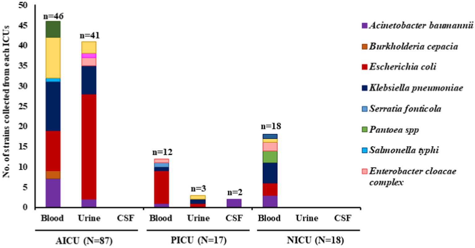
Figure 1. Schematic diagram of Gram-negative organisms recovered from different intensive care units along with their source of isolation. AICU, Anesthesia intensive care unit; PICU, pediatric intensive care unit; NICU, neonatal intensive care unit; CSF, cerebrospinal fluid; N = total number of isolates in each ICU; n = number of isolates in each specimen.
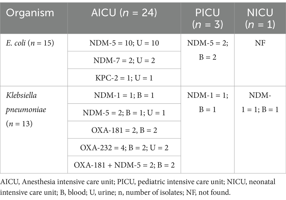
Table 1. Detailed breakup of carbapenemases found in study isolates, units, and source of isolation.
3.2 Antimicrobial susceptibility and genotypic characterization of study isolates collected from ICUs
E. coli and K. pneumoniae (n = 70) exhibited varying level of non-susceptibility toward different groups of antimicrobials (% resistant K. pneumoniae vs. % resistant E. coli) such as piperacillin-tazobactam: 73% vs. 56%; doripenem, imipenem, meropenem: 68% vs. 40%; amikacin: 55% vs. 17%; ciprofloxacin: 77% vs. 90%; levofloxacin: 77% vs. 94%; trimethoprim-sulphamethoxazole: 41% vs. 63%; and colistin: 14% vs. 2% (Supplementary Table S1). K. pneumoniae isolates exhibited higher non-susceptibility toward carbapenems compared to E. coli. Twenty-eight Enterobacterales, that is, E. coli (n = 15) and K. pneumoniae (n = 13), exhibited non-susceptibility toward the carbapenem group of drugs (doripenem, imipenem, and meropenem). They were assessed for the presence of carbapenemase genes by conventional PCR and validated by whole-genome sequencing (WGS).
Three types of carbapenem-resistant genes, namely, blaKPC-2 (class-A serine carbapenemase), blaNDM (class-B metallo-β-lactamase), and blaOXA-48-like (class-D serine carbapenemase), were noted among the ICUs. Three variants of blaNDM were found, namely, blaNDM-1 (n = 3/28), blaNDM-5 (n = 16/28), and blaNDM-7 (n = 2/28). blaNDM-5 was the dominant variant and was mainly identified in E. coli (n = 12) collected from urine (n = 10) and in a few K. pneumoniae (n = 4) in blood (n = 3), urine (n = 1) (Table 1). blaNDM-7 was found only in uropathogenic E. coli at AICU (Table 2). Two variants of blaOXA-48-like: blaOXA-181 (n = 4) and blaOXA-232 (n = 4) were identified in K. pneumoniae only, from blood. Two K. pneumoniae isolates (AGA0002 & AGA0038) from AICU exhibited the presence of dual carbapenemases, blaNDM-5 with blaOXA-181. One E. coli (AGA0014) harbored blaKPC-2; however, none of the E. coli harbored blaOXA-48-like carbapenemase (Table 1; Figures 2A, 3A). Isolates were resistant to meropenem, with MICs ranging between 8 and >128 mg/L (Figures 2A, 3A; Tables 2, 3), and isolates harboring dual carbapenemases (blaNDM-5 and blaOXA-181) exhibited an increased meropenem MIC (>128 mg/L) (Figure 2A; Table 2).
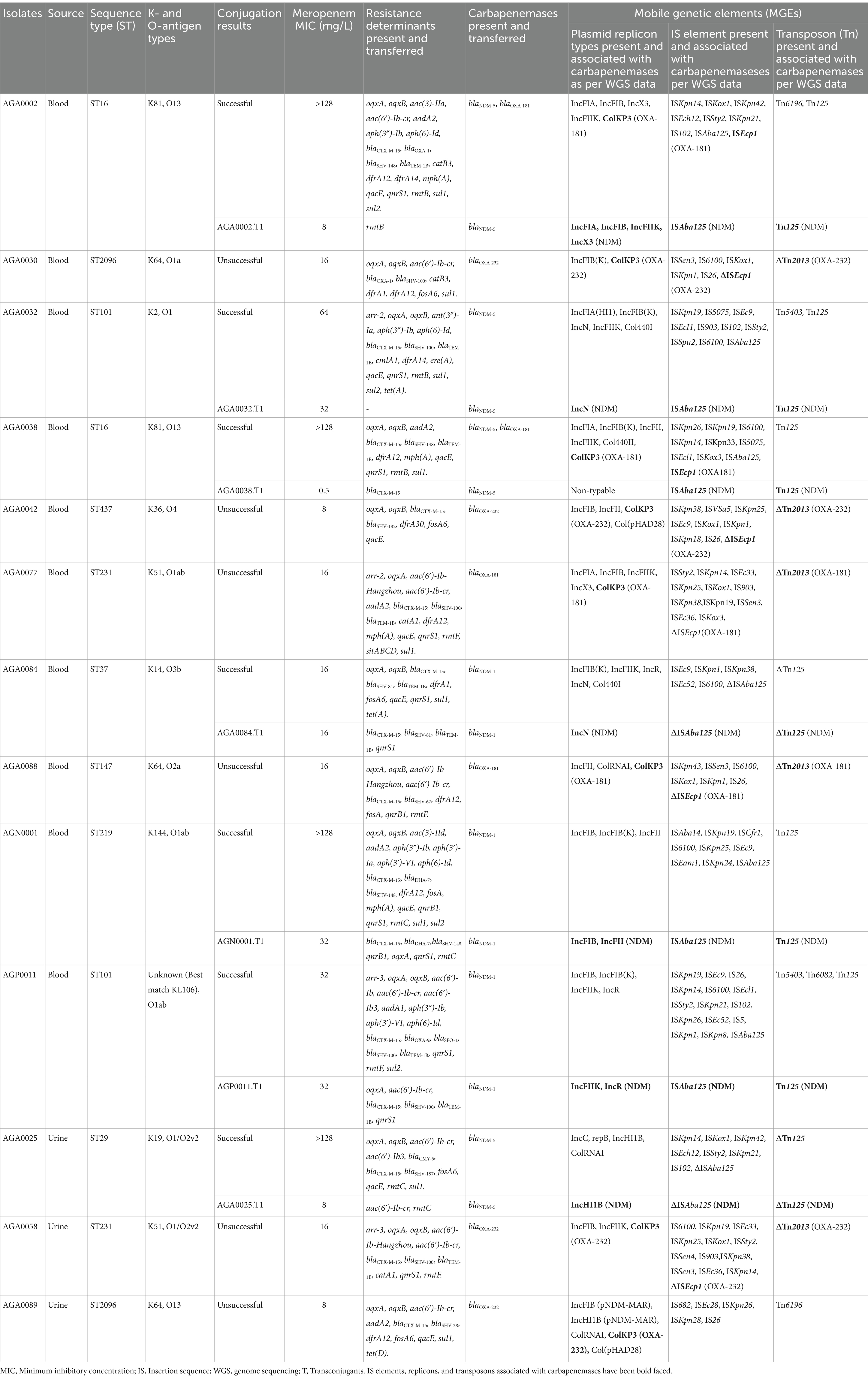
Table 2. Genotypic characterization, transmissibility of carbapenemases, and their association with mobile genetic elements among carbapenemase-producing Klebsiella pneumoniae isolates collected from anesthesia, pediatric, and neonatal intensive care units.
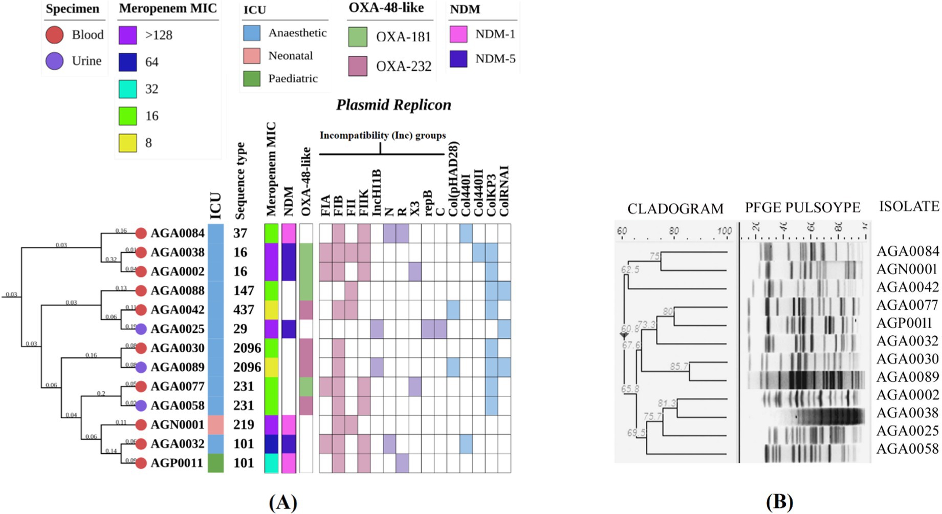
Figure 2. Molecular typing, core genome phylogeny, and genomic characterization of carbapenem-resistant Klebsiella pneumoniae isolated from different ICUs. (A) Core genome phylogeny of study isolates among themselves along with their source of isolation, specimen type, sequence types, meropenem MIC, carbapenemase genes, and replicons, (B) Analysis of pulsed-field gel electrophoresis using XbaI digestion pattern based on Dice’s similarity coefficient and UPGMA (the position tolerance and optimization was set at 1.5 and 1.5%, respectively). MIC, Minimum inhibitory concentration; ICU, Intensive care unit. PFGE for AGA0088 could not be performed, but WGS for this isolate was done, and hence it was included in the core phylogeny.
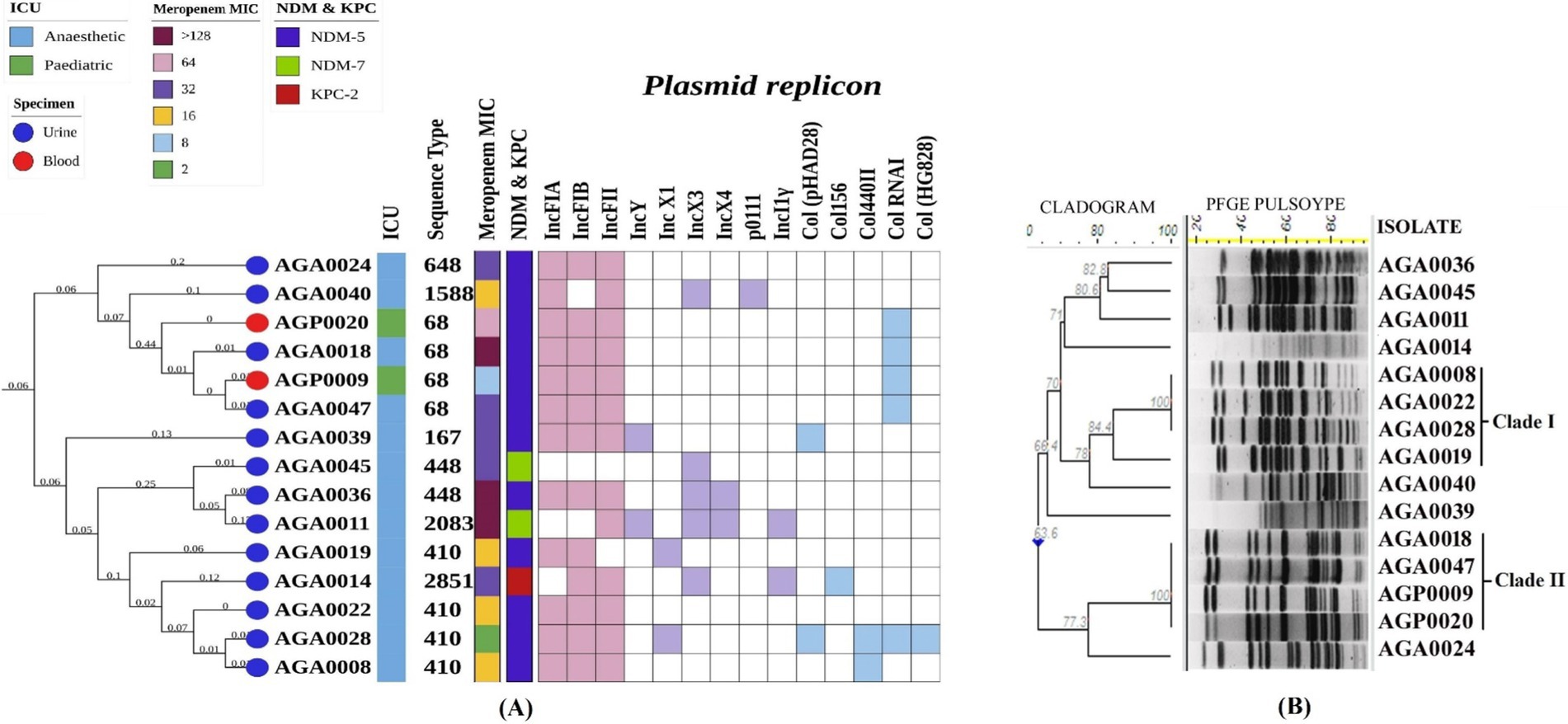
Figure 3. Molecular typing, core genome phylogeny, and genomic characterization of carbapenem-resistant E. coli isolated from different ICUs. (A) Core genome phylogeny of study isolates among themselves along with their source of isolation, specimen type, sequence types, meropenem MIC, carbapenemase genes, and replicons, (B) Analysis of pulsed-field gel electrophoresis using XbaI digestion pattern based on Dice’s similarity coefficient and UPGMA (the position tolerance and optimization was set at 1.5 and 1.5% respectively). Abbreviations: MIC: Minimum inhibitory concentration; ICU: Intensive care unit.
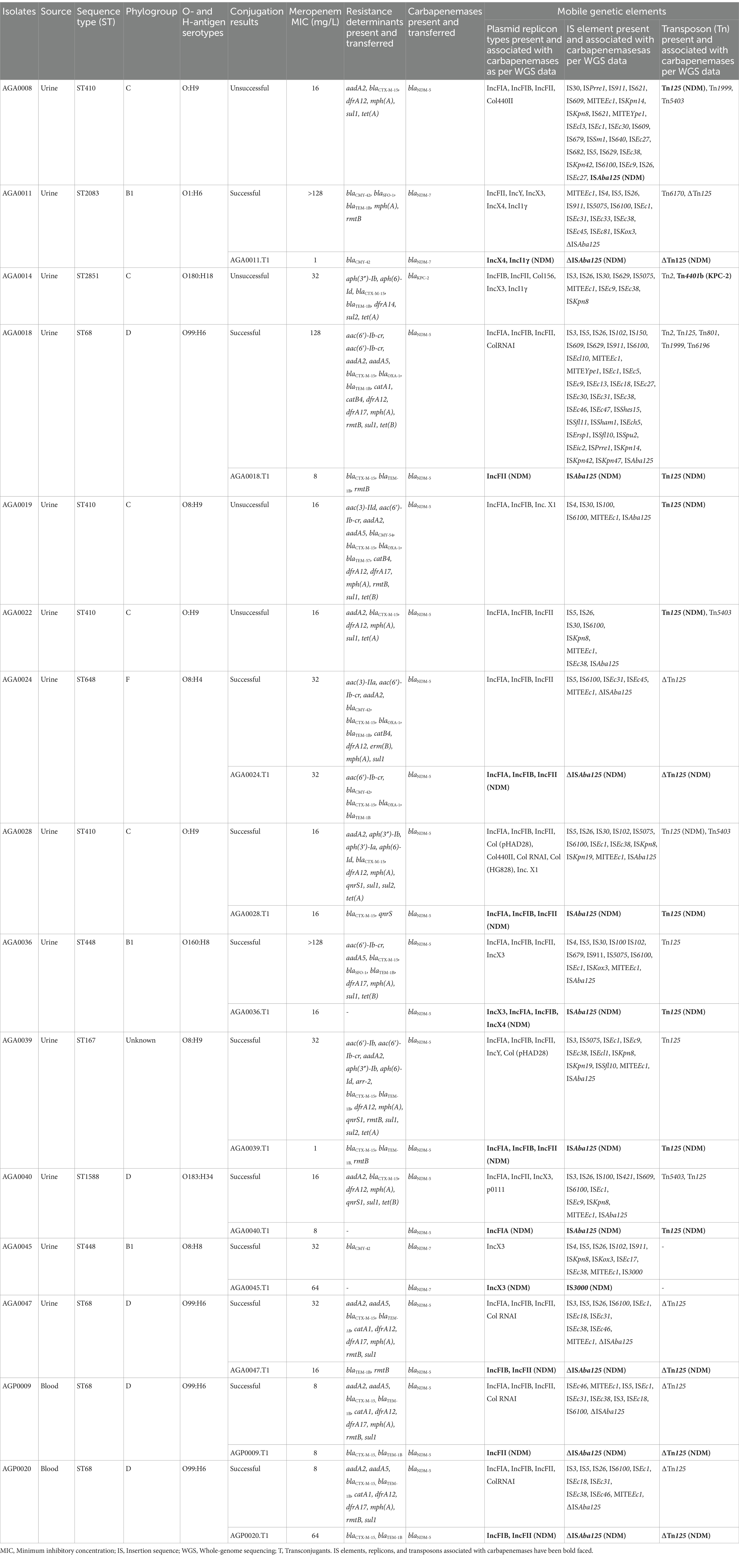
Table 3. Genotypic characterization, transmissibility of carbapenemases, and their association with mobile genetic elements among carbapenemase-producing E. coli isolates collected from anesthesia, pediatric, and neonatal intensive care units.
Additionally, the isolates harbored different types of resistance genes such as: blaCTX-M-15, blaOXA-1, blaTEM-1B,33,57, blaSHV-1,67,81,83,100,148,167, blaCMY-6,42,54, blaDHA-7 (β-lactamase genes), aac(6′)-Ib, aac(6′)-Ib-cr, rmtB, rmtC (aminoglycoside resistance genes), oqxA, oqxB, qnrS1, and aac(6′)-Ib-cr (fluoroquinolone resistance genes). Apart from this, several other genes imparting resistance to different antimicrobials, such as chloramphenicol and trimethoprim, were found in the genomes (Tables 2, 3).
3.3 Molecular typing of carbapenemase-producing isolates
Carbapenemase-producing K. pneumoniae and E. coli were diverse as per PFGE band patterns (Figures 2B, 3B). However, in E. coli, two clonal clades were identified. In clade I, the clonal isolates (AGA0008, AGA0019, AGA0022, and AGA0028) were recovered from the same ICU (AICU), and the clones appear to persist in the ICU environment over 4 months. In clade II, two isolates (AGP0009 and AGA0018) were collected within 1 month from two different ICUs (PICU and AICU) while the other two isolates (AGA0047 and AGP0020) were collected after 3 and 7 months of the initial isolation, exhibiting the potential of this clone to spread and persist in both the ICUs (Supplementary Table S2). Such findings indicated an inter-ICU spread of carbapenemase-producing E. coli isolates (Table 3; Figure 3B).
Nine different STs, namely, ST16 (n = 2), ST101 (n = 2), ST231 (n = 2), ST2096 (n = 2), ST29, ST37, ST147, ST219, and ST437 (Table 2; Figure 2A), were noted in K. pneumoniae isolates, while eight different STs, namely, ST68 (n = 4), ST410 (n = 4), ST448 (n = 2), ST167, ST648, ST1588, ST2083, and ST2851 (Table 3; Figure 3A), were noted in E. coli isolates. A core phylogeny for K. pneumoniae and E. coli was carried out (Figures 2A, 3A). Branching patterns of the phylogeny trees for both E. coli and K. pneumoniae were similar to the PFGE cladogram for the majority of isolates, while they varied for a few (E. coli: AGA0014, AGA0040, and AGA0039; K. pneumoniae: AGA0024, AGA0042, AGA0058, AGA0077, and AGA0084). Core phylogeny of the E. coli isolates exhibited two clades, concordant with PFGE results (Figures 3A,B). These clades (clades I and II) were found to cluster ST410 (n = 4) and ST68 (n = 4), respectively, again reinforcing the fact of inter-ICU spread.
Apart from PFGE and MLST, K-/O-antigen (K. pneumoniae) and O-/H-antigen (E. coli) serotyping were performed. Study K. pneumoniae exhibited eight diverse serotypes. K64 is the prevalent serotype (n = 3) followed by K51 (n = 2) and K81 (n = 2) (Table 2). Among the E. coli isolates, O8 (n = 4) and O99 (n = 4) were the most commonly encountered serotypes (Table 3).
3.4 Transmissibility of carbapenemases
Conjugal transfer of carbapenemase genes (blaNDM-1,5,7) was successful in the majority of isolates. However, few isolates (AGA0008, AGA0019, and AGA0022) with blaNDM-5 did not show successful conjugation. Isolates co-harboring blaNDM-5 and blaOXA-181 transferred only blaNDM-5. None among blaOXA-181,232 and blaKPC-2 showed conjugal transfer (Tables 2, 3).
3.5 Genetic environment of carbapenemase genes and their association with different MGEs
Analysis of the genetic environment is based on findings from WGS data and is restricted to the size of the contigs containing the carbapenemase genes. Primer walking was performed as and when required. Transmissibility of the carbapenemase genes and association with different MGEs were assessed by conjugation and PCR-based assay.
3.5.1 blaNDM-1,5,7
The genetic environment of blaNDM-1 and blaNDM-5 is similar in all the cases except for minute variations. The generic background noted in these isolates is in the following order: ISAba125➔blaNDM-1,5➔bleomycin resssistance gene (bleMBL)➔phosphoribosyl anthranilate (trpF)➔disulfide reductase (dsbD) (Figures 4A,B). Truncation of ISAba125 with ISKpn26 (AGN0001), presence of IS1 and ISSpu2 between ISAba125 and blaNDM-5 have been noted in AGA0025 and AGP0011, respectively (Figures 4A,B). Downstream of blaNDM-7 exhibited similarity with blaNDM-5, but upstream of blaNDM-7 has IS5-like-tnpA followed by IS3000-tnpA instead of ISAba125 (Figure 4C). These differences in the IS elements have been depicted in Figures 4A–C.
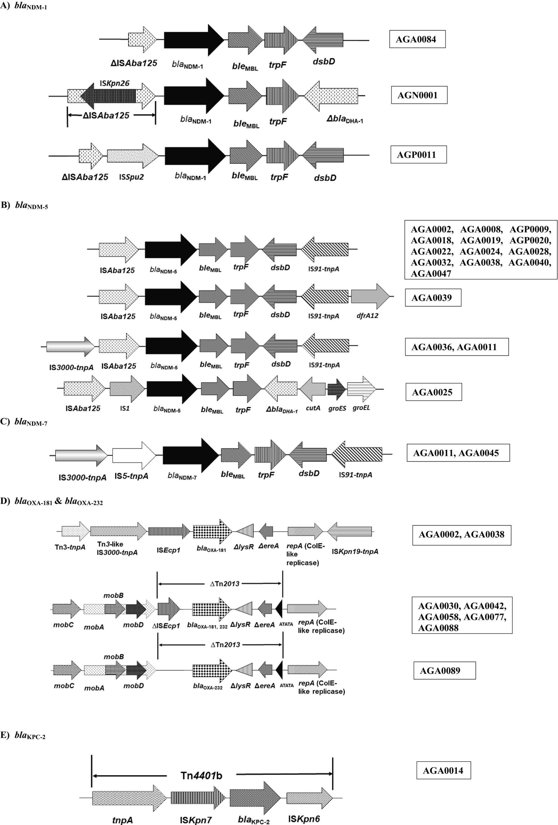
Figure 4. Schematic presentation of mobile genetic elements associated with carbapenemases found in this study. (A) blaNDM-1; (B) blaNDM-5; (C) blaNDM-7; (D) blaOXA-181 and blaOXA-232; (E) blaKPC-2. Target site duplications (ATATA) generated by the insertion of Tn2013 are indicated by black triangles. bla, beta-lactamase; mobA, mobB, mobC, and mobD, mobilization relaxosome proteins; ∆lysR, truncated LysR-type transcriptional regulator; ∆ereA, truncated erythromycin esterase; repA, replicase; Tn, transposon; IS, insertion sequence; tnpA, transposase; bleMBL; bleomycin resistance gene; trpF, N-(5́-phosphoribosyl) anthranilate isomerase; dsbD, Disulfide bond oxidoreductase D; ∆, denotes deletion or truncation.
blaNDM-bearing isolates harbor different types of plasmid replicons (Tables 2, 3; Figures 2A, 3A). The prevalence of IncFIA/FIB/FII and IncFIB has been noted in E. coli and K. pneumoniae isolates, respectively (Figures 2A, 3A). TCs harboring blaNDM-1 and blaNDM-5 exhibited the presence of IncFIB, IncFII, IncFIIK, IncN, IncR, and IncHI1B, IncN, IncX3, respectively, while IncX4 and IncI1γ were found in the TCs of blaNDM-7 (Tables 2, 3). Out of 12 blaNDM-5-possessing E. coli, 6 isolates exhibited a strong association of blaNDM-5 with IncFIA, IncFIB, and IncFII (Table 3).
Genomes of blaNDM-1,5,7 revealed the presence of different types of IS elements listed in Tables 2, 3. IS1, ISKpn26, ISAba125, ISSpu2, and IS3000 were found in the immediate upstream of the blaNDM and were associated with blaNDM (Figures 4A–C).
Other than IS elements, NDM-harboring isolates carried different types of transposons, not necessarily associated with blaNDM (Tables 2, 3). Of these transposons, Tn125, Tn5403, and Tn6196 were found in both the study isolates, E. coli and K. pneumoniae. However, Tn125 was found to be associated with NDM among the study isolates (Tables 2, 3).
3.5.2 blaOXA-181, 232
blaOXA-181,232, found in this study, exhibited a similar genetic background. The presence of a mobilization relaxosome, followed by an intact/ truncated ISEcp1, was noted upstream, and a truncated lysR, truncated ereA, and a replicase protein (ColKP3) downstream of blaOXA-181,232, except for one isolate (AGA0089) with deletion of ISEcp1 (Table 2; Figure 4D).
Similar to blaNDM-producing isolates, blaOXA-181,232-producing isolates also harbor different types of plasmid replicons (Table 2), predominantly IncFIB and ColKP3 (Figure 2A). Conjugal transfer of blaOXA-181,232 was unsuccessful, and the presence of ColKP3 was noted downstream of blaOXA-181,232 as per WGS data and primer walking.
blaOXA-181,232-harboring isolates exhibited the presence of different types of IS elements and transposons (Table 2). ColKP3, ISEcp1, and Tn2013 were associated with blaOXA-181,232 within the study isolates.
3.5.3 blaKPC-2
One blaKPC-2-bearing E. coli had blaKPC-2 bracketed between ISKpn7 (upstream) and ISKpn6 (downstream) of Tn4401b (Figure 4E). This isolate harbored various IS elements and replicon types (Table 3). However, conjugal transfer of blaKPC-2 was unsuccessful; hence, the replicon associated with blaKPC-2 in the study isolate could not be determined.
3.6 Comparative genome analysis of study isolates with relevant carbapenemase-producing isolates
Core genome phylogeny trees for E. coli (Figure 5) and K. pneumoniae (Figure 6) were prepared with isolates from different parts of India and Bangladesh. Both E. coli and K. pneumoniae study isolates showed relatedness with other isolates from Chhattisgarh, New Delhi, Gujarat, Chennai, Kolkata, Vellore, Mysore, and Bhubaneswar, and the neighboring country, Bangladesh. The phylogenetic tree for E. coli comprised 43 isolates, including 15 isolates from this study (Figure 5). Study isolates were diverse and scattered throughout the phylogeny. Isolates for this analysis were equally distributed between blood (n = 21) and urine (n = 22). In this collection, the prevalence of two epidemic clones, such as ST410 (n = 14, isolated from blood and urine) and ST167 (n = 12, from blood), was noted. It was noted that all isolates, that is, both study and others, primarily harbored blaNDM-5 (n = 39) with few blaNDM-7 (n = 3), and blaKPC-2 was only found in the study isolate, AGA0014. One uropathogenic E. coli (AGA0036) from this study was distantly related to an uropathogenic isolate from Bangladesh. Mostly, the study isolates from blood and urine showed relatedness with other Indian isolates of blood and urine origin, respectively. Although exceptions were noted, wherein uropathogenic study isolates (AGA0011, AGA0024, AGA0036, and AGA0039) showed relatedness with other Indian isolates of blood origin.
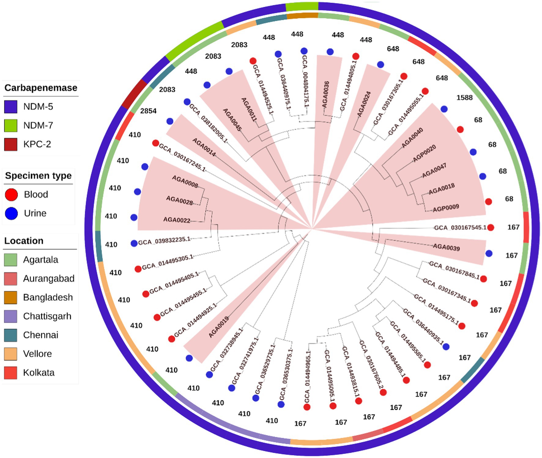
Figure 5. Core genome phylogeny of 43 E. coli using Roary (v3.13.0) and iTOL (v7). Isolates are colored at the endpoint according to specimen type, followed by sequence types (in numerical value). The additional two outer circles denote the place of collection (inner circle) and the presence of blaNDM variants and blaKPC-2 carbapenemases (outer circle). Clades containing isolates from this study are highlighted in peach.
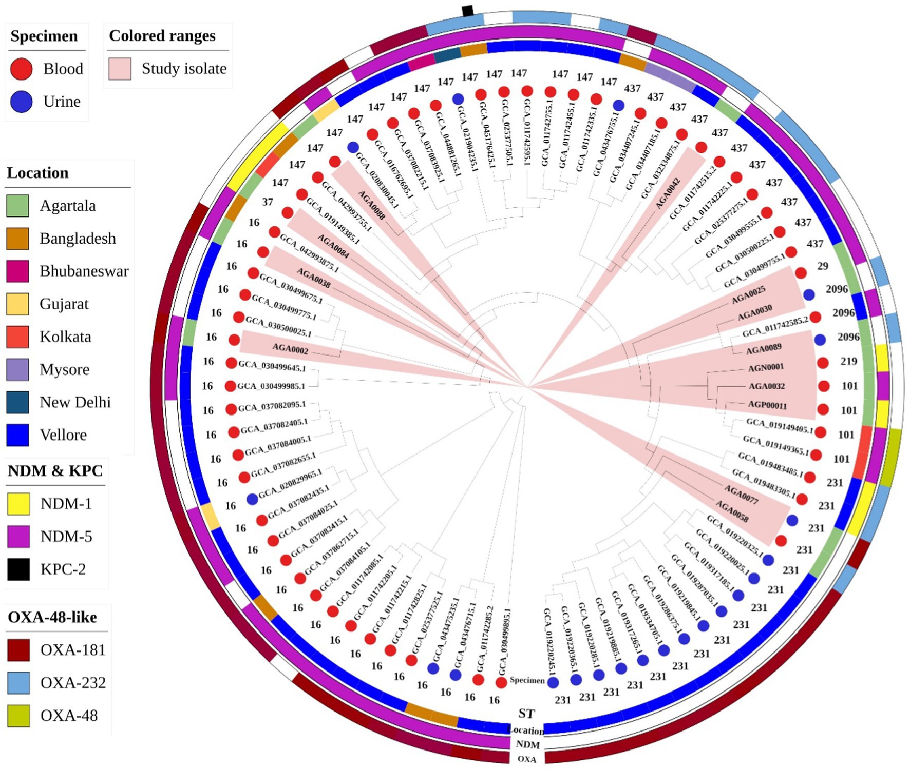
Figure 6. Core genome phylogeny of 79 Klebsiella pneumoniae using Roary (v3.13.0) and iTOL (v7). Isolates are colored at the endpoint according to specimen type, followed by sequence types (in numerical value). The additional three outer rings (from inside to the out): circle-1 represents the location of collection (inner circle), circle-2 represents class-B carbapenemases (middle circle), and circle 3 represents class-D carbapenemases (outer circle). KPC-2 was present in one isolate and has been described as a black box outside the outer ring. Clades containing isolates from this study are highlighted in peach.
The phylogenetic tree of K. pneumoniae comprised 79 isolates, including 13 isolates from this study (Figure 6). Similar to the E. coli study isolates, K. pneumoniae was also diverse. Unlike the phylogenetic tree for E. coli (Figure 5), the K. pneumoniae phylogenetic tree was primarily composed of isolates from blood (n = 58/79, 73%). AMR high-risk clones ST16 (n = 27) is the dominant ST, followed by ST231 (n = 16) and ST147 (n = 15) distributed between both blood and urine. In this collection, two carbapenemases, namely, blaNDM-5 (n = 43) and blaOXA-181 (n = 44) are the prevalent ones followed by isolates harboring multiple carbapenemases, namely, blaNDM-1 and blaOXA-232 (n = 2), blaNDM-5 and blaOXA-181 (n = 20), blaNDM-5 and blaOXA-48 (n = 2). The presence of blaNDM-5 along with blaKPC-2 (n = 1) was rarely noted. Multiple carbapenemase-harboring isolates were largely of blood origin. Analogous to the phylogenetic tree of E. coli, relatedness of study isolates from urine (AGA0058 and AGA0089) and blood (AGA0088) with isolates from India (blood) and Bangladesh (urine), respectively, was observed (Figure 6).
4 Discussion
HGT, an essential driver of bacterial evolution along with its mediators, MGEs, has mobilized different genes, which have benefited other bacteria to meet the evolutionary challenges of combating antimicrobials (Soucy et al., 2015). A web of genomic exchange has been noted among organisms, which has not been limited to species but has traversed to different genera and even phyla, indicating a re-evaluation of the “species concept” (Goldenfeld and Woese, 2007). Involvement of MGEs in the spread of carbapenem resistance has been documented worldwide, including various regions of India. As MGEs can facilitate the spread of resistance determinants between isolates and even species, studying them is crucial for understanding the transmission of resistance. Hence, this study assessed carbapenemase-associated MGEs and their interplay in the spread of carbapenem resistance within E. coli and K. pneumoniae.
Study isolates exhibited moderate (40%) to high (68%) level resistance to carbapenems, which is in contrast with previous reports from Northeast India, wherein low-level resistance (5–19%) to carbapenems was noted among Enterobacterales (Chellapandi et al., 2017; Sharma et al., 2021; Ralte et al., 2022). Study isolates carried many resistance genes, emphasizing the MDR nature of the isolates. Three carbapenemases (blaNDM-1,5,7, blaKPC-2, and blaOXA-181,232) were noted in this study. The prevalence of blaNDM-5 in both genera of this study indicated an allelic shift of blaNDM-1 to blaNDM-5, a phenomenon also documented in other global studies (Wu et al., 2019). NDM-5 has two mutations, V88L and M154L, compared to NDM-1 (Wu et al., 2019). V88L has been reported to exhibit 4-fold higher carbapenemase activity than NDM-1 (Hornsey et al., 2011). Further, M154L is responsible for higher zinc affinity, providing stability to the enzyme, thereby conferring higher resistance than NDM-1 (Bahr et al., 2018). Enhanced stability and increased carbapenemase activity of blaNDM-5 perhaps established this as a dominant allele over blaNDM-1. Report of blaKPC-2-harboring E. coli has been documented from India (Das et al., 2021) with no available genome description in public repositories such as NCBI. To our knowledge, this is the first study from India documenting a blaKPC-2-harboring E. coli genome.
In this study, isolates co-harboring blaNDM-5 and blaOXA-181 exhibited increased meropenem MIC (>128 mg/L), as also noted in previous studies (Naha et al., 2021; Bianco et al., 2022). The variability of carbapenemases was higher in isolates from AICU, as overall isolation of Enterobacterales was higher in this ICU than others. Previous studies from this region documented the presence of a single carbapenemase, that is, blaNDM-1 (Mukherjee et al., 2019) or blaOXA-48 (Ralte et al., 2022) or blaKPC-2 (Das et al., 2021). However, this study reports the presence of multiple carbapenemases (blaNDM-1,5,7, blaKPC-2, and blaOXA-181,232) from a single hospital and also the presence of dual carbapenemases within an isolate. The presence of multiple carbapenemases warrants a threat, and strict infection control needs to be ensured.
Study isolates were diverse, as found from PFGE and core genome phylogeny, and belonged to different STs. Differences in branching patterns for core phylogeny and PFGE noted in very few isolates of this study are due to the fact that PFGE and core genome phylogeny use two different approaches for genomic typing. PFGE compares band patterns generated due to the enzymatic digestion of genomic DNA, leading to a less significant view of genomic differences between isolates. On the other hand, core genome phylogeny is based on the sequences of core genes present in the isolates, providing a more detailed depiction of genetic relatedness (Gona et al., 2020). In this study, carbapenemases were found in high-risk international clones of K. pneumoniae (ST16, ST101, ST147, and ST231) and uropathogenic high-risk MDR clones of E. coli (ST167, ST410, and ST648), as also reported in other studies (Nittayasut et al., 2021; Bhattacharjee et al., 2023) highlighting their propensity for transboundary spread. K. pneumoniae ST2096 isolated from the ICUs is now being considered as an emerging hypervirulent MDR clone worldwide (Shankar et al., 2022). The presence of hypervirulent serotypes, such as K2 and K64, in high-risk international clones, ST101 and ST147, respectively, increases the spread of resistance and virulence. Association of different carbapenemases with the STs, as seen in this study, corroborates findings from various parts of India and the globe (Naha et al., 2021, 2022; Bhattacharjee et al., 2023; Shukla et al., 2023). The diversity of study isolates signifies that the spread of carbapenemases is not through specific clones or lineages, but via involvement of MGEs.
Comparative core genome phylogenetic analysis showed similarities of study isolates with isolates from the Indian cities - Kolkata, Chennai, Vellore, Aurangabad, Mysore, Bhubaneshwar, and the state, Chhattisgarh along with the neighboring country, Bangladesh. Similarities of isolates from blood with those from urine, as seen through the phylogenetic analysis, indicated the potential of the study isolates to cause invasive systemic infections (Shah et al., 2022; Bhattacharjee et al., 2023; Mueller and Tainter, 2025). India is a large country with distinct geographies and also sharp differences in health indicators. In spite of such differences, isolates exhibited relatedness with other Indian isolates, which is indicative of the spread of these isolates across different parts of India.
Study genomes harbored an assemblage of different plasmids. Study E. coli exhibited dominant association of blaNDM with IncFIA, IncFIB, IncFII (primarily), along with IncX3, IncX4, IncI1γ. On the other hand, study K. pneumoniae showed an association of blaNDM with different replicons such as IncFIA, IncFIB, IncFII, IncN, IncR, and IncHI1B. Globally, blaNDM has been reported in at least 20 types of replicons (Wu et al., 2019) with continent-specific dominance of certain replicons, namely, America (IncFI), Asia (South, East, and West) (IncA/C, IncR, IncHI2, IncX3, and IncY), and Europe (IncX1/X4/X6) (Li et al., 2023). Previous studies have noted association of NDM with F-plasmids (Partridge et al., 2018) along with IncX3 and IncHI1 replicons (Wu et al., 2019). Recent studies have documented association of NDM with the hybrid IncFIA/FIB/FII plasmid (Zhao et al., 2022; Bhattacharjee et al., 2023), as also noted in isolates from this study. Conjugal transfer and involvement of NDM with diverse replicons among the study isolates highlighted the dissemination potential of blaNDM across various genera.
Recently, it has been noted that blaNDM-bearing plasmids harbor different virulence genes along with ARGs, forming a sizeable hybrid plasmid (>200 kb). pNDM-MAR is one such plasmid (Villa et al., 2012), which is noted in one study isolate (AGA0089), composed of two replicons, namely, IncFIB and IncHI1B. Virulence and resistance in the same plasmid allow bacteria to persist, colonize, and spread resistance in hostile conditions. In contrast to blaNDM, blaOXA-181,232 found in the study isolates is restricted to the non-conjugative ColKP3 replicon, corroborating with previous reports (Potron et al., 2011; Pitout et al., 2019; Shankar et al., 2019; Naha et al., 2021). blaOXA-181,232 has been reported in limited numbers of plasmid replicons (ColE2, IncN1, IncT, IncX3, and ColKP3), majority of which are non-self-transmissible. Previous studies have shown transfer of blaOXA-181,232 plasmids with aid of helper plasmids (Pitout et al., 2019; Naha et al., 2021). In addition, clonal outbreaks of blaOXA-181,232 have also been reported (Pitout et al., 2019; Guzmán-Puche et al., 2021; Boyd et al., 2022). The study isolates co-harboring blaOXA-181 and blaNDM-5, did not show transfer of blaOXA-181. blaOXA-181,232 and blaNDM-5 were present on separate plasmids, suggesting an independent acquisition of two carbapenemase-harboring plasmids at different time points. Restricted mobility of the Col plasmids probably limited the spread of this gene compared to blaNDM. Another carbapenemase, blaKPC-2, was found in one E. coli, borne on a non-conjugative plasmid, as documented in a previous study (Naha et al., 2020). WGS data revealed the presence of various replicons in this isolate, but the replicon associated with blaKPC-2 could not be determined due to the non-conjugative nature of the plasmid and the limitation of short-read sequencing.
Association of IS elements and transposons with the carbapenemases has been noted in this study. Carbapenemases (blaNDM, blaOXA-181,232, and blaKPC-2) exhibited an ancestral genetic context. blaNDM was bracketed between ISAba125 and bleMBL, corroborating with other global studies, implying a prominent role of the IS element in the mobilization of this gene (Acman et al., 2022). ISAba125, being part of Tn125, indicates the active role of this transposon in the spread of blaNDM, as also documented in previous studies (Wu et al., 2019; Acman et al., 2022; Li et al., 2023). Tn125 has undergone minute variations leading to a complex genetic context (truncation by two copies of the same IS elements, such as IS26, IS903, and IS3000) (Toleman et al., 2012), but over the years the mobility of this transposon has been retained.
blaOXA-181,232 were present in association with an intact or truncated ISEcp1 and ∆lysR-∆ereA upstream and downstream, respectively, on an intact or truncated Tn2013 of a ColKP3 plasmid, comparable to studies from various parts of the world (Pitout et al., 2019; Naha et al., 2021). Deletion/insertion and truncation of this IS element restrict the spread of these genes (Pitout et al., 2019; Naha et al., 2021). In contrast to the results of this study and other global data, a study on MGEs of blaOXA-181,232 from North and South India exhibited a highly diverse genetic background involving different IS elements such as ISX4, IS1, IS3, ISKpn1, etc. (Shankar et al., 2019). It has been found that the spread of blaOXA-181,232 among Enterobacterales is driven by ISEcp1 (Pitout et al., 2019). blaOXA-181,232 has been associated with different types of transposons, namely, Tn2013, Tn2016, Tn6360, Tn6237, and Tn51098 (Pitout et al., 2019) but the occurrence of ISEcp1 among the study isolates indicated the presence of blaOXA-181,232 within Tn2013, the widely reported transposon responsible for both transboundary and interspecies spread of blaOXA-181,232 among Enterobacterales (Pitout et al., 2019). blaKPC-2 in this study has been found between ISKpn7 and ISKpn6 within Tn4401b. Out of 8 variants of Tn4401 (Tn4401a- Tn4401h) (Tang et al., 2022), Tn4401b has been considered to play a crucial role in the spread of blaKPC-2, while other transposons such as Tn1721, and IS26-like transposon are also emerging as vehicles for dissemination of blaKPC-2 in Southern Asia (Tang et al., 2022). The study isolate retained involvement of Tn4401b, similar to a previous study from India (Naha et al., 2020).
This study reports various MGEs, which included both plasmids and transposons associated with carbapenem-resistant genes. The spread of blaNDM across several countries within 15 years is evidence of the capability of the MGEs (such as Tn3, IS5, IS26, ISAba125, IncF-type, and IncX-type plasmids) in the spread of this gene. Initial studies indicated that the ISAba125 is solely responsible for the spread of blaNDM, but lately other IS elements such as IS26 have gained attention (Wu et al., 2019). IS26 is one such MGE readily found in the majority of the genomes. IS26 is responsible for the formation of large fusion plasmids harboring a collection of resistance genes, sometimes also generating tandem repeats of resistance genes such as blaNDM in a single plasmid. It can also integrate into the promoter region of a gene and can cause constitutive expression of the gene, leading to increased resistance (Harmer and Hall, 2024). Association of blaNDM with specific plasmids (primarily IncF plasmids) has also played a significant role in its spread. Contrary to blaNDM, the spread of blaOXA-181,232 and blaKPC-2 has been restricted to specific geographical regions with few sporadic cases in other parts of the globe (Pitout et al., 2019; Ding et al., 2023). This study also noted that the spread of blaNDM has primarily been driven by plasmids and IS elements, but in the case of blaOXA-181,232 and blaKPC-2, though MGEs play an essential role, the spread of these carbapenemases is associated with specific clonal lineages. Spread of KPC through sequence type 258 (ST258) and ST11 has been reported worldwide (Marsh et al., 2019; Yuan et al., 2024). Global dissemination of OXA-48 and its derivatives is often associated with specific high-risk clones, such as ST14, ST15, ST147, ST231, ST307 (K. pneumoniae), and ST38 and ST410 (E. coli) (Pitout et al., 2019). The contribution of MGEs is more critical in the spread of resistance than bacterial lineages, leading to the emergence of new resistant clones (Caliskan-Aydogan and Alocilja, 2023).
In conclusion, this study highlighted the association of HGT and different MGEs in the mobilization of carbapenemases. The use of short-read sequencing limits the detailed characterization of the carbapenem-resistant plasmids. Similarity of MGEs (plasmid profiles, transposons, and IS elements) for the carbapenemases (particularly blaNDM) of this study is concordant with reports across the globe indicating their efficiency to transfer in diverse lineages and existence as part of the global gene pool. Other than this, MGEs also provide information about the local selective pressures that influence gene distribution via HGT. The occurrence of fusion plasmids (pNDM-MAR) exhibiting integration of virulence with resistance genes has already been noted. The presence of such plasmids is worrisome as this expedite the spread of carbapenem resistance and virulence. On this account, surveillance of these genes and MGEs remains very relevant for infection control.
Data availability statement
The datasets presented in this study can be found in online repositories. Genome sequence data have been deposited at NCBI under BioProject No. PRJNA790720. Antimicrobial resistance pattern of E. coli & K. pneumoniae and detailed patient data can be found in the Supplementary material.
Ethics statement
The studies involving humans were approved by the Institutional Ethics Committee of the ICMR-National Institute of Cholera and Enteric Diseases (A-1/2019-IEC). The studies were conducted in accordance with the local legislation and institutional requirements. Written informed consent for participation was obtained from the participants or the participants’ legal guardians/next of kin in accordance with the national legislation and institutional requirements.
Author contributions
SM: Formal analysis, Methodology, Validation, Writing – original draft, Writing – review & editing, Data curation, Investigation, Software, Visualization. SN: Data curation, Formal analysis, Investigation, Methodology, Software, Validation, Visualization, Writing – original draft, Writing – review & editing. JC: Data curation, Investigation, Writing – review & editing. SD: Data curation, Investigation, Writing – review & editing. HK: Writing – review & editing, Formal analysis. TM: Formal analysis, Writing – review & editing, Resources. SB: Formal analysis, Resources, Writing – review & editing, Conceptualization, Funding acquisition, Methodology, Project administration, Supervision, Validation, Writing – original draft.
Funding
The author(s) declare that financial support was received for the research and/or publication of this article. This study was supported by the Indian Council of Medical Research (ICMR), India, extramural funding (grant number: NER/67/2019-ECD-I). SM and SN were recipients of a fellowship from ICMR. The funding agency did not play any role in the study design, data collection, analysis, interpretation, writing of the manuscript, or the decision to submit the work for publication.
Acknowledgments
We acknowledge the staff of the anesthesia, pediatric, and neonatal intensive care units who cared for the patients and helped collect patient information. We thank Surjendu Bikash Sasmal for his laboratory assistance and data maintenance. We also acknowledge the Indian Council of Medical Research (ICMR), India, for extramural funding to support the work.
Conflict of interest
The authors declare that the research was conducted in the absence of any commercial or financial relationships that could be construed as a potential conflict of interest.
The author(s) declared that they were an editorial board member of Frontiers, at the time of submission. This had no impact on the peer review process and the final decision.
Generative AI statement
The author(s) declare that no Gen AI was used in the creation of this manuscript.
Publisher’s note
All claims expressed in this article are solely those of the authors and do not necessarily represent those of their affiliated organizations, or those of the publisher, the editors and the reviewers. Any product that may be evaluated in this article, or claim that may be made by its manufacturer, is not guaranteed or endorsed by the publisher.
Supplementary material
The Supplementary material for this article can be found online at: https://www.frontiersin.org/articles/10.3389/fmicb.2025.1543427/full#supplementary-material
Footnotes
1. ^https://iris.who.int/bitstream/handle/10665/376776/9789240093461-eng.pdf
2. ^https://static.pib.gov.in/WriteReadData/specificdocs/documents/2023/jun/doc202362208601.pdf
3. ^https://www.diatheva.com/product/pbrt-2-0-kit/
8. ^https://cge.food.dtu.dk/services/PlasmidFinder/
References
Acman, M., Wang, R., van Dorp, L., Shaw, L. P., Wang, Q., Luhmann, N., et al. (2022). Role of mobile genetic elements in the global dissemination of the carbapenem resistance gene blaNDM. Nat. Commun. 13:1131. doi: 10.1038/s41467-022-28819-2
Al-Zahrani, I. A., and Alsiri, B. A. (2018). The emergence of carbapenem-resistant Klebsiella pneumoniae isolates producing OXA-48 and NDM in the southern (Asir) province, Saudi Arabia. Saudi Med. J. 39, 23–30. doi: 10.15537/smj.2018.1.21094
Bahr, G., Vitor-Horen, L., Bethel, C. R., Bonomo, R. A., González, L. J., and Vila, A. J. (2018). Clinical evolution of New Delhi Metallo-β-lactamase (NDM) optimizes resistance under Zn(II) deprivation. Antimicrob. Agents Chemother. 62, e01849–e01817. doi: 10.1128/AAC.01849-17
Bhattacharjee, A., Sands, K., Mitra, S., Basu, R., Saha, B., Clermont, O., et al. (2023). A decade-long evaluation of neonatal Septicaemic Escherichia coli: clonal lineages, genomes, and New Delhi Metallo-Beta-lactamase variants. Microbiol. Spectr. 11:e0521522. doi: 10.1128/spectrum.05215-22
Bianco, G., Boattini, M., Comini, S., Casale, R., Iannaccone, M., Cavallo, R., et al. (2022). Occurrence of multi-carbapenemases producers among carbapenemase-producing Enterobacterales and in vitro activity of combinations including cefiderocol, ceftazidime-avibactam, meropenem-vaborbactam, and aztreonam in the COVID-19 era. Eur. J. Clin. Microbiol. Infect. Dis. 41, 573–580. doi: 10.1007/s10096-022-04408-5
Boyd, S. E., Holmes, A., Peck, R., Livermore, D. M., and Hope, W. (2022). OXA-48-like β-lactamases: global epidemiology, treatment options, and development pipeline. Antimicrob. Agents Chemother. 66:e0021622. doi: 10.1128/aac.00216-22
Caliskan-Aydogan, O., and Alocilja, E. C. (2023). A review of carbapenem resistance in enterobacterales and its detection techniques. Microorganisms 11:1491. doi: 10.3390/microorganisms11061491
Chellapandi, K., Dutta, T. K., Sharma, I., De Mandal, S., Kumar, N. S., and Ralte, L. (2017). Prevalence of multi drug resistant enteropathogenic and enteroinvasive Escherichia coli isolated from children with and without diarrhea in northeast Indian population. Ann. Clin. Microbiol. Antimicrob. 16:49. doi: 10.1186/s12941-017-0225-x
CLSI (2024). Performance Standards for Antimicrobial Susceptibility Testing. 34th ed. CLSI supplement M100. Clinical and Laboratory Standards Institute.
Cui, X., Zhang, H., and Du, H. (2019). Carbapenemases in Enterobacteriaceae: detection and antimicrobial therapy. Front. Microbiol. 10:1823. doi: 10.3389/fmicb.2019.01823
da Silva, G. C., Gonçalves, O. S., Rosa, J. N., França, K. C., Bossé, J. T., Santana, M. F., et al. (2021). Mobile genetic elements drive antimicrobial resistance gene spread in Pasteurellaceae species. Front. Microbiol. 12:773284. doi: 10.3389/fmicb.2021.773284
Das, B. J., Wangkheimayum, J., Singha, K. M., Bhowmik, D., Dhar, D., and Bhattacharjee, A. (2021). Propagation of blaKPC-2 within two sequence types of Escherichia coli in a tertiary referral hospital of Northeast India. Gene Rep. 24:101283. doi: 10.1016/j.genrep.2021.101283
Ding, L., Shen, S., Chen, J., Tian, Z., Shi, Q., Han, R., et al. (2023). Klebsiella pneumoniae carbapenemase variants: the new threat to global public health. Clin. Microbiol. Rev. 36:e0000823. doi: 10.1128/cmr.00008-23
Espinal, P., Fugazza, G., López, Y., Kasma, M., Lerman, Y., Malhotra-Kumar, S., et al. (2011). Dissemination of an NDM-2-producing Acinetobacter baumannii clone in an Israeli rehabilitation center. Antimicrob. Agents Chemother. 55, 5396–5398. doi: 10.1128/AAC.00679-11
Goldenfeld, N., and Woese, C. (2007). Biology’s next revolution. Nature 445:369. doi: 10.1038/445369a
Gona, F., Comandatore, F., Battaglia, S., Piazza, A., Trovato, A., Lorenzin, G., et al. (2020). Comparison of core-genome MLST, coreSNP and PFGE methods for Klebsiella pneumoniae cluster analysis. Microb. Genom. 6:e000347. doi: 10.1099/mgen.0.000347
Gorrie, C. L., Mirčeta, M., Wick, R. R., Judd, L. M., Lam, M. M. C., Gomi, R., et al. (2022). Genomic dissection of Klebsiella pneumoniae infections in hospital patients reveals insights into an opportunistic pathogen. Nat. Commun. 13:3017. doi: 10.1038/s41467-022-30717-6
Grönthal, T., Österblad, M., Eklund, M., Jalava, J., Nykäsenoja, S., Pekkanen, K., et al. (2018). Sharing more than friendship - transmission of NDM-5 ST167 and CTX-M-9 ST69 Escherichia coli between dogs and humans in a family, Finland, 2015. Euro Surveill. 23:1700497. doi: 10.2807/1560-7917.ES.2018.23.27.1700497
Guzmán-Puche, J., Jenayeh, R., Pérez-Vázquez, M., Manuel-Causse, N., Asma, F., Jalel, B., et al. (2021). Characterization of OXA-48-producing Klebsiella oxytoca isolates from a hospital outbreak in Tunisia. J. Glob. Antimicrob. Resist. 24, 306–310. doi: 10.1016/j.jgar.2021.01.008
Harmer, C. J., and Hall, R. M. (2024). IS26 and the IS26 family: versatile resistance gene movers and genome reorganizers. Microbiol. Mol. Biol. Rev. 88:e0011922. doi: 10.1128/mmbr.00119-22
Hornsey, M., Phee, L., and Wareham, D. W. (2011). A novel variant, NDM-5, of the New Delhi metallo-β-lactamase in a multidrug-resistant Escherichia coli ST648 isolate recovered from a patient in the United Kingdom. Antimicrob. Agents Chemother. 55, 5952–5954. doi: 10.1128/AAC.05108-11
Khedkar, S., Smyshlyaev, G., Letunic, I., Maistrenko, O. M., Coelho, L. P., Orakov, A., et al. (2022). Landscape of mobile genetic elements and their antibiotic resistance cargo in prokaryotic genomes. Nucleic Acids Res. 50, 3155–3168. doi: 10.1093/nar/gkac163
Lee, C.-R., Lee, J. H., Park, K. S., Kim, Y. B., Jeong, B. C., and Lee, S. H. (2016). Global dissemination of Carbapenemase-producing Klebsiella pneumoniae: epidemiology, genetic context, treatment options, and detection methods. Front. Microbiol. 7:895. doi: 10.3389/fmicb.2016.00895
Li, Y., Yang, Y., Wang, Y., Walsh, T. R., Wang, S., and Cai, C. (2023). Molecular characterization of blaNDM-harboring plasmids reveal its rapid adaptation and evolution in the Enterobacteriaceae. One Health Adv. 1:30. doi: 10.1186/s44280-023-00033-9
Liu, H., Shi, K., Wang, Y., Zhong, W., Pan, S., Zhou, L., et al. (2024). Characterization of antibiotic resistance genes and mobile genetic elements in Escherichia coli isolated from captive black bears. Sci. Rep. 14:2745. doi: 10.1038/s41598-024-52622-2
MacKinnon, M. C., Sargeant, J. M., Pearl, D. L., Reid-Smith, R. J., Carson, C. A., Parmley, E. J., et al. (2020). Evaluation of the health and healthcare system burden due to antimicrobial-resistant Escherichia coli infections in humans: a systematic review and meta-analysis. Antimicrob. Resist. Infect. Control 9:200. doi: 10.1186/s13756-020-00863-x
Marsh, J. W., Mustapha, M. M., Griffith, M. P., Evans, D. R., Ezeonwuka, C., Pasculle, A. W., et al. (2019). Evolution of outbreak-causing Carbapenem-resistant Klebsiella pneumoniae ST258 at a tertiary care hospital over 8 years. mBio 10:e01945-19. doi: 10.1128/mbio.01945-19
Martin, R. M., and Bachman, M. A. (2018). Colonization, infection, and the accessory genome of Klebsiella pneumoniae. Front. Cell. Infect. Microbiol. 8:4. doi: 10.3389/fcimb.2018.00004
Mueller, M., and Tainter, C. R. (2025). Escherichia coli infection. in StatPearls, Treasure Island, FL: StatPearls Publishing). Available online at: http://www.ncbi.nlm.nih.gov/books/NBK564298/ (Accessed March 3, 2025).
Mukherjee, S., Bhattacharjee, A., Naha, S., Majumdar, T., Debbarma, S. K., Kaur, H., et al. (2019). Molecular characterization of NDM-1-producing Klebsiella pneumoniae ST29, ST347, ST1224, and ST2558 causing sepsis in neonates in a tertiary care hospital of north-East India. Infect. Genet. Evol. 69, 166–175. doi: 10.1016/j.meegid.2019.01.024
Naha, S., Sands, K., Mukherjee, S., Dutta, S., and Basu, S. (2022). A 12 year experience of colistin resistance in Klebsiella pneumoniae causing neonatal sepsis: two-component systems, efflux pumps, lipopolysaccharide modification and comparative phylogenomics. J. Antimicrob. Chemother. 77, 1586–1591. doi: 10.1093/jac/dkac083
Naha, S., Sands, K., Mukherjee, S., Roy, C., Rameez, M. J., Saha, B., et al. (2020). KPC-2-producing Klebsiella pneumoniae ST147 in a neonatal unit: clonal isolates with differences in colistin susceptibility attributed to AcrAB-TolC pump. Int. J. Antimicrob. Agents 55:105903. doi: 10.1016/j.ijantimicag.2020.105903
Naha, S., Sands, K., Mukherjee, S., Saha, B., Dutta, S., and Basu, S. (2021). OXA-181-like Carbapenemases in Klebsiella pneumoniae ST14, ST15, ST23, ST48, and ST231 from septicemic neonates: coexistence with NDM-5, Resistome, transmissibility, and genome diversity. mSphere 6, e01156–e01120. doi: 10.1128/mSphere.01156-20
Navon-Venezia, S., Kondratyeva, K., and Carattoli, A. (2017). Klebsiella pneumoniae: a major worldwide source and shuttle for antibiotic resistance. FEMS Microbiol. Rev. 41, 252–275. doi: 10.1093/femsre/fux013
Nittayasut, N., Yindee, J., Boonkham, P., Yata, T., Suanpairintr, N., and Chanchaithong, P. (2021). Multiple and high-risk clones of extended-Spectrum cephalosporin-resistant and blaNDM-5-harbouring uropathogenic Escherichia coli from cats and dogs in Thailand. Antibiotics 10:1374. doi: 10.3390/antibiotics10111374
Paczosa, M. K., and Mecsas, J. (2016). Klebsiella pneumoniae: going on the offense with a strong defense. Microbiol. Mol. Biol. Rev. 80, 629–661. doi: 10.1128/MMBR.00078-15
Partridge, S. R., Kwong, S. M., Firth, N., and Jensen, S. O. (2018). Mobile genetic elements associated with antimicrobial resistance. Clin. Microbiol. Rev. 31, e00088–e00017. doi: 10.1128/CMR.00088-17
Pitout, J. D. D., Peirano, G., Kock, M. M., Strydom, K.-A., and Matsumura, Y. (2019). The global ascendency of OXA-48-type Carbapenemases. Clin. Microbiol. Rev. 33, e00102–e00119. doi: 10.1128/CMR.00102-19
Potron, A., Nordmann, P., Lafeuille, E., Al Maskari, Z., Al Rashdi, F., and Poirel, L. (2011). Characterization of OXA-181, a carbapenem-hydrolyzing class D beta-lactamase from Klebsiella pneumoniae. Antimicrob. Agents Chemother. 55, 4896–4899. doi: 10.1128/AAC.00481-11
Ralte, V. S. C., Loganathan, A., Manohar, P., Sailo, C. V., Sanga, Z., Ralte, L., et al. (2022). The emergence of Carbapenem-resistant gram-negative Bacteria in Mizoram, Northeast India. Microbiol. Res. 13, 342–349. doi: 10.3390/microbiolres13030027
Shah, A., Shetty, A., Victor, D., and Kodali, S. (2022). Klebsiella pneumoniae infection as a mimicker of multiple metastatic lesions. Cureus 14:e32669. doi: 10.7759/cureus.32669
Shankar, C., Mathur, P., Venkatesan, M., Pragasam, A. K., Anandan, S., Khurana, S., et al. (2019). Rapidly disseminating blaOXA-232 carrying Klebsiella pneumoniae belonging to ST231 in India: multiple and varied mobile genetic elements. BMC Microbiol. 19:137. doi: 10.1186/s12866-019-1513-8
Shankar, C., Vasudevan, K., Jacob, J. J., Baker, S., Isaac, B. J., Neeravi, A. R., et al. (2022). Hybrid plasmids encoding antimicrobial resistance and virulence traits among Hypervirulent Klebsiella pneumoniae ST2096 in India. Front. Cell. Infect. Microbiol. 12:875116. doi: 10.3389/fcimb.2022.875116
Sharma, M., Chetia, P., Puzari, M., Neog, N., Phukan, U., and Borah, A. (2021). Carbapenem resistance among common enterobacteriaceae clinical isolates in part of north-east India. Available online at: http://www.eurekaselect.com (Accessed February 16, 2025).
Shukla, S., Desai, S., Bagchi, A., Singh, P., Joshi, M., Joshi, C., et al. (2023). Diversity and distribution of β-lactamase genes circulating in Indian isolates of multidrug-resistant Klebsiella pneumoniae. Antibiotics 12:449. doi: 10.3390/antibiotics12030449
Soucy, S. M., Huang, J., and Gogarten, J. P. (2015). Horizontal gene transfer: building the web of life. Nat. Rev. Genet. 16, 472–482. doi: 10.1038/nrg3962
Taggar, G., Attiq Rheman, M., Boerlin, P., and Diarra, M. S. (2020). Molecular epidemiology of carbapenemases in enterobacteriales from humans, animals, food and the environment. Antibiotics 9:693. doi: 10.3390/antibiotics9100693
Tang, Y., Li, G., Shen, P., Zhang, Y., and Jiang, X. (2022). Replicative transposition contributes to the evolution and dissemination of KPC-2-producing plasmid in Enterobacterales. Emerg. Microbes Infect. 11, 113–122. doi: 10.1080/22221751.2021.2013105
Tenover, F. C., Arbeit, R. D., Goering, R. V., Mickelsen, P. A., Murray, B. E., Persing, D. H., et al. (1995). Interpreting chromosomal DNA restriction patterns produced by pulsed-field gel electrophoresis: criteria for bacterial strain typing. J. Clin. Microbiol. 33, 2233–2239. doi: 10.1128/jcm.33.9.2233-2239.1995
Toleman, M. A., Spencer, J., Jones, L., and Walsh, T. R. (2012). blaNDM-1 is a chimera likely constructed in Acinetobacter baumannii. Antimicrob. Agents Chemother. 56, 2773–2776. doi: 10.1128/AAC.06297-11
Villa, L., Poirel, L., Nordmann, P., Carta, C., and Carattoli, A. (2012). Complete sequencing of an IncH plasmid carrying the blaNDM-1, blaCTX-M-15 and qnrB1 genes. J. Antimicrob. Chemother. 67, 1645–1650. doi: 10.1093/jac/dks114
Wang, L., Zhu, M., Yan, C., Zhang, Y., He, X., Wu, L., et al. (2023). Class 1 integrons and multiple mobile genetic elements in clinical isolates of the Klebsiella pneumoniae complex from a tertiary hospital in eastern China. Front. Microbiol. 14:985102. doi: 10.3389/fmicb.2023.985102
Wick, R. R., Judd, L. M., Gorrie, C. L., and Holt, K. E. (2017). Unicycler: Resolving bacterial genome assemblies from short and long sequencing reads. PLoS Comput. Biol. 13:e1005595. doi: 10.1371/journal.pcbi.1005595
Wu, W., Feng, Y., Tang, G., Qiao, F., McNally, A., and Zong, Z. (2019). NDM Metallo-β-lactamases and their bacterial producers in health care settings. Clin. Microbiol. Rev. 32, e00115–e00118. doi: 10.1128/CMR.00115-18
Wyres, K. L., and Holt, K. E. (2018). Klebsiella pneumoniae as a key trafficker of drug resistance genes from environmental to clinically important bacteria. Curr. Opin. Microbiol. 45, 131–139. doi: 10.1016/j.mib.2018.04.004
Yigit, H., Queenan, A. M., Anderson, G. J., Domenech-Sanchez, A., Biddle, J. W., Steward, C. D., et al. (2001). Novel Carbapenem-hydrolyzing β-lactamase, KPC-1, from a Carbapenem-resistant strain of Klebsiella pneumoniae. Antimicrob. Agents Chemother. 45, 1151–1161. doi: 10.1128/AAC.45.4.1151-1161.2001
Yuan, Y., Lu, Y., Cao, L., Fu, Y., Li, Y., and Zhang, L. (2024). Genetic characteristics of clinical carbapenem-resistant Klebsiella pneumoniae: epidemic ST11 KPC-2-producing strains and non-negligible NDM-5-producing strains with diverse STs. Sci. Rep. 14:24296. doi: 10.1038/s41598-024-74307-6
Keywords: plasmids, insertion sequence element, transposons, blaNDM-1,5,7, blaOXA-181,232, blaKPC-2-harboring Escherichia coli, core genome phylogeny
Citation: Mitra S, Naha S, Chakraborty J, De S, Kaur H, Majumdar T and Basu S (2025) Diversity of mobile genetic elements in carbapenem-resistant Enterobacterales isolated from the intensive care units of a tertiary care hospital in Northeast India. Front. Microbiol. 16:1543427. doi: 10.3389/fmicb.2025.1543427
Edited by:
John Osei Sekyere, University of Pretoria, South AfricaReviewed by:
Fengxia Yang, Agro-Environmental Protection Institute, ChinaJennifer Joseph Moussa, Lebanese American University, Lebanon
Shyamalima Saikia, Dibrugarh University, India
Copyright © 2025 Mitra, Naha, Chakraborty, De, Kaur, Majumdar and Basu. This is an open-access article distributed under the terms of the Creative Commons Attribution License (CC BY). The use, distribution or reproduction in other forums is permitted, provided the original author(s) and the copyright owner(s) are credited and that the original publication in this journal is cited, in accordance with accepted academic practice. No use, distribution or reproduction is permitted which does not comply with these terms.
*Correspondence: Sulagna Basu, YmFzdXMubmljZWRAZ292Lmlu; c3VwYWJhc3VAeWFob28uY28uaW4=
†These authors share first authorship
‡ORCID: Shravani Mitra, orcid.org/0009-0009-3434-9677
Sharmi Naha, orcid.org/0000-0001-7518-9264
Harpreet Kaur, orcid.org/0000-0001-7143-8075
Sulagna Basu, orcid.org/0000-0002-7811-7140
 Shravani Mitra
 Sharmi Naha
 Joy Chakraborty
 Subhadeep De1
 Harpreet Kaur
 Sulagna Basu