- 1School of Integrated Chinese and Western Medicine, Hunan University of Chinese Medicine, Changsha, China
- 2Key Laboratory of Hunan Province for Integrated Traditional Chinese and Western Medicine on Prevention and Treatment of Cardio-Cerebral Diseases, Hunan University of Chinese Medicine, Changsha, China
- 3Department of Endocrine, Yueyang Traditional Chinese Medicine Hospital, Yueyang, China
- 4School of Traditional Chinese Medicine, Hunan University of Chinese Medicine, Changsha, China
Background: Hyperuricemia (HUA), found widely in humans and birds, is a key physiological factor responsible for the development of gout. In recent years, the relationship between the gut microbiota and HUA has garnered significant attention from researchers. This study aims to explore the current research hotspots, knowledge gaps, and future research trends regarding the gut microbiota and HUA.
Methods: We performed a thorough search of the literature on gut flora and HUA published between 2005 and 2024 using the Web of Science and PubMed databases. The resulting data were analyzed using VOSviewer, CiteSpace, and Bibliometrix.
Results: Including 735 papers in total, the study found that the number of publications in the subject increased significantly between 2020 and 2024, with 2024 being the year with the highest number of publications. The primary research countries are highlighted as China and the United States, with institutions such as the University of California, San Diego, and Qingdao University making significant contributions. Sanjay K. Nigam and Chenyang Lu have made the most important contributions as authors. Keywords analysis highlighted high-frequency terms including “gastrointestinal microbiome,” “uric acid,” “hyperuricemia,” “inflammation,” “gout,” and “probiotics.” In the visualization map of the keyword timeline, emerging research hotspots include “diets,” “dietary fiber,” “fecal microbiota transplantation,” and “gut-kidney axis.”
Conclusion: This study is the first to conduct a quantitative literature analysis in the field of gut microbiota in HUA, revealing that the core research hotspots include disease-related microbiota characteristics, probiotic therapy, microecological intervention, and the gut-distal target organ axis. The emerging hotspots focus on dietary supplementation, fecal microbiota transplantation (FMT) treatment strategies, and in-depth research on the above organ axes. Provide valuable guidance for future research directions.
1 Introduction
Hyperuricemia (HUA) is a metabolic disorder characterized by elevated levels of uric acid (UA) in the bloodstream that exceed the normal physiological range (Li et al., 2020). UA, the final product of purine metabolism, is typically regulated by the body through excretion via the kidneys and intestines (Fathallah-Shaykh and Cramer, 2014). Elevated blood levels of UA, resulting in HUA, can occur due to increased production or decreased excretion. The global rise in HUA incidence, driven by lifestyle changes and the westernization of diets, has emerged as a significant public health issue. Notably, global research indicates that while the prevalence of HUA differs among regions and ethnicities, it is generally on the rise (Dehlin et al., 2020). The prevalence rates of HUA among adults in the United States, Finland, Australia, South Korea, and French Polynesia are reported to be 20.1%, 48%, 16.6%, 11.4%, and 71.6%, respectively (Kim et al., 2018; Chen-Xu et al., 2019; Pathmanathan et al., 2021; Timsans et al., 2023; Pascart et al., 2024). Additionally, HUA is more common in males than females, with its incidence rising with age. Clinically, HUA is a direct contributor to urological conditions, such as gout, kidney stones, and renal insufficiency (Jordan et al., 2019; Narang et al., 2021). Moreover, it is linked to a heightened risk of cardiovascular diseases such as hypertension, coronary heart disease, atherosclerosis, and heart failure (Yu et al., 2017; Lee et al., 2019; Kimura et al., 2021; Zheng et al., 2024). HUA is also associated with insulin resistance and may contribute to the development of type 2 diabetes (Wan et al., 2016; Zhang Y. et al., 2023). Therefore, the management and treatment of HUA are vital for enhancing patient quality of life and preventing associated complications.
The intestinal microbiota, a complex assemblage of microbial species in the human gut, is essential for the host’s metabolism and immune system, having co-evolved with humans for millennia (Thursby and Juge, 2017; Rinninella et al., 2019). The variety, homeostasis, and adaptability of the intestinal microbiota, as well as their mutualistic relationships with the host, have been influenced by a prolonged co-evolutionary process that dictates the complicated relationships between the gut microbiota and the health of the host (Luckey, 1972). The host benefits from various metabolic functions provided by the gut microbiota, which arise from the anaerobic fermentation of undigested dietary elements, like short-chain fatty acids (SCFAs), or metabolic products originating from both the microbes and the host (Clemente et al., 2012; Sun et al., 2025). Research has shown that changes in gut microflora composition and function are associated with the onset of metabolic diseases, including obesity, diabetes, and cardiovascular disease (Xu et al., 2024). The connection between gut flora and HUA has recently attracted considerable interest from researchers and healthcare professionals (Zhou X. et al., 2024). Research demonstrates that the gut flora (such as Firmicutes and Actinobacteria) can metabolize UA into xanthine or SCFAs (Liu et al., 2023). Additionally, dysbiosis within the gut microbiota may compromise intestinal barrier integrity and increase permeability, which in turn can influence UA excretion (Xu et al., 2019; Chen et al., 2020; Wang et al., 2022).
Researchers worldwide have made significant strides in exploring the connection between gut microbiota and HUA in recent years. Nonetheless, there have been no significant reports of bibliometric analysis or visualization studies in this field. The trajectory and progress of research on HUA and gut microbiota can be revealed through bibliometric analysis at a macro level. Consequently, we carried out a bibliometric analysis with the help of VOSviewer, CiteSpace, and Bibliometrix to investigate the research trends and emerging areas in the gut microbiome and HUA, thus making clear the themes that typify the junction of these two crucial research areas.
2 Materials and methods
2.1 Data collection
We searched PubMed and the Web of Science Core Collection (WOS) for all studies on the gut microbiota and HUA published between 1 January 2005, and 31 December 2024. On 15 February 2025, we completed all searches to prevent bias in the quantity of documents resulting from database upgrades (Zhu et al., 2025). Using the terms “gastrointestinal microbiomes,” “hyperuricemia,” and “uric acid” (as well as their MeSH synonyms), and adding a few more phrases that have been reported to be associated with the gastrointestinal microbiome (Yang et al., 2022), we expanded the scope of the searches. The terms for hyperuricemia and gut microbiota are provided in Supplementary Appendix 1.
2.2 Inclusion and exclusion criteria
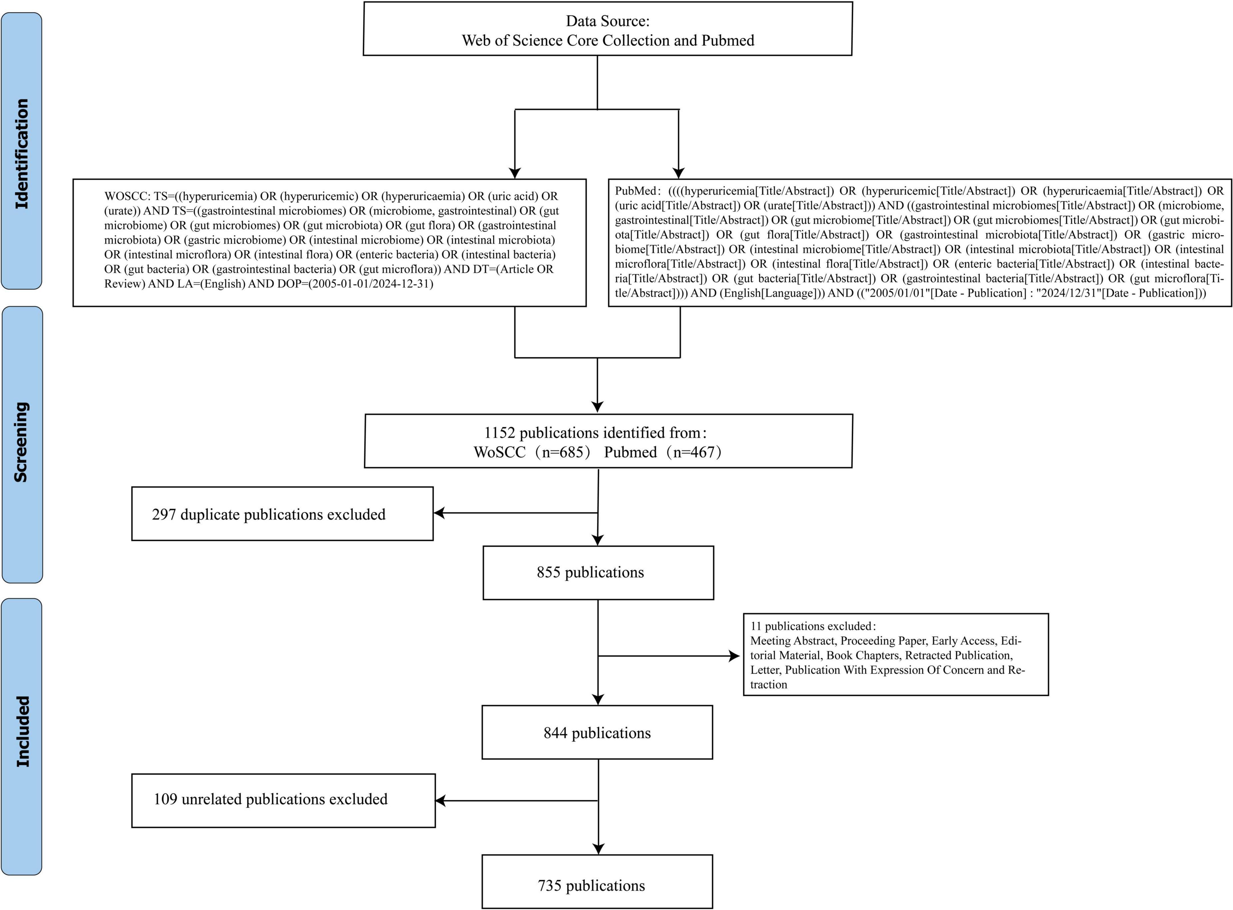
Inclusion criteria: All original articles and reviews in English related to “intestinal flora” and “HUA.” Exclusion criteria: Duplicated literature, literature not related to “intestinal flora” and “HUA” research, as well as meeting abstracts, proceeding papers, early access, editorial material, book chapters, retracted publications, and letters. In this study, Jingjing Yang and Jing Chen independently screened and excluded studies. Any discrepancies that arose were resolved by Dingxiang Li, who made the final decisions. A total of 735 papers were included, including 130 reviews and 605 original articles. Figure 1 illustrates the flowchart of the literature search and screening process.
2.3 Data analysis and visualization
The export of the qualifying publications “Full Record and Cited References” is performed in either “Plain Text File” format, with the filename “download*.txt.” The “Plain Text File” was loaded into VOSviewer, CiteSpace, and Bibliometrix1 for graphing. Before analysis, synonymous terms were unified using “thesaurus_terms.txt” in VOSviewer 1.6.19 to create thesaurus files. This process included synonyms (e.g., “gut microbiome” and “gastrointestinal microbiome”), singular and plural forms (e.g., “broiler chicken” and “broiler chickens”), and different expressions (e.g., “xanthine oxidase” and “Xanthine oxidase enzyme”). Excel 2021 software produces tables for sorting various counts. We used VOSviewer 1.6.19 to summarize the leading authors, co-cited authors, countries/regions, institutions, journals, co-cited references, keywords, and associated knowledge maps. For the dual-map overlay of journals, we employed CiteSpace V (version 6.3 R3, downloaded from https://citespace.podia.com/). The settings for CiteSpace V were configured as follows: cluster labels (12), journal labels (8), arcs α (3), citing journal titles (min pubs: 10), and cited journal titles (min cites: 10).
3 Results
3.1 Publication and citation trends
A total of 735 publications addressing the gastrointestinal microbiome and HUA were analyzed for this research; 130 of these were reviews, while the remaining 605 were articles. Figure 2 displays the trends in publications and annual citations for research on the gut microflora and hyperuricemia from 2005 to 2024. The bar graphs represent the number of publications, whereas the line graphs represent annual citations. Figure 2 illustrates that between 2005 and 2012, fewer than 10 papers were published annually in the area of gut flora and HUA, reflecting a low level of research activity. From 2013 to 2019, there was a slow growth in the number of publications, with 15–35 papers published each year, and a corresponding increase in citations. Notably, there was a remarkable increase in the number of publications in the field from 2020 to 2024, indicating that the interest of researchers in exploring the link between gut microbiota and HUA is growing, with 2024 emerging as the peak year for publications. This trend demonstrates that the study of gut microbiota and HUA has received increasing attention over the past 5 years and is expected to continue to be a key area of research.
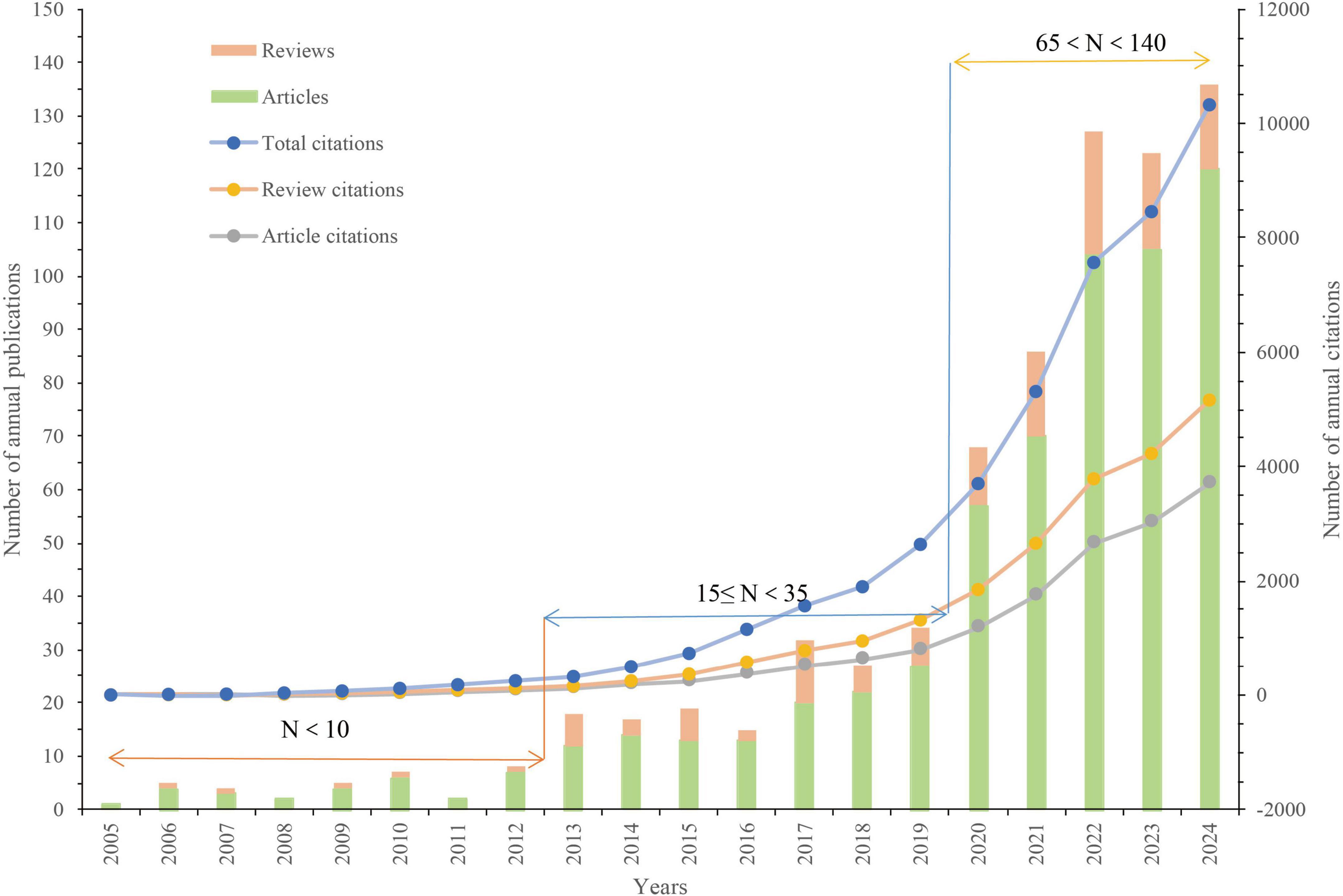
Figure 2. Trends in publications and annual citations for research on the gut microflora and hyperuricemia from 2005 to 2024.
3.2 Countries/regions and institutions
A collective of 72 distinct countries/regions participated in the study of the gastrointestinal microbiome and HUA. China had the highest number of publications (n = 413), followed by the United States (n = 126), Japan (n = 24), Italy (n = 21), and Germany (n = 20) (Table 1). According to Table 1, the United States has the highest total number of citations (n = 6,591), which is much higher than the number of citations for China (n = 5,200) and other high-output countries. Figure 3 displays the international collaboration networks of nations/regions. This map includes 30 countries/regions, each having published at least five articles. The size of the nodes represents the volume of publications, with larger nodes indicating a higher number of publications. Nodes sharing the same color denote a cluster with significant collaboration. The arcs illustrate the cooperation between different countries or regions, with thicker arcs signifying stronger ties. The figure reveals that both China and the United States have collaborative relationships with several countries, with the most frequent collaboration between China and the United States.
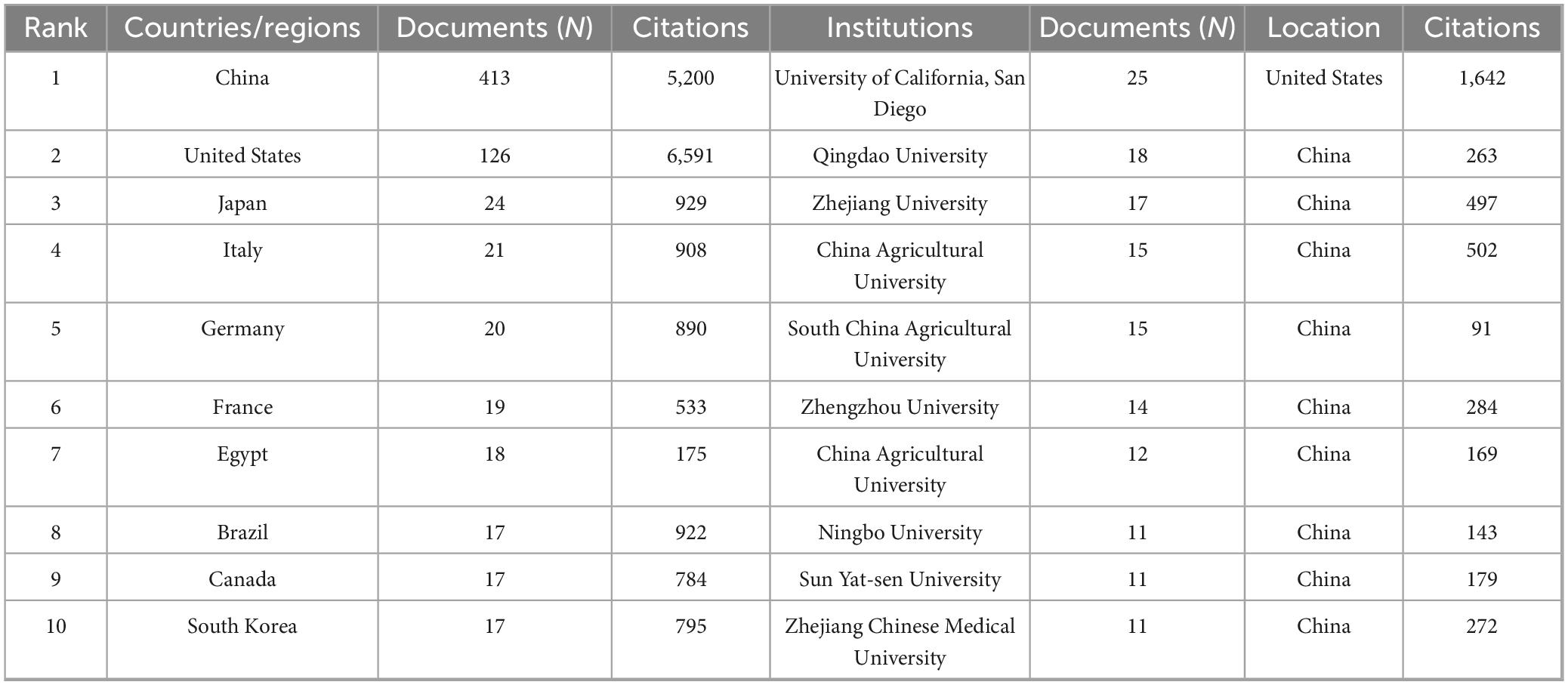
Table 1. Top 10 countries/regions and institutions investigating the intestinal microbiota and hyperuricemia.
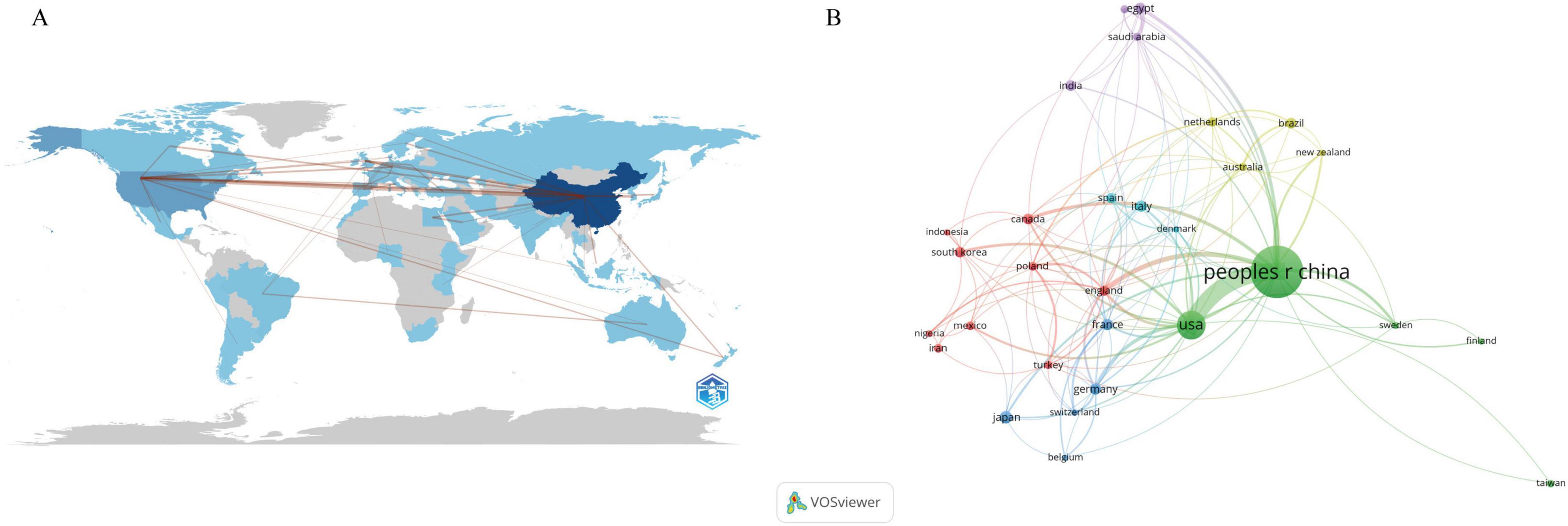
Figure 3. (A) The global publications’ geographic distribution map. (B) The global publications’ nations/regions collaboration map for the intestinal microbiota and hyperuricemia.
A total of 1,265 institutions researched gut microbiota and HUA. The top 10 institutions with the highest output published a total of 149 articles (Table 1). The University of California, San Diego (UCSD) ranked first in terms of the number of publications and citations, with 25 articles. Qingdao University and Zhejiang University followed with 18 and 17 articles, respectively. Notably, 9 of the top 10 institutions in terms of publication volume are from China. Figure 4 shows the cooperative network among different institutions. The size of a node indicates the number of documents published by the institution. The larger the node, the more documents are published. The more curves there are, the more cooperative institutions there are; the thicker the curve, the closer the cooperation among the institutions. Figure 4 illustrates that the Chinese Academy of Sciences has the highest number of collaboration linkages, indicating that this institution has the most extensive collaborative network. In terms of international collaboration, the UCSD has engaged in close collaborations with top-ranking institutions, such as Qingdao University and the Chinese Academy of Sciences.
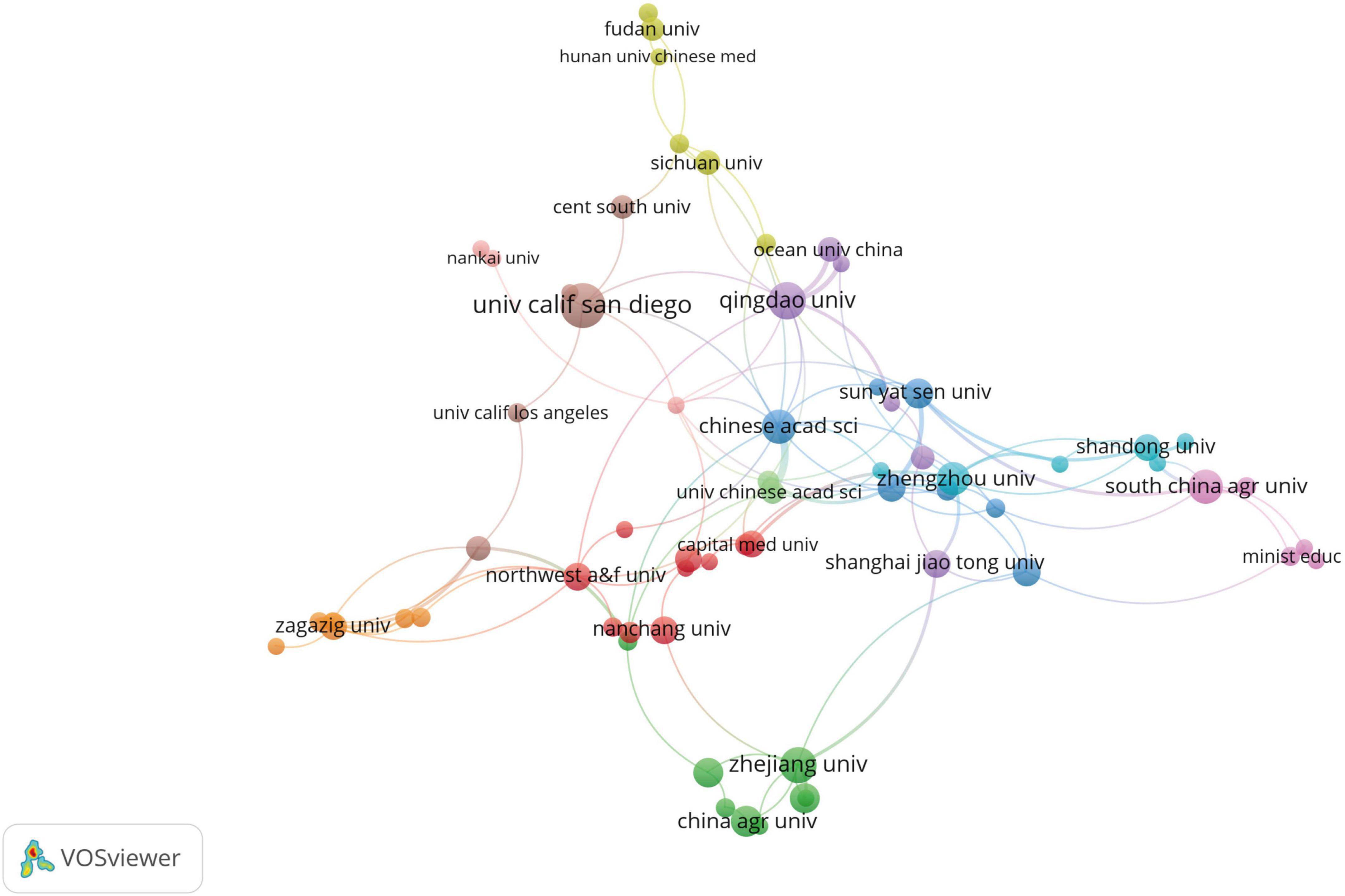
Figure 4. Collaborative network visualization of institutions involved in researching the intestinal microbiota and hyperuricemia.
3.3 Authors and co-cited authors
A total of 4,622 authors participated in studies on HUA and the gut flora (Table 2). Sanjay K. Nigam from the University of California, San Diego, was the top contributor, publishing 19 papers. Subsequently, Chenyang Lu and Xiurong Su from Ningbo University each published nine papers. Additionally, we analyzed the number of co-citations among the authors. Most of the top 10 co-cited authors were American. Nicola Dalbeth, Jing Wang, Zhuang Guo, Sanjay K., and Richard J. Johnson, with more than 80 co-citations. Nigam was among the top five for co-cited. Notably, Sanjay K. Nigam ranked highly in both co-cited and publication output. Supplementary Figure 1 provides a visual representation of the analysis results, highlighting the associations among countries, affiliations, and authors.
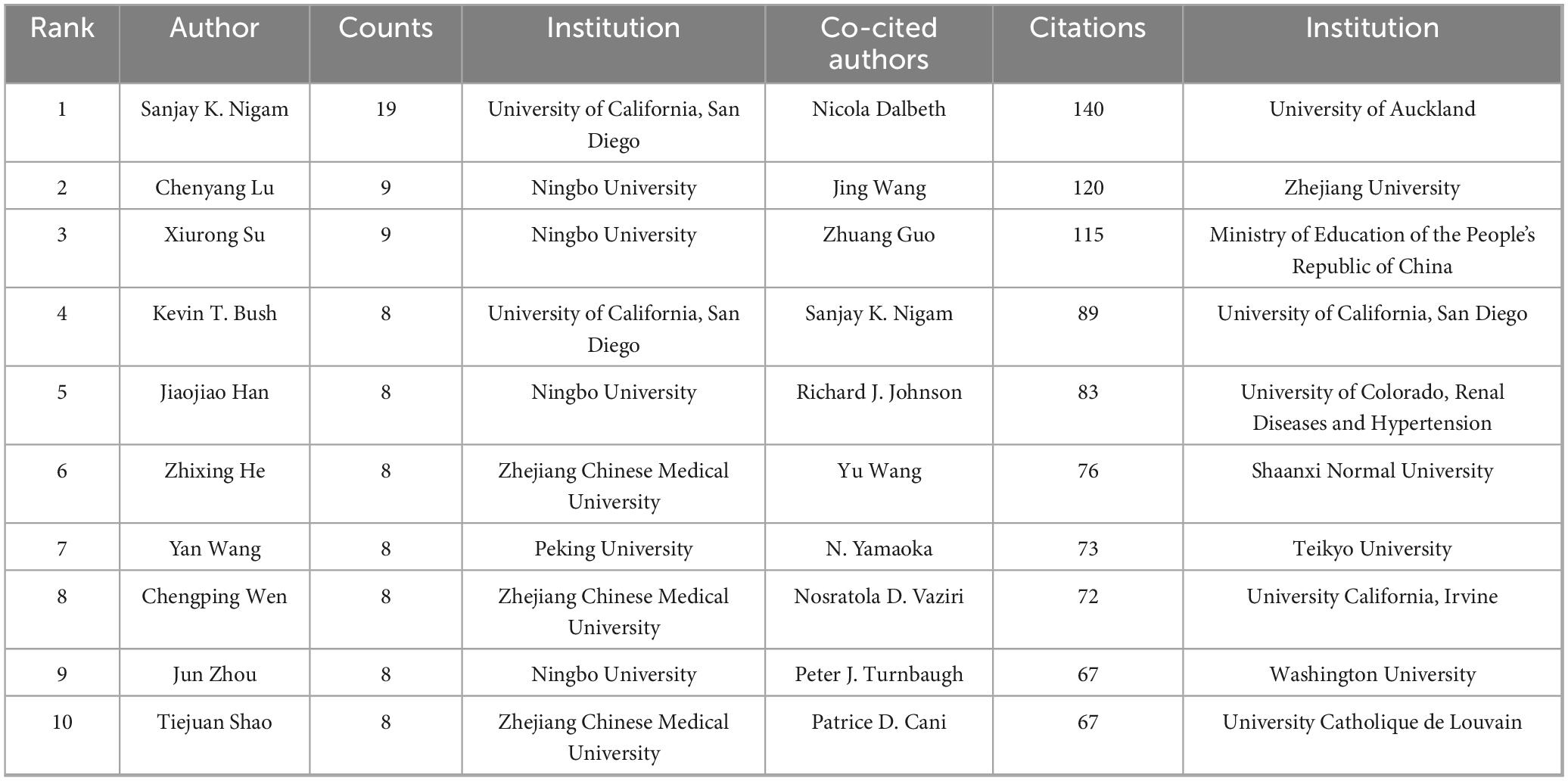
Table 2. Top 10 authors with high productivity and co-citation in studies on the gut microflora and hyperuricemia.
3.4 Journal analysis
Research papers on gut microbiota and HUA have been published in 369 different journals. Table 3 lists the top 10 journals ranked by the number of articles and co-cited counts. The journal Nutrients published the highest number of research articles, totaling 24, followed by Frontiers in Microbiology and Food & Function, which published 21 and 18 articles, respectively. Furthermore, all 10 journals with the highest citation counts have been cited more than 350 times, with PLoS One leading the way with a total of 883 citations, followed by Scientific Reports and Nature, which have been co-cited 648 and 631 times, respectively. According to the overlay visualization of journal maps (Supplementary Figure 2), studies published in veterinary/animal science journals predominantly cited papers in molecular biology/genetics journals. Similarly, research studies published in Molecular, Biology, and Immunology journals cited papers in Environmental, Toxicology, and Nutrition journals, as well as in Molecular, Biology, and Genetics journals and Health, Nursing, and Medicine journals. Furthermore, studies published in Medicine, Medical, and Clinical journals primarily cited papers in Molecular, Biology, and Genetics journals and Health, Nursing, and Medicine journals.
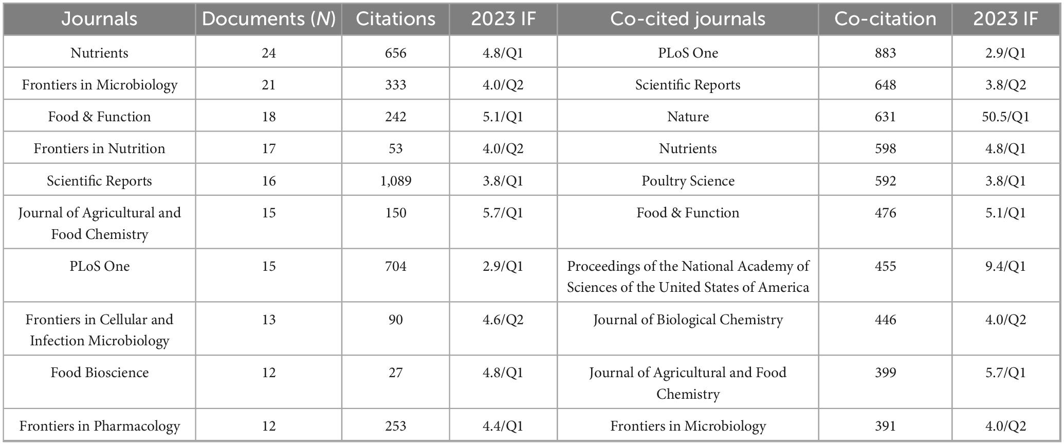
Table 3. Top 10 journals and co-cited journals for research on the gut microflora and hyperuricemia.
3.5 Reference analysis
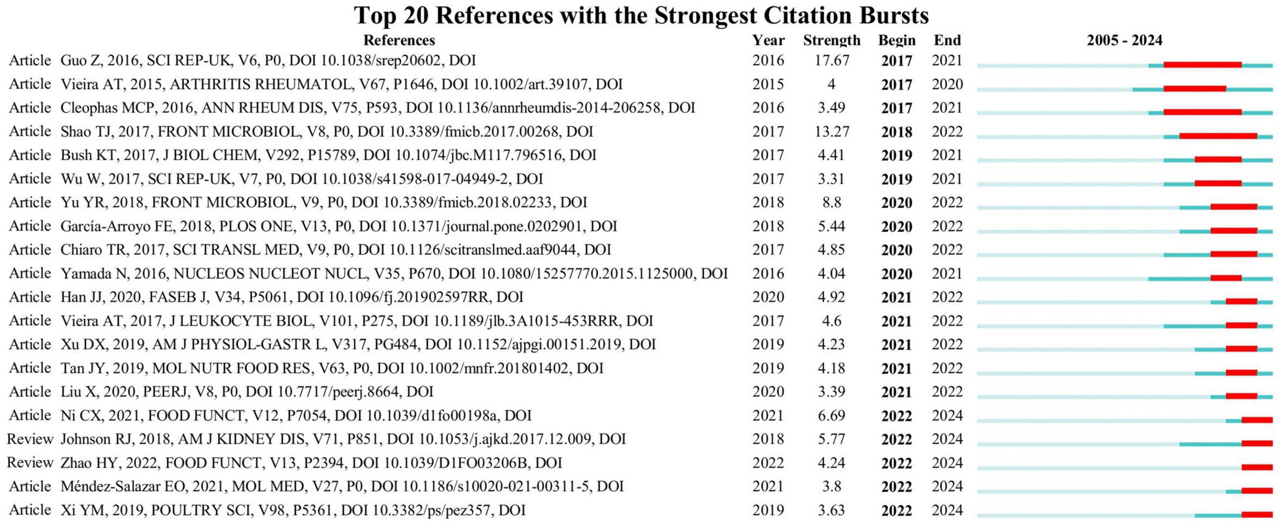
Citation burst analysis identifies significant and impactful literature from a specific time frame by pinpointing studies that experience a spike in citations during that period. Figure 5 shows the top 20 references with the strongest citation bursts, including 3 clinical studies, 15 experimental studies, and 2 reviews. The blue line depicts the timeline, and the red segments indicate the times when the references had bursts. Citation strength for the top 20 references spanned from 3.55 to 17.67. The citation explosion in this field began in 2017. Furthermore, two reviews and three articles are presently experiencing a burst.
3.6 Keyword analysis
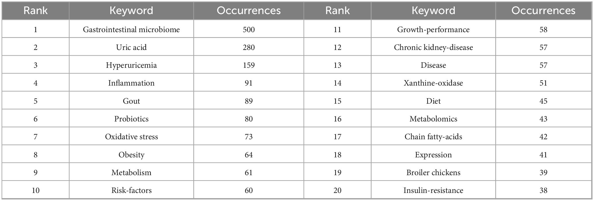
High-frequency keywords can indicate evolving research frontiers within certain knowledge domains. Using VOSviewer, we identified 3,421 keywords, 109 of which appeared at least 10 times. Table 4 shows the top 20 high-frequency keywords. Among the keywords, “gastrointestinal microbiome” was the most frequently appeared (n = 500), followed by “uric-acid” (n = 280), “hyperuricemia” (n = 159), “inflammation” (n = 91), “gout” (n = 90), and “probiotics” (n = 80). Figure 6 depicts the co-occurrence of keywords that appeared more than 10 times. Each node’s size reflects how often it co-occurs, and the connections illustrate the relationships between these co-occurring keywords. Each link’s thickness reflects the frequency of co-occurrence between two keywords, with the same color representing a tighter cluster. Figure 6 results in the formation of five distinct clusters. Cluster 1, represented in red, concentrates on the pathogenesis within the field and its interactions with other diseases. This cluster encompasses 32 keywords, including gastrointestinal microbiome, UA, obesity, risk factors, diet, insulin resistance, and SCFA. Cluster 2, depicted in green, highlights the role of probiotics in regulating UA levels and their importance in improving gut health and growth performance in broiler chickens. This cluster comprises 30 keywords, such as probiotics, growth performance, broiler chickens, antioxidant activities, lactobacillus, immunity, inulin, and intestinal morphology. Cluster 3, shown in blue, addresses the molecular mechanisms pertinent to this field and the application of fecal microbiota transplantation. It includes 19 keywords, such as inflammation, oxidative stress, dysbiosis, NLRP3 inflammasome, and fecal microbiota transplantation. Cluster 4, illustrated in yellow, examines the mechanisms through which natural products modulate gut microbiota to ameliorate HUA and gout. This cluster includes 18 keywords such as hyperuricemia, gout, metabolism, xanthine oxidase, extract, polysaccharides, and pathway. Cluster 5 (purple cluster) focuses on the interactions between this field and kidney diseases, including 10 keywords such as chronic kidney disease, metabolites, kidney, impact, uremic toxins, and progression. To study the trend of theme changes in the field, we constructed a topic evolution map and overlay visualization using the Bibliometrix package in the R software environment and VOSviewer (Figure 7). Figure 7 displays the topic evolution and a high-frequency keyword overlay map, with colors indicating the average publication year. The analysis revealed that from 2012 to 2018, the field primarily focused on macro issues, including intestinal gut health status in HUA. From 2019 to 2024, the areas of “gut-kidney axis,” “SCFAs,” “antioxidant activities,” “fecal microbiota transplantation,” “probiotics,” “diet,” and “untargeted metabolomics” are notably emerging, as marked in yellow, focusing on intervention mechanisms between HUA and gut flora.
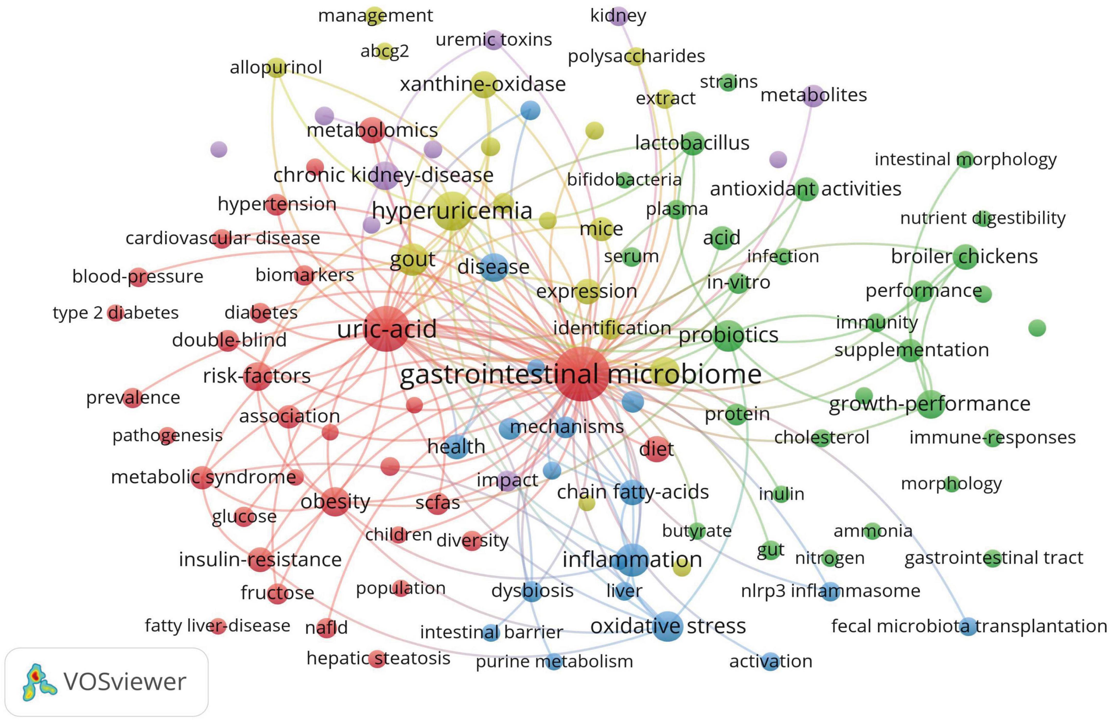
Figure 6. The co-occurrence network diagram of keywords related to gut microbiota and hyperuricemia.

Figure 7. (A) The topic evolution map of hyperuricemia and gut flora. (B) Overlay visualization of hyperuricemia and gut flora.
Keyword clustering can visually present the topic distribution in the research field. In this study, the LSI clustering method of CiteSpace software was used to obtain a reasonable clustering graph (Figure 8, Q = 0.4542, S = 0.7577). A Q value exceeding 0.3 signifies a notable cluster structure. An S value above 0.5 suggests effective clustering, while values over 0.7 denote high reliability. As shown in Figure 8A, 10 major research clusters have been formed in this field. Different clusters contain distinct keywords and themes. Clusters with smaller label values encompass a broader range of keywords, indicating more diverse research content. The 10 clustering clusters are as follows: #0 gut microbiota, #1 hyperuricemic nephropathy, #2 intestinal morphology, #3 animal models, #4 gut-kidney axis, #5 chronic kidney disease, #6 gut microbiome, #7 systems biology, #8 gastrointestinal tract, and #9 fecal microbiota transplantation. Figure 8B is a timeline visualization map created by CiteSpace. The timeline visualization shows the first appearance time of each important keyword and the dynamic changes of research hotspots, reflecting the evolution of research topics. As shown in Figure 8B, from 2005 to 2012, researchers have begun to recognize that gut microbiota may be related to UA metabolism (e.g., gut microflora, microbiome, and UA). However, the research is still in its infancy. From 2013 to 2018, this field focused on the changes in the gut microbiota and related mechanisms of hyperuricemia, as well as the changes in the gut microbiota associated with metabolic diseases and high levels of UA (e.g., gut microbiota, double blind, oxidative stress, and metabolic syndrome). Since 2019, the research focus in this field has shifted to the microbial intervention effects and mechanisms of hyperuricemia (e.g., probiotics, prebiotics, extract, diets, and therapy). In addition, the gut-kidney axis, hyperuricemic nephropathy, SCFAs, butyrate, etc., have become the focus of research in recent years.
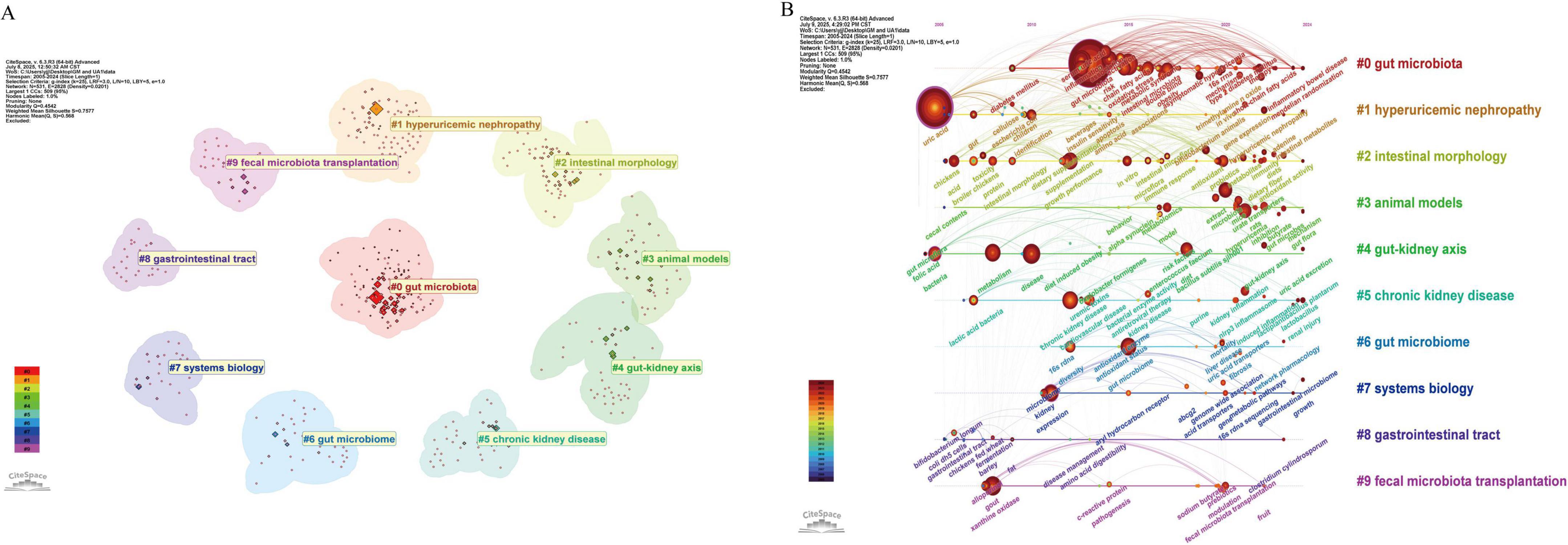
Figure 8. (A) Keyword clustering graph of hyperuricemia and gut flora. (B) Timeline visualization graph of keywords of hyperuricemia and gut flora.
4 Discussion
4.1 Basic information
A total of 736 papers on HUA and gut flora were included in this study. The publication volume in this field can be divided into three phases: a low activity period (2005–2012), a slow growth period (2013–2019), and an active period (2020–2024). This increasing trend reflects the growing interest and contributions of researchers in the field. The advancement and use of new technologies like metagenomics and high-throughput sequencing undoubtedly aid the growth in this discipline (Qin et al., 2010; Weinstock, 2012). Several nations or institutions have repeatedly carried out gut microbiota-related initiatives and achieved ground-breaking results, which have also offered direction and established the groundwork for the research into the connection between gut flora and HUA. For instance, the European Commission launched the Human Gut Metagenome Project in 2008, and the National Institutes of Health in the United States published the Human Microbiome Project (HMP) in 2007 (Turnbaugh et al., 2007; The Human Microbiome Project Consortium, 2012).
4.1.1 Countries/regions and institutions
China ranks first in the number of publications in this field, followed by the United States. The publication output of these two countries far exceeds that of others, indicating the significant interest of researchers in both countries and their substantial investment in research related to gut microbiota and HUA. Among the top 10 institutions in terms of publications, the top institution in terms of both publications and citations is UCSD in the United States, and the remaining 9 institutions are from China. UCSD has close collaboration with leading institutions such as Qingdao University and the Chinese Academy of Sciences. Professor Changgui Li from the Department of Endocrinology and Metabolism at the Affiliated Hospital of Qingdao University, Professor Huiyong Yin from the Chinese Academy of Sciences, and expert Robert Terkeltaub from UCSD worked together to discover differentially abundant metabolites and pathways underlying infrequent gout flares (InGFs) and frequent gout flares (FrGFs) through metabolomics and to establish a predictive model via machine learning (ML) algorithms (Wang M. et al., 2023). Notably, Robert Terkeltaub contributed to the American College of Rheumatology’s gout management guidelines (Khanna et al., 2012). Additionally, Professor Changgui Li serves as co-chair of the Asia–Pacific Gout Consortium (APGC)2 and plays a leading role in the field of gout and HUA, potentially laying the groundwork for collaboration among various organizations.
4.1.2 Authors
Analysis of authors and co-cited authors demonstrates that Sanjay K. Nigam is an influential writer who has made significant contributions to the fields of HUA and the intestinal microbiota. A 2015 review by Sanjay K. Nigam, published in Physiological Reviews, stands out as a representative work. This review underscores the essential functions of organic anion transporter 1 (OAT1) and OAT3 in the metabolism and processing of gut microbiome metabolites (Nigam et al., 2015). These transporters are predominantly expressed in the proximal tubule cells of the kidney, where they facilitate the transport of organic anions, such as UA, from the bloodstream into cells, subsequently expelling them through the cell’s apical membrane into the urine. OAT1 and OAT3 are particularly critical in the transmembrane transport of UA, a process they facilitate by exchanging dihydroxyacetate within the cell (Otani et al., 2017). In 2019, Sanjay K. Nigam’s team constructed a co-expression network of the gut-liver-kidney (GLK) axis, revealing interactions between transport proteins (e.g., OAT1, OAT3, and URAT1) and metabolizing enzymes (e.g., CYP4A11 and UGT2B4) associated with UA metabolism, emphasizing the role of these genes in regulating UA levels and intestinal flora metabolite transport (Rosenthal et al., 2019). In 2020 and 2022, it was noted that OAT1 and OAT3 are involved in UA reabsorption and excretion in renal proximal tubules and interact with gut flora metabolites (e.g., indoleacetic acid and 4-hydroxyphenylacetic acid) to affect UA levels, which in turn affects the progression of CKD (Engelhart et al., 2020; Granados et al., 2022; Jamshidi and Nigam, 2022). In 2023, it was found that gut microbiota metabolites, including tryptophan derivatives, could activate the host’s OATs and aryl hydrocarbon receptor (AHR), impacting UA secretion and excretion, thus creating a remote sensing and signaling mechanism between UA metabolism and intestinal flora, which is particularly important in chronic kidney disease (Nigam and Granados, 2023). In addition, loss of function of transporter proteins such as OAT1 leads to changes in the composition and function of the intestinal flora, thereby affecting UA metabolism (Ermakov et al., 2023). The research of Professors Sanjay K. Nigam et al. revealed the core bridging role of OAT1/OAT3 in the interaction between UA metabolism and intestinal microbiota, constructed the theoretical framework of the GLK axis, and laid a revolutionary theoretical foundation for the mechanism research, prevention, and treatment of hyperuricemia and chronic kidney disease.
4.1.3 Journals
A total of 736 documents appeared in 369 different journals, with major contributions from respected sources like Nutrients, Frontiers in Microbiology, and Food & Function. Interestingly, the Nutrients stood out as a primary focus, with a significant number of published studies and citations. This recognition confirms the Nutrients’ position as a significant outlet for sharing research findings in the field of gut microbiota and HUA and highlights the publication’s importance in this sector.
4.1.4 References
References with the strongest citation bursts analysis reveal the most influential references in the field. The citation boom period in this field is relatively concentrated, mainly occurring between 2017 and 2022. The citation research content that emerged at this stage was quite rich, including the description of the microbiota of clinical patients and microecological intervention strategies (including probiotics for lowering UA, dietary fiber for promoting the generation of SCFAs, tuna oligopeptides for repairing the intestinal barrier, etc.) (Guo et al., 2016; Shao et al., 2017; García-Arroyo et al., 2018; Han et al., 2020). Published in 2016 in Scientific Reports, the paper “Intestinal Microbiota Distinguish Gout Patients from Healthy Humans” experienced the strongest citation burst (strength = 17.67) between 2017 and 2021. This is a clinical cohort study, which revealed significant differences in the intestinal microflora between gout patients and healthy individuals. Bacteroides xylanisolvens and Bacteroides caccae were more abundant in gout patients, whereas Bifidobacterium pseudocatenulatum and Faecalibacterium prausnitzii were less abundant (Guo et al., 2016). The second strongest citation burst (strength = 13.27) article, titled “Combined Signature of the Fecal Microbiome and Metabolome in Patients with Gout,” was published by Shao et al. (2017) in the journal Frontiers in Microbiology. This study examined the fecal microbiome signatures and revealed an increase in pathogens, including Erysipelatoclostridium, Rhodococcus, Anaerolineaceae, and Bacteroides. The metabolome signatures included altered metabolites that may play a role in inflammatory responses, purine metabolism, and UA excretion (Shao et al., 2017). The third most intense citation burst (strength = 8.8) is an article titled “Alterations of the Gut Microbiome Associated With the Treatment of Hyperuricaemia in Male Rats,” published by Yu et al. (2018) in the journal Frontiers in Microbiology. The study investigates the effects of allopurinol and benzbromarone on the gut microbiota of male rats with hyperuricaemia, revealing specific alterations in bacterial genera and metabolic pathways associated with nucleotide and lipid metabolism (Yu et al., 2018). In addition, several papers that are still in the explosive period (2022–2024) indicate that the functional differences of gout subtypes in the microbiota and the deepening of probiotic mechanisms in this field have been studied (Méndez-Salazar et al., 2021; Ni et al., 2021).
4.2 Research hotspots and trends
The analysis of keywords can highlight the essential topics, focal points, and trends in a research domain, aiding researchers in grasping the knowledge framework and potential future directions of the field. By analyzing the high-frequency keywords, co-occurrence of keywords, keyword clustering, and timeline visualization maps in the research field of intestinal flora in hyperuricemia, the primary focuses of current studies can be identified.
4.2.1 Features of the gut microbiota in individuals with HUA
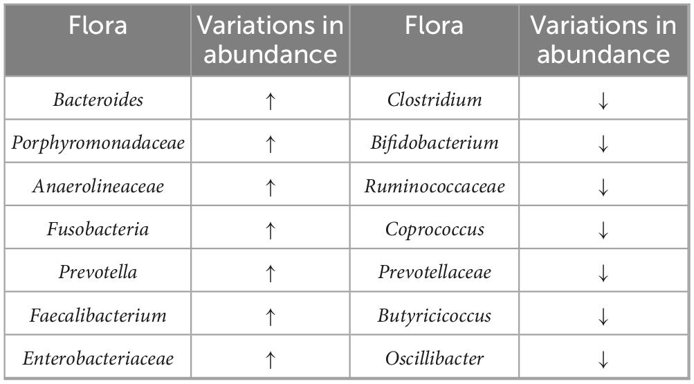
Through keyword co-occurrence and timeline visualization graphs, a large number of microbiota names, such as “gut microbiota,” “Bifidobacteria,” and “Escherichia coli,” were discovered, indicating that various types of microbiota have always been the focus of attention in this field. Multiple studies have shown that compared with healthy individuals, there are significant differences in the diversity and composition of the gut microbiota in patients with HUA (Table 5 and Supplementary Table 1). These differences are not only reflected in the “rise and fall” of specific bacterial genera, but also reflect the dual impact of the imbalance in the interaction between the microbiota and the host on metabolism and inflammation. Firstly, the enrichment of SCFAs-producing genera such as Alistipes, Faecalibacterium, and Roseburia has been repeatedly reported (Lin et al., 2021; Yuan et al., 2022). These bacteria may constitute a compensatory mechanism by which the body attempts to alleviate hyperuric acid-related metabolic disorders by maintaining the integrity of the intestinal barrier and inhibiting inflammatory responses. Secondly, multiple studies have reported that serum UA levels are positively correlated with the abundance of Bacteroides in patients with HUA/gout (Guo et al., 2016; Shao et al., 2017; Méndez-Salazar et al., 2021). Although research reports have found that Bacteroides are one of the main enterotypes in the healthy population of South Korea, and 5-hydroxyisourate hydrolase, involved in the conversion of UA to allantoin, is enriched in this type of intestinal type (Lim et al., 2014). However, most studies have found that Bacteroides are involved in pro-inflammatory effects under disease conditions. For instance, B. caccae have been identified as biomarkers of inflammatory bowel disease (IBD) (Wei et al., 2001). Additionally, an increased presence of Bacteroides spp. has been linked to various autoimmune diseases, including systemic lupus erythematosus (Hevia et al., 2014), rheumatoid arthritis (Zhang et al., 2015), and type 1 diabetes (Davis-Richardson and Triplett, 2015). It is suggested that the amplification of Bacteroides may simultaneously trigger chronic inflammation. Third, the general reduction of probiotics such as Bifidobacteria (Guo et al., 2016; Lin et al., 2021; Méndez-Salazar et al., 2021; Yang et al., 2021; Ul-Haq et al., 2022). Bifidobacteria are renowned for their probiotic properties and anti-inflammatory effects, and their depletion may contribute to the overall dysbiosis and elevated inflammatory levels in HUA patients (Ruiz et al., 2017). In addition, the imbalance of opportunistic pathogenic bacteria (such as Porphyromonas and Enterobacteriaceae) and SCFA-producing beneficial bacteria (such as Clostridium and Ruminococcus) further amplifies the risk of inflammation. It is worth noting that the abundance changes of Prevotella show contradictory results in different studies (Ul-Haq et al., 2022; Martínez-Nava et al., 2023). This discrepancy highlights the influence of confounding factors such as disease stage, diet, and testing methods on the dynamics of the microbiota (Wu et al., 2011). In conclusion, the intestinal microbiota of patients with HUA shows a pattern of “pro-inflammatory-anti-inflammatory” bacterial growth and decline. This imbalance pattern may either be the result of metabolic disorders or exacerbate hyperuricemia and inflammation through gut-axis feedback. In the future, it is necessary to combine longitudinal cohorts and standardized analyses to clarify the causal roles and individual heterogeneity of the changes in the microbiota. Meanwhile, the specific mechanism of action of Bacteroides under different physiological and pathological conditions should be further explored to provide more comprehensive theoretical support for the prevention and treatment of hyperuricemia and related diseases.
4.2.2 Mechanisms by which probiotics regulate uric acid levels
Through high-frequency keywords, keyword clustering, and citation burst analysis, we found that “probiotics,” “prebiotics,” and “therapy” are important research topics in the field of gut microbiota in hyperuricemia. The mechanism of probiotics in treating HUA includes altering the composition of the intestinal microbiota. Probiotics regulate the composition of the gut microbiota by increasing the abundance of beneficial bacteria (such as Bifidobacteria, Prevotella, Firmicutes, and SCFA-producing bacteria) while reducing pathogenic or pro-inflammatory bacteria (such as Bacteroidetes and Enterococcus). For instance, researchers have shown that Lactobacillus plantarum LLY-606 and L. plantarum TCI227 enhance the production of SCFAs and decrease the abundance of harmful bacteria such as Escherichia/Shigella (Chien et al., 2022; Shi et al., 2023). This modulation of the gut microbiota is considered a key mechanism underlying the therapeutic efficacy of probiotics. Secondly, it inhibits the activity of XOD. XOD is a pivotal enzyme in the biosynthesis of UA. Several probiotic strains (including Lactobacillus rhamnosus Fmb14, L. plantarum Q7, and Lactobacillus DM9218) have been shown to inhibit XOD activity in the liver, subsequently reducing UA concentrations (Wang et al., 2019; Ni et al., 2021; Cao et al., 2022; Li et al., 2022; Zhao et al., 2022; Shi et al., 2023). Additionally, probiotics can modulate the expression of UA transporter proteins, such as ABCG2, GLUT9, and URAT1 (Zhao et al., 2022; Wang Z. et al., 2023). These observations underscore the potential of probiotics to exert a direct influence on UA metabolism. Furthermore, numerous probiotics demonstrate notable anti-inflammatory properties by diminishing the expression of pro-inflammatory cytokines, including IL-1β, TNF-α, and IL-6. For instance, L. rhamnosus Fmb14 and L. plantarum LLY-606 have been shown to downregulate IL-1β and TNF-α levels, thereby mitigating inflammation associated with HUA (Li et al., 2022; Shi et al., 2023). Supplementary Table 2 details the specific mechanisms.
In addition, keyword co-occurrence reveals that the high-frequency keywords “growth performance,” “broiler chickens,” and “probiotics” are linked to each other. It is indicated that adding probiotics to regulate the UA level in broilers and improve their growth performance and meat quality is also a research hotspot on this topic. For instance, studies have shown that Lactobacillus farciminis CNMA67-4R, Clostridium butyricum CBM 588, multi-strain probiotics, and symbiotics (Bacillus subtilis, inulin, and Saccharomyces cerevisiae) significantly reduced the UA levels in the feces of broiler chickens, which is the primary substrate for ammonia production. Moreover, L. farciminis CNMA67-4R and the symbiotics decreased the urine nitrogen ratio in the feces, thereby leading to a reduction in ammonia emissions from the chickens (Such et al., 2021; Such et al., 2023). The reduction of ammonia emissions is of considerable importance for environmental protection and animal welfare, and it can also enhance the growth performance and immune function of broiler chickens. The timeline visualization map shows that “fecal microbiota transplantation,” “fermentation,” and “extract” are the key research topics in recent years. It indicates that, in addition to probiotics, other microbiota-based treatments [such as microbiota transplantation and microbial fermentation extracts (MFEs)] have received extensive attention. Liu et al. (2020) found that FMT effectively modulated the gut microbiota in rats with high-purine-induced HUA, leading to improvements in metabolic parameters. Furthermore, in patients with gout, washing microbiota transplantation decreases serum UA levels, is related to a reduction in both the frequency and duration of acute gout flares, lowers diamine oxidase and endotoxin levels, and helps improve their impaired intestinal barrier function (Xie et al., 2022). Treatment with Lactobacillus acidophilus fermented dandelion (LAFD) has been shown to restore imbalances in the gut microbial ecosystem and reverse alterations in Bacteroidetes/Firmicutes, Muribaculaceae, and Lachnospiraceae in HUA mice (Ma et al., 2023). In addition, research indicates that certain MFEs did not exhibit systemic toxicity even at high doses, suggesting a favorable safety and efficacy profile for the treatment and prevention of HUA (Chen et al., 2017). In conclusion, the technique of bacterial colony transplantation and its associated microbial fermentation products hold promise for the treatment of HUA.
However, in practical applications, probiotics face multiple challenges in the management of HUA. Firstly, ensuring a high viable bacterial count and stability of probiotics during processing, storage, transportation, and after passing through harsh gastrointestinal environments such as gastric acid and bile is crucial. Although encapsulation technology can enhance targeted delivery and viability, its effectiveness and repeatability still need to be optimized (Broeckx et al., 2016; Cassani et al., 2020; Sun et al., 2023). Secondly, significant individual differences in therapeutic effects exist, influenced by the composition of the host’s intestinal flora, genetic background, and lifestyle factors such as diet and medication (Sun et al., 2024; Wang Q. et al., 2024). This highlights the necessity of personalized strain and dosage selection. Finally, the current regulatory and standardization framework is still not perfect. There is a lack of unified standards for strain identification, evaluation, and quality control. It is urgent to implement stricter quality control procedures (such as ensuring the consistency between the actual content of the product and the label statement and preventing contamination) and adopt genomic assessment methods to verify the safety and accuracy of strains, to enhance product quality, safety, and consumer trust (Salvetti et al., 2016; Kolaček et al., 2017).
4.2.3 Microecological interventions
Keyword co-occurrence and timeline visualization analysis reveal that “polysaccharides,” “diet,” and “dietary fiber” are important research topics in this field, indicating that both traditional Chinese medicine (TCM) and dietary patterns regard the intestinal flora as the hub target for regulating HUA. They exert their effects through a two-way intervention strategy: on the one hand, they inhibit “dangerous bacteria” such as Bacteroidetes and Enterobacteriaceae that produce endotoxins and promote inflammation; on the other hand, they increase beneficial bacterial species such as Lactobacillus, Bifidobacterium, and Roseburia that produce SCFAs and strengthen the gut-kidney axis. Ultimately, they jointly reduce the UA load, decrease oxidative stress and inflammation, and achieve multi-layered benefits from the structure of the microbiota to the host’s metabolic-immune-kidney function.
The active ingredients of TCM and the intestinal flora synergistically intervene in UA homeostasis through a “bidirectional metabolism-regulation” model. Some bioactive ingredients in TCM are not effectively absorbed in the digestive tract, causing low bioavailability (Gong et al., 2020). On the one hand, TCM is first enzymatically hydrolyzed by the microbiota to generate more easily absorbed active metabolites, such as baicalin being converted into baicalein before entering the bloodstream (Noh et al., 2016), and the host metabolism is indirectly regulated through SCFAs and other microbiota products (Wang et al., 2017); on the other hand, active ingredients reshape the structure of the microbiota. Tea polyphenols, berberine, etc., can inhibit harmful bacteria, promote the growth of beneficial bacteria, improve microecology, and UA metabolism (Yan et al., 2020; Chen Q. et al., 2023; Wang L. et al., 2023). This bidirectional network provides new ideas for explaining the mechanism of action of TCM and developing microbiogenic therapies. However, most of the existing evidence remains at the animal model level, and the causal chain has not yet been fully verified in the human body. Among polyphenols, resveratrol (from Polygonum cuspidatum) was shown to increase Lactobacillus spp. and SCFA-producing bacteria (e.g., Clostridium and Bifidobacterium), while reducing Bacteroides and pro-inflammatory cytokines (IL-6, IL-1β, and TNF-α) (Zhou Y. et al., 2024). Chlorogenic acid (from Lonicera japonica) was observed to inhibit TMAO-synthesizing bacteria (Faecalibaculum and Blautia) and activate the PI3K/AKT/mTOR pathway, reducing renal fibrosis and oxidative stress (Zhou et al., 2022). Furthermore, ferulic acid (from Ligusticum chuanxiong) remodeled gut microbiota in HUA rats, while inhibiting the TLR4/NF-κB pathway to reduce UA absorption (Zhang N. et al., 2023). Among flavonoids, quercetin (from Taxillus chinensis) reduced hepatic XOD activity and enhanced purine degradation in HUA mice, with Lactobacillus aviarius implicated in UA metabolism modulation (Li D. et al., 2023). Myricetin-Nobiletin Hybrid (from waxberry and Citrus reticulata) regulated glycerophospholipid metabolism and increased norank_f_Muribaculaceae abundance in HUA mice, ameliorating renal damage (Li Y. et al., 2023). Saffron flavonoid extract (from Crocus sativus) reversed HUA-induced dysbiosis by enriching beneficial genera (Roseburia and Clostridium sp.) and suppressing pathogens (Alloprevotella and Parabacteroides) in rats, while enhancing antioxidant activity (Chen et al., 2022). Regarding alkaloids, berberine (from Coptis chinensis) upregulated colonic ABCG2 and reduced Bacteroidetes in HUA rats, enriching Lactobacillus and promoting UA excretion (Chen Q. et al., 2023). The classification of active ingredients in TCM, as well as their mechanisms of action and targeted bacterial communities, is shown in Supplementary Table 3.
In addition, diuretic and turbidium-reducing herbs have also demonstrated consistent effects in animal models, such as enhancing probiotics, reducing pathogenic bacteria, and lowering UA. Kidney tea has been observed to significantly enhance the abundance of Roseburia and Enterorhabdus, while reducing the abundance of Ileibacterium and UBA1819 in HUA model mice (Chen Y. et al., 2023). In addition, Chicory promotes probiotic growth and pathogen reduction to aid UA excretion in HUA quail (Bian et al., 2020). Additionally, Camellia sinensis increases the abundance of Ruminococcus and Lactobacillus, decreases the abundance of Bacteroides and E. coli, and regulates UA metabolism in HUA mice (Wu et al., 2022). Although preclinical studies based on rodent models suggest that TCM and its components may intervene in HUA by regulating the intestinal flora and UA metabolism, there is still significant uncertainty regarding its clinical transformation. The key limitation lies in the lack of high-quality clinical trials to verify the mechanism of action mediated by microorganisms. It is difficult to establish a causal relationship in multi-component compound prescriptions where specific components drive microbial changes and mechanically reduce UA. The methodological heterogeneity existing in preclinical studies (such as differences in HUA induction regimens, administration doses/dosage forms, and microbiota analysis techniques) restricts the direct comparability and universality of the results. Furthermore, the long-term sustainability impact of significant baseline microbiota differences among individuals on intervention responses and induced changes urgently needs in-depth exploration. Future research should enhance high-quality clinical trials, adopt uniform standards, systematically evaluate the impact of TCM on HUA, and take into account individual differences in microbiota to formulate personalized treatment plans. Figure 9 demonstrates the mechanisms by which probiotics and TCM mitigate HUA through modulation of the gut microbiota.
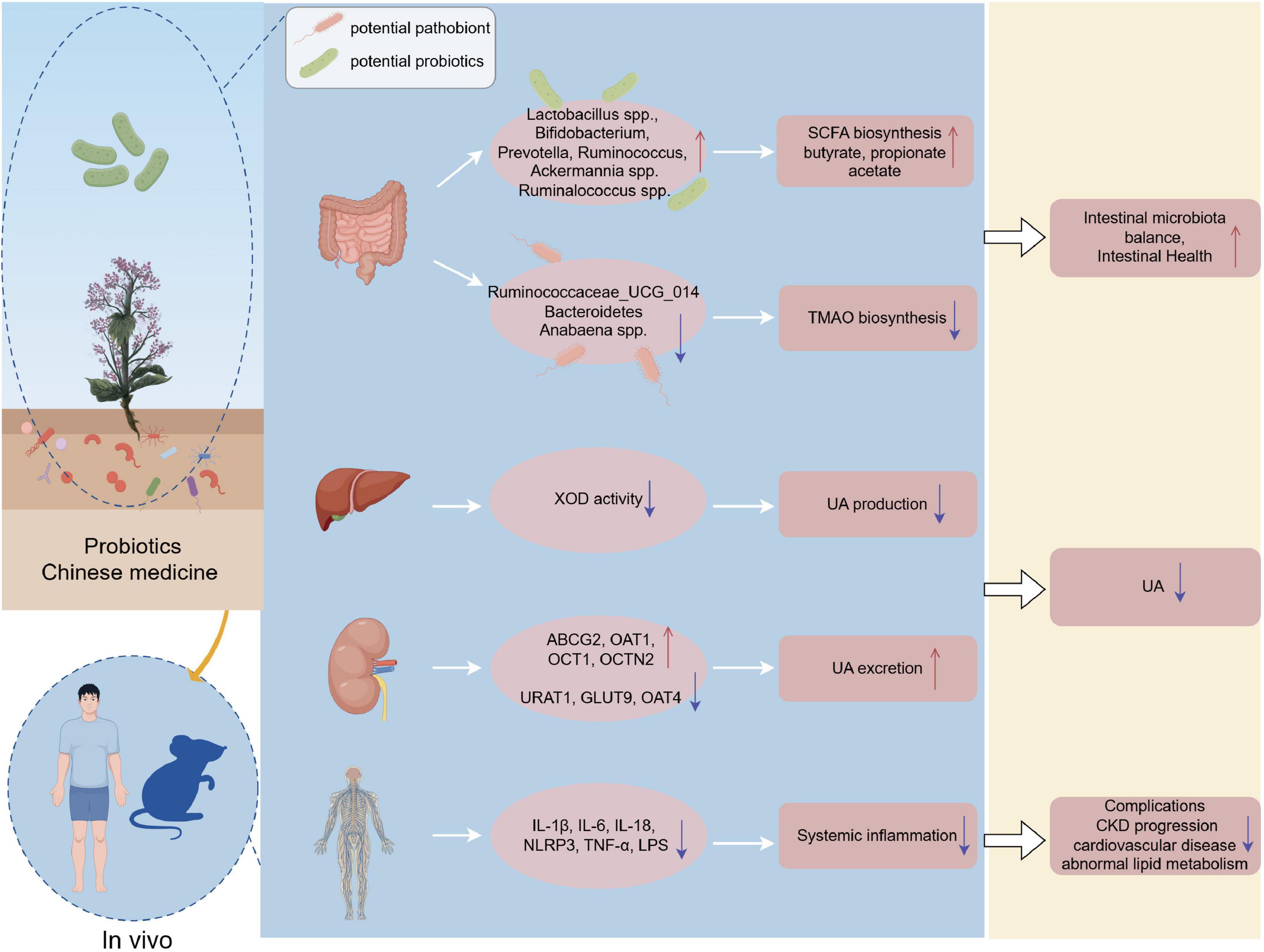
Figure 9. Mechanism of probiotics and TCM in regulating gut microbiota to alleviate hyperuricemia. Probiotics and TCM components influence gut microbiota by promoting beneficial bacteria and inhibiting harmful ones, while also modulating metabolic pathways to lower UA levels, reduce inflammation, and enhance gut health. Created by FigDraw.
Different dietary patterns significantly influence the composition and function of gut microbiota, which in turn affects the management of HUA. A study has demonstrated that dietary patterns characterized by high intake of animal protein, fat, and alcohol are positively correlated with the prevalence of HUA, whereas diets abundant in fruits, vegetables, legumes, and grains are inversely associated with HUA prevalence (Teng et al., 2015). The key components of these dietary patterns regulate UA levels through specific interactions with gut microbiota and their metabolic outputs. For example, Western dietary patterns, characterized by elevated intake of saturated fats and animal proteins, induce dysbiosis of the intestinal flora (Huang et al., 2013). The microbiota characteristics of animal models induced by long-term high-fat and high-fructose diets also support this view (Zhang N. et al., 2023). This dysbiosis increases intestinal permeability and stimulates the release of pro-inflammatory mediators (e.g., LPS, TNF-α, and IL-1β) and diminishes the production of SCFAs, particularly butyrate, which has anti-inflammatory properties and may enhance renal UA excretion (Chu et al., 2021; Li et al., 2021; Xu et al., 2021). In addition, researchers have found that SCFAs enhance the excretion of intestinal UA by activating peroxisome proliferator-activated receptor γ (PPARγ), which binds to the ABCG2 promoter (Adeyanju et al., 2021; Li M. et al., 2023). Conversely, the Mediterranean dietary (e.g., fruits, vegetables, nuts, whole grains, and beans) pattern, abundant in vitamins, minerals, polyphenols, dietary fiber, and monounsaturated fatty acids (MUFAs), fosters the proliferation of Bifidobacteria and Lactobacillus, and improve gut health by increasing the production of SCFAs while mitigating cells inflammatory responses lowering concentrations of TMAO, thus exerting a favorable effect on UA reduction (Chrysohoou et al., 2011; Chatzipavlou et al., 2014; Cariello et al., 2020; Soldán et al., 2024). Research has demonstrated that in hyperuricemia nephropathy rats, there was a significant increase in Blautia, Enterococcus, and Faecalibaculum associated with TMAO production. TMAO activates PI3K/AKT/mTOR signaling pathway, induces local inflammatory reaction in the kidney, aggravates kidney fibrosis, destabilizes UA transport proteins, and diminishes the kidney’s UA excretion capacity (Zhou et al., 2022). In addition, The Dietary Approaches to Stop Hypertension (DASH) diet, which emphasizes plant-based foods rich in whole grains, fruits, vegetables, and low-fat dairy products, has been shown to significantly enhance the abundance of beneficial gut microbiota such as Bifidobacterium and Lactobacillus, while reducing the prevalence of harmful bacteria, including LPS-producing Enterobacteriaceae. These beneficial microbial populations contribute to the reduction of UA levels through the production of SCFAs and the inhibition of XOD activity (Martínez et al., 2010; Gong et al., 2018; Ashaolu, 2020; Aslam et al., 2020). These findings underscore the significant role of dietary patterns in managing HUA by modulating the composition and function of the gut microbiome. Furthermore, selecting diets rich in dietary fiber and polyphenols may aid in lowering UA levels and alleviating symptoms in individuals with HUA.
4.2.4 Quadruple-axis synergistic regulation of uric acid homeostasis by gut microbiota
Timeline visualization analysis revealed that “liver,” “kidney,” “gut-kidney axis,” etc. have also been research hotspots in recent years. It has attracted much attention in recent years that the gut microbiota, through a multi-dimensional network composed of four axes including gut-kidney, gut-liver, gut-joint, and gut-brain, collaboratively regulates UA homeostasis in distant organs. On the gut-kidney axis, probiotics (such as L. rhamnosus) directly upregulate colonic UA transporters (such as ABCG2), accelerating UA excretion to reduce blood UA (Wang H. et al., 2024). Its metabolites, SCFAs, indirectly maintain UA balance by strengthening the intestinal barrier and inhibiting the TLR4/NLRP3 inflammatory pathway (Bi et al., 2024; Wang Q. et al., 2024; Lv et al., 2025). In the gut-liver axis, specific strains (Lactobacillus reuteri and Lactobacillus johnsonii) also promote UA excretion by enhancing barrier function and transporter protein expression, while further reducing blood UA by utilizing purine metabolism and using UA as a carbon source (Kasahara et al., 2023; Hussain et al., 2024; Han et al., 2025). The combination of probiotics with ursolic acid and oleanolic acid can reshape the structure of the microbiota, synergistically inhibit liver inflammatory pathways, and optimize UA metabolism (Ma et al., 2020). On the gut-joint axis, the barrier disruption mediated by TLR4/NLRP3 inflammation hinders UA excretion, while 7-ketocholic acid promotes epithelial repair by inhibiting FXR (Li et al., 2024; Wang Q. et al., 2024). Probiotics enrich Lactobacillus and Faecalibacterium through tryptophan metabolism, enhance the UA transport function of the colon, and indirectly reduce the risk of hyperuricemia. The gut-brain axis enhances the barrier through strains such as L. reuteri, upregulates the production of ABCG2 and SCFAs, transmits signals to the central nervous system via the vagus nerve, and regulates the synthesis and excretion of UA (Cryan et al., 2019; Wang et al., 2020; Hussain et al., 2024). In conclusion, the collaborative network of microbiota, metabolites, transport proteins, and distal organs provides a new microbiogenic intervention strategy for hyperuricemia.
4.3 Strengths and limitations
As the first thorough systematic bibliometric analysis of the gut microbiota and HUA, our work offers a wealth of insights and directions for academic investigators and clinical professionals alike. However, this study has the following limitations. Firstly, the research data were sourced solely from the WOS and PubMed databases, possibly resulting in incomplete data and outcomes. Secondly, as the bibliometric analysis tool of this study relies primarily on English literature, it may neglect non-English literature, potentially impacting a comprehensive understanding of global research activities. Thirdly, existing research shows that differences influence variations in gut microbiota characteristics among HUA patients in study subjects, sample sizes, detection methods, geographic locations, and dietary habits. The gut microbiota composition in animal models of HUA varies, likely due to differences in animal types and preparation methods. These variations may lead to inadequacies in concluding. Finally, bibliometric analysis needs to keep pace with actual research activities, and the updating of citation data takes time, potentially resulting in inadequate responsiveness to emerging research areas or hot topics.
5 Conclusion
In this extensive bibliometric analysis, we employed CiteSpace, VOSviewer, and Bibliometrix to systematically analyze a substantial corpus of research concerning HUA and the gut microbiota. Overall, our research has revealed four important research hotspots in this field, including microbiota characteristics, probiotic therapy, microecological intervention, and the gut-distal target organ axis. The focus of emerging hotspots is on dietary supplementation, microbiota transplantation treatment strategies, and extensive research on the organ axes discussed above. Moreover, although the modulation of the gut microbiota to improve HUA and its related diseases shows great potential, there are still some shortcomings in understanding broader mechanisms, long-term effects, and clinical applications. Future research should consider conducting large-scale, long-term clinical trials to assess the efficacy and safety of different microbiome therapies and utilize multi-omics technologies (including but not limited to proteomics and transcriptomics) to explore a wide range of molecular mechanisms to address these gaps.
Data availability statement
The original contributions presented in this study are included in this article/Supplementary material, further inquiries can be directed to the corresponding authors.
Author contributions
JY: Formal analysis, Visualization, Funding acquisition, Writing – review & editing, Writing – original draft, Data curation, Software. JC: Writing – original draft, Software, Formal analysis, Writing – review & editing, Data curation, Visualization. DL: Supervision, Writing – original draft, Writing – review & editing, Investigation, Validation, Conceptualization. QW: Validation, Methodology, Software, Writing – review & editing, Writing – original draft, Visualization. YZ: Software, Conceptualization, Writing – review & editing, Methodology, Writing – original draft, Data curation. YL: Conceptualization, Resources, Methodology, Writing – review & editing, Project administration, Writing – original draft, Funding acquisition. YD: Supervision, Conceptualization, Methodology, Writing – original draft, Resources, Project administration, Funding acquisition, Writing – review & editing.
Funding
The author(s) declare that financial support was received for the research and/or publication of this article. This research was funded by the 2022 National Key Research and Development Program “Modernization of Traditional Chinese Medicine,” Key Special Project of China (Nos. 2022YFC3501200 and 2022YFC3501202), the Science and Technology Innovation Program of Hunan Province (No. 2020RC4050), the 2024 Hunan Provincial Graduate Student Research and Innovation Program (No. CX20240738), and the 2023 General Project of Chinese Medicine Research Topics in Hunan Province (No. B2023057).
Acknowledgments
We express our gratitude to the reviewers for their valuable feedback. Thanks to HOME for the Researchers for assisting with the mechanism map (ID: PSWWW14114).
Conflict of interest
The authors declare that the research was conducted in the absence of any commercial or financial relationships that could be construed as a potential conflict of interest.
Generative AI statement
The authors declare that no Generative AI was used in the creation of this manuscript.
Publisher’s note
All claims expressed in this article are solely those of the authors and do not necessarily represent those of their affiliated organizations, or those of the publisher, the editors and the reviewers. Any product that may be evaluated in this article, or claim that may be made by its manufacturer, is not guaranteed or endorsed by the publisher.
Supplementary material
The Supplementary Material for this article can be found online at: https://www.frontiersin.org/articles/10.3389/fmicb.2025.1620561/full#supplementary-material
Abbreviations
HUA, hyperuricemia; UA, uric acid; WOS, Web of Science; SCFAs, short-chain fatty acids; XOD, xanthine oxidase; FMT, fecal microbiota transplantation; NLRP3, NOD-like receptor pyrin domain-containing 3; TMAO, trimethylamine N-oxide; PI3K, phosphatidylinositol 3-kinase; AKT, protein kinase B; mTOR, mammalian target of rapamycin; LPS, lipopolysaccharides; IL-1β, interleukin-1 beta; TNF-α, tumor necrosis factor alpha; CRE, creatinine; BUN, blood urea nitrogen; ALT, alanine aminotransferase; AST, aspartate aminotransferase; OAT1, organic anion transporter 1; OCT1, organic cation transporter 1; OCTN2, organic cation transporter N2; GLUT9, glucose transporter 9; OAT4, organic anion transporter 4; URAT1, urate transporter 1; NADPH, nicotinamide adenine dinucleotide phosphate; GSH-PX, glutathione peroxidase; MDA, malondialdehyde; DAO, diamine oxidase; D-Lac, D-lactate; HMANGO-C, Hmong microbiome and gout, obesity, vitamin C; Uox-KO, urate oxidase knockout.
Footnotes
References
Adeyanju, O., Badejogbin, O., Areola, D., Olaniyi, K., Dibia, C., Soetan, O., et al. (2021). Sodium butyrate arrests pancreato-hepatic synchronous uric acid and lipid dysmetabolism in high fat diet fed Wistar rats. Biomed. Pharmacother. 133:110994. doi: 10.1016/j.biopha.2020.110994
Ashaolu, T. (2020). Soy bioactive peptides and the gut microbiota modulation. Appl. Microbiol. Biotechnol. 104, 9009–9017. doi: 10.1007/s00253-020-10799-2
Aslam, H., Marx, W., Rocks, T., Loughman, A., Chandrasekaran, V., Ruusunen, A., et al. (2020). The effects of dairy and dairy derivatives on the gut microbiota: A systematic literature review. Gut Microbes 12:1799533. doi: 10.1080/19490976.2020.1799533
Bi, C., Zhang, L., Liu, J., and Chen, L. (2024). Lactobacillus paracasei 259 alleviates hyperuricemia in rats by decreasing uric acid and modulating the gut microbiota. Front. Nutr. 11:1450284. doi: 10.3389/fnut.2024.1450284
Bian, M., Wang, J., Wang, Y., Nie, A., Zhu, C., Sun, Z., et al. (2020). Chicory ameliorates hyperuricemia via modulating gut microbiota and alleviating LPS/TLR4 axis in quail. Biomed. Pharmacother. 131:110719. doi: 10.1016/j.biopha.2020.110719
Broeckx, G., Vandenheuvel, D., Claes, I., Lebeer, S., and Kiekens, F. (2016). Drying techniques of probiotic bacteria as an important step towards the development of novel pharmabiotics. Int. J. Pharm. 505, 303–318. doi: 10.1016/j.ijpharm.2016.04.002
Cao, J., Bu, Y., Hao, H., Liu, Q., Wang, T., Liu, Y., et al. (2022). Effect and potential mechanism of Lactobacillus plantarum Q7 on Hyperuricemia in vitro and in vivo. Front. Nutr. 9:954545. doi: 10.3389/fnut.2022.954545
Cariello, M., Contursi, A., Gadaleta, R., Piccinin, E., De Santis, S., Piglionica, M., et al. (2020). Extra-virgin olive oil from apulian cultivars and intestinal inflammation. Nutrients 12:1084. doi: 10.3390/nu12041084
Cassani, L., Gomez-Zavaglia, A., and Simal-Gandara, J. (2020). Technological strategies ensuring the safe arrival of beneficial microorganisms to the gut: From food processing and storage to their passage through the gastrointestinal tract. Food Res. Int. 129:108852. doi: 10.1016/j.foodres.2019.108852
Chatzipavlou, M., Magiorkinis, G., Koutsogeorgopoulou, L., and Kassimos, D. (2014). Mediterranean diet intervention for patients with hyperuricemia: A pilot study. Rheumatol. Int. 34, 759–762. doi: 10.1007/s00296-013-2690-7
Chen, N., Wang, R., Li, H., Wang, W., Wang, L., Yin, X., et al. (2022). Flavonoid extract of saffron by-product alleviates hyperuricemia via inhibiting xanthine oxidase and modulating gut microbiota. Phytother. Res. 36, 4604–4619. doi: 10.1002/ptr.7579
Chen, Q., Li, D., Wu, F., He, X., Zhou, Y., Sun, C., et al. (2023). Berberine regulates the metabolism of uric acid and modulates intestinal flora in hyperuricemia rats model. Comb. Chem. High Throughput Screen. 26, 2057–2066. doi: 10.2174/1386207326666221124093228
Chen, Q., Li, D., Wu, F., He, X., Zhou, Y., Sun, C., et al. (2023). Berberine regulates the metabolism of uric acid and modulates intestinal flora in hyperuricemia rats model. Comb. Chem. High Throughput Screen. 26, 2057–2066. doi: 10.2174/1386207326666221124093228
Chen, Y., Pei, C., Chen, Y., Xiao, X., Zhang, X., Cai, K., et al. (2023). Kidney tea ameliorates hyperuricemia in mice via altering gut microbiota and restoring metabolic profile. Chem. Biol. Interact. 376:110449. doi: 10.1016/j.cbi.2023.110449
Chen, R., Chen, M., Chen, Y., Hsiao, C., Chen, H., Chen, S., et al. (2017). Evaluating the urate-lowering effects of different microbial fermented extracts in hyperuricemic models accompanied with a safety study. J. Food Drug Anal. 25, 597–606. doi: 10.1016/j.jfda.2016.07.003
Chen, X., Ge, H., Lei, S., Jiang, Z., Su, J., He, X., et al. (2020). Dendrobium officinalis six nostrum ameliorates urate under-excretion and protects renal dysfunction in lipid emulsion-induced hyperuricemic rats. Biomed. Pharmacother. 132:110765. doi: 10.1016/j.biopha.2020.110765
Chen-Xu, M., Yokose, C., Rai, S., Pillinger, M., and Choi, H. (2019). Contemporary prevalence of gout and hyperuricemia in the United States and Decadal Trends: The national health and nutrition examination survey, 2007-2016. Arthritis Rheumatol. 71, 991–999. doi: 10.1002/art.40807
Chien, C., Chien, Y., Lin, Y., Lin, Y., Chan, S., Hu, W., et al. (2022). Supplementation of Lactobacillus plantarum (TCI227) prevented potassium-oxonate-induced hyperuricemia in rats. Nutrients 14:4832. doi: 10.3390/nu14224832
Chrysohoou, C., Skoumas, J., Pitsavos, C., Masoura, C., Siasos, G., Galiatsatos, N., et al. (2011). Long-term adherence to the Mediterranean diet reduces the prevalence of hyperuricaemia in elderly individuals, without known cardiovascular disease: The Ikaria study. Maturitas 70, 58–64. doi: 10.1016/j.maturitas.2011.06.003
Chu, Y., Sun, S., Huang, Y., Gao, Q., Xie, X., Wang, P., et al. (2021). Metagenomic analysis revealed the potential role of gut microbiome in gout. NPJ Biofilms Microbiomes 7:66. doi: 10.1038/s41522-021-00235-2
Clemente, J., Ursell, L., Parfrey, L., and Knight, R. (2012). The impact of the gut microbiota on human health: An integrative view. Cell 148, 1258–1270. doi: 10.1016/j.cell.2012.01.035
Cryan, J., O’Riordan, K., Cowan, C., Sandhu, K., Bastiaanssen, T., Boehme, M., et al. (2019). The microbiota-gut-brain axis. Physiol. Rev. 99, 1877–2013. doi: 10.1152/physrev.00018.2018
Davis-Richardson, A., and Triplett, E. W. (2015). A model for the role of gut bacteria in the development of autoimmunity for type 1 diabetes. Diabetologia 58, 1386–1393. doi: 10.1007/s00125-015-3614-8
Dehlin, M., Jacobsson, L., and Roddy, E. (2020). Global epidemiology of gout: Prevalence, incidence, treatment patterns and risk factors. Nat. Rev. Rheumatol. 16, 380–390. doi: 10.1038/s41584-020-0441-1
Engelhart, D., Granados, J., Shi, D., Saier, M. Jr., Baker, M., Abagyan, R., et al. (2020). Systems biology analysis reveals eight SLC22 transporter subgroups, including OATs, OCTs, and OCTNs. Int. J. Mol. Sci. 21:1791. doi: 10.3390/ijms21051791
Ermakov, V., Granados, J., and Nigam, S. (2023). Remote effects of kidney drug transporter OAT1 on gut microbiome composition and urate homeostasis. JCI Insight 8:e172341. doi: 10.1172/jci.insight.172341
Fathallah-Shaykh, S., and Cramer, M. (2014). Uric acid and the kidney. Pediatr. Nephrol. 29, 999–1008. doi: 10.1007/s00467-013-2549-x
García-Arroyo, F., Gonzaga, G., Muñoz-Jiménez, I., Blas-Marron, M., Silverio, O., Tapia, E., et al. (2018). Probiotic supplements prevented oxonic acid-induced hyperuricemia and renal damage. PLoS One 13:e0202901. doi: 10.1371/journal.pone.0202901
Gong, L., Cao, W., Chi, H., Wang, J., Zhang, H., Liu, J., et al. (2018). Whole cereal grains and potential health effects: Involvement of the gut microbiota. Food Res. Int. 103, 84–102. doi: 10.1016/j.foodres.2017.10.025
Gong, X., Li, X., Bo, A., Shi, R., Li, Q., Lei, L., et al. (2020). The interactions between gut microbiota and bioactive ingredients of traditional Chinese medicines: A review. Pharmacol. Res. 157:104824. doi: 10.1016/j.phrs.2020.104824
Granados, J., Bhatnagar, V., and Nigam, S. (2022). Blockade of organic anion transport in humans after treatment with the drug probenecid leads to major metabolic alterations in plasma and urine. Clin. Pharmacol. Ther. 112, 653–664. doi: 10.1002/cpt.2630
Guo, Z., Zhang, J., Wang, Z., Ang, K., Huang, S., Hou, Q., et al. (2016). Intestinal microbiota distinguish gout patients from healthy humans. Sci. Rep. 6:20602. doi: 10.1038/srep20602
Han, J., Wang, X., Tang, S., Lu, C., Wan, H., Zhou, J., et al. (2020). Protective effects of tuna meat oligopeptides (TMOP) supplementation on hyperuricemia and associated renal inflammation mediated by gut microbiota. FASEB J. 34, 5061–5076. doi: 10.1096/fj.201902597RR
Han, R., Wang, Z., Li, Y., Ke, L., Li, X., Li, C., et al. (2025). Gut microbiota Lactobacillus johnsonii alleviates hyperuricemia by modulating intestinal urate and gut microbiota-derived butyrate. Chin. Med. J. [Online ahead of print]. doi: 10.1097/CM9.0000000000003603
Hevia, A., Milani, C., López, P., Cuervo, A., Arboleya, S., Duranti, S., et al. (2014). Intestinal dysbiosis associated with systemic lupus erythematosus. mBio 5:e01548-e14. doi: 10.1128/mBio.01548-14
Huang, E., Devkota, S., Moscoso, D., Chang, E., and Leone, V. (2013). The role of diet in triggering human inflammatory disorders in the modern age. Microbes Infect. 15, 765–774. doi: 10.1016/j.micinf.2013.07.004
Hussain, A., Rui, B., Ullah, H., Dai, P., Ahmad, K., Yuan, J., et al. (2024). Limosilactobacillus reuteri HCS02-001 attenuates hyperuricemia through gut microbiota-dependent regulation of uric acid biosynthesis and excretion. Microorganisms 12:637. doi: 10.3390/microorganisms12040637
Jamshidi, N., and Nigam, S. (2022). Drug transporters OAT1 and OAT3 have specific effects on multiple organs and gut microbiome as revealed by contextualized metabolic network reconstructions. Sci. Rep. 12:18308. doi: 10.1038/s41598-022-21091-w
Jordan, D., Choi, H., Verbanck, M., Topless, R., Won, H., Nadkarni, G., et al. (2019). No causal effects of serum urate levels on the risk of chronic kidney disease: A Mendelian randomization study. PLoS Med. 16:e1002725. doi: 10.1371/journal.pmed.1002725
Kasahara, K., Kerby, R., Zhang, Q., Pradhan, M., Mehrabian, M., Lusis, A., et al. (2023). Gut bacterial metabolism contributes to host global purine homeostasis. Cell Host Microbe 31, 1038–1053.e10. doi: 10.1016/j.chom.2023.05.011
Khanna, D., Khanna, P., Fitzgerald, J., Singh, M., Bae, S., Neogi, T., et al. (2012). 2012 American College of Rheumatology guidelines for management of gout. Part 2: Therapy and antiinflammatory prophylaxis of acute gouty arthritis. Arthritis Care Res. 64, 1447–1461. doi: 10.1002/acr.21773
Kim, Y., Kang, J., and Kim, G. (2018). Prevalence of hyperuricemia and its associated factors in the general Korean population: An analysis of a population-based nationally representative sample. Clin. Rheumatol. 37, 2529–2538. doi: 10.1007/s10067-018-4130-2
Kimura, Y., Tsukui, D., and Kono, H. (2021). Uric acid in inflammation and the pathogenesis of atherosclerosis. Int. J. Mol. Sci. 22:12394. doi: 10.3390/ijms222212394
Kolaček, S., Hojsak, I., Berni Canani, R., Guarino, A., Indrio, F., Orel, R., et al. (2017). Commercial probiotic products: A call for improved quality control. A position paper by the ESPGHAN Working Group for probiotics and prebiotics. J. Pediatr. Gastroenterol. Nutr. 65, 117–124. doi: 10.1097/MPG.0000000000001603
Lee, S., Park, W., Suh, Y., Lim, M., Kwon, S., Lee, J., et al. (2019). Association of serum uric acid with cardiovascular disease risk scores in Koreans. Int. J. Environ. Res. Public Health 16:4632. doi: 10.3390/ijerph16234632
Li, D., Zhang, M., Teng Zhu La, A., Lyu, Z., Li, X., Feng, Y., et al. (2023). Quercetin-enriched Lactobacillus aviarius alleviates hyperuricemia by hydrolase-mediated degradation of purine nucleosides. Pharmacol. Res. 196:106928. doi: 10.1016/j.phrs.2023.106928
Li, L., Zhang, Y., and Zeng, C. (2020). Update on the epidemiology, genetics, and therapeutic options of hyperuricemia. Am. J. Transl. Res. 12, 3167–3181.
Li, M., Wu, X., Guo, Z., Gao, R., Ni, Z., Cui, H., et al. (2023). Lactiplantibacillus plantarum enables blood urate control in mice through degradation of nucleosides in gastrointestinal tract. Microbiome 11:153. doi: 10.1186/s40168-023-01605-y
Li, T., Ding, N., Guo, H., Hua, R., Lin, Z., Tian, H., et al. (2024). A gut microbiota-bile acid axis promotes intestinal homeostasis upon aspirin-mediated damage. Cell Host Microbe 32, 191–208.e9. doi: 10.1016/j.chom.2023.12.015
Li, X., Chen, Y., Gao, X., Wu, Y., El-Seedi, H., Cao, Y., et al. (2021). Antihyperuricemic effect of green alga ulva lactuca ulvan through regulating urate transporters. J. Agric. Food Chem. 69, 11225–11235. doi: 10.1021/acs.jafc.1c03607
Li, Y., Pu, L., Li, Y., Zhu, G., and Wu, Z. (2023). Design, synthesis and evaluation of a myricetin and nobiletin hybrid compound for alleviating hyperuricemia based on metabolomics and gut microbiota. RSC Adv. 13, 21448–21458. doi: 10.1039/d3ra03188h
Li, Y., Zhu, J., Lin, G., Gao, K., Yu, Y., Chen, S., et al. (2022). Probiotic effects of Lacticaseibacillus rhamnosus 1155 and Limosilactobacillus fermentum 2644 on hyperuricemic rats. Front. Nutr. 9:993951. doi: 10.3389/fnut.2022.993951
Lim, M., Rho, M., Song, Y., Lee, K., Sung, J., and Ko, G. (2014). Stability of gut enterotypes in Korean monozygotic twins and their association with biomarkers and diet. Sci. Rep. 4:7348. doi: 10.1038/srep07348
Lin, S., Zhang, T., Zhu, L., Pang, K., Lu, S., Liao, X., et al. (2021). Characteristic dysbiosis in gout and the impact of a uric acid-lowering treatment, febuxostat on the gut microbiota. J. Genet. Genomics 48, 781–791. doi: 10.1016/j.jgg.2021.06.009
Liu, X., Lv, Q., Ren, H., Gao, L., Zhao, P., Yang, X., et al. (2020). The altered gut microbiota of high-purine-induced hyperuricemia rats and its correlation with hyperuricemia. PeerJ 8:e8664. doi: 10.7717/peerj.8664
Liu, Y., Jarman, J., Low, Y., Augustijn, H., Huang, S., Chen, H., et al. (2023). A widely distributed gene cluster compensates for uricase loss in hominids. Cell 186, 3400–3413.e20. doi: 10.1016/j.cell.2023.06.010
Luckey, T. (1972). Introduction to intestinal microecology. Am. J. Clin. Nutr. 25, 1292–1294. doi: 10.1093/ajcn/25.12.1292
Lv, J., Hao, P., Zhou, Y., Liu, T., Wang, L., Song, C., et al. (2025). Role of the intestinal flora-immunity axis in the pathogenesis of rheumatoid arthritis-mechanisms regulating short-chain fatty acids and Th17/Treg homeostasis. Mol. Biol. Rep. 52:617. doi: 10.1007/s11033-025-10714-w
Ma, C., Yang, X., Lv, Q., Yan, Z., Chen, Z., Xu, D., et al. (2020). Soluble uric acid induces inflammation via TLR4/NLRP3 pathway in intestinal epithelial cells. Iran J. Basic Med. Sci. 23, 744–750. doi: 10.22038/ijbms.2020.44948.10482
Ma, Q., Chen, M., Liu, Y., Tong, Y., Liu, T., Wu, L., et al. (2023). Lactobacillus acidophilus fermented dandelion improves hyperuricemia and regulates gut microbiota. Fermentation-Basel 9:352. doi: 10.3390/fermentation9040352
Martínez, I., Kim, J., Duffy, P., Schlegel, V., and Walter, J. (2010). Resistant starches types 2 and 4 have differential effects on the composition of the fecal microbiota in human subjects. PLoS One 5:e15046. doi: 10.1371/journal.pone.0015046
Martínez-Nava, G., Méndez-Salazar, E., Vázquez-Mellado, J., Zamudio-Cuevas, Y., Francisco-Balderas, A., Martínez-Flores, K., et al. (2023). The impact of short-chain fatty acid-producing bacteria of the gut microbiota in hyperuricemia and gout diagnosis. Clin. Rheumatol. 42, 203–214. doi: 10.1007/s10067-022-06392-9
Méndez-Salazar, E., Vázquez-Mellado, J., Casimiro-Soriguer, C., Dopazo, J., Çubuk, C., Zamudio-Cuevas, Y., et al. (2021). Taxonomic variations in the gut microbiome of gout patients with and without tophi might have a functional impact on urate metabolism. Mol. Med. 27:50. doi: 10.1186/s10020-021-00311-5
Narang, R., Gamble, G., Topless, R., Cadzow, M., Stamp, L., Merriman, T., et al. (2021). Assessing the relationship between serum urate and urolithiasis using mendelian randomization: An analysis of the UK Biobank. Am. J. Kidney Dis. 78, 210–218. doi: 10.1053/j.ajkd.2020.11.018
Ni, C., Li, X., Wang, L., Li, X., Zhao, J., Zhang, H., et al. (2021). Lactic acid bacteria strains relieve hyperuricaemia by suppressing xanthine oxidase activity via a short-chain fatty acid-dependent mechanism. Food Funct. 12, 7054–7067. doi: 10.1039/d1fo00198a
Nigam, S., Bush, K., Martovetsky, G., Ahn, S., Liu, H., Richard, E., et al. (2015). The organic anion transporter (OAT) family: A systems biology perspective. Physiol. Rev. 95, 83–123. doi: 10.1152/physrev.00025.2013
Nigam, S., and Granados, J. (2023). OAT, OATP, and MRP Drug transporters and the remote sensing and signaling theory. Annu. Rev. Pharmacol. Toxicol. 63, 637–660. doi: 10.1146/annurev-pharmtox-030322-084058
Noh, K., Kang, Y., Nepal, M., Jeong, K., Oh, D., Kang, M., et al. (2016). Role of intestinal microbiota in baicalin-induced drug interaction and its pharmacokinetics. Molecules 21:337. doi: 10.3390/molecules21030337
Otani, N., Ouchi, M., Hayashi, K., Jutabha, P., and Anzai, N. (2017). Roles of organic anion transporters (OATs) in renal proximal tubules and their localization. Anat. Sci. Int. 92, 200–206. doi: 10.1007/s12565-016-0369-3
Pascart, T., Wasik, K., Preda, C., Chune, V., Torterat, J., Prud’homme, N., et al. (2024). The gout epidemic in French Polynesia: A modelling study of data from the Ma’i u’u epidemiological survey. Lancet Glob. Health 12, e685–e696. doi: 10.1016/S2214-109X(24)00012-3
Pathmanathan, K., Robinson, P., Hill, C., and Keen, H. (2021). The prevalence of gout and hyperuricaemia in Australia: An updated systematic review. Semin. Arthritis Rheum. 51, 121–128. doi: 10.1016/j.semarthrit.2020.12.001
Qin, J., Li, R., Raes, J., Arumugam, M., Burgdorf, K., Manichanh, C., et al. (2010). A human gut microbial gene catalogue established by metagenomic sequencing. Nature 464, 59–65. doi: 10.1038/nature08821
Rinninella, E., Raoul, P., Cintoni, M., Franceschi, F., Miggiano, G., Gasbarrini, A., et al. (2019). What is the healthy gut microbiota composition? A changing ecosystem across age, environment, diet, and diseases. Microorganisms 7:14. doi: 10.3390/microorganisms7010014
Rosenthal, S., Bush, K., and Nigam, S. K. A. (2019). Network of SLC and ABC transporter and DME genes involved in remote sensing and signaling in the gut-liver-kidney axis. Sci. Rep. 9:11879. doi: 10.1038/s41598-019-47798-x
Ruiz, L., Delgado, S., Ruas-Madiedo, P., Sánchez, B., and Margolles, A. (2017). Bifidobacteria and their molecular communication with the immune system. Front. Microbiol. 8:2345. doi: 10.3389/fmicb.2017.02345
Salvetti, E., Orrù, L., Capozzi, V., Martina, A., Lamontanara, A., Keller, D., et al. (2016). Integrate genome-based assessment of safety for probiotic strains: Bacillus coagulans GBI-30, 6086 as a case study. Appl. Microbiol. Biotechnol. 100, 4595–4605. doi: 10.1007/s00253-016-7416-9
Shao, T., Shao, L., Li, H., Xie, Z., He, Z., and Wen, C. (2017). Combined signature of the fecal microbiome and metabolome in patients with gout. Front. Microbiol. 8:268. doi: 10.3389/fmicb.2017.00268
Shi, R., Ye, J., Fan, H., Xiao, C., Wang, D., Xia, B., et al. (2023). Lactobacillus plantarum LLY-606 supplementation ameliorates hyperuricemia via modulating intestinal homeostasis and relieving inflammation. Food Funct. 14, 5663–5677. doi: 10.1039/d2fo03411e
Soldán, M., Argalášová, Ľ,Hadvinová, L., Galileo, B., and Babjaková, J. (2024). The effect of dietary types on gut microbiota composition and development of non-communicable diseases: A narrative review. Nutrients 16:3134. doi: 10.3390/nu16183134
Such, N., Csitári, G., Stankovics, P., Wágner, L., Koltay, I., Farkas, V., et al. (2021). Effects of probiotics and wheat bran supplementation of broiler diets on the ammonia emission from excreta. Animals 11:2703. doi: 10.3390/ani11092703
Such, N., Mezőlaki, Á,Rawash, M., Tewelde, K., Pál, L., Wágner, L., et al. (2023). Diet composition and using probiotics or symbiotics can modify the urinary and faecal nitrogen ratio of broiler chicken’s excreta and also the dynamics of in vitro ammonia emission. Animals 13:332. doi: 10.3390/ani13030332
Sun, L., Ni, C., Zhao, J., Wang, G., and Chen, W. (2024). Probiotics, bioactive compounds and dietary patterns for the effective management of hyperuricemia: A review. Crit. Rev. Food Sci. Nutr. 64, 2016–2031. doi: 10.1080/10408398.2022.2119934
Sun, L., Zhang, M., Zhao, J., Chen, W., and Wang, G. (2025). The human gut microbiota and uric acid metabolism: Genes, metabolites, and diet. Crit. Rev. Food Sci. Nutr. [Online ahead of print]. doi: 10.1080/10408398.2025.2475238
Sun, Q., Yin, S., He, Y., Cao, Y., and Jiang, C. (2023). Biomaterials and encapsulation techniques for probiotics: Current status and future prospects in biomedical applications. Nanomaterials 13:2185. doi: 10.3390/nano13152185
Teng, G., Pan, A., Yuan, J., and Koh, W. (2015). Food sources of protein and risk of incident gout in the Singapore Chinese Health Study. Arthritis Rheumatol. 67, 1933–1942. doi: 10.1002/art.39115
The Human Microbiome Project Consortium (2012). A framework for human microbiome research. Nature 486, 215–221. doi: 10.1038/nature11209
Thursby, E., and Juge, N. (2017). Introduction to the human gut microbiota. Biochem. J. 474, 1823–1836. doi: 10.1042/BCJ20160510
Timsans, J., Kauppi, J., Kerola, A., Lehto, T., Kautiainen, H., and Kauppi, M. (2023). Hyperuricaemia: Prevalence and association with mortality in an elderly Finnish population. BMJ Open 13:e072110. doi: 10.1136/bmjopen-2023-072110
Turnbaugh, P., Ley, R., Hamady, M., Fraser-Liggett, C., Knight, R., and Gordon, J. (2007). The human microbiome project. Nature 449, 804–810. doi: 10.1038/nature06244
Ul-Haq, A., Lee, K., Seo, H., Kim, S., Jo, S., Ko, K., et al. (2022). Characteristic alterations of gut microbiota in uncontrolled gout. J. Microbiol. 60, 1178–1190. doi: 10.1007/s12275-022-2416-1
Wan, X., Xu, C., Lin, Y., Lu, C., Li, D., Sang, J., et al. (2016). Uric acid regulates hepatic steatosis and insulin resistance through the NLRP3 inflammasome-dependent mechanism. J. Hepatol. 64, 925–932. doi: 10.1016/j.jhep.2015.11.022
Wang, H., Mei, L., Deng, Y., Liu, Y., Wei, X., Liu, M., et al. (2019). Lactobacillus brevis DM9218 ameliorates fructose-induced hyperuricemia through inosine degradation and manipulation of intestinal dysbiosis. Nutrition 62, 63–73. doi: 10.1016/j.nut.2018.11.018
Wang, H., Zheng, Y., Yang, M., Wang, L., Xu, Y., You, S., et al. (2024). Gut microecology: Effective targets for natural products to modulate uric acid metabolism. Front. Pharmacol. 15:1446776. doi: 10.3389/fphar.2024.1446776
Wang, J., Chen, Y., Zhong, H., Chen, F., Regenstein, J., Hu, X., et al. (2022). The gut microbiota as a target to control hyperuricemia pathogenesis: Potential mechanisms and therapeutic strategies. Crit. Rev. Food Sci. Nutr. 62, 3979–3989. doi: 10.1080/10408398.2021.1874287
Wang, L., Gou, X., Ding, Y., Liu, J., Wang, Y., Wang, Y., et al. (2023). The interplay between herbal medicines and gut microbiota in metabolic diseases. Front. Pharmacol. 14:1105405. doi: 10.3389/fphar.2023.1105405
Wang, M., Li, R., Qi, H., Pang, L., Cui, L., Liu, Z., et al. (2023). Metabolomics and machine learning identify metabolic differences and potential biomarkers for frequent versus infrequent gout flares. Arthritis Rheumatol. 75, 2252–2264. doi: 10.1002/art.42635
Wang, Q., Liang, J., Zou, Q., Wang, W., Yan, G., Guo, R., et al. (2024). Tryptophan metabolism-regulating probiotics alleviate hyperuricemia by protecting the gut barrier integrity and enhancing colonic uric acid excretion. J. Agric. Food Chem. 72, 26746–26761. doi: 10.1021/acs.jafc.4c07716
Wang, S., Yu, Y., and Adeli, K. (2020). Role of gut microbiota in neuroendocrine regulation of carbohydrate and lipid metabolism via the microbiota-gut-brain-liver axis. Microorganisms 8:527. doi: 10.3390/microorganisms8040527
Wang, Y., Shou, J., Li, X., Zhao, Z., Fu, J., He, C., et al. (2017). Berberine-induced bioactive metabolites of the gut microbiota improve energy metabolism. Metabolism 70, 72–84. doi: 10.1016/j.metabol.2017.02.003
Wang, Z., Song, L., Li, X., Xiao, Y., Huang, Y., Zhang, Y., et al. (2023). Lactiplantibacillus pentosus P2020 protects the hyperuricemia and renal inflammation in mice. Front. Nutr. 10:1094483. doi: 10.3389/fnut.2023.1094483
Wei, B., Dalwadi, H., Gordon, L., Landers, C., Bruckner, D., Targan, S., et al. (2001). Molecular cloning of a Bacteroides caccae TonB-linked outer membrane protein identified by an inflammatory bowel disease marker antibody. Infect. Immun. 69, 6044–6054. doi: 10.1128/IAI.69.10.6044-6054.2001
Weinstock, G. (2012). Genomic approaches to studying the human microbiota. Nature 489, 250–256. doi: 10.1038/nature11553
Wu, D., Chen, R., Li, Q., Lai, X., Sun, L., Zhang, Z., et al. (2022). Tea (Camellia sinensis) ameliorates hyperuricemia via uric acid metabolic pathways and gut microbiota. Nutrients 14:2666. doi: 10.3390/nu14132666
Wu, G., Chen, J., Hoffmann, C., Bittinger, K., Chen, Y., Keilbaugh, S., et al. (2011). Linking long-term dietary patterns with gut microbial enterotypes. Science 334, 105–108. doi: 10.1126/science.1208344
Xie, W., Yang, X., Deng, Z., Zheng, Y., Zhang, R., Wu, L., et al. (2022). Effects of washed microbiota transplantation on serum uric acid levels, symptoms, and intestinal barrier function in patients with acute and recurrent gout: A Pilot Study. Dig. Dis. 40, 684–690. doi: 10.1159/000521273
Xu, D., Lv, Q., Wang, X., Cui, X., Zhao, P., Yang, X., et al. (2019). Hyperuricemia is associated with impaired intestinal permeability in mice. Am. J. Physiol. Gastrointest Liver Physiol. 317, G484–G492. doi: 10.1152/ajpgi.00151.2019
Xu, Y., Cao, X., Zhao, H., Yang, E., Wang, Y., Cheng, N., et al. (2021). Impact of Camellia japonica bee pollen polyphenols on hyperuricemia and gut microbiota in potassium oxonate-induced mice. Nutrients 13:2665. doi: 10.3390/nu13082665
Xu, Y., Tian, J., Song, Y., Zhang, B., and Wang, J. (2024). Metabolic syndrome’s new therapy: Supplement the gut microbiome. World J. Diabetes 15, 793–796. doi: 10.4239/wjd.v15.i4.793
Yan, R., Ho, C., and Zhang, X. (2020). Interaction between tea polyphenols and intestinal microbiota in host metabolic diseases from the perspective of the gut-brain axis. Mol. Nutr. Food Res. 64:e2000187. doi: 10.1002/mnfr.202000187
Yang, H., Xiu, W., Liu, J., Yang, Y., Hou, X., Zheng, Y., et al. (2021). Gut microbiota characterization in patients with asymptomatic hyperuricemia: Probiotics increased. Bioengineered 12, 7263–7275. doi: 10.1080/21655979.2021.1976897
Yang, J., Deng, Y., Cai, Y., Liu, Y., Peng, L., Luo, Z., et al. (2022). Mapping trends and hotspot regarding gastrointestinal microbiome and neuroscience: A bibliometric analysis of global research (2002-2022). Front. Neurosci. 16:1048565. doi: 10.3389/fnins.2022.1048565
Yu, F., Shi, Y., Cheng, H., Huang, X., and Liu, S. (2017). An observational study on the relationship between serum uric acid and hypertension in a Northern Chinese population aged 45 to 59 years. Medicine 96:e6773. doi: 10.1097/MD.0000000000006773
Yu, Y., Liu, Q., Li, H., Wen, C., and He, Z. (2018). Alterations of the gut microbiome associated with the treatment of hyperuricaemia in male rats. Front. Microbiol. 9:2233. doi: 10.3389/fmicb.2018.02233
Yuan, X., Chen, R., Zhang, Y., Lin, X., and Yang, X. (2022). Altered gut microbiota in children with hyperuricemia. Front. Endocrinol. 13:848715. doi: 10.3389/fendo.2022.848715
Zhang, N., Zhou, J., Zhao, L., Zhao, Z., Wang, S., Zhang, L., et al. (2023). Ferulic acid supplementation alleviates hyperuricemia in high-fructose/fat diet-fed rats via promoting uric acid excretion and mediating the gut microbiota. Food Funct. 14, 1710–1725. doi: 10.1039/d2fo03332a
Zhang, X., Zhang, D., Jia, H., Feng, Q., Wang, D., Liang, D., et al. (2015). The oral and gut microbiomes are perturbed in rheumatoid arthritis and partly normalized after treatment. Nat. Med. 21, 895–905. doi: 10.1038/nm.3914
Zhang, Y., Tang, Z., Tong, L., Wang, Y., and Li, L. (2023). Serum uric acid and risk of diabetic neuropathy: A genetic correlation and mendelian randomization study. Front. Endocrinol. 14:1277984. doi: 10.3389/fendo.2023.1277984
Zhao, H., Chen, X., Zhang, L., Meng, F., Zhou, L., Pang, X., et al. (2022). Lacticaseibacillus rhamnosus Fmb14 prevents purine induced hyperuricemia and alleviate renal fibrosis through gut-kidney axis. Pharmacol. Res. 182:106350. doi: 10.1016/j.phrs.2022.106350
Zheng, Y., Chen, Z., Yang, J., Zheng, J., Shui, X., Yan, Y., et al. (2024). The role of hyperuricemia in cardiac diseases: Evidence, controversies, and therapeutic strategies. Biomolecules 14:753. doi: 10.3390/biom14070753
Zhou, X., Ji, S., Chen, L., Liu, X., Deng, Y., You, Y., et al. (2024). Gut microbiota dysbiosis in hyperuricaemia promotes renal injury through the activation of NLRP3 inflammasome. Microbiome 12:109. doi: 10.1186/s40168-024-01826-9
Zhou, X., Zhang, B., Zhao, X., Lin, Y., Zhuang, Y., Guo, J., et al. (2022). Chlorogenic acid prevents hyperuricemia nephropathy via regulating TMAO-related gut microbes and inhibiting the PI3K/AKT/mTOR Pathway. J. Agric. Food Chem. 70, 10182–10193. doi: 10.1021/acs.jafc.2c03099
Zhou, Y., Zeng, Y., Wang, R., Pang, J., Wang, X., Pan, Z., et al. (2024). Resveratrol improves hyperuricemia and ameliorates renal injury by modulating the gut microbiota. Nutrients 16:1086. doi: 10.3390/nu16071086
Keywords: gut microbiota, hyperuricemia, CiteSpace, VOSviewer, Bibliometrix, bibliometric, trends, hotspots
Citation: Yang J, Chen J, Li D, Wu Q, Zhang Y, Li Y and Deng Y (2025) Hyperuricemia and the gut microbiota: current research hotspots and future trends. Front. Microbiol. 16:1620561. doi: 10.3389/fmicb.2025.1620561
Received: 30 April 2025; Accepted: 28 July 2025;
Published: 14 August 2025.
Edited by:
Shanshan Hu, Anhui Agricultural University, ChinaCopyright © 2025 Yang, Chen, Li, Wu, Zhang, Li and Deng. This is an open-access article distributed under the terms of the Creative Commons Attribution License (CC BY). The use, distribution or reproduction in other forums is permitted, provided the original author(s) and the copyright owner(s) are credited and that the original publication in this journal is cited, in accordance with accepted academic practice. No use, distribution or reproduction is permitted which does not comply with these terms.
*Correspondence: Yihui Deng, ZGVuZ3lpaHVpMDZAMTI2LmNvbQ==; Yujia Li, MzkzODI0MTgxQHFxLmNvbQ==
†These authors have contributed equally to this work
 Jingjing Yang
 Jing Chen3†
 Yanan Zhang
 Yihui Deng