 Qurat ul ain Zahra
 Qaiser Ali Khan
 Zhaofeng Luo
- 1Core Facility Center for Life Sciences, Department of Biochemistry and Molecular Biology, School of Life Sciences, University of Sciences and Technology of China, Hefei, China
- 2Hefei National Lab for Physical Sciences at the Microscale and the Centers for Biomedical Engineering, University of Science and Technology of China, Hefei, China
- 3Institute of Chemistry of New Materials, Universität Osnabrück, Osnabrück, Germany
Cancer is a life-threatening concern worldwide. Sensitive and early-stage diagnostics of different cancer types can make it possible for patients to get through the best available treatment options to combat this menace. Among several new detection methods, aptamer-based biosensors (aptasensors) have recently shown promising results in terms of sensitivity, identification, or detection of either cancerous cells or the associated biomarkers. In this mini-review, we have summarized the most recent (2016–2020) developments in different approaches belonging to optical aptasensor technologies being widely employed for their simple operation, sensitivity, and early cancer diagnostics. Finally, we shed some light on limitations, advantages, and current challenges of aptasensors in clinical diagnostics, and we elaborated on some future perspectives.
Background and Introduction
Cancer is one of the main life-threatening concerns both in developed and developing countries around the world. Stomach, breast, liver, colorectal, and lung cancer are the most common cancer types causing a high mortality rate every year (1). Abnormal, uncontrolled cell division, apoptotic resistance, and accumulation of increasing genetic mutations are the leading causes of tumor development. Specific proteins are expressed on the surface of cancerous cells, which are not expressed by healthy normal cells or sometimes expressed in smaller amounts. These surface proteins are known as cancer biomarkers and are used to detect cancer (2). Since cancer is a deadly disease, sensitive and early-stage diagnostics can make it possible for the patients to get through the best available treatment options to combat this menace for longer survival (3). Currently, different cancer diagnostic tests are available including mammography, colonoscopy, cervical cytology, prostate-specific antigen, immunohistochemistry, molecular detection, cancer imaging (IHC), and many more, and all have some associated limitations that may produce unauthentic results (4, 5). Bing et al. screened a novel BG2 (G-rich) aptamer by systematic evolution of ligands by exponential enrichment (cell-SELEX). Their ssDNA aptamer as molecular probe could isolate alkaline phosphatase heterodimers (from cell lysate) present on the surface of various cancer cells (both in vivo and in vitro) (6). In another report, Bing et al. introduced a wy-5a aptamer that specifically binds prion proteins (reference markers) on tumor cells and tissues. They demonstrated that it could serve well as a probe in diagnostics and therapy of breast and prostate cancers (7). Consequently, there was an increasing demand to develop a precise, cost-effective, sensitive, and early-stage cancer detection method for various cancer types to prevent malignancies.
Oligonucleotide aptamers are single-stranded, short, nucleic acid (DNA or RNA) sequences with an ability to rearrange into a three-dimensional unique structure for highly specific binding to their particular targets ranging from proteins (8, 9) to even whole cells (10). A variety of aptamers in the last two decades have been screened against different cancer biomarker proteins overexpressed on the tumor surface. Some high-affinity aptamers specific for certain cancer biomarkers are used in early cancer diagnostic sensing platforms (11). The targets are purified for further detection by aptamers, which are used as ligands making targets a potential new biomarker. The binding of aptamers to unknown molecular signatures of a particular cell leads to the discovery of potential biomarkers (12). Aptamers as biorecognition elements in biosensors have shaped a new kind of sensing technology known as aptasensors (13) and exhibit an exceptional recognition ability against their particular target types (14). They can be used in a variety of applications such as the detection of chemicals, disease markers, and foodborne pathogens (15). Aptasensors are classified based on their signal transduction modalities including electrical, micromechanical, mass-sensitive, and optical aptasensors (16). Numerous optical aptasensor types have been developed to bind and identify different cancer biomarkers or cells. In this mini-review, we have focused on recent advances in different types of optical aptasensors being used for early detection of cancer.
Optical Aptasensors in Early Cancer Detection
Optical methods are advantageous because they show a quick response, simple operation, and high sensitivity. Optical aptasensors involve aptamers as a biorecognition element along with various optical approaches as signal transduction element (17). Optical aptasensors can be classified based on their luminescence changes and light absorption as a result of interaction with different analytes. These aptasensors usually have minute reagent requirements, cost-effectiveness, simple labeling, and swift procedures (18). Optical aptasensors are categorized based on different optical detection methods used to diagnose different cancer types at early stages and are discussed below. Aptamer name, their particular target cancer biomarkers/cells, and the limit of detection/sensitivity have been summarized in Table 1 for all (2016–2020) reports discussed in this mini-review.
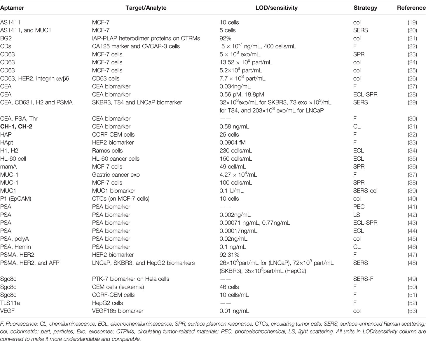
Table 1 Aptasensors reported for early cancer detection.
Fluorescence-Based Optical Aptasensors
Fluorescence is an optical approach commonly employed to construct aptasensors for their low costs, high sensitivity, operation simplicity, and high efficiency (54). Lei et al. introduced a “nanodoctor” known as “smart split aptamer-based activatable theranostic probe (SATP)” for in vivo cancer imaging that not only can activate fluorescence signals as a result of interaction with its analyte but also releases the drug (50). A graphene oxide-based label-free aptasensor for quantitative diagnostics of rare CCRF-CEM cells was employed by Xiao et al. CTCESA-based (cell-triggered cyclic enzymatic signal amplification) fluorescent aptasensors show a better selectivity and sensitivity for clinical and preclinical cancer detection in comparison to normal fluorescence-based aptasensors (32). Hamd-Ghadareh et al. constructed an antibody-ssDNA aptamer-based fluorescence sandwich-type ultrasensitive biosensor for CA125 early detection (22). A fluorescent “turn on” aptasensor based on fluorophore-labeled protein-aptamers and MoS2 (molybdenum disulfide) nanosheets was assembled by Zhao et al. for a highly sensitive and rapid CEA protein detection (27). In addition, Lai et al., Tan et al., and many others have recently published articles based on fluorescent aptasensors (51, 52). Another label-free, versatile “turn on” fluorescent aptasensor for HER2 early detection was fabricated by Zhang et al. (33). Exosomes for gastric cancer detection can be efficiently identified by a method designed by Huang et al. (37). A multiplex, competitive aptasensor based on fluorescent nanoparticles count was proposed by Pei et al. to detect various cancer biomarkers (30). Li et al. designed a platform not only efficient in exosomal protein profiling but also filling the technological innovation gap to facilitate the exosomal detection assays and shed light on methods for early detection of cancer such as liquid biopsy (47). An aptamer dependent fluorescence polarization technique was established by Zhang et al. that allows direct quantification of exosomes in human plasma without separation. It minimizes the operation time by simplifying the quantification without losing exosomes from the sample during separation (55).
Chemiluminescence Based Optical Aptasensors
A phenomenon where emitted light from a substance is not because of heat is called luminescence, which can be categorized based on the energy (trigger) sources into electrochemiluminescence (ECL) and chemiluminescence (CL). Both are widely used in the development of aptasensors employed in cancer biomarkers detection (56). Several CL aptasensors for early cancer detection have already been introduced (57). It is considered the most sensitive optical approach because of excellent results (58). Jie and Jie described a quantum dots (QDs) nanocluster based ECL signal probe with a great potential for early cancer diagnosis in clinical samples (34). Wang et al. designed a ratiometric dual-signaling electrochemiluminescence aptasensor exhibiting good reproducibility, high selectivity, and stability against their target cancer cells among non-target cancer cells (35). A simple, all in one, cost-effective, easy to operate, and portable medical platform to be used in hospitals and homes was designed by Khang et al. for early-stage breast cancer diagnostics (31). Another competitive, novel GO@AuNRs-GOD-SA nanoprobe based ECL aptasensor for PSA detection was demonstrated by Cao et al. (44). It is considered to be an excellent advancement for the detection of trace-level disease biomarkers. Kim et al. fabricated a fast biosensor, based on a dual aptamer system connected by a 5 adenine linker, to be used for rapid and accurate PSA quantification (46).
Surface Plasmon Resonance (SPR) Based Aptasensors
Surface plasmon resonance (SPR) based biosensing is advantageous because of label-free and kinetic studies exploring properties that are not offered by many other systems, giving direct and real-time detection of targets. SPR based assays are widely used to detect several types of cancer cells and biomarkers due to their high sensitivity (59). Li et al. proposed that MUC-1 aptamer (Mucin 1 protein) functionalized gold nanorods (AuNRs) have the ability to specially recognize MCF-7 cells via specific interactions that can be further processed by their unique localized surface plasmon resonance (LSPR) spectra. Their biosensor can be employed to detect human breast cancer at early stages (38). Electrochemical and SPR assays were combined by Guo et al. to examine the detection kinetics, which revealed significant outcomes for CEA detection by using their developed aptasensor based on the AgNCs@Apt@UiO-66 nanocomposite. Their SPR aptasensor is considered to have good performance, regenerate ability, selectivity, acceptable reproducibility, high sensitivity, and stability (28). A bi-functional (electrochemical-SPR) aptasensor with exceptional electrochemical action of MoS2QDs@g-C3N4 nanosheets and good SPR enactment of CS-AuNPs was combined by Duan et al. to make a 2D MoS2QDs@g-C3N4@CS-AuNPs nanocomposite. The authors expected satisfactory results of their sensor for the detection of cancer markers in clinical applications (43). A highly effective and sensitive SPR aptasensor for exosomal detection was invented by Wang et al., which is based on dual AuNPs assisted signal amplification. The approach finds promising practical applications in clinical and biological studies (23). Loyez et al. devised multiple narrowband resonances (near-infrared wavelength range), an all-fiber SPR aptasensor that takes five minutes for the detection of metastatic breast cancer cells. Supplementary addition of functionalized AuNPs enhances the 2-fold performance of the aptasensor (36).
Surface-Enhanced Raman Scattering (SERS)-Based Aptasensors
SERS spectroscopy has emerged as a promising tool for characterization in the field of nanoscience, i.e., widely investigated in cancer-related applications (60–62). The major advantages of SERS imaging are the mapping of a sample with a high spatial resolution (< 0.5 microns in the visible range) and the capability of multiplexed analysis (63). The ultrasensitive vibrational spectroscopic technique SERS can be used to detect several target molecules in a single experiment (60, 64). Ning et al. synthesized the aptamer-based SERS detection probes based on gold–silver–silver core-shell–shell nanotrepangs (GSSNTs) nanotags and magnetic beads for simultaneous detection of multiple cancer-related exosomes: the biomarkers (PSMA, Her2, and AFP proteins) for the prostate cancer cell line (LNCaP), breast cancer cell line (SKBR3), and hepatocellular cancer cell line (HepG2) (48). For the simultaneous detection of multiple kinds of exosomes (SKBR3, T84, and LNCaP), three different SERS probes types were designed to have three different types of Raman reporters and aptamers by Weng et al. and the principle of SERS detection (29). Liang et al. fabricated a series of aptamer-charged SERS probes (AS1411 and MUC1) for targeting cancer cells (MCF-7), and their results show the limit of detection (LOD) up to five cancer cells (20). Lately, the SERS spectroscopy method has been combined with other techniques for attaining maximum information from a sample. Li et al. fabricated a SERS-colorimetric dual-mode aptasensor for cancer biomarker MUC1 detection. The SERS probes were fabricated by using modified gold-silver core-shell nanoparticles with Raman reporters and the sequence of MUC1. The SERS probes report both SERS and colorimetric signals simultaneously (39). Bamrungsap et al. combined SERS and fluorescence nanotags assembled-system using a layer-by-layer process. The nanotags consisting of gold-silver nanorods, aptamers, and fluorophore-labeled aptamer for SERS signal generation, targeting ligands and fluorescence imaging, respectively. The dual-mode sensor system was successful for highly sensitive and specific cancer (cervical cancer) diagnostics (49).
Colorimetric Aptasensors
Colorimetric-based aptasensors have been used for the detection of disease biomarkers, due to their simplicity, ease of use, accessibility, and point-of-care detection (65, 66). The colorimetric method is a promising technique due to the possibility of detection by simply visual color change (67). Xu et al. developed a highly sensitive colorimetric-based aptasensor for the detection of exosomes obtained from breast and pancreatic cancer cells. In this novel approach, the specific detection was accelerated by horseradish peroxidase (HRP) accelerated dopamine polymerization, and sensitivity was enhanced by in situ deposition of polydopamine around exosomes particles (26). Shayesteh et al. developed a label-free colorimetric aptasensor, using poly-adenine aptamer and gold nanoparticles for sensitive detection of prostate-specific antigen (PSA) tumor marker. The concentration of PSA (5ng/ml) was detected by the naked eye with the color change (45). Dong et al. proposed a novel highly selective calorimetric based aptasensor strategy for detection of the vascular endothelial growth factor165 (VEGF165) in human serum (53). Colorimetric aptasensor based on gold nanoparticles aggregation developed by Borghei et al. was shown to have good results for the detection of rare circulating cancer cells. In this method, aptamer desorbed from solution due to specific binding of AS1411 aptamer to cancer cells, which resulted in the solution color change from purple to red (19). Xia et al. designed a fast and label-free DNA-capped-Single-Walled Carbon Nanotubes based aptasensor for exosomes detection through visible inspection. The exosomes were obtained from MCF-7 and breast cancer patient’s serum. The ability to detect exosomes in a homogenous system in combination with excluding complicated rinsing procedure is the key advantage of this proposed method (25). Wang et al. demonstrated single-stranded DNA (ssDNA) with graphitic carbon nitride nanosheets (g-C3N4 NSs) hybrid aptasensor for the colorimetric detection of exosomes originated by a breast cancer cell line (MCF-7) and a control cell line (MCF-10A). The intrinsic peroxidase-like activity of g-C3N4 NSs was enhanced by ssDNA (24). Shen et al. fabricated a colorimetric aptasensor for the detection and isolation of circulating tumor-related materials and is based on aptamer functionalized magnetic nanoparticles and endogenous alkaline phosphatase signal amplification. Their method exhibited great potential for clinical samples and is considered to find promising applications in point-of-care testing (21).
Other Optical Aptasensors
Terahertz radiation (THR) finds useful applications in cancer imaging (68). To overcome THR shortcomings regarding cancer cell and biomarkers detection, new technology has recently emerged known as terahertz chemical microscopy (TCM). However TCM has been reported to detect metastatic breast cancer cells, and only limited reports has been published (69). A photoelectrochemical (PEC) aptasensor fabricated by Zhou et al. (2017) is based on reduced graphene oxide-functionalized iron oxyhydroxide (FeOOH-rGO) as the photoactive material for the detection of PSA (prostate-specific antigen). Accuracy, specificity, and stability of the system were comparable to the commercially used PSA ELISA (enzyme-linked immunosorbent assay) kit (41). Liu et al. (2019) reported an ultrasensitive, activatable light-scattering (LS) method for PSA detection in real samples. The working mechanism of the aptasensor is based on target stimuli-responsive aggregation of AuNPs, which are responsible for lighting up the light-scattering signals (42).
Aptasensors in Clinical Diagnostics
An early cancer diagnosis is particularly an active research area because early detection can help to improve patient survival and disease prognosis. For this purpose, very sensitive and stable methods are needed for early cancer diagnosis (70). The main advantages of using aptasensors for clinical diagnostics are high selectivity and specificity, and low cost of production (71). The stability, ability of easy modification, and capability of fast development (animal-free) make nucleic acid aptamers detection methods widely functional compared to traditional antibody-based detection methods. And the nucleic acid aptamers can be used against a wide spectrum of targets (71, 72). The smaller size of aptamers compared to antibodies improves transport and tissue penetration (72). However one of the main disadvantages of the aptasensor is restricting each aptasensor to one marker or cell type (73). The development of an increasing number of published articles on aptamers for oncological diseases detection shows increased interest and progress in aptamer technology. Despite all the advantages, traditional immunoassays are still the dominant technology in the field of clinical diagnostics (70, 71). Nevertheless, this knowledge utilization for clinical practices has been challenging and the process has been very slow. There are many challenges if the aptasensor based sensing platform is to be used commercially. For example, improved signal-to-noise ratio and a high level of confidence in signal detection must be recognized (74). The compatibility of aptasensor assay with current equipment of diagnostics units is also an issue and is for the reason of the fragility of aptasensors (75). Many published reports investigated the sensing in buffer or diluted biological fluids; however, the goal should be the detection of biomarkers in a raw biological fluid. The cost of the whole sensing system should also be considered, for example, the cost of TCM components (laser) is expensive and uses a bulky femtosecond laser setup (11). Aptasensors after resolving all the above-discussed obstacles can be one of the most important early cancer detection tools (74).
Conclusion and Future Perspectives
Since early cancer detection has significant roles to increase available treatment options for the longer survival of patients, advances in various types of optical aptasensors for the detection of cancer cells and biomarkers or exosomes have been comprehensively summarized in this mini-review. Fluorescence-based label-free/labeled (e.g., FRET-based) aptasensors in combination with different nanomaterials/dyes, etc. as fluorophores, and quenchers to quench (change) the fluorescence properties as a result of specific interactions, have gained increasing attention. ECL and CL owing to their wide-ranging calibration and low background signals have recently been broadly exploited. Other types of optical aptasensors based on SPR, TCM, and SERS, etc. are also highly recommended for early-stage cancer diagnostics. Colorimetric methods combined with several different latest strategies (e.g., nanoparticles, etc.) implicate the simplest aptasensors and can be analyzed easily with the naked eye.
Several optical aptasensors reported for cancer early detection exhibit good performance in terms of selectivity and sensitivity, yet commercially available aptasensors just appear as the tips of some icebergs when we compare them to the mighty academic literature available in this area. Some new methods are still in lab trials with the early results favoring their commercial applications outside labs. However, some important technological issues and challenges still need to be addressed or improved. First, only a limited number of good specificity/sensitivity aptamers are available against a certain type of cancer cells/biomarkers, and more aptamers need to be screened that could target multiple cancer biomarkers without any complexity or off-target recognition in biological samples. Second, aptasensors need further investigations for clinical applications with real, undiluted (raw) biological samples with a primary focus on aptamer specificity, high sensitivity, cost-effectiveness, and simple operation. Overall, it is evident that the full bloom of optical aptasensor technology for cancer diagnostics is still on the way to a bright future.
Author Contributions
All authors listed have made a substantial, direct, and intellectual contribution to the work and approved it for publication.
Funding
We acknowledge the financial support from “the Fundamental Research Funds for the Central Universities (No. YD2070002013)” and the National Natural Science Foundation of China (No. 31570755).
Conflict of Interest
The authors declare that the research was conducted in the absence of any commercial or financial relationships that could be construed as a potential conflict of interest.
References
1. Cancer molecular markers: A guide to cancer detection and management. Semin Cancer Biol (2018) 52(1):39–55. doi: 10.1016/j.semcancer.2018.02.002
2. Hanahan D, Weinberg RA. Hallmarks of cancer: the next generation. Cell (2011) 144(5):646–74. doi: 10.1016/j.cell.2011.02.013
3. Crosby D, Lyons N, Greenwood E, Harrison S, Hiom S, Moffat J, et al. A roadmap for the early detection and diagnosis of cancer. Lancet Oncol (2020) 21(11):1397–9. doi: 10.1016/S1470-2045(20)30593-3
4. Chen X, Gole J, Gore A, He Q, Lu M, Min J, et al. Non-invasive early detection of cancer four years before conventional diagnosis using a blood test. Nat Commun (2020) 11(1):1–10. doi: 10.1038/s41467-020-17316-z
5. Liu M, Yu X, Chen Z, Yang T, Yang D, Liu Q, et al. Aptamer selection and applications for breast cancer diagnostics and therapy. J Nanobiotechnol (2017) 15(1):1–16. doi: 10.1186/s12951-017-0311-4
6. Bing T, Shen L, Wang J, Wang L, Liu X, Zhang N, et al. Aptameric Probe Specifically Binding Protein Heterodimer Rather Than Monomers. Adv Sci (2019) 6(11):1900143. doi: 10.1002/advs.201900143
7. Bing T, Wang J, Shen L, Liu X, Shangguan D. Prion Protein Targeted by a Prostate Cancer Cell Binding Aptamer, a Potential Tumor Marker? ACS Appl Bio Mat (2020) 3(5):2658–65. doi: 10.1021/acsabm.0c00024
8. Tuerk C, Gold L. Systematic evolution of ligands by exponential enrichment: RNA ligands to bacteriophage T4 DNA polymerase. Science (1990) 249(4968):505–10. doi: 10.1126/science.2200121
9. Bock LC, Griffin LC, Latham JA, Vermaas EH, Toole JJ. Selection of single-stranded DNA molecules that bind and inhibit human thrombin. Nature (1992) 355(6360):564–6. doi: 10.1038/355564a0
10. Shangguan D, Li Y, Tang Z, Cao ZC, Chen HW, Mallikaratchy P, et al. Aptamers evolved from live cells as effective molecular probes for cancer study. Proc Natl Acad Sci (2006) 103(32):11838–43. doi: 10.1073/pnas.0602615103
11. Hassan EM, DeRosa MC. Recent advances in cancer early detection and diagnosis: Role of nucleic acid based aptasensors. TrAC Trends Anal Chem (2020) 124:115806. doi: 10.1016/j.trac.2020.115806
12. Bing T, Zhang N, Shangguan D. Cell-SELEX, an Effective Way to the Discovery of Biomarkers and Unexpected Molecular Events. Adv Biosyst (2019) 3(12):1900193. doi: 10.1002/adbi.201900193
13. Eivazzadeh-Keihan R, Pashazadeh-Panahi P, Baradaran B, Maleki A, Hejazi M, Mokhtarzadeh A, et al. Recent advances on nanomaterial based electrochemical and optical aptasensors for detection of cancer biomarkers. TrAC Trends Anal Chem (2018) 100:103–15. doi: 10.1016/j.trac.2017.12.019
14. Feng C, Dai S, Wang L. Optical aptasensors for quantitative detection of small biomolecules: A review. Biosens Bioelectron (2014) 59:64–74. doi: 10.1016/j.bios.2014.03.014
15. Nikhil B, Pawan J, Nello F, Pedro E. Introduction to biosensors. Essays Biochem (2016) 60(1):1–8. doi: 10.1042/EBC20150001
16. Lim Y, Kouzani A, Duan W. Aptasensors: a review. J Biomed Nanotechnol (2010) 6(2):93–105. doi: 10.1166/jbn.2010.1103
17. Sadeghi AS, Ansari N, Ramezani M, Abnous K, Mohsenzadeh M, Taghdisi SM, et al. Optical and electrochemical aptasensors for the detection of amphenicols. Biosens Bioelectron (2018) 118:137–52. doi: 10.1016/j.bios.2018.07.045
18. Pashazadeh P, Mokhtarzadeh A, Hasanzadeh M, Hejazi M, Hashemi M, de la Guardia M. Nano-materials for use in sensing of salmonella infections: recent advances. Biosens Bioelectron (2017) 87:1050–64. doi: 10.1016/j.bios.2016.08.012
19. Borghei YS, Hosseini M, Dadmehr M, Hosseinkhani S, Ganjali MR, Sheikhnejad R. Visual detection of cancer cells by colorimetric aptasensor based on aggregation of gold nanoparticles induced by DNA hybridization. Anal Chim Acta (2016) 904:92–7. doi: 10.1016/j.aca.2015.11.026
20. Liang D, Jin Q, Yan N, Feng J, Wang J, Tang X. SERS Nanoprobes in Biologically Raman Silent Region for Tumor Cell Imaging and In Vivo Tumor Spectral Detection in Mice. Adv Biosyst (2018) 2:1800100. doi: 10.1002/adbi.201800100
21. Shen L, Jia K, Bing T, Zhang Z, Zhen X, Liu X, et al. Detection of circulating tumor-related materials by aptamer capturing and endogenous enzyme-signal amplification. Anal Chem (2020) 92(7):5370–8. doi: 10.1021/acs.analchem.0c00051
22. Hamd-Ghadareh S, Salimi A, Fathi F, Bahrami S. An amplified comparative fluorescence resonance energy transfer immunosensing of CA125 tumor marker and ovarian cancer cells using green and economic carbon dots for bio-applications in labeling, imaging and sensing. Biosens Bioelectron (2017) 96:308–16. doi: 10.1016/j.bios.2017.05.003
23. Wang Q, Zou L, Yang X, Liu X, Nie W, Zheng Y, et al. Direct quantification of cancerous exosomes via surface plasmon resonance with dual gold nanoparticle-assisted signal amplification. Biosens Bioelectron (2019) 135:129–36. doi: 10.1016/j.bios.2019.04.013
24. Wang YM, Liu JW, Adkins GB, Shen W, Trinh MP, Duan LY, et al. Enhancement of the Intrinsic Peroxidase-Like Activity of Graphitic Carbon Nitride Nanosheets by ssDNAs and Its Application for Detection of Exosomes. Anal Chem (2017) 89:12327–33. doi: 10.1021/acs.analchem.7b03335
25. Xia Y, Liu M, Wang L, Yan A, He W, Chen M, et al. A visible and colorimetric aptasensor based on DNA-capped single-walled carbon nanotubes for detection of exosomes. Biosens Bioelectron (2017) 92:8–15. doi: 10.1016/j.bios.2017.01.063
26. Xu L, Chopdat R, Li D, Al-Jamal KT. Development of a simple, sensitive and selective colorimetric aptasensor for the detection of cancer-derived exosomes. Biosens Bioelectron (2020) 169:112576. doi: 10.1016/j.bios.2020.112576
27. Zhao L, Cheng M, Liu G, Lu H, Gao Y, Yan X, et al. A fluorescent biosensor based on molybdenum disulfide nanosheets and protein aptamer for sensitive detection of carcinoembryonic antigen. Sensors Actuators B: Chem (2018) 273:185–90. doi: 10.1016/j.snb.2018.06.004
28. Guo C, Su F, Song Y, Hu B, Wang M, He L, et al. Aptamer-templated silver nanoclusters embedded in zirconium metal–organic framework for bifunctional electrochemical and SPR aptasensors toward carcinoembryonic antigen. ACS Appl Mat Interfaces (2017) 9(47):41188–99. doi: 10.1021/acsami.7b14952
29. Weng Z, Zong S, Wang Y, Li N, Li L, Lu J, et al. Screening and multiple detection of cancer exosomes using an SERS-based method. Nanoscale (2018) 10:9053–62. doi: 10.1039/C7NR09162A
30. Pei X, Wu X, Xiong J, Wang G, Tao G, Ma Y, et al. Competitive aptasensor for the ultrasensitive multiplexed detection of cancer biomarkers by fluorescent nanoparticle counting. Analyst (2020) 145(10):3612–9. doi: 10.1039/D0AN00239A
31. Khang H, Cho K, Chong S, Lee JH. All-in-one dual-aptasensor capable of rapidly quantifying carcinoembryonic antigen. Biosens Bioelectron (2017) 90:46–52. doi: 10.1016/j.bios.2016.11.043
32. Xiao K, Liu J, Chen H, Zhang S, Kong J. A label-free and high-efficient GO-based aptasensor for cancer cells based on cyclic enzymatic signal amplification. Biosens Bioelectron (2017) 91:76–81. doi: 10.1016/j.bios.2016.11.057
33. Zhang M, Gao G, Ding Y, Deng C, Xiang J, Wu H. A fluorescent aptasensor for the femtomolar detection of epidermal growth factor receptor-2 based on the proximity of G-rich sequences to Ag nanoclusters. Talanta (2019) 199:238–43. doi: 10.1016/j.talanta.2019.02.014
34. Jie G, Jie G. Sensitive electrochemiluminescence detection of cancer cells based on a CdSe/ZnS quantum dot nanocluster by multibranched hybridization chain reaction on gold nanoparticles. RSC Adv (2016) 6(29):24780–5. doi: 10.1039/C6RA00750C
35. Wang Y-Z, Hao N, Feng Q-M, Shi H-W, Xu J-J, Chen H-Y. A ratiometric electrochemiluminescence detection for cancer cells using g-C3N4 nanosheets and Ag–PAMAM–luminol nanocomposites. Biosens Bioelectron (2016) 77:76–82. doi: 10.1016/j.bios.2015.08.057
36. Loyez M, Hassan EM, Lobry M, Liu F, Caucheteur C, Wattiez R, et al. Rapid detection of circulating breast cancer cells using a Multiresonant optical fiber Aptasensor with plasmonic amplification. ACS Sensors (2020) 5(2):454–63. doi: 10.1021/acssensors.9b02155
37. Huang R, He L, Li S, Liu H, Jin L, Chen Z, et al. A simple fluorescence aptasensor for gastric cancer exosome detection based on branched rolling circle amplification. Nanoscale (2020) 12(4):2445–51. doi: 10.1039/C9NR08747H
38. Li Y, Zhang Y, Zhao M, Zhou Q, Wang L, Wang H, et al. A simple aptamer-functionalized gold nanorods based biosensor for the sensitive detection of MCF-7 breast cancer cells. Chem Commun (2016) 52(20):3959–61. doi: 10.1039/C6CC01014H
39. Li N, Zong S, Zhang Y, Wang Z, Wang Y, Zhu K, et al. A SERS-colorimetric dual-mode aptasensor for the detection of cancer biomarker MUC1. Anal Bioanal Chem (2020) 412:5707–18. doi: 10.1007/s00216-020-02790-7
40. Zhu L, Feng X, Yang S, Wang J, Pan Y, Ding J, et al. Colorimetric detection of immunomagnetically captured rare number CTCs using mDNA-wrapped single-walled carbon nanotubes. Biosens Bioelectron (2020) 172:112780. doi: 10.1016/j.bios.2020.112780
41. Zhou Q, Lin Y, Shu J, Zhang K, Yu Z, Tang D. Reduced graphene oxide-functionalized FeOOH for signal-on photoelectrochemical sensing of prostate-specific antigen with bioresponsive controlled release system. Biosens Bioelectron (2017) 98:15–21. doi: 10.1016/j.bios.2017.06.033
42. Liu G, Feng D-Q, Li Z, Feng Y. Target-activatable gold nanoparticle-based aptasensing for protein biomarkers using stimuli-responsive aggregation. Talanta (2019) 192:112–7. doi: 10.1016/j.talanta.2018.08.034
43. Duan F, Zhang S, Yang L, Zhang Z, He L, Wang M. Bifunctional aptasensor based on novel two-dimensional nanocomposite of MoS2 quantum dots and g-C3N4 nanosheets decorated with chitosan-stabilized Au nanoparticles for selectively detecting prostate specific antigen. Anal Chim Acta (2018) 1036:121–32. doi: 10.1016/j.aca.2018.06.070
44. Cao J-T, Yang J-J, Zhao L-Z, Wang Y-L, Wang H, Liu Y-M, et al. Graphene oxide@ gold nanorods-based multiple-assisted electrochemiluminescence signal amplification strategy for sensitive detection of prostate specific antigen. Biosens Bioelectron (2018) 99:92–8. doi: 10.1016/j.bios.2017.07.050
45. Shayesteh OH, Ghavami R. A novel label-free colorimetric aptasensor for sensitive determination of PSA biomarker using gold nanoparticles and a cationic polymer in human serum. Spectrochim Acta - Part A: Mol Biomol Spectrosc (2020) 226:117644. doi: 10.1016/j.saa.2019.117644
46. Kim M, Kim K, Lee JH. A cost-effective and rapid aptasensor with chemiluminescence detection for the early diagnosis of prostate cancer. Microchem J (2020) 155(2020):104763. doi: 10.1016/j.microc.2020.104763
47. Li B, Liu C, Pan W, Shen J, Guo J, Luo T, et al. Facile fluorescent aptasensor using aggregation-induced emission luminogens for exosomal proteins profiling towards liquid biopsy. Biosens Bioelectron (2020) 168:112520. doi: 10.1016/j.bios.2020.112520
48. Ning CF, Wang L, Tian YF, Yin BC, Ye BC. Multiple and sensitive SERS detection of cancer-related exosomes based on gold-silver bimetallic nanotrepangs. Analyst (2020) 145:2795–804. doi: 10.1039/C9AN02180A
49. Bamrungsap S, Treetong A, Apiwat C, Wuttikhun T, Dharakul T. SERS-fluorescence dual mode nanotags for cervical cancer detection using aptamers conjugated to gold-silver nanorods. Microchim Acta (2016) 183:249–56. doi: 10.1007/s00604-015-1639-9
50. Lei Y, Tang J, Shi H, Ye X, He X, Xu F, et al. Nature-inspired smart DNA nanodoctor for activatable in vivo cancer imaging and in situ drug release based on recognition-triggered assembly of split aptamer. Anal Chem (2016) 88(23):11699–706. doi: 10.1021/acs.analchem.6b03283
51. Tan J, Lai Z, Zhong L, Zhang Z, Zheng R, Su J, et al. A graphene oxide-based fluorescent aptasensor for the turn-on detection of CCRF-CEM. Nanoscale Res Lett (2018) 13(1):1–8. doi: 10.1186/s11671-018-2525-2
52. Lai Z, Tan J, Wan R, Tan J, Zhang Z, Hu Z, et al. An ‘activatable’aptamer-based fluorescence probe for the detection of HepG2 cells. Oncol Rep (2017) 37(5):2688–94. doi: 10.3892/or.2017.5527
53. Dong J, He L, Wang Y, Yu F, Yu S, Liu L, et al. A highly sensitive colorimetric aptasensor for the detection of the vascular endothelial growth factor in human serum. Spectrochim Acta - Part A: Mol Biomol Spectrosc (2020) 226:117622. doi: 10.1016/j.saa.2019.117622
54. Musumeci D, Platella C, Riccardi C, Moccia F, Montesarchio D. Fluorescence sensing using DNA aptamers in cancer research and clinical diagnostics. Cancers (2017) 9(12):174. doi: 10.3390/cancers9120174
55. Zhang Z, Tang C, Zhao L, Xu L, Zhou W, Dong Z, et al. Aptamer-based fluorescence polarization assay for separation-free exosome quantification. Nanoscale (2019) 11(20):10106–13. doi: 10.1039/C9NR01589B
56. Kou X, Zhang X, Shao X, Jiang C, Ning L. Recent advances in optical aptasensor technology for amplification strategies in cancer diagnostics. Anal Bioanal Chem (2020) 1–15. doi: 10.1007/s00216-020-02774-7
57. Eivazzadeh-Keihan R, Pashazadeh P, Hejazi M, de la Guardia M, Mokhtarzadeh A. Recent advances in nanomaterial-mediated bio and immune sensors for detection of aflatoxin in food products. TrAC Trends Anal Chem (2017) 87:112–28. doi: 10.1016/j.trac.2016.12.003
58. Park L, Kim J, Lee JH. Role of background observed in aptasensor with chemiluminescence detection. Talanta (2013) 116:736–42. doi: 10.1016/j.talanta.2013.07.072
59. Nguyen HH, Park J, Kang S, Kim M. Surface plasmon resonance: a versatile technique for biosensor applications. Sensors (2015) 15(5):10481–510. doi: 10.3390/s150510481
60. Guerrini L, Alvarez-Puebla RA. Surface-enhanced raman spectroscopy in cancer diagnosis, prognosis and monitoring. Cancers (2019) 11(6):748.
61. Vendrell M, Maiti KK, Dhaliwal K, Chang YT. Surface-enhanced Raman scattering in cancer detection and imaging Trends Biotechnol (2013) 31(4):249–57.
62. Moisoiu V, Stefancu A, Gulei D, Boitor R, Magdo L, Raduly L, et al. SERS-based differential diagnosis between multiple solid malignancies: Breast, colorectal, lung, ovarian and oral cancer. Int J Nanomed (2019) 14:6165–78. doi: 10.2147/IJN.S198684
63. Blanco-Formoso M, Alvarez-Puebla RA. Cancer diagnosis through sers and other related techniques. Int J Mol Sci (2020) 21(6):2253. doi: 10.3390/ijms21062253
64. Dougan JA, Faulds K. Surface enhanced Raman scattering for multiplexed detection. Analyst (2012) 137(3):545–54.
65. De La Rica R, Stevens MM. Plasmonic ELISA for the ultrasensitive detection of disease biomarkers with the naked eye. Nat Nanotechnol (2012) 7:821–4. doi: 10.1038/nnano.2012.186
66. Wang D, Guo R, Wei Y, Zhang Y, Zhao X, Xu Z. Sensitive multicolor visual detection of telomerase activity based on catalytic hairpin assembly and etching of Au nanorods. Biosens Bioelectron (2018) 122:247–53. doi: 10.1016/j.bios.2018.09.064
67. Akshaya K, Arthi C, Pavithra AJ, Poovizhi P, Antinate SS, Hikku GS, et al. Bioconjugated gold nanoparticles as an efficient colorimetric sensor for cancer diagnostics. Photodiagnosis Photodyn Ther (2020) 30:101699.
68. Walther M, Fischer BM, Ortner A, Bitzer A, Thoman A, Helm H. Chemical sensing and imaging with pulsed terahertz radiation. Anal Bioanal Chem (2010) 397(3):1009–17. doi: 10.1007/s00216-010-3672-1
69. Hassan EM, Mohamed A, DeRosa MC, Willmore WG, Hanaoka Y, Kiwa T, et al. High-sensitivity detection of metastatic breast cancer cells via terahertz chemical microscopy using aptamers. Sensors Actuators B: Chem (2019) 287:595–601. doi: 10.1016/j.snb.2019.02.019
70. Hong P, Li W, Li J. Applications of aptasensors in clinical diagnostics. Sensors (2012) 12(2):1181–93.
71. Hong P, Li W, Li J Applications of aptasensors in clinical diagnostics Sensors (2012) 12(2):1181–93.
72. Mairal T, Cengiz Özalp V, Lozano Sánchez P, Mir M, Katakis I, O’Sullivan CK. Aptamers: Molecular tools for analytical applications. Anal Bioanal Chem (2008) 390:989–1007. doi: 10.1007/s00216-007-1346-4
73. Thiviyanathan V, Gorenstein DG. Aptamers and the next generation of diagnostic reagents. PROTEOMICS–Clin Appl (2012) 6(11-12):563–73.
74. Liu LS, Wang F, Ge Y, Lo PK. Recent Developments in Aptasensors for Diagnostic Applications. ACS Appl Mat Interfaces (2020). doi: 10.1021/acsami.0c14788
Keywords: optical aptasensors, early cancer diagnostics, cancer biomarkers, fluorescence aptasensors, chemiluminescence, SERS cancer aptasensors, colorimetric aptasensors, SPR aptasensors
Citation: Zahra Qua, Khan QA and Luo Z (2021) Advances in Optical Aptasensors for Early Detection and Diagnosis of Various Cancer Types. Front. Oncol. 11:632165. doi: 10.3389/fonc.2021.632165
Received: 22 November 2020; Accepted: 04 January 2021;
Published: 25 February 2021.
Edited by:
Yingyan Yu, Shanghai Jiao Tong University, ChinaReviewed by:
Tao Bing, Institute of Chemistry (CAS), ChinaXinhui Lou, Capital Normal University, China
Copyright © 2021 Zahra, Khan and Luo. This is an open-access article distributed under the terms of the Creative Commons Attribution License (CC BY). The use, distribution or reproduction in other forums is permitted, provided the original author(s) and the copyright owner(s) are credited and that the original publication in this journal is cited, in accordance with accepted academic practice. No use, distribution or reproduction is permitted which does not comply with these terms.
*Correspondence: Zhaofeng Luo, bHpmQHVzdGMuZWR1LmNu