- 1Department of First Clinical Medical College, Shandong University of Traditional Chinese Medicine, Shandong Provincial Qianfoshan Hospital, Jinan, Shandong, China
- 2Department of Oncology, The First Affiliated Hospital of Shandong First Medical University and Shandong Provincial Qianfoshan Hospital, Shandong Lung Cancer Institute, Jinan, China
- 3Department of Gastroenterology, Shandong Provincial Hospital Affiliated to Shandong First Medical University, Jinan, China
- 4Department of Radiology, People’s Hospital of Guangrao County, Dongying, Shandong, China
- 5Shandong Medicine and Health Key Laboratory of Cardiac Electrophysiology and Arrhythmia, Department of Cardiology, The First Affiliated Hospital of Shandong First Medical University & Shandong Provincial Qianfoshan Hospital, Jinan, Shandong, China
Objective: This retrospective study was conducted to delineate our experience in managing perioperative antithrombotic agents in patients receiving antithrombotic therapy underwent percutaneous biopsy and microwave ablation (B+MWA) for lung ground-glass opacities (GGOs).
Methods: The study comprised 67 patients with GGOs who receiving antithrombotic therapy underwent B+MWA sessions from January 1, 2020, to May 31, 2022. During the perioperative period, patients who received rivaroxaban as a bridging drug were assigned to Group A, and who interrupted the antithrombotic therapy were assigned to Group B. Information about the technical success rate, positive biopsy rate, local control rates, and major bleeding and thrombotic complications were collected and analyzed.
Results: Group A comprised 36 patients (19 males; mean age, 67.97 ± 8.49 years), while Group B comprised 31 patients (12 males; mean age, 65.48 ± 4.32 years). The technical success rate was 100%. The positive biopsy rates were 94.44% and 96.77%, respectively. In group A and B, the overall local control rates at 6, 18, and 24 months were 100.0% vs. 100.0%, 94.44% (34/36) vs. 96.77% (30/31), and 86.11% (31/36) vs. 87.10% (27/31), with no significant difference between the two groups (p = 0.2156). During the perioperative period, a single case of lower extremity venous thrombosis was identified in Group A, while three cases of lower extremity venous thrombosis, one case of new-onset cerebral infarction, and one case of new-onset pulmonary embolism were identified in Group B, with no statistically significant difference in the overall incidence of bleeding and thrombotic complications between the two groups.
Conclusions: Compared with direct interruption of antithrombotic therapy, the use of rivaroxaban in the perioperative period of B+MWA in patients with GGOs who are receiving antithrombotic therapy can reduce the incidence of severe thrombotic complications without increasing the risk of bleeding, with a satisfactory effectiveness.
Highlights
The significant finding of the study is that bridging oral anticoagulant therapy along with synchronous CT-guided percutaneous biopsy and microwave ablation should be considered as an alternative treatment for lung ground-glass opacities in patients receiving antithrombotic therapy. This study adds the research data of microwave ablation for patients receiving antithrombotic therapy.
Introduction
Ground-glass opacity (GGO) is imaging manifestations on computed tomography (CT) that appear as obscure shadows covering the underlying bronchial structure and vascular structure of the lung. GGO is a nonspecific imaging parameter for early-stage pulmonary cancer (1, 2). Highly suspicious lung ground-glass opacities (HSML-GGOs) are generally defined as nonsolid pulmonary nodules >6mm or with increasing solid components that persist during a 6–9 months follow-up, which are difficult to detect on chest X-ray. However, with the wide application of computed tomography (CT) in recent decades, HSML-GGOs have attracted increasing attention (3, 4). HSML-GGOs usually have irregular margins, complex density, and spiculate or lobular outline (5). These imaging features make it difficult to distinguish malignant GGOs from some benign nodules, such as tuberculoma or inflammatory pseudotumor (6, 7). Moreover, due to the biological characteristics of GGOs, traditional imaging examinations such as enhanced CT, magnetic resonance imaging, Positron Emission Tomography-CT, etc., cannot well distinguish benign and malignant GGOs (5, 8). Currently, while the management of HSML-GGOs remains a topic of contention and is the subject of ongoing research, long-term follow-up data indicate that clinical guidelines recommend a tailored approach. For certain patients, this involves a period of surveillance, whereas others may require tissue sampling, including resection, based on identified risk factors (9, 10). The precise diagnosis of HSML-GGOs depends on pathological examination, which was obtained by surgical resection and percutaneous pulmonary biopsy (11–13). Percutaneous pulmonary biopsy technique has seen significant advancement, yet parenchymal hemorrhage persists as an inescapable complication (14, 15). Unlike solid nodules, the occurrence of pulmonary hemorrhage tends to have a greater impact on the visualization and localization of GGOs on CT scans. This interference can lead to unsuccessful procedure outcomes or an elevated false-negative rate in pathology results, a concern that is particularly acute for patients undergoing antithrombotic therapy. However, for patients necessitating antithrombotic treatment, prematurely discontinuing antithrombotic therapy can precipitate life-threatening thrombotic complications, particularly in elderly patients with malignant tumors who are more likely to develop thrombotic complications because of the hypercoagulable state (16, 17). Consequently, judicious management of anticoagulant medications during the period of B+MWA for GGOs is of paramount importance (18, 19). A strategy involving rivaroxaban as a bridging antithrombotic agent has been extensively investigated in clinical practice (20, 21). The objective of this study is to assess the safety and efficacy of rivaroxaban bridging in patients with GGOs underwent CT-guided percutaneous B+MWA procedure. This study will provide fresh evidence to inform the use of rivaroxaban in patients undergoing percutaneous pulmonary biopsy while on antithrombotic therapy, thereby guiding clinical decision-making.
Materials and methods
Patients
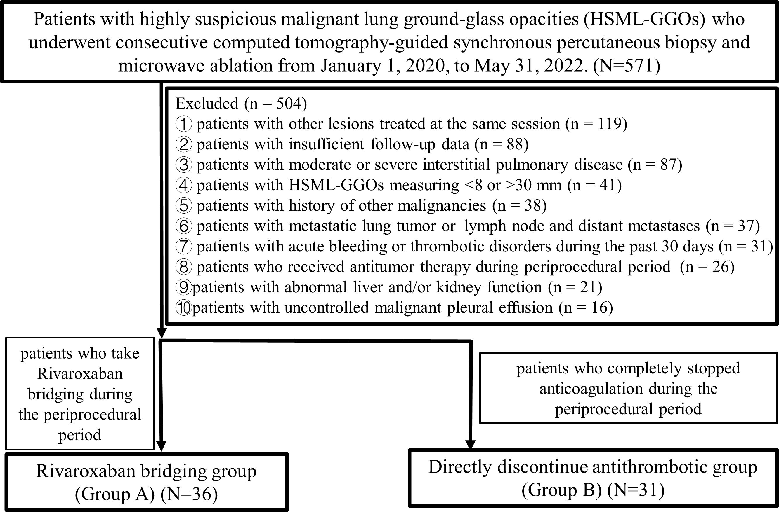
This study consisted of 486 patients on antithrombotic therapy with GGOs who underwent 571 consecutive B+MWA treatments from January 1, 2020, to May 31, 2022. These patients were unsuitable candidates for surgery or radiotherapy. The inclusion criteria were as follows: 1) patients with ground-glass nodule diagnosed as HSML-GGOs; 2) those with GGOs measuring 8–30 mm; and 3) those with an Eastern Cooperative Oncology Group performance status (ECOG PS) score of ≤ 2. The exclusion criteria were as follows: 1) patients with other lesions treated at the same session; 2) those with metastatic lung tumor or primary lung cancer with lymph node and distant metastases; 3) those with abnormal liver and/or kidney function; 4) those with acute bleeding or thrombotic disorders during the past 30 days; 5) those with moderate or severe interstitial pulmonary disease; 6) those with uncontrolled malignant pleural effusion; 7) those who received other antitumor therapy during the peri-procedural period; 8) those with history of other malignancies; and 9) those who were lost to follow-up. In total, 67 patients (comprising 31 males and 36 females; with a mean age of 66.82 ± 7.62 years, ranging from 50 to 79 years) were enrolled in the study. The process of patient selection is delineated in Figure 1. Patients who received rivaroxaban bridging during their hospitalization for B+MWA treatment were allocated to Group A, while those who ceased antithrombotic therapy directly during their hospitalization were placed in Group B. Prior to treatment, all patients underwent a multidisciplinary consultation involving specialists from thoracic surgery, oncology, respiratory diseases, radiotherapy, interventional radiology, imaging, and pathology. The treatment plan was subsequently formulated in accordance with expert consensus (22).
This study complied with the standards of the Declaration of Helsinki and obtained approval from the Institutional Ethics Committee of Shandong Provincial Qianfoshan Hospital, Jinan, Shandong, China. The Institutional Review Board waived the requirement for informed consent since this was a retrospective study.
Synchronous percutaneous biopsy and microwave ablation
The procedure of CT-guided B+MWA for patients with pulmonary nodules has been extensively documented in previous studies (23, 24). The approach here is to place the ablation antenna in a satisfactory position and then place the biopsy coaxial needle and the biopsy needle according to the preoperative plan. Ablation is strategically timed to facilitate microvascular hemostasis and thrombosis within the ablation area, thereby mitigating the risk of bleeding that may arise during the subsequent biopsy. Should the biopsy inadvertently result in excessive bleeding that compromises the visibility of the GGOs on CT imaging, the accuracy of the antenna’s position is reassessed based on its relative location to the preoperative reference markers, and ablation is then carried out accordingly. Specifically, it is divided into the following steps: 1). After local anesthesia with 5–10 mL of 2% lidocaine, a 19-G (Gauge) antenna was inserted, followed by a coaxial guiding needle (17 G, GMT Medical) along the designated path or the ablation antenna; 2). Ablation was performed for 1–2 min at 30–50 W power. CT was performed again to observe changes at the ablation site. The position of the needle tip was confirmed before the 18-G biopsy needle was inserted along the coaxial catheter, and samples were collected. If severe pulmonary bleeding was observed, ablation with a power of 30–50 W was applied for 1–2 min to promote hemostasis; 3). After performing the B+MWA, as described previously, optimal circumferential ablated margins of ≥5 mm for GGO margins were achieved; 4). The needle tract was ablated to promote hemostasis and reduce metastasis. Vessels with a diameter > 2 mm distributed along the ablation antenna path were ablated for 1–2 min at the intersection of the path and the vessel to reduce bleeding. It was not advisable to have vessels >3 mm in diameter within the preprocedural designed puncture path.
All participants were advised to bed overnight. The important observations included: 1) abnormal hypotension and tachycardia (systolic pressure < 90 mmHg, heart rate >100 beats/min); 2) unusual hemoptysis or any other manifestations or symptoms suggestive of bleeding, such as severe chest pain, dyspnea, or vertigo; 3) In such instances, immediate hemoglobin level assessment and chest CT scan were conducted, while electrocardiogram and blood pressure monitoring continued. Appropriate fluid resuscitation and hemostatic measures were initiated, and blood transfusion was considered if hemodynamic instability persisted; 4) A routine postoperative chest CT scan was scheduled 24 hours post-surgery. If the bleeding range did not diminish within 24 hours postoperatively, a CT scan was repeated every three days postoperatively until a trend of hemorrhage reduction was evident.
Management of anticoagulants in the perioperative period
The perioperative period was defined as spanning from 5 days prior to the B+MWA procedure to 1-month post-procedure. Within this timeframe, we exclusively monitored for procedure-related bleeding and thrombotic complications. All patients discontinued all antithrombotic medications 5 days prior to procedure. Patients in group A were administered rivaroxaban (10 mg once a day) bridging, whereas patients in group B did not receive any anticoagulants. The original antithrombotic therapy could be reinitiated in patients who met the following criteria 3 days post-procedure: 1) A chest CT scan performed 24 hours or 3 days post-procedure demonstrated a reduction in the bleeding range; 2) The patient did not present with hemoptysis; 3) The patient exhibited no other new bleeding complications, such as epistaxis, gingival bleeding, visceral bleeding, or central bleeding. Patients who restarted antithrombotic therapy were to be monitored closely throughout the perioperative period. In the event of bleeding complications, immediate further examination and treatment would be pursued to avert fatal bleeding. Should thrombotic complications arise during the perioperative period, consultations with relevant departments would be solicited, and appropriate medication adjustments or treatments would be implemented.
Response criteria and follow-up
Technical success was defined as completion of MWA with sample collection as planned before procedure. Positive biopsy rate was defined as diagnostic pathological findings, including atypical adenomatoid hyperplasia (AAH), adenocarcinoma in situ (AIS), inflammatory tissue, and malignant tissue. Chest enhanced CT was performed at 1-, 3-, 6-, 12-, 18- and 24-months after procedure. Complete ablation rate was used to describe lesion control rate. The severity of complications was evaluated according to the National Cancer Institute Common Terminology Criteria for Adverse Events (CTCAE; version 5.0), and grade ≥3 complications were defined as major complications (25).
Statistical analysis
Statistical analyses were performed using SPSS version 20.0. Categorical variables were compared using Fisher’s exact test, whereas continuous variables were compared using the unpaired Student’s t-test (parametric) or Mann-Whitney U test (nonparametric). P values <0.05 in a two-tailed test were considered statistically significant. All data used in this study were recorded for reference purposes.
Results
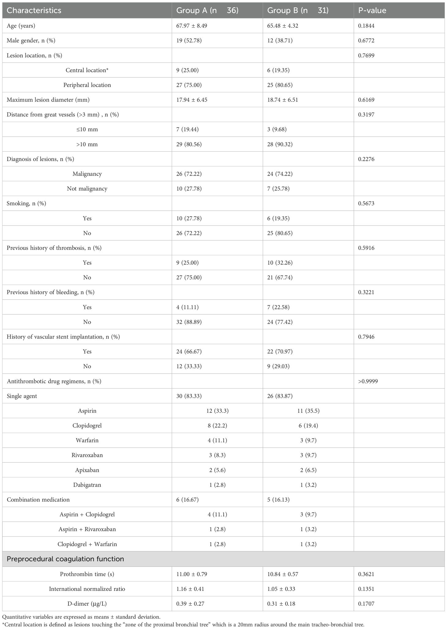
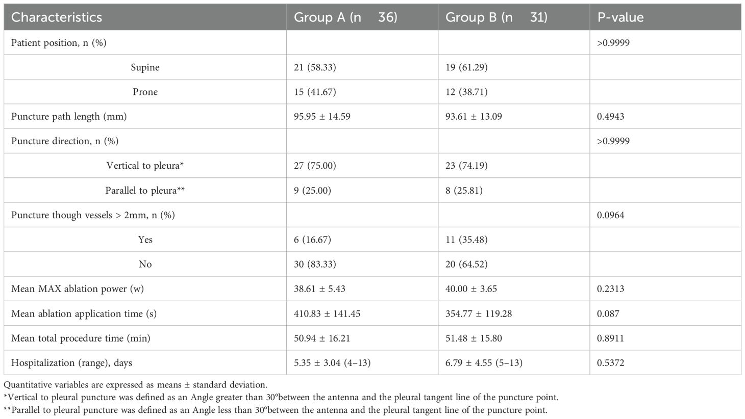
Table 1 shows the baseline characteristics of the patients. There were 36 patients (19 men; mean age, 67.97 ± 8.49 years) in Group A and 31 patients (12 men; mean age, 65.48 ± 4.32 years) in Group B. There were no significant differences between the two groups in terms of the age, gender, lesion’s location, lesion’s diameter, distance of lesion to great vessels, lesion’s diagnosis, smoking rate, previous history of thrombosis and bleeding, history of vascular stent implantation, and preprocedural coagulation function. The preprocedural coagulation function was defined as the coagulation profile assessed within 5 days before the procedure. Table 2 shows the procedural characteristics. There are no significant differences in patient position, puncture path length, puncture direction, cases of puncture though vessel’s diameter > 2mm, mean maximum ablation power, mean ablation application duration, mean total procedure time, and hospitalization days between the two groups.
Efficacy
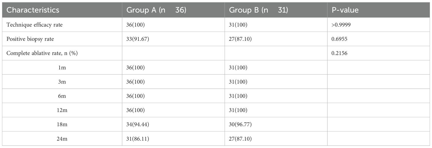
Table 3 shows the treatment efficacy of two groups. The mean follow-up time was 22.4 ± 7.9 months (range, 15.5–39.8 months). The technical success rate was 100%. In group A and group B, 8.33% (3/36) and 12.90% (4/31) of the lesions were pathologically diagnosed as alveolar tissue or carbonized fibrous tissue, which were considered pathologically negative. The rates of pathological positivity for the two groups were 91.67% (33/36) and 87.10% (27/31), respectively, with no statistically significant difference (p=0.6955). The rates of positive malignancy were 72.22% (26/36) and 77.42% (24/31) of two groups, respectively, with no statistically significant difference (p=0.2276). At 6-, 18-, and 24-months post-procedure, the local control rates for group A and group B were 100.0% vs. 100.0%, 94.44% (34/36) vs. 96.77% (30/31), and 86.11% (31/36) vs. 87.10% (27/31), respectively, with no significant differences (p=0.2156).
Bleeding and thrombosis-related complications
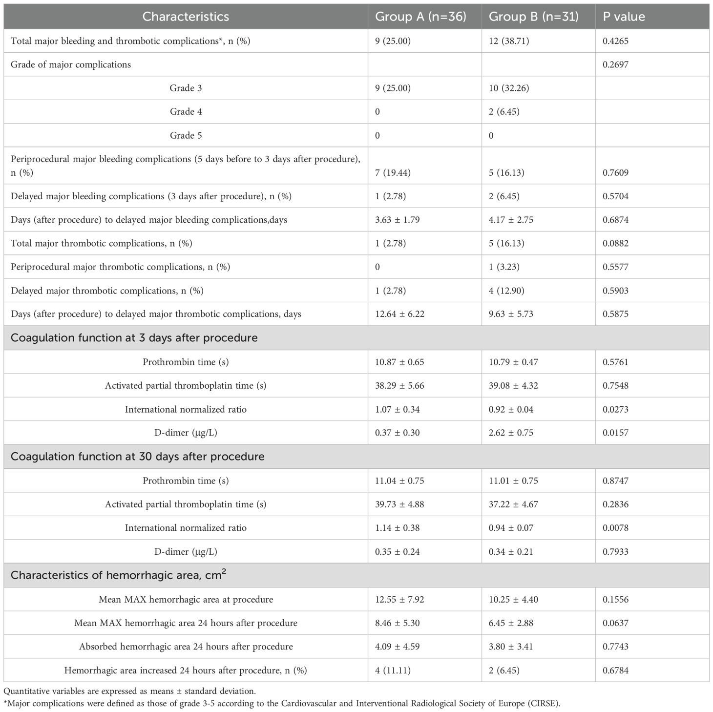
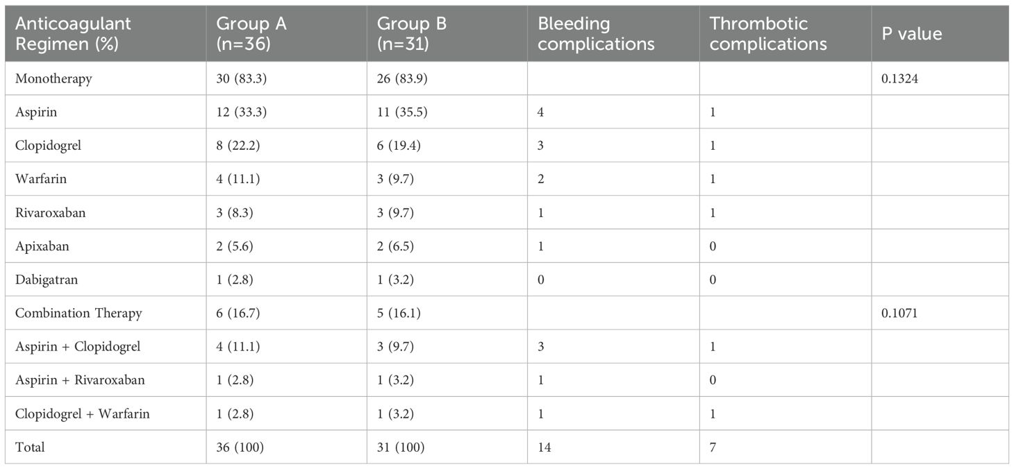
Table 4 shows the details of bleeding and thrombosis complications. The overall incidence of major complications was 9/36 (25.00%) in group A and 12/31 (38.71%) in group B. In group B, there were two cases of grade 4 thrombotic complications—one case of cerebral infarction and one case of pulmonary embolism. The two patients recovered after adequate anticoagulant treatment and hospitalization observation, without the need for interventional embolization or surgical intervention. No grade 5 complications occurred. The incidence and severity of major complications did not differ significantly between the two groups. Major bleeding complications were observed in 15 patients (8 in group A and 7 in group B), and major thrombotic complications occurred in 6 patients (1 in group A and 5 in group B), with no significant difference in incidence. Statistically significant differences were found in International normalized ratio (INR) and D-dimer levels between the two groups on the 3rd day post-procedure, as well as INR on the 30th day post-procedure. The maximum bleeding area in group A was slightly larger than that in group B both during the procedure and 24 hours post- procedure, although the difference was not statistically significant. Figure 2 presents the serial imaging findings and corresponding pathological specimens from a 67-year-old male patient with long-term aspirin use, demonstrating the perioperative and postoperative course following synchronous biopsy and MWA of a highly suspicious malignant GGO in the left lung. Table 5 presents an analysis of perioperative bleeding and thrombotic complication risks based on different preoperative anticoagulation regimens. When anticoagulation strategy was analyzed as a variable, no statistically significant differences were observed in perioperative bleeding or thrombotic complication rates between the two groups.
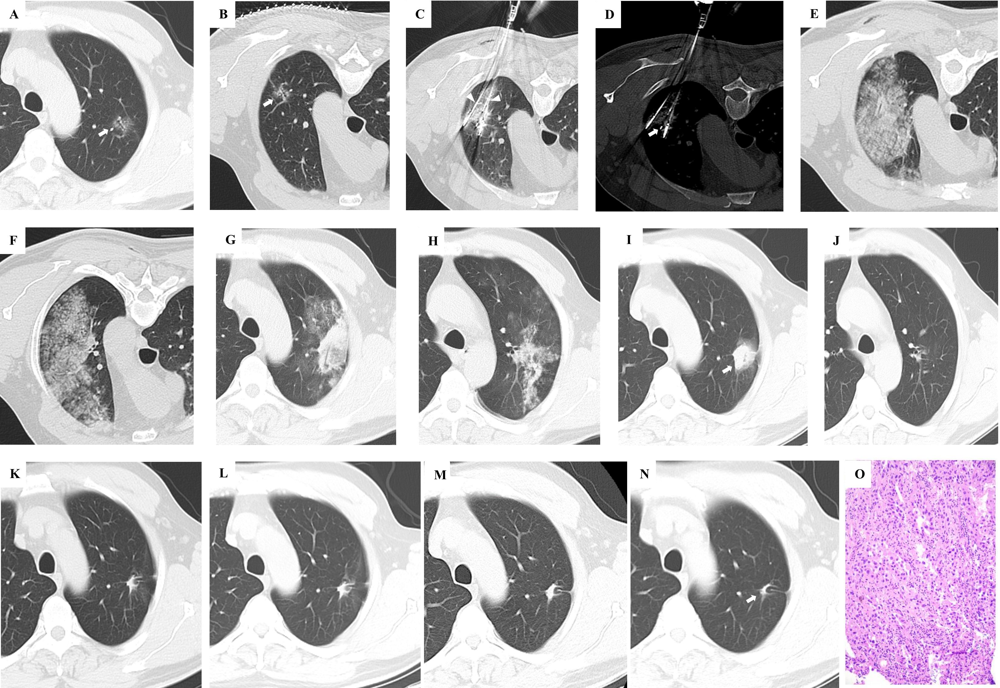
Figure 2. This 67-year-old male patient, with a history of long-term oral aspirin use post-percutaneous coronary intervention, presented with a highly suspicious malignant GGO in the left lung lobe. Upon expressing a clear preference against surgical resection, CT-guided synchronous percutaneous biopsy and microwave ablation (MWA) were chosen as an alternative therapeutic approach. (A) A GGO with a maximum diameter of 18.1mm is identified in the left upper lung lobe (indicated by the white arrow). (B) For the procedure, the patient was positioned prone. Imaging was aligned, and a puncture plan was meticulously outlined. (C) Two antennas were strategically placed at the predetermined locations as per the preprocedural plan. (D) The biopsy needle was accurately positioned at the designated site in accordance with the preprocedural plan. (E) Following the removal of the antenna and biopsy needle, a significant area of pulmonary parenchymal hemorrhage was noted. (F) Extensive hemorrhage within the lung parenchyma was observed at various CT scan levels. (G) A 24-hour follow-up CT scan revealed that the majority of the parenchymal hemorrhage had been resorbed. (H) A 24-hour CT scan at different levels also showed substantial absorption of the parenchymal hemorrhage. (I) One-month post-procedure, a CT scan indicated that the pulmonary parenchymal hemorrhage had been entirely resolved. The ablation zone completely enveloped the original GGO, deemed a successful ablation. (J) Parenchymal hemorrhage at multiple CT levels had also been fully resorbed. (K) At three months, a CT scan demonstrated that the ablation area had commenced shrinkage, with no residual or recurrent GGO detected. (L) A six-month CT scan revealed ongoing shrinkage of the ablation area. (M) The twelve-month CT scan indicated continued reduction in the size of the ablation area. (N) By eighteen months, the ablation area had transformed into a scar-like lesion, signifying the stability of the treatment outcome. (O) The pathology of this GGO was invasive adenocarcinoma.
Discussion
Image-guided thermal ablation is increasingly popular for the treatment of pulmonary GGOs. Current clinical evidence suggests that GGO-like lung cancer has a very low rate of lymph node and distant metastasis, making local lesion inactivation a viable treatment option (2, 26, 27). Furthermore, GGO-like lung cancer often as multiple GGOs, which would benefit from the ultra-minimally invasive, repeatable, and high local control rates associated with thermal ablation techniques (28, 29). Synchronous B+MWA is a commonly employed method for pulmonary GGOs (30–32). CT-guided puncture relies on the excellent visualization of GGOs on CT scans and precise puncture technique. However, the pulmonary parenchymal bleeding surrounding the GGOs caused by the puncture and biopsy can obscure the CT visualization of the nodules and potentially impacting treatment effectiveness (33, 34). Patients on antithrombotic therapy are at a higher risk of developing bleeding during puncture due to abnormal platelet function (35, 36). However, arbitrarily stopping antithrombotic medications can lead to an increased risk of thrombotic diseases (37, 38). These patients may develop several challenges: 1) Pulmonary parenchymal bleeding can impede the localization of GGOs; 2) The use of hemostatic agents following bleeding may further promote the development of thrombotic complications; 3) Persistent bleeding can result in extended hospital stays and other complications. Consequently, appropriate management of antithrombotic therapy during the perioperative period is crucial for patients with GGOs who are on antithrombotic therapy.
The present study enrolled patients with pulmonary GGOs receiving antithrombotic therapy who underwent B+MWA procedures. A case-control analysis revealed no significant baseline difference between Group A patients who received rivaroxaban as a bridging therapy and Group B patients who abruptly ceased antithrombotic therapy during the perioperative period. The technical success rate was 100%, with satisfactory rates of positive biopsy rate and local control rate. Perioperative complications showed a similar incidence of bleeding events in both groups, though the incidence of thrombotic events was higher in Group B than in Group A, a difference that didn’t reach statistical significance. The thrombotic events in Group B comprised two severe complications: one case of cerebral infarction and one case of pulmonary embolism, with mild sequelae following appropriate treatment. Previous study by Anderson R et al. has highlighted the high risk of thrombosis in lung cancer patients and the potential benefits of antiplatelet agents in cancer adjuvant therapy (39). A few studies have reported that patients with a history of thrombotic disease experienced a significantly increased risk of subsequent thrombotic complications after cessation of continuous antithrombotic therapy, and a strategy of replacing antithrombotic drugs with perioperative anticoagulants is recommended (40–42). The lower incidence of thrombotic complications in Group A patients may be attributed to the use of rivaroxaban as a bridging therapy during the interruption of antithrombotic therapy and the prompt resumption of antithrombotic therapy thereafter. Chen et al. reported in case-based narrative review that the decision to discontinue or maintain antithrombotic therapy should be informed by the individual patient’s risks of bleeding and thrombosis, as well as the specific procedural risks involved. For procedures with a low risk of bleeding, anticoagulant therapy can be safely maintained without disruption. Conversely, when anticoagulant therapy cannot be safely continued, careful consideration should be given to the timing of interruption and resumption of therapy, along with the potential need for bridging therapy to mitigate any increased risk of thrombosis (43). Similarly, many studies also support the use of anticoagulant bridging antithrombotic agents to manage the balance of bleeding and thrombotic complications in patients receiving antithrombotic therapy during the perioperative period (44–46). In summary, perioperative rivaroxaban bridging therapy for B+MWA in patients with pulmonary nodules on antithrombotic therapy effectively reduces the incidence of thrombotic complications while maintaining an acceptable rate of bleeding complications.
The present study has several limitations. Firstly, it was conducted at a single center, which means the findings may not be uniformly replicable in other clinical settings. Secondly, the study excluded patients with pulmonary GGOs measuring <8 mm and >30 mm, as well as those with multiple lesions treated using the same procedure. These exclusions could potentially explain the high technical success rate and low complication rate observed in our research. Thirdly, our sample size was limited by the inclusion of various antithrombotic drug classes, without accounting for the metabolic characteristics of each medicine. Additionally, the conventional coagulation tests presented in this study, including PT, APTT, INR, and D-dimer, are not sensitive indicators of rivaroxaban’s anticoagulant activity. It is therefore recommended that specific anti-factor Xa activity assays be considered essential monitoring tools for assessing the anticoagulant effects of rivaroxaban in future prospective studies. Furthermore, randomized studies with larger sample sizes are warranted, and standardized management protocols for rivaroxaban bridging therapy should be carefully redefined and optimized in subsequent research.
In conclusion, for patients with pulmonary GGOs who are on antithrombotic therapy, the use of oral rivaroxaban as a bridging strategy during perioperative period for simultaneous CT-guided percutaneous B+MWA is both safe and effective. This approach can lower the incidence of severe thrombotic complications during the perioperative period, compared to the abrupt discontinuation of antithrombotic therapy during this time.
Data availability statement
The datasets used and/or analyzed during the current study are available from the corresponding author on reasonable request.
Ethics statement
The studies involving humans were approved by Institutional Ethics Committee of Shandong Provincial Qianfoshan Hospital, Jinan, Shandong, China. The studies were conducted in accordance with the local legislation and institutional requirements. Written informed consent for participation was not required from the participants or the participants’ legal guardians/next of kin in accordance with the national legislation and institutional requirements.
Author contributions
NW: Data curation, Writing – original draft. GC: Data curation, Supervision, Investigation, Writing – review & editing. BJ: Data curation, Writing – original draft. WL: Data curation, Writing – original draft. JX: Data curation, Investigation, Writing – review & editing. JWX: Data curation, Investigation, Writing – review & editing. GX: Writing – review & editing, Data curation. XY: Writing – review & editing.
Funding
The author(s) declare that financial support was received for the research and/or publication of this article. This study was funded by the Project of Zhongguancun Precision Medicine Foundation (2024-JZYX-00538) and Shandong Provincial Natural Science Foundation Young Project (ZR2024QH100). Hubei Chen Xiaoping Science and Technology Development Foundation Young Scientists Special Fund, Grant/Award Number: CXPJJH125001-2519.
Conflict of interest
The authors declare that the research was conducted in the absence of any commercial or financial relationships that could be construed as a potential conflict of interest.
The reviewer JL declared a shared parent affiliation with the authors NW, BJ, WL, JWX, GX, XY to the handling editor at the time of review.
Generative AI statement
The author(s) declare that no Generative AI was used in the creation of this manuscript.
Publisher’s note
All claims expressed in this article are solely those of the authors and do not necessarily represent those of their affiliated organizations, or those of the publisher, the editors and the reviewers. Any product that may be evaluated in this article, or claim that may be made by its manufacturer, is not guaranteed or endorsed by the publisher.
References
1. Ram D, Egan H, and Ramanathan T. Ground glass opacity: can we correlate radiological and histological features to plan clinical decision making? Gen Thorac Cardiovasc Surg. (2022) 70:971–6. doi: 10.1007/s11748-022-01826-2
2. Bou-Samra P, Chang A, Kelly N, Yudien M, Galandarova A, Ibrahimli A, et al. Ground-glass opacities: a problem bound to get more challenging. Quant Imaging Med Surg. (2023) 13:5447–50. doi: 10.21037/qims-23-797
3. Lancaster HL, Heuvelmans MA, and Oudkerk M. Low-dose computed tomography lung cancer screening: Clinical evidence and implementation research. J Intern Med. (2022) 292:68–80. doi: 10.1111/joim.13480
4. Liang ZR, Ye M, Lv FJ, Fu BJ, Lin RY, Li WJ, et al. Differential diagnosis of benign and Malignant patchy ground-glass opacity by thin-section computed tomography. BMC Cancer. (2022) 22:1206. doi: 10.1186/s12885-022-10338-4
5. Cai Y, Chen T, Zhang S, Tan M, and Wang J. Correlation exploration among CT imaging, pathology and genotype of pulmonary ground-glass opacity. J Cell Mol Med. (2023) 27:2021–31. doi: 10.1111/jcmm.17797
6. Qin Y, Xu Y, Ma D, Tian Z, Huang C, Zhou X, et al. Clinical characteristics of resected solitary ground-glass opacities: Comparison between benign and Malignant nodules. Thorac Cancer. (2020) 11:2767–74. doi: 10.1111/1759-7714.13575
7. Liang ZR, Lv FJ, Fu BJ, Lin RY, Li WJ, Chu ZG, et al. Reticulation sign on thin-section CT: utility for predicting invasiveness of pure ground-glass nodules. AJR Am J Roentgenol. (2023) 221:69–78. doi: 10.2214/AJR.22.28892
8. Cho H, Lee HY, Kim J, Kim HK, Choi JY, Um SW, et al. Pure ground glass nodular adenocarcinomas: Are preoperative positron emission tomography/computed tomography and brain magnetic resonance imaging useful or necessary? J Thorac Cardiovasc Surg. (2015) 150:514–20. doi: 10.1016/j.jtcvs.2015.06.024
9. Cardillo G, Petersen RH, Ricciardi S, Patel A, Lodhia JV, Gooseman MR, et al. European guidelines for the surgical management of pure ground-glass opacities and part-solid nodules: Task Force of the European Association of Cardio-Thoracic Surgery and the European Society of Thoracic Surgeons. Eur J Cardiothorac Surg. (2023) 64:ezad386. doi: 10.1093/ejcts/ezad222
10. Abdallat M, Jonigk D, and Khalil HA. Pure ground-glass opacity of the lung: making the case for a lasting cure. J Thorac Dis. (2024) 16:2684–6. doi: 10.21037/jtd-23-1706
11. Faiella E, Messina L, Castiello G, Bernetti C, Pacella G, Altomare C, et al. Augmented reality 3D navigation system for percutaneous CT-guided pulmonary ground-glass opacity biopsies: a comparison with the standard CT-guided technique. J Thorac Dis. (2022) 14:247–56. doi: 10.21037/jtd-21-1285
12. Aokage K, Suzuki K, Saji H, Wakabayashi M, Kataoka T, Sekino Y, et al. Segmentectomy for ground-glass-dominant lung cancer with a tumour diameter of 3 cm or less including ground-glass opacity (JCOG1211): a multicentre, single-arm, confirmatory, phase 3 trial. Lancet Respir Med. (2023) 11:540–9. doi: 10.1016/S2213-2600(23)00041-3
13. Wang N, Xue T, Zheng W, Shao Z, Liu Z, Dai F, et al. Safety and efficacy of percutaneous biopsy and microwave ablation in patients with pulmonary nodules on antithrombotic therapy: A study with rivaroxaban bridging. Thorac Cancer. (2024) 15:1989–99. doi: 10.1111/1759-7714.15425
14. Chang JM and Yoen H. Breast biopsy and hematoma associated with antithrombotic therapy. Radiology. (2023) 306:87–9. doi: 10.1148/radiol.221871
15. Huang Z, Chen J, Xie F, Liu S, Zhou Y, Shi M, et al. Cone-beam computed tomography-guided cryobiopsy combined with conventional biopsy for ground glass opacity-predominant pulmonary nodules. Respiration. (2024) 103:32–40. doi: 10.1159/000535236
16. Kaye AD, Manchikanti L, Novitch MB, Mungrue IN, Anwar M, Jones MR, et al. Responsible, safe, and effective use of antithrombotics and anticoagulants in patients undergoing interventional techniques: american society of interventional pain physicians (ASIPP) guidelines. Pain Physician. (2019) 22:S75–S128.
17. Ma Y, Li G, Li X, Gao Y, Ding T, Yang G, et al. Clinical characteristics and prognostic analysis of Lung Cancer patients with Hypercoagulability: A single-center, retrospective, real-world study. J Cancer. (2021) 12:2968–74. doi: 10.7150/jca.46600
18. Wang TF, Sanfilippo KM, Douketis J, Falanga A, Karageorgiou J, Maraveyas A, et al. Peri-procedure management of antithrombotic agents and thrombocytopenia for common procedures in oncology: Guidance from the SSC of the ISTH. J Thromb Haemost. (2022) 20:3026–38. doi: 10.1111/jth.15896
19. Lyons MD, Pope B, and Alexander J. Perioperative management of antithrombotic therapy. JAMA. (2024) 332:420–1. doi: 10.1001/jama.2024.5880
20. Ortel TL. Perioperative management of patients on chronic antithrombotic therapy. Blood. (2012) 120:4699–705. doi: 10.1182/blood-2012-05-423228
21. Renda G, Pecen L, Patti G, Ricci F, Kotecha D, Siller-Matula JM, et al. Antithrombotic management and outcomes of patients with atrial fibrillation treated with NOACs early at the time of market introduction: Main results from the PREFER in AF Prolongation Registry. Intern Emerg Med. (2021) 16:591–9. doi: 10.1007/s11739-020-02442-9
22. Ye X, Fan W, Wang Z, Wang J, Wang H, Niu L, et al. Clinical practice guidelines on image-guided thermal ablation of primary and metastatic lung tumors (2022 edition). J Cancer Res Ther. (2022) 18:1213–30. doi: 10.4103/jcrt.jcrt_880_22
23. Wang N, Xu J, Xue G, Han C, Zhang H, Zhao W, et al. Synchronous computed tomography-guided percutaneous biopsy and microwave ablation for highly suspicious Malignant lung ground-glass opacities adjacent to mediastinum. Int J Hyperthermia. (2023) 40:2193362. doi: 10.1080/02656736.2023.2193362
24. Li B, Bie Z, Li Y, Guo R, Wang C, Li X, et al. Synchronous percutaneous core-needle biopsy and microwave ablation for stage I non-small cell lung cancer in patients with Idiopathic pulmonary fibrosis: initial experience. Int J Hyperthermia. (2023) 40:2270793. doi: 10.1080/02656736.2023.2270793
25. Freites-Martinez A, Santana N, Arias-Santiago S, and Viera A. Using the common terminology criteria for adverse events (CTCAE - version 5.0) to evaluate the severity of adverse events of anticancer therapies. Actas Dermosifiliogr (Engl Ed). (2021) 112:90–2. doi: 10.1016/j.ad.2019.05.009
26. Liu S, Zhu X, Qin Z, Xu J, Zeng J, Chen J, et al. Computed tomography-guided percutaneous cryoablation for lung ground-glass opacity: A pilot study. J Cancer Res Ther. (2019) 15:370–4. doi: 10.4103/jcrt.JCRT_299_18
27. Meng M, Huang G, Wang J, Li W, Ni Y, Zhang T, et al. Facilitating combined biopsy and percutaneous microwave ablation of pulmonary ground-glass opacities using lipiodol localisation. Eur Radiol. (2023) 33:3124–32. doi: 10.1007/s00330-023-09486-3
28. Yang X, Ye X, Lin Z, Jin Y, Zhang K, Dong Y, et al. Computed tomography-guided percutaneous microwave ablation for treatment of peripheral ground-glass opacity-Lung adenocarcinoma: A pilot study. J Cancer Res Ther. (2018) 14:764–71. doi: 10.4103/jcrt.JCRT_269_18
29. Xue G, Li Z, Wang G, Wei Z, and Ye X. Computed tomography-guided percutaneous microwave ablation for pulmonary multiple ground-glass opacities. J Cancer Res Ther. (2021) 17:811–3. doi: 10.4103/jcrt.jcrt_531_21
30. Kong F, Bie Z, Li Y, Li B, Guo R, Wang C, et al. Synchronous microwave ablation followed by core-needle biopsy via a coaxial cannula for highly suspected Malignant lung ground-glass opacities: A single-center, single-arm retrospective study. Thorac Cancer. (2021) 12:3216–22. doi: 10.1111/1759-7714.14189
31. Cao P, Wei Z, Xue G, Wang N, Li Z, Hu Y, et al. Complications of synchronous microwave ablation and biopsy versus microwave ablation alone for pulmonary sub-solid nodules: a retrospective, large sample, case-control study. Quant Imaging Med Surg. (2024) 14:7218–28. doi: 10.21037/qims-24-906
32. Chen Z, Zeng J, Lin Y, Zhang X, Wu X, Yong Y, et al. Synchronous computed tomography-guided percutaneous transthoracic needle biopsy and microwave ablation for highly suspicious Malignant pulmonary ground-glass nodules. Respiration. (2024) 103:388–96. doi: 10.1159/000538743
33. Zhu L, Huang J, Jin C, Zhou A, Chen Y, Zhang B, et al. Retrospective cohort study on the correlation analysis among peri-procedural factors, complications, and local tumor progression of lung tumors treated with CT-guided microwave ablation. J Thorac Dis. (2023) 15:6915–27. doi: 10.21037/jtd-23-1799
34. Xu S, Bie ZX, Li YM, Li B, Peng JZ, Kong FL, et al. Computed tomography-guided microwave ablation for non-small cell lung cancer patients on antithrombotic therapy: a retrospective cohort study. Quant Imaging Med Surg. (2022) 12:3251–63. doi: 10.21037/qims-21-1043
35. Uchino K, Tateishi R, Wake T, Kinoshita MN, Nakagomi R, Nakatsuka T, et al. Radiofrequency ablation of liver tumors in patients on antithrombotic therapy: A case-control analysis of over 10,000 treatments. J Vasc Interv Radiol. (2021) 32:869–77. doi: 10.1016/j.jvir.2021.02.021
36. Scherer FD, Dressler C, Valles GA, and Nast A. Risk of complications due to antithrombotic agents in cutaneous surgery: a systematic review and meta-analysis. J Dtsch Dermatol Ges. (2021) 19:1421–32. doi: 10.1111/ddg.14579
37. Douketis JD, Spyropoulos AC, Murad MH, Arcelus JI, Dager WE, Dunn AS, et al. Perioperative management of antithrombotic therapy: an american college of chest physicians clinical practice guideline. Chest. (2023) 162:e207–43. doi: 10.1016/j.chest.2022.07.025
38. Li YK, Guo CG, and Cheung KS. Risk of post colonoscopy thromboembolic events: A real-world cohort study. Clin Gastroenterol Hepatol. (2023) 21:3051–3059.e4. doi: 10.1016/j.cgh.2022.09.021
39. Anderson R, Rapoport BL, Steel HC, and Theron AJ. Pro-tumorigenic and thrombotic activities of platelets in lung cancer. Int J Mol Sci. (2023) 24:11927. doi: 10.3390/ijms241511927
40. Fujikawa T, Takahashi R, and Naito S. Perioperative antithrombotic management of patients who receive direct oral anticoagulants during gastroenterological surgery. Ann Gastroenterol Surg. (2020) 4:301–9. doi: 10.1002/ags3.12328
41. Fujikawa T. Perioperative antithrombotic management during gastroenterological surgery in patients with thromboembolic risks: current status and future prospects. Cureus. (2022) 14:e23471. doi: 10.7759/cureus.23471
42. Scherer FD, Nast A, Gaskins M, Werner RN, and Dressler C. Perioperative management of antithrombotic drugs in skin surgery - A survey of dermatologists in Germany. J Dtsch Dermatol Ges. (2022) 20:941–50. doi: 10.1111/ddg.14758
43. Chen AT, Patel M, and Douketis JD. Perioperative management of antithrombotic therapy: a case-based narrative review. Intern Emerg Med. (2022) 17:25–35. doi: 10.1007/s11739-021-02866-x
44. Ning YJ, Wan ZX, Meng J, and Wang XP. A systemic review and meta-analysis of the effects of perioperative anticoagulant and antiplatelet therapy on bleeding complications in robot-assisted prostatectomy. Eur Rev Med Pharmacol Sci. (2022) 26:2085–97. doi: 10.26355/eurrev_202203_28356
45. Jiang SH, Hukamdad M, Gould A, Bhaskara M, Chiu RG, Sadeh M, et al. Effect of perioperative anticoagulant prophylaxis in patients with traumatic subdural hematoma and a history of anticoagulant use: a propensity-matched National Trauma Data Bank analysis. Neurosurg Focus. (2023) 55:E3. doi: 10.3171/2023.7.FOCUS23346
46. Manchikanti L, Sanapati MR, Nampiaparampil D, et al. Perioperative management of antiplatelet and anticoagulant therapy in patients undergoing interventional techniques: 2024 updated guidelines from the american society of interventional pain physicians (ASIPP). Pain Physician. (2024) 27:S1–S94. doi: 10.36076/ppj.2024.7.S1
Keywords: microwave ablation, biopsy, ground-glass opacities, antithrombotic therapy, complication
Citation: Wang N, Chen G, Jin B, Lu W, Xu J, Xu J, Xue G and Ye X (2025) Enhanced management strategy of synchronous percutaneous biopsy and microwave ablation in patients with lung ground-glass opacities undergoing antithrombotic treatment: a clinical perspective on our experience. Front. Oncol. 15:1554365. doi: 10.3389/fonc.2025.1554365
Received: 07 February 2025; Accepted: 07 July 2025;
Published: 23 July 2025.
Edited by:
Roberto Minici, University Hospital Renato Dulbecco, ItalyReviewed by:
Tino Schneider, Cantonal Hospital St.Gallen, SwitzerlandJinpeng Li, Shandong First Medical University and Shandong Academy of Medical Sciences, China
Copyright © 2025 Wang, Chen, Jin, Lu, Xu, Xu, Xue and Ye. This is an open-access article distributed under the terms of the Creative Commons Attribution License (CC BY). The use, distribution or reproduction in other forums is permitted, provided the original author(s) and the copyright owner(s) are credited and that the original publication in this journal is cited, in accordance with accepted academic practice. No use, distribution or reproduction is permitted which does not comply with these terms.
*Correspondence: Jingwen Xu, eHVqaW5nd2VuMjIzMjggQDEyNi5jb20=; Guoliang Xue, eGc0MzkyQDE2My5jb20=; Xin Ye, eWV4aW50YWlhbjIwMjBAMTYzLmNvbQ==
†These authors have contributed equally to this work
 Nan Wang
 Guoqiang Chen1†
 Guoliang Xue
 Xin Ye