- 1Medicinal and Pharmaceutical Chemistry Department, (Pharmacology Group), National Research Centre, Giza, Egypt
- 2Pharmacology and Toxicology Department, Faculty of Pharmacy, Cairo University, Cairo, Egypt
- 3School of Pharmacy, Newgiza University, Giza, Egypt
- 4Pharmacognosy Department, National Research Centre, Giza, Egypt
- 5Pathology Department, National Research Centre, Giza, Egypt
- 6Department of Biology, Darb University College, Jazan University, Jazan, Saudi Arabia
Aims: Dipeptidyl peptidase-4 enzyme (DPP-4) was reported to be associated with immune stimulation, resistance to anti-neoplastic agents and lipid accumulation. Dysregulated DPP-4 expression was reported in various malignant tumors such as hepatocellular carcinoma. Hence, the influence of sitagliptin, an inhibitor of DPP-4 enzyme, was performed in vitro (HepG2 cells) and in vivo (mouse model of hepatic preneoplasia).
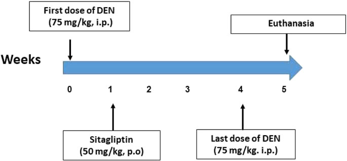
Main methods: The effect of sitagliptin was investigated in vitro via MTT assay. The in vivo model of hepatic preneoplasia was conducted by weekly intraperitoneal injection of 75 mg/kg of diethylnitrosamine (DEN) for five successive weeks. Mice were treated daily with sitagliptin (50 mg/kg, p.o.) starting 1 week after DEN injection till the end of the experiment.
Key findings: Sitagliptin exerted a significant cytotoxic effect on HepG2 cells, which was dependent on elevating mRNA expression of p53 and BAX/BCL2. Sitagliptin also improved serum liver enzymes and attenuated histopathological alterations in mice. These changes were accompanied by reducing liver GGT, DPP-4, CYP2E1, GGT-P, NF-κB and PCNA along with increasing CYP3A4. Furthermore, sitagliptin attenuated DEN-induced liver DNA damage and inflammation.
Significance: These findings shed the light on the role of DPP-inhibitors in the future of cancer therapy and management.
1 Introduction
The liver is a key metabolic organ that detoxifies natural and foreign harmful chemicals, which makes it highly susceptible to carcinogenesis. Hepatocellular carcinoma (HCC) is a common type of liver cancer, which is aggressive and associated with rapid progression and poor prognosis (Memon et al., 2020). It develops via multistep processes linked to different genetic changes that eventually lead to malignant transformation of liver cells (Sarkar et al., 2020). HCC is mostly linked to extrinsic factors that directly or indirectly prompt DNA damage and chromosomal aberrations. Accumulation of uncontrolled DNA damage, results in formation of expanding foci of dysplastic hepatocytes, which progress to liver malignancy (Buitrago-Molina et al., 2013).
Treatment of HCC is complex with limited surgical resection due to risk of postoperative liver failure. Although liver transplantation is considered the best choice to increase the survival, it cannot be performed in all cases due to insufficient organs and incompatibility (Memon et al., 2020). High mortality and adverse effects associated with chemotherapy and radiotherapy highlights the need for alternative therapy in cancer management (Assar et al., 2021).
Injection of diethylnitrosamine (DEN) is used experimentally to induce HCC (Memon et al., 2020; Sarkar et al., 2020). DEN is a genotoxic compound which is bio-activated by cytochrome P4502E1 (CYP2E1) producing mutagenic DNA adducts and production of reactive oxygen species (ROS), thus promoting hepatocarcinogenesis (Kang et al., 2007; Memon et al., 2020; Sarkar et al., 2020).
Dipeptidyl peptidase 4 (DPP-4) enzyme is one of type II transmembrane glycoproteins that cleaves the X-proline or X-alanine dipeptides from the N-terminus of polypeptides such as chemokines, neuropeptides and peptide hormones (Zhao, 2019; Kawakubo et al., 2020). DPP-4 inactivates incretins including glucagon-like peptide-1 (GLP-1) and gastric inhibitory polypeptide (GIP) regulating blood glucose level (Onoyama et al., 2015; Katakami et al., 2018). DPP-4 enzyme associates with several proteins, so it plays a crucial role in cancer biology and may be considered as a useful marker for different tumors (Havre et al., 2008). In addition, DPP-4 enzyme was reported to be involved in tumor metastasis and invasion. Moreover, DDP-4 may act as either a tumor suppressor or promoter via its role with chemokines and cytokines (Almagthali et al., 2019). Previous data refer to the role of DPP-4 in the development of various chronic liver diseases like non-alcoholic fatty liver disease, hepatitis C virus infection and hepatocellular carcinoma. DPP-4 is also present in hepatic stem cells and plays a key role in hepatic regeneration (Itou et al., 2013). Studies on DPP-4 enzyme and its role in liver cancer development still did not provide a conclusive answer regarding its role in HCC (Yu et al., 2020; Busek et al., 2022).
Sitagliptin is a selective inhibitor of DPP4 that is used in management of type II diabetes (Mest and Mentlein, 2005). Recently, sitagliptin has demonstrated a hepatoprotective activity in thioacetamide-induced acute liver injury in mice (El-Kashef and Serrya, 2019), rat liver steatosis (Shen et al., 2018) and mice diabetic liver disease (Wang et al., 2021). Therefore, the present study was conducted to investigate the possible effects of the sitagliptin on in vitro HepG2 cells and DEN-induced hepatic preneoplasia in mice.
2 Materials and methods
2.1 In vitro study
Human hepatocellular carcinoma cell line HepG2 cells were obtained from Karolinska Institute, Sweden with the help of Dr. Stig Linder. Minimum Essential Medium (MEM) medium 10% fetal bovine serum (Gibco, United States) was used for culturing the cells and then they were maintained in an incubator at 37°C with a humidified atmosphere of 5% CO2.
2.2 Cytotoxic effect of sitagliptin on HepG2 cells
A Laminar flow cabinet biosafety class II level (Baker, SG403INT, Sanford, ME, United States) was used for conduction of all in vitro procedures in a sterile area. Fetal bovine serum (10%), 1% L-glutamine and 1% antibiotic-antimycotic mixture were all added to MEM media and used for the suspension of HepG2 cells, then cells were kept at 37°C under 5% CO2. A 96-well plate was used for seeding the cells at a density of 10 × 103 cells/well at 37°C for 24 h under 5% CO2 using a water-jacketed CO2 incubator (Sheldon, TC2323, Cornelius, OR, United States).
Sitagliptin (3.90–250 µmole, concentration) was added to wells and cultured for 48 h, then aspiration of the medium was done followed by incubation with 40 µL 3-(4,5-dimethylthiazol-2-yl)-2,5-diphenyl tetrazolium bromide (MTT) salt (2.5 μg/mL) for 4 h. After that, 200 µL of 10% sodium dodecyl sulfate (SDS) dissolved in deionized water were added to all wells to dissolve the formed crystals as well as stop the reaction and then incubated at 37°C overnight. The development of purple formazan is an indication of cell viability (Mosmann, 1983).
Wells containing cells only served as the negative control. Doxorubicin (200 µm) was used as a positive control that is reported to give 100% lethality when used under the same conditions (Thabrew et al., 2005; EL-Menshawi et al., 2010). The absorbance was read at 595 nm using microplate multi-well reader (Bio-Rad Laboratories Inc., model 3350, Hercules, California, United States) with reference wavelength set at 620 nm. The resulting color absorbance is directly proportional to viable cell number in each sample. Dimethyl sulfoxide (DMSO<0.2%) was used as a vehicle for all treatments. The %viability was estimated using the following formula:
where AT = absorbance of treatment and AC = absorbance of negative control.
2.3 mRNA expression of BCL2-associated X protein (BAX), B-cell lymphoma 2 (BCL2), and tumor protein (p53) in HepG2 cells using real-time PCR technique
To isolate total RNA from liver cancer cell lines, RNeasy Mini Kit (Qiagen, Hilden, Germany) supplemented with DNaseI (Qiagen) digestion step were used as reported by the manufacturer’s protocol. StepOne™ Real-Time PCR System was used to determine cDNA copy number of the liver cell line (Applied Biosystems, Thermo Fisher Scientific, Waltham, MA United States). The primer sequences are as follows: β-actin, F-CAT CCG CAA AGA CCT GTA CG, R-CCT GCT TGC TGA TCC ACA TC, accession no, DQ407611.1, BAX, F-GAT GAC CCT CTG ACC CTA GC, R-CGG GCA TTA AAG AGC TGG AC, accession no, NM_001291430.2; BCL2, F-CAA GTG TTC CGC GTG ATT GA, R-CAG AGG AAA AGC AAC GGG G, accession no, KY098799.1 and p53, F-TGG CCA TCT ACA AGC AGT CA, R-GGT ACA GTC AGA GCC AAC CT, accession no, AB082923.1. The 2−ΔΔCT method was used for the relative quantification of the target to the reference (Ramadan et al., 2019).
β-actin was selected as the housekeeping gene due to its consistent expression under our experimental conditions, as previously validated in HepG2 cells treated with pharmacologic agents (Ramadan et al., 2019).
Housekeeping genes are favored for two reasons: they encode proteins that undergo stabilizing selection to maintain metabolic function, and they exhibit sufficient variability to detect a range of variants in the isolate collection. The nucleotide substitutions in housekeeping genes accumulate relatively slowly and remain constant enough for the method to be ideal for global epidemiology. Those housekeeping proteins are used as internal controls with presumed stability and no changes in physiological condition (Li and Shen, 2013; Muñoz et al., 2014).
2.4 In vivo study
2.4.1 Animals
A total of forty male albino mice weighing 20–25 g were obtained from the animal breeding unit at the National Research Centre (Giza, Egypt). Mice were housed in laboratory standardized conditions of proper ventilation, relative humidity (55% ± 5%) room temperature (23°C ± 2°C), and a 12-h light/dark cycle. Animals were provided free access to standard chow diet and water. Approval for this investigation was granted by the Ethics Committee for Animal Experimentation at the Faculty of Pharmacy, Cairo University (Permit Number: PT 2198) and the Ethics Committee for Animal Experimentation at the National Research Centre, Giza, Egypt (18047).
2.4.2 Chemicals and drugs
Diethylnitrosamine and sitagliptin were purchased from Sigma Aldrich (St. Louis, MO, United States) and Merck Sharp and Dohme Ltd. (Hertfordshire, United Kingdom), respectively.
2.4.3 Experimental design
Animals were randomly assigned into four groups of 10 mice each (Figure 1). Group 1 (Control): mice received saline intraperitoneally (i.p.). Group 2 (Sita): mice received sitagliptin (50 mg/kg, p.o.) dissolved in distilled water (Gault et al., 2015) for 4 weeks. Group 3 (DEN): Mice were given five weekly intraperitoneal injections of 75 mg/kg DEN dissolved in saline (Eitah et al., 2023). Group 4 (DEN + Sita): mice received DEN as in group 3, combined with daily oral sitagliptin (50 mg/kg) 1 week post-DEN injection and continued for 4 weeks.
For the calculation of the weight change percentage, the following formula was used: (Lee et al., 2012).
The experiment was terminated after the fifth week post-DEN induction. Blood samples were collected under light anesthesia, and then the mice were euthanized by cervical dislocation. Liver was rapidly excised, washed with ice-cold saline, dried, and weighed to record the absolute liver weight as well as relative liver weight (liver weight per 100 g of animal weight). Tissue homogenate (10%) was prepared from the right lobe for biochemical analysis. The left lobe was isolated for histopathological investigation.
2.4.4 Biochemical analysis
Commercially available colorimetric kits were used for the determination of serum levels of aspartate aminotransferase (AST) and alanine aminotransferase (ALT) obtained from Teco Diagnostic (Anaheim, United States) and alkaline phosphatase (ALP) obtained from BioAssay Systems (Hayward, United States). However, mouse-specific ELISA kits were purchased from LifeSpan BioSciences, Inc. (North America) to determine the tissue level of gamma glutamyl transferase (GGT), DPP-4, tumor necrosis factor alpha (TNF-α), CYP2E1, and CYP3A4. ELISA kits for tissue interleukin (IL)-6 were provided from Mybiosource (San Diego, CA, United States). Procedural steps were strictly conducted as per the manual provided by the manufacturer.
2.4.5 Comet assay
The modification of the method done by Blasiak et al. (2004) was used to perform the comet assay. Low melting-point agarose (1:10 v/v) was mixed with liver cells of each treatment group, and then pipetted to pre-coated slides with normal-melting-point agarose. In a dark environment, the slides were kept flat for 30 min at 4°C. Low melting point agarose (third layer) was then pipetted on slides and left to solidify at 4°C for 30 min. A pre-chilled lysis solution was used to keep the slides at 4°C for 60 min. Freshly prepared alkaline unwinding solution was used for immersion of the slides for 60 min away from light at room temperature. An electrophoresis run at 0.8 V/cm and 300 m Amps was applied to slides for 30 min at 4°C. Neutralizing solution was used for rinsing the slides, and then the slides were immersed in 70% ethanol and then air-dried. Slides were stained using ethidium bromide, then a Zeiss epifluorescence microscope (510–560 nm, barrier filter 590 nm) was used for visualization at 400x magnification. According to Augustyniak et al. (2016), 400 cells per group were scored. Random selection of cells that are non-overlapping was done. Subsequently, cells were visually assigned a score on an arbitrary scale of 0–3; where class 0 = no damage or tail, class 1 = tail length < nuclear diameter, class 2 = tail length ranging from 1 and 2x nuclear diameter, class 3 = tail length 2x larger than nuclear diameter (Fairbairn et al., 1995).
2.4.6 Liver histopathology
Liver tissues were flushed and fixed for 48 h in 10% neutral buffered formalin. Samples were prepared to be embedded in paraffin, then cut into 5 µm thick serial sections using the rotatory microtome and mounted on glass slides for staining with hematoxylin and eosin (H&E). Sections were examined using an Olympus CX41 research microscope. Sample fixation, staining and all standard procedures were done according to Culling (2013). A CCD digital camera (Olympus SC100) which is attached to the microscope was used for the slide tissue microphotography. Digital photomicrography for the hepatic tissue sections was done at different power magnifications.
2.4.7 Proliferating cell nuclear antigen (PCNA), placental glutathione S-transferase (GST-P), and nuclear factor kappa B (NF-κB) immunostaining
Immunohistochemical staining of paraffin-embedded tissue sections (5-μm) was performed as per the manual instructions. H2O2 (3%) was used for 20 min for the treatment of the deparaffinized retrieved tissue sections. This was followed by incubation with anti-GST3/GST pi antibody (Cat no. ab153949, Abcam, Cambridge, United Kingdom), anti-PCNA (Cat no. MA5-11358, Thermo Fisher Scientific Inc., Rockford, IL, United States) and anti-p-NF-κB p65 antibody (Cat no. GTX54672, GeneTex Inc., Hsinchu, Taiwan) overnight at 4°C. After washing, incubation with secondary antibody HRP Envision kit (DAKO) was done for 20 min. Then, washing with PBS was followed by incubation with diaminobenzidine for 10 min. Counterstaining with hematoxylin was done after washing with PBS, then dehydrating and clearing in xylene was done, and finally cover-slipping for microscopic examination. Leica Microsystems (GmbH, Germany) was used for analysis of data via a high-definition microscopic imaging system. Brownish cytoplasmic staining represents GST-P + immune reaction, while brownish nuclear staining represents PCNA + immune reaction. Regarding NF-κB, tissue images are considered positive immunoreactive by brownish nuclear and cytoplasmic staining. For quantitative measurement of area percentage for GST-P, PCNA, and NF-κB, six non-overlapping fields were randomly selected and scanned per tissue section of each sample in the immunostained sections.
2.4.8 Morphometric analysis
A Leica DM-LB microscope with a JVC color video camera connected to a computer system was utilized as part of an image analysis system (Leica Qwin 500 Image analyzer, LEICA Imaging Systems Ltd., Cambridge, United Kingdom) for morphometric measurement at a magnification of ×100 on standard hematoxylin. Once the light source was adjusted, the slides to be inspected were put on the microscope. Additionally, the lighting was successfully evaluated and modified. A green mask, known as a binary image, automatically covered a lined tube that encircled the chosen regions of each parameter. The attributes of these binary images were automatically rendered in a table that included the total, mean, standard deviation, standard error, and the minimum and maximum areas measured in micrometers. Twenty fields on each slide were used for area measurement, with subsequent mean and standard error determination for all parameters examined.
2.5 Statistical analysis
For in vitro investigation, statistical analysis was carried out with the SPSS 11 program using regression analysis to compare samples with the negative control. In vivo data were compared using one-way ANOVA and a Tukey’s post hoc test with GraphPad Prism software version 6 (San Diego, CA, United States). Values of p < 0.05 were regarded as significant.
3 Results
3.1 In vitro experiment
3.1.1 Cytotoxic effect of sitagliptin on HepG2 cells
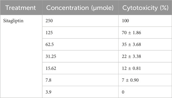
Sitagliptin exhibited a cytotoxic effect on HepG2 cells in a dose dependent manner, with IC50: 89 µm, while at 3.9 µm sitagliptin did not exert any cytotoxic effect (Table 1).
3.1.2 Effect of sitagliptin on the mRNA expression of BAX, BCL2 and p53 in HepG2 cells
Figure 2 illustrates a significant reduction in BCL2 expression level was reported upon treatment of HepG2 cells with the standard anticancer drug doxorubicin in a concentration of 200 µmole or with sitagliptin (IC50: 89 μM; or IC35; 62.5 µM) to reach 44%, 43%, and 58%, respectively, as compared to the negative control, untreated cancer cells. However, a significant upregulation in BAX expression level was observed in hepatic cancer cell lines treated with doxorubicin (200 µm) or sitagliptin (IC50: 89 μM; or IC35; 62.5 µM) to reach 4.23, 4.15, and 2.72 folds, respectively, as compared to the untreated HepG2 cells. Moreover, a significant increase in p53 expression level was observed upon treatment with doxorubicin 200 µmole or sitagliptin (IC50: 89 μM; or IC35; 62.5 µM) to reach 4.98, 4.31, and 3.22 folds, respectively, as compared to the untreated HepG2 cells. Similarly, a significant elevation in BAX/BCL2 ratio to reach 9.43, 9.76, and 4.69 folds, respectively, was observed in hepatic cancer cell lines treated with doxorubicin 200 µm or sitagliptin (IC50: 89 μM; or IC35; 62.5 µM) as compared to the negative control, untreated cancer cell lines.
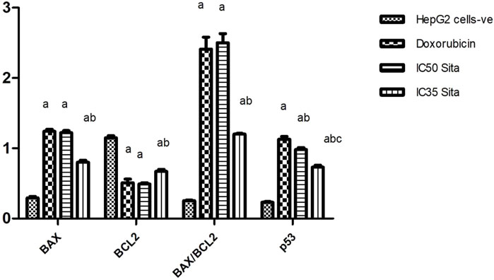
Figure 2. Effect of sitagliptin on the mRNA expression of BAX, BCL2, BAX/BCL2 ratio, and p53 in HepG2 cells. a sig. from negative control HepG2 cells value, b sig. from doxorubicin, c sig. from IC 50 sitagliptin at P < 0.01. Each value represents mean ± standard error of mean (S.E.M.) (n = 4). BAX, BCL2-associated X protein; BCL2, B-cell lymphoma 2 (BCL2), P53, tumor protein.
3.2 In vivo experiment
Noteworthy, animals given sitagliptin alone did not exhibit any significant changes in all measured parameters.
3.2.1 Effect of sitagliptin on final body weight in male mice expressing hepatic preneoplastic lesions induced by DEN
Repeated injections of DEN to mice significantly depressed the final body weight to reach 53% as compared with the control value. Treatment with sitagliptin (50 mg/kg, p.o.) did not produce any significant change in the final body weight as compared to DEN-treated animals (Figure 3A). DEN injection resulted in a significant decrease in the body weight change % to reach −8.0 ± 0.60, while the administration of sitagliptin resulted in a significant slight rise in the body weight change % to be 0.90 ± 0.70, (Figure 3B).
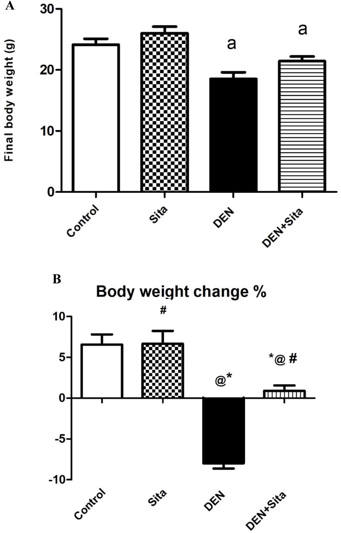
Figure 3. (A) Effect of sitagliptin on final body weight in male mice expressing hepatic preneoplastic lesions induced by DEN. a sig. from control at p < 0.05. Each value represents mean ± standard error of mean (S.E.M.) (n = 10). (B) Effect of sitagliptin on body weight change % in male mice expressing hepatic preneoplastic lesions induced by DEN. * sig. from control, @ sig. from Sita, # sig. from DEN, at p < 0.05. Each value represents mean ± standard error of mean (S.E.M.) (n = 10).
3.2.2 Effect of sitagliptin on absolute and relative liver weights in male mice expressing hepatic preneoplastic lesions induced by DEN
DEN-injected animals exhibited a marked decrease in absolute and relative liver weights to reach 33% and 59% as compared to the control group values, respectively. However, treatment with sitagliptin only succeeded to normalize the relative liver weight (Figure 4).
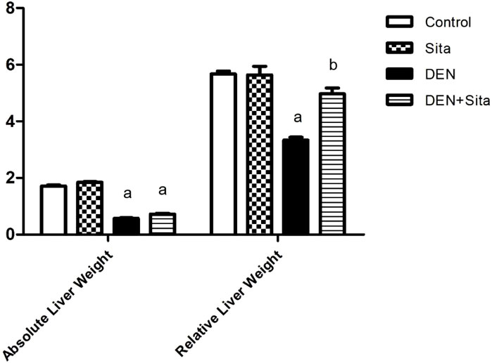
Figure 4. Effect of sitagliptin on absolute and relative liver weights in male mice expressing hepatic preneoplastic lesions induced by DEN. a sig. from control. b sig from DEN at p < 0.05. Each value represents mean ± standard error of mean (S.E.M.) (n = 10).
3.2.3 Effect of sitagliptin on serum liver enzymes in male mice expressing hepatic preneoplastic lesions induced by DEN
Figure 5 shows a prominent rise in serum AST, ALT and ALP activities in DEN group to reach 3.70, 3.67 and 6.31 folds, respectively as compared to the control group. Treatment with sitagliptin normalized serum ALT level and decreased serum levels of AST and ALP to reach 38% and 21%, respectively as compared to DEN group values.

Figure 5. Effect of sitagliptin on serum AST (A), ALT (B), and ALP (C) in male mice expressing hepatic preneoplastic lesions induced by DEN. a sig. from control. b sig. from DEN at p < 0.05. Each value represents mean ± standard error of mean (S.E.M.) (n = 10). AST (Aspartate transaminase); ALT (Alanine transaminase); ALP (Alkaline phosphatase).
3.2.4 Effect of sitagliptin on tissue GGT, DDP-4, TNF-α and IL-6 in male mice expressing hepatic preneoplastic lesions induced by DEN
A profound rise of GGT, DPP-4, TNF-α and IL-6 tissue levels to reach 9.73, 9.40, 9.41 and 9.70 folds, respectively as compared to control group was observed in DEN-injected mice (Figure 6). In contrast, treatment with sitagliptin restored tissue GGT to normal value and significantly decreased tissue levels of DPP-4, TNF-α and IL-6 to reach 21%, 20% and 17%, respectively as compared to DEN group.
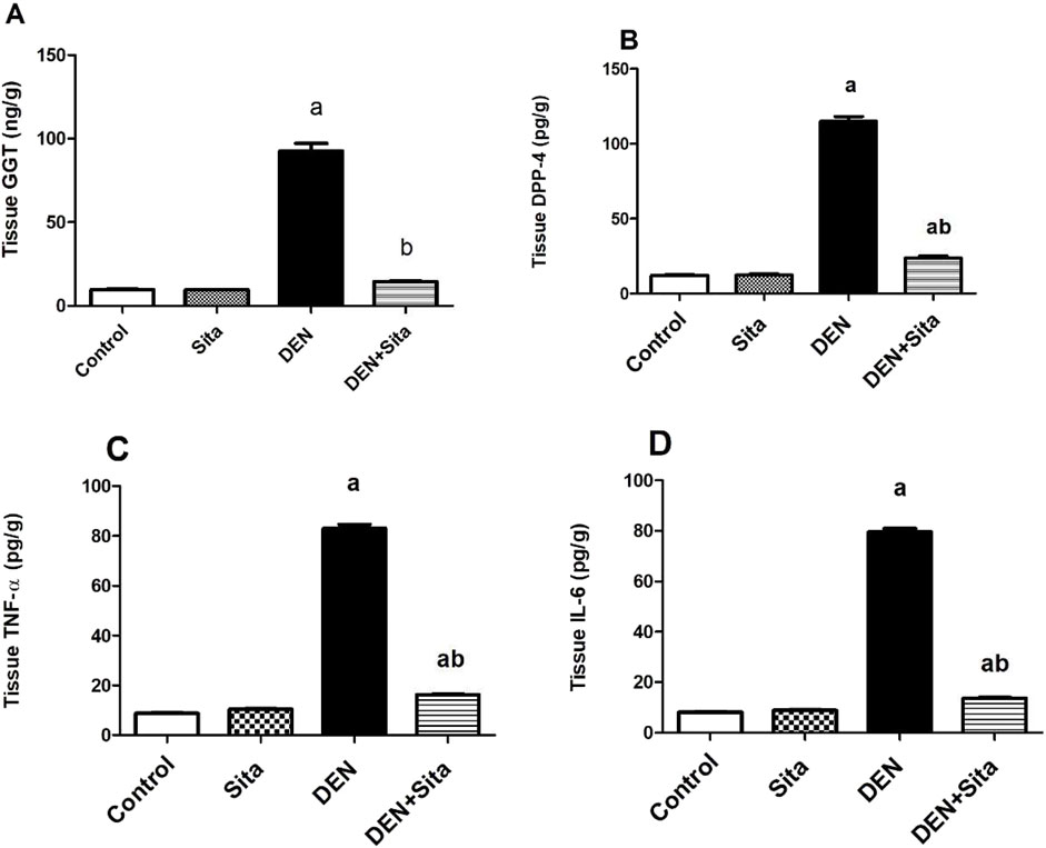
Figure 6. Effect of sitagliptin on tissue GGT (A), DDP-4 (B), TNF-α (C) and IL-6 (D) in male mice expressing hepatic preneoplastic lesions induced by DEN. a sig. from control. b sig. from DEN at p < 0.05. Each value represents mean ± standard error of mean (S.E.M.) (n = 10). GGT (Gamma glutamyl transpeptidase); DPP-4 (Dipeptidyl peptidase-4); TNF-α (Tumor necrosis factor alpha); IL-6 (Interleukin-6).
3.2.5 Effect of sitagliptin on liver CYP2E1 and CYP3A4 levels in male mice expressing hepatic preneoplastic lesions induced by DEN
Figure 7 shows that DEN induced a significant increment in tissue CYP2E1 level to reach 6.65 folds along with a significant decline in tissue CYP3A4 level to reach 21% as compared to control group. However, sitagliptin treatment attenuated these changes as evidenced by decreased CYP2E1 level to 28% and increased CYP3A4 level to 3.23 folds as compared to DEN-injected animals.
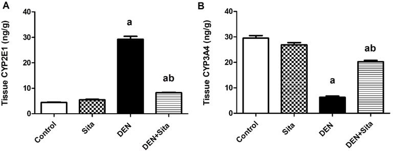
Figure 7. Effect of sitagliptin on liver CYP2E1 (A) and CYP3A4 (B) levels in male mice expressing hepatic preneoplastic lesions induced by DEN. a sig. from control. b sig. from DEN at p < 0.05. Each value represents mean ± standard error of mean (S.E.M.) (n = 10). CYP2E1 (Cytochrome P 2E1); CYP3A4 (Cytochrome P 3A4).
3.2.6 Effect of sitagliptin on DNA damage in male mice expressing hepatic preneoplastic lesions induced by DEN
Data in Table 2 and Figure 8 reveal that liver tissues from DEN group exhibited marked DNA damage to reach 3.3 folds as compared to the control group. Sitagliptin administration to DEN-injected mice significantly ameliorated this DNA damage to reach 63% that of DEN group value.

Table 2. Effect of sitagliptin on DNA damage in male mice expressing hepatic preneoplastic lesions induced by DEN.
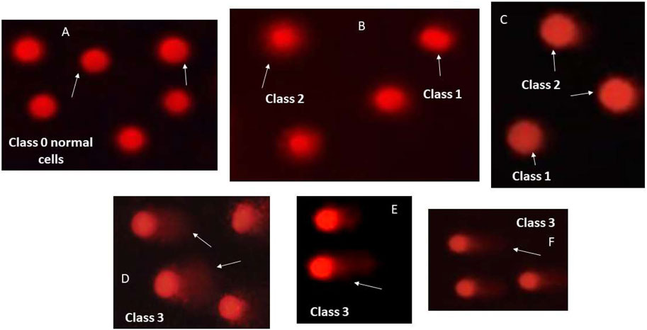
Figure 8. Visual scores of DNA damage using comet assay. Representative images of comet assay illustrating different types of cell classes. (A) class 0 normal cells without tail; (B,C) class 1 and 2 tailed cells; and (D–F): class 3 cells with extremely tailed cells.
3.2.7 Effect of sitagliptin on histopathological and morphometric alterations in male mice expressing hepatic preneoplastic lesions induced by DEN
Sections from control and sitagliptin alone groups revealed preserved architecture (Figures 9A–D). Hepatocytes are distributed regularly in single-cell thick plates radiating from a central vein. Sinusoids were observed between liver cell plates. However, sections from DEN group showed eosinophilic hepatocellular altered foci, bile ductular proliferation, bizarre nuclei, and inflammatory cell aggregates (Figures 9E,F). The borders of the cellular altered foci were irregular, discrete and distinguished from the surrounding tissue. The cells in the eosinophilic altered foci were large having eosinophilic cytoplasm with large nuclei in the center containing prominent nucleoli. Clear hepatocellular altered foci consisted of enlarged cells with balloon-like appearance having very pale to clear eosinophilic cytoplasm with central nuclei. Marked enlargement of the nucleus, hyperchromasia, pleomorphism (size and shape variation) and bizarre nuclei were also noticed (Figure 9F). Dilated central vein and inflammatory aggregates were observed in DEN-injected mice tissue sections (Figure 9G). Prominent nucleoli and intranuclear inclusions were also observed in DEN group sections (Figure 9H). Moreover, a significant rise in altered hepatocellular foci, nuclear hyperchromasia/pleomorphism, ductular proliferation, cholestasis, and inflammatory cell aggregates area percentages to reach 88.70, 18.57, 5.88, 21.79 and 17.38 folds, respectively was observed in DEN group as compared to control values (Figure 9K).
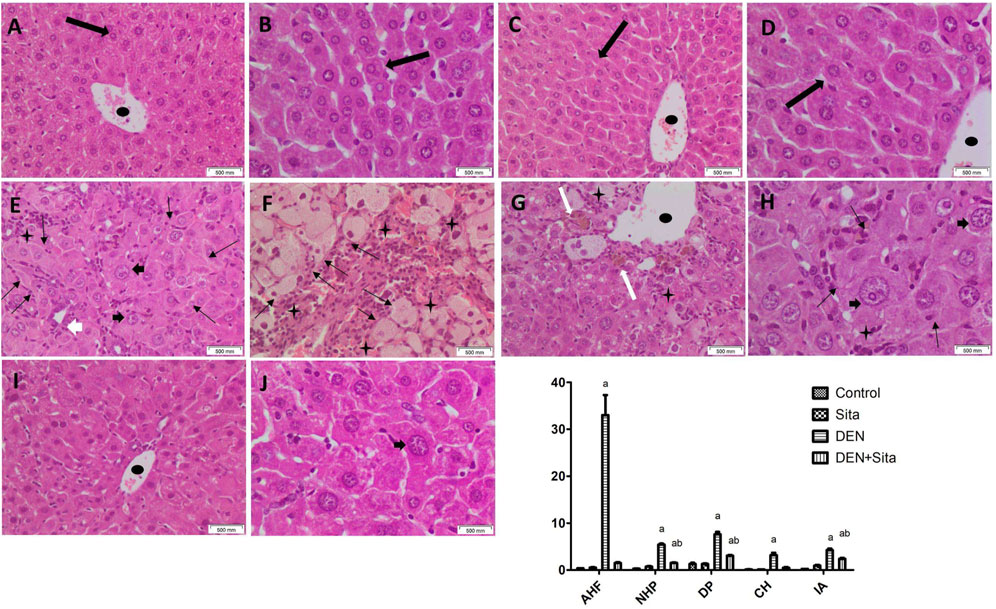
Figure 9. Effect of sitagliptin on histopathological and morphometric alterations in male mice expressing hepatic preneoplastic lesions induced by DEN. Photomicrographs of liver tissue sections stained with H&E. (A,B) (x200 and x400, respectively): Control group showing preserved architecture with hepatocytes distributed regularly in single cell thick plates radiating from a central vein. (C,D) (x200 and x400, respectively): Sitagliptin alone group showing normal liver architecture. (E–G) (x200): diseased group showing eosinophilic AHF having discrete but irregular borders, clear AHF, IA, CH and dilated central vein. (H) (x400): DEN-injected mice showing marked NHP, bizarre nuclei together with intra-nuclear inclusions. (I,J) (x200 and x400, respectively): DEN-injected mice treated with sitagliptin showing apparently normal hepatocytes with minimal residual nuclear size variation. AHF (thin black arrow), normal (thick black arrow), IA (black star), central vein (black oval shape), NHP (black arrow heads), CH (thick white arrow) and DP (white arrowhead). (K) Morphometric analysis (area%). a sig. from control. b sig from DEN at p < 0.05. Each value represents mean ± standard error of mean (S.E.M.) (10 sections per group). AHF (Altered Hepatocellular Foci); NHP (Nuclear Hyperchromasia and Pleomorphism); DP (Ductular Proliferation); CH (Cholestasis); IA (Inflammatory Aggregates).
Sections from sitagliptin treatment group illustrated restoration of the preserved liver architecture (Figures 9I,J). The hepatocytes are regularly distributed in single-cell thick plates radiating from the central vein. Neither hepatocellular altered foci nor bizarre nuclei were observed, only minimal residual nuclear size variation was noticed. A marked decline in area percentages of nuclear ductular proliferation, hyperchromasia/pleomorphism, and inflammatory aggregates to reach 40%, 28% and 48%, respectively was also observed in liver tissue sections of mice treated with sitagliptin compared to DEN group (Figure 9K).
3.2.8 Effect of sitagliptin effect on GST-P, PCNA and NF-κB immunoreactivity in male mice expressing hepatic preneoplastic lesions induced by DEN
As presented in Figures 10A,B, liver sections from control and sitagliptin alone mice showed homogenous GST-P distribution with few GST-P-positive cells. However, DEN-injected mice exhibited a massive positive cytoplasmic immunoreactivity for GST-P to 406 folds as compared to the control group (Figure 10C). Sitagliptin treatment attenuated these effects as indicated by lesser GST-P immunoreactivity to 21% as compared to DEN group (Figure 10D).
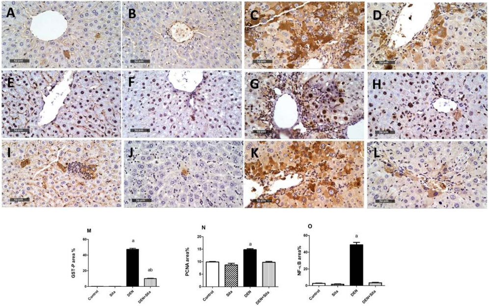
Figure 10. Effect of sitagliptin on GST-P, PCNA, and NF-κB immunoreactivity in male mice expressing hepatic preneoplastic lesions induced by DEN. Mice liver tissue sections A-D stained for GST-P. (A) Control, (B) Sita, (C) DEN, and (D) DEN + Sita. Liver tissue sections (E–H) stained for PCNA. (E) Control, (F) Sita, (G) DEN, and (H) DEN + Sita. Liver tissue sections (I–L) stained for NF-κB. (I) Control, (J) Sita, (K) DEN, and (L) DEN + Sita. (M–O) the area% of GST-P: Glutathione S transferase, PCNA: Proliferating cellular nuclear antigen, and NF-κB: Nuclear factor Kappa B, respectively. a sig. from control. b sig from DEN at p < 0.05. Each value represents the mean ± standard error of mean (S.E.M.) (10 sections per group).
Similarly, liver sections from control and sitagliptin alone mice showed few PCNA-positive cells (Figures 10E,F). In contrast, DEN caused a noticeable increase in PCNA-positive cells to 1.5 folds compared with the control groups (Figure 10G). The sitagliptin treatment group manifested a clear decrease in PCNA-positive cells to 65.97% compared with the DEN group (Figure 10H).
Liver sections from control and sitagliptin alone mice demonstrated negative immunohistochemical reaction for NF-κB (Figures 10I,J). However, NF-κB was upregulated in DEN-treated animals as evidenced by positive immune reaction (18 fold) as compared to the control group (Figure 10K). Sections from sitagliptin-treated mice revealed a weaker positive expression of 7% as compared to the DEN group (Figure 10L).
4 Discussion
HCC, a leading cause of cancer-related mortality, typically arises in the background of chronic liver disease such as hepatitis or cirrhosis. Due to its asymptomatic progression and late-stage diagnosis, current therapeutic strategies are often inadequate, necessitating the exploration of safer and more effective chemopreventive agents (Li et al., 2021; You et al., 2021).
Results of the present study revealed that treatment of HepG2 cells with sitagliptin induced a dose-dependent cytotoxic effect with an IC50 of 89 µmole. In addition, sitagliptin treatment also resulted in a marked downregulation of the mRNA expression of the anti-apoptotic BCL2 along with a significant upregulation of the expression of the pro-apoptotic BAX, the tumor suppressor p53 and BAX/BCL2 ratio in HepG2 cells. In line, a previous study reported the cytotoxic effect of sitagliptin in HepG2 cells via its anti-proliferative and apoptogenic effects (Sarkar et al., 2017). In contrast, Sliwinska et al. (2015) reported that sitagliptin did not affect HepG2 cell viability in the dose range 0.01–200 μmole.
DEN is a chemical agent that has been extensively used for the experimental induction of hepatic preneoplasia and cancer (Gupta et al., 2010; Alsahli et al., 2021). DEN activation occurs through the CYP 450 system in the liver, especially CYP2E1, and produces DNA-adducts via an alkylation mechanism (You et al., 2021). Our results confirmed that injection of DEN to mice produced a significant depression in the final body, absolute and relative liver weights, as previously reported in the literature (Li et al., 2022). However, sitagliptin treatment succeeded in normalizing relative liver weight only. The present results also confirmed that DEN injection to mice induced marked alterations in liver function as previously reported (Memon et al., 2020). Treatment with sitagliptin attenuated DEN-induced increases in serum AST, ALT, and ALP. In agreement with our findings, sitagliptin was stated to improve liver function in non-alcoholic fatty liver disease in mice (Xu et al., 2017) and hepatic ischemia reperfusion in rats (Abdel-Gaber et al., 2019).
GGT catalyzes the main step in the degradation of reduced glutathione and is considered as a marker of liver injury (Kim et al., 2018). Elevated GGT activity leads to increased free radical production (Alam et al., 2018; Jiang et al., 2018). Furthermore, elevated GGT is associated with increased cancer risk, and may be implicated in the activation of pro-oncogenes or the inactivation of tumor-suppressor genes (Moreira et al., 2015). The present results show that sitagliptin treatment decreased tissue GGT in DEN-injected mice.
DPP-4 enzyme is an essential biomarker in cancer disease biology (Kawakita et al., 2021; Busek et al., 2022). DPP-4 enzyme is responsible for the cleavage of the N-terminal dipeptides of proline or alanine-containing peptides like incretins. In addition, DPP-4 was reported to play an important role in inflammation, immune system function, and resistance to anticancer agents (Itou et al., 2013). Moreover, high expression of the DPP-4 enzyme may contribute to the progression of liver cancer (Nishina et al., 2018).
In addition, tumor size, stage, and proliferation in HCC were related to DPP-4 enzyme level (Busek et al., 2022). Herein, tissue DPP-4 enzyme was significantly reduced after treatment with sitagliptin, which is sitagliptin’s core mechanism of action.
Cytokines are pleiotropic hormones of the immune system that play a fundamental role in the initiation, maintenance, and progression of cancer (Lee and Margolin, 2011). Neoplastic cells and tumor-associated macrophages secrete numerous cytokines as well as angiogenic growth factors that promote tumor development (Khan et al., 2017; Alsahli et al., 2021). NF-κB signaling is a key arm in the inflammatory process of the tumor microenvironment, as it regulates many genes encoding for interleukins, inducible effector enzymes, and chemokines (Zhang et al., 2021). The current data reveal that sitagliptin treatment produced a prominent reduction in DEN-induced increases in tissue TNF-α, IL-6, and NF-κB p65. These results confirm that the anti-inflammatory activity of sitagliptin is mediated through suppression of NF-κB signaling, as previously reported in DEN-injected rats (Jiang et al., 2018).
Sitagliptin’s inhibition of DPP-4 may modulate inflammatory signaling pathways, including NF-κB, through indirect effects on cytokine release and immune cell recruitment (Jiang et al., 2018).
A previous study reported that treatment with sitagliptin inhibited NF-κB p53 activation (Zheng et al., 2018). Another previous study showed that anagliptin prevented LPS-induced elevations of inflammatory cytokines in macrophages, adipocytes, and the liver in mice through suppression of NF-κB transcriptional activity (Shinjo et al., 2015).
The various metabolic activities that CYP3A4 catalyzes result in the inactivation and removal of the majority of drugs (Maideen, 2019). Further, CYP2E1 plays a critical role in the activation of many carcinogenic agents, including DEN (Gao et al., 2017). The current results show that sitagliptin treatment decreased CYP2E1 and increased CYP3A4 levels in the liver of DEN-injected mice. In parallel with current results, sitagliptin was reported to increase the expression of CYP2E1 in mice fed with a methionine/choline-deficient diet (Jung et al., 2014). Sitagliptin is predominantly eliminated via urine and feces, with 79% of the dose actively secreted unchanged in urine (Gu et al., 2020). The CYP3A4 isozyme is responsible for the limited oxidative metabolism of sitagliptin (Mistry et al., 2008), which may explain the effect of sitagliptin on CYP3A4 levels observed in the present study. The increase in CYP3A4 expression observed in DEN-injected mice following sitagliptin treatment may stem from its anti-inflammatory and hepatoprotective properties, potentially mitigating cytokine-induced suppression of CYP3A4 expression.
Most drugs are inactivated and eliminated due to the various metabolic functions mediated by CYP3A4 (Maideen, 2019). Moreover, CYP2E1 is an essential enzyme in the bioactivation of numerous carcinogens such as DEN (Gao et al., 2017). The present data suggest that sitagliptin treatment reduced liver CYP2E1 and increased CYP3A4 in the livers of DEN-injected mice. The observed induction of CYP3A4 by sitagliptin could be attributed to sitagliptin’s anti-inflammatory and hepatoprotective effects, thereby inhibiting the cytokine-induced suppression of CYP3A4 that is often observed in hepatic inflammation and preneoplasia. Inflammatory cytokines, including IL-6 and TNF, have also been found to inhibit the nuclear receptors (PXR, CAR, etc.) associated with CYP3A4 gene expression. The reduction of IL-6 and TNF-α due to sitagliptin, as seen in our study, might also have restored and/or stimulated CYP3A4 expression. This is in line with previous studies showing that DPP-4 inhibitors preserved or even increased CYP3A4 levels via suppression of inflammation (Jiang et al., 2018; Kawakita et al., 2021). Moreover, because sitagliptin is eliminated primarily unchanged and only minimally via CYP3A4-mediated metabolism (Mistry et al., 2008), we would not expect it to inhibit the activity of this enzyme and perhaps even maintain its hepatic expression when exposed to toxic injury.
Our results also show that sitagliptin treatment resulted in a significant reduction of DEN-induced liver DNA damage. This finding is in line with a study demonstrating that sitagliptin reduced total body irradiation-induced DNA double-strand breaks in the hematopoietic cells in mice via decreasing the expression of γH2AX (Wang et al., 2020), which is a concrete and sensitive molecular marker for examining DNA damage (Mah et al., 2010). Furthermore, sitagliptin markedly reduced the ratio of 8-hydroxy-2' –deoxyguanosine (8-OHdG) positive cells, another marker of oxidative DNA damage found in the liver samples isolated from animals with non-alcoholic steatohepatitis and HCC (Onoyama et al., 2015; You et al., 2021).
The results of the histopathological and morphometric analysis further confirmed our findings. Sitagliptin decreased the formation of inflammatory aggregates, nuclear hyperchromasia, and pleomorphism in liver sections. In addition, sitagliptin mitigated the altered hepatocellular foci, cholestasis, and ductular proliferation.
It was documented that GST-P is highly expressed in preneoplastic lesions and HCC induced by genotoxic carcinogens (Zhu et al., 1998; Gamal-Eldeen et al., 2016). Overexpression of GST-P in response to DEN signifies the initiation of liver carcinogenesis (Assar et al., 2021). Herein, sitagliptin treatment significantly decreased the immunoexpression of GST-P in the liver of DEN-treated mice. In agreement, Okura et al. (2017) reported that the size and number of GST-P-positive preneoplastic lesions significantly decrease after sitagliptin treatment. This could be related to its inhibitory effect on hepatic vascular endothelial growth factor (VEGF) and cluster of differentiation 31 (CD31) mRNA expressions. Proliferation has a crucial role during the initiation and promotion of carcinogenesis (Tablas et al., 2018). Synthesis of PCNA, a marker of cell proliferation, is initiated in the nucleus in late G1 phase and remains during the S phase (Arboatti et al., 2018). In the present study, sitagliptin reduced PCNA liver expression in DEN-injected mice, indicating its anti-proliferative activity.
5 Conclusion
Our findings demonstrate that sitagliptin exerted a significant cytotoxic effect on HepG2 cells, which was dependent on elevating mRNA expression of p53 and BAX/BCL2. Sitagliptin improved serum liver enzymes and attenuated histopathological changes in DEN-induced hepatic preneoplasia in mice via inhibition of the DPP-4 enzyme. These changes were accompanied by reducing liver GGT, CYP2E1, GGT-P, NF-κB, and PCNA along with increasing CYP3A4. Furthermore, sitagliptin attenuated DEN-induced liver DNA damage and inflammation. These findings suggest a promising effect of sitagliptin in the management of hepatic cancer initiation and progression.
Data availability statement
The original contributions presented in the study are included in the article; further inquiries can be directed to the corresponding authors.
Ethics statement
Ethical approval was not required for the studies on humans in accordance with the local legislation and institutional requirements because only commercially available established cell lines were used. The animal study was approved by Approval for this investigation was granted by the Ethics Committee for Animal Experimentation at the Faculty of Pharmacy, Cairo University (Permit Number: PT 2198) and the Ethics Committee for Animal Experimentation at the National Research Centre, Giza, Egypt (18047). The study was conducted in accordance with the local legislation and institutional requirements.
Author contributions
HE: Conceptualization, Data curation, Formal Analysis, Investigation, Methodology, Writing – original draft. RS: Supervision, Validation, Visualization, Writing – review and editing. YM: Funding acquisition, Project administration, Supervision, Validation, Writing – review and editing. AG: Formal Analysis, Methodology, Visualization, Writing – review and editing. KM: Funding acquisition, Supervision, Validation, Writing – review and editing. AE-S: Investigation, Methodology, Supervision, Writing – review and editing. AnA: Validation, Writing – review and editing. AlA: Validation, Writing – review and editing. HN: Funding acquisition, Project administration, Supervision, Validation, Writing – review and editing.
Funding
The author(s) declare that financial support was received for the research and/or publication of this article. National Research Centre (Giza, Egypt) has funded this study (12020103).
Acknowledgments
The authors gratefully acknowledge the funding of the Deanship of Graduate Studies and Scientific Research, Jazan University, Saudi Arabia.
Conflict of interest
The authors declare that the research was conducted in the absence of any commercial or financial relationships that could be construed as a potential conflict of interest.
Generative AI statement
The author(s) declare that no Generative AI was used in the creation of this manuscript.
Publisher’s note
All claims expressed in this article are solely those of the authors and do not necessarily represent those of their affiliated organizations, or those of the publisher, the editors and the reviewers. Any product that may be evaluated in this article, or claim that may be made by its manufacturer, is not guaranteed or endorsed by the publisher.
References
Abdel-Gaber, S. A., Geddawy, A., and Moussa, R. A. (2019). The hepatoprotective effect of sitagliptin against hepatic ischemia reperfusion-induced injury in rats involves Nrf-2/HO-1 pathway. Pharmacol. Rep. 71 (6), 1044–1049. doi:10.1016/j.pharep.2019.06.006
Alam, S., Ghosh, J., Mustafa, G., Kamal, M., and Ahmad, N. (2018). Effect of sitagliptin on hepatic histological activity and fibrosis of nonalcoholic steatohepatitis patients: a 1-year randomized control trial. Hepatic Med. 10, 23–31. doi:10.2147/HMER.S158053
Almagthali, A. G., Alkhaldi, E. H., Alzahrani, A. S., Alghamdi, A. K., Alghamdi, W. Y., and Kabel, A. M. (2019). Dipeptidyl peptidase-4 inhibitors: anti-diabetic drugs with potential effects on cancer. Diabetes Metabolic Syndrome 13 (1), 36–39. doi:10.1016/j.dsx.2018.08.012
Alsahli, M. A., Almatroodi, S. A., Almatroudi, A., Khan, A. A., Anwar, S., Almutary, A. G., et al. (2021). 6-Gingerol, a major ingredient of ginger attenuates diethylnitrosamine -Induced liver injury in rats through the modulation of oxidative stress and anti-inflammatory activity. Mediat. Inflamm. 2021, 6661937. doi:10.1155/2021/6661937
Arboatti, A. S., Lambertucci, F., Sedlmeier, M. G., Pisani, G., Monti, J., De, M., et al. (2018). Diethylnitrosamine increases proliferation in early stages of hepatic carcinogenesis in insulin-treated type 1 diabetic mice. Biomed Res. Int. 2018, 9472939. doi:10.1155/2018/9472939
Assar, D. H., Mokhbatly, A. A., Ghazy, E. W., Ragab, A. E., Asa, S. A., Abdo, W., et al. (2021). Ameliorative effects of Aspergillus awamori against the initiation of hepatocarcinogenesis induced by diethylnitrosamine in a rat model: regulation of Cyp19 and p53 gene expression. Antioxidants 10, 922. doi:10.3390/antiox10060922
Augustyniak, M., Gladysz, M., and Dziewięcka, M. (2016). The comet assay in insects-Status, prospects and benefits for science. Mutat. Res. - Rev. Mutat. Res. 767, 67–76. doi:10.1016/j.mrrev.2015.09.001
Blasiak, J., Arabski, M., Krupa, R., Wozniak, K., Zadrozny, M., Kasznicki, J., et al. (2004). DNA damage and repair in type 2 diabetes mellitus. Mutat. Res. 554, 297–304. doi:10.1016/j.mrfmmm.2004.05.011
Buitrago-Molina, L. E., Marhenke, S., Longerich, T., Sharma, A. D., Boukouris, A. E., Geffers, R., et al. (2013). The degree of liver injury determines the role of p21 in liver regeneration and hepatocarcinogenesis in mice. Hepatology 58 (3), 1143–1152. doi:10.1002/hep.26412
Busek, P., Duke-cohan, J. S., and Sedo, A. (2022). Does DPP-IV inhibition offer new avenues for therapeutic intervention in malignant disease. Cancers (Basel) 14 (9), 2072. doi:10.3390/cancers14092072
Culling, C. F. (2013). Handbook of histopathological and histochemical techniques. 3rd ed. London, UK: Butterworths.
Eitah, H. E., Attia, H. N., Soliman, A. A. F., Gamal El Din, A. A., Mahmoud, K., Sayed, R. H., et al. (2023). Vitamin D ameliorates diethylnitrosamine-induced liver preneoplasia: a pivotal role of CYP3A4/CYP2E1 via DPP-4 enzyme inhibition. Toxicol. Appl. Pharmacol. 458, 116324. doi:10.1016/j.taap.2022.116324
El-Kashef, D. H., and Serrya, M. S. (2019). Sitagliptin ameliorates thioacetamide-induced acute liver injury via modulating TLR4/NF-KB signaling pathway in mice. Life Sci. 228, 266–273. doi:10.1016/j.lfs.2019.05.019
El-menshawi, B. S., Fayad, W., Mahmoud, K., El-hallouty, S. M., El-manawaty, M., Olofsson, M. H., et al. (2010). Screening of natural products for therapeutic activity against solid tumors. Indian J. Exp. Biol. 48 (3), 258–264.
Fairbairn, D. W., Olive, P. L., and O’Neill, K. L. (1995). The comet assay: a comprehensive review. Mutat. Research/Reviews Genet. Toxicol. 339 (1), 37–59. doi:10.1016/0165-1110(94)00013-3
Gamal-Eldeen, A. M., Moustafa, D., El-Daly, S. M., El-Hussieny, E. A., Saleh, S., Khoobchandani, M., et al. (2016). Photothermal therapy mediated by gum Arabic-conjugated gold nanoparticles suppresses liver preneoplastic lesions in mice. J. Photochem. Photobiol. B 163, 47–56. doi:10.1016/j.jphotobiol.2016.08.009
Gao, J., Wang, G. J., Wang, Z., Gao, N., Li, J., Zhang, Y. F., et al. (2017). High CYP2E1 activity correlates with hepatofibrogenesis induced by nitrosamines. Oncotarget 8 (68), 112199–112210. doi:10.18632/oncotarget.22937
Gault, V. A., Lennox, R., and Flatt, P. R. (2015). Sitagliptin, a dipeptidyl peptidase-4 inhibitor, improves recognition memory, oxidative stress and hippocampal neurogenesis and upregulates key genes involved in cognitive decline. Diabetes, Obes. Metabolism 17 (4), 403–413. doi:10.1111/dom.12432
Gu, N., Park, S. I., Chung, H., Jin, X., Lee, S., and Kim, T. E. (2020). Possibility of pharmacokinetic drug interaction between a DPP-4 inhibitor and a SGLT2 inhibitor. Transl. Clin. Pharmacol. 28 (1), 17–33. doi:10.12793/tcp.2020.28.e4
Gupta, C., Vikram, A., Tripathi, D. N., Ramarao, P., and Jena, G. B. (2010). Antioxidant and antimutagenic effect of quercetin against DEN induced hepatotoxicity in rat. Phytothearpy Res. 24 (1), 119–128. doi:10.1002/ptr.2883
Havre, P. A., Abe, M., Urasaki, Y., Ohnuma, K., Morimoto, C., and Dang, N. H. (2008). The role of CD26/dipeptidyl peptidase IV in cancer. Front. Biosci. 13 (5), 1634–1645. doi:10.2741/2787
Itou, M., Kawaguchi, T., Taniguchi, E., and Sata, M. (2013). Dipeptidyl peptidase-4: a key player in chronic liver disease. World J. Gastroenterology 19 (15), 2298–2306. doi:10.3748/wjg.v19.i15.2298
Ji, S. B., Park, S. Y., Bae, S., Seo, H. J., Kim, S. E., Lee, G. M., et al. (2021). Comprehensive investigation of stereoselective food drug interaction potential of resveratrol on nine P450 and six UGT isoforms in human liver microsomes. Pharmaceutics 13 (9), 1419. doi:10.3390/pharmaceutics13091419
Jiang, W., Wen, D., Cheng, Z., Yang, Y., Zheng, G., and Yin, F. (2018). Effect of sitagliptin, a DPP-4 inhibitor, against DENA-induced liver cancer in rats mediated via NF-κB activation and inflammatory cytokines. J. Biochem. Mol. Toxicol. 32 (12), e22220. doi:10.1002/jbt.22220
Jung, Y. A., Choi, Y. K., Jung, G. S., Seo, H. Y., Kim, H. S., Jang, B. K., et al. (2014). Sitagliptin attenuates methionine/choline-deficient diet-induced steatohepatitis. Diabetes Res. Clin. Pract. 105 (1), 47–57. doi:10.1016/j.diabres.2014.04.028
Kang, J. S., Wanibuchi, H., Morimura, K., Gonzalez, F. J., and Fukushima, S. (2007). Role of CYP2E1 in diethylnitrosamine-induced hepatocarcinogenesis in vivo. Cancer Res. 67 (23), 11141–11146. doi:10.1158/0008-5472.CAN-07-1369
Katakami, N., Mita, T., Irie, Y., Takahara, M., Matsuoka, T., Gosho, M., et al. (2018). Effect of sitagliptin on tissue characteristics of the carotid wall in patients with type 2 diabetes: a post hoc sub - analysis of the sitagliptin preventive study of intima - media thickness evaluation (SPIKE). Cardiovasc. Diabetol. 17, 24. doi:10.1186/s12933-018-0666-3
Kawakita, E., Koya, D., and Kanasaki, K. (2021). Cd26/dpp-4: type 2 diabetes drug target with potential influence on cancer biology. Cancers 13 (9), 2191. doi:10.3390/cancers13092191
Kawakubo, M., Tanaka, M., Ochi, K., Watanabe, A., Saka-Tanaka, M., Kanamori, Y., et al. (2020). Dipeptidyl peptidase-4 inhibition prevents nonalcoholic steatohepatitis-associated liver fibrosis and tumor development in mice independently of its anti-diabetic effects. Scintific Rep. 10 (1), 983. doi:10.1038/s41598-020-57935-6
Khan, F., Khan, T. J., Kalamegam, G., Pushparaj, P. N., Chaudhary, A., Abuzenadah, A., et al. (2017). Anti-cancer effects of Ajwa dates (phoenix dactylifera L.) in diethylnitrosamine induced hepatocellular carcinoma in wistar rats. BMC Complementary Altern. Med. 17 (1), 418–10. doi:10.1186/s12906-017-1926-6
Kim, J. G., Chang, K., Choo, E. H., Lee, J. M., and Seung, K. B. (2018). Serum gamma-glutamyl transferase is a predictor of mortality in patients with acute myocardial infarction. Med. Baltim. 97 (29), e11393. doi:10.1097/MD.0000000000011393
Lee, S., and Margolin, K. (2011). Cytokines in cancer immunotherapy. Cancers (Basel) 3 (4), 3856–3893. doi:10.3390/cancers3043856
Lee, S., Nam, H., Kim, J., Cho, H., Jang, Y., Lee, E., et al. (2012). Body weight changes of laboratory animals during transportation. Asian-Australas J. Anim. Sci. 25 (2), 286–290. doi:10.5713/ajas.2011.11227
Li, G., Qi, L., Chen, H., and Tian, G. (2022). Involvement of NF-κB/PI3K/AKT signaling pathway in the protective effect of prunetin against a diethylnitrosamine induced hepatocellular carcinogenesis in rats. J. Biochem. Mol. Toxicol. 36, e23016. doi:10.1002/jbt.23016
Li, R., and Shen, Y. (2013). An old method facing a new challenge: re-visiting housekeeping proteins as internal reference control for neuroscience research. Life Sci. 92 (13), 747–751. doi:10.1016/j.lfs.2013.02.014
Li, S., Li, Y., Sun, H., Jiang, Y., Pan, K., Su, Y., et al. (2021). Mulberry fruit polysaccharides alleviate diethylnitrosamine/phenobarbital-induced hepatocarcinogenesis in vivo: the roles of cell apoptosis and inflammation. Bioengineered 12 (2), 11599–11611. doi:10.1080/21655979.2021.1993716
Mah, L. J., El-Osta, A., and Karagiannis, T. C. (2010). gammaH2AX: a sensitive molecular marker of DNA damage and repair. Leukemia 24, 679–686. doi:10.1038/leu.2010.6
Maideen, N. M. P. (2019). Drug interactions of dipeptidyl peptidase 4 inhibitors involving CYP enzymes and P-gp efflux pump. World J. Meta-Analysis 7 (4), 156–161. doi:10.13105/wjma.v7.i4.156
Memon, A., Pyao, Y., Jung, Y., Lee, J.Il., and Lee, W. K. (2020). A modified protocol of diethylnitrosamine administration in mice to model hepatocellular carcinoma. Int. J. Mol. Sci. 21 (15), 5461. doi:10.3390/ijms21155461
Mest, H. J., and Mentlein, R. (2005). Dipeptidyl peptidase inhibitors as new drugs for the treatment of type 2 diabetes. Diabetologia 48 (4), 616–620. doi:10.1007/s00125-005-1707-5
Mistry, G. C., Bergman, A. J., Zheng, W., Hreniuk, D., Zinny, M. A., Gottesdiener, K. M., et al. (2008). Sitagliptin, an dipeptidyl peptidase-4 inhibitor, does not alter the pharmacokinetics of the sulphonylurea, glyburide, in healthy subjects. Br. J. Clin. Pharmacol. 66 (1), 36–42. doi:10.1111/j.1365-2125.2008.03148.x
Moreira, A. J., Rodrigues, G., Bona, S., Thadeu, C., Marroni, C. A., Mauriz, J. L., et al. (2015). Oxidative stress and cell damage in a model of precancerous lesions and advanced hepatocellular carcinoma in rats. Toxicol. Rep. 2, 333–340. doi:10.1016/j.toxrep.2014.11.015
Mosmann, T. (1983). Rapid colorimetric assay for cellular growth and survival: application to proliferation and cytotoxicity assays. J. Immunol. Methods 65 (1–2), 55–63. doi:10.1016/0022-1759(83)90303-4
Muñoz, R., de las Rivas, B., and Curiel, J. A. (2014). “Identification methods: multilocus sequence typing of food microorganisms,” in Encyclopedia of food microbiology. Editors C. A. Batt, and M. L. Tortorello 2nd ed. (Academic Press), 300–309. doi:10.1016/B978-0-12-384730-0.00412-2
Nair, A. B., and Jacob, S. (2016). A simple practice guide for dose conversion between animals and human. J. Basic Clin. Pharm. 7 (2), 27–31. doi:10.4103/0976-0105.177703
Nishina, S., Yamauchi, A., Kawaguchi, T., Kaku, K., Goto, M., Sasaki, K., et al. (2018). Dipeptidyl peptidase 4 inhibitors reduce hepatocellular carcinoma by activating lymphocyte chemotaxis in mice. Cell. Mol. Gastroenterology Hepatology 7 (1), 115–134. doi:10.1016/j.jcmgh.2018.08.008
Okura, Y., Namisaki, T., Moriya, K., Kitade, M., Takeda, K., Kaji, K., et al. (2017). Combined treatment with dipeptidyl peptidase-4 inhibitor (sitagliptin) and angiotensin-II type 1 receptor blocker (losartan) suppresses progression in a non-diabetic rat model of steatohepatitis. Hepatology Res. 47 (12), 1317–1328. doi:10.1111/hepr.12860
Onoyama, T., Koda, M., Okamoto, T., Kishina, M., Matono, T., Sugihara, T., et al. (2015). Therapeutic effects of the dipeptidyl peptidase-IV inhibitor, sitagliptin, on non-alcoholic steatohepatitis in FLS-ob/ob male mice. Mol. Med. Rep. 12 (5), 6895–6902. doi:10.3892/mmr.2015.4329
Ramadan, M. A., Shawkey, A. E., Rabeh, M. A., and Abdellatif, A. O. (2019). Expression of P53, BAX, and BCL-2 in human malignant melanoma and squamous cell carcinoma cells after tea tree oil treatment in vitro. Cytotechnology 71 (1), 461–473. doi:10.1007/s10616-018-0287-4
Sarkar, M., Dey, S., and Giri, B. (2017). Antiproliferative and apoptogenic efficacy of antidiabetic drugs metformin and sitagliptin against MCF7 and HepG2 cancer cells: a comparative mollecular study. J. Drug Deliv. Ther. 7 (6), 11–21. doi:10.22270/jddt.v7i6.1519
Sarkar, S., Bhattacharjee, P., Ghosh, T., and Bhadra, K. (2020). Pharmaceutical efficacy of harmalol in inhibiting hepatocellular carcinoma. Future J. Pharm. Sci. 6, 29. doi:10.1186/s43094-020-00045-x
Shen, T., Xu, B., Lei, T., Chen, L., Zhang, C., and Ni, Z. (2018). Sitagliptin reduces insulin resistance and improves rat liver steatosis via the SIRT1/AMPKα pathway. Exp. Ther. Med. 16 (4), 3121–3128. doi:10.3892/etm.2018.6554
Shinjo, T., Nakatsu, Y., Iwashita, M., Sano, T., Sakoda, H., Ishihara, H., et al. (2015). DPP-IV inhibitor anagliptin exerts anti-inflammatory effects on macrophages, adipocytes, and mouse livers by suppressing NF-κB activation. Am. J. Physiology Endocrinol. Metabolism 309 (3), E214–E223. doi:10.1152/ajpendo.00553.2014
Sliwinska, A., Rogalska, A., Marczak, A., Kasznicki, J., and Drzewoski, J. (2015). Metformin, but not sitagliptin, enhances WP 631-induced apoptotic HepG2 cell death. Toxicol. Vitro 29 (5), 1116–1123. doi:10.1016/j.tiv.2015.04.019
Tablas, M. B., Goto, R. L., Caetano, B. F. R., Santos, S. A. A., and Barbisan, L. F. (2018). Vitamin D 3 suppresses the early stages of chemically induced hepatocarcinogenesis in rats: a dose-response analysis. Nutrire 43 (1), 12–11. doi:10.1186/s41110-018-0065-2
Thabrew, M. I., Mitry, R. R., Morsy, M. A., and Hughes, R. D. (2005). Cytotoxic effects of a decoction of Nigella sativa, Hemidesmus indicus and Smilax glabra on human hepatoma HepG2 cells. Life Sci. 77 (12), 1319–1330. doi:10.1016/j.lfs.2005.01.022
Wang, M., Dong, Y., Wu, J., Li, H., Zhang, J., Lu, L., et al. (2020). Sitagliptin mitigates total body irradiation-induced hematopoietic injury in mice. Oxidative Med. Cell. Longev. 2020, 8308616. doi:10.1155/2020/8308616
Wang, X., Ke, J., Zhu, Y. J., Cao, B., Yin, R. L., Wang, Y., et al. (2021). Dipeptidyl peptidase-4 (DPP4) inhibitor sitagliptin alleviates liver inflammation of diabetic mice by acting as a ROS scavenger and inhibiting the NFκB pathway. Cell Death Discov. 7 (1), 236. doi:10.1038/s41420-021-00625-7
Xu, B., Shen, T., Chen, L., Xia, J., Zhang, C., Wang, H., et al. (2017). The effect of sitagliptin on lipid metabolism of fatty liver mice and related mechanisms. Med. Sci. Monit. 23, 1363–1370. doi:10.12659/msm.900033
You, Y., Zhu, F., Li, Z., Zhang, L. F., Xie, Y., Chinnathambi, A., et al. (2021). Phyllanthin prevents diethylnitrosamine (DEN) induced liver carcinogenesis in rats and induces apoptotic cell death in HepG2 cells. Biomed. and Pharmacother. 137, 111335. doi:10.1016/J.BIOPHA.2021.111335
Yu, H., Mei, X., Su, P., Jin, G., and Zhou, H. (2020). A poor prognosis in human hepatocellular carcinoma is associated with low expression of DPP4. Braz. J. Med. Biol. Res. 53 (4), 1–8. doi:10.1590/1414-431x20209114
Zhang, Y., Li, X., and Li, X. (2021). Curcumae ameliorates diethylnitrosamine-induced hepatocellular carcinoma via alteration of oxidative stress, inflammation and gut microbiota. J. Inflamm. Res. 14, 5551–5566. doi:10.2147/JIR.S330499
Zhao, Y. (2019). CD26 in autoimmune diseases: the other side of “moonlight protein.”. Int. Immunopharmacol. 75, 105757. doi:10.1016/j.intimp.2019.105757
Zheng, W., Zhou, J., Song, S., Kong, W., Xia, W., Chen, L., et al. (2018). Dipeptidyl-peptidase 4 inhibitor sitagliptin ameliorates hepatic insulin resistance by modulating inflammation and autophagy in ob/ob mice. Int. J. Endocrinol. 19, 8309723. doi:10.1155/2018/8309723
Zhou, C., Zhou, S., Wang, J., Xie, L., Lv, Z., Zhao, Y., et al. (2024). Safety, tolerability, pharmacokinetics and pharmacokinetic-pharmacodynamic modeling of cetagliptin in patients with type 2 diabetes mellitus. Front. Endocrinol. (Lausanne) 15, 1359407. doi:10.3389/fendo.2024.1359407
Keywords: CYP2E1, CYP3A4, diethylnitrosamine, DPP-4, hepatic preneoplasia, sitagliptin
Citation: Eitah HE, Sayed RH, Maklad YA, Gamal el Din AA, Mahmoud K, El-Sahar AE, Alhejely A, Abdulbaqi AA and Naeim Attia H (2025) Dipeptidyl peptidase-4 enzyme inhibition and its impacts on hepatic preneoplasia: a new avenue for liver cancer management. Front. Pharmacol. 16:1559303. doi: 10.3389/fphar.2025.1559303
Received: 14 January 2025; Accepted: 27 June 2025;
Published: 25 July 2025.
Edited by:
David E. Stec, University of Mississippi Medical Center, United StatesReviewed by:
Xinning Yang, United States Food and Drug Administration, United StatesVladimir Lj Jakovljevic, University of Kragujevac, Serbia
Copyright © 2025 Eitah, Sayed, Maklad, Gamal el Din, Mahmoud, El-Sahar, Alhejely, Abdulbaqi and Naeim Attia. This is an open-access article distributed under the terms of the Creative Commons Attribution License (CC BY). The use, distribution or reproduction in other forums is permitted, provided the original author(s) and the copyright owner(s) are credited and that the original publication in this journal is cited, in accordance with accepted academic practice. No use, distribution or reproduction is permitted which does not comply with these terms.
*Correspondence: Hebatollah E. Eitah, aGViYXRvbGxhaGVpdGFoMDI3QGdtYWlsLmNvbQ==, aGUuYWJkZWxhYWxAbnJjLnNjaS5lZw==
†ORCID: Hebatollah E. Eitah, orcid.org/0000-0002-6936-5150
 Hebatollah E. Eitah
 Rabab H. Sayed
 Yousreya A. Maklad1
 Ayman E. El-Sahar
 Amal A. Abdulbaqi
 Hanan Naeim Attia