- 1Nanjing University of Chinese Medicine, Nanjing, China
- 2School of Elderly Care Services and Management, Nanjing University of Chinese Medicine, Nanjing, China
- 3Union Laboratory of Traditional Chinese Medicine for Brain Science and Gerontology, Nanjing University of Chinese Medicine, Nanjing, China
- 4Jiangsu Association of Medicated Diet, Nanjing, China
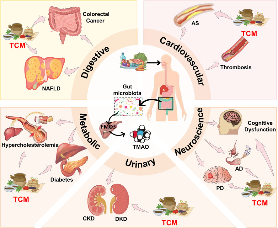
Trimethylamine N-oxide (TMAO), a metabolite derived from gut microbiota, has been implicated in the pathogenesis of various chronic diseases, including cardiovascular, digestive, neurological, and renal disorders. This review explores the complex mechanisms by which TMAO contributes to disease progression, including its role in inflammation, oxidative stress, and metabolic disorders. The study focused on the potential of traditional Chinese medicine (TCM) to regulate TMAO levels and mitigate its adverse effects. TCM interventions, through modulation of gut microbiota and inhibition of key enzymes like flavin-containing monooxygenase 3 (FMO3), offer promising therapeutic avenues. Despite the positive outcomes observed in preliminary studies, further research is needed to fully elucidate the mechanisms by which TCM interacts with TMAO and to establish its efficacy in clinical settings.
1 Introduction
Trimethylamine N-oxide (TMAO) is a bioactive molecule derived from metabolites of the gut microbiota, which is converted from trimethylamine (TMA) by the flavin containing monooxygenase 3 (FMO3) in the host liver. Studies have indicated a positive correlation between TMAO levels and various chronic non-communicable diseases, including insulin resistance, atherosclerotic plaque formation, diabetes, cancer, heart failure, hypertension, chronic kidney disease, liver disease, neurodegeneration, and Alzheimer’s disease (Coutinho-Wolino et al., 2021). Dietary choline is associated with increased plasma TMAO concentrations, thereby raising the likelihood of adverse cardiovascular events, metabolic disorders, neurological diseases, and renal diseases (Agus et al., 2021). Traditional Chinese Medicine (TCM) is a holistic medical system that utilizes natural plant or animal-based substances as methods for treating diseases and has a history of over five thousand years. With increased international exchange, TCM has also been adopted in many other countries, such as the United States, Canada, Finland, Australia, the United Kingdom, and others, and encompasses a variety of therapeutic practices. Specifically, TCM includes herbal preparations, acupuncture, moxibustion, dietary therapy (medicinal cuisine), tuina (therapeutic massage), and other traditional approaches. In this review, “TCM” primarily refers to these modalities, with particular emphasis on herbal interventions and dietary therapy, while also acknowledging the important role of practices such as acupuncture in the prevention and treatment of diseases related to TMAO (Wu et al., 2012).
1.1 Sources of TMAO
In the human body, TMA is a significant precursor of TMAO. TMA is primarily formed in the gut through the enzymatic metabolism of certain dietary compounds present in foods such as peanuts, dairy products, liver, egg yolks, and other full-fat dietary items, which all characterized by high levels of choline and carnitine, being both important precursors of TMA and TMAO (Wu K. et al., 2020). Choline, a trimethylamine-containing compound, as part of the phosphatidylcholine head group, can be found in various foods. Phosphatidylcholine, also known as lecithin, is a fundamental component of membranes and the neurotransmitter acetylcholine. Phospholipase D can catalyze the conversion of lecithin to choline, which is a reversible transformation (Fennema et al., 2016). Human milk and soy-derived infant formula contain substantial amounts of free choline, while beef liver, cauliflower, and peanuts (Zeisel et al., 2003) contain several choline compounds (phosphatidylcholine, phosphocholine, sphingomyelin, etc.). Studies have shown that the key enzyme responsible for producing TMA is choline TMA-lyase (CutC), which is a glycyl radical enzyme requiring the activating protein CutD to assist its function. These two components work together to catalyze the cleavage of choline, generating TMA and acetaldehyde (Yoo et al., 2021). The process of TMA production through the choline TMA-lyase complex, namely, CutC and CutD (often collectively referred to as CUTC), represents the primary pathway by which gut microbiota convert dietary choline into TMA (Craciun and Balskus, 2012).
L-carnitine is present in red meat and dairy products (Feller and Rudman, 1988). Serratia bacteria and Acinetobacter calcoaceticus that found in the human gut can cleave the 3-hydroxybutyryloxy group of L-carnitine to directly produce TMA (Meadows and Wargo, 2015). Certain gut microbial communities possess two enzyme systems, namely, the CNTA/B systems, which are composed of the Rieske-type oxygenase CntA and the electron transfer reductase CntB. These two subunits collaborate to degrade L-carnitine in vitro, thereby generating TMA (Zhu et al., 2014). Moreover, within the body, carnitine can be metabolized into γ-butyrobetaine and crotonobetaine through the enzymatic action of L-carnitine dehydrogenase (Meadows and Wargo, 2015) and γ-butyrobetaine CoA transferase (Koeth et al., 2014), while choline can be oxidized into betaine by choline dehydrogenase and betaine aldehyde dehydrogenase. Betaine itself is a substance abundant in wheat bran, wheat germ, and spinach (Zeisel et al., 2003). These compounds can also serve as precursors for the formation of TMA and TMAO (Wang et al., 2019). Betaine can be reduced and cleaved into TMA and acetate in a coupled redox process (Stickland reaction) by betaine reductase (Naumann et al., 1983). In addition to these common sources, TMA can also be derived from dietary ergothioneine found in foods such as legumes, mushrooms, and liver (Cheah and Halliwell, 2012). Ergothionase catalyzes the degradation of ergothioneine to produce TMA and the by-product thiosulfonate (Muramatsu et al., 2013). Once the above precursors are converted into TMA through complex actions by gut microbiota and enzymes, a small portion of TMA can be directly metabolized into TMAO and dimethylamine (DMA) by bacteria in the gut. The remainder can be transported to the liver via the portal vein circulation, and in there it would be oxidized into TMAO by the host liver flavin monooxygenases (FMO1 and FMO3) (Lang et al., 1998; Descamps et al., 2019). Finally, TMAO and TMA can also be directly acquired from fish and other seafood (Tang and Hazen, 2017). Compared to freshwater fish, the concentration of TMA in marine fish (Bain et al., 2005) (such as cod, halibut, herring, and skate) is higher.
1.2 Metabolism of TMAO
After being produced in the body, the majority of TMA is absorbed via passive diffusion across the intestinal cell membrane. Subsequently, almost 95% of TMA is oxidized to TMAO in the liver. Before excretion, TMAO—and any unmetabolized TMA—enters the plasma and is transported to body tissues (such as the lungs, liver, kidneys, muscles, and heart) where it accumulates as an osmolyte compound. Relatively, TMA and TMAO are most likely to accumulate in the lungs and kidneys, followed by the liver, then muscles, with the heart being the least likely organ for accumulation (Smith et al., 1994). Eventually, both TMA and TMAO are mixed with urine in the kidneys via the circulatory system and excreted from the body. Organic cation transporter 2 (OCT2), situated on the basolateral membrane of renal tubular cells, serves as a crucial uptake transporter for TMAO, with over 90% of TMAO being excreted in the urine after renal metabolism (Bennett et al., 2013; Canyelles et al., 2018). Approximately 4% of the remaining TMAO is excreted via faeces, and less than 1% is expelled through respiration. Research indicates that most orally ingested TMAO can be absorbed by extrahepatic tissues without microbial or hepatic processing. Under the action of TMAO reductase, some TMAO can be reduced back to TMA in the intestine. Additionally, certain bacteria have the ability to convert TMA and TMAO into DMA and formaldehyde through trimethylamine dehydrogenase (TMADH) and TMAO demethylase.
2 Treatment approaches related to TMAO
The above content suggests that the majority of TMA is absorbed into the hepatic portal venous circulation via passive diffusion across the intestinal cell membrane and subsequently converted to TMAO by hepatic FMO3, and TMAO elimination can be achieved by targeting specific pathways, including inhibition of TMA precursor production, suppression of TMA production, and blocking the conversion of TMA to TMAO.
2.1 Inhibition of TMA and TMAO production
By supplementing with broad-spectrum antibiotics (e.g., ciprofloxacin and metronidazole) (Tang et al., 2013), it is possible to inhibit microbial groups that can convert choline, betaine, and L-carnitine into TMA (Wang et al., 2011). Although antibiotics initially suppress TMAO levels, the long-term persistence of this effect remains unknown. Unlike this mechanism, Meldonium is an anti-ischaemic and anti-atherosclerotic drug that can competitively inhibit not only butyrobetaine hydroxylase but also the reabsorption of L-carnitine in the kidneys via the carnitine/organic cation transporter protein (OCTN2). Moreover, it also reduces TMAO concentration in human plasma by increasing urinary excretion (Dambrova et al., 2013). In terms of the microbial metabolic regulation, a structural analogue of choline, 3,3-dimethyl-1-butanol (DMB), does not impede choline uptake into cells. However, it can restrain the formation of TMA by microorganisms through inhibiting the activity of microbial TMA lyases, such as CutC (Yang Y. et al., 2022), and reducing the formation of TMA in various human microbiomes. Irreversible covalent inhibitors targeting CutC lyase also exert analogous effects. Previous studies have demonstrated that fluoromethylcholine (FMC), a non-lethal, microbe-friendly inhibitor of CutC lyase, significantly reduces plasma TMAO levels by irreversibly modifying the enzymatic pair CutC/D within gut microbiota harboring the corresponding gene cluster (Benson et al., 2023). The research by Nilaksh Gupta and colleagues found that iodomethylcholine (IMC) can inhibit the production of microbial TMA in hosts and reduce plasma TMAO levels, demonstrating over 10,000-fold greater potency (sub-nanomolar IC50) compared to previously reported microbial choline TMA lyase inhibitors (Gupta et al., 2020). In addition, the reversible competitive inhibitor betaine aldehyde can lower plasma TMAO levels by specifically targeting the gut microbial enzyme CutC (Orman et al., 2019). DMB treatment significantly lowers plasma TMAO levels and prevents cardiac dysfunction (Wang et al., 2015), without affecting body weight and dyslipidemia (Chen K. et al., 2017). It is worth noting that in addition to the strategies of directly intervening in metabolic pathways mentioned above, Enalapril, an angiotensin-converting enzyme inhibitor (ACE-I), represents a new method of reducing plasma TMAO levels, although the underlying mechanism has not been discovered (Konop et al., 2018). Additionally, recent studies have confirmed that inhibiting host enzyme FMO3 has a positive impact on reducing circulating TMAO levels and diet-enhanced atherosclerosis. However, adverse side effects may be brought by inhibiting FMO3, including liver inflammation and trimethylaminuria (fish odour syndrome) (Wang et al., 2015).
2.2 Microbiota-based treatments to reduce TMAO
TMAO represents a critical microbial metabolite derived from the gut microbiota’s metabolism of dietary nutrients. Consequently, reducing TMAO levels via microbiota-targeted approaches has emerged as a significant research focus for treating associated diseases. Wang et al. demonstrated that berberine (BBR) reduces gut microbial TMA biosynthesis, ultimately lowering plasma TMAO levels, by decreasing the abundance of CutC/D-expressing bacterial taxa, including Lachnospira, Lachnospiraceae group, Lachnospiraceae group, Clostridia, Lachnoclostridium, and Ruminococcus (Wang et al., 2024). In contrast, puerarin (PU) acts by targeting specific TMA-producing species. It specifically inhibits the membrane function of Prevotella copri, thereby diminishing its capacity to generate TMA and subsequently reducing TMAO levels (Authors, 2024). Furthermore, remodeling the gut microbiota structure can reduce the colonization and metabolic activity of bacteria that produce TMA precursors, leading to decreased TMAO concentrations. Recent research revealed that Akkermansia muciniphila, a mucin-degrading bacterium with probiotic properties, secretes the antimicrobial peptide Amuc. This peptide inhibits the growth of TMA-producing bacteria such as Anaerococcus hydrogenalis, resulting in reduced TMAO levels (Li et al., 2024). The bioactive xanthone mangiferin lowers plasma TMAO by reshaping gut microbial composition; it promotes the growth of beneficial taxa including Akkermansia, Parabacteroides, and Bifidobacteriaceae, while concurrently reducing the relative abundance of the pathobiont genus Helicobacter (He Z. et al., 2023). Additionally, Lactiplantibacillus plantarum ZDY04 achieves therapeutic effects by modulating gut microbiota structure, significantly decreasing serum TMAO content and cecal TMA levels (Qiu et al., 2018). Collectively, these findings indicate that microbiota-targeted therapy offers a promising therapeutic paradigm for the precise intervention of TMAO-related diseases.
2.3 Negative effects
TMAO is a stable, non-volatile and odourless oxidised product (Wang et al., 2011), whereas TMA is a volatile gas with a fishy odour. Mutations or inhibitions of human FMO3 that prevent TMAO production can lead to the accumulation of TMA, which is excreted in excess in urine, sweat, and breath, smelling like rotten fish (Zeisel and Warrier, 2017). Thus, inhibiting the conversion of TMA to TMAO by reducing FMO3 expression could result in fish odour syndrome. Moreover, while antibiotic treatment can suppress plasma TMAO levels, such as with metronidazole and ciprofloxacin, continued use of antibiotics to lower TMAO concentrations can lead to resistance in bacterial strains. Moreover, antibiotics can kill beneficial bacteria as well as harmful ones, leading to gut dysbiosis, and after stopping antibiotics, TMAO levels rise again. Long-term use of Meldonium may cause side effects such as hypoxia, dizziness, and reduced blood supply (Benedetto et al., 2008). Taking ACE-Is may impair kidney function, potentially resulting in renal failure and electrolyte imbalance, and enalapril treatment may cause increased water intake (Konop et al., 2018). Additionally, the accumulation of TMA and its unpleasant odour in individuals with FMO3 gene defection who suffer from fish odour syndrome diminishes the potential of FMO3 as an inhibitory therapeutic target. Therefore, this paper will analyze the advantages of Traditional Chinese Medicine in treating diseases related to TMAO.
3 The role of TMAO in various diseases and the therapeutic effects of traditional Chinese medicine
Multiple studies have shown that TMAO is involved in the occurrence and development of various chronic diseases, including cardiovascular, digestive, neurological, kidney diseases, and metabolic disorders. Modern medical drugs have been widely used for treatment, but there are various side effects. At the same time, TCM has been proven to have significant potential and remarkable effects in improving and treating chronic diseases. Based on its multi-component and multi-target characteristics, TCM offers a unique therapeutic approach for regulating TMAO levels and mitigating the progression of related diseases. In this review, herbal medicine is identified as one of the primary modalities of TCM for treating TMAO-related conditions, demonstrating strong potential in modulating the gut microbiota, inhibiting key enzymes such as FMO3, and reducing systemic inflammation and oxidative stress. For example, berberine, baicalin, and curcumin can lower serum TMAO by altering gut microbial composition or intervening in metabolic pathways (Liu et al., 2020; Wang et al., 2024). Dietary and nutritional interventions can influence TMAO production by promoting a healthier gut microbiota. Studies have shown that dietary fiber, a nutrient digested and absorbed in the colon, can significantly lower TMAO levels in human peripheral blood (Xie et al., 2025). Other modalities, such as acupuncture and moxibustion, are believed to improve organ function and overall regulation by modulating neuroimmune pathways and reducing inflammatory responses, although their specific impact on TMAO requires further investigation (Sun et al., 2024). The combination and individualized use of these TCM approaches provide both theoretical and clinical foundations for the comprehensive prevention and treatment of TMAO-related chronic diseases. The subsequent sections of this review will further explore the therapeutic potential and mechanisms of TCM in addressing TMAO-related conditions.
3.1 Digestive system and metabolic diseases
Numerous studies indicate that TMAO is closely associated with digestive system diseases such as colorectal cancer (CRC) and non-alcoholic fatty liver disease (NAFLD). TMAO activates its intracellular potential receptors, protein kinase R-like endoplasmic reticulum kinase (PERK), which further activates NLRP3 and NF-κB that mediate pro-inflammatory responses. TMAO also induces reactive oxygen species through oxidative stress, altering the invasion and migration of tumour cells, thereby affecting CRC progression (Duizer and de Zoete, 2023). Moreover, TMAO can directly enhance the onset of NAFLD through oxidative stress or, due to disorders in hepatic lipid metabolism and inflammation, affect bile acid production, alter liver TG levels, influence cholesterol transport and glucose and energy balance, thus exacerbating hepatic steatosis. Metabolic disorders are an increasingly severe global health issue, closely linked to changes in the intestinal microbiome. TMAO is an important regulator of lipid metabolism and is closely related to the pathogenesis of metabolic diseases such as hypercholesterolemia and diabetes (Agus et al., 2021). TMAO inhibits reverse cholesterol transport (RCT), reduces hepatic bile acid transporters, and alters bile acid synthesis, leading to impaired cholesterol elimination and hypercholesterolemia, thereby increasing cardiovascular disease risk (Koeth et al., 2013). Furthermore, TMAO promotes diabetes by elevating fasting insulin, increasing insulin resistance (HOMA-IR), and inducing adipose tissue inflammation, contributing to glucose metabolism dysfunction and heightened cardiovascular event risks in diabetic patients (Subramaniam and Fletcher, 2018).
3.1.1 Colorectal cancer
Beginning from abnormal crypts, they evolve into precancerous lesions (polyps), eventually progressing to colorectal cancer over an estimated 10–15 years. Currently, most colorectal cancers are supposed to originate from stem cells or stem cell-like cells (Medema, 2013; Nassar and Blanpain, 2016). These cancer stem cells are the result of accumulating genetic and epigenetic changes that inactivate tumor suppressor genes and activate oncogenes. Located at the base of colonic crypts, these cancer stem cells are crucial for tumor initiation and maintenance (Medema, 2013; Nassar and Blanpain, 2016). Studies have found that TMAO may share numerous gene pathways, including immune system, cell cycle, and Wnt signaling pathways, with CRC, indicating a clear link between TMAO concentration and CRC (Xu et al., 2015), however, its specific mechanism requires further investigation. Existing research suggests TMAO influences CRC through inflammation induction, oxidative stress, and DNA damage. One way TMAO promotes colorectal cancer is by inducing inflammation. In a long-term choline-fed mouse experiment, TMAO administration leds to NF-κB-mediated pro-inflammatory responses (Seldin et al., 2016). Enhanced TMAO levels can promote the initiation of the NF-κB pathway and improve the expression of pro-inflammatory genes, including chemokines, adhesion molecules, and inflammatory cytokines. The NLRP3 inflammasome, closely related to CRC, is activated by TMAO-induced endothelial inflammation mediated by mitochondrial reactive oxygen species (ROS) (Chen M. L. et al., 2017). Another study showed that TMAO promotes IBD progression by inhibiting ATG16L1-induced autophagy in colonic epithelial cells, thereby activating the NLRP3 inflammasome (Yue et al., 2017). Moreover, a potential receptor for TMAO may be PERK, which was identified in hepatocytes (Chen S. et al., 2019). Activation of PERK may subsequently lead to the activation of NLRP3 and NF-κB (Chen S. et al., 2019). Specifically, in CRC cells, TMAO has been proved to promote proliferation and potential angiogenesis by upregulating vascular endothelial growth factor A (Yang S. et al., 2022). Therefore, TMAO affects CRC by activating PERK, subsequently activating NLRP3 and NF-κB (Figure 1). In systemic circulation, increased TMAO levels are associated with oxidative stress and induce the production of superoxide, a type of ROS (Li T. et al., 2017). Oxidative stress can render tumor cells insensitive to anti-proliferative signals, apoptosis, and anchorage-independent growth, which alters tumor cell invasion and migration through epigenetic and metabolic mechanisms, further contributing to colorectal cancer development and progression (Zińczuk et al., 2020). Consequently, TMAO also participates in the formation of NOCs, leading to DNA damage and epigenetic changes, indicating a potential role of DNA damage in TMAO’s carcinogenic effects (Oellgaard et al., 2017).
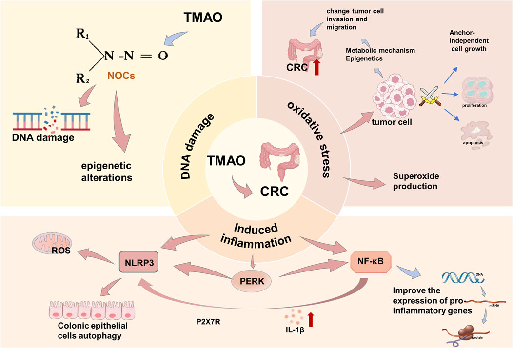
Figure 1. Mechanism of TMAO inducing CRC. TMAO promotes the occurrence and development of CRC by inducing inflammation, oxidative stress, and DNA damage.
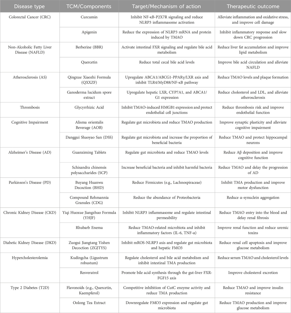
Regarding TCM treatment, there are currently no definitive research conclusions about specific TCM components or compounds treating CRC by affecting TMAO. However, as mentioned above, TMAO influences CRC development through the induction of inflammation, oxidative stress, and DNA damage, with NLRP3 inflammasomes being noteworthy in the context of inflammation induction. Clinical studies have demonstrated that curcumin inhibits NLRP3 inflammasome activation via NF-κB-induced P2X7R signalling in macrophages (Kong et al., 2016), and apigenin significantly reduces the mRNA and protein expression of TMAO-induced NLRP3 (Yamagata et al., 2019), which suggests that TCM could potentially treat CRC by impacting the NLRP3 inflammasome, providing a potential pathway.
3.1.2 Non-alcoholic fatty liver disease
A key characteristic of NAFLD is hepatic steatosis, mainly driven by obesity, insulin resistance (IR), and adipose tissue (AT) dysfunction. Unhealthy lifestyles and diets high in sugar and fat contribute to obesity and increased liver fat, directly leading to steatosis (Boden, 2006; Powell et al., 2021). IR is characterized by a poor response to insulin whereby glucose uptake is impaired regardless of insulin levels (Boucher et al., 2014). Dysfunctional AT leads to low adiponectin and high leptin levels, causing hepatic insulin resistance and increased lipolysis, further accelarating the development and progression of NAFLD. Studies show that NAFLD patients have higher serum TMAO levels, which correlate positively with steatosis severity (Chen Y. M. et al., 2016; León-Mimila et al., 2021). TMAO is believed to promote NAFLD through four pathways: enhancing oxidative stress (Li X. et al., 2021), impairing glucose tolerance in the liver (Wong et al., 2016; Tang and Hazen, 2017), increasing the expression of proteins related to the unfolded protein response (GRP78, XBP1, Derlin-1) and triggering hepatic lipid metabolism disorders and inflammation (Shi C. et al., 2022), and disrupting bile acid cycling. Specifically, TMAO blocks bile acid-activated farnesoid X receptor (FXR) signaling and reduces key bile acid synthesis enzymes (Cyp7a1, Cyp27a1), limiting bile acid production and increasing fatty liver risk (Koeth et al., 2013; Tan et al., 2019) (Figure 2).
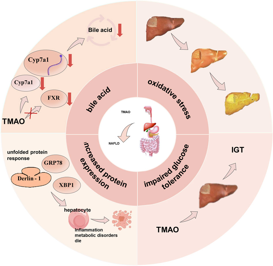
Figure 2. Mechanism of TMAO inducing NAFLD. TMAO causes NAFLD through four pathways: by affecting oxidative stress, by affecting bile acid production, by promoting increased protein expression, and by directly acting on the liver to reduce glucose tolerance.
A pivotal aspect of TMAO’s influence on the NAFLD development is bile acid circulation, which provides a new perspective on TCM. Clinical studies have shown that berberine (BBR), a natural plant alkaloid and major pharmacological component in the Chinese herb Coptis chinensis Franch, significantly increases secondary or total bile acid content and activates intestinal FXR signalling by accumulating taurocholic acid (TCA) (Tian et al., 2019). Quercetin significantly lowers caecal total bile acid levels (Nie et al., 2019), and Rubus idaeus L can promote the expression of bile acid synthesis genes (Matziouridou et al., 2016). Therefore, applying Chinese medicine to regulate bile acid metabolism to treat NAFLD holds substantial promise for the future.
3.1.3 Hypercholesterolemia
Hypercholesterolemia is a systemic metabolic disease characterized by abnormal lipid metabolism due to genetic factors, high-fat intake, lack of exercise, etc. Relevant studies indicate that TMAO levels are abnormally high in hypercholesterolemia patients, suggesting a close link between TMAO and the pathogenesis of hypercholesterolemia (Dehghan et al., 2020). Previous research shows that TMAO’s impact on hypercholesterolemia is strongly associated with changes in BA metabolism (Duval et al., 2006; Khan et al., 2014; Chen M. L. et al., 2016; Ding et al., 2018). TMAO reduces hepatic bile acid transporter proteins and BA synthesis, effectively decreasing the bile acid pool and promoting a primary pathway for CHO elimination—altering bile acid synthesis, thereby inducing hypercholesterolemia. Studies have shown that TMAO impacts CHO elimination through various mechanisms, leading to hypercholesterolemia. Initially, TMAO promotes foam cell formation through scavenger receptors in macrophages and downregulates main BA synthesizing enzymes cyp7a1 and cyp27a1, reducing intracellular BA levels and affecting hepatic CHO, BA production, and bile secretion, leading to hypercholesterolemia. TMAO also decreases the mRNA expression of Niemann-Pick C1 (NPC1L1) and ATP-binding cassette (ABC) G5/G8, inhibiting intestinal CHO absorption (Koeth et al., 2013). Other studies found that TMAO induces hypercholesterolemia by promoting intracellular CHO accumulation—enhancing macrophage cholesterol accumulation via microbiota-dependent pathways involving the increased expression of pro-atherogenic scavenger receptor proteins CD36 and SRA on the cell surface (Wang et al., 2011). Additionally, FMO3 is a negative regulator of macrophage reverse cholesterol transport and a major pathway for TMAO production. FMO3 also contributes to metabolic anomalies by affecting CHO. Experiments that knocking down hepatic FMO3 in LDL receptor-deficient mice demonstrate that FMO3 knockdown can alter bile secretion, intestinal absorption, and constrains hepatic oxysterol and cholesterol ester production in cholesterol-fed mice (Shih et al., 2015; Warrier et al., 2015). FMO3 also impairs cholesterol flux into the TICE pathway, triggering hypercholesterolemia. These studies suggest that FMO3 and TMAO are critical targets for treating hypercholesterolemia (Canyelles et al., 2018), and inhibiting TMAO generation is a crucial treatment method. There are two possible mechanisms to inhibit TMAO production: inhibiting TMA production and reducing FMO3 expression or activity (Figure 3).
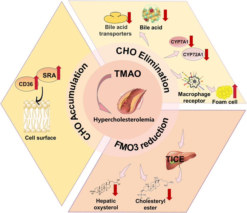
Figure 3. Mechanism of TMAO inducing hypercholesterolemia. TMAO leads to Hypercholesterolemia by affecting cholesterol (CHO) levels and reducing the FMO content.
Modern research indicates that antibiotics, choline analogues such as DMB, probiotics, plant sterols, and statins have demonstrated effectiveness in treating hypercholesterolemia. These treatments employ different mechanisms, with some targeting the TMA/FMO3/TMAO pathway and others working through alternative routes. Antibiotics, for instance, reduce TMAO levels by inhibiting the microbial production of TMA from dietary precursors. Broad-spectrum antibiotics like ciprofloxacin and metronidazole can almost completely suppress TMAO levels by targeting microbial populations responsible for converting choline, betaine, and L-carnitine into TMA (Wang et al., 2011; Tang et al., 2013). Despite their initial effectiveness, the long-term impact of antibiotics on TMAO levels remains uncertain. Prolonged antibiotic use can lead to the emergence of resistant bacterial strains, increased risks of obesity and cardiovascular events, and intestinal dysbiosis, which disrupts the balance of gut microbiota. Once antibiotics are discontinued, TMAO levels often rise again. Furthermore, antibiotics not only kill harmful bacteria but also affect beneficial ones, further complicating their use as a long-term solution. In contrast, the choline analogue DMB functions as a non-lethal inhibitor of TMA production. It suppresses microbial TMA lyase activities, such as the biological choline TMA lyase CutC (Yang Y. et al., 2022), without interfering with choline uptake into cells. This inhibition reduces TMA formation in cultured microbes, lowers TMA production across various microbial communities in the human body (Wang et al., 2015), and decreases levels of TMA, TMAO, acetate, and propionate in vivo. Although promising, DMB’s chemical structure requires further refinement to enhance its effectiveness and safety (Korpela et al., 2016; Heianza et al., 2019). In addition, specific probiotic strains, including Escherichia coli ZDY01 and Lactobacillus plantarum ZDY04, also demonstrate promising capabilities in reducing circulating TMAO concentrations (Qiu et al., 2018). Despite potential benefits, probiotic applications in disease management remain contentious. Critical research gaps persist in elucidating gut colonization mechanisms and complex interactions between probiotic strains and existing gut microbiota, necessitating comprehensive scientific exploration (Seguro et al., 2021). Unlike the aforementioned treatments, plant sterols and statins do not target the TMA/FMO3/TMAO pathway. Plant sterols, natural molecules derived from plants, offer a non-pharmacological approach to managing abnormal blood lipid levels. While they can help prevent or control hypercholesterolemia, they are not a substitute for pharmaceutical interventions in more severe cases. On the other hand, statins are widely used drugs that inhibit serum cholesterol levels by suppressing 3-hydroxy-3-methylglutaryl-CoA reductase (Sirtori, 2014), thereby reducing the synthesis of mevalonate and cholesterol. Despite their clinical effectiveness, statins are associated with several adverse effects, including myopathy, hyperglycemia, abnormal liver enzymes, and cognitive impairments, which can limit their long-term use.
Traditional Chinese medicine also shows significant efficacy in treating hypercholesterolemia through the TMAO pathway, with superior advantages. Ligustrum lucidum W.T.Aiton (LR), also known as kudingcha, is a flavonoid-rich tea-like plant. LR not only prevents the formation of choline-induced TMA and TMAO but also lowers serum TMAO levels by affecting the gut microbiome (Liu S. et al., 2021), thereby modifying the prevalence of specific microbial taxa and modulating their functional characteristics. Concurrently, LR may influence the molecular mechanisms of cholesterol and BA metabolism. As mentioned earlier, TMAO is closely related to changes in BA metabolism. Studies indicate that LR extract reduces liver and serum cholesterol, increases faecal cholesterol and BA excretion, thus effectively alleviating hypercholesterolemia. LR has historically served as a traditional tea in China, characterized by its economic accessibility, ubiquitous availability, simple preparation method, and minimal adverse reactions, akin to several natural products (Wang et al., 2015; Chen M. L. et al., 2016). Therefore, LR may have greater potential in the prevention of hypercholesterolemia. Resveratrol, an anthraquinone terpene compound, is mainly obtained from the TCM Reynoutria japonica Houtt. Studies suggest that resveratrol reduces TMA production by reshaping the gut microbiome in mice and modifying the microbial communities in ApoE, thus inhibiting TMAO synthesis and lowering TMAO levels to mitigate TMAO-induced hypercholesterolemia. As previously mentioned, TMAO affects cholesterol metabolism by influencing BA biosynthesis pathways (Koeth et al., 2013), and research by Chen et al. showed that resveratrol significantly decreases ileal FGF15 mRNA and protein levels, leading to increased BA synthesis. This indicates that resveratrol can induce hepatic BA synthesis via the gut-liver FXR-FGF15 axis, thereby mitigating TMAO-induced hypercholesterolemia (Chen M. L. et al., 2016).
3.1.4 Diabetes
Diabetes is a metabolic disorder characterized by persistent hyperglycemia due to impaired insulin secretion or cellular responsiveness. The gut microbiota plays a critical role in type 2 diabetes (T2D) pathogenesis, with T2D patients showing dysbiosis, disrupted intestinal barriers, and abnormal TMAO production and absorption (Wang et al., 2011; Tang et al., 2013). Elevated circulating TMAO is significantly linked to higher T2D risk (Fang et al., 2021b). Mechanistically, TMAO promotes T2D by increasing fasting insulin, HOMA-IR, and glucose intolerance, and inducing adipose tissue inflammation (Gao et al., 2014; Dambrova et al., 2016). It impairs insulin signaling and hepatic glucose metabolism, affecting glycogen synthesis and gluconeogenesis (Kalagi et al., 2022) (Figure 4). TMAO’s production depends on FMO3, and FMO3 polymorphisms may influence T2D risk (Yamazaki and Shimizu, 2013). Nonetheless, the exact mechanism linking circulating TMAO levels to T2D has yet to be fully clarified.
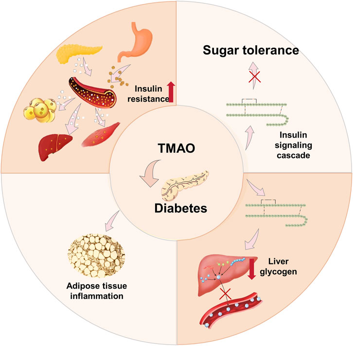
Figure 4. Mechanism of TMAO inducing diabetes. TMAO leads to T2D by increasing insulin resistance (HOMA-IR), inducing adipose tissue inflammation, and aggravating the blockade of the insulin signaling cascade. In addition, TMAO is also associated with genes in the insulin signaling pathway.
Recent studies suggest that DMB and dietary indoles can prevent and treat diabetes via the TMAO pathway. First, DMB, a choline analogue, is found in balsamic vinegar, olive oil, grape seed oil, and red wine. Studies have found that DMB treatment does not affect body weight or lipid abnormalities but significantly reduces plasma TMAO levels and prevents cardiac dysfunction (Chen K. et al., 2017). DMB can mitigate foam cell formation and atherosclerotic plaque development by lowering TMAO levels. However, DMB has been demonstrated solely as an effective non-lethal bactericidal agent, with no proven effect on improving glucose homeostasis and insulin sensitivity. Whereas DMB shows promise in treating cardiovascular disease and T2D, its chemical structure leaves room for improvement. Secondly, dietary indoles can effectively lower TMAO levels and inhibit FMO3 activity, however, their lack of specificity arises from their role as potent 5-HT agonists, with the ability to cross the blood-brain barrier and potentially induce adverse psychological effects (Cashman et al., 1999; Chen J. et al., 2016; Zajdel et al., 2016). Clinically, T2D is commonly treated by insulin injection. Although it shows good efficacy, fear of needles contributes to poor adherence, leading to inadequate blood sugar control and recovery hindrance. Non-invasive alternatives include inhaled or oral insulin; however, challenges exist in using these routes (Zajdel et al., 2016). Clinically, metformin and sulphonylureas are commonly used anti-diabetes drugs. Metformin remains the most widely prescribed antidiabetic agent, particularly for obese and overweight patients. However, metformin exerts no direct effects on β-cells; moreover, in the absence of weight reduction, there is no substantial improvement in muscle insulin sensitivity. Sulphonylureas are secreagogues that treat T2D by triggering the endogenous insulin secretion of pancreatic β-cells. However, sulphonylureas do not have long-term protective effects on β-cell function and may accelerate β-cell failure. Moreover, sulphonylureas, particularly older generations, have a high incidence of causing hypoglycaemia and adverse effects such as weight gain (Gao et al., 2014).
Meanwhile, TCM showed encouraging results in treating diabetes through the TMAO pathway. Flavonoids can lower TMAO levels by inhibiting TMA production, while oolong tea can reduce TMAO levels by reducing FMO3 levels. Studies show an interaction between flavonoids and the gut microbiota, which is directly related to TMAO (Hua et al., 2022). Flavonoids are widely present in a variety of Chinese medicines such as Styphnolobium japonicum (L.) Schott, Carthamus tinctorius, Scutellaria baicalensis Georgi, Pueraria montana var,Lonicera japonica Thunb., Citrus reticulata Blanco, Chrysanthemum × morifolium (Ramat.) Hemsl., Epimedium sagittatum (Siebold and Zucc.) Maxim, Ginkgo biloba L., and China’s ten famous traditional green teas, including Lu’an melon seed tea. CutC can break down dietary choline and betaine to generate TMA. Hence, inhibiting CutC activity can suppress TMA production, thereby lowering TMAO levels and achieving anti-diabetic effects. An experiment docking 16 flavonoids from Lu’an melon seed tea with CutC revealed that kaempferol 3-O-rutinoside (Hua et al., 2022), quercetin 3-O-rhamnosidyl galactoside, kaempferol 3-O-rhamnosidyl galactoside, and myricetin 3-O-galactoside can bind with CutC to regulate its activity, suppress TMA production, reduce TMAO levels, and achieve anti-diabetic effects. Additionally, flavonoids, abundant in various plants, fruits, vegetables, and leaves, exhibit a wide range of medicinal properties, such as anticancer, antioxidant, anti-inflammatory, and antiviral activities. They also offer neuroprotective and cardioprotective benefits. Therefore, the advantages of using flavonoids to treat diabetes are more pronounced (Ullah et al., 2020). Oolong tea, a tea variety unique to China, has shown significant efficacy in preventing diabetes. Experiments have indicated that oolong tea extract can lower TMAO formation capacity by remodeling the gut microbiota and downregulating the elevation in FMO3 induced by carnitine (Chen P. Y. et al., 2019). Moreover, FMO3 also reduces lipogenesis and gluconeogenesis through modulating PPARα expression and activity, highlighting the significant efficacy of oolong tea in diabetes prevention (Shih et al., 2015).
3.2 Cardiovascular and neurological disorders
Recent research has established that elevated plasma TMAO concentrations demonstrate a significant correlation with an augmented risk of atherosclerotic thrombotic cardiovascular disease (CVD), rendering it a significant pathogenic determinant for cardiovascular, peripheral, and cerebrovascular diseases (Janeiro et al., 2018). TMAO promotes atherosclerosis by upregulating macrophage scavenger receptors (Al-Rubaye et al., 2019), inducing cholesterol accumulation, inflammation, foam cell formation, endothelial dysfunction, and thrombosis (Zhen et al., 2023). Elevated levels of TMAO precursors, such as choline and betaine, are also linked to higher CVD prevalence and poor outcomes. Additionally, TMAO is closely associated with neurological disorders (Parker et al., 2020); it crosses the blood-brain barrier (BBB), and impairs synaptic plasticity and cognitive function by downregulating the mTOR pathway, causing hippocampal neuron loss and synaptic damage (Chen S. et al., 2019).
3.2.1 Atherosclerosis (AS)
In recent years, numerous studies and experiments have shown a close relationship between high levels of TMAO and adverse cardiovascular events caused by atherosclerosis (Komaroff, 2018). TMAO accelerates aortic lesion formation by disrupting cholesterol and bile acid metabolism. In animal models, use of the choline TMA lyase inhibitor iodomethyl choline (IMC) increases bacterial cholesterol metabolite loss and decreases intestinal sterol transport protein Niemann-Pick C1-Like 1 (NPC1L1) expression, reshaping gut microbiota and reducing hepatic cholesterol accumulation, while upregulating CYP7A1 and other bile acid-related genes (Pathak et al., 2020). Mechanistically, TMAO induces oxidative stress and activates the ROS-TXNIP-NLRP3 inflammasome, increasing inflammatory cytokines (IL-1β, IL-18), impairing endothelial nitric oxide synthase (eNOS) and nitric oxide (NO) production, and leading to endothelial dysfunction (Sun et al., 2016). Clinical data show that higher plasma TMAO is associated with increased risk of major adverse cardiovascular events (MACE) and mortality (Guasti et al., 2021). TMAO is now considered both a driver and prognostic marker of atherosclerosis progression to CVD, mainly by affecting lipid metabolism, inflammation, and endothelial function (Wang et al., 2011; Koeth et al., 2013). TMA, produced by gut microbes, is oxidized to TMAO in the liver via FMO3(Tang et al., 2013; Shih et al., 2019), triggering chronic inflammation and arterial damage that promote atherosclerotic lesions (Fatkhullina et al., 2018; Komaroff, 2018) (Figure 5A).
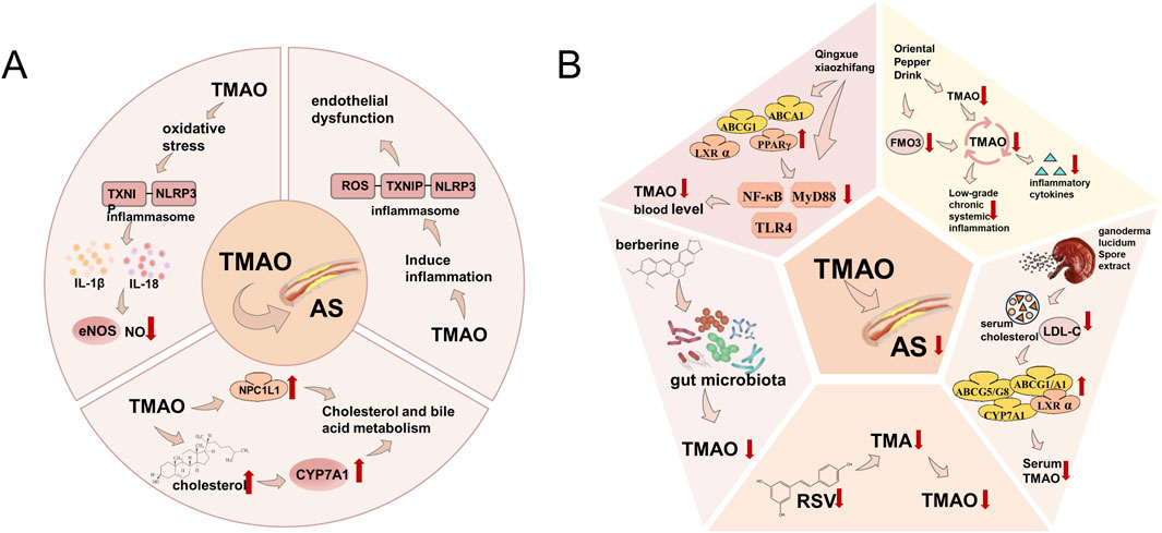
Figure 5. (A) Mechanism of TMAO inducing AS. TMAO participates in the occurrence and development of atherosclerosis by inducing oxidative stress, inflammation, and altering the metabolic mechanisms of cholesterol and bile acid in the human body. (B) Treatment of AS caused by TMAO by TCM. Traditional Chinese medicine can reduce TMAO by inhibiting inflammation, directly or through inhibiting TMA, regulating gut microbiota, lowering blood cholesterol and LDL-C levels, and reducing FMO3 expression.
In recent years, mechanistically-based small molecule inhibitors targeting the primary bacterial enzyme TMA lyase have been developed, presenting potential as anti-atherosclerotic thrombotic agents (Pathak et al., 2020). TCM can modulate lipid metabolism through altering levels of TMAO. For example, Zingiber officinale Roscoe exhibits anti-atherosclerosis effects. Moreover, results from various animal model experiments have indicated that resveratrol (RSV) can mitigate TMAO-induced AS by suppressing TMA formation, with the hepatic FXR-fibroblast growth factor 15 (FGF15) axis serving a critical role in resveratrol-induced bile acid (BA) synthesis (Chen M. L. et al., 2016). Numerous types of Chinese herbs can regulate lipid metabolism in the body (Li et al., 2021c). TMAO is formed through the super-metabolism of dietary substrates containing trimethylamine groups, making its production highly dependent on the composition of the gut microbiome (Falony et al., 2015). Altering dietary habits to reduce the intake of TMAO precursors (such as choline and carnitine) or modifying the composition and function of the gut microbiome can diminish TMAO formation (Iglesias-Carres et al., 2021). A high-choline diet raises TMAO levels and atherosclerosis in animal populations. TMAO may partially mediate the well-documented correlation between red meat intake and cardiovascular disease risk. Therefore, lower blood TMAO levels due to fruit and vegetable intake could explain the cardioprotective effects observed (Sun et al., 2016). Berberine, a bioactive alkaloid derived from traditional Chinese herbal medicines, demonstrates the capacity to suppress TMAO production through modulation of the gut microbiome’s microbial composition. Berberine’s anti-atherosclerotic efficacy potentially stems from its ability to reduce TMAO production, making it an excellent modulator for inhibiting atherosclerotic plaque development (Chen and Wang, 2021). Berberine has emerged as a compound with the potential to lower atherosclerosis risk (Etxeberria et al., 2015). Qing-Xue-Xiao-Zhi Formula (QXXZF) demonstrates significant anti-atherosclerotic effects in vivo and in vitro by improving lipid metabolism and inhibiting inflammation. It reduces blood TMAO concentration, helping to elucidate its potential mechanisms for atherosclerosis protection. QXXZF can treat atherosclerosis by upregulating the ABCA1/ABCG1-PPARγ/LXR axis and inhibiting the TLR4/MyD88/NF-κB signalling pathway (Li et al., 2021d). Ganoderma lucidum (Leyss. ex Fr.) Karst spore extract has hypolipidaemic and anti-atherosclerotic effects on hyperlipidaemic rabbits, lowering blood cholesterol and low-density lipoproteins while reducing arterial plaque area, and upregulating LXR, CYP7A1, and ABCA1/G1 in the liver, intestine, and macrophages (Lai et al., 2020). Ganoderma lucidum spore extract decreases TG, TC, LDL levels, and serum TMAO in heart failure rats induced by high TMAO levels (Liu Y. et al., 2021). The Alisma orientalis Beverage (AOB), a TCM made from various herbal plants, has long been used to treat metabolic syndrome and AS (Liu et al., 2023). In an atherosclerosis model established in male apolipoprotein E-deficient mice fed a high-fat diet (HFD), multiple interventions were applied. Data analysis revealed that after 8 weeks of HFD, AOB-treated mice exhibited significantly reduced inflammatory cytokine expression and AS development. Furthermore, AOB lowered serum TMAO and hepatic FMO3 expression (Figure 5B). Diminishing circulating TMAO levels can mitigate inflammatory cytokine release, thereby attenuating chronic low-grade systemic inflammation and consequently reducing the risk of HFD-induced atherosclerosis. The anti-atherosclerotic effects of AOB are related to changes in the gut microbiome and reduced gut microbiome metabolite TMAO, indicating AOB’s potential therapeutic value in AS treatment (Zhu et al., 2020).
3.2.2 Thrombosis
Research indicates that adverse cardiovascular outcomes, including arterial thrombosis and mortality (Tang et al., 2013; Li X. S. et al., 2017; Haghikia et al., 2018), are linked to TMAO (Zhu et al., 2018). TMAO promotes platelet hyperactivity, elevates thrombosis risk, and directly regulates thrombotic diseases. After vascular endothelial dysfunction, the exposure of collagen and tissue factor triggers thrombus formation (Furie and Furie, 2008), while vascular calcification (VC) increases vessel rigidity and facilitates thrombosis (Lee et al., 2020). TMAO also induces endothelial dysfunction by disrupting junction proteins, activating the NLRP3 inflammasome to release high-mobility group box 1 protein (HMGB1), and altering endothelial permeability (Singh et al., 2019). Furthermore, TMAO exacerbates VC through dose-dependent vascular smooth muscle cell calcification and activation of the NLRP3 inflammasome and NF-κB signaling (Zhang et al., 2020), thereby further promoting thrombosis (Figure 6).
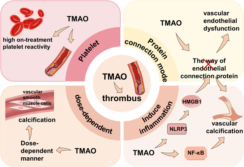
Figure 6. Mechanism of TMAO inducing thrombus. TMAO participates in thrombosis by directly causing high platelet reactivity, disrupting protein connections, activating inflammatory pathways, exacerbating vascular calcification, and promoting vascular calcification in a dose-dependent manner.
Given that TMAO can induce thrombosis directly or indirectly through endothelial damage and vascular calcification, lowering TMAO levels and inhibiting factors leading to thrombosis might be effective in treating CVD through drugs or relevant medical interventions. Nonetheless, further research is needed to elucidate specific therapeutic mechanisms. Moreover, HMGB1, a key mediator of TMAO-induced endothelial dysfunction, might serve as a significant target for treating endothelial dysfunction and its related cardiovascular diseases. Cell control experiments and analyses demonstrate that glycyrrhizic acid, a known HMGB1 binder, which reduces HMGB1 expression induced by TMAO, can be used to treat the disruption of cell junction proteins (Singh et al., 2019).
Numerous studies have concluded that TMAO is a non-traditional risk factor for CVD in patients with CKD (Bennett et al., 2013). For these patients with both CVD and CKD, standard clinical interventions for managing CVD do not improve cardiovascular outcomes. Consequently, CVD represents the predominant cause of mortality among patients with CKD (Levey et al., 1998). Experiments have demonstrated that TMAO exhibits acute positive inotropic and lusitropic effects on human and mouse myocardium, significantly increasing intracellular calcium in ventricular cardiomyocytes (Oakley et al., 2020). However, research indicates that chronically elevated contractility and intracellular calcium levels increase cardiac energy consumption, e ultimately leading to heart failure (Böhm et al., 2010).
3.2.3 Cognitive dysfunction
Synaptic plasticity is the activity-dependent change in the strength of neuron connections (Magee and Grienberger, 2020), long believed to be a fundamental component of learning and memory. TMAO reduces synaptic plasticity and induces cognitive dysfunction by promoting endoplasmic reticulum stress and directly binding to activate PERK, which damages synaptic plasticity (Zhang et al., 2007; Chen S. et al., 2019), leading to damage of synaptic plasticity. TMAO also impairs synaptic plasticity through the mammalian target of rapamycin (mTOR)/70-kDa ribosomal protein S6 kinase (p70S6K) pathway, both key regulators of protein translation and synaptic plasticity (Lipton and Sahin, 2014; Zhou et al., 2023). Studies show that elevated TMAO decreases mTOR and p70S6K expression, causes hippocampal neuron loss and synaptic ultrastructural damage, and worsens cognitive function in mice, with higher TMAO leading to more severe deficits (Li et al., 2018). Intervention with L. plantarum alongside memantine reduced Aβ1-42 and Aβ1-40, protected hippocampal neurons, improved synaptic plasticity, and decreased TMAO production, alleviating cognitive impairment in AD mice (Wang et al., 2020). However, clinical trials found that memantine treatment may worsen stuttering and language problems in autistic children (Alaghband-Rad et al., 2013).
In TCM, TMAO mainly alleviates cognitive impairment by improving synaptic plasticity. Liu et al. found through experiments that compared to the TMAO group, synaptic structural damage significantly improved in the AOB group, characterized by regular synaptic morphology, enhanced vesicle distribution within the presynaptic region, and more obvious synaptic clefts. This indicates that AOB mitigates TMAO-induced cognitive impairment by improving synaptic plasticity and regulating synaptic-related proteins (Liu et al., 2023). Furthermore, Jin et al. found that the combination of Danggui Shaoyao San (DSS) and its decoction formula can diminish the prevalence of detrimental gut microbiota (Jin et al., 2023), thereby improving cognitive and learning capacities. Through Nissl staining and Western blot analysis of the integrity of hippocampal neurons and synaptic protein expression, they found that DSS and the decoction formula group showed reduced damage to hippocampal neurons and increased expression levels of synapsin I (P < 0.05) and PSD95 (P < 0.01) proteins. Meanwhile, alpha and beta diversity analyses indicated that the richness and diversity of gut microbiota species in DSS and decoction formula groups were similar to the sham operation group, signifying significant recovery effects (P < 0.05). This suggests that DSS may mitigate cognitive impairment by modulating gut microbiota and increasing the proportion of beneficial bacteria, thereby reducing TMAO production. Additionally, baicalin and berberine have also been shown to lower TMAO levels (Liu et al., 2020). Bazi Bushen capsule alleviates cognitive deficits by inhibiting cellular senescence, secreting SASP factor, and regulating microglial activation and polarization mechanisms, thereby reducing the decline of synaptic function and protecting neurons (Ji et al., 2022).
3.2.4 Alzheimer’s disease (AD)
AD is a progressive neurodegenerative disorder of the central nervous system in the elderly, marked by cognitive impairment and characterized by cerebral amyloid plaques (mainly amyloid β, Aβ) and neurofibrillary tangles (NFT) (Naseri et al., 2019). TMAO promotes Aβ aggregation and stabilizes aggregates by redistributing water and enhancing hydrogen bonding (Kumari et al., 2018). TMAO levels are correlated with hippocampal Aβ plaques, and Aβ accumulation is an early pathological change in AD (Jack et al., 2019). Additionally, TMAO increases platelet reactivity, promoting Aβ release and neuroinflammation (Zhu et al., 2016; Borroni et al., 2002). TMAO also enhances tau aggregation into NFTs by stabilizing hydrogen bonds, reducing the aggregation threshold and lag phase (Levine et al., 2015). Furthermore, TMAO activates astrocytes to secrete pro-inflammatory mediators, leading to neuroinflammation (Heneka et al., 2010; Brunt et al., 2021) Finally, TMAO induces mitochondrial dysfunction, neuronal aging, and mitochondrial damage in the hippocampus, contributing to AD pathology (Swerdlow et al., 2010; 2014; Swerdlow et al., 2017; Li et al., 2018) (Figure 7).
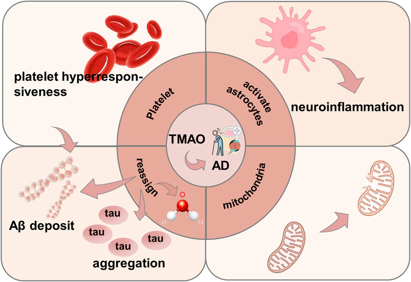
Figure 7. Mechanism of TMAO inducing AD. TMAO promotes AD by directly promoting platelet hyperreactivity, activating astrocytes to induce neuroinflammation, inducing mitochondrial damage, promoting tau protein aggregation and A β deposition.
Clinically, common AD medications are acetylcholinesterase inhibitors (AChEIs). However, a trial involving 22,845 AD patients showed that those treated with AChEIs had higher risks of appetite disorders, insomnia, or depression compared to those receiving a placebo (Bittner et al., 2023), indicating that modern clinical drugs come with certain side effects.
Traditional Chinese medicine treats AD primarily by regulating gut microbiota balance to inhibit TMAO production. Guanxinning tablets, an oral compound preparation composed of Salvia miltiorrhiza Bunge and Pueraria montana var. lobata (Willd.) Maesen and S.M.Almeida ex Sanjappa and Predeep [Fabaceae], exhibit potential in mitigating TMAO concentrations and enhancing gut microbiota composition (Zhang et al., 2021). Xanthoceraside (XAN), extracted from the husks of Xanthoceras sorbifolium Bunge, was found by Zhou et al. to alleviate gut microbiota imbalance and modulate levels of microbial-derived metabolites to mitigate AD (Zhou et al., 2022). However, the precise mechanism by which XAN treats AD through altering microbial-derived metabolite levels remains unclear, though it may relate to reduced TMAO levels. Schisandra chinensis (Turcz.) Baill, isolated from Schisandra chinensis polysaccharides (SCP), and preventative electroacupuncture can regulate gut microbiota by increasing the proportion of beneficial bacteria, thereby suppressing harmful bacteria and reducing TMAO production (He et al., 2021; Fu et al., 2023), preventing the onset of AD. In addition, some traditional Chinese medicine prescriptions can improve the clinical symptoms of AD through other effects. For example, Danggui-Shaoyao-san prescription plays an active and effective role in improving oxidative stress and neuroinflammation in APP/PS1 mice and ultimately improving cognitive deficits, which is conducive to the improvement of AD (Wu Q. et al., 2020). A natural Pterocarpus indicus plant antitoxin, Medicarpin, can alleviate cognitive and memory dysfunction in AD patients by influencing the cholinergic system, neuronal apoptosis and synaptic function (Li D. et al., 2021).
3.2.5 Parkinson’s disease (PD)
PD is a prevalent neurodegenerative disorder affecting the elderly (Khan et al., 2019). Numerous researchers have demonstrated that TMAO serves as an early biomarker for Parkinson’s disease (Chung et al., 2021). TMAO promotes PD progression by inducing neuroinflammation through microglial activation. Elevated serum TMAO not only activates astrocytes in the striatum and hippocampus of the PD models in mice but also promotes M1-type polarization of microglia (Quan et al., 2023), thereby initiating neuroinflammatory cascades. Microglia exhibit phenotypic plasticity, capable of transitioning between the pro-inflammatory M1 phenotype and the anti-inflammatory M2 phenotype (Ji et al., 2018). Qiao and colleagues demonstrated that elevated serum TMAO significantly upregulated mRNA expression of pro-inflammatory M1 microglial markers (CD16, CD32, and iNOS) in the TMAO + MPTP experimental model, suggesting a potential mechanism by which high TMAO levels intensify neuroinflammatory processes in PD through M1 microglial polarization (Qiao et al., 2023). Additionally, TMAO can induce α-synuclein misfolding, affecting neuron cells. Aggregated forms of α-Synuclein in neurons or glial cells are pathological markers of PD (Lücking and Brice, 2000). α-Synuclein is a small (14 kDa), highly conserved presynaptic protein abundant throughout the brain (Maroteaux et al., 1988). Through small-angle X-ray scattering (SAXS) experiments, Uversky et al. analyzed that TMAO induces α-synuclein misfolding (Uversky et al., 2001), leading to PD. Jamal et al. further confirmed through replica exchange molecular dynamics (REMD) simulations that TMAO promotes α-synuclein dense folding (Jamal et al., 2017). Moreover, TMAO can reduce the levels of the neurotransmitter 5-HT, crucial for mood regulation and cognition in the brain (Ni et al., 2021). Quan et al. studied the effect of TMAO on 1-methyl-4-phenyl-1,2,3,6-tetrahydropyridine (MPTP)-induced PD model mice (Quan et al., 2023), determining 5-HT levels and its metabolites in the striatum via high-performance liquid chromatography to explore TMAO’s effect on striatal neurotransmitters. Results showed a significant reduction in 5-HT levels in the TMAO + MPTP group compared to the MPTP group, indicating that TMAO diminishes neurotransmitter 5-HT levels, influencing the onset of PD.
Modern medical research suggests L-DOPA is effective for PD treatment. However, prolonged L-DOPA use may lead to motor disorders (Ahlskog and Muenter, 2001), manifesting as involuntary, purposeless, irregular, and repetitive motor phenomena involving limb, axial, and facial musculature. Additionally, antibiotics like metronidazole and ciprofloxacin are frequently used for neurological disorders (Tang et al., 2013), but continuous antibiotic use to reduce TMAO can disrupt intestinal ecology, affecting beneficial gut flora, and TMAO levels can reappear a month or more after stopping antibiotics.
However, TCM offers unique roles in treating PD by reducing the abundance of gut flora that produces TMA, thus indirectly inhibiting TMAO production. TMA is a precursor to TMAO (Galland, 2014), which is oxidized after absorption by intestinal epithelium to form TMAO. Two main TMA biosynthetic mechanisms have been described involving a specialized ethyl radical enzyme (Craciun and Balskus, 2012), The first pathway comprises CutC with its activator CutD, utilizing choline as a substrate, while the second involves a two-component Rieske-type oxygenase/reductase system (CntA/B). Rath et al. established a key gene database for principal TMA synthesis pathways (Rath et al., 2017), encoding CutC and carnitine oxygenase (CntA), to investigate microbial community TMA formation potential. Through 16S rRNA gene sequence analysis, they found that Clostridium cluster XIVa and Proteobacteria contain CutC genes, with genes encoding CntA/B found in γ- and β-Proteobacteria. Buyang Huanwu Decoction (BHD) is a renowned TCM formula comprising Astragalus mongholicus Bunge, Angelica sinensis (Oliv.) Diels, Paeonia lactiflora Pall, Conioselinum anthriscoides ‘Chuanxiong’, Carthamus tinctorius L, Prunus persica (L.) Batsch, and Earthworm (Fu et al., 2022). Hu et al. reported that BHD reduces Lachnospiraceae abundance (Hu et al., 2024), a phylogenetically heterogeneous taxon within the Firmicutes phylum of Clostridium cluster XIVa (Ruan, 2013). Consequently, BHD reduces Lachnospiraceae abundance, inhibiting TMA production and thus decreasing TMAO levels. Wan et al. found that Astragalus mongholicus not only reduces α-synuclein aggregation in the striatum but also decreases Proteobacteria’s relative abundance in PD models (Wan et al., 2022), indicating Astragalus mongholicus inhibits TMAO production while reducing α-syn aggregation leading to PD. An ancient PD treatment is Compound Dihuang Granule (CDG), composed of seven herbs: Rehmannia glutinosa (Gaertn.) Libosch. ex DC., Paeonia lactiflora, Uncaria rhynchophylla (Miq.) Miq., Pearl Shell, Salvia miltiorrhiza, Acorus verus (L.) Raf., and Scorpion. He et al. found decreased Proteobacteria in PD mice after CDG administration (He Z. Q. et al., 2023), suggesting CDG indirectly reduces TMAO production by lowering Proteobacteria. Polysaccharides and ginsenosides from Panax quinquefolius L restore gut microbiota composition (Zhou et al., 2021), reducing Escherichia coli abundance. Furthermore, YeaW, a ubiquitous enzyme in E. coli, has been proposed as a key enzyme for the third major metabolic pathway of carnitine to TMA conversion (Koeth et al., 2014). Piperine (PIP) stimulates autophagy by inhibiting PI3K/AKT/mTOR activation (Yu et al., 2024), degrading α-synuclein accumulation in PD rat colons and substantia nigra. PIP administration reduces E. coli to 6.37%, demonstrating its inhibitory effect on TMAO production. Besides TCM treatment, acupuncture also benefits PD treatment. Acupuncture can improve gut microbiota imbalance; Jang et al. found after acupuncture treatment in PD mice, a reduction in Proteobacteria in the gut, indicating decreased TMAO levels (Jang et al., 2020) (Figure 8).
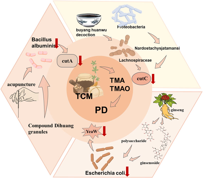
Figure 8. Treatment of AS caused by TMAO by TCM. TCM reduces the TMA level by regulating different intestinal flora and thus reduces the TMAO level to treat PD.
3.3 Urinary system diseases
Plasma levels of TMAO have been found to have a clear association with various human urinary system diseases and serve as a key biomarker for several kidney diseases (Chang et al., 2021). Clinical investigations have consistently demonstrated significantly increased plasma TMAO levels among patients diagnosed with chronic kidney disease (CKD). TMAO suppresses megalin expression in renal tubular epithelial cells through PI3K and ERK signaling pathways, reducing albumin uptake by these cells. Additionally, TMAO induces oxidative stress and activates NLRP3 inflammasomes through the MAPK pathway, thereby impairing renal function and inducing renal fibrosis (Andrikopoulos et al., 2023). TMAO is also a significant factor in diabetic kidney disease (DKD) progression, with elevated plasma TMAO levels leading to NLRP3 inflammasome formation and activation in endothelial cells, which then results in endothelial dysfunction, increased monocyte adhesion, and production of pro-inflammatory cytokines in blood vessels, eventually progressing to vascular oxidative stress and DKD.
3.3.1 Chronic kidney disease (CKD)
Metabolomic analysis shows that chronic kidney disease (CKD) patients have higher plasma TMAO concentrations (Prokopienko et al., 2019). In HFD-induced mice, elevated TMAO mediates renal fibrosis and dysfunction via oxidative stress and inflammation (Sun et al., 2017). As impaired renal function and fibrosis are key features of CKD (Xu et al., 2017; Wang et al., 2022), TMAO is considered a risk factor. Mechanistically, TMAO inhibits megalin expression via PI3K/ERK, reducing albumin uptake and promoting tubular cell dysfunction, which triggers tubulointerstitial inflammation and fibrosis (Kapetanaki et al., 2022). Through promoting p38 phosphorylation in the MAPK pathway, TMAO activates inflammatory pathways and upregulates NOX4 to enhance oxidative stress and activate NLRP3 inflammasomes, resulting in renal inflammation. TMAO also activates the NLRP3-IL-1β axis, promoting inflammatory chemotaxis and cytokine release. Increased oxidative stress and cytokines ultimately lead to tubulointerstitial damage and renal function deterioration, triggering CKD (Lai et al., 2022). Furthermore, TMAO enhances CaOx crystal deposition via oxidative stress and autophagy-related cell death, impairs renal function, and promotes kidney stones (Dong et al., 2022). It also triggers fibroblast proliferation and renal fibrosis through PERK/Akt/mTOR and NLRP3/NF-κB signaling, advancing CKD (Kapetanaki et al., 2021) (Figure 9A).
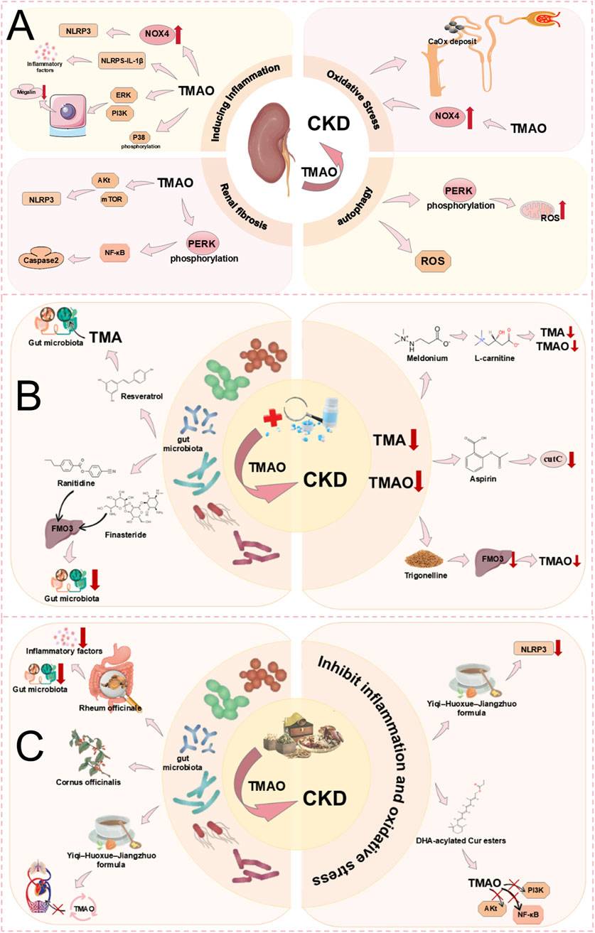
Figure 9. (A) Mechanism of TMAO inducing CKD. TMAO promotes renal fibrosis leading to CKD by inducing autophagy, inducing inflammation, and oxidative stress. (B) Treatment of CKD caused by TMAO by modern medical drugs. TMAO acts with modern medical drugs to treat CKD through two ways: changing the intestinal microflora and reducing TMA or TMAO precursor levels. (C) Treatment of CKD caused by TMAO by TCM. TMAO and TCM treat CKD through two ways: changing intestinal microflora and inhibiting inflammation and oxidative stress.
Numerous studies have indicated that TMAO can be a target for the diagnosis and treatment of CKD (Tomlinson and Wheeler, 2017). TMAO is produced by the oxidation of TMA, therefore inhibiting TMA synthesis can effectively reduce TMAO levels. TMAO is related to the abundance of gut microbiota, with several bacterial families, such as Enterobacteriaceae, involved in TMA-TMAO production, making the direct regulation of gut microbiota one method to modulate TMAO levels (Zixin et al., 2022). Medications like ranitidine and finasteride, as FMO substrates, competitively inhibit TMA binding, consequently diminishing TMAO generation and potentially mitigating CKD progression. Additionally, ranitidine and finasteride can significantly decrease Enterobacteriaceae, altering the gut microbiota and offering potential renal protective mechanisms (Zixin et al., 2022). Meldonium can reduce the gut microbiota-dependent production of TMA/TMAO from L-carnitine, thereby lowering TMAO levels and potentially delaying CKD, thereby lowering TMAO levels and potentially delaying CKD (Kuka et al., 2014). However, prolonged use of Meldonium may lead to adverse effects such as hypoxia, dizziness, and a lack of blood supply (Benedetto et al., 2008). A nutritional supplement, RSV, lowers TMAO levels by reducing the gut microbial production of TMA, thus delaying CKD (Song et al., 2020). Aspirin reduces TMAO levels through inhibition of microbial TMA lyase activity, thus delaying CKD progression (Zixin et al., 2022). Trigonelline, an alkaloid extracted from fenugreek seeds, can reduce hepatic FMO3 activity, inhibit TMA oxidation, and decrease TMAO production, thereby slowing the progression of CKD (Yong et al., 2023) (Figure 9B). However, inhibiting FMO3 can also bring about side effects such as liver inflammation and fish odour syndrome (trimethylaminuria) (Wang et al., 2015). Despite significant advances in modern medicine for treating CKD, the side effects associated with the use of modern medications cannot be overlooked.
Additionally, clinical studies have shown that TCM has a significant effect on the treatment of CKD. Curcumin diester acylated with DHA can markedly reduce TMAO levels. DHA-acylated Curcumin diester potently suppresses inflammation, apoptosis, and oxidative stress by interrupting the TMAO-mediated PI3K/Akt/NF-κB signaling cascade (Shi H. H. et al., 2022). This can slow the progression of CKD. The Yi Qi Huo Xue Jiang Zhuo formula (YHJF) consists of five traditional Chinese herbs: Astragalus mongholicus, Angelica sinensis, Rheum officinale Baill, Salvia miltiorrhiza, and Scleromitrion diffusum (Willd.) R.J.Wang. YHJF demonstrates anti-inflammatory properties by suppressing NLRP3 inflammasome activation, thereby preventing TMAO level escalation in 5/6 nephrectomised mice. YHJF can also alter the gut microbiota and reverse gut permeability, preventing increased transport of TMAO into the circulation and retarding the progression of CKD to some extent (Liu et al., 2022). Rheum officinale enema can also reduce TMAO and TMA levels in the serum of 5/6Nx CKD rats by decreasing certain TMAO-related bacteria, inhibit the expression of inflammatory markers (interleukin-6, tumour necrosis factor-α, and interferon-γ), alleviate renal interstitial fibrosis, and slow the progression of CKD (Ji et al., 2021). Additionally, Cornus officinalis Siebold and Zucc. demonstrates potential CKD prevention through comprehensive modulation of gut microbiota and targeted regulation of uraemic toxins (including TMAO) (Du et al., 2022) (Figure 9C).
3.3.2 Diabetic kidney disease (DKD)
According to the definition, DKD pathogenesis is characterized by compromised renal function, manifesting as either decreased glomerular filtration rate or elevated urinary albumin excretion, or both (Gheith et al., 2016). The Framingham Heart Study suggests that TMAO might be a surrogate marker for GFR. Experiments by Xu and others have also demonstrated that a lower GFR leads to higher TMAO levels (Xu et al., 2017). Therefore, TMAO is highly likely to be a significant risk factor in the onset of DKD. Gut-derived TMAO induces apoptosis by increasing intracellular mROS levels and further promoting NLRP3 assembly activation, with the underlying mechanism being the regulation of the intracellular mROS-NLRP3 axis to activate cytokinesis and release inflammatory factors, thus promoting DKD (Yi et al., 2023). In rats with CKD, augmented TMAO concentrations precipitate vascular oxidative stress and inflammatory cascades, culminating in endothelial impairment. Inflammatory processes and microvascular endothelial compromise constitute pivotal pathogenetic mechanisms in diabetic nephropathy. Therefore, increased TMAO levels can lead CKD patients to develop DKD further. Additionally, TMAO can promote the development of DKD by inducing inflammation (NF-κB, NLRP3, TNF-α, IL-1β, IL-6), oxidative stress, and fibrosis in the renal system (Fang et al., 2021b). The promotion of renal-related inflammatory mechanisms occurs through TMAO activating NLRP3 inflammasomes and NF-κB signal transduction, with NF-κB playing a role in the DKD process by promoting vascular inflammation and oxidative stress, becoming a potential pathogenic mechanism in DKD (Huang et al., 2023) (Figure 10A).
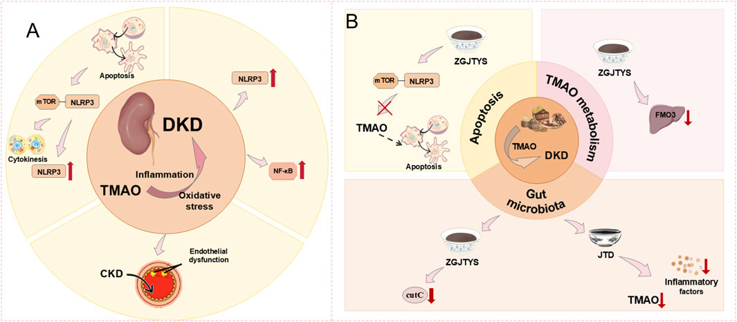
Figure 10. (A) Mechanism of TMAO inducing DKD. TMAO leads to DKD by inducing inflammation and promoting oxidative stress. (B) Treatment of DKD caused by TMAO by TCM. TCM treats DKD by acting with TMAO by regulating intestinal microbes, promoting cell apoptosis, and affecting TMAO metabolism.
Experiments have demonstrated that elevated serum TMAO levels are positively correlated with the risk of DKD in patients, with TMAO potentially being a biological marker for DKD (Huang et al., 2023). Therefore, targeting TMAO could be a way to treat DKD. However, clear research results regarding the interaction of current modern medical drugs with TMAO to treat DKD are not yet available. Recent studies, however, have shown that high levels of circulating TMAO may exacerbate DKD, suggesting that choline-TMA lyase inhibitors (such as DMB, IMC) might have potential in improving DKD (Fang et al., 2021a). More detailed mechanisms are yet to be widely researched. Encouragingly, there is currently evidence that TCM has a positive effect on the treatment of DKD. The Zuogui-Jiangtang-Yishen Decoction (ZGJTYS) is composed of nine Chinese herbs, including Astragalus mongholicus, Salvia miltiorrhiza, Dioscorea polystachya Turcz., Coptis chinensis, Achyranthes bidentata Blume, Leonurus japonicus Houtt, Rehmannia glutinosa, Cornus officinalis, and Zea mays L. It operates from three aspects to provide treatment effects. ZGJTYS may impact the expression of CutC by regulating gut microbiota and inhibit liver FMO3 levels to reduce TMAO. ZGJTYS demonstrates the capacity to directly attenuate TMAO concentrations in both plasma and renal tissue. Moreover, ZGJTYS mitigates diabetic kidney disease progression by suppressing TMAO-mediated apoptotic mechanisms through modulation of the mROS-NLRP3 inflammatory pathway (Yi et al., 2023). The Jiangtang Decoction (JTD) consists of five herbs, including Euphorbia pekinensis Rupr., Salvia miltiorrhiza, Astragalus mongholicus, leeches, and Cistanche deserticola Ma. JTD reduces TMAO levels and shows alleviating effects on inflammatory factors (such as NLRP3, IL-6, and IL-17A), effectively improving the progression of DKD. JTD regulates the composition of gut microbiota, thereby reducing the potential role of gut microbiota-mediated uraemic toxins (including TMAO) and inflammation in promoting the development of DKD. Although experimental results demonstrate a significant association between JTD and gut microbiota, the underlying mechanisms require further research (Hong et al., 2023).
In the study of kidney diseases, TMAO often induces kidney dysfunction and a decrease in glomerular filtration rate through pathways like activating autophagy, inducing inflammation, promoting renal interstitial fibrosis, inducing apoptosis, and oxidative stress. This mediates the onset and progression of renal diseases such as CKD and DKD. Notably, while modern medical drugs are often accompanied by some adverse reactions, the application of TCM shows certain advantages in treating kidney diseases. Therefore, the extensive use of TCM could potentially become an effective means of treating kidney-related diseases. However, up to now, the deeper pathogenic mechanisms between TMAO, CKD, and DKD, along with the therapeutic mechanisms of TCM interacting with TMAO, have not been fully elucidated. Therefore, further investigation into the development of TMAO and kidney diseases is required (Figure 10B).
4 Conclusion and outlook
TMAO, a key metabolite of gut microbiota, has been identified by numerous studies as a common pathological basis for multisystem chronic diseases. Its production relies on the action of CutC in gut microbiota and the oxidation by hepatic FMO3 enzyme. In digestive diseases, TMAO promotes CRC progression through inflammatory responses and oxidative stress. It activates PERK to further stimulate NLRP3 and NF-κB pathways, driving CRC development via ROS-mediated oxidative stress. Alternatively, TMAO may increase NAFLD risk by inducing oxidative stress, impairing glucose tolerance, or reducing bile acid synthesis. In cardiovascular diseases, elevated plasma TMAO upregulates scavenger receptors on macrophages to promote cholesterol deposition. Concurrently, TMAO activates the ROS-TXNIP-NLRP3 pathway, causing endothelial inflammatory dysfunction and accelerating atherosclerotic plaque formation. It also disrupts tight junction proteins through inflammation induction, exacerbates VC via NLRP3 inflammasome and NF-κB signaling activation, and promotes thrombosis. In neurodegenerative diseases, TMAO penetrates the BBB, impairing synaptic plasticity through PERK activation or mTOR pathway inhibition (Zhou et al., 2023), while promoting Aβ and tau protein aggregation in Alzheimer’s and Parkinson’s disease progression. As a key biomarker in urinary system diseases, TMAO suppresses Megalin expression via PI3K and ERK signaling, activates the MAPK-NLRP3 pathway to induce renal oxidative stress and inflammation, and ultimately leads to renal fibrosis.
In TMAO-targeted therapies, TCM demonstrates multi-target intervention capabilities. BBR from Coptis chinensis inhibits TMA oxidation by activating intestinal FXR signaling. Flavonoids competitively block CutC enzyme activity, reducing TMA production at its source. Guanxinning Tablets modulate gut microbiota, while SCP decrease TMA precursor generation. For metabolic and inflammatory regulation, ZGJTYS suppresses TMAO-induced renal apoptosis by inhibiting the mROS-NLRP3 axis. Curcumin inhibits NLRP3 inflammasome activation via P2X7R signaling, and apigenin reduces NLRP3 expression to improve cholesterol metabolism disorders. Synergistic effects of TCM formulations are particularly notable: JTD alleviates inflammatory cytokines, while AOB mitigates TMAO-induced cognitive impairment by enhancing synaptic plasticity and regulating synaptic proteins (Zhu et al., 2020; Liu et al., 2023).
In modern medical approaches, prolonged antibiotic use and Meldonium carry significant side effects. While FMO3 inhibitors reduce TMAO levels, they risk adverse effects like hepatic inflammation, and individuals with FMO3 gene defects may develop trimethylaminuria (fish odour syndrome). In contrast, TCM achieves therapeutic outcomes with reduced toxicity risks. For instance, berberine lowers serum TMAO while maintaining gut microbiota homeostasis, and Guanxinning Tablets provide cardiovascular protection comparable to statins without typical statin-related side effects.
However, current research on TCM interventions for TMAO-related diseases faces notable limitations. The mechanism of XAN in treating AD via microbiota metabolite regulation remains unclear, as does ZGJTYS’s anti-renal apoptosis mechanism. Additionally, studies exploring TCM-TMAO interactions in systemic disease management remain scarce, highlighting the need for broader experimental and clinical validation.
Existing literature shows promising results but requires more comprehensive experimental and clinical verification. Future research should focus on elucidating TMAO’s pathogenic mechanisms and exploring TCM’s therapeutic potential. Clinically, personalized treatment strategies could be developed by integrating modern medicine with TCM or combining herbal and synthetic drugs to enhance therapeutic efficacy and improve patient outcomes.
The effects of various TCM therapies on different diseases induced by TMAO, including their therapeutic targets and outcomes, are summarized and presented in the following table.
Author contributions
ZQ: Writing – original draft, Writing – review and editing. WW: Writing – original draft, Writing – review and editing. XY: Writing – original draft, Writing – review and editing. XW: Writing – review and editing. AD: Writing – review and editing. YH: Writing – review and editing. JT: Writing – review and editing. SJ: Writing – review and editing. PZ: Writing – review and editing. CQ: Writing – review and editing. XZ: Writing – review and editing. SZ: Writing – review and editing. YW: Writing – review and editing. ZS: Writing – review and editing. MS: Writing – review and editing. MW: Supervision, Writing – review and editing. SS: Supervision, Writing – review and editing. BZ: Funding acquisition, Writing – review and editing.
Funding
The author(s) declare that financial support was received for the research and/or publication of this article. This work was supported by National Science Foundation of China (82405262; 82405266), Natural Science Foundation of Jiangsu Province of China (BK20230447), Jiangsu Traditional Chinese Medicine Science and Technology Development Project (QN202202), Jiangsu Province of Youth Science and Technology Talent Nurturing Project (JSTJ-2024-452), NATCM’s Project of High-level Construction of Key TCM Disciplines (GSPZDXK-FJXYB018), Luo LinXiu Teacher Development Fund Project (LLX202312), Nanjing University of Traditional Chinese Medicine College Student Innovation Training Program Plan (202410315059Z), Nanjing University of Traditional Chinese Medicine College Student Innovation Training Program Plan (202310315111Y), Jiangsu province Graduate Research and Practice Innovation Program (CX10315).
Conflict of interest
The authors declare that the research was conducted in the absence of any commercial or financial relationships that could be construed as a potential conflict of interest.
Generative AI statement
The author(s) declare that no Generative AI was used in the creation of this manuscript.
Publisher’s note
All claims expressed in this article are solely those of the authors and do not necessarily represent those of their affiliated organizations, or those of the publisher, the editors and the reviewers. Any product that may be evaluated in this article, or claim that may be made by its manufacturer, is not guaranteed or endorsed by the publisher.
References
Authors, (2024). Puerarin alleviates atherosclerosis via the inhibition of Prevotella copri and its trimethylamine production. Gut 73 (12), 1934–1943. doi:10.1136/gutjnl-2024-331880
Agus, A., Clément, K., and Sokol, H. (2021). Gut microbiota-derived metabolites as central regulators in metabolic disorders. Gut 70 (6), 1174–1182. doi:10.1136/gutjnl-2020-323071
Ahlskog, J. E., and Muenter, M. D. (2001). Frequency of levodopa-related dyskinesias and motor fluctuations as estimated from the cumulative literature. Mov. Disord. 16 (3), 448–458. doi:10.1002/mds.1090
Alaghband-Rad, J., Nikvarz, N., Tehrani-Doost, M., and Ghaeli, P. (2013). Memantine-induced speech problems in two patients with autistic disorder. Daru 21 (1), 54. doi:10.1186/2008-2231-21-54
Al-Rubaye, H., Perfetti, G., and Kaski, J. C. (2019). The role of microbiota in cardiovascular risk: focus on trimethylamine oxide. Curr. Probl. Cardiol. 44 (6), 182–196. doi:10.1016/j.cpcardiol.2018.06.005
Andrikopoulos, P., Aron-Wisnewsky, J., Chakaroun, R., Myridakis, A., Forslund, S. K., Nielsen, T., et al. (2023). Evidence of a causal and modifiable relationship between kidney function and circulating trimethylamine N-oxide. Nat. Commun. 14 (1), 5843. doi:10.1038/s41467-023-39824-4
Bain, M. A., Fornasini, G., and Evans, A. M. (2005). Trimethylamine: metabolic, pharmacokinetic and safety aspects. Curr. Drug Metab. 6 (3), 227–240. doi:10.2174/1389200054021807
Benedetto, U., Sciarretta, S., Roscitano, A., Fiorani, B., Refice, S., Angeloni, E., et al. (2008). Preoperative angiotensin-converting enzyme inhibitors and acute kidney injury after coronary artery bypass grafting. Ann. Thorac. Surg. 86 (4), 1160–1165. doi:10.1016/j.athoracsur.2008.06.018
Bennett, B. J., de Aguiar Vallim, T. Q., Wang, Z., Shih, D. M., Meng, Y., Gregory, J., et al. (2013). Trimethylamine-N-oxide, a metabolite associated with atherosclerosis, exhibits complex genetic and dietary regulation. Cell Metab. 17 (1), 49–60. doi:10.1016/j.cmet.2012.12.011
Benson, T. W., Conrad, K. A., Li, X. S., Wang, Z., Helsley, R. N., Schugar, R. C., et al. (2023). Gut microbiota-derived trimethylamine N-oxide contributes to abdominal aortic aneurysm through inflammatory and apoptotic mechanisms. Circulation 147 (14), 1079–1096. doi:10.1161/circulationaha.122.060573
Bittner, N., Funk, C. S. M., Schmidt, A., Bermpohl, F., Brandl, E. J., Algharably, E. E. A., et al. (2023). Psychiatric adverse events of acetylcholinesterase inhibitors in Alzheimer’s disease and Parkinson’s dementia: systematic review and meta-analysis. Drugs Aging 40 (11), 953–964. doi:10.1007/s40266-023-01065-x
Boden, G. (2006). Fatty acid-induced inflammation and insulin resistance in skeletal muscle and liver. Curr. Diab Rep. 6 (3), 177–181. doi:10.1007/s11892-006-0031-x
Böhm, M., Swedberg, K., Komajda, M., Borer, J. S., Ford, I., Dubost-Brama, A., et al. (2010). Heart rate as a risk factor in chronic heart failure (SHIFT): the association between heart rate and outcomes in a randomised placebo-controlled trial. Lancet 376 (9744), 886–894. doi:10.1016/s0140-6736(10)61259-7
Borroni, B., Akkawi, N., Martini, G., Colciaghi, F., Prometti, P., Rozzini, L., et al. (2002). Microvascular damage and platelet abnormalities in early Alzheimer’s disease. J. Neurol. Sci. 203–204, 189–193. doi:10.1016/s0022-510x(02)00289-7
Boucher, J., Kleinridders, A., and Kahn, C. R. (2014). Insulin receptor signaling in normal and insulin-resistant states. Cold Spring Harb. Perspect. Biol. 6 (1), a009191. doi:10.1101/cshperspect.a009191
Brunt, V. E., LaRocca, T. J., Bazzoni, A. E., Sapinsley, Z. J., Miyamoto-Ditmon, J., Gioscia-Ryan, R. A., et al. (2021). The gut microbiome-derived metabolite trimethylamine N-oxide modulates neuroinflammation and cognitive function with aging. Geroscience 43 (1), 377–394. doi:10.1007/s11357-020-00257-2
Canyelles, M., Tondo, M., Cedó, L., Farràs, M., Escolà-Gil, J. C., and Blanco-Vaca, F. (2018). Trimethylamine N-oxide: a link among diet, gut microbiota, gene regulation of liver and intestine cholesterol homeostasis and HDL function. Int. J. Mol. Sci. 19 (10), 3228. doi:10.3390/ijms19103228
Cashman, J. R., Xiong, Y., Lin, J., Verhagen, H., van Poppel, G., van Bladeren, P. J., et al. (1999). In vitro and in vivo inhibition of human flavin-containing monooxygenase form 3 (FMO3) in the presence of dietary indoles. Biochem. Pharmacol. 58 (6), 1047–1055. doi:10.1016/s0006-2952(99)00166-5
Chang, Y. C., Chu, Y. H., Wang, C. C., Wang, C. H., Tain, Y. L., and Yang, H. W. (2021). Rapid detection of gut microbial metabolite trimethylamine N-Oxide for chronic kidney disease prevention. Biosens. (Basel) 11 (9), 339. doi:10.3390/bios11090339
Cheah, I. K., and Halliwell, B. (2012). Ergothioneine; antioxidant potential, physiological function and role in disease. Biochim. Biophys. Acta 1822 (5), 784–793. doi:10.1016/j.bbadis.2011.09.017
Chen, J., Tao, L. X., Xiao, W., Ji, S. S., Wang, J. R., Li, X. W., et al. (2016a). Design, synthesis and biological evaluation of novel chiral oxazino-indoles as potential and selective neuroprotective agents against Aβ25-35-induced neuronal damage. Bioorg Med. Chem. Lett. 26 (15), 3765–3769. doi:10.1016/j.bmcl.2016.05.061
Chen, K., Zheng, X., Feng, M., Li, D., and Zhang, H. (2017a). Gut microbiota-dependent metabolite trimethylamine N-oxide contributes to cardiac dysfunction in Western diet-induced obese mice. Front. Physiol. 8, 139. doi:10.3389/fphys.2017.00139
Chen, M. L., Yi, L., Zhang, Y., Zhou, X., Ran, L., Yang, J., et al. (2016b). Resveratrol attenuates Trimethylamine-N-Oxide (TMAO)-induced atherosclerosis by regulating TMAO synthesis and bile acid metabolism via remodeling of the gut microbiota. mBio 7 (2), e02210–e02215. doi:10.1128/mBio.02210-15
Chen, M. L., Zhu, X. H., Ran, L., Lang, H. D., Yi, L., and Mi, M. T. (2017b). Trimethylamine-N-Oxide induces vascular inflammation by activating the NLRP3 inflammasome through the SIRT3-SOD2-mtROS signaling pathway. J. Am. Heart Assoc. 6 (9), e006347. doi:10.1161/jaha.117.006347
Chen, P. Y., Li, S., Koh, Y. C., Wu, J. C., Yang, M. J., Ho, C. T., et al. (2019a). Oolong tea extract and citrus peel polymethoxyflavones reduce transformation of l-Carnitine to Trimethylamine-N-Oxide and decrease vascular inflammation in l-Carnitine feeding mice. J. Agric. Food Chem. 67 (28), 7869–7879. doi:10.1021/acs.jafc.9b03092
Chen, S., Henderson, A., Petriello, M. C., Romano, K. A., Gearing, M., Miao, J., et al. (2019b). Trimethylamine N-Oxide binds and activates PERK to promote metabolic dysfunction. Cell Metab. 30 (6), 1141–1151.e5. doi:10.1016/j.cmet.2019.08.021
Chen, Y., and Wang, M. (2021). New insights of anti-hyperglycemic agents and traditional Chinese medicine on gut microbiota in type 2 diabetes. Drug Des. Devel Ther. 15, 4849–4863. doi:10.2147/dddt.S334325
Chen, Y. M., Liu, Y., Zhou, R. F., Chen, X. L., Wang, C., Tan, X. Y., et al. (2016c). Associations of gut-flora-dependent metabolite trimethylamine-N-oxide, betaine and choline with non-alcoholic fatty liver disease in adults. Sci. Rep. 6, 19076. doi:10.1038/srep19076
Chung, S. J., Rim, J. H., Ji, D., Lee, S., Yoo, H. S., Jung, J. H., et al. (2021). Gut microbiota-derived metabolite trimethylamine N-oxide as a biomarker in early Parkinson’s disease. Nutrition 83, 111090. doi:10.1016/j.nut.2020.111090
Coutinho-Wolino, K. S., de F., Cardozo, de Oliveira Leal, V., Mafra, D., and Stockler-Pinto, M. B. (2021). Can diet modulate trimethylamine N-oxide (TMAO) production? What do we know so far? Eur. J. Nutr. 60 (7), 3567–3584. doi:10.1007/s00394-021-02491-6
Craciun, S., and Balskus, E. P. (2012). Microbial conversion of choline to trimethylamine requires a glycyl radical enzyme. Proc. Natl. Acad. Sci. U. S. A. 109 (52), 21307–21312. doi:10.1073/pnas.1215689109
Dambrova, M., Latkovskis, G., Kuka, J., Strele, I., Konrade, I., Grinberga, S., et al. (2016). Diabetes is associated with higher trimethylamine N-oxide plasma levels. Exp. Clin. Endocrinol. Diabetes 124 (4), 251–256. doi:10.1055/s-0035-1569330
Dambrova, M., Skapare-Makarova, E., Konrade, I., Pugovics, O., Grinberga, S., Tirzite, D., et al. (2013). Meldonium decreases the diet-increased plasma levels of trimethylamine N-oxide, a metabolite associated with atherosclerosis. J. Clin. Pharmacol. 53 (10), 1095–1098. doi:10.1002/jcph.135
Dehghan, P., Farhangi, M. A., Nikniaz, L., Nikniaz, Z., and Asghari-Jafarabadi, M. (2020). Gut microbiota-derived metabolite trimethylamine N-oxide (TMAO) potentially increases the risk of obesity in adults: an exploratory systematic review and dose-response meta-analysis. Obes. Rev. 21 (5), e12993. doi:10.1111/obr.12993
Descamps, H. C., Herrmann, B., Wiredu, D., and Thaiss, C. A. (2019). The path toward using microbial metabolites as therapies. EBioMedicine 44, 747–754. doi:10.1016/j.ebiom.2019.05.063
Ding, L., Chang, M., Guo, Y., Zhang, L., Xue, C., Yanagita, T., et al. (2018). Trimethylamine-N-oxide (TMAO)-Induced atherosclerosis is associated with bile acid metabolism. Lipids Health Dis. 17 (1), 286. doi:10.1186/s12944-018-0939-6
Dong, F., Jiang, S., Tang, C., Wang, X., Ren, X., Wei, Q., et al. (2022). Trimethylamine N-oxide promotes hyperoxaluria-induced calcium oxalate deposition and kidney injury by activating autophagy. Free Radic. Biol. Med. 179, 288–300. doi:10.1016/j.freeradbiomed.2021.11.010
Du, J., Yang, M., Zhang, Z., Cao, B., Wang, Z., and Han, J. (2022). The modulation of gut microbiota by herbal medicine to alleviate diabetic kidney disease - a review. Front. Pharmacol. 13, 1032208. doi:10.3389/fphar.2022.1032208
Duizer, C., and de Zoete, M. R. (2023). The role of microbiota-derived metabolites in colorectal cancer. Int. J. Mol. Sci. 24 (9), 8024. doi:10.3390/ijms24098024
Duval, C., Touche, V., Tailleux, A., Fruchart, J. C., Fievet, C., Clavey, V., et al. (2006). Niemann-pick C1 like 1 gene expression is down-regulated by LXR activators in the intestine. Biochem. Biophys. Res. Commun. 340 (4), 1259–1263. doi:10.1016/j.bbrc.2005.12.137
Etxeberria, U., Arias, N., Boqué, N., Macarulla, M. T., Portillo, M. P., Martínez, J. A., et al. (2015). Reshaping faecal gut microbiota composition by the intake of trans-resveratrol and quercetin in high-fat sucrose diet-fed rats. J. Nutr. Biochem. 26 (6), 651–660. doi:10.1016/j.jnutbio.2015.01.002
Falony, G., Vieira-Silva, S., and Raes, J. (2015). Microbiology meets big data: the case of gut microbiota-derived trimethylamine. Annu. Rev. Microbiol. 69, 305–321. doi:10.1146/annurev-micro-091014-104422
Fang, Q., Liu, N., Zheng, B., Guo, F., Zeng, X., Huang, X., et al. (2021a). Roles of gut microbial metabolites in diabetic kidney disease. Front. Endocrinol. (Lausanne) 12, 636175. doi:10.3389/fendo.2021.636175
Fang, Q., Zheng, B., Liu, N., Liu, J., Liu, W., Huang, X., et al. (2021b). Trimethylamine N-Oxide exacerbates renal inflammation and fibrosis in rats with diabetic kidney disease. Front. Physiol. 12, 682482. doi:10.3389/fphys.2021.682482
Fatkhullina, A. R., Peshkova, I. O., Dzutsev, A., Aghayev, T., McCulloch, J. A., Thovarai, V., et al. (2018). An Interleukin-23-Interleukin-22 axis regulates intestinal microbial homeostasis to protect from diet-induced atherosclerosis. Immunity 49 (5), 943–957.e9. doi:10.1016/j.immuni.2018.09.011
Feller, A. G., and Rudman, D. (1988). Role of carnitine in human nutrition. J. Nutr. 118 (5), 541–547. doi:10.1093/jn/118.5.541
Fennema, D., Phillips, I. R., and Shephard, E. A. (2016). Trimethylamine and trimethylamine N-Oxide, a flavin-containing monooxygenase 3 (FMO3)-mediated host-microbiome metabolic axis implicated in health and disease. Drug Metab. Dispos. 44 (11), 1839–1850. doi:10.1124/dmd.116.070615
Fu, J., Li, J., Sun, Y., Liu, S., Song, F., and Liu, Z. (2023). In-depth investigation of the mechanisms of Schisandra chinensis polysaccharide mitigating Alzheimer’s disease rat via gut microbiota and feces metabolomics. Int. J. Biol. Macromol. 232, 123488. doi:10.1016/j.ijbiomac.2023.123488
Fu, X., Sun, Z., Long, Q., Tan, W., Ding, H., Liu, X., et al. (2022). Glycosides from buyang huanwu decoction inhibit atherosclerotic inflammation via JAK/STAT signaling pathway. Phytomedicine 105, 154385. doi:10.1016/j.phymed.2022.154385
Furie, B., and Furie, B. C. (2008). Mechanisms of thrombus formation. N. Engl. J. Med. 359 (9), 938–949. doi:10.1056/NEJMra0801082
Galland, L. (2014). The gut microbiome and the brain. J. Med. Food 17 (12), 1261–1272. doi:10.1089/jmf.2014.7000
Gao, X., Liu, X., Xu, J., Xue, C., Xue, Y., and Wang, Y. (2014). Dietary trimethylamine N-oxide exacerbates impaired glucose tolerance in mice fed a high fat diet. J. Biosci. Bioeng. 118 (4), 476–481. doi:10.1016/j.jbiosc.2014.03.001
Gheith, O., Farouk, N., Nampoory, N., Halim, M. A., and Al-Otaibi, T. (2016). Diabetic kidney disease: world wide difference of prevalence and risk factors. J. Nephropharmacol 5 (1), 49–56. Available online at: https://pubmed.ncbi.nlm.nih.gov/28197499/.
Guasti, L., Galliazzo, S., Molaro, M., Visconti, E., Pennella, B., Gaudio, G. V., et al. (2021). TMAO as a biomarker of cardiovascular events: a systematic review and meta-analysis. Intern Emerg. Med. 16 (1), 201–207. doi:10.1007/s11739-020-02470-5
Gupta, N., Buffa, J. A., Roberts, A. B., Sangwan, N., Skye, S. M., Li, L., et al. (2020). Targeted inhibition of gut microbial trimethylamine N-Oxide production reduces renal tubulointerstitial fibrosis and functional impairment in a murine model of chronic kidney disease. Arterioscler. Thromb. Vasc. Biol. 40 (5), 1239–1255. doi:10.1161/atvbaha.120.314139
Haghikia, A., Li, X. S., Liman, T. G., Bledau, N., Schmidt, D., Zimmermann, F., et al. (2018). Gut microbiota-dependent trimethylamine N-Oxide predicts risk of cardiovascular events in patients with stroke and is related to proinflammatory monocytes. Arterioscler. Thromb. Vasc. Biol. 38 (9), 2225–2235. doi:10.1161/atvbaha.118.311023
He, C., Huang, Z. S., Yu, C. C., Wang, X. S., Jiang, T., Wu, M., et al. (2021). Preventive electroacupuncture ameliorates D-galactose-induced Alzheimer’s disease-like inflammation and memory deficits, probably via modulating the microbiota-gut-brain axis. Iran. J. Basic Med. Sci. 24 (3), 341–348. doi:10.22038/ijbms.2021.49147.11256
He, Z., Zhu, H., Liu, J., Kwek, E., Ma, K. Y., and Chen, Z. Y. (2023a). Mangiferin alleviates trimethylamine-N-oxide (TMAO)-Induced atherogenesis and modulates gut microbiota in mice. Food Funct. 14 (20), 9212–9225. doi:10.1039/d3fo02791k
He, Z. Q., Huan, P. F., Wang, L., and He, J. C. (2023b). Compound dihuang granule changes gut microbiota of MPTP-induced Parkinson’s disease mice via inhibiting TLR4/NF-κB signaling. Neurochem. Res. 48 (12), 3610–3624. doi:10.1007/s11064-023-04004-9
Heianza, Y., Zheng, Y., Ma, W., Rimm, E. B., Albert, C. M., Hu, F. B., et al. (2019). Duration and life-stage of antibiotic use and risk of cardiovascular events in women. Eur. Heart J. 40 (47), 3838–3845. doi:10.1093/eurheartj/ehz231
Heneka, M. T., Rodríguez, J. J., and Verkhratsky, A. (2010). Neuroglia in neurodegeneration. Brain Res. Rev. 63 (1-2), 189–211. doi:10.1016/j.brainresrev.2009.11.004
Hong, J., Fu, T., Liu, W., Du, Y., Bu, J., Wei, G., et al. (2023). Jiangtang decoction ameliorates diabetic kidney disease through the modulation of the gut microbiota. Diabetes Metab. Syndr. Obes. 16, 3707–3725. doi:10.2147/dmso.S441457
Hu, J., Li, P., Zhao, H., Ji, P., Yang, Y., Ma, J., et al. (2024). Alterations of gut microbiota and its correlation with the liver metabolome in the process of ameliorating Parkinson’s disease with buyang huanwu decoction. J. Ethnopharmacol. 318 (Pt A), 116893. doi:10.1016/j.jep.2023.116893
Hua, F., Zhou, P., Bao, G. H., and Ling, T. J. (2022). Flavonoids in Lu’an GuaPian tea as potential inhibitors of TMA-Lyase in acute myocardial infarction. J. Food Biochem. 46 (7), e14110. doi:10.1111/jfbc.14110
Huang, Y., Zhu, Z., Huang, Z., and Zhou, J. (2023). Elevated serum trimethylamine oxide levels as potential biomarker for diabetic kidney disease. Endocr. Connect. 12 (8), e220542. doi:10.1530/ec-22-0542
Iglesias-Carres, L., Hughes, M. D., Steele, C. N., Ponder, M. A., Davy, K. P., and Neilson, A. P. (2021). Use of dietary phytochemicals for inhibition of trimethylamine N-oxide formation. J. Nutr. Biochem. 91, 108600. doi:10.1016/j.jnutbio.2021.108600
Jack, C. R., Wiste, H. J., Botha, H., Weigand, S. D., Therneau, T. M., Knopman, D. S., et al. (2019). The bivariate distribution of amyloid-β and tau: relationship with established neurocognitive clinical syndromes. Brain 142 (10), 3230–3242. doi:10.1093/brain/awz268
Jamal, S., Kumari, A., Singh, A., Goyal, S., and Grover, A. (2017). Conformational ensembles of α-Synuclein derived peptide with different osmolytes from temperature replica exchange sampling. Front. Neurosci. 11, 684. doi:10.3389/fnins.2017.00684
Janeiro, M. H., Ramírez, M. J., Milagro, F. I., Martínez, J. A., and Solas, M. (2018). Implication of trimethylamine N-Oxide (TMAO) in disease: potential biomarker or new therapeutic target. Nutrients 10 (10), 1398. doi:10.3390/nu10101398
Jang, J. H., Yeom, M. J., Ahn, S., Oh, J. Y., Ji, S., Kim, T. H., et al. (2020). Acupuncture inhibits neuroinflammation and gut microbial dysbiosis in a mouse model of Parkinson’s disease. Brain Behav. Immun. 89, 641–655. doi:10.1016/j.bbi.2020.08.015
Ji, C., Li, Y., Mo, Y., Lu, Z., Lu, F., Lin, Q., et al. (2021). Rhubarb enema decreases circulating trimethylamine N-Oxide level and improves renal fibrosis accompanied with gut microbiota change in chronic kidney disease rats. Front. Pharmacol. 12, 780924. doi:10.3389/fphar.2021.780924
Ji, C., Wei, C., Li, M., Shen, S., Zhang, S., Hou, Y., et al. (2022). Bazi bushen capsule attenuates cognitive deficits by inhibiting microglia activation and cellular senescence. Pharm. Biol. 60 (1), 2025–2039. doi:10.1080/13880209.2022.2131839
Ji, J., Xue, T. F., Guo, X. D., Yang, J., Guo, R. B., Wang, J., et al. (2018). Antagonizing peroxisome proliferator-activated receptor γ facilitates M1-to-M2 shift of microglia by enhancing autophagy via the LKB1-AMPK signaling pathway. Aging Cell 17 (4), e12774. doi:10.1111/acel.12774
Jin, Y., Liang, S., Qiu, J., Jin, J., Zhou, Y., Huang, Y., et al. (2023). Intestinal flora study reveals the mechanism of danggui Shaoyao San and its decomposed recipes to improve cognitive dysfunction in the rat model of Alzheimer’s disease. Front. Cell Infect. Microbiol. 13, 1323674. doi:10.3389/fcimb.2023.1323674
Kalagi, N. A., Thota, R. N., Stojanovski, E., Alburikan, K. A., and Garg, M. L. (2022). Association between plasma trimethylamine N-Oxide levels and type 2 diabetes: a case control study. Nutrients 14 (10), 2093. doi:10.3390/nu14102093
Kapetanaki, S., Kumawat, A. K., Persson, K., and Demirel, I. (2021). The fibrotic effects of TMAO on human renal fibroblasts is mediated by NLRP3, caspase-1 and the PERK/Akt/mTOR pathway. Int. J. Mol. Sci. 22 (21), 11864. doi:10.3390/ijms222111864
Kapetanaki, S., Kumawat, A. K., Persson, K., and Demirel, I. (2022). TMAO suppresses megalin expression and albumin uptake in human proximal tubular cells via PI3K and ERK signaling. Int. J. Mol. Sci. 23 (16), 8856. doi:10.3390/ijms23168856
Khan, A. U., Akram, M., Daniyal, M., and Zainab, R. (2019). Awareness and current knowledge of Parkinson’s disease: a neurodegenerative disorder. Int. J. Neurosci. 129 (1), 55–93. doi:10.1080/00207454.2018.1486837
Khan, M. T., Nieuwdorp, M., and Bäckhed, F. (2014). Microbial modulation of insulin sensitivity. Cell Metab. 20 (5), 753–760. doi:10.1016/j.cmet.2014.07.006
Koeth, R. A., Levison, B. S., Culley, M. K., Buffa, J. A., Wang, Z., Gregory, J. C., et al. (2014). γ-Butyrobetaine is a proatherogenic intermediate in gut microbial metabolism of L-carnitine to TMAO. Cell Metab. 20 (5), 799–812. doi:10.1016/j.cmet.2014.10.006
Koeth, R. A., Wang, Z., Levison, B. S., Buffa, J. A., Org, E., Sheehy, B. T., et al. (2013). Intestinal microbiota metabolism of L-carnitine, a nutrient in red meat, promotes atherosclerosis. Nat. Med. 19 (5), 576–585. doi:10.1038/nm.3145
Komaroff, A. L. (2018). The microbiome and risk for atherosclerosis. JAMA 319 (23), 2381–2382. doi:10.1001/jama.2018.5240
Kong, F., Ye, B., Cao, J., Cai, X., Lin, L., Huang, S., et al. (2016). Curcumin represses NLRP3 inflammasome activation via TLR4/MyD88/NF-κB and P2X7R signaling in PMA-induced macrophages. Front. Pharmacol. 7, 369. doi:10.3389/fphar.2016.00369
Konop, M., Radkowski, M., Grochowska, M., Perlejewski, K., Samborowska, E., and Ufnal, M. (2018). Enalapril decreases rat plasma concentration of TMAO, a gut bacteria-derived cardiovascular marker. Biomarkers 23 (4), 380–385. doi:10.1080/1354750x.2018.1432689
Korpela, K., Salonen, A., Virta, L. J., Kekkonen, R. A., Forslund, K., Bork, P., et al. (2016). Intestinal microbiome is related to lifetime antibiotic use in Finnish pre-school children. Nat. Commun. 7, 10410. doi:10.1038/ncomms10410
Kuka, J., Liepinsh, E., Makrecka-Kuka, M., Liepins, J., Cirule, H., Gustina, D., et al. (2014). Suppression of intestinal microbiota-dependent production of pro-atherogenic trimethylamine N-oxide by shifting L-carnitine microbial degradation. Life Sci. 117 (2), 84–92. doi:10.1016/j.lfs.2014.09.028
Kumari, A., Rajput, R., Shrivastava, N., Somvanshi, P., and Grover, A. (2018). Synergistic approaches unraveling regulation and aggregation of intrinsically disordered β-amyloids implicated in Alzheimer’s disease. Int. J. Biochem. Cell Biol. 99, 19–27. doi:10.1016/j.biocel.2018.03.014
Lai, P., Cao, X., Xu, Q., Liu, Y., Li, R., Zhang, J., et al. (2020). Ganoderma lucidum spore ethanol extract attenuates atherosclerosis by regulating lipid metabolism via upregulation of liver X receptor alpha. Pharm. Biol. 58 (1), 760–770. doi:10.1080/13880209.2020.1798471
Lai, Y., Tang, H., Zhang, X., Zhou, Z., Zhou, M., Hu, Z., et al. (2022). Trimethylamine-N-Oxide aggravates kidney injury via activation of p38/MAPK signaling and upregulation of HuR. Kidney Blood Press Res. 47 (1), 61–71. doi:10.1159/000519603
Lang, D. H., Yeung, C. K., Peter, R. M., Ibarra, C., Gasser, R., Itagaki, K., et al. (1998). Isoform specificity of trimethylamine N-oxygenation by human flavin-containing monooxygenase (FMO) and P450 enzymes: selective catalysis by FMO3. Biochem. Pharmacol. 56 (8), 1005–1012. doi:10.1016/s0006-2952(98)00218-4
Lee, S. J., Lee, I. K., and Jeon, J. H. (2020). Vascular calcification-new insights into its mechanism. Int. J. Mol. Sci. 21 (8), 2685. doi:10.3390/ijms21082685
León-Mimila, P., Villamil-Ramírez, H., Li, X. S., Shih, D. M., Hui, S. T., Ocampo-Medina, E., et al. (2021). Trimethylamine N-oxide levels are associated with NASH in obese subjects with type 2 diabetes. Diabetes Metab. 47 (2), 101183. doi:10.1016/j.diabet.2020.07.010
Levey, A. S., Beto, J. A., Coronado, B. E., Eknoyan, G., Foley, R. N., Kasiske, B. L., et al. (1998). Controlling the epidemic of cardiovascular disease in chronic renal disease: what do we know? What do we need to learn? Where do we go from here? National kidney foundation task force on cardiovascular disease. Am. J. Kidney Dis. 32 (5), 853–906. doi:10.1016/s0272-6386(98)70145-3
Levine, Z. A., Larini, L., LaPointe, N. E., Feinstein, S. C., and Shea, J. E. (2015). Regulation and aggregation of intrinsically disordered peptides. Proc. Natl. Acad. Sci. U. S. A. 112 (9), 2758–2763. doi:10.1073/pnas.1418155112
Li, C., Stražar, M., Mohamed, A. M. T., Pacheco, J. A., Walker, R. L., Lebar, T., et al. (2024). Gut microbiome and metabolome profiling in framingham heart study reveals cholesterol-metabolizing bacteria. Cell 187 (8), 1834–1852.e19. doi:10.1016/j.cell.2024.03.014
Li, D., Cai, C., Liao, Y., Wu, Q., Ke, H., Guo, P., et al. (2021a). Systems pharmacology approach uncovers the therapeutic mechanism of medicarpin against scopolamine-induced memory loss. Phytomedicine 91, 153662. doi:10.1016/j.phymed.2021.153662
Li, D., Ke, Y., Zhan, R., Liu, C., Zhao, M., Zeng, A., et al. (2018). Trimethylamine-N-oxide promotes brain aging and cognitive impairment in mice. Aging Cell 17 (4), e12768. doi:10.1111/acel.12768
Li, T., Chen, Y., Gua, C., and Li, X. (2017a). Elevated circulating trimethylamine N-Oxide levels contribute to endothelial dysfunction in aged rats through vascular inflammation and oxidative stress. Front. Physiol. 8, 350. doi:10.3389/fphys.2017.00350
Li, X., Hong, J., Wang, Y., Pei, M., Wang, L., and Gong, Z. (2021b). Trimethylamine-N-Oxide pathway: a potential target for the treatment of MAFLD. Front. Mol. Biosci. 8, 733507. doi:10.3389/fmolb.2021.733507
Li, X. S., Obeid, S., Klingenberg, R., Gencer, B., Mach, F., Räber, L., et al. (2017b). Gut microbiota-dependent trimethylamine N-oxide in acute coronary syndromes: a prognostic marker for incident cardiovascular events beyond traditional risk factors. Eur. Heart J. 38 (11), 814–824. doi:10.1093/eurheartj/ehw582
Li, Y., Ji, X., Wu, H., Li, X., Zhang, H., and Tang, D. (2021c). Mechanisms of traditional Chinese medicine in modulating gut microbiota metabolites-mediated lipid metabolism. J. Ethnopharmacol. 278, 114207. doi:10.1016/j.jep.2021.114207
Li, Y., Zhang, L., Ren, P., Yang, Y., Li, S., Qin, X., et al. (2021d). Qing-Xue-Xiao-Zhi formula attenuates atherosclerosis by inhibiting macrophage lipid accumulation and inflammatory response via TLR4/MyD88/NF-κB pathway regulation. Phytomedicine 93, 153812. doi:10.1016/j.phymed.2021.153812
Lipton, J. O., and Sahin, M. (2014). The neurology of mTOR. Neuron 84 (2), 275–291. doi:10.1016/j.neuron.2014.09.034
Liu, J., Zhang, T., Wang, Y., Si, C., Wang, X., Wang, R. T., et al. (2020). Baicalin ameliorates neuropathology in repeated cerebral ischemia-reperfusion injury model mice by remodeling the gut microbiota. Aging (Albany NY) 12 (4), 3791–3806. doi:10.18632/aging.102846
Liu, J., Zhou, S., Wang, Y., Liu, J., Sun, S., Sun, Y., et al. (2023). ZeXieYin formula alleviates TMAO-Induced cognitive impairment by restoring synaptic plasticity damage. J. Ethnopharmacol. 314, 116604. doi:10.1016/j.jep.2023.116604
Liu, S., He, F., Zheng, T., Wan, S., Chen, J., Yang, F., et al. (2021a). Ligustrum robustum alleviates atherosclerosis by decreasing serum TMAO, modulating gut microbiota, and decreasing bile acid and cholesterol absorption in mice. Mol. Nutr. Food Res. 65 (14), e2100014. doi:10.1002/mnfr.202100014
Liu, T., Lu, X., Gao, W., Zhai, Y., Li, H., Li, S., et al. (2022). Cardioprotection effect of Yiqi-Huoxue-Jiangzhuo formula in a chronic kidney disease mouse model associated with gut microbiota modulation and NLRP3 inflammasome inhibition. Biomed. Pharmacother. 152, 113159. doi:10.1016/j.biopha.2022.113159
Liu, Y., Lai, G., Guo, Y., Tang, X., Shuai, O., Xie, Y., et al. (2021b). Protective effect of Ganoderma lucidum spore extract in trimethylamine-N-oxide-induced cardiac dysfunction in rats. J. Food Sci. 86 (2), 546–562. doi:10.1111/1750-3841.15575
Lücking, C. B., and Brice, A. (2000). Alpha-synuclein and Parkinson’s disease. Cell Mol. Life Sci. 57 (13-14), 1894–1908. doi:10.1007/pl00000671
Magee, J. C., and Grienberger, C. (2020). Synaptic plasticity forms and functions. Annu. Rev. Neurosci. 43, 95–117. doi:10.1146/annurev-neuro-090919-022842
Maroteaux, L., Campanelli, J. T., and Scheller, R. H. (1988). Synuclein: a neuron-specific protein localized to the nucleus and presynaptic nerve terminal. J. Neurosci. 8 (8), 2804–2815. doi:10.1523/jneurosci.08-08-02804.1988
Matziouridou, C., Marungruang, N., Nguyen, T. D., Nyman, M., and Fåk, F. (2016). Lingonberries reduce atherosclerosis in Apoe(-/-) mice in association with altered gut microbiota composition and improved lipid profile. Mol. Nutr. Food Res. 60 (5), 1150–1160. doi:10.1002/mnfr.201500738
Meadows, J. A., and Wargo, M. J. (2015). Carnitine in bacterial physiology and metabolism. Microbiol. Read. 161 (6), 1161–1174. doi:10.1099/mic.0.000080
Medema, J. P. (2013). Cancer stem cells: the challenges ahead. Nat. Cell Biol. 15 (4), 338–344. doi:10.1038/ncb2717
Muramatsu, H., Matsuo, H., Okada, N., Ueda, M., Yamamoto, H., Kato, S., et al. (2013). Characterization of ergothionase from burkholderia sp. HME13 and its application to enzymatic quantification of ergothioneine. Appl. Microbiol. Biotechnol. 97 (12), 5389–5400. doi:10.1007/s00253-012-4442-0
Naseri, N. N., Wang, H., Guo, J., Sharma, M., and Luo, W. (2019). The complexity of tau in Alzheimer’s disease. Neurosci. Lett. 705, 183–194. doi:10.1016/j.neulet.2019.04.022
Nassar, D., and Blanpain, C. (2016). Cancer stem cells: basic concepts and therapeutic implications. Annu. Rev. Pathol. 11, 47–76. doi:10.1146/annurev-pathol-012615-044438
Naumann, E., Hippe, H., and Gottschalk, G. (1983). Betaine: new oxidant in the stickland reaction and methanogenesis from betaine and l-Alanine by a clostridium sporogenes-methanosarcina barkeri coculture. Appl. Environ. Microbiol. 45 (2), 474–483. doi:10.1128/aem.45.2.474-483.1983
Ni, Y., Hu, L., Yang, S., Ni, L., Ma, L., Zhao, Y., et al. (2021). Bisphenol A impairs cognitive function and 5-HT metabolism in adult male mice by modulating the microbiota-gut-brain axis. Chemosphere 282, 130952. doi:10.1016/j.chemosphere.2021.130952
Nie, J., Zhang, L., Zhao, G., and Du, X. (2019). Quercetin reduces atherosclerotic lesions by altering the gut microbiota and reducing atherogenic lipid metabolites. J. Appl. Microbiol. 127 (6), 1824–1834. doi:10.1111/jam.14441
Oakley, C. I., Vallejo, J. A., Wang, D., Gray, M. A., Tiede-Lewis, L. M., Shawgo, T., et al. (2020). Trimethylamine-N-oxide acutely increases cardiac muscle contractility. Am. J. Physiol. Heart Circ. Physiol. 318 (5), H1272–h1282. doi:10.1152/ajpheart.00507.2019
Oellgaard, J., Winther, S. A., Hansen, T. S., Rossing, P., and von Scholten, B. J. (2017). Trimethylamine N-oxide (TMAO) as a new potential therapeutic target for insulin resistance and cancer. Curr. Pharm. Des. 23 (25), 3699–3712. doi:10.2174/1381612823666170622095324
Orman, M., Bodea, S., Funk, M. A., Campo, A. M., Bollenbach, M., Drennan, C. L., et al. (2019). Structure-guided identification of a small molecule that inhibits anaerobic choline metabolism by human gut bacteria. J. Am. Chem. Soc. 141 (1), 33–37. doi:10.1021/jacs.8b04883
Parker, A., Fonseca, S., and Carding, S. R. (2020). Gut microbes and metabolites as modulators of blood-brain barrier integrity and brain health. Gut Microbes 11 (2), 135–157. doi:10.1080/19490976.2019.1638722
Pathak, P., Helsley, R. N., Brown, A. L., Buffa, J. A., Choucair, I., Nemet, I., et al. (2020). Small molecule inhibition of gut microbial choline trimethylamine lyase activity alters host cholesterol and bile acid metabolism. Am. J. Physiol. Heart Circ. Physiol. 318 (6), H1474–h1486. doi:10.1152/ajpheart.00584.2019
Powell, E. E., Wong, V. W., and Rinella, M. (2021). Non-alcoholic fatty liver disease. Lancet 397 (10290), 2212–2224. doi:10.1016/s0140-6736(20)32511-3
Prokopienko, A. J., West, R. E., Schrum, D. P., Stubbs, J. R., Leblond, F. A., Pichette, V., et al. (2019). Metabolic activation of flavin monooxygenase-mediated Trimethylamine-N-Oxide formation in experimental kidney disease. Sci. Rep. 9 (1), 15901. doi:10.1038/s41598-019-52032-9
Qiao, C. M., Quan, W., Zhou, Y., Niu, G. Y., Hong, H., Wu, J., et al. (2023). Orally induced high serum level of trimethylamine N-oxide worsened glial reaction and neuroinflammation on MPTP-induced acute Parkinson’s disease model mice. Mol. Neurobiol. 60 (9), 5137–5154. doi:10.1007/s12035-023-03392-x
Qiu, L., Tao, X., Xiong, H., Yu, J., and Wei, H. (2018). Lactobacillus plantarum ZDY04 exhibits a strain-specific property of lowering TMAO via the modulation of gut microbiota in mice. Food Funct. 9 (8), 4299–4309. doi:10.1039/c8fo00349a
Quan, W., Qiao, C. M., Niu, G. Y., Wu, J., Zhao, L. P., Cui, C., et al. (2023). Trimethylamine N-Oxide exacerbates neuroinflammation and motor dysfunction in an acute MPTP mice model of Parkinson’s disease. Brain Sci. 13 (5), 790. doi:10.3390/brainsci13050790
Rath, S., Heidrich, B., Pieper, D. H., and Vital, M. (2017). Uncovering the trimethylamine-producing bacteria of the human gut microbiota. Microbiome 5 (1), 54. doi:10.1186/s40168-017-0271-9
Ruan, J. (2013). Bergey’s manual of systematic bacteriology (second edition) volume 5 and the study of actinomycetes systematic in China. Wei Sheng Wu Xue Bao 53 (6), 521–530.
Seguro, F. S., Silva, C., Moura, C. M. B., Conchon, M., Fogliatto, L., Funke, V. A. M., et al. (2021). Recommendations for the management of cardiovascular risk in patients with chronic myeloid leukemia on tyrosine kinase inhibitors: risk assessment, stratification, treatment and monitoring. Hematol. Transfus. Cell Ther. 43 (2), 191–200. doi:10.1016/j.htct.2020.04.009
Seldin, M. M., Meng, Y., Qi, H., Zhu, W., Wang, Z., Hazen, S. L., et al. (2016). Trimethylamine N-Oxide promotes vascular inflammation through signaling of mitogen-activated protein kinase and nuclear factor-κB. J. Am. Heart Assoc. 5 (2), e002767. doi:10.1161/jaha.115.002767
Shi, C., Pei, M., Wang, Y., Chen, Q., Cao, P., Zhang, L., et al. (2022a). Changes of flavin-containing monooxygenases and trimethylamine-N-oxide may be involved in the promotion of non-alcoholic fatty liver disease by intestinal microbiota metabolite trimethylamine. Biochem. Biophys. Res. Commun. 594, 1–7. doi:10.1016/j.bbrc.2022.01.060
Shi, H. H., Chen, L. P., Wang, C. C., Zhao, Y. C., Wang, Y. M., Xue, C. H., et al. (2022b). Docosahexaenoic acid-acylated curcumin diester alleviates cisplatin-induced acute kidney injury by regulating the effect of gut microbiota on the lipopolysaccharide- and trimethylamine-N-oxide-mediated PI3K/Akt/NF-κB signaling pathway in mice. Food Funct. 13 (11), 6103–6117. doi:10.1039/d1fo04178a
Shih, D. M., Wang, Z., Lee, R., Meng, Y., Che, N., Charugundla, S., et al. (2015). Flavin containing monooxygenase 3 exerts broad effects on glucose and lipid metabolism and atherosclerosis. J. Lipid Res. 56 (1), 22–37. doi:10.1194/jlr.M051680
Shih, D. M., Zhu, W., Schugar, R. C., Meng, Y., Jia, X., Miikeda, A., et al. (2019). Genetic deficiency of flavin-containing monooxygenase 3 (Fmo3) protects against thrombosis but has only a minor effect on plasma lipid levels-brief report. Arterioscler. Thromb. Vasc. Biol. 39 (6), 1045–1054. doi:10.1161/atvbaha.119.312592
Singh, G. B., Zhang, Y., Boini, K. M., and Koka, S. (2019). High mobility group box 1 mediates TMAO-induced endothelial dysfunction. Int. J. Mol. Sci. 20 (14), 3570. doi:10.3390/ijms20143570
Sirtori, C. R. (2014). The pharmacology of statins. Pharmacol. Res. 88, 3–11. doi:10.1016/j.phrs.2014.03.002
Smith, J. L., Wishnok, J. S., and Deen, W. M. (1994). Metabolism and excretion of methylamines in rats. Toxicol. Appl. Pharmacol. 125 (2), 296–308. doi:10.1006/taap.1994.1076
Song, J. Y., Shen, T. C., Hou, Y. C., Chang, J. F., Lu, C. L., Liu, W. C., et al. (2020). Influence of resveratrol on the cardiovascular health effects of chronic kidney disease. Int. J. Mol. Sci. 21 (17), 6294. doi:10.3390/ijms21176294
Subramaniam, S., and Fletcher, C. (2018). Trimethylamine N-oxide: breathe new life. Br. J. Pharmacol. 175 (8), 1344–1353. doi:10.1111/bph.13959
Sun, G., Yin, Z., Liu, N., Bian, X., Yu, R., Su, X., et al. (2017). Gut microbial metabolite TMAO contributes to renal dysfunction in a mouse model of diet-induced obesity. Biochem. Biophys. Res. Commun. 493 (2), 964–970. doi:10.1016/j.bbrc.2017.09.108
Sun, X., Jiao, X., Ma, Y., Liu, Y., Zhang, L., He, Y., et al. (2016). Trimethylamine N-oxide induces inflammation and endothelial dysfunction in human umbilical vein endothelial cells via activating ROS-TXNIP-NLRP3 inflammasome. Biochem. Biophys. Res. Commun. 481 (1-2), 63–70. doi:10.1016/j.bbrc.2016.11.017
Sun, X., Zhang, A., Pang, B., Wu, Y., Shi, J., Zhang, N., et al. (2024). Electroacupuncture pretreatment alleviates spasticity after stroke in rats by inducing the NF-κB/NLRP3 signaling pathway and the gut-brain axis. Brain Res. 1822, 148643. doi:10.1016/j.brainres.2023.148643
Swerdlow, R. H., Burns, J. M., and Khan, S. M. (2010). The Alzheimer’s disease mitochondrial Cascade hypothesis. J. Alzheimers Dis. 20 (Suppl. 2), S265–S279. doi:10.3233/jad-2010-100339
Swerdlow, R. H., Burns, J. M., and Khan, S. M. (2014). The Alzheimer’s disease mitochondrial cascade hypothesis: progress and perspectives. Biochim. Biophys. Acta 1842 (8), 1219–1231. doi:10.1016/j.bbadis.2013.09.010
Swerdlow, R. H., Koppel, S., Weidling, I., Hayley, C., Ji, Y., and Wilkins, H. M. (2017). Mitochondria, cybrids, aging, and Alzheimer’s disease. Prog. Mol. Biol. Transl. Sci. 146, 259–302. doi:10.1016/bs.pmbts.2016.12.017
Tan, X., Liu, Y., Long, J., Chen, S., Liao, G., Wu, S., et al. (2019). Trimethylamine N-Oxide aggravates liver steatosis through modulation of bile acid metabolism and inhibition of farnesoid X receptor signaling in nonalcoholic fatty liver disease. Mol. Nutr. Food Res. 63 (17), e1900257. doi:10.1002/mnfr.201900257
Tang, W. H., and Hazen, S. L. (2017). Microbiome, trimethylamine N-oxide, and cardiometabolic disease. Transl. Res. 179, 108–115. doi:10.1016/j.trsl.2016.07.007
Tang, W. H., Wang, Z., Levison, B. S., Koeth, R. A., Britt, E. B., Fu, X., et al. (2013). Intestinal microbial metabolism of phosphatidylcholine and cardiovascular risk. N. Engl. J. Med. 368 (17), 1575–1584. doi:10.1056/NEJMoa1109400
Tian, Y., Cai, J., Gui, W., Nichols, R. G., Koo, I., Zhang, J., et al. (2019). Berberine directly affects the gut microbiota to promote intestinal farnesoid X receptor activation. Drug Metab. Dispos. 47 (2), 86–93. doi:10.1124/dmd.118.083691
Tomlinson, J. A. P., and Wheeler, D. C. (2017). The role of trimethylamine N-oxide as a mediator of cardiovascular complications in chronic kidney disease. Kidney Int. 92 (4), 809–815. doi:10.1016/j.kint.2017.03.053
Ullah, A., Munir, S., Badshah, S. L., Khan, N., Ghani, L., Poulson, B. G., et al. (2020). Important flavonoids and their role as a therapeutic agent. Molecules 25 (22), 5243. doi:10.3390/molecules25225243
Uversky, V. N., Li, J., and Fink, A. L. (2001). Trimethylamine-N-oxide-induced folding of alpha-synuclein. FEBS Lett. 509 (1), 31–35. doi:10.1016/s0014-5793(01)03121-0
Wan, G. H., Wei, X. J., Li, J. Y., Yang, X., Yu, J. H., Liu, J. F., et al. (2022). Effects of Nardostachys jatamansi on gut microbiota of rats with Parkinson’s disease. Zhongguo Zhong Yao Za Zhi 47 (2), 499–510. doi:10.19540/j.cnki.cjcmm.20210922.701
Wang, Q. J., Shen, Y. E., Wang, X., Fu, S., Zhang, X., Zhang, Y. N., et al. (2020). Concomitant memantine and Lactobacillus plantarum treatment attenuates cognitive impairments in APP/PS1 mice. Aging (Albany NY) 12 (1), 628–649. doi:10.18632/aging.102645
Wang, Y., Feng, Y., Li, M., Yang, M., Shi, G., Xuan, Z., et al. (2022). Traditional Chinese medicine in the treatment of chronic kidney diseases: theories, applications, and mechanisms. Front. Pharmacol. 13, 917975. doi:10.3389/fphar.2022.917975
Wang, Z., Bergeron, N., Levison, B. S., Li, X. S., Chiu, S., Jia, X., et al. (2019). Impact of chronic dietary red meat, white meat, or non-meat protein on trimethylamine N-oxide metabolism and renal excretion in healthy men and women. Eur. Heart J. 40 (7), 583–594. doi:10.1093/eurheartj/ehy799
Wang, Z., Klipfell, E., Bennett, B. J., Koeth, R., Levison, B. S., Dugar, B., et al. (2011). Gut flora metabolism of phosphatidylcholine promotes cardiovascular disease. Nature 472 (7341), 57–63. doi:10.1038/nature09922
Wang, Z., Roberts, A. B., Buffa, J. A., Levison, B. S., Zhu, W., Org, E., et al. (2015). Non-lethal inhibition of gut microbial trimethylamine production for the treatment of atherosclerosis. Cell 163 (7), 1585–1595. doi:10.1016/j.cell.2015.11.055
Wang, Z., Shao, Y., Wu, F., Luo, D., He, G., Liang, J., et al. (2024). Berberine ameliorates vascular dysfunction by downregulating TMAO-endoplasmic reticulum stress pathway via gut microbiota in hypertension. Microbiol. Res. 287, 127824. doi:10.1016/j.micres.2024.127824
Warrier, M., Shih, D. M., Burrows, A. C., Ferguson, D., Gromovsky, A. D., Brown, A. L., et al. (2015). The TMAO-generating enzyme flavin monooxygenase 3 is a central regulator of cholesterol balance. Cell Rep. 10 (3), 326–338. doi:10.1016/j.celrep.2014.12.036
Wong, C. R., Nguyen, M. H., and Lim, J. K. (2016). Hepatocellular carcinoma in patients with non-alcoholic fatty liver disease. World J. Gastroenterol. 22 (37), 8294–8303. doi:10.3748/wjg.v22.i37.8294
Wu, K., Yuan, Y., Yu, H., Dai, X., Wang, S., Sun, Z., et al. (2020a). The gut microbial metabolite trimethylamine N-oxide aggravates GVHD by inducing M1 macrophage polarization in mice. Blood 136 (4), 501–515. doi:10.1182/blood.2019003990
Wu, M., Fang, M., Hu, Y., and Wang, X. (2012). Four types of traditional Chinese medicine inducing epileptic seizures. Seizure - Eur. J. Epilepsy 21 (5), 311–315. doi:10.1016/j.seizure.2012.02.010
Wu, Q., Chen, Y., Gu, Y., Fang, S., Li, W., Wang, Q., et al. (2020b). Systems pharmacology-based approach to investigate the mechanisms of Danggui-Shaoyao-san prescription for treatment of Alzheimer’s disease. BMC Complement. Med. Ther. 20 (1), 282. doi:10.1186/s12906-020-03066-4
Xie, H., Jiang, J., Cao, S., Xu, X., Zhou, J., Zhang, R., et al. (2025). The role of gut microbiota-derived trimethylamine N-Oxide in the pathogenesis and treatment of mild cognitive impairment. Int. J. Mol. Sci. 26 (3), 1373. doi:10.3390/ijms26031373
Xu, K. Y., Xia, G. H., Lu, J. Q., Chen, M. X., Zhen, X., Wang, S., et al. (2017). Impaired renal function and dysbiosis of gut microbiota contribute to increased trimethylamine-N-oxide in chronic kidney disease patients. Sci. Rep. 7 (1), 1445. doi:10.1038/s41598-017-01387-y
Xu, R., Wang, Q., and Li, L. (2015). A genome-wide systems analysis reveals strong link between colorectal cancer and trimethylamine N-oxide (TMAO), a gut microbial metabolite of dietary meat and fat. BMC Genomics 16 (Suppl. 7), S4. doi:10.1186/1471-2164-16-s7-s4
Yamagata, K., Hashiguchi, K., Yamamoto, H., and Tagami, M. (2019). Dietary apigenin reduces induction of LOX-1 and NLRP3 expression, leukocyte adhesion, and acetylated low-density lipoprotein uptake in human endothelial cells exposed to Trimethylamine-N-Oxide. J. Cardiovasc Pharmacol. 74 (6), 558–565. doi:10.1097/fjc.0000000000000747
Yamazaki, H., and Shimizu, M. (2013). Survey of variants of human flavin-containing monooxygenase 3 (FMO3) and their drug oxidation activities. Biochem. Pharmacol. 85 (11), 1588–1593. doi:10.1016/j.bcp.2013.03.020
Yang, S., Dai, H., Lu, Y., Li, R., Gao, C., and Pan, S. (2022a). Trimethylamine N-Oxide promotes cell proliferation and angiogenesis in colorectal cancer. J. Immunol. Res. 2022, 7043856. doi:10.1155/2022/7043856
Yang, Y., Zeng, Q., Gao, J., Yang, B., Zhou, J., Li, K., et al. (2022b). High-circulating gut microbiota-dependent metabolite trimethylamine N-oxide is associated with poor prognosis in pulmonary arterial hypertension. Eur. Heart J. Open 2 (5), oeac021. doi:10.1093/ehjopen/oeac021
Yi, Z. Y., Peng, Y. J., Hui, B. P., Liu, Z., Lin, Q. X., Zhao, D., et al. (2023). Zuogui-jiangtang-yishen decoction prevents diabetic kidney disease: intervene pyroptosis induced by trimethylamine n-oxide through the mROS-NLRP3 axis. Phytomedicine 114, 154775. doi:10.1016/j.phymed.2023.154775
Yong, C., Huang, G. S., Ge, H. W., Sun, Q. M., Gao, K., and Zhou, E. C. (2023). Effect of traditional Chinese medicine in attenuating chronic kidney disease and its complications by regulating gut microbiota-derived metabolite trimethylamine N-oxide: a review. Zhongguo Zhong Yao Za Zhi 48 (2), 321–328. doi:10.19540/j.cnki.cjcmm.20220726.501
Yoo, W., Zieba, J. K., Foegeding, N. J., Torres, T. P., Shelton, C. D., Shealy, N. G., et al. (2021). High-fat diet-induced colonocyte dysfunction escalates microbiota-derived trimethylamine N-oxide. Science 373 (6556), 813–818. doi:10.1126/science.aba3683
Yu, L., Hu, X., Xu, R., Zhao, Y., Xiong, L., Ai, J., et al. (2024). Piperine promotes PI3K/AKT/mTOR-mediated gut-brain autophagy to degrade α-Synuclein in Parkinson’s disease rats. J. Ethnopharmacol. 322, 117628. doi:10.1016/j.jep.2023.117628
Yue, C., Yang, X., Li, J., Chen, X., Zhao, X., Chen, Y., et al. (2017). Trimethylamine N-oxide prime NLRP3 inflammasome via inhibiting ATG16L1-induced autophagy in colonic epithelial cells. Biochem. Biophys. Res. Commun. 490 (2), 541–551. doi:10.1016/j.bbrc.2017.06.075
Zajdel, P., Marciniec, K., Satała, G., Canale, V., Kos, T., Partyka, A., et al. (2016). N1-Azinylsulfonyl-1H-indoles: 5-HT6 receptor antagonists with procognitive and antidepressant-like properties. ACS Med. Chem. Lett. 7 (6), 618–622. doi:10.1021/acsmedchemlett.6b00056
Zeisel, S. H., Mar, M. H., Howe, J. C., and Holden, J. M. (2003). Concentrations of choline-containing compounds and betaine in common foods. J. Nutr. 133 (5), 1302–1307. doi:10.1093/jn/133.5.1302
Zeisel, S. H., and Warrier, M. (2017). Trimethylamine N-Oxide, the microbiome, and heart and kidney disease. Annu. Rev. Nutr. 37, 157–181. doi:10.1146/annurev-nutr-071816-064732
Zhang, F., Xu, Y., Shen, L., Huang, J., Xu, S., Li, J., et al. (2021). GuanXinNing tablet attenuates Alzheimer’s disease via improving gut microbiota, host metabolites, and neuronal apoptosis in rabbits. Evid. Based Complement. Altern. Med. 2021, 9253281. doi:10.1155/2021/9253281
Zhang, X., Li, Y., Yang, P., Liu, X., Lu, L., Chen, Y., et al. (2020). Trimethylamine-N-Oxide promotes vascular calcification through activation of NLRP3 (nucleotide-binding domain, leucine-rich-containing family, pyrin domain-containing-3) inflammasome and NF-κB (nuclear factor κB) signals. Arterioscler. Thromb. Vasc. Biol. 40 (3), 751–765. doi:10.1161/atvbaha.119.313414
Zhang, X., Szabo, E., Michalak, M., and Opas, M. (2007). Endoplasmic reticulum stress during the embryonic development of the central nervous system in the mouse. Int. J. Dev. Neurosci. 25 (7), 455–463. doi:10.1016/j.ijdevneu.2007.08.007
Zhen, J., Zhou, Z., He, M., Han, H. X., Lv, E. H., Wen, P. B., et al. (2023). The gut microbial metabolite trimethylamine N-oxide and cardiovascular diseases. Front. Endocrinol. (Lausanne) 14, 1085041. doi:10.3389/fendo.2023.1085041
Zhou, H., Zhao, J., Liu, C., Zhang, Z., Zhang, Y., and Meng, D. (2022). Xanthoceraside exerts anti-Alzheimer’s disease effect by remodeling gut microbiota and modulating microbial-derived metabolites level in rats. Phytomedicine 98, 153937. doi:10.1016/j.phymed.2022.153937
Zhou, R., He, D., Xie, J., Zhou, Q., Zeng, H., Li, H., et al. (2021). The synergistic effects of polysaccharides and ginsenosides from American ginseng (Panax quinquefolius L.) ameliorating cyclophosphamide-induced intestinal immune disorders and gut barrier dysfunctions based on microbiome-metabolomics analysis. Front. Immunol. 12, 665901. doi:10.3389/fimmu.2021.665901
Zhou, S., Liu, J., Sun, Y., Xu, P., Liu, J. L., Sun, S., et al. (2023). Dietary choline metabolite TMAO impairs cognitive function and induces hippocampal synaptic plasticity declining through the mTOR/P70S6K/4EBP1 pathway. Food Funct. 14 (6), 2881–2895. doi:10.1039/d2fo03874a
Zhu, B., Zhai, Y., Ji, M., Wei, Y., Wu, J., Xue, W., et al. (2020). Alisma orientalis beverage treats atherosclerosis by regulating gut microbiota in ApoE(-/-) mice. Front. Pharmacol. 11, 570555. doi:10.3389/fphar.2020.570555
Zhu, W., Buffa, J. A., Wang, Z., Warrier, M., Schugar, R., Shih, D. M., et al. (2018). Flavin monooxygenase 3, the host hepatic enzyme in the metaorganismal trimethylamine N-oxide-generating pathway, modulates platelet responsiveness and thrombosis risk. J. Thromb. Haemost. 16 (9), 1857–1872. doi:10.1111/jth.14234
Zhu, W., Gregory, J. C., Org, E., Buffa, J. A., Gupta, N., Wang, Z., et al. (2016). Gut microbial metabolite TMAO enhances platelet hyperreactivity and thrombosis risk. Cell 165 (1), 111–124. doi:10.1016/j.cell.2016.02.011
Zhu, Y., Jameson, E., Crosatti, M., Schäfer, H., Rajakumar, K., Bugg, T. D., et al. (2014). Carnitine metabolism to trimethylamine by an unusual Rieske-type oxygenase from human microbiota. Proc. Natl. Acad. Sci. U. S. A. 111 (11), 4268–4273. doi:10.1073/pnas.1316569111
Zińczuk, J., Maciejczyk, M., Zaręba, K., Pryczynicz, A., Dymicka-Piekarska, V., Kamińska, J., et al. (2020). Pro-oxidant enzymes, redox balance and oxidative damage to proteins, lipids and DNA in colorectal cancer tissue. Is oxidative stress dependent on tumour budding and inflammatory infiltration? Cancers (Basel) 12 (6), 1636. doi:10.3390/cancers12061636
Zixin, Y., Lulu, C., Xiangchang, Z., Qing, F., Binjie, Z., Chunyang, L., et al. (2022). TMAO as a potential biomarker and therapeutic target for chronic kidney disease: a review. Front. Pharmacol. 13, 929262. doi:10.3389/fphar.2022.929262
Glossary
TMAO Trimethylamine N-oxide
TCM Traditional Chinese Medicine
FMO3 flavin-containing monooxygenase 3
TMA trimethylamine
CutC choline TMA lyase
DMA dimethylamine
OCT2 Organic cation transporter 2
TMADH trimethylamine dehydrogenase
ACE-I angiotensin-converting enzyme inhibitor
CRC colorectal cancer
NAFLD non-alcoholic fatty liver disease
PERK protein kinase R-like endoplasmic reticulum kinase
IR insulin resistance
AT adipose tissue
FXR farnesoid X receptor
BBR berberine
TCA taurocholic acid
CVD cardiovascular disease
AS Atherosclerosis
NPC1L1 Niemann-Pick C1-Like one
IL interleukin
eNOS endothelial nitric oxide synthase
NO nitric oxide
NAC N-acetylcysteine
MACE major adverse cardiovascular events
RSV resveratrol
FGF15 FXR-fibroblast growth factor 15
BA bile acid
QXXZF Qing-Xue-Xiao-Zhi Formula
AOB Alisma orientalis Beverage
HFD high-fat diet
VC vascular calcification
HMGB1 high-mobility group box 1 protein
BBB blood-brain barrier
mTOR mammalian target of rapamycin
p70S6K 70-kDa ribosomal protein S6 kinase
4E-BP1 4E-binding protein 1
LTP long-term potentiation
AD Alzheimer’s Disease
NFT neurofibrillary tangles
Aβ amyloid β
ROS reactive oxygen species
AChEIs acetylcholinesterase inhibitors
XAN Xanthoceraside
SCP Schisandra chinensis polysaccharides
PD Parkinson’s Disease
SAXS small-angle X-ray scattering
REMD replica exchange molecular dynamics
BHD Buyang Huanwu Decoction
CDG Dihuang Granule
PIP Piperine
CKD chronic kidney disease
DKD diabetic kidney disease
YHJF Yi Qi Huo Xue Jiang Zhuo formula
ZGJTYS Zuogui-Jiangtang-Yishen Decoction
JTD Jiangtang Decoction
RCT reverse cholesterol transport
NPC1L1 Niemann-Pick C1
ABC ATP-binding cassette
CHO cholesterol
LR Ligustrum robustum
T2D type 2 diabetes
DMB 3,3-dimethyl-1-butanol
Keywords: trimethylamine N-oxide, gut microbiota, chronic diseases, traditional Chinese medicine, inflammation, oxidative stress, metabolic disorders
Citation: Qin Z, Wu W, Yang X, Wang X, Ding A, Huang Y, Tang J, Jiang S, Zhang P, Qian C, Zhang X, Zhou S, Wang Y, Song Z, Sun M, Wang M, Shen S and Zhu B (2025) The role of trimethylamine N-oxide in disease pathogenesis and the therapeutic potential of traditional Chinese medicine. Front. Pharmacol. 16:1592524. doi: 10.3389/fphar.2025.1592524
Received: 12 March 2025; Accepted: 07 July 2025;
Published: 24 July 2025.
Edited by:
Jinwei Zhang, University of Exeter, United KingdomReviewed by:
Jun Xue, Fudan University, ChinaLuis A. Constantino-Jonapa, National Polytechnic Institute, Mexico
Copyright © 2025 Qin, Wu, Yang, Wang, Ding, Huang, Tang, Jiang, Zhang, Qian, Zhang, Zhou, Wang, Song, Sun, Wang, Shen and Zhu. This is an open-access article distributed under the terms of the Creative Commons Attribution License (CC BY). The use, distribution or reproduction in other forums is permitted, provided the original author(s) and the copyright owner(s) are credited and that the original publication in this journal is cited, in accordance with accepted academic practice. No use, distribution or reproduction is permitted which does not comply with these terms.
*Correspondence: Mingqiang Wang, d2FuZ21xQG5qdWNtLmVkdS5jbg==; Shuang Shen, c2hlbnNodWFuZ0BuanVjbS5lZHUuY24=; Boran Zhu, emh1Ym9yYW5Abmp1Y20uZWR1LmNu
†These authors have contributed equally to this work
 Zizhen Qin
 Wanning Wu1†
 Shuang Shen
 Boran Zhu