- 1Department of Traumatic Orthopedics, Ganzhou Traditional Chinese Medicine Hospital Affiliated to Jiangxi University of Chinese Medicine, Ganzhou, China
- 2Clinical Medical College, Jiangxi University of Chinese Medicine, Nanchang, China
- 3Department of Medical Cardiology, Affiliated Hospital of Jiangxi University of Chinese Medicine, Nanchang, China
Cardiovascular diseases (CVDs) have emerged as one of the leading causes of global mortality and health burden, with their prevalence and mortality rates demonstrating a persistent upward trend, thereby posing significant challenges to public health worldwide. Tanshinone IIA (Tan IIA), the principal lipophilic bioactive component isolated from Salvia miltiorrhiza Bunge, has gained substantial recognition in cardiovascular therapeutics. Accumulating evidence from recent investigations has demonstrated that Tan IIA exhibits multi-target pharmacological properties and modulates diverse signaling pathways in cardiovascular protection, positioning it as a promising candidate in natural product-based drug discovery. The therapeutic efficacy is mediated through multiple mechanisms, including but not limited to anti-atherosclerotic effects, lipid homeostasis regulation, anti-arrhythmic properties, myocardial functional enhancement, and hemodynamic stabilization. This comprehensive review systematically elucidates the molecular mechanisms and therapeutic targets underlying Tan IIA’s cardio-protective effects, particularly focusing on its anti-inflammatory, antioxidant, anti-atherosclerotic, and myocardial preservation properties. Furthermore, we critically evaluate its current clinical applications and propose potential directions for future research to optimize its therapeutic potential in cardiovascular medicine.
1 Introduction
Cardiovascular diseases (CVDs) represent a predominant cause of global morbidity and mortality, characterized by intricate pathological mechanisms involving multiple interrelated factors, including chronic inflammation, oxidative stress, atherosclerosis, myocardial fibrosis, and ventricular remodeling (Greco et al., 2025; Ueyama et al., 2025). Despite significant advancements in modern medicine, including the development of innovative anti-platelet therapies, sophisticated vascular interventional techniques, and targeted pharmacological agents, critical challenges persist in cardiovascular management. These challenges encompass high recurrence rates, irreversible myocardial damage, and adverse drug effects. For example, a particularly pressing concern is the development of chronic heart failure secondary to post-myocardial infarction ventricular remodeling, which remains a formidable clinical challenge. Consequently, investigating natural compounds with multi-target therapeutic effects and favorable safety profiles has emerged as a pivotal research focus in cardiovascular medicine.
Tanshinone IIA (Tan IIA), the principal bioactive lipophilic constituent extracted from Salvia miltiorrhiza Bunge, exhibits a broad spectrum of pharmacological properties encompassing anti-inflammatory, antioxidant, anti-atherosclerotic, and cardioprotective activities (Chen, 2024; Hu K. B. et al., 2023). Recent advancements in elucidating Tan IIA’s molecular mechanisms have significantly enhanced its therapeutic potential in CVDs management, garnering considerable attention in pharmacological research. Mechanistic studies have revealed that Tan IIA not only modulates inflammatory cytokine cascades but also potentiates endogenous antioxidant systems and attenuates myocardial ischemia-reperfusion injury (Peng et al., 2023; Wu X. et al., 2023). Furthermore, emerging evidence highlights its regulatory capacity in microRNA expression profiles and inhibitory effects on myocardial fibrotic processes, underscoring its unique multi-target therapeutic characteristics (Li S. et al., 2023; Qian et al., 2023). This comprehensive review systematically examines the molecular mechanisms underlying Tan IIA’s cardiovascular protective effects, with the dual objectives of delineating its precise pharmacological targets and expanding the therapeutic horizons of traditional Chinese medicine in cardiovascular therapeutics.
2 Essential characteristics of Tan IIA
The precise timeline for the initial synthesis of Tan IIA remains unclear in the scientific literature. The structural characterization of Tan IIA (PubChem CID: 164676, chemical structure illustrated in Figure 1) was first established by Kakisawa in the 1960s (Lee et al., 1987), marking the commencement of extensive research into its synthesis and structural analogs. However, the inherent lipophilic nature of Tan IIA presents significant pharmaceutical challenges, particularly its poor aqueous solubility and consequent low bioavailability, which substantially impeded its formulation development and clinical implementation. Pharmacokinetic studies have identified two primary contributing factors to its limited oral bioavailability: P-glycoprotein-mediated intestinal efflux and extensive first-pass hepatic metabolism (Yu et al., 2007). To address these limitations, including suboptimal intestinal absorption and delayed clinical onset, structural modifications have been pursued by medicinal chemists. A significant breakthrough was achieved in 1978 when Qian et al. developed a semi-synthetic sulfonated derivative of Tan IIA, converting it into a sodium salt form. This modification markedly enhanced its aqueous solubility, enabling its formulation as an injectable preparation for intravenous administration in cardiovascular and cerebrovascular disease management. This advancement represented a pivotal milestone in Tan IIA’s clinical translation. Despite its demonstrated multi-target therapeutic potential, Tan IIA’s clinical application continues to face pharmacological challenges, particularly regarding its bioavailability and short half-life. The sulfonated derivative, while addressing solubility issues, introduced new limitations due to its excessive hydrophilicity, resulting in rapid renal clearance and incomplete therapeutic utilization. Consequently, the optimization of Tan IIA’s solubility profile while maintaining its therapeutic efficacy remains an active area of pharmaceutical research and development.
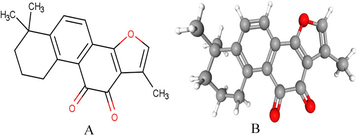
Figure 1. Chemical structure of Tan IIA. (A) Plane chemical structure of Tan IIA. (B) Stereochemical chemical structure of Tan IIA.
Recent advancements in pharmaceutical technology have revolutionized Tan IIA delivery strategies, primarily through structural modification and innovative nano-delivery systems. Yan et al. demonstrated that porous silica-based solid dispersions significantly enhance Tan IIA’s oral bioavailability by optimizing dissolution kinetics while ensuring formulation stability (Yan et al., 2015). Parallel research by Liu et al. revealed that low-molecular-weight chitosan solid dispersions offer a promising approach to improve dissolution rates and oral bioavailability in rats (Liu et al., 2013). In a notable advancement, Luo et al. engineered a chitosan/montmorillonite composite microsphere system, leveraging its unique interlayer architecture to achieve a superior loading capacities and sustained-release properties (Luo et al., 2019). Lipid-based nanocarriers have emerged as a particularly promising strategy. Ashour et al. developed sophisticated lipid nanocapsules that utilize biocompatible lipid matrices to enhance Tan IIA’s oral bioavailability through efficient drug encapsulation and controlled release mechanisms (Ashour et al., 2020). Similarly, Zhu et al. achieved remarkable progress in Tan IIA mesoporous silica nanoparticles functionalized with polyethyleneimine-polyethylene glycol, demonstrating optimal dispersibility, particle size distribution, and sustained release characteristics (Zhu et al., 2021). Innovative delivery systems have been developed for specific therapeutic applications. Ye et al. engineered an injectable borneol-modified Tan IIA liposomal formulation, demonstrating enhanced bioavailability and superior brain tissue penetration compared to conventional Tan IIA sulfonate preparations (Wang et al., 2020b). Furthermore, transdermal delivery has been advanced through nanocrystal-porous silica composite cataplasms, showing excellent skin permeability in rats (Gu et al., 2021). Microemulsion technology has also shown promise, with Ma et al. developing a Tan IIA-loaded microemulsion that effectively circumvents first-pass metabolism (Ma et al., 2022). Targeting specific pathological conditions, Zhang et al. designed discoidal reconstituted high-density lipoproteins as precision nanocarriers for atherosclerotic plaque targeting (Zhang et al., 2011). Zhan et al. fabricated a self-dissolving microneedle loaded with Tan IIA, which was shown to effectively suppress human skin fibroblast proliferation (Zhan et al., 2023). Besides, cutting-edge biomaterials have further expanded Tan IIA’s therapeutic potential. Fan et al. pioneered an injectable liquid metal-sodium alginate composite, achieving stable drug release and demonstrating efficacy in intrapericardial administration for CVDs (Fan et al., 2024). While these technological breakthroughs have significantly addressed solubility and absorption limitations, ongoing research efforts remain crucial to develop more sophisticated dosage forms that can further optimize Tan IIA’s bioavailability and therapeutic efficacy in clinical settings.
Tan IIA possesses a fused aromatic ring system with diketone groups, conferring high lipophilicity that facilitates its passive diffusion across cell membranes. This property enables intracellular accumulation and interaction with hydrophobic targets, including mitochondria and nuclear receptors, underpinning its anti-inflammatory, antioxidant, and anti-apoptotic effects (Yang et al., 2023). The compound’s tissue distribution profile like preferential accumulation in lipid-rich organs such as adipose tissue, liver, and brain, contributes to its therapeutic efficacy in CVDs (Guo et al., 2020). Furthermore, its stability in lipid-rich environments prolongs its pharmacological action, ensuring sustained target inhibition. Structurally, Tan IIA’s planar quinone core restricts conformational flexibility but enhances binding affinity to protein kinase domains through hydrophobic interactions. This rigidity enables selective binding to key proteins, such as those involved in angiogenesis, and modulates L-type calcium channels to suppress Ca2+ influx, thereby exerting anti-arrhythmic and vasodilatory effects (Ansari et al., 2021). The pharmacological mechanisms of Tan IIA are intrinsically linked to its physicochemical properties such as lipophilicity, redox activity, and structural rigidity, which collectively govern its cellular permeability, target-binding specificity, metabolic stability, and antioxidant/anti-inflammatory activities. Optimizing these characteristics through structural modifications or advanced formulation strategies remains pivotal for enhancing its clinical efficacy.
3 The clinical use of Tan IIA in the treatment of CVDs
Clinical evidence has demonstrated that both Tanshinone IIA (Tan IIA) and its water-soluble derivative sodium Tanshinone IIA sulfonate (STS) exhibit significant therapeutic benefits in CVDs. In a multicenter, randomized controlled trial involving 372 patients with non-ST elevation acute coronary syndrome undergoing percutaneous coronary intervention (PCI), STS administration significantly reduced peri-procedural myocardial injury and decreased the incidence of short-term major adverse cardiac events (MACEs) (Mao et al., 2021). A clinical study of 300 acute myocardial infarction patients post-PCI demonstrated that Tan IIA injection effectively improved coronary microcirculation, attenuated ventricular remodeling, enhanced cardiac function, and reduced MACE occurrence (Lu et al., 2021). In addition, a prospective randomized trial with 101 ST-elevated myocardial infarction patients showed that STS treatment markedly prevented adverse left ventricular remodeling and limited neutrophil-mediated myocardial damage in the infarct zone (Mao et al., 2019). Besides, another randomized controlled trial comprising 72 participants with either unstable angina or non-ST-elevation myocardial infarction revealed that STS therapy significantly improved cardiac function while reducing inflammatory markers, including high-sensitivity C-reactive protein (hs-CRP), interleukin-6 (IL-6), monocyte chemotactic protein-1 (MCP-1), and soluble CD40 ligand (sCD40L) (Li et al., 2017). These well-designed clinical studies collectively demonstrate that Tan IIA represents a safe and effective therapeutic option for various cardiovascular conditions. However, further large-scale clinical investigations are warranted to fully elucidate its therapeutic potential and optimize clinical applications.
4 Mechanism and target of Tan IIA in treating CVDs
4.1 Atherosclerosis
Atherosclerosis, a prevalent and pathologically complex cardiovascular disorder, is characterized by the progressive accumulation of lipid deposits and fibroproliferative changes within the arterial wall, leading to luminal stenosis, vascular stiffening, and compromised hemodynamic function (Libby, 2021). This chronic inflammatory disease process involves the formation of atheromatous plaques, which are complex structures comprising lipid cores, vascular smooth muscle cells (VSMCs), and inflammatory infiltrates including macrophages and lymphocytes within the arterial intima (Certo et al., 2024). The pathophysiological progression of atherosclerosis typically initiates with endothelial dysfunction, followed by the development of fatty streaks that evolve into advanced plaques, ultimately resulting in significant luminal obstruction or complete vascular occlusion. The distribution of atherosclerotic lesions predominantly affects large and medium-sized arteries, with a particular predilection for coronary, cerebrovascular, and peripheral arterial systems. The course of atherosclerotic plaques involves progressive calcification and vascular remodeling, contributing to arterial rigidity and impaired vascular compliance. Of particular clinical significance is the phenomenon of plaque vulnerability, characterized by thin fibrous caps, large lipid cores, and intense inflammatory activity, which predisposes to plaque rupture and subsequent thromboembolic complications. In recent years, Tan IIA has emerged as a promising therapeutic agent in atherosclerosis management.
4.1.1 Suppressing VSMCs proliferation and migration
VSMCs play a pivotal role in the pathogenesis of atherosclerosis, contributing significantly to arterial stiffening and luminal narrowing. This complex pathological process is mediated through multiple interconnected biological mechanisms and signaling pathways. The proliferative activity of VSMCs results in cellular hyperplasia, leading to fibrous cap formation that critically influences the stability of atherosclerotic plaques. Furthermore, migratory VSMCs not only augment the cellular density within the intimal layer but also engage in intricate interactions with infiltrating immune cells, thereby contributing to the development of more sophisticated plaque architecture. The cellular aggregation within the plaque microenvironment facilitates lipid deposition and perpetuates inflammatory cascades. The proliferation and migration of VSMCs represent dynamic and interrelated processes, wherein proliferating VSMCs can maintain their proliferative capacity in novel microenvironments, while migratory cells possess the potential to establish new cell populations within the intima (Yu et al., 2024). This reciprocal cellular behavior profoundly influences the morphological evolution and functional characteristics of atherosclerotic plaques. Specific growth factors, particularly transforming growth factor-β (TGF-β) and platelet-derived growth factor (PDGF), exert their biological effects through receptor-mediated activation of intracellular signaling cascades, thereby orchestrating VSMC proliferation and migration (Wang et al., 2022). These molecular mediators are typically upregulated in response to vascular endothelial injury and subsequent inflammatory processes.
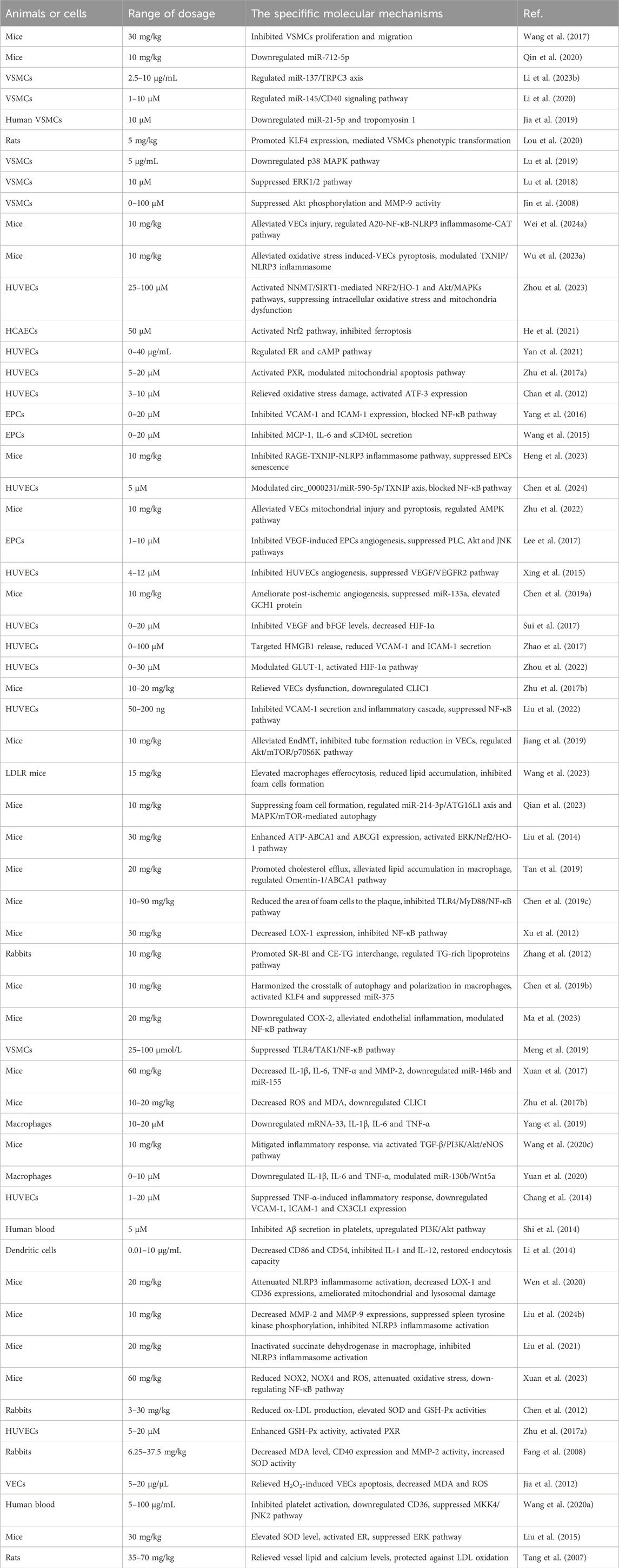
Tan IIA has demonstrated significant efficacy in mitigating atherosclerotic lesions in mice through the inhibition of VSMC proliferation and migration (Wang et al., 2017). Emerging evidence has established microRNAs (miRs) as crucial regulatory molecules in the pathogenesis of atherosclerosis. Mechanistic study revealed that Tan IIA exerted its anti-atherosclerotic effects by suppressing VSMCs proliferation through down-regulating miR-712-5p (Qin et al., 2020). Furthermore, Li et al. demonstrated that Tan IIA effectively inhibits oxidized-low density lipoprotein (ox-LDL)-induced VSMC proliferation and migration via modulation of the miR-137/TRPC3 axis (Li W. et al., 2023). In a separate investigation, Li et al. elucidated that Tan IIA attenuated homocysteine-induced VSMCs proliferation through regulation of the miR-145/CD40 pathway (Li et al., 2020). The therapeutic potential of Tan IIA extends to metabolic stress conditions, as evidenced by its ability to ameliorate high glucose-induced VSMCs proliferation and migration via downregulation of miR-21-5p and tropomyosin 1 expression (Jia et al., 2019). Lou et al. provided additional mechanistic insight, demonstrating that Tan IIA suppresses VSMCs proliferation and migration by up-regulating Krüppel-like factor 4 (KLF4) expression and mediating VSMC phenotypic modulation in rat models (Lou et al., 2020). The pharmacological effects of Tan IIA also encompass the regulation of hormonal and inflammatory pathways. Experimental study has shown that Tan IIA administration effectively suppresses angiotensin II (Ang II)-induced VSMCs proliferation and autophagy through inhibition of the p38 MAPK pathway (Lu et al., 2019). Similarly, Lu et al. reported that Tan IIA attenuates advanced glycation end products (AGEs)-induced VSMCs proliferation and migration by inhibiting the ERK1/2 signaling cascade (Lu et al., 2018). Additionally, Tan IIA inhibited TNF-α-induced VSMCs proliferation and migration via suppression of protein kinase B (Akt) phosphorylation and matrix metalloproteinase-9 (MMP-9) activity (Jin et al., 2008).
4.1.2 Improving vascular endothelial dysfunction
Vascular endothelial cells (VECs), forming the innermost layer of the vascular wall, serve as critical regulators of vascular homeostasis through multiple essential functions. These include maintenance of vascular permeability, hemodynamic regulation, synthesis of vasoprotective mediators such as nitric oxide (NO), and immunomodulatory activities (Wei X. et al., 2024). Under physiological conditions, VECs play a pivotal role in maintaining vascular health by preventing inflammatory processes and thrombus formation. However, various pathological stimuli, including hypertension, hyperglycemia, dyslipidemia, tobacco exposure, and inflammatory cytokines, can induce endothelial dysfunction, thereby initiating the atherosclerotic cascade. The pathogenesis involves chemokines and cell adhesion molecules up-regulating, which serve as inflammatory mediators that recruit circulating immune cells, particularly monocytes and lymphocytes, into the vascular intima, establishing a chronic inflammatory state. Dysfunctional VECs facilitate the transendothelial migration and subsequent oxidative modification of LDL within the subendothelial space. The resulting ox-LDL not only perpetuates the inflammatory response but also induces further endothelial injury (Panduga et al., 2024). Furthermore, endothelial dysfunction exacerbates atherogenesis through the dysregulation of vasoactive substances, characterized by diminished NO bioavailability and altered endothelin-1 (ET-1) secretion, coupled with enhanced platelet activation and prothrombotic tendency.
Wei et al. demonstrated that STS attenuates human umbilical vein endothelial cells (HUVECs) injury in mice through modulation of the A20-nuclear factor kappa B (NF-κB)-NOD-like receptor family pyrin domain containing 3 (NLRP3) inflammasome-catalase (CAT) axis (Wei W. et al., 2024). In a complementary study, Wu et al. revealed that Tan IIA protects against oxidative stress-induced VECs pyroptosis in mice by regulating the thioredoxin-interacting protein (TXNIP)/NLRP3 inflammasome pathway (Wu Q. et al., 2023). Recognizing homocysteine (Hcy) as an independent atherogenic factor, Zhou et al. reported that STS mitigates Hcy-induced VECs injury through suppression of intracellular oxidative stress and mitochondrial dysfunction. This protective effect is mediated via activation of the nicotinamide N-methyltransferase (NNMT)/sirtuin 1 (SIRT1)-dependent nuclear factor erythroid 2-related factor 2 (Nrf2)/heme oxygenase-1 (HO-1) and Akt/MAPK signaling cascades (Zhou et al., 2023). Similarly, Tan IIA has been shown to protect human coronary artery endothelial cells (HCAECs) by inhibiting ferroptosis through Nrf2 pathway activation (He et al., 2021). Moreover, Tan IIA exhibits potent protective effects against oxidative stress-induced HUVECs injury, mediated through estrogen receptor (ER) and cyclic adenosine monophosphate (cAMP) signaling pathways (Yan et al., 2021). The mitochondrial pathway has been identified as another crucial mechanism of Tan IIA’s endothelial protection. Zhu et al. demonstrated that Tan IIA alleviates hydrogen peroxide (H2O2)-induced HUVECs injury through regulation of mitochondrial apoptotic pathway (Zhu H. et al., 2017). This finding was corroborated by Chan et al., who showed that Tan IIA pretreatment reduces H2O2-induced HUVECs apoptosis by attenuating oxidative stress through elevating activating transcription factor-3 (ATF-3) expression (Chan et al., 2012). Endothelial progenitor cells (EPCs), the precursor cells of VECs, play a critical role in vascular repair through their mobilization from bone marrow to peripheral circulation in response to physiological and pathological stimuli. Yang et al. demonstrated that Tan IIA exerts anti-inflammatory effects in TNF-α-stimulated EPCs by down-regulating vascular cell adhesion molecule-1 (VCAM-1) and intercellular adhesion molecule-1 (ICAM-1) expression through inhibition of the NF-κB pathway (Yang et al., 2016). Additionally, Tan IIA has been shown to enhance TNF-α-induced EPCs proliferation, migration, adhesion, and vasculogenic capacity while suppressing the release of inflammatory mediators, including MCP-1, IL-6, and sCD40L (Wang et al., 2015). Heng et al. reported that Tan IIA preserves EPCs proliferation and differentiation capacity by maintaining CAT activity through inhibition of the receptor for advanced glycation end products (RAGE)-TXNIP-NLRP3 inflammasome pathway (Heng et al., 2023). Chen et al. further elucidated that Tan IIA attenuates ox-LDL-induced VECs injury through modulation of the circ_0000231/miR-590-5p/TXNIP axis, mediated by NF-κB pathway inhibition (Chen et al., 2024). Considering the critical role of mitochondrial dysfunction in atherogenesis, Zhu et al. demonstrated that Tan IIA maintains mitochondrial homeostasis and suppresses mitochondrial reactive oxygen species (ROS) overproduction through AMP-activated protein kinase (AMPK) pathway regulation, thereby alleviating VECs pyroptosis in atherosclerotic mice (Zhu et al., 2022).
Emerging evidence indicates that angiogenesis triggered by VECs injury plays a pivotal role in the pathogenesis of atherosclerosis. Lee et al. demonstrated that Tan IIA effectively inhibits vascular endothelial growth factor (VEGF)-induced EPCs angiogenesis through suppression of the phospholipase C (PLC), Akt, and JNK pathways (Lee et al., 2017). The anti-angiogenic properties of Tan IIA are further mediated through inhibition of the VEGF/VEGF receptor 2 (VEGFR2) signaling cascade in VECs (Xing et al., 2015). Chen et al. provided mechanistic insights into the therapeutic potential of STS, showing its ability to ameliorate post-ischemic angiogenesis in mice through dual modulation of miR-133a suppression and GTP cyclohydrolase 1 (GCH1) protein upregulation (Chen L. et al., 2019). Furthermore, Tan IIA has been shown to attenuate hypoxia-induced angiogenesis by down-regulating VEGF and basic fibroblast growth factor (bFGF) expression through inhibition of hypoxia-inducible factor 1α (HIF-1α) (Sui et al., 2017). Zhao et al. reported that STS alleviates hypoxic trophoblast-induced HUVECs dysfunction through targeted inhibition of high mobility group box 1 (HMGB1) release and subsequent reduction of VCAM-1 and ICAM-1 secretion (Zhao et al., 2017). The protective effects of Tan IIA on hypoxia-induced endothelial dysfunction extend to the glucose metabolism regulating, as evidenced by its ability to modulate glucose transporter 1 (GLUT-1) expression through HIF-1α pathway activation (Zhou et al., 2022). The involvement of intracellular chloride channel 1 (CLIC1) in oxidative stress and inflammatory responses has been elucidated by Zhu et al., who demonstrated that STS ameliorates VECs dysfunction in atherosclerotic mice through CLIC1 downregulation (Zhu J. et al., 2017). Additionally, Tan IIA micelles have shown significant endothelial protective effects by inhibiting VCAM-1 secretion and inflammatory cascades through suppression of the NF-κB pathway (Liu et al., 2022). Endothelial-mesenchymal transition (EndMT), a dynamic cellular process characterized by endothelial cell transformation into mesenchymal phenotypes under pathological stimuli, has emerged as a critical mechanism in atherosclerotic progression. Recent investigation has revealed that Tan IIA attenuates bleomycin-induced EndMT by preserving VECs tube formation capacity through modulating Akt/mammalian target of rapamycin (mTOR)/p70S6K pathway (Jiang et al., 2019). This finding underscores the therapeutic potential of Tan IIA in modulating endothelial plasticity during atherogenesis.
4.1.3 Inhibiting macrophage-derived foam cells formation
Foam cells, representing a crucial cellular component in atherogenesis, are characterized by the excessive intracellular accumulation of lipids, primarily cholesterol esters, within macrophages or VSMCs following the recognition and uptake of ox-LDL. The formation of these lipid-laden cells is mediated through specific surface receptors, including cluster of differentiation 36 (CD36) and scavenger receptor-A (SR-A), which facilitate the internalization of ox-LDL (Saki et al., 2024). The transformation of macrophages into foam cells is marked by the progressive accumulation of cytoplasmic lipid droplets, creating a distinctive foamy appearance. This cellular transformation represents a hallmark event in the development of atherosclerotic lesions. Foam cells contribute significantly to the chronic inflammatory milieu within the vascular wall through the sustained release of chemokines, and proteolytic enzymes.
The pathological significance of foam cells extends beyond their inflammatory role. As these cells proliferate and eventually undergo necrosis, they exacerbate local inflammation and contribute to plaque instability. Furthermore, foam cells secrete MMPs, which degrade the extracellular matrix components of the vascular wall, potentially leading to plaque rupture or calcification, these processes collectively increase the risk of thrombotic complications (Li et al., 2024). Consequently, therapeutic strategies targeting foam cell formation represent a crucial approach in the management of atherosclerosis, offering potential for plaque stabilization and prevention of disease progression.
Tan IIA has demonstrated significant anti-atherosclerotic potential in LDL receptor (LDLR) knockout mice through its ability to reduce lipid accumulation and inhibit macrophage-derived foam cell formation by elevating macrophage efferocytosis (Wang et al., 2023). Qian et al. elucidated that Tan IIA attenuates atherosclerosis progression by modulating lipid metabolism and suppressing foam cells formation through regulation of the miR-214-3p/autophagy-related protein-16-like protein 1 (ATG16L1) axis and MAPK/mTOR-mediated autophagy pathways (Qian et al., 2023). Complementing these findings, Liu et al. demonstrated that Tan IIA inhibits atherogenesis and reduces cholesterol accumulation in macrophage-derived foam cells through up-regulating ATP-binding cassette transporter A1 (ABCA1) and ABCG1 expression, mediated by activation of the ERK/Nrf2/HO-1 signaling cascade (Liu et al., 2014). Omentin-1 is a novel adipocytokine that possesses a protective role in the cardiovascular system. The therapeutic effects of Tan IIA extend to the regulation of adipocytokine signaling, as evidenced by Tan et al.'s report that Tan IIA attenuates atherosclerosis in apolipoprotein E-deficient (ApoE−/−) mice by promoting cholesterol efflux and alleviating lipid accumulation in macrophages through modulation of the Omentin-1/ABCA1 pathway (Tan et al., 2019). Chen et al. provided further mechanistic insights, showing that Tan IIA stabilizes vulnerable atherosclerotic plaques by reducing foam cells accumulation through inhibition of the TLR4/MyD88/NF-κB pathway (Chen Z. et al., 2019). The regulation of scavenger receptor activity represents another mechanism of Tan IIA’s effect, as demonstrated by Xu et al., who reported that Tan IIA suppresses ox-LDL uptake and inhibits plaque formation in ApoE−/− mice by down-regulating lectin-like ox-LDL receptor-1 (LOX-1) expression via NF-κB pathway inhibition (Xu et al., 2012). Besides, Zhang et al. revealed additional cholesterol metabolism pathways influenced by Tan IIA, showing its ability to ameliorate atherosclerosis in rabbits by targeting foam cells formation through promotion of scavenger receptor class B type I (SR-BI) and cholesteryl ester (CE)-triglyceride (TG) interchange, along with regulation of TG-rich lipoprotein metabolism (Zhang et al., 2012). Furthermore, the role of macrophage polarization in atherogenesis has been increasingly recognized, with M1-type macrophages demonstrating impaired lipid processing capacity, leading to foam cells formation and subsequent plaque progression. Tan IIA has been shown to modulate this process through its ability to alleviate atherosclerosis in ApoE−/− mice by orchestrating the crosstalk between autophagy and macrophage polarization via KLF4 activation and miR-375 suppression (Chen W. et al., 2019).
4.1.4 Inhibiting inflammatory reaction
Inflammatory response plays a pivotal role in the pathogenesis and progression of atherosclerosis, as well as in the destabilization and rupture of atherosclerotic plaques. This inflammatory process not only exacerbates vascular stenosis and arterial stiffening but also dramatically contributes to both the progression and instability of atherosclerotic plaques. During the initial phases of atherosclerosis, pro-inflammatory cytokines are released, which subsequently trigger endothelial cells activation, leukocyte recruitment, and lipid accumulation (Kong et al., 2022). Additionally, chemokines such as MCP-1 facilitate the migration of monocytes into the arterial wall, amplifying the inflammatory cascade and accelerating atherosclerotic plaque formation (Bianconi et al., 2018; Sun et al., 2023). The chronic inflammatory milieu promotes the proliferation of VSMCs and the deposition of extracellular matrix components, ultimately leading to vascular fibrosis. Importantly, inflammation not only fosters plaque development but also compromises the structural integrity of the fibrous cap, rendering plaques vulnerable to rupture. Such plaque disruption can precipitate thrombotic events, resulting in acute coronary syndromes, including unstable angina. Furthermore, MMPs, particularly MMP-2 and MMP-9, contribute to plaque instability by degrading the extracellular matrix components of the vascular wall, thereby weakening the fibrous cap and increasing the propensity for plaque rupture (Liu B. et al., 2024).
Cyclooxygenase-2 (COX-2), a key enzyme in the arachidonic acid epoxidase pathway, plays a crucial role in inflammatory processes and atherosclerosis progression. Its over-expression has been implicated in promoting atherosclerotic plaque instability. Recent studies have elucidated the anti-atherosclerotic mechanisms of Tan IIA through its modulation of various inflammatory pathways. Ma et al. demonstrated that Tan IIA exerts anti-atherosclerotic effects by down-regulating COX-2 expression and mitigating endothelial inflammation through NF-κB pathway modulation (Ma et al., 2023). This finding is further supported by Meng et al., who reported that Tan IIA significantly attenuates lipopolysaccharide (LPS)-induced inflammatory responses in VSMCs via suppression of the TLR4/transforming growth factor-β-activated kinase 1 (TAK1)/NF-κB signaling axis (Meng et al., 2019). The anti-inflammatory properties of Tan IIA extend to its regulatory effects on cytokine expression. Xuan et al. observed reduced levels of inflammatory mediators in cardiac and aortic tissues of ApoE−/− mice following Tan IIA treatment, mediated through downregulation of miR-146b and miR-155 (Xuan et al., 2017). Similarly, Yang et al. demonstrated that Tan IIA alleviates ox-LDL-induced inflammatory responses in macrophages by suppressing mRNA-33 and pro-inflammatory cytokines (Yang et al., 2019). The molecular mechanisms underlying Tan IIA’s protective effects involve multiple signaling pathways. Studies have identified its action through the TGF-β/phosphatidylinositol 3-kinase (PI3K)/Akt/endothelial nitric oxide synthase (eNOS) pathway (Wang et al., 2020c), and its regulation of wingless-type MMTV integration site family member 5a (Wnt5a)-mediated inflammation (Awan et al., 2022). Yuan et al. specifically reported that Tan IIA inhibits adipogenesis and inflammatory responses in ox-LDL-induced macrophages by modulating the miR-130b/Wnt5a axis (Yuan et al., 2020). Tan IIA also demonstrates significant effects on cellular interactions in atherosclerosis. It inhibits monocyte adhesion to VECs by downregulating VCAM-1, ICAM-1, and CX3CL1 expression (Chang et al., 2014), and modulates platelet-derived amyloid β peptide (Aβ) secretion through PI3K/Akt pathway activation (Shi et al., 2014). Furthermore, it regulates dendritic cell function by reducing CD86 and CD54 expression while inhibiting IL-1 and IL-12 release (Li et al., 2014). Recent advances in transcriptomic analysis have provided deeper insights into Tan IIA’s mechanisms. Chen et al. employed sequencing technology to identify non-coding RNA expression patterns in atherosclerotic lesions of Tan IIA-treated ApoE−/− mice. Their findings revealed the involvement of multiple signaling pathways, including Ras, Rap1, MAPK, cAMP, and T cell receptor pathways. The competitive endogenous RNA network analysis identified key anti-inflammatory nodes: circ-Tns3/let-7d-5p/Ctsl, circ-Wdr91/miR-378a-5p/Msr1, and circ-Cd84/miR-30c/Tlr2 (Chen et al., 2020).
The NLRP3 inflammasome plays a pivotal role in mediating immune responses and contributing to the pathogenesis of various inflammatory diseases. This multiprotein complex can be activated by diverse pathogen-associated molecular patterns (PAMPs) and damage-associated molecular patterns (DAMPs), leading to caspase-1 activation and subsequent maturation and release of pro-inflammatory cytokines, these molecular events trigger inflammatory cascades that significantly contribute to atherosclerosis (Yang et al., 2024). Given its central role in inflammatory processes, the NLRP3 inflammasome has emerged as a promising therapeutic target for atherosclerosis intervention. Recent studies have demonstrated the therapeutic potential of Tan IIA in modulating NLRP3 inflammasome activity. Experimental evidence shows that Tan IIA attenuates atherosclerotic progression in ApoE−/− mice through multiple mechanisms: suppression of NLRP3 inflammasome activation, downregulation of LOX-1 and CD36 expression, and amelioration of mitochondrial and lysosomal dysfunction (Wen et al., 2020). Similarly, STS has been shown to exert anti-atherosclerotic effects in ApoE−/− mice by reducing MMP-2 and MMP-9 expression, inhibiting spleen tyrosine kinase phosphorylation, and suppressing NLRP3 inflammasome activation (Liu H. H. et al., 2024). Further mechanistic insights into Tan IIA’s anti-inflammatory properties were provided by Liu et al., who demonstrated that Tan IIA ameliorates LPS-induced inflammatory responses in mice through a unique mechanism involving succinate dehydrogenase inactivation in macrophages, mediated by NLRP3 inflammasome inhibition (Liu et al., 2021). These findings collectively highlight the therapeutic potential of targeting the NLRP3 inflammasome in atherosclerosis management.
4.1.5 Suppressing oxidative stress damage
Oxidative stress represents a pathophysiological state characterized by an imbalance between the generation and elimination of ROS, resulting in the accumulation of these reactive molecules and subsequent cytotoxic effects. This imbalance may arise from either excessive endogenous production of oxygen free radicals or increased intake of exogenous oxidants. In the context of atherosclerosis, oxidative stress primarily contributes to disease pathogenesis through three distinct mechanisms: oxidative modification of cellular components, promotion of cellular proliferation, and modulation of vascular gene expression (Dos Santos et al., 2022). At the vascular level, oxidative stress induces the oxidative modification of ox-LDL, activates VSMCs and macrophages, and alters the expression of key adhesion molecules and chemokines, including VCAM-1 and MCP-1. Notably, emerging evidence suggests a dual role of oxidative stress in atherosclerosis development. While severe oxidative stress promotes atherogenesis, mild oxidative stress appears to play a protective role by physiologically regulating vascular gene expression and inducing the expression of anti-atherosclerotic genes, thereby maintaining vascular homeostasis (Shao et al., 2024). This paradoxical relationship between oxidative stress and atherosclerosis highlights the complexity of redox biology in vascular pathophysiology. The intensity and duration of oxidative stress appear to be critical determinants of its biological effects, with low-level oxidative stress potentially serving as an adaptive mechanism, while excessive oxidative stress contributes to vascular damage and disease progression.
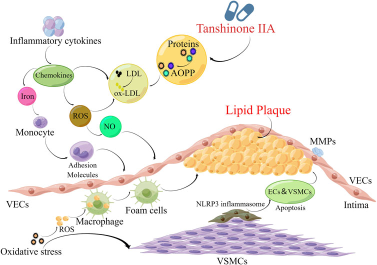
Nicotinamide adenine dinucleotide phosphate (NADPH) oxidase, a critical enzyme complex involved in multifarious physiological processes including cellular pathway and host defense, plays a pivotal role in redox biology by catalyzing the production of ROS from NADPH. These ROS molecules serve as important mediators in immune responses, apoptotic processes, and signal transduction pathways. Recent studies have extensively investigated the antioxidative properties of Tan IIA and its derivatives in the context of atherosclerosis. The antioxidative mechanisms of Tan IIA have been demonstrated through multiple experimental models. In porphyromonas gingivalis-induced atherosclerosis, Tan IIA ameliorates disease progression by reducing NOX2 and NOX4 expression, decreasing ROS production, and attenuating oxidative stress through NF-κB pathway downregulation (Xuan et al., 2023). Chen et al. further elucidated that Tan IIA inhibits atherosclerotic lesion formation in hyperlipidemic rabbits by reducing ox-LDL generation while enhancing the activities of key antioxidant enzymes, superoxide dismutase (SOD) and glutathione peroxidase (GSH-Px) (Chen et al., 2012). The therapeutic potential of STS was demonstrated by Zhu et al., who reported its ability to reduce ROS and malondialdehyde (MDA) levels in atherosclerotic mice through CLIC1 downregulation (Zhu J. et al., 2017). Similarly, Fang et al. observed that Tan IIA mitigates atherogenesis in rabbits by modulating oxidative stress markers, including decreased MDA levels and CD40 expression, reduced MMP-2 activity, and increased SOD activity (Fang et al., 2008). Moreover, the antioxidative effects of Tan IIA extend to its interaction with nuclear receptors. Pregnane X receptor (PXR) is a member of the nuclear receptor superfamily, and its activation has shown significant effects in anti-oxidative stress and protection of cardiovascular health (Shen et al., 2024). Zhu et al. demonstrated that Tan IIA protects HUVECs against H2O2-induced oxidative stress by enhancing GSH-Px activity through PXR activation (Zhu H. et al., 2017). This finding is complemented by Jia et al.'s observation that Tan IIA reduces endothelial cells apoptosis by decreasing MDA and ROS levels in oxidative stress conditions (Jia et al., 2012). At the molecular level, Tan IIA exhibits unique mechanisms in modulating oxidative stress-related pathways. It alleviates oxidative damage and inhibits platelet activation by down-regulating CD36 expression through suppression of the MAPK kinase 4 (MKK4)/JNK2 signaling pathway (Wang H. et al., 2020). Furthermore, in ovariectomized ApoE−/− mice, Tan IIA demonstrates ER-mediated antioxidative effects by elevating serum SOD levels and suppressing ERK pathway activation (Liu et al., 2015). The comprehensive antioxidative properties of Tan IIA are further evidenced by its ability to attenuate atherosclerotic calcification and regulate vascular lipid and calcium homeostasis through protection against superoxide anion-induced LDL oxidation (Tang et al., 2007). These multifaceted mechanisms contribute to Tan IIA’s overall anti-atherosclerotic effects, as generalized in Figure 2 and Table 1.
4.2 Hyperlipidemia
Primary hyperlipidemia, a condition with significant genetic underpinnings, arises from either monogenic or polygenic defects affecting key components of lipoprotein metabolism, including receptors, enzymes, and apolipoproteins (Tomlinson, 2025). A prominent example is familial hypercholesterolemia, which typically results from genetic mutations affecting cell surface lipoprotein receptors, lipoprotein lipase functionality, or structural components of lipoproteins and apolipoproteins. These genetic alterations lead to congenital lipid metabolism disorders, ultimately manifesting as hyperlipidemia (Maidman et al., 2024). The pathogenesis of primary hyperlipidemia often involves complex gene-environment interactions, although the precise mechanisms remain incompletely understood. Genetic polymorphisms can significantly influence lipid metabolism pathways, while environmental factors may modulate gene expression and metabolic processes. Dietary habits play an essential role in the progression of hyperlipidemia, with excessive consumption of high-fat foods and caloric intake leading to fat accumulation that surpasses the body’s metabolic capacity. Furthermore, high carbohydrate intake can stimulate insulin secretion and enhance hepatic synthesis of very low-density lipoprotein, predisposing individuals to hypertriglyceridemia. Obesity represents a significant risk factor for hyperlipidemia, as increased adipose tissue mass disrupts lipid homeostasis and elevates circulating lipid levels. This condition often coexists with other metabolic disorders, including diabetes mellitus and hypertension, which can further exacerbate lipid metabolism abnormalities. Additionally, various systemic diseases can induce secondary hyperlipidemia through multiple mechanisms. Conditions such as diabetes mellitus, hypothyroidism, non-alcoholic fatty liver disease, nephrotic syndrome, and gout can interfere with normal lipid metabolic processes, leading to dyslipidemia (Driessen et al., 2023; Liu X. et al., 2024).
Recent studies have elucidated the multifaceted mechanisms through which Tan IIA and its derivatives modulate lipid metabolism and alleviate hyperlipidemia. Jia et al. demonstrated that Tan IIA ameliorates hyperlipidemia in rat models by modulating hepatic lipid metabolism through regulation of key enzymes and receptors, including cytochrome P450 family 7 subfamily A polypeptide 1 (CYP7A1), LDLR, sterol regulatory element binding protein-2 (SREBP-2), and lecithin cholesterol acyltransferase (LCAT), additionally, Tan IIA influenced macrophage cholesterol efflux through regulation of ABCA1 and CD36 expression (Jia L. Q. et al., 2016). Further mechanistic insights revealed that Tan IIA reduces lipid deposition by suppressing miR-33a and modulating the SREBP-2/proprotein convertase subtilisin/kexin type 9 (PCSK9) pathway (Jia L. et al., 2016). The therapeutic potential of STS has been demonstrated in various experimental models. Zhong et al. reported that STS protects against high-fat diet-induced hyperlipidemia by enhancing antioxidant capacity through activation of the Nrf2/HO-1 pathway (Zhong et al., 2020). Liu et al. further demonstrated that STS improves vascular dysfunction in hyperlipidemic mice by inhibiting spleen tyrosine kinase phosphorylation through modulation of the NLRP3 inflammasome-MMP2/9 pathway (Liu H. H. et al., 2024). At the cellular level, Tan IIA exhibits significant effects on lipid homeostasis and ER stress. Experimental evidence shows that Tan IIA reduces lipid accumulation by alleviating ER stress-induced unfolded protein response through activation of the peroxisome proliferator-activated receptor α (PPARα)/fibroblast growth factor 21 (FGF21) axis (Pi et al., 2024). In HepG2 cells, Tan IIA demonstrates protective effects against palmitate-induced lipid accumulation and apoptosis by suppressing ER stress markers, including glucose-regulated protein 78 (GRP78), activating transcription factor 6 (ATF6), and C/EBP homologous protein (CHOP) (Wang et al., 2020d). Furthermore, Gao et al. reported that Tan IIA attenuates hepatic lipid accumulation by down-regulating lipogenic genes such as fatty acid synthase (FASN), acetyl-CoA carboxylase-1 (ACC1), and stearoyl-CoA desaturase-1 (SCD1) through modulation of the liver X receptor α (LXRα)/SREBP1 pathway (Gao et al., 2021). The interplay between inflammation and lipid metabolism represents another therapeutic target of Tan IIA. Huang et al. demonstrated that Tan IIA reduces plasma lipid levels by ameliorating oxidative stress and inflammation through PPARγ activation and TLR4 downregulation (Huang et al., 2019). Similarly, Li et al. found that STS inhibits lipogenesis and moderates fat accumulation by suppressing pro-inflammatory cytokines through activation of the SIRT1/AMP-activated protein kinase α 1 (PRKAα1) pathway (Li X. X. et al., 2019).
4.3 Hypertension
Hypertension represents a systemic vascular disorder rather than a localized vascular lesion, involving multiple organ systems including the cerebral, coronary, renal, and retinal circulations (Mandorfer et al., 2025). The pathogenesis of hypertension involves complex interactions between vascular structural changes and neurohormonal regulation. Progressive vasoconstriction and luminal narrowing of resistance vessels, characterized by arterial wall thickening and reduced elasticity, lead to increased peripheral vascular resistance and subsequent elevation of blood pressure (BP). Besides, alterations in central nervous system function and dysregulation of neurotransmitter systems, including norepinephrine, dopamine, and enkephalins, can induce sympathetic nervous system hyperactivity. This results in elevated plasma catecholamine concentrations, which potentiate vasoconstriction of resistance arterioles and contribute to sustained BP elevation (Kim and Thiruvengadam, 2024). Additionally, activation of the renin-angiotensin-aldosterone system (RAAS) in response to various stimuli plays a pivotal role in the development and progression of hypertension. Renal pathophysiology significantly contributes to hypertension through mechanisms involving sodium and water retention, increased cardiac output, and subsequent elevation of peripheral vascular resistance. Furthermore, various modifiable risk factors have been identified in the etiology of hypertension, including chronic tobacco use, excessive dietary sodium intake, obesity, sleep deprivation, and chronic psychological stress (Faraci and Scheer, 2024). These factors interact with genetic predispositions to influence vascular tone regulation, fluid homeostasis, and neuroendocrine function, ultimately contributing to the development of sustained hypertension.
Pharmacological modulation of calcium (Ca2+) and large-conductance calcium-activated potassium (BKCa) channels represents a crucial mechanism for regulating vascular tone under both physiological and pathological conditions. STS has demonstrated significant hypotensive effects in experimental models, primarily through its dual action on BKCa channel activation and Ca2+ channel inhibition. The vasorelaxant properties of STS are mediated through multiple mechanisms, including BKCa channel activation, retardation of Ca2+ channels, and inhibition of Ca2+ influx in VSMCs (Zhang X. D. et al., 2018). At the molecular level, Tan et al. demonstrated that STS activates BKCa channels by enhancing the membrane expression of the α subunit in both HEK293 cells and VSMCs (Tan et al., 2014). This BKCa-dependent vasodilatory effect has been specifically observed in the mesenteric arteries of spontaneously hypertensive rats (Zhou et al., 2019). Furthermore, Yang et al. confirmed that STS activates high-conductance Ca2+-activated K+ channels in porcine coronary artery smooth muscle cells, providing additional evidence for its potassium channel-mediated vasodilatory effects (Yang et al., 2008). The vasodilatory mechanisms of Tan IIA extend beyond ion channel modulation. Wang et al. revealed that Tan IIA enhances eNOS expression in HUVECs through the transient receptor potential vanilloid 4 (TRPV4)-NO-protein kinase G (PKG) signaling pathway (Wang P. et al., 2024). This finding is complemented by Fan et al.'s observation that Tan IIA’s vasodilatory effects are modulated through ER-dependent eNOS activation and calcium mobilization (Fan et al., 2011). Interestingly, STS exhibits unique vascular effects in pregnancy-related conditions, demonstrating effective dilation of uterine arteries and direct vasodilatory effects on vascular resistance arteries in pregnant rats through NO-independent mechanisms (Morton et al., 2015). Additionally, Yu et al. demonstrated that Tan IIA exerts antihypertensive effects and inhibits cerebrovascular cell proliferation in hypertensive rats by attenuating ET-1 expression through phosphoinositide-dependent kinase 1 (PDK1) inactivation (Yu et al., 2015).
4.4 Myocardial injury and myocardial infarction (MI)
MI primarily results from the obstruction of coronary blood supply, leading to myocardial hypoxia and ischemia. The predominant etiology of MI is coronary atherosclerosis, which causes narrowing or complete occlusion of coronary arteries. This obstruction impedes adequate supply of oxygenated blood and essential nutrients to myocardium, resulting in ischemic injury. Prolonged or severe ischemia can induce metabolic disturbances and cardiomyocyte damage, ultimately leading to cellular necrosis, which represents a core pathological feature of MI (Zhai et al., 2025). The progression of MI involves distinct pathological phases. During the ischemic phase, cardiomyocytes remain viable but sustain significant damage. In the subsequent necrotic phase, these cells undergo complete functional loss, releasing intracellular enzymes and organelles that trigger inflammatory responses and tissue remodeling. Histopathological examination reveals characteristic features of necrotic cardiomyocytes, including cellular swelling, organelle disintegration, and nuclear degeneration (Wei et al., 2025). A major therapeutic challenge in MI is ischemia/reperfusion (I/R) injury, wherein the reintroduction of oxygen-rich blood to hypoxic cardiac tissue paradoxically exacerbates cellular damage through mechanisms involving calcium overload, as excessive extracellular calcium influx leads to intracellular calcium accumulation and increased cardiomyocyte death (Ghanta et al., 2025). The pathophysiology of MI also involves complex inflammatory and oxidative processes. Tissue damage triggers cell membrane degradation and increased production of arachidonic acid metabolites, which recruit leukocytes to the injured site. These inflammatory cells adhere to vascular endothelium, further exacerbating endothelial injury. Concurrently, an imbalance between ROS production and antioxidant defenses initiates lipid peroxidation cascades. Oxygen free radicals impair ATP production, induce inflammatory mediator release, and cause membrane damage, collectively contributing to cellular death and myocardial injury progression.
Chen et al. demonstrated that STS attenuates endotoxin-induced cardiomyocyte pyroptosis and autophagy in mice through inhibiting NLRP3 inflammasome activation (Chen P. et al., 2021). This finding is complemented by Hu et al.'s research showing that STS ameliorates ischemia-induced myocardial inflammation in canine models by modulating NLRP3 inflammasome activation through restoration of PPAR-α expression via the JAK2-STAT3 pathway (Hu et al., 2015). The therapeutic potential of Tan IIA extends to its interaction with IGF-2R, a critical mediator in myocardial injury pathogenesis. Experimental evidence shows that Tan IIA inhibits Ang II-induced apoptosis in rat H9c2 cardiomyocytes and prevents subsequent cardiac remodeling by suppressing β-catenin nuclear translocation and IGF-2R inactivation (Chen et al., 2017). Weng et al. further demonstrated that Tan IIA protects H9c2 cardiomyocytes from injury through IGF-2R inhibition mediated by PI3K/Akt pathway activation (Weng et al., 2015). The antioxidative properties of STS contribute significantly to its cardioprotective effects. Yan et al. reported that STS reduces myocardial apoptosis in murine models by mitigating oxidative stress through modulation of the Keap1-Nrf2 pathway (Yan et al., 2018). These findings collectively highlight the multifaceted mechanisms through which STS and Tan IIA exert their protective effects against myocardial injury and infarction.
MI initiates a cascade of pathological cardiac remodeling that often progresses to HF. Zhang et al. demonstrated that STS attenuates post-MI pathological remodeling in mice through multiple mechanisms, including reduction of myocardial necrosis, suppression of inflammatory responses, and promotion of angiogenesis (Zhang B. et al., 2022). Chai et al. further elucidated that Tan IIA inhibits cardiomyocyte pyroptosis in a rat HF model following acute MI by down-regulating key inflammatory mediators IL-1β, pro-IL-1β, NLRP3, and caspase-1 through suppression of the TLR4/NF-κB p65 pathway (Chai et al., 2023). Additionally, STS has shown cardioprotective effects in isoproterenol-induced MI models by modulating fatty acid β-oxidation (Wei et al., 2013). The ER, crucial for protein synthesis and processing in cardiomyocytes, plays a significant role in MI pathophysiology. Under conditions of hypoxia, nutrient deprivation, or calcium imbalance, ER stress response is activated. Prolonged ER stress can lead to dysfunction and subsequent cardiomyocyte damage. Tan IIA has been shown to mitigate cardiomyocyte injury by alleviating ER stress through upregulation of SIRT1 expression (Wu S. et al., 2023). The tumor suppressor phosphatase and tensin homolog (PTEN) has emerged as a key regulator of cardiomyocyte apoptosis. Wang et al. reported that Tan IIA improves cardiac function in MI mice by promoting angiogenesis through modulation of miR-499-5p and PTEN expression (Wang and Wu, 2022). This finding is supported by Zhang et al.'s demonstration that Tan IIA inhibits apoptosis in rat H9c2 cardiomyocytes by up-regulating miR-152-3p and down-regulating PTEN (Zhang et al., 2016). Recent advances in understanding post-MI cardiac repair have highlighted the critical role of macrophage reprogramming. Modulation of macrophage metabolic pathways and functional phenotypes has shown potential in promoting cardiac repair and improving cardiac function (Hu et al., 2024; Xie et al., 2024). Gao et al. demonstrated that Tan IIA facilitates cardiac repair in post-MI mice by reprogramming macrophage phenotypes through inactivation of the phosphoglycerate kinase 1 (PGK1)/pyruvate dehydrogenase kinase 1 (PDHK1) pathway and remodeling macrophage energy metabolism (Gao et al., 2024).
While reperfusion therapy remains the gold standard treatment for myocardial ischemia, it inevitably induces myocardial I/R injury (Zhu et al., 2023). A systematic meta-analysis revealed that Tan IIA exhibits significant cardioprotective effects in rat models of I/R injury at doses exceeding 5 mg/kg, primarily via suppression of oxidative stress (Zhang X. et al., 2024). The protective mechanisms of Tan IIA and its derivatives against I/R injury involve multiple molecular pathways. Zhong et al. demonstrated that Tan IIA alleviates cardiac microvascular I/R injury by reducing mitochondrial apoptosis through activation of the SIRT1-PGC1α pathway (Zhong et al., 2019). This finding is complemented by Long et al.'s research showing that STS attenuates microvascular I/R injury through inhibition of fibrinogen-like protein 2 (FGL2) expression, fibrin deposition, and inflammatory responses via mediating Akt and NF-κB pathways (Long et al., 2015). Li et al. further reported that Tan IIA ameliorates myocardial I/R injury by suppressing NLRP3 inflammasome activation and regulating Th17/Treg cells differentiation (Li et al., 2022). The antioxidative and anti-inflammatory properties of STS contribute to its cardioprotective effects, as evidenced by its ability to enhance HO-1 activity and mitigate I/R injury (Wei et al., 2014). Fang et al. demonstrated that Tan IIA improves myocardial ischemia by reducing cardiomyocyte apoptosis and modulating apoptotic markers caspase-3, Cyto c, and Apaf-1 in myocardial tissue (Fang et al., 2021). Moreover, the ataxia-telangiectasia mutated (ATM) kinase, a crucial regulator of DNA damage response and genomic stability, has emerged as a potential therapeutic target in myocardial injury (Kukreja, 2014). Sang et al. showed that Tan IIA protects against I/R injury in H9c2 cardiomyocytes via ATM-mediated activation of the GADD45/ORC pathway (Sang et al., 2024). Besides, the composition of extracellular matrix changes significantly, collagen secretion decreases while hyaluronic acid accumulation increases competitively during acute MI, activated cardiac fibroblasts produce hyaluronic acid through hyaluronan synthase 2 (HAS2) (Little et al., 2025). Tan IIA has been shown to mitigate I/R injury in human AC16 cardiac cells by targeting the HAS2/fibroblast growth factor 9 (FGF9) axis, thereby reducing inflammation and oxidative stress (Wang Y. et al., 2024). Furthermore, Tan IIA protects against mitochondrial dysfunction through modulation of voltage-dependent anion channel 1 (VDAC1), an essential regulator of mitochondrial-cytoplasmic exchange. Hu et al. demonstrated that Tan IIA inhibits ferroptosis and apoptosis in H9c2 cardiomyocytes by down-regulating VDAC1 and preventing oxidative stress (Hu T. et al., 2023). The cardioprotective effects of Tan IIA extend to mitochondrial preservation. It protects H9c2 cardiomyocytes against anoxia/reoxygenation injury by inhibiting mPTP opening and apoptosis through upregulation of 14-3-3η, a protein that played a momentous protective role in cardiomyocytes (Zhang Z. et al., 2018). Wen et al. further demonstrated that Tan IIA alleviates I/R injury by regulating autophagy and maintaining mitochondrial function through 14-3-3η-mediated modulation of the Akt/Beclin1 pathway (Wen et al., 2023). Emerging therapeutic strategies utilizing mesenchymal stem cells (MSCs) have shown promise in CVDs treatment. Li et al. found that exosomes derived from Tan IIA-pretreated MSCs exert cardioprotective effects in myocardial I/R models by up-regulating miR-223-5p and inactivating CCR2, a protein-coding gene highly expressed in cardiomyocytes (Li S. et al., 2023).
Interestingly, Tan IIA demonstrates significant myocardial protective effects by modulating the crosstalk between the NLRP3 inflammasome and the Nrf2 antioxidant pathway, with its efficacy exhibiting a clear dose-dependent pattern. Mechanistically, Nrf2 activation is more responsive to oxidative stress, whereas NLRP3 inhibition requires higher Tan IIA concentrations to effectively suppress the inflammatory signaling cascade. At low doses, Tan IIA preferentially enhances antioxidant defenses, markedly up-regulating SOD activity and reducing MDA levels, while exerting only a modest inhibitory effect on NLRP3 inflammasome activation (Chen P. et al., 2021). In contrast, medium-dose Tan IIA achieves a balanced modulation of both pathways, simultaneously reducing myeloperoxidase (MPO) activity and IL-1β levels while significantly restoring ATP content, indicating coordinated anti-inflammatory and antioxidant actions (Vidal-Gomez et al., 2025). However, at high doses, Tan IIA may excessively suppress inflammatory responses, potentially compromising immune defense mechanisms (Wu S. et al., 2023). Collectively, Tan IIA mediates cardioprotection through a dynamic Nrf2-NLRP3 interaction network, exhibiting dose-dependent preferential effects: low-dose regimens favor anti-oxidative activity, high-dose treatments enhance anti-inflammatory efficacy, and intermediate doses promote synergistic regulation. These findings provide a mechanistic rationale for optimizing Tan IIA dosing in clinical applications. Future studies should further elucidate tissue-specific dose-response relationships and evaluate long-term safety profiles to facilitate translational development. A comprehensive summary of the myocardial protective effect of Tan IIA through crosstalk among NLRP3 inflammasome, NF-κB, and Nrf2 pathways is presented in Figure 3.
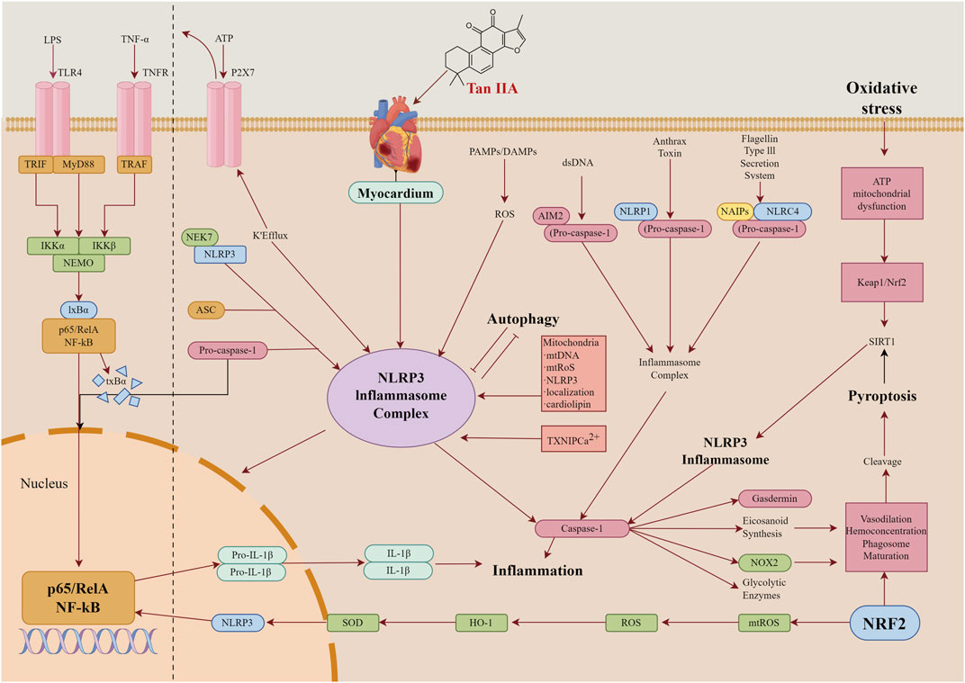
Figure 3. Tan IIA mediate cross-talk among NLRP3 inflammasome, NF-κB, and Nrf2 pathways in myocardial protection.
4.5 Cardiac hypertrophy, fibrosis, and heart failure (HF)
Cardiac hypertrophy, characterized by an increase in myocardial cells volume and consequent thickening of the ventricular wall, represents a complex adaptive response that can be either physiological or pathological in nature. Physiological hypertrophy, exemplified by the athlete’s heart, contrasts with pathological hypertrophy commonly observed in conditions such as hypertension and valvular heart disease. The pathogenesis of pathological myocardial hypertrophy primarily involves activation of neuroendocrine systems, particularly the RAAS and sympathetic nervous system, along with dysregulation of cellular signaling pathways including calcium ion channels and MAPK cascades (Chen et al., 2025). Myocardial fibrosis, characterized by excessive deposition of collagen and extracellular matrix components, represents another critical pathological process in cardiac remodeling. This fibrotic transformation, frequently associated with aging, obesity, and diabetes mellitus, significantly impairs cardiac function through multiple mechanisms. The accumulation of fibrous tissue increases myocardial stiffness, compromising ventricular diastolic function and impairing proper cardiac filling, ultimately reducing stroke volume and cardiac output (Skorka et al., 2025; Zhang et al., 2023). Furthermore, myocardial fibrosis disrupts the typical cellular architecture and electrophysiological properties of cardiac tissue, creating a substrate for arrhythmias that further compromise cardiac efficiency and contribute to the progression of HF (Lee et al., 2025). The excessive extracellular matrix deposition also physically restricts cardiomyocyte contractility, reducing myocardial contractile force and exacerbating cardiac dysfunction (Hosseinzadeh et al., 2024). The interplay between myocardial hypertrophy and fibrosis creates a vicious cycle in HF pathophysiology. Hypertrophic changes increase cardiac wall stress, promoting further cardiomyocyte enlargement and subsequent fibrotic transformation. The resulting fibrosis exacerbates myocardial stiffness and dysfunction, creating a self-perpetuating cycle of cardiac remodeling and functional deterioration. This complex interaction between structural and functional alterations underlies the progressive nature of HF development.
Tan IIA has demonstrated significant cardioprotective effects against cardiac hypertrophy and fibrosis through multiple molecular mechanisms. Experimental studies have revealed that Tan IIA attenuates cardiac hypertrophy in rat models by down-regulating collagen type I α-1 (COL1A1) mRNA expression through inhibition of the cystatin C (Cys-C)/Wnt pathway (Feng et al., 2017). Jiang et al. further elucidated Tan IIA’s potent anti-fibrotic properties, which are mediated through modulation of both TGF-β/Smad and MAPK/ERK pathways (Jiang et al., 2024). The therapeutic potential of Tan IIA extends to Ang II-induced cardiac fibrosis, where it enhances NO production and eNOS phosphorylation while inhibiting ERK phosphorylation (Chan et al., 2011). Zhang et al. demonstrated that Tan IIA alleviates acid-induced fibrosis by targeting the Nrf2-NLRP3 pathway, thereby reducing oxidative stress and inflammatory responses (Zhang W. et al., 2024). In the context of hypertension-induced cardiac remodeling, Tan IIA exhibits multifaceted protective effects. It mitigates left ventricular myocardial changes by reducing MMP-9 and tissue inhibitor of metalloproteinase-1 (TIMP-1) expression, thereby improving cardiac function in renovascular hypertensive rats (Fang et al., 2010). Pang et al. reported that Tan IIA inhibits hypertension-induced left ventricular hypertrophy and fibrosis by modulating the TGF-β/Smads pathway while reducing MDA levels and enhancing SOD activity (Pang et al., 2014). Additionally, Jiang et al. found that Tan IIA suppresses cardiomyocyte hypertrophy and apoptosis in spontaneously hypertensive rats through regulation of Bcl-2, Bax, and p53 expression (Jiang et al., 2013). The antioxidative properties of Tan IIA contribute significantly to its cardioprotective effects. It improves cardiac function and reduces fibrosis in hypertensive models by decreasing NADPH oxidase activity, a major source of ROS in the heart (Wang et al., 2011). This mechanism is further supported by studies showing Tan IIA’s ability to inhibit H2O2-stimulated cardiac fibroblasts through suppression of collagen synthesis and NADPH oxidase activity (Wang et al., 2013). Tan IIA’s anti-hypertrophic effects are also mediated through inflammation modulation. It attenuates transverse aortic constriction-induced cardiac hypertrophy by reducing IL-6 and TNF-α levels, decreasing MDA content, and enhancing SOD activity via SIRT1 activation (Feng et al., 2016). Furthermore, Zhang et al. demonstrated that Tan IIA inhibits galectin-3 expression through modulation of N6-methyladenosine (m6A) methylation, thereby attenuating cardiac hypertrophy (Zhang M. et al., 2022). At the molecular level, Tan IIA influences microRNA regulation of cardiac remodeling. It inhibits hypertrophy and collagen deposition in rat heart tissues through upregulation of miR-618 (Yan et al., 2022). Mao et al. further reported that Tan IIA reduces collagen deposition in human cardiac fibroblasts by modulating MMP-2 and MMP-9 expression and regulating elastin deposition (Mao et al., 2014). The cardioprotective effects of Tan IIA extend to adrenergic stress-induced cardiac remodeling. It mitigates isoproterenol-induced cardiac hypertrophy by reducing brain natriuretic peptide (BNP), atrial natriuretic peptide (ANP), and β-myosin heavy chain (β-MHC) levels through suppression of the calcineurin/NFATc3 pathway (Tan et al., 2011).
Emerging evidence has highlighted the crucial role of gut microbiota dysbiosis and gut-brain axis dysfunction in the pathogenesis of HF. Zhu et al. demonstrated that Tan IIA significantly attenuates myocardial apoptosis and fibrosis, subsequently improving cardiac hypertrophy and function in mice. This cardioprotective effect is mediated through inhibition of inflammatory responses and restoration of intestinal barrier integrity via modulation of the gut-brain axis (Zhu et al., 2024). The therapeutic potential of Tan IIA extends to chemotherapy-induced cardiotoxicity. Xu et al. reported that Tan IIA ameliorates doxorubicin-induced myocardial structural alterations and myofibrillar damage in HF mice through activation of the ERK1/2 signaling pathway (Xu et al., 2022). Wang et al. further elucidated that Tan IIA mitigates doxorubicin-induced HF by restoring autophagic flux through regulation of the Beclin1/lysosomal-associated membrane protein-1 (LAMP1) pathway (Wang et al., 2019). Innovative drug delivery systems have been developed to enhance Tan IIA’s therapeutic efficacy. Zhao et al. designed ROS-responsive triphenylphosphine-modified Tan IIA micelles, which demonstrated superior cardioprotective effects in doxorubicin-induced HF by reducing oxidative stress and inflammatory cytokine infiltration through mitochondrial activation in cardiomyocytes (Zhao et al., 2025). The molecular mechanisms underlying Tan IIA’s cardioprotective effects involve multiple signaling pathways. Experimental evidence shows that Tan IIA protects against HF by inhibiting myocardial apoptosis and promoting autophagy through activation of the AMPK-mTOR signaling cascade (Zhang et al., 2019). Chen et al. further demonstrated that Tan IIA attenuates cardiac dysfunction and fibrosis in HF models by reducing TGF-β, α-SMA, and MMP-9 expression in cardiac fibroblasts through suppression of oxidative stress (Chen R. et al., 2021). In the context of pressure overload-induced HF, Tan IIA exhibits significant therapeutic potential. Chronic pressure overload, characterized by increased resistance during cardiac contraction and relaxation, leads to pathological cardiomyocyte hypertrophy and subsequent cardiac insufficiency. Li et al. demonstrated that Tan IIA alleviates ventricular remodeling and improves cardiac function in HF mice via restraining inflammatory responses and cardiomyocyte apoptosis (Li X. et al., 2019).
4.6 Arrhythmia
Arrhythmia is defined as a disturbance in the frequency and/or rhythm of cardiac contractions resulting from abnormalities in the initiation and/or propagation of electrical impulses within the heart. The underlying mechanisms primarily involve irregularities in impulse formation and conduction, which can be influenced by various pathological factors, including pharmacological effects, drug toxicity, electrolyte imbalances, anesthesia, surgical interventions, cardiac catheterization, and autonomic nervous system dysfunction (Mondejar-Parreno et al., 2025). Arrhythmia arises when the electrical signals responsible for regulating cardiac contractions are delayed or obstructed. This disruption can occur due to either malfunctioning specialized cardiac pacemaker cells or impaired transmission of electrical impulses through the heart’s conduction system (Zeitler et al., 2024). Additionally, arrhythmia may result from ectopic electrical activity originating in other regions of the heart or abnormal propagation of impulses from pacemaker cells, both of which can interfere with the heart’s regular rhythmic activity. Furthermore, insufficient coronary blood flow can compromise the metabolic and functional integrity of cardiomyocytes, leading to alterations in myocardial electrophysiological properties and subsequent arrhythmogenesis (Yang et al., 2025). Electrolytes play a critical role in maintaining the regular electrophysiological activity of cardiomyocytes. Imbalances in the concentrations of key ions such as potassium, sodium, and calcium can significantly affect cardiomyocyte excitability, automaticity, and conductivity, thereby predisposing to arrhythmia.
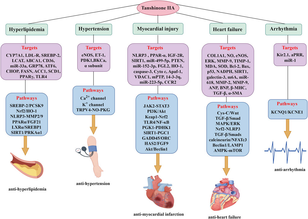
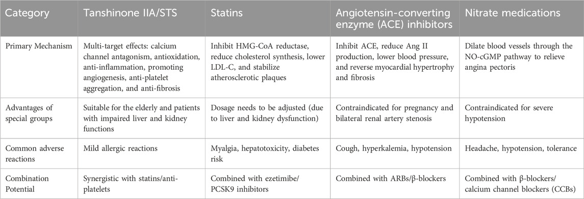
He et al. demonstrated that Tan IIA effectively ameliorates atrial fibrillation in rabbits, with its anti-arrhythmic properties primarily attributed to the modulation of atrial electrophysiology. Specifically, Tan IIA prolongs atrial postrepolarization refractoriness (aPRR) and moderately increases interatrial conduction time (He et al., 2016). Shan et al. further elucidated that Tan IIA reduces the duration of arrhythmia and lowers the incidence of ventricular tachycardia (VT) and ventricular fibrillation (VF) in rats. This effect is mediated through the upregulation of Kir2.1 expression by inhibiting miR-1, which expressed in the heart that regulates various ion channels and myocardial electrical activity under pathological conditions (Shan et al., 2009). Sun et al. identified Tan IIA as a potential activator of the human cardiac slow delayed-rectifier K+ current (IKs). Their findings revealed that Tan IIA activates human cardiac KCNQ1/KCNE1 potassium channels in HEK 293 cells by altering the channels’ kinetic properties (Sun et al., 2008). Despite these promising findings, research on the anti-arrhythmic effects of Tan IIA remains limited, and the precise pharmacological mechanisms warrant further investigation. A comprehensive summary of the effects and molecular targets regulated by Tan IIA in treating CVDs is presented in Figure 4 and Table 2. A comprehensive summary of Tan IIA’s efficacy against conventional cardiovascular drugs for CVDs is presented in Table 3.
5 Conclusion and future perspective
In conclusion, Tan IIA represents a valuable natural compound with immense potential in cardiovascular medicine. Its multifaceted pharmacological properties, including potent anti-inflammatory, antioxidant, and anti-atherosclerotic effects, position it as a compelling candidate for managing a spectrum of CVDs such as atherosclerosis, acute myocardial infarction, and coronary heart disease. Recent advancements have elucidated Tan IIA’s ability to modulate key signaling pathways implicated in cardiovascular pathology, including the inhibition of pro-inflammatory cytokine expression, reduction of oxidative stress, and stabilization of atherosclerotic plaques. By targeting multiple pathological processes simultaneously, Tan IIA demonstrates remarkable versatility and therapeutic efficacy. As research continues to evolve, Tan IIA holds the potential to revolutionize cardiovascular treatment paradigms, offering innovative, integrative therapeutic strategies that could significantly improve clinical outcomes for patients with CVDs. Nevertheless, the potential adverse effects associated with prolonged and/or high-dose administration of Tan IIA warrant careful consideration and should not be overlooked. Some individuals may exhibit allergic reactions to Tan IIA, manifesting as pruritus, erythema, swelling, and cutaneous eruptions following administration. In severe cases, these hypersensitivity reactions may be accompanied by respiratory distress. Tan IIA may also exert irritative effects on the gastrointestinal mucosa, potentially inducing epigastric pain, nausea, emesis, abdominal discomfort, and diarrhea. Prolonged administration on an empty stomach may result in gastric mucosal injury. Due to its pharmacologic properties of promoting blood circulation, Tan IIA administration may lead to accelerated systemic circulation, potentially causing facial flushing in some patients. Furthermore, it may interfere with the coagulation cascade, potentially resulting in coagulation disorders. This may present as cutaneous ecchymosis, ocular hemorrhage, hematuria, menorrhagia, or gastrointestinal bleeding. In severe instances, it may precipitate critical hemorrhagic events, including but not limited to cerebral hemorrhage. Upon the occurrence of the aforementioned adverse effects, immediate discontinuation of Tan IIA is warranted, followed by appropriate symptomatic management.
Despite its considerable promise, the clinical application of Tan IIA faces several challenges. Key limitations include its low bioavailability, insufficient clinical data, and an incomplete understanding of its therapeutic mechanisms and molecular targets. To date, most studies have been confined to animal models or small-scale clinical trials, underscoring the need for large-scale, high-quality clinical studies to validate its safety and efficacy. Moving forward, interdisciplinary collaboration and innovative research methodologies will be critical to unlocking the full therapeutic potential of Tan IIA. Future research should prioritize optimizing drug delivery systems, elucidating multi-target synergistic mechanisms, and developing highly effective derivative which not only improves the bioavailability of Tan IIA, but also enhances its pharmacological activity and reduces its adverse effects. The development of new derivatives and compounds containing Tan IIA such as Compound Danshen Dripping Pills is of great significance and is expected to provide a new option for treating CVDs (Wang R. et al., 2024). These efforts will be essential to bridge the gap between traditional Chinese medicine and modern therapeutics, ultimately facilitating the transformation of Tan IIA into a widely adopted clinical treatment for CVDs.
Author contributions
QW: Funding acquisition, Conceptualization, Writing – review and editing, Supervision. LD: Methodology, Software, Investigation, Writing – original draft. CG: Investigation, Writing – original draft, Methodology, Software. HZ: Software, Methodology, Writing – original draft, Investigation. HJ: Software, Methodology, Investigation, Writing – original draft.
Funding
The author(s) declare that financial support was received for the research and/or publication of this article. This work was supported by the National Natural Science Foundation of China (82374367), Jiangxi Provincial Natural Science Foundation (20242BAB26163 and 20232BAB206144), Jiangxi Province Key Laboratory of Traditional Chinese Medicine for Cardiovascular Diseases (20242BCC32096), Ganzhou Key Research and Development Program Project (GZ2024YLJ153), NATCM's Project of High-level Construction of Key TCM Disciplines (zyyzdxk-2023113), Project of Key Discipline Construction Fund of Jiangxi University of Chinese Medicine (2023jzzdxk032), Science and Technology Innovation Team Development Program of Jiangxi University of Chinese Medicine (CXTD22011), National Traditional Chinese Medicine Inheritance and Innovation Center Construction Project.
Acknowledgments
The figures in this article were created by Figdraw.
Conflict of interest
The authors declare that the research was conducted in the absence of any commercial or financial relationships that could be construed as a potential conflict of interest.
Generative AI statement
The author(s) declare that no Generative AI was used in the creation of this manuscript.
Publisher’s note
All claims expressed in this article are solely those of the authors and do not necessarily represent those of their affiliated organizations, or those of the publisher, the editors and the reviewers. Any product that may be evaluated in this article, or claim that may be made by its manufacturer, is not guaranteed or endorsed by the publisher.
References
Ansari, M. A., Khan, F. B., Safdari, H. A., Almatroudi, A., Alzohairy, M. A., Safdari, M., et al. (2021). Prospective therapeutic potential of Tanshinone IIA: an updated overview. Pharmacol. Res. 164, 105364. doi:10.1016/j.phrs.2020.105364
Ashour, A. A., Ramadan, A. A., Abdelmonsif, D. A., and El-Kamel, A. H. (2020). Enhanced oral bioavailability of Tanshinone IIA using lipid nanocapsules: formulation, in-vitro appraisal and pharmacokinetics. Int. J. Pharm. 586, 119598. doi:10.1016/j.ijpharm.2020.119598
Awan, S., Lambert, M., Imtiaz, A., Alpy, F., Tomasetto, C., Oulad-Abdelghani, M., et al. (2022). Wnt5a promotes lysosomal cholesterol egress and protects against atherosclerosis. Circ. Res. 130, 184–199. doi:10.1161/CIRCRESAHA.121.318881
Bianconi, V., Sahebkar, A., Atkin, S. L., and Pirro, M. (2018). The regulation and importance of monocyte chemoattractant protein-1. Curr. Opin. Hematol. 25, 44–51. doi:10.1097/MOH.0000000000000389
Certo, M., Rahimzadeh, M., and Mauro, C. (2024). Immunometabolism in atherosclerosis: a new understanding of an old disease. Trends Biochem. Sci. 49, 791–803. doi:10.1016/j.tibs.2024.06.003
Chai, R., Ye, Z., Xue, W., Shi, S., Wei, Y., Hu, Y., et al. (2023). Tanshinone IIA inhibits cardiomyocyte pyroptosis through TLR4/NF-κB p65 pathway after acute myocardial infarction. Front. Cell Dev. Biol. 11, 1252942. doi:10.3389/fcell.2023.1252942
Chan, P., Chen, Y. C., Lin, L. J., Cheng, T. H., Anzai, K., Chen, Y. H., et al. (2012). Tanshinone IIA attenuates H₂O₂ -induced injury in human umbilical vein endothelial cells. Am. J. Chin. Med. 40, 1307–1319. doi:10.1142/S0192415X12500966
Chan, P., Liu, J. C., Lin, L. J., Chen, P. Y., Cheng, T. H., Lin, J. G., et al. (2011). Tanshinone IIA inhibits angiotensin II-induced cell proliferation in rat cardiac fibroblasts. Am. J. Chin. Med. 39, 381–394. doi:10.1142/S0192415X11008890
Chang, C. C., Chu, C. F., Wang, C. N., Wu, H. T., Bi, K. W., Pang, J. H., et al. (2014). The anti-atherosclerotic effect of Tanshinone IIA is associated with the inhibition of TNF-alpha-induced VCAM-1, ICAM-1 and CX3CL1 expression. Phytomedicine 21, 207–216. doi:10.1016/j.phymed.2013.09.012
Chen, J., Xing, Y., Sun, J., Liu, Y., Lang, Z., Zhang, L., et al. (2025). Hypertrophic cardiomyopathy: genes and mechanisms. Front. Biosci. Landmark Ed. 30, 25714. doi:10.31083/FBL25714
Chen, L., He, W., Peng, B., Yuan, M., Wang, N., Wang, J., et al. (2019a). Sodium Tanshinone IIA sulfonate improves post-ischemic angiogenesis in hyperglycemia. Biochem. Biophys. Res. Commun. 520, 580–585. doi:10.1016/j.bbrc.2019.09.106
Chen, P., An, Q., Huang, Y., Zhang, M., and Mao, S. (2021a). Prevention of endotoxin-induced cardiomyopathy using sodium Tanshinone IIA sulfonate: involvement of augmented autophagy and NLRP3 inflammasome suppression. Eur. J. Pharmacol. 909, 174438. doi:10.1016/j.ejphar.2021.174438
Chen, R., Chen, W., Huang, X., and Rui, Q. (2021b). Tanshinone IIA attenuates heart failure via inhibiting oxidative stress in myocardial infarction rats. Mol. Med. Rep. 23, 404. doi:10.3892/mmr.2021.12043
Chen, S. (2024). Biosynthesis of natural products from medicinal plants: challenges, progress and prospects. Chin. Herb. Med. 16, 1–2. doi:10.1016/j.chmed.2024.01.001
Chen, W., Guo, S., Li, X., Song, N., Wang, D., and Yu, R. (2020). The regulated profile of noncoding RNAs associated with inflammation by Tanshinone IIA on atherosclerosis. J. Leukoc. Biol. 108, 243–252. doi:10.1002/JLB.3MA0320-327RRR
Chen, W., Li, X., Guo, S., Song, N., Wang, J., Jia, L., et al. (2019b). Tanshinone IIA harmonizes the crosstalk of autophagy and polarization in macrophages via miR-375/KLF4 pathway to attenuate atherosclerosis. Int. Immunopharmacol. 70, 486–497. doi:10.1016/j.intimp.2019.02.054
Chen, W., Tang, F., Xie, B., Chen, S., Huang, H., and Liu, P. (2012). Amelioration of atherosclerosis by Tanshinone IIA in hyperlipidemic rabbits through attenuation of oxidative stress. Eur. J. Pharmacol. 674, 359–364. doi:10.1016/j.ejphar.2011.10.040
Chen, Y. F., Day, C. H., Lee, N. H., Chen, Y. F., Yang, J. J., Lin, C. H., et al. (2017). Tanshinone IIA inhibits beta-catenin nuclear translocation and IGF-2R activation via estrogen receptors to suppress angiotensin II-Induced H9c2 cardiomyoblast cell apoptosis. Int. J. Med. Sci. 14, 1284–1291. doi:10.7150/ijms.20396
Chen, Z., Gao, X., Jiao, Y., Qiu, Y., Wang, A., Yu, M., et al. (2019c). Tanshinone IIA exerts anti-inflammatory and immune-regulating effects on vulnerable atherosclerotic plaque partially via the TLR4/MyD88/NF-κB signal pathway. Front. Pharmacol. 10, 850. doi:10.3389/fphar.2019.00850
Chen, Z., Zhao, J., Wang, S., and Li, Q. (2024). Tanshinone IIA attenuates ox-LDL-induced endothelial cell injury by inhibiting NF-kapaB pathway via circ_0000231/miR-590-5p/TXNIP axis. Chem. Biol. Drug Des. 103, e14394. doi:10.1111/cbdd.14394
Dos Santos, L., Bertoli, S. R., Avila, R. A., and Marques, V. B. (2022). Iron overload, oxidative stress and vascular dysfunction: evidences from clinical studies and animal models. Biochim. Biophys. Acta Gen. Subj. 1866, 130172. doi:10.1016/j.bbagen.2022.130172
Driessen, S., Francque, S. M., Anker, S. D., Castro Cabezas, M., Grobbee, D. E., Tushuizen, M. E., et al. (2023). Metabolic dysfunction-associated steatotic liver disease and the heart. Hepatology. doi:10.1097/HEP.0000000000000735
Fan, G., Zhu, Y., Guo, H., Wang, X., Wang, H., and Gao, X. (2011). Direct vasorelaxation by a novel phytoestrogen tanshinone IIA is mediated by nongenomic action of estrogen receptor through endothelial nitric oxide synthase activation and calcium mobilization. J. Cardiovasc Pharmacol. 57, 340–347. doi:10.1097/FJC.0b013e31820a0da1
Fan, L., Qu, H., Wang, B., Li, H. Z., Yang, W. W., Guo, H., et al. (2024). Delivery of liquid metal particles and tanshinone IIA into the pericardial cavity for myocardial infarction treatment. J. Mater Chem. B 12, 11916–11925. doi:10.1039/d4tb01274g
Fang, J., Xu, S. W., Wang, P., Tang, F. T., Zhou, S. G., Gao, J., et al. (2010). Tanshinone II-A attenuates cardiac fibrosis and modulates collagen metabolism in rats with renovascular hypertension. Phytomedicine 18, 58–64. doi:10.1016/j.phymed.2010.06.002
Fang, Y., Duan, C., Chen, S., Liu, Z., Jiang, B., Ai, W., et al. (2021). Tanshinone-IIA inhibits myocardial infarct via decreasing of the mitochondrial apoptotic signaling pathway in myocardiocytes. Int. J. Mol. Med. 48, 158. doi:10.3892/ijmm.2021.4991
Fang, Z. Y., Lin, R., Yuan, B. X., Yang, G. D., Liu, Y., and Zhang, H. (2008). Tanshinone IIA downregulates the CD40 expression and decreases MMP-2 activity on atherosclerosis induced by high fatty diet in rabbit. J. Ethnopharmacol. 115, 217–222. doi:10.1016/j.jep.2007.09.025
Faraci, F. M., and Scheer, F. (2024). Hypertension: causes and consequences of circadian rhythms in blood pressure. Circ. Res. 134, 810–832. doi:10.1161/CIRCRESAHA.124.323515
Feng, J., Chen, H. W., Pi, L. J., Wang, J., and Zhan, D. Q. (2017). Protective effect of tanshinone IIA against cardiac hypertrophy in spontaneously hypertensive rats through inhibiting the Cys-C/Wnt signaling pathway. Oncotarget 8, 10161–10170. doi:10.18632/oncotarget.14328
Feng, J., Li, S., and Chen, H. (2016). Tanshinone IIA inhibits myocardial remodeling induced by pressure overload via suppressing oxidative stress and inflammation: possible role of silent information regulator 1. Eur. J. Pharmacol. 791, 632–639. doi:10.1016/j.ejphar.2016.09.041
Gao, S., Yang, Z., Li, D., Wang, B., Zheng, X., Li, C., et al. (2024). Intervention of tanshinone IIA on the PGK1-PDHK1 pathway to reprogram macrophage phenotype after myocardial infarction. Cardiovasc Drugs Ther. 38, 1359–1373. doi:10.1007/s10557-023-07520-6
Gao, W. Y., Chen, P. Y., Hsu, H. J., Lin, C. Y., Wu, M. J., and Yen, J. H. (2021). Tanshinone IIA downregulates lipogenic gene expression and attenuates lipid accumulation through the modulation of LXRα/SREBP1 pathway in HepG2 cells. Biomedicines 9, 326. doi:10.3390/biomedicines9030326
Ghanta, S. N., Kattamuri, L. P. V., Odueke, A., and Mehta, J. L. (2025). Molecular insights into ischemia-reperfusion injury in coronary artery disease: mechanisms and therapeutic implications: a comprehensive review. Antioxidants (Basel) 14, 213. doi:10.3390/antiox14020213
Greco, A., Finocchiaro, S., Spagnolo, M., Faro, D. C., Mauro, M. S., Raffo, C., et al. (2025). Lipoprotein(a) as a pharmacological target: premises, promises, and prospects. Circulation 151, 400–415. doi:10.1161/CIRCULATIONAHA.124.069210
Gu, X., Guo, J., Mai, Y., Niu, Y., Chen, J., Zhao, Q., et al. (2021). Improved transdermal permeability of tanshinone IIA from cataplasms by loading onto nanocrystals and porous silica. Pharm. Dev. Technol. 26, 1061–1072. doi:10.1080/10837450.2021.1980800
Guo, R., Li, L., Su, J., Li, S., Duncan, S. E., Liu, Z., et al. (2020). Pharmacological activity and mechanism of tanshinone IIA in related diseases. Drug Des. Devel Ther. 14, 4735–4748. doi:10.2147/DDDT.S266911
He, L., Liu, Y. Y., Wang, K., Li, C., Zhang, W., Li, Z. Z., et al. (2021). Tanshinone IIA protects human coronary artery endothelial cells from ferroptosis by activating the NRF2 pathway. Biochem. Biophys. Res. Commun. 575, 1–7. doi:10.1016/j.bbrc.2021.08.067
He, Z., Sun, C., Xu, Y., and Cheng, D. (2016). Reduction of atrial fibrillation by tanshinone IIA in chronic heart failure. Biomed. Pharmacother. 84, 1760–1767. doi:10.1016/j.biopha.2016.10.110
Heng, Y. Y., Shang, H. J., Zhang, X. Z., and Wei, W. (2023). Sodium tanshinone IIA sulfonate ameliorates neointima by protecting endothelial progenitor cells in diabetic mice. BMC Cardiovasc Disord. 23, 446. doi:10.1186/s12872-023-03485-4
Hosseinzadeh, A., Pourhanifeh, M. H., Amiri, S., Sheibani, M., Irilouzadian, R., Reiter, R. J., et al. (2024). Therapeutic potential of melatonin in targeting molecular pathways of organ fibrosis. Pharmacol. Rep. 76, 25–50. doi:10.1007/s43440-023-00554-5
Hu, D., Li, R., Li, Y., Wang, M., Wang, L., Wang, S., et al. (2024). Inflammation-targeted nanomedicines alleviate oxidative stress and reprogram macrophages polarization for myocardial infarction treatment. Adv. Sci. (Weinh) 11, e2308910. doi:10.1002/advs.202308910
Hu, K. B., Lu, X. M., Wang, H. Y., Liu, H. L., Wu, Q. Y., Liao, P., et al. (2023a). Effects and mechanisms of tanshinone IIA on PTSD-Like symptoms. Phytomedicine 120, 155032. doi:10.1016/j.phymed.2023.155032
Hu, Q., Wei, B., Wei, L., Hua, K., Yu, X., Li, H., et al. (2015). Sodium tanshinone IIA sulfonate ameliorates ischemia-induced myocardial inflammation and lipid accumulation in beagle dogs through NLRP3 inflammasome. Int. J. Cardiol. 196, 183–192. doi:10.1016/j.ijcard.2015.05.152
Hu, T., Zou, H. X., Le, S. Y., Wang, Y. R., Qiao, Y. M., Yuan, Y., et al. (2023b). Tanshinone IIA confers protection against myocardial ischemia/reperfusion injury by inhibiting ferroptosis and apoptosis via VDAC1. Int. J. Mol. Med. 52, 109. doi:10.3892/ijmm.2023.5312
Huang, L., Ding, W., Wang, M. Q., Wang, Z. G., Chen, H. H., Chen, W., et al. (2019). Tanshinone IIA ameliorates non-alcoholic fatty liver disease through targeting peroxisome proliferator-activated receptor gamma and toll-like receptor 4. J. Int. Med. Res. 47, 5239–5255. doi:10.1177/0300060519859750
Jia, L., Song, N., Yang, G., Ma, Y., Li, X., Lu, R., et al. (2016a). Effects of tanshinone IIA on the modulation of miR-33a and the SREBP-2/Pcsk9 signaling pathway in hyperlipidemic rats. Mol. Med. Rep. 13, 4627–4635. doi:10.3892/mmr.2016.5133
Jia, L. Q., Yang, G. L., Ren, L., Chen, W. N., Feng, J. Y., Cao, Y., et al. (2012). Tanshinone IIA reduces apoptosis induced by hydrogen peroxide in the human endothelium-derived EA.hy926 cells. J. Ethnopharmacol. 143, 100–108. doi:10.1016/j.jep.2012.06.007
Jia, L. Q., Zhang, N., Xu, Y., Chen, W. N., Zhu, M. L., Song, N., et al. (2016b). Tanshinone IIA affects the HDL subfractions distribution not serum lipid levels: involving in intake and efflux of cholesterol. Arch. Biochem. Biophys. 592, 50–59. doi:10.1016/j.abb.2016.01.001
Jia, S., Ma, W. D., Zhang, C. Y., Zhang, Y., Yao, Z. H., Quan, X. H., et al. (2019). Tanshinone IIA attenuates high glucose induced human VSMC proliferation and migration through miR-21-5p-mediated tropomyosin 1 downregulation. Arch. Biochem. Biophys. 677, 108154. doi:10.1016/j.abb.2019.108154
Jiang, F. L., Leo, S., Wang, X. G., Li, H., Gong, L. Y., Kuang, Y., et al. (2013). Effect of tanshinone IIA on cardiomyocyte hypertrophy and apoptosis in spontaneously hypertensive rats. Exp. Ther. Med. 6, 1517–1521. doi:10.3892/etm.2013.1339
Jiang, Y., Hu, F., Li, M., and Li, Q. (2024). Tanshinone IIA ameliorates the development of dermal fibrosis in systemic sclerosis. Clin. Exp. Pharmacol. Physiol. 51, e13834. doi:10.1111/1440-1681.13834
Jiang, Y., Hu, F., Li, Q., Shen, C., Yang, J., and Li, M. (2019). Tanshinone IIA ameliorates the bleomycin-induced endothelial-to-mesenchymal transition via the Akt/mTOR/p70S6K pathway in a murine model of systemic sclerosis. Int. Immunopharmacol. 77, 105968. doi:10.1016/j.intimp.2019.105968
Jin, U. H., Suh, S. J., Chang, H. W., Son, J. K., Lee, S. H., Son, K. H., et al. (2008). Tanshinone IIA from Salvia miltiorrhiza BUNGE inhibits human aortic smooth muscle cell migration and MMP-9 activity through AKT signaling pathway. J. Cell Biochem. 104, 15–26. doi:10.1002/jcb.21599
Kim, J. H., and Thiruvengadam, R. (2024). Hypertension in an ageing population: diagnosis, mechanisms, collateral health risks, treatments, and clinical challenges. Ageing Res. Rev. 98, 102344. doi:10.1016/j.arr.2024.102344
Kong, P., Cui, Z. Y., Huang, X. F., Zhang, D. D., Guo, R. J., and Han, M. (2022). Inflammation and atherosclerosis: signaling pathways and therapeutic intervention. Signal Transduct. Target Ther. 7, 131. doi:10.1038/s41392-022-00955-7
Kukreja, R. C. (2014). Ataxia-telangiectasia mutated kinase: a potential new target for suppressing inflammation in heart failure? J. Am. Heart Assoc. 3, e001591. doi:10.1161/JAHA.114.001591
Lee, A. R., Wu, W. L., Chang, W. L., Lin, H. C., and King, M. L. (1987). Isolation and bioactivity of new tanshinones. J. Nat. Prod. 50, 157–160. doi:10.1021/np50050a004
Lee, H. P., Liu, Y. C., Chen, P. C., Tai, H. C., Li, T. M., Fong, Y. C., et al. (2017). Tanshinone IIA inhibits angiogenesis in human endothelial progenitor cells in vitro and in vivo. Oncotarget 8, 109217–109227. doi:10.18632/oncotarget.22649
Lee, W. C., Fang, H. Y., Chen, H. C., Lin, Y. S., Chen, M. C., and Liu, P. Y. (2025). Atrial cardiomyopathy-associated arrhythmia and the impact of sirtuin signaling pathway: a narrative review. Curr. Vasc. Pharmacol. 23. doi:10.2174/0115701611336403250122100104
Li, B., Lu, M., Wang, H., Sheng, S., Guo, S., Li, J., et al. (2024). Macrophage ferroptosis promotes MMP2/9 overexpression induced by hemin in hemorrhagic plaque. Thromb. Haemost. 124, 568–580. doi:10.1055/a-2173-3602
Li, D., Yang, Z., Gao, S., Zhang, H., and Fan, G. (2022). Tanshinone IIA ameliorates myocardial ischemia/reperfusion injury in rats by regulation of NLRP3 inflammasome activation and Th17 cells differentiation. Acta Cir. Bras. 37, e370701. doi:10.1590/acb370701
Li, H. Z., Lu, Y. H., Huang, G. S., Chen, Q., Fu, Q., and Li, Z. L. (2014). Tanshinone II A inhibits dendritic cell-mediated adaptive immunity: potential role in anti-atherosclerotic activity. Chin. J. Integr. Med. 20, 764–769. doi:10.1007/s11655-012-1213-9
Li, S., Jiao, Y., Wang, H., Shang, Q., Lu, F., Huang, L., et al. (2017). Sodium tanshinone IIA sulfate adjunct therapy reduces high-sensitivity C-reactive protein level in coronary artery disease patients: a randomized controlled trial. Sci. Rep. 7, 17451. doi:10.1038/s41598-017-16980-4
Li, S., Yang, K., Cao, W., Guo, R., Liu, Z., Zhang, J., et al. (2023a). Tanshinone IIA enhances the therapeutic efficacy of mesenchymal stem cells derived exosomes in myocardial ischemia/reperfusion injury via up-regulating miR-223-5p. J. Control Release 358, 13–26. doi:10.1016/j.jconrel.2023.04.014
Li, W., Gao, Z., and Guan, Q. L. (2023b). Tan IIA mitigates vascular smooth muscle cell proliferation and migration induced by ox-LDL through the miR-137/TRPC3 axis. Kaohsiung J. Med. Sci. 39, 596–604. doi:10.1002/kjm2.12663
Li, X., Xiang, D., Shu, Y., Zeng, X., and Li, Y. (2019a). Mitigating effect of tanshinone IIA on ventricular remodeling in rats with pressure overload-induced heart failure. Acta Cir. Bras. 34, e201900807. doi:10.1590/s0102-865020190080000007
Li, X. X., Lu, X. Y., Zhang, S. J., Chiu, A. P., Lo, L. H., Largaespada, D. A., et al. (2019b). Sodium tanshinone IIA sulfonate ameliorates hepatic steatosis by inhibiting lipogenesis and inflammation. Biomed. Pharmacother. 111, 68–75. doi:10.1016/j.biopha.2018.12.019
Li, Y., Chen, F., Guo, R., Jia, S., Li, W., and Zhang, B. (2020). Tanshinone ⅡA inhibits homocysteine-induced proliferation of vascular smooth muscle cells via miR-145/CD40 signaling. Biochem. Biophys. Res. Commun. 522, 157–163. doi:10.1016/j.bbrc.2019.11.055
Libby, P. (2021). The changing landscape of atherosclerosis. Nature 592, 524–533. doi:10.1038/s41586-021-03392-8
Little, D. T., Howard, C. M., Pendergraft, E., Brittian, K. R., Audam, T. N., Lukudu, E. W., et al. (2025). Activated cardiac fibroblasts are a primary source of high-molecular-weight hyaluronan production. Am. J. Physiol. Cell Physiol. 328, C939–C953. doi:10.1152/ajpcell.00786.2024
Liu, B., Su, L., Loo, S. J., Gao, Y., Khin, E., Kong, X., et al. (2024a). Matrix metallopeptidase 9 contributes to the beginning of plaque and is a potential biomarker for the early identification of atherosclerosis in asymptomatic patients with diabetes. Front. Endocrinol. (Lausanne) 15, 1369369. doi:10.3389/fendo.2024.1369369
Liu, H. H., Wei, W., Wu, F. F., Cao, L., Yang, B. J., Fu, J. N., et al. (2024b). Sodium tanshinone IIA sulfonate protects vascular relaxation in ApoE-knockout mice by inhibiting the SYK-NLRP3 inflammasome-MMP2/9 pathway. BMC Cardiovasc Disord. 24, 354. doi:10.1186/s12872-024-03990-0
Liu, M., Liu, S., Zhu, X., Sun, Y., Su, L., Yu, H., et al. (2022). Tanshinone IIA-loaded micelles functionalized with rosmarinic acid: a novel synergistic anti-inflammatory strategy for treatment of atherosclerosis. J. Pharm. Sci. 111, 2827–2838. doi:10.1016/j.xphs.2022.05.007
Liu, Q. Y., Zhang, Z. H., Jin, X., Jiang, Y. R., and Jia, X. B. (2013). Enhanced dissolution and oral bioavailability of tanshinone IIA base by solid dispersion system with low-molecular-weight chitosan. J. Pharm. Pharmacol. 65, 839–846. doi:10.1111/jphp.12047
Liu, Q. Y., Zhuang, Y., Song, X. R., Niu, Q., Sun, Q. S., Li, X. N., et al. (2021). Tanshinone IIA prevents LPS-induced inflammatory responses in mice via inactivation of succinate dehydrogenase in macrophages. Acta Pharmacol. Sin. 42, 987–997. doi:10.1038/s41401-020-00535-x
Liu, X., Guo, C. Y., Ma, X. J., Wu, C. F., Zhang, Y., Sun, M. Y., et al. (2015). Anti-inflammatory effects of tanshinone IIA on atherosclerostic vessels of ovariectomized ApoE mice are mediated by estrogen receptor activation and through the ERK signaling pathway. Cell Physiol. Biochem. 35, 1744–1755. doi:10.1159/000373986
Liu, X., Yang, J., Yan, Y., Li, Q., and Huang, R. L. (2024c). Unleashing the potential of adipose organoids: a revolutionary approach to combat obesity-related metabolic diseases. Theranostics 14, 2075–2098. doi:10.7150/thno.93919
Liu, Z., Wang, J., Huang, E., Gao, S., Li, H., Lu, J., et al. (2014). Tanshinone IIA suppresses cholesterol accumulation in human macrophages: role of heme oxygenase-1. J. Lipid Res. 55, 201–213. doi:10.1194/jlr.M040394
Long, R., You, Y., Li, W., Jin, N., Huang, S., Li, T., et al. (2015). Sodium tanshinone IIA sulfonate ameliorates experimental coronary no-reflow phenomenon through down-regulation of FGL2. Life Sci. 142, 8–18. doi:10.1016/j.lfs.2015.10.018
Lou, G., Hu, W., Wu, Z., Xu, H., Yao, H., Wang, Y., et al. (2020). Tanshinone II A attenuates vascular remodeling through klf4 mediated smooth muscle cell phenotypic switching. Sci. Rep. 10, 13858. doi:10.1038/s41598-020-70887-1
Lu, J., Shan, J., Liu, N., Ding, Y., and Wang, P. (2019). Tanshinone IIA can inhibit angiotensin II-Induced proliferation and autophagy of vascular smooth muscle cells via regulating the MAPK signaling pathway. Biol. Pharm. Bull. 42, 1783–1788. doi:10.1248/bpb.b19-00053
Lu, M., Luo, Y., Hu, P., Dou, L., and Huang, S. (2018). Tanshinone IIA inhibits AGEs-induced proliferation and migration of cultured vascular smooth muscle cells by suppressing ERK1/2 MAPK signaling. Iran. J. Basic Med. Sci. 21, 83–88. doi:10.22038/IJBMS.2017.20100.5276
Lu, Y., Yan, Y., and Liu, X. (2021). Effects of alprostadil combined with tanshinone IIa injection on microcirculation disorder, outcomes, and cardiac function in AMI patients after PCI. Ann. Palliat. Med. 10, 97–103. doi:10.21037/apm-20-2147
Luo, C., Yang, Q., Lin, X., Qi, C., and Li, G. (2019). Preparation and drug release property of tanshinone IIA loaded chitosan-montmorillonite microspheres. Int. J. Biol. Macromol. 125, 721–729. doi:10.1016/j.ijbiomac.2018.12.072
Ma, H., Hu, Z. C., Long, Y., Cheng, L. C., Zhao, C. Y., and Shao, M. K. (2022). Tanshinone IIA microemulsion protects against cerebral ischemia reperfusion injury via regulating H3K18ac and H4K8ac in vivo and in vitro. Am. J. Chin. Med. 50, 1845–1868. doi:10.1142/S0192415X22500781
Ma, X., Zhang, L., Gao, F., Jia, W., and Li, C. (2023). Salvia miltiorrhiza and tanshinone IIA reduce endothelial inflammation and atherosclerotic plaque formation through inhibiting COX-2. Biomed. Pharmacother. 167, 115501. doi:10.1016/j.biopha.2023.115501
Maidman, S. D., Hegele, R. A., and Rosenson, R. S. (2024). The emerging potential of apolipoprotein C-III inhibition for ASCVD prevention: a state-of-the-art review. Curr. Atheroscler. Rep. 27, 3. doi:10.1007/s11883-024-01258-8
Mandorfer, M., Abraldes, J. G., and Berzigotti, A. (2025). Non-invasive assessment of portal hypertension: liver stiffness and beyond. JHEP Rep. 7, 101300. doi:10.1016/j.jhepr.2024.101300
Mao, S., Taylor, S., Chen, Q., Zhang, M., and Hinek, A. (2019). Sodium tanshinone IIA sulfonate prevents the adverse left ventricular remodelling: focus on polymorphonuclear neutrophil-derived granule components. J. Cell Mol. Med. 23, 4592–4600. doi:10.1111/jcmm.14306
Mao, S., Wang, L., Zhao, X., Guo, L., Lin, Q., Wang, X., et al. (2021). Efficacy of sodium tanshinone IIA sulfonate in patients with Non-ST elevation acute coronary syndrome undergoing percutaneous coronary intervention: results from a multicentre, controlled, randomized trial. Cardiovasc Drugs Ther. 35, 321–329. doi:10.1007/s10557-020-07077-8
Mao, S., Wang, Y., Zhang, M., and Hinek, A. (2014). Phytoestrogen, tanshinone IIA diminishes collagen deposition and stimulates new elastogenesis in cultures of human cardiac fibroblasts. Exp. Cell Res. 323, 189–197. doi:10.1016/j.yexcr.2014.02.001
Meng, Z., Si, C. Y., Teng, S., Yu, X. H., and Li, H. Y. (2019). Tanshinone IIA inhibits lipopolysaccharide‑induced inflammatory responses through the TLR4/TAK1/NF‑κB signaling pathway in vascular smooth muscle cells. Int. J. Mol. Med. 43, 1847–1858. doi:10.3892/ijmm.2019.4100
Mondejar-Parreno, G., Sanchez-Perez, P., Cruz, F. M., and Jalife, J. (2025). Promising tools for future drug discovery and development in antiarrhythmic therapy. Pharmacol. Rev. 77, 100013. doi:10.1124/pharmrev.124.001297
Morton, J. S., Andersson, I. J., Cheung, P. Y., Baker, P., and Davidge, S. T. (2015). The vascular effects of sodium tanshinone IIA sulphonate in rodent and human pregnancy. PLoS One 10, e0121897. doi:10.1371/journal.pone.0121897
Panduga, S., Vasishta, S., Subramani, R., Vincent, S., Mutalik, S., and Joshi, M. B. (2024). Epidrugs in the clinical management of atherosclerosis: mechanisms, challenges and promises. Eur. J. Pharmacol. 980, 176827. doi:10.1016/j.ejphar.2024.176827
Pang, H., Han, B., Yu, T., and Peng, Z. (2014). The complex regulation of tanshinone IIA in rats with hypertension-induced left ventricular hypertrophy. PLoS One 9, e92216. doi:10.1371/journal.pone.0092216
Peng, L., Fan, M., Li, J., and Chen, W. (2023). Evidence quality assessment of sodium tanshinone IIA sulfonate injection intervention coronary heart disease angina pectoris: an overview of systematic reviews and meta-analyses. Med. Baltim. 102, e35509. doi:10.1097/MD.0000000000035509
Pi, D., Liang, Z., Pan, J., Zhen, J., Zheng, C., Fan, W., et al. (2024). Tanshinone IIA inhibits the endoplasmic reticulum stress-induced unfolded protein response by activating the PPARα/FGF21 axis to ameliorate nonalcoholic steatohepatitis. Antioxidants (Basel) 13, 1026. doi:10.3390/antiox13091026
Qian, Y., He, Y., Qiong, A., and Zhang, W. (2023). Tanshinone IIA regulates MAPK/mTOR signal-mediated autophagy to alleviate atherosclerosis through the miR-214-3p/ATG16L1 axis. Int. Heart J. 64, 945–954. doi:10.1536/ihj.23-087
Qin, Y., Zheng, B., Yang, G. S., Zhou, J., Yang, H. J., Nie, Z. Y., et al. (2020). Tanshinone ⅡA inhibits VSMC inflammation and proliferation in vivo and in vitro by downregulating miR-712-5p expression. Eur. J. Pharmacol. 880, 173140. doi:10.1016/j.ejphar.2020.173140
Saki, N., Haybar, H., Maniati, M., Davari, N., Javan, M., and Moghimian-Boroujeni, B. (2024). Modification macrophage to foam cells in atherosclerosis disease: some factors stimulate or inhibit this process. J. Diabetes Metab. Disord. 23, 1687–1697. doi:10.1007/s40200-024-01482-8
Sang, Y., Du, J., Zulikala, D., and Sang, Z. (2024). Mechanistic analysis of tanshinone IIA's regulation of the ATM/GADD45/ORC signaling pathway to reduce myocardial ischemia-reperfusion injury. Front. Pharmacol. 15, 1510380. doi:10.3389/fphar.2024.1510380
Shan, H., Li, X., Pan, Z., Zhang, L., Cai, B., Zhang, Y., et al. (2009). Tanshinone IIA protects against sudden cardiac death induced by lethal arrhythmias via repression of microRNA-1. Br. J. Pharmacol. 158, 1227–1235. doi:10.1111/j.1476-5381.2009.00377.x
Shao, R., Chen, R., Zheng, Q., Yao, M., Li, K., Cao, Y., et al. (2024). Oxidative stress disrupts vascular microenvironmental homeostasis affecting the development of atherosclerosis. Cell Biol. Int. 48, 1781–1801. doi:10.1002/cbin.12239
Shen, Z., Wang, J., Chen, Y., Fang, P., Yuan, A., Chen, A. F., et al. (2024). Activation of nuclear receptor pregnane-X-receptor protects against abdominal aortic aneurysm by inhibiting oxidative stress. Redox Biol. 77, 103397. doi:10.1016/j.redox.2024.103397
Shi, C., Zhu, X., Wang, J., and Long, D. (2014). Tanshinone IIA promotes non-amyloidogenic processing of amyloid precursor protein in platelets via estrogen receptor signaling to phosphatidylinositol 3-kinase/Akt. Biomed. Rep. 2, 500–504. doi:10.3892/br.2014.263
Skorka, P., Piotrowski, J., Bakinowska, E., Kielbowski, K., and Pawlik, A. (2025). The role of signalling pathways in myocardial fibrosis in hypertrophic cardiomyopathy. Rev. Cardiovasc Med. 26, 27152. doi:10.31083/RCM27152
Sui, H., Zhao, J., Zhou, L., Wen, H., Deng, W., Li, C., et al. (2017). Tanshinone IIA inhibits β-catenin/VEGF-mediated angiogenesis by targeting TGF-β1 in normoxic and HIF-1α in hypoxic microenvironments in human colorectal cancer. Cancer Lett. 403, 86–97. doi:10.1016/j.canlet.2017.05.013
Sun, D. D., Wang, H. C., Wang, X. B., Luo, Y., Jin, Z. X., Li, Z. C., et al. (2008). Tanshinone IIA: a new activator of human cardiac KCNQ1/KCNE1 (I(Ks)) potassium channels. Eur. J. Pharmacol. 590, 317–321. doi:10.1016/j.ejphar.2008.06.005
Sun, J., Chen, X., Wang, Y., Song, Y., Pan, B., Fan, B., et al. (2023). Neuroprotective effects of Longxue Tongluo capsule on ischemic stroke rats revealed by LC-MS/MS-based metabolomics approach. Chin. Herb. Med. 15, 430–438. doi:10.1016/j.chmed.2022.12.010
Tan, X., Li, J., Wang, X., Chen, N., Cai, B., Wang, G., et al. (2011). Tanshinone IIA protects against cardiac hypertrophy via inhibiting calcineurin/NFATc3 pathway. Int. J. Biol. Sci. 7, 383–389. doi:10.7150/ijbs.7.383
Tan, X. Q., Cheng, X. L., Yang, Y., Yan, L., Gu, J. L., Li, H., et al. (2014). Tanshinone II-A sodium sulfonate (DS-201) enhances human BKCa channel activity by selectively targeting the pore-forming alpha subunit. Acta Pharmacol. Sin. 35, 1351–1363. doi:10.1038/aps.2014.85
Tan, Y. L., Ou, H. X., Zhang, M., Gong, D., Zhao, Z. W., Chen, L. Y., et al. (2019). Tanshinone IIA promotes macrophage cholesterol efflux and attenuates atherosclerosis of apoE-/- mice by Omentin-1/ABCA1 pathway. Curr. Pharm. Biotechnol. 20, 422–432. doi:10.2174/1389201020666190404125213
Tang, F., Wu, X., Wang, T., Wang, P., Li, R., Zhang, H., et al. (2007). Tanshinone II A attenuates atherosclerotic calcification in rat model by inhibition of oxidative stress. Vasc. Pharmacol. 46, 427–438. doi:10.1016/j.vph.2007.01.001
Tomlinson, B. (2025). An up-to-date review of emerging biologic therapies for hypercholesterolemia. Expert Opin. Biol. Ther. 25, 69–78. doi:10.1080/14712598.2024.2442455
Ueyama, H. A., Akita, K., Kiyohara, Y., Takagi, H., Briasoulis, A., Wiley, J., et al. (2025). Optimal strategy for complete revascularization in ST-Segment elevation myocardial infarction and multivessel disease: a network meta-analysis. J. Am. Coll. Cardiol. 85, 19–38. doi:10.1016/j.jacc.2024.09.1231
Vidal-Gomez, X., Vergori, L., Dubois, S., Gagnadoux, F., Henni, S., Veerapen, R., et al. (2025). NLRP3, conveyed via extracellular vesicles from metabolic syndrome patients, is involved in atherosclerosis development. Cell Commun. Signal 23, 284. doi:10.1186/s12964-025-02296-8
Wang, B., Ge, Z., Cheng, Z., and Zhao, Z. (2017). Tanshinone IIA suppresses the progression of atherosclerosis by inhibiting the apoptosis of vascular smooth muscle cells and the proliferation and migration of macrophages induced by ox-LDL. Biol. Open 6, 489–495. doi:10.1242/bio.024133
Wang, H., Zhong, L., Mi, S., Song, N., Zhang, W., and Zhong, M. (2020a). Tanshinone IIA prevents platelet activation and down-regulates CD36 and MKK4/JNK2 signaling pathway. BMC Cardiovasc Disord. 20, 81. doi:10.1186/s12872-019-01289-z
Wang, J., Cai, J., Wang, X., Zhu, G., Feng, Y., Chen, H., et al. (2020b). An injectable liposome for sustained release of tanshinone IIA to the treatment of acute blunt muscle injury by augmenting autophagy and alleviating oxidative stress. Am. J. Transl. Res. 12, 4189–4203.
Wang, J., He, X., Chen, W., Zhang, N., Guo, J., Liu, J., et al. (2020c). Tanshinone IIA protects mice against atherosclerotic injury by activating the TGF-β/PI3K/Akt/eNOS pathway. Coron. Artery Dis. 31, 385–392. doi:10.1097/MCA.0000000000000835
Wang, J., Hu, R., Yin, C., and Xiao, Y. (2020d). Tanshinone IIA reduces palmitate-induced apoptosis via inhibition of endoplasmic reticulum stress in HepG2 liver cells. Fundam. Clin. Pharmacol. 34, 249–262. doi:10.1111/fcp.12510
Wang, J., Zhang, Y., Feng, X., Du, M., Li, S., Chang, X., et al. (2023). Tanshinone IIA alleviates atherosclerosis in LDLR(-/-) mice by regulating efferocytosis of macrophages. Front. Pharmacol. 14, 1233709. doi:10.3389/fphar.2023.1233709
Wang, M., Li, S., Liu, H., Liu, M., Zhang, J., Wu, Y., et al. (2022). Large-conductance Ca2 +-activated K+ channel β1-subunit maintains the contractile phenotype of vascular smooth muscle cells. Front. Cardiovasc Med. 9, 1062695. doi:10.3389/fcvm.2022.1062695
Wang, P., Gu, Y., Lu, J., Song, M., Hou, W., Li, P., et al. (2024a). Endothelial TRPV4 channel mediates the vasodilation induced by tanshinone IIA. Chem. Biol. Interact. 402, 111181. doi:10.1016/j.cbi.2024.111181
Wang, P., Wu, X., Bao, Y., Fang, J., Zhou, S., Gao, J., et al. (2011). Tanshinone IIA prevents cardiac remodeling through attenuating NAD (P)H oxidase-derived reactive oxygen species production in hypertensive rats. Pharmazie 66, 517–524.
Wang, P., Zhou, S., Xu, L., Lu, Y., Yuan, X., Zhang, H., et al. (2013). Hydrogen peroxide-mediated oxidative stress and collagen synthesis in cardiac fibroblasts: blockade by tanshinone IIA. J. Ethnopharmacol. 145, 152–161. doi:10.1016/j.jep.2012.10.044
Wang, R., Hu, J., Li, Y., and Yin, H. (2024b). Compound danshen dripping pills combined with isosorbide mononitrate for angina pectoris: a systematic review and a Meta-analysis. Chin. Herb. Med. 16, 622–637. doi:10.1016/j.chmed.2023.12.005
Wang, X., Li, C., Wang, Q., Li, W., Guo, D., Zhang, X., et al. (2019). Tanshinone IIA restores dynamic balance of autophagosome/autolysosome in doxorubicin-induced cardiotoxicity via targeting Beclin1/LAMP1. Cancers (Basel) 11, 910. doi:10.3390/cancers11070910
Wang, X., and Wu, C. (2022). Tanshinone IIA improves cardiac function via regulating miR-499-5p dependent angiogenesis in myocardial ischemic mice. Microvasc. Res. 143, 104399. doi:10.1016/j.mvr.2022.104399
Wang, X. X., Yang, J. X., Pan, Y. Y., and Zhang, Y. F. (2015). Protective effects of tanshinone ⅡA on endothelial progenitor cells injured by tumor necrosis factor-α. Mol. Med. Rep. 12, 4055–4062. doi:10.3892/mmr.2015.3969
Wang, Y., Sun, W., Shen, L., Yu, P., Shen, Q., Zhou, Y., et al. (2024c). Tanshinone IIA protects Ischemia/reperfusion-induced cardiomyocyte injury by inhibiting the HAS2/FGF9 axis. Cardiol. Res. Pract. 2024, 2581638. doi:10.1155/2024/2581638
Wei, B., Li, W. W., Ji, J., Hu, Q. H., and Ji, H. (2014). The cardioprotective effect of sodium tanshinone IIA sulfonate and the optimizing of therapeutic time window in myocardial ischemia/reperfusion injury in rats. Atherosclerosis 235, 318–327. doi:10.1016/j.atherosclerosis.2014.05.924
Wei, B., You, M. G., Ling, J. J., Wei, L. L., Wang, K., Li, W. W., et al. (2013). Regulation of antioxidant system, lipids and fatty acid beta-oxidation contributes to the cardioprotective effect of sodium tanshinone IIA sulphonate in isoproterenol-induced myocardial infarction in rats. Atherosclerosis 230, 148–156. doi:10.1016/j.atherosclerosis.2013.07.005
Wei, L., Wan, N., Zhu, W., Liu, C., Chen, Z., Rong, W., et al. (2025). Inflammatory adhesion mediates myocardial segmental necroptosis induced by mixed lineage kinase domain-like protein in acute myocardial infarction. Cell Commun. Signal 23, 32. doi:10.1186/s12964-025-02031-3
Wei, W., Heng, Y. Y., Wu, F. F., Dong, H. Y., Zhang, P. F., Li, J. X., et al. (2024a). Sodium tanshinone IIA sulfonate alleviates vascular senescence in diabetic mice by modulating the A20-NFκB-NLRP3 inflammasome-catalase pathway. Sci. Rep. 14, 17665. doi:10.1038/s41598-024-68169-1
Wei, X., Wang, L., Xing, Z., Chen, P., He, X., Tuo, X., et al. (2024b). Glutamine synthetase accelerates re-endothelialization of vascular grafts by mitigating endothelial cell dysfunction in a rat model. Biomaterials 314, 122877. doi:10.1016/j.biomaterials.2024.122877
Wen, J., Chang, Y., Huo, S., Li, W., Huang, H., Gao, Y., et al. (2020). Tanshinone IIA attenuates atherosclerosis via inhibiting NLRP3 inflammasome activation. Aging (Albany NY) 13, 910–932. doi:10.18632/aging.202202
Wen, L., Cheng, X., Fan, Q., Chen, Z., Luo, Z., Xu, T., et al. (2023). TanshinoneⅡA inhibits excessive autophagy and protects myocardium against ischemia/reperfusion injury via 14-3-3η/Akt/Beclin1 pathway. Eur. J. Pharmacol. 954, 175865. doi:10.1016/j.ejphar.2023.175865
Weng, Y. S., Wang, H. F., Pai, P. Y., Jong, G. P., Lai, C. H., Chung, L. C., et al. (2015). Tanshinone IIA prevents Leu27IGF-II-Induced cardiomyocyte hypertrophy mediated by estrogen receptor and subsequent Akt activation. Am. J. Chin. Med. 43, 1567–1591. doi:10.1142/S0192415X15500895
Wu, Q., Guan, Y. B., Zhang, K. J., Li, L., and Zhou, Y. (2023a). Tanshinone IIA mediates protection from diabetes kidney disease by inhibiting oxidative stress induced pyroptosis. J. Ethnopharmacol. 316, 116667. doi:10.1016/j.jep.2023.116667
Wu, S., Lu, D., Gajendran, B., Hu, Q., Zhang, J., Wang, S., et al. (2023b). Tanshinone IIA ameliorates experimental diabetic cardiomyopathy by inhibiting endoplasmic reticulum stress in cardiomyocytes via SIRT1. Phytother. Res. 37, 3543–3558. doi:10.1002/ptr.7831
Wu, X., Fan, M., Wei, S., and Guo, D. (2023c). The efficacy and safety of sodium tanshinone ⅡA sulfonate injection in the treatment of unstable angina pectoris: a systematic review and meta-analysis. PLoS One 18, e0290841. doi:10.1371/journal.pone.0290841
Xie, B., Li, J., Lou, Y., Chen, Q., Yang, Y., Zhang, R., et al. (2024). Reprogramming macrophage metabolism following myocardial infarction: a neglected piece of a therapeutic opportunity. Int. Immunopharmacol. 142, 113019. doi:10.1016/j.intimp.2024.113019
Xing, Y., Tu, J., Zheng, L., Guo, L., and Xi, T. (2015). Anti-angiogenic effect of tanshinone IIA involves inhibition of the VEGF/VEGFR2 pathway in vascular endothelial cells. Oncol. Rep. 33, 163–170. doi:10.3892/or.2014.3592
Xu, L., He, D., Wu, Y., Shen, L., Wang, Y., and Xu, Y. (2022). Tanshinone IIA inhibits cardiomyocyte apoptosis and rescues cardiac function during doxorubicin-induced cardiotoxicity by activating the DAXX/MEK/ERK1/2 pathway. Phytomedicine 107, 154471. doi:10.1016/j.phymed.2022.154471
Xu, S., Liu, Z., Huang, Y., Le, K., Tang, F., Huang, H., et al. (2012). Tanshinone II-A inhibits oxidized LDL-induced LOX-1 expression in macrophages by reducing intracellular superoxide radical generation and NF-κB activation. Transl. Res. 160, 114–124. doi:10.1016/j.trsl.2012.01.008
Xuan, Y., Gao, Y., Huang, H., Wang, X., Cai, Y., and Luan, Q. X. (2017). Tanshinone IIA attenuates atherosclerosis in apolipoprotein E knockout mice infected with Porphyromonas gingivalis. Inflammation 40, 1631–1642. doi:10.1007/s10753-017-0603-8
Xuan, Y., Yu, C., Ni, K., Congcong, L., Lixin, Q., and Qingxian, L. (2023). Protective effects of tanshinone IIA on porphyromonas gingivalis-induced atherosclerosis via the downregulation of the NOX2/NOX4-ROS mediation of NF-κB signaling pathway. Microbes Infect. 25, 105177. doi:10.1016/j.micinf.2023.105177
Yan, H. M., Sun, E., Cui, L., Jia, X. B., and Jin, X. (2015). Improvement in oral bioavailability and dissolution of tanshinone IIA by preparation of solid dispersions with porous silica. J. Pharm. Pharmacol. 67, 1207–1214. doi:10.1111/jphp.12423
Yan, N., Xiao, C., Wang, X., Xu, Z., and Yang, J. (2022). Tanshinone IIA from Salvia miltiorrhiza exerts anti-fibrotic effects on cardiac fibroblasts and rat heart tissues by suppressing the levels of pro-fibrotic factors: the key role of miR-618. J. Food Biochem. 46, e14078. doi:10.1111/jfbc.14078
Yan, Q., Mao, Z., Hong, J., Gao, K., Niimi, M., Mitsui, T., et al. (2021). Tanshinone IIA stimulates cystathionine gamma-Lyase expression and protects endothelial cells from oxidative injury. Antioxidants (Basel) 10, 1007. doi:10.3390/antiox10071007
Yan, S. H., Zhao, N. W., Geng, Z. R., Shen, J. Y., Liu, F. M., Yan, D., et al. (2018). Modulations of Keap1-Nrf2 signaling axis by TIIA ameliorated the oxidative stress-induced myocardial apoptosis. Free Radic. Biol. Med. 115, 191–201. doi:10.1016/j.freeradbiomed.2017.12.001
Yang, C., Lei, X., and Li, J. (2019). Tanshinone IIA reduces oxidized low-density lipoprotein-induced inflammatory responses by downregulating microRNA-33 in THP-1 macrophages. Int. J. Clin. Exp. Pathol. 12, 3791–3798.
Yang, C., Mu, Y., Li, S., Zhang, Y., Liu, X., and Li, J. (2023). Tanshinone IIA: a Chinese herbal ingredient for the treatment of atherosclerosis. Front. Pharmacol. 14, 1321880. doi:10.3389/fphar.2023.1321880
Yang, J., Li, L., Hu, Y., Li, Z., and Hua, W. (2025). Novel electroactive therapeutic platforms for cardiac arrhythmia management. Adv. Sci. (Weinh), e2500061. doi:10.1002/advs.202500061
Yang, J. X., Pan, Y. Y., Ge, J. H., Chen, B., Mao, W., Qiu, Y. G., et al. (2016). Tanshinone II A attenuates TNF-α-Induced expression of VCAM-1 and ICAM-1 in endothelial progenitor cells by blocking activation of NF-κB. Cell Physiol. Biochem. 40, 195–206. doi:10.1159/000452537
Yang, S., Li, M., Lian, G., Wu, Y., Cui, J., and Wang, L. (2024). ABHD8 antagonizes inflammation by facilitating chaperone-mediated autophagy-mediated degradation of NLRP3. Autophagy 21, 338–351. doi:10.1080/15548627.2024.2395158
Yang, Y., Cai, F., Li, P. Y., Li, M. L., Chen, J., Chen, G. L., et al. (2008). Activation of high conductance Ca(2+)-activated K(+) channels by sodium tanshinoneII-A sulfonate (DS-201) in porcine coronary artery smooth muscle cells. Eur. J. Pharmacol. 598, 9–15. doi:10.1016/j.ejphar.2008.09.013
Yu, X. Y., Lin, S. G., Zhou, Z. W., Chen, X., Liang, J., Liu, P. Q., et al. (2007). Role of P-glycoprotein in the intestinal absorption of tanshinone IIA, a major active ingredient in the root of Salvia miltiorrhiza Bunge. Curr. Drug Metab. 8, 325–340. doi:10.2174/138920007780655450
Yu, Y., Cai, Y., Yang, F., Yang, Y., Cui, Z., Shi, D., et al. (2024). Vascular smooth muscle cell phenotypic switching in atherosclerosis. Heliyon 10, e37727. doi:10.1016/j.heliyon.2024.e37727
Yu, Z. L., Wang, J. N., Wu, X. H., Xie, H. J., Han, Y., Guan, Y. T., et al. (2015). Tanshinone IIA prevents rat basilar artery smooth muscle cells proliferation by inactivation of PDK1 during the development of hypertension. J. Cardiovasc Pharmacol. Ther. 20, 563–571. doi:10.1177/1074248415574743
Yuan, L., Li, Q., Zhang, Z., Liu, Q., Wang, X., and Fan, L. (2020). Tanshinone IIA inhibits the adipogenesis and inflammatory response in ox-LDL-challenged human monocyte-derived macrophages via regulating miR-130b/WNT5A. J. Cell Biochem. 121, 1400–1408. doi:10.1002/jcb.29375
Zeitler, E. P., Johnson, A. E., Cooper, L. B., Steinberg, B. A., and Houston, B. A. (2024). Atrial fibrillation and heart failure with reduced ejection fraction: new assessment of an old problem. JACC Heart Fail 12, 1528–1539. doi:10.1016/j.jchf.2024.06.016
Zhai, Z., Yang, C., Yin, W., Liu, Y., Li, S., Ye, Z., et al. (2025). Engineered strategies to interfere with macrophage fate in myocardial infarction. ACS Biomater. Sci. Eng. 11, 784–805. doi:10.1021/acsbiomaterials.4c02061
Zhan, Y., Xu, X., Luo, X., Liu, R., Lin, Y., Zhao, P., et al. (2023). Preparation of tanshinone II(A) self-soluble microneedles and its inhibition on proliferation of human skin fibroblasts. Chin. Herb. Med. 15, 251–262. doi:10.1016/j.chmed.2022.10.002
Zhang, B., Yu, P., Su, E., Jia, J., Zhang, C., Xie, S., et al. (2022a). Sodium tanshinone IIA sulfonate improves adverse ventricular remodeling Post-MI by reducing myocardial necrosis, modulating inflammation, and promoting angiogenesis. Curr. Pharm. Des. 28, 751–759. doi:10.2174/1381612828666211224152440
Zhang, H., Jin, B., You, X., Yi, P., Guo, H., Niu, L., et al. (2023). Pharmacodynamic advantages and characteristics of traditional Chinese medicine in prevention and treatment of ischemic stroke. Chin. Herb. Med. 15, 496–508. doi:10.1016/j.chmed.2023.09.003
Zhang, M., Chen, Y., Chen, H., Shen, Y., Pang, L., Wu, W., et al. (2022b). Tanshinone IIA alleviates cardiac hypertrophy through m6A modification of galectin-3. Bioengineered 13, 4260–4270. doi:10.1080/21655979.2022.2031388
Zhang, W., Feng, J., Liu, R., Xiang, T., and Wu, X. (2024a). Tanshinone IIA regulates NRF2/NLRP3 signal pathway to restrain oxidative stress and inflammation in uric acid-induced HK-2 fibrotic models. Endocr. Metab. Immune Disord. Drug Targets 25, 721–731. doi:10.2174/0118715303315786240926075342
Zhang, W., Li, J., Liu, J., Wu, Z., Xu, Y., and Wang, J. (2012). Tanshinone IIA-loaded reconstituted high density lipoproteins: atherosclerotic plaque targeting mechanism in a foam cell model and pharmacokinetics in rabbits. Pharmazie 67, 324–330.
Zhang, W. L., Xiao, Y., Liu, J. P., Wu, Z. M., Gu, X., Xu, Y. M., et al. (2011). Structure and remodeling behavior of drug-loaded high density lipoproteins and their atherosclerotic plaque targeting mechanism in foam cell model. Int. J. Pharm. 419, 314–321. doi:10.1016/j.ijpharm.2011.07.039
Zhang, X., Jiang, H., Zhang, L., Chen, C., Xing, M., Du, D., et al. (2024b). Efficacy of tanshinone IIA in rat models with myocardial ischemia-reperfusion injury: a systematic mini-review and meta-analysis. PeerJ 12, e17885. doi:10.7717/peerj.17885
Zhang, X., Wang, Q., Wang, X., Chen, X., Shao, M., Zhang, Q., et al. (2019). Tanshinone IIA protects against heart failure post-myocardial infarction via AMPKs/mTOR-dependent autophagy pathway. Biomed. Pharmacother. 112, 108599. doi:10.1016/j.biopha.2019.108599
Zhang, X. D., He, C. X., Cheng, J., Wen, J., Li, P. Y., Wang, N., et al. (2018a). Sodium tanshinone II-A sulfonate (DS-201) induces vasorelaxation of rat mesenteric arteries via inhibition of L-Type Ca(2+) channel. Front. Pharmacol. 9, 62. doi:10.3389/fphar.2018.00062
Zhang, Z., He, H., Qiao, Y., Huang, J., Wu, Z., Xu, P., et al. (2018b). Tanshinone IIA pretreatment protects H9c2 cells against anoxia/reoxygenation injury: involvement of the translocation of Bcl-2 to mitochondria mediated by 14-3-3η. Oxid. Med. Cell Longev. 2018, 3583921. doi:10.1155/2018/3583921
Zhang, Z., Li, Y., Sheng, C., Yang, C., Chen, L., and Sun, J. (2016). Tanshinone IIA inhibits apoptosis in the myocardium by inducing microRNA-152-3p expression and thereby downregulating PTEN. Am. J. Transl. Res. 8, 3124–3132.
Zhao, M., Feng, Y., Xiao, J., Liang, J., Yin, Y., and Chen, D. (2017). Sodium tanshinone IIA sulfonate prevents hypoxic trophoblast-induced endothelial cell dysfunction via targeting HMGB1 release. J. Biochem. Mol. Toxicol. 31. doi:10.1002/jbt.21903
Zhao, Y., Wang, J., Zhang, Z., Kong, L., Liu, M., Chen, M., et al. (2025). A ROS-responsive TPP-modified tanshinone IIA micelle improves DOX-induced heart failure. Int. J. Pharm. 672, 125318. doi:10.1016/j.ijpharm.2025.125318
Zhong, J., Ouyang, H., Sun, M., Lu, J., Zhong, Y., Tan, Y., et al. (2019). Tanshinone IIA attenuates cardiac microvascular ischemia-reperfusion injury via regulating the SIRT1-PGC1α-mitochondrial apoptosis pathway. Cell Stress Chaperones 24, 991–1003. doi:10.1007/s12192-019-01027-6
Zhong, L., Ding, W., Zeng, Q., He, B., Zhang, H., Wang, L., et al. (2020). Sodium tanshinone IIA sulfonate attenuates erectile dysfunction in rats with hyperlipidemia. Oxid. Med. Cell Longev. 2020, 7286958. doi:10.1155/2020/7286958
Zhou, Y., Liu, X., Zhang, X., Wen, J., Cheng, J., Li, P., et al. (2019). Decreased vasodilatory effect of tanshinone ⅡA sodium sulfonate on mesenteric artery in hypertension. Eur. J. Pharmacol. 854, 365–371. doi:10.1016/j.ejphar.2019.04.049
Zhou, Y., Zhang, H., Huang, Y., Wu, S., and Liu, Z. (2022). Tanshinone IIA regulates expression of glucose transporter 1 via activation of the HIF‑1α signaling pathway. Mol. Med. Rep. 26, 328. doi:10.3892/mmr.2022.12844
Zhou, Z. Y., Shi, W. T., Zhang, J., Zhao, W. R., Xiao, Y., Zhang, K. Y., et al. (2023). Sodium tanshinone IIA sulfonate protects against hyperhomocysteine-induced vascular endothelial injury via activation of NNMT/SIRT1-mediated NRF2/HO-1 and AKT/MAPKs signaling in human umbilical vascular endothelial cells. Biomed. Pharmacother. 158, 114137. doi:10.1016/j.biopha.2022.114137
Zhu, H., Chen, Z., Ma, Z., Tan, H., Xiao, C., Tang, X., et al. (2017a). Tanshinone IIA protects endothelial cells from H₂O₂-induced injuries via PXR activation. Biomol. Ther. Seoul. 25, 599–608. doi:10.4062/biomolther.2016.179
Zhu, J., Chen, H., Guo, J., Zha, C., and Lu, D. (2022). Sodium tanshinone IIA sulfonate inhibits vascular endothelial cell pyroptosis via the AMPK signaling pathway in atherosclerosis. J. Inflamm. Res. 15, 6293–6306. doi:10.2147/JIR.S386470
Zhu, J., Xu, Y., Ren, G., Hu, X., Wang, C., Yang, Z., et al. (2017b). Tanshinone IIA sodium sulfonate regulates antioxidant system, inflammation, and endothelial dysfunction in atherosclerosis by downregulation of CLIC1. Eur. J. Pharmacol. 815, 427–436. doi:10.1016/j.ejphar.2017.09.047
Zhu, P. C., Shen, J., Qian, R. Y., Xu, J., Liu, C., Hu, W. M., et al. (2023). Effect of tanshinone IIA for myocardial ischemia/reperfusion injury in animal model: preclinical evidence and possible mechanisms. Front. Pharmacol. 14, 1165212. doi:10.3389/fphar.2023.1165212
Zhu, T., Chen, J., Zhang, M., Tang, Z., Tong, J., Hao, X., et al. (2024). Tanshinone IIA exerts cardioprotective effects through improving gut-brain axis post-myocardial infarction. Cardiovasc Toxicol. 24, 1317–1334. doi:10.1007/s12012-024-09928-4
Keywords: Tanshinone IIA, cardiovascular diseases, therapeutic mechanisms, molecular targets, clinical use
Citation: Du L, Guan C, Zhang H, Jia H and Wan Q (2025) Harnessing the therapeutic value of Tanshinone IIA: a breakthrough therapy in cardiovascular diseases. Front. Pharmacol. 16:1620152. doi: 10.3389/fphar.2025.1620152
Received: 29 April 2025; Accepted: 24 June 2025;
Published: 04 July 2025.
Edited by:
Nirmal Parajuli, Henry Ford Health System, United StatesReviewed by:
Wen Wang, Capital Medical University, ChinaSixuan Chen, Minxi Vocational & Technical College, China
Copyright © 2025 Du, Guan, Zhang, Jia and Wan. This is an open-access article distributed under the terms of the Creative Commons Attribution License (CC BY). The use, distribution or reproduction in other forums is permitted, provided the original author(s) and the copyright owner(s) are credited and that the original publication in this journal is cited, in accordance with accepted academic practice. No use, distribution or reproduction is permitted which does not comply with these terms.
*Correspondence: Qiang Wan, d2FucWlhbmcxMDk1NTkxNDBAMTYzLmNvbQ==
 Lanxiang Du1
 Qiang Wan