- 1Department of Pharmaceutics, Hygia Institute of Pharmacy, Lucknow, Uttar Pradesh, India
- 2Department of Biotechnology, College of Life and Applied Sciences, Yeungnam University, Gyeongsan, Gyeongsangbuk-do, Republic of Korea
- 3Department of Pharmaceutical Sciences, School of Health Sciences and Technology UPES, Dehradun, Uttarakhand, India
The global rise in diabetes mellitus has been paralleled by an increase in associated complications, notably impaired Wound Healing. Non-healing diabetic wounds are driven by multifactorial pathogenesis involving hyperglycemia, immune dysfunction, impaired angiogenesis, bacterial infections, and increased oxidative stress. Traditionally, a variety of plant-derived extracts and phytochemicals such as quercetin, curcumin, and paeoniflorin have been employed in the treatment of diabetic wounds worldwide. These agents exert their therapeutic effects primarily through antioxidant, antibacterial, anti-inflammatory, and pro-angiogenic mechanisms and properties, typically with minimal side effects. Recent advancements have highlighted the potential of integrating phytoconstituents with metal nanoparticles to enhance Wound Healing efficacy. Nanoformulations improve targeted phytochemical delivery and offer synergistic benefits due to intrinsic antimicrobial and antioxidant properties, enhanced antioxidant activity, and high biocompatibility. Similarly, polymeric nanocarrier-based delivery systems have emerged as a promising strategy to address the limitations of conventional wound treatments, promoting faster and more efficient healing in diabetic patients. This review comprehensively discusses the pathophysiology and clinical challenges associated with diabetic Wound Healing, explores the therapeutic potential of key phytochemicals, and presents the current progress in nanoparticle-based delivery systems (metallic and polymeric) for diabetic wound management. Additionally, it provides an update on recent patents and clinical trials involving phytoconstituents and their formulations for the treatment of diabetic wounds.
1 Introduction
Diabetes is a chronic disease that occurs from either insufficient insulin production by the pancreas or ineffective insulin action by the body (Sultana et al., 2025). Uncontrolled diabetes leads to hyperglycemia, which over time damages the kidneys, arteries, nerves, and causes cancer-related issues (McDermott et al., 2023). Hyperglycemia is caused by the malfunctioning or destruction of the pancreas and insulin-producing β-cells, leading to inadequate insulin secretion.
Compared to high-income countries, diabetes is rising more quickly in low- and middle-income countries. Recently, it was projected by the International Diabetes Federation that 10% of the global population suffered from diabetes in 2021; by 2030 and 2045, that number is expected to rise to 643 million and 783 million, respectively (Monteiro-Soares and Santos, 2023; Khattak et al., 2025). In 2021, kidney disease due to diabetes caused over 2 million deaths (WHO, 2024). Additionally, it was anticipated to a rise in diabetes cases globally due to modern lifestyles, obesity, physical inactivity, and population growth. This can lead to several serious long-term complications, for instance, neuropathy, nephropathy, retinopathy, cardiovascular diseases, and skin ulcers (Singh et al., 2025). Delayed or nonhealing wounds are among the major consequences associated with hyperglycemia. Many factors can delay healing mechanisms, for example, age-related changes in normal physiological function and unfavorable environmental conditions (Patel et al., 2019). Patients with diabetes may sustain relatively modest wounds, but these wounds might develop into chronic, nonhealing ulcers that cause further infection, gangrene, and occasionally amputation. The highest amputation rate has been reported in diabetic patients (Greenhalgh, 2003). Also, long-term elevated blood glucose levels are common among diabetics, and this can seriously harm the brain, blood vessels, and immune system (Li et al., 2022a; Yuan et al., 2022). Furthermore, chronic inflammation is more likely to develop in diabetics who have wounds. Both acute and chronic diabetic wounds are characterized by slow, uncoordinated, and partial wound healing (Chen et al., 2020; Li et al., 2024). Impaired and untimely wound healing can result in diabetic foot ulcers (DFU) (Iqbal et al., 2024).
The connection between diabetes and chronic wounds is linked with DFU or diabetic wounds. It is one of the most perilous and recurrent chronic consequences connected with diabetes (Wang H. et al., 2021; Kurkela et al., 2022). The prevalence of DFU in Brazil is 21.0%, whereas Southeast Asia has a range of 10.0%–30.0%, while from 1.0% to 17.0% is found in Europe, and the incidence varies from 5.0% to 20.0% in the Middle East or North Africa (Kalan et al., 2019; Khattak et al., 2025). DFU has a global occurrence rate of 9.1–26.1 million per annum (Mohsin et al., 2024). Approximately 15–25 percent of diabetics encounter DFU, which is an open wound located on the bottom edge of the foot.
Likewise, the process of Wound Healing is intricate and has a brain system (Li et al., 2022a; Yuan et al., 2022). As a result, diabetics who have wounds are more likely to develop chronic involves several steps, including contraction, angiogenesis, fibroplasia, inflammation, coagulation, and tissue remodeling (Baveloni et al., 2025). Also, the complex origins and elevated risk of infection, diabetic wounds are difficult to treat, and these wounds heal more slowly and are more prone to reappear than other types of injuries (Chen et al., 2020; Li et al., 2024).
Furthermore, delayed or nonhealing wounds are among the major consequences associated with hyperglycemia. Many factors can delay healing mechanisms, such as age-related changes in normal physiological function and unfavorable environmental conditions (Patel et al., 2019). There is a possibility that diabetic patients have relatively minor wounds, but these minor wounds may lead to chronic, nonhealing ulcers that are responsible for further contamination and can lead to gangrene and sometimes amputation. The highest amputation rate has been reported in diabetic patients (McDermott et al., 2023). The complex phenomenon of Wound Healing takes place when the anatomic properties of the skin are lost, and the barrier function of the skin is impaired (Spampinato et al., 2020). Subsequently, chronic nonhealing wounds seriously affect the wellbeing and efficiency of patients; hence, they are considered one of the most critical and recurring medical problems in diabetic patients (Shoham et al., 2018). Furthermore, most non-healing wounds exhibit bacterial colonization and biofilm development (Sari et al., 2025). Therefore, antimicrobial therapy is often required for successful wound healing (Rybka et al., 2022). Also, erythema and redness are typical indicators of delayed healing, which can progress to a systemic infection if left untreated. As a result of systemic infection, sepsis may occur, followed by multiorgan failure and eventual death (Price et al., 2021). Because of their complexity and protracted healing phase, diabetic wounds pose a substantial problem in the medical industry. Therefore, there is an urgent need for cutting-edge wound healing treatments that might help diabetic patients heal more quickly and efficiently, encourage tissue regeneration that can enhance Wound Healing.
Medicinal plants are rich in bioactive compounds that act as both antimicrobial agents and free radical scavengers, hence, beneficial for Wound Healing and skin rejuvenation (Thangapazham et al., 2016; Barku, 2018). The strengths of modern scientific techniques can be combined with the development of new functional compounds with increased therapeutic potential to develop plant-based Wound Healing drugs (Barku, 2018).
In this review, we have covered various topics, including the pathophysiology of Wound Healing, factors affecting Wound Healing, some of the phytochemicals that are effective in treating diabetic wounds, their molecular targets, as well as their mechanisms of action. Additionally, this review study will allow researchers to gain a deeper understanding of recent advancements in nanotechnology-based developments for active treatment strategies for diabetic wounds.
2 Pathophysiology of diabetic wounds
The phenomenon of Wound Healing is divided into four distinct phases. These phases include hemostasis, inflammation, proliferation, and remodeling, as shown in Figure 1.
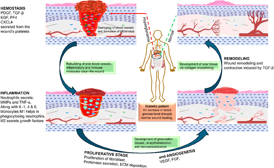
Figure 1. A diabetic patient’s Wound Healing process (PDGF-Platelet-Derived Growth Factor, TGF β- Transforming Growth Factor β, TNF α- Tissue Necrosis Factor, EGF- Epidermal Growth Factor, IL- Interleukins, MMPs-matrix metalloproteases, VEGF- Vascular endothelial growth factor, CXCL4-chemokine (C-X-C motif) ligand 4).
Hemostasis (Phase I) is a clotting process in which platelets reach the injury site to adhere to collagen type 1 and activate, releasing glycoproteins that cause their aggregation (Oguntibeju, 2019) and resulting in the release of transforming growth factor-β (TGF-β), platelet-derived growth factor (PDGF), endothelial growth factor (EGF), and fibroblast growth factor (FGF) (Ezhilarasu et al., 2020). The interaction of growth factors causes thrombin production, which in turn stimulates fibrinogen formation and the intrinsic coagulation cascade. In addition, when blood vessels are injured, they constrict within minutes, and reducing the level of bleeding can be accomplished by taking various steps that allow hemostasis to occur (Clark, 1994; Oguntibeju, 2019). In case of diabetes, high blood glucose and oxidative stress interfere with platelet aggregation and clotting factors due to hyperglycemia and advanced glycation products, which increase the sensitivity of platelets for spontaneous aggregation, causing rapid consumption and increased probability of reduced availability during hemostasis (Rodriguez and Johnson, 2020) and cause endothelial dysfunction, interfering with hemostasis (Patel et al., 2019). These factors also affect the secretion of TGF-β, PDGF, EGF, and FGF.
Inflammation (Phase II)- Immediately after hemostasis, the inflammation phase starts the movement of inflammatory agents at the injury site (Ezhilarasu et al., 2020). It may take up to 2 weeks or more for the inflammation to subside. Constriction of blood vessels and platelet aggregation, followed by phagocytosis to produce inflammation at the targeted site, are the characteristics of this phase (Oguntibeju, 2019), which occurs due to the release of inflammatory particles such as histamine, prostaglandin, and leukotrienes to promote angiogenesis and cell permeability at the site of the wound (Jonidi Shariatzadeh et al., 2025). One of the major hallmarks of wounds in diabetic patients is the presence of a prolonged inflammatory phase. It often results in delayed healing processes and a risk of chronic wounds. It is caused by the dysregulated function of macrophages and neutrophils as discussed in detail in the next section.
Proliferation (Phase III): The proliferation phase lasts from a few days to a few weeks. This phase is characterized by angiogenesis and the formation of an extracellular matrix (ECM), such as collagens, granulation, and epithelialization (Khatoon et al., 2024). During tissue granulation, fibroblasts form a bed of collagen by transforming into the myofibroblast phenotype, with an augmented alpha-smooth muscle actin (α-SMA) cytoskeleton, which is critical for promoting Wound Healing (van de Water et al., 2013). To reduce the contraction of wound edges, they are pulled together, and new epidermal tissues are formed at the wound site (Fitridge et al., 2024). Delayed angiogenesis is one of the major steps for the development of chronic wounds in case of patients suffering from diabetes. This further limits the supply of nutrients and oxygen to hypoxic tissue (due to the deranged function of microcapillaries because of hyperglycemia), finally leading to delayed tissue regeneration.
Remodeling (Phase IV): The last phase is reported to persist from 3 weeks to 24 months. Throughout the time of remodeling, new collagens are synthesized, along with increased tissue tensile strength (Oguntibeju, 2019). In patients suffering from diabetic complications, as indicated in the next section, there are microvascular complications in the peripheral tissues, and impaired angiogenesis leads to delays in the remodeling phase. Extracellular matrix formation is also hindered due to an altered microenvironment, especially due to dysregulated microphages (Krzyszczyk et al., 2018), which is further complicated by microbial infections. This may lead to ulceration, or, if healing occurs, poor collagen strength in the healed wound, increasing the probability of re-ulceration (Fitridge et al., 2024).
3 Complications associated with diabetic wound healing
Compared with nondiabetic patients, diabetes mellitus patients are prone to infections and tend to have more severe infections, as shown in Figure 2. There is compelling evidence that diabetic patients are more likely to have certain infections (Holt et al., 2024). Micro- and macrovascular disease and inadequate angiogenesis are likely contributing factors (Singh et al., 2025). Long-term hyperglycemia is strongly associated with microvascular damage, i.e., neuropathy, nephropathy, and retinopathy, which reduce life expectancy, microvascular complications, and other aspects of quality of life (Singh et al., 2025). The clotting process is disrupted in diabetes, and the inflammatory response is sometimes exaggerated and prolonged, resulting in reduced angiogenesis, weakened wound contraction, and decreased wound strength. Diabetic wounds cause hypoxia, which intensifies inflammation. Thus, numerous factors alter the wound-healing process in diabetic patients, resulting in several complications (Fitridge et al., 2024) Figure 3.
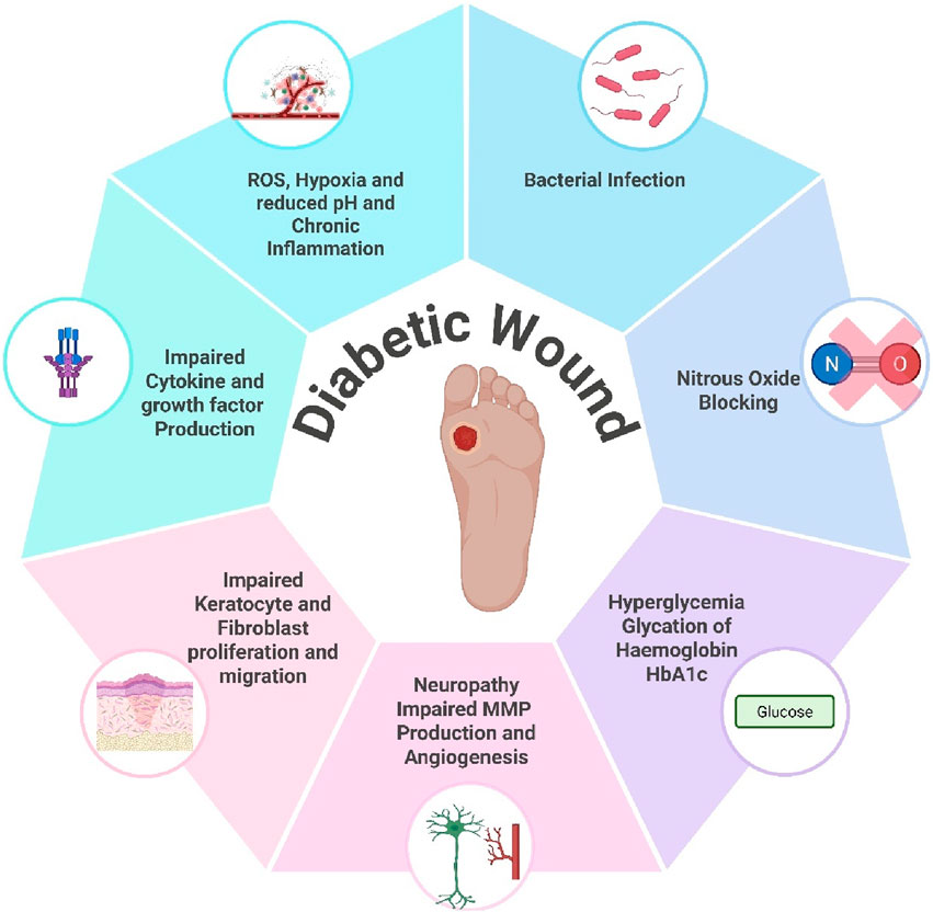
Figure 3. Major factors affecting wound healing in diabetic patients (made using biorender.com).
3.1 Poor blood circulation and oxidative stress
Higher blood glucose levels cause the blood to thicken, which impairs blood circulation and makes it harder for blood cells to move through the body. As a result, the body parts do not receive an adequate blood supply, resulting in oxygen and nutrient deficiency, which impairs cellular metabolism and delays the healing process (Chen et al., 2016).
Additionally, the development of sugar-derived highly oxidant substances can obstruct the healing process of wounds in diabetic patients by inhibiting inflammation and proliferation. These compounds are called advanced glycation end products (AGEs) or glycotoxins (Kang et al., 2021). Free sugars are frequently very reactive and are subject to non-enzymatic degradation under physiological conditions, which leads to the generation of intermediary compounds that can react with other molecules, potentially causing harmful effects. This group of intermediary and advanced products is referred to as glycotoxins (Monteiro-Alfredo and Matafome, 2022). It is a diverse category of compounds that include AGEs and their precursors, most of which are highly reactive intermediary compounds.
Furthermore, free radicals are generated by AGEs, resulting in an imbalance between free radicals and antioxidants in the body, which causes oxidative stress, tissue damage, and slow Wound Healing. When free radicals react with proinflammatory cytokines, they promote the production of serine proteinases and matrix metalloproteinases that damage and disable components of the extracellular matrix (ECM) and growth factors required for normal cell function (Fitridge et al., 2024).
3.2 Impaired immune response
Cellular dysfunctions such as defective and impaired T lymphocytes, chemotaxis, phagocytosis, and bactericidal capacity also play important roles in the delayed Wound Healing of diabetic patients (Houreld, 2014). The Wound Healing rate becomes sluggish because of compromised immune function, resulting in an increased risk of infection. When such infections are left untreated, many severe complications, including sepsis and gangrene, can occur. Abnormally extended activation and dysregulated apoptosis of neutrophils in a high-glucose wound microenvironment lead to an abnormally high release of neutrophil extracellular traps (NETs), resulting in NETosis. This phenomenon ultimately culminates in reduced angiogenesis and prolonged inflammation in the wound (Xiao et al., 2024). Changes in Macrophage phenotypes in high glucose and oxidative microenvironment lead to exclusive proinflammatory M1 phenotype, promoting inflammation and delaying wound healing in the case of diabetes. Therapeutic approaches promoting conversion of M1 macrophages to an Anti-inflammatory M2 phenotype or promotion of recruitment of M2 macrophages to the wound microenvironment can lead to the resolution of inflammation and hastened healing (Fu et al., 2020; Wu et al., 2022). Although Mast cells play a role in wound healing but their role in impaired diabetic wounds is not well defined. In diabetic wounds, infiltrating cells release elevated levels of inflammatory cytokines, such as tumor necrosis factor-alpha (TNF-α), interleukin-6 (IL-6), and interleukin-1β (IL-1β), which persist at high concentrations for extended periods, thereby prolonging the inflammatory phase (Oyebode et al., 2023).
3.3 Neuropathy
Diabetic complications damage the nerve, reducing the ability to feel or perceive wounds and their pain (neuropathy) in the limbs, feet, or other parts of the body. Consequently, diabetic patients who have cuts, burns, and blisters that are untreated are more likely to become seriously infected, and their wounds heal more slowly. Moreover, individuals with diabetes are prone to small problems that may become major problems if they are not detected. Neuropathy is common in the hands and feet (Feldman et al., 2019). In addition, dysregulation of secretion of neuropeptides from the nerves, especially substance P and Calcitonin gene-related peptide, has effects on vasodilation and migration of cells to the wound tissue as well as enhances the expression of NO, a potent vasodilator. Substance P is also involved in the activation of macrophages and the secretion of cytokines. The detailed role of all neuropeptides in wound healing is discussed in Table 1.
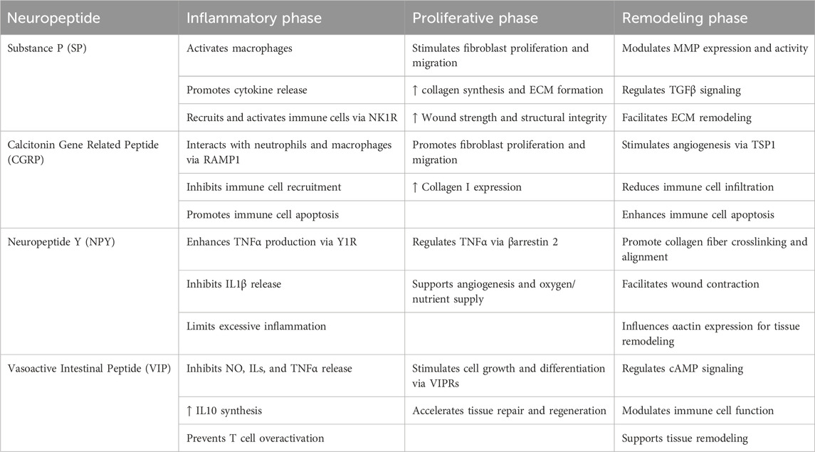
Table 1. Effect of Neuropeptides secreted from Neurons on wound healing ((Xing et al., 2024)).
3.4 Malformation of the ECM
The formation of the extracellular matrix (ECM), an important healing mediator, contributes to the tissue being structurally stable (Houreld, 2014), facilitates transduction of the signal and coordinates cell‒cell and cell‒matrix interactions. In diabetic wounds, the ECM is distorted due to the disturbed interaction between the ECM and growth factors (such as TGF-β and VEGF), which are necessary for normal cell function. This, in turn, results in impaired proliferation, migration, and differentiation of fibroblasts, and there is an imbalance between matrix-degrading enzymes, MMPs, and their inhibitors, and tissue inhibitor metalloproteinases. The balance between matrix synthesis and degradation is essential for collagen synthesis and maintenance in ECM. Therefore, diabetes results in decreased collagen production and increased metabolism, or sometimes a combination of both (Tombulturk et al., 2024).
Therefore, it is now understood that the delayed or impaired healing of wounds that occur in patients with diabetes is caused by both molecular and cellular abnormalities involving a reduction in the host’s immune response, neuropathy, damage from reactive oxygen species, and advanced glycation end products, abnormalities in fibroblast and epidermal cell functions, hypoxia, impaired angiogenesis, and elevated levels of MMPs. Nevertheless, it remains a challenge for healthcare professionals to make an accurate diagnosis, select effective treatments, and prevent wound recurrence.
3.5 Wound microbiota
Nearly 50% of diabetic wounds are complicated by the presence of a diverse microbial population (Prompers et al., 2007). Chronic wounds are generally inhabited by microorganisms of the following species, i.e., Staphylococcus spp., Pseudomonas spp., Corynebacterium spp., Enterococcus spp., Streptococcus spp., and Cutibacterium spp. With increased severity. Cells present in the wound and adjacent tissues undergo programmed cell death through various mechanisms induced by high oxidative stress. These processes lead to the conversion of chronic wounds into necrotic wounds. Wound management necessitates extreme measures such as amputation in the worst cases, to save the patient. This condition is further complicated by the presence of various bacteria and fungi, most notably biofilm-forming bacteria such as S. aureus (MRSA) and P. aeruginosa (Pranantyo et al., 2024). These biofilms protect bacteria from the host immune system as well as antibiotic therapy thereby prolonging their residence on wounds and development of antibiotic resistance (Razdan et al., 2022). This condition necessitates the use and development of topical therapies able to target a matrix of inflammation, oxidative stress, biofilms, and microbial contaminations.
3.6 Clinical approaches for the treatment of diabetic wounds
The clinical management of diabetic wounds utilizes a multifaceted approach that involves a combination of USFDA-approved therapies, adjunctive therapies, and some experimental approaches. The therapy is initiated with wound assessment and proceeds with key aspects such as offloading pressure from the wound, debridement, infection prevention/control and glycemic control. Topical treatment, such as ointments and dressings, plays a very important role in controlling local infection and promoting wound healing.
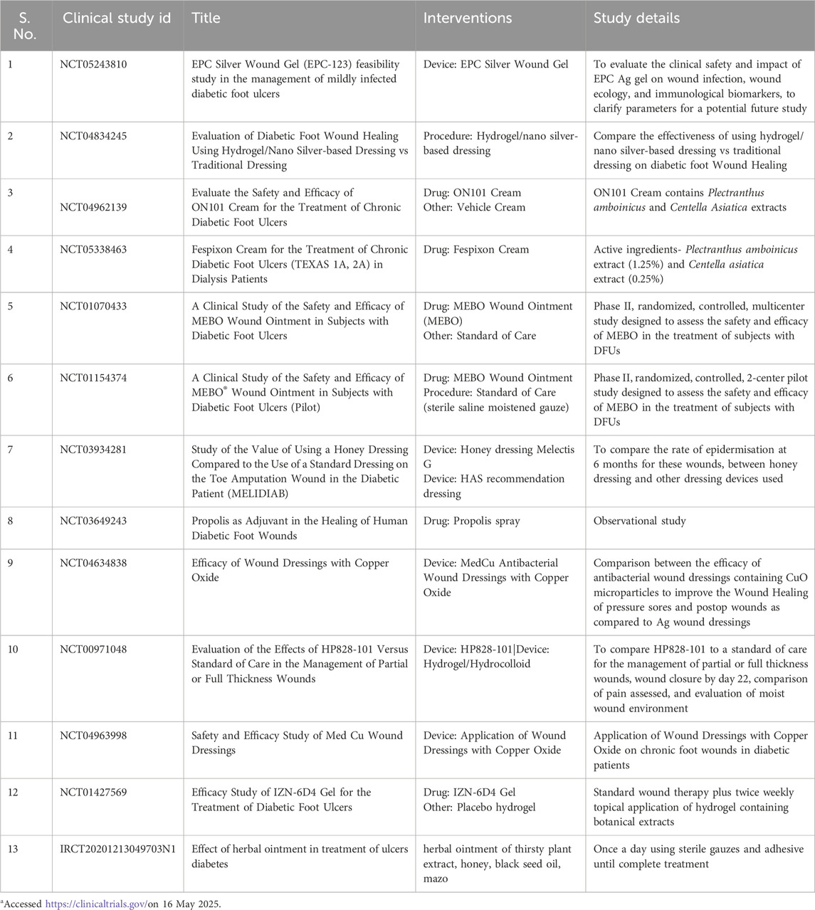
Treatment of diabetic wounds includes growth factors, acellular matrix, bioengineered tissue, negative-pressure therapy, stem cell therapy, and topical and hyperbaric oxygen therapy (Elsayed et al., 2023). Many of these treatment options are not backed up by robust clinical trials. FDA-approved therapies include Becaplermin, containing platelet-derived growth factor, PDGF, which was one of the first therapies to be approved by the USFDA for Diabetic foot ulcer. In addition to that, Apigraf and Dermagraft have also been approved as a wound dressing for Diabetic foot ulcers lasting more than 4 weeks. The major disadvantage of these dressings cannot be used for wounds with infections (Barakat et al., 2021). Integra’s Omnigraft Dermal Regeneration Matrix, originally approved for burns, also got approval for DFU. One of the recent additions to the list is SkinTE, an organoid-like culture obtained from harvested skin for the therapy of Wagner grade 1 diabetic foot ulcer (Armstrong et al., 2023).
Antibiotics are also a mainstay for therapy in the presence of microbial infections. Apart from parental and oral administration of antibiotics, topical antibiotics have also been used for the therapy of microbial infection in diabetic wounds. Recently, the USFDA has given Pravibismane approval for the topical therapy of diabetic wounds contaminated with microbial infections (Lipsky et al., 2024). The drug is different from current microbial therapy due to its novel structure and effectiveness against biofilm formation.
4 Phytoconstituents used for the topical therapy of diabetic wounds
Medicinal plants contain various phytoconstituents, including micronutrients, amino acids, proteins, resins, mucilage’s, essential oils, terpenes, and/or triterpenoids, sterols, saponins, carotenoids, alkaloids, flavonoids, tannins, and phenolic acids, which have significant roles in therapeutic activity (Teoh and Das, 2018). In recent years, increasing evidence has shown that phytochemicals can be useful in enhancing Wound Healing both acutely and chronically (Shah and Amini-Nik, 2017). Topical applications of natural medicinal products and plant extracts have long been utilized for wound management. The discovery and preclinical studies of phytochemicals suggest that phytoconstituents could be beneficial for Wound Healing, skin regeneration, and may prevent and/or treat a variety of other deadly diseases (Karma et al., 2025). The mechanism of phytochemical-mediated improved diabetic Wound Healing includes antimicrobial, antioxidant, debridement, and anti-inflammatory effects, and may provide a hydrated environment. Figure 4 represents the various phytoconstituents and their probable molecular targets. The majority of Wound Healing pharmaceutical products are plant-based; only 20% are mineral-based, and 10% are animal-based (Ahmed et al., 2018). Various natural compounds that can have wound-healing properties are discussed below.
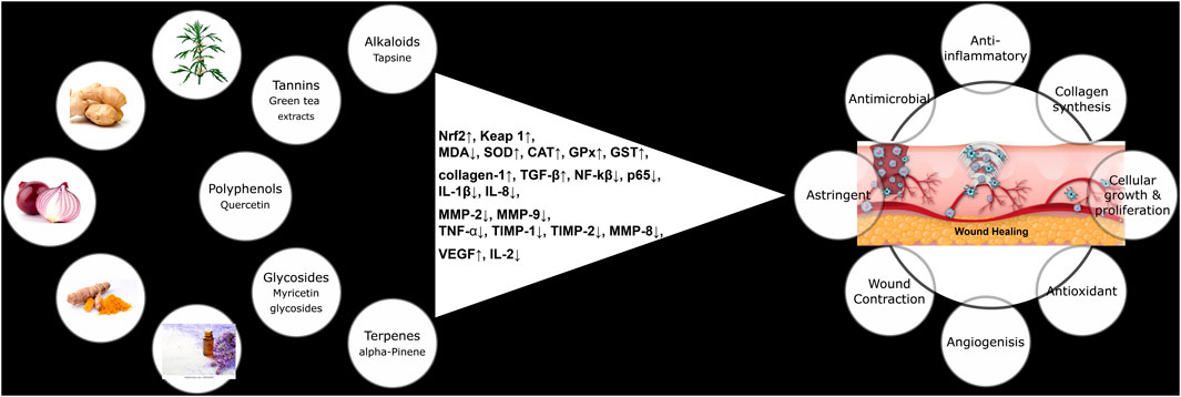
Figure 4. Different phytochemicals, their molecular targets, and pharmacological effects in the context of diabetic wound healing.
4.1 Flavonoids and polyphenols
Several bioactive compounds found in plants are known to accelerate healing. Flavonoids are among the most important bioactive constituents in plants (Ahmad et al., 2017). Flavonoids have a polyphenolic structure (Ullah et al., 2020) and are well known for their ability to modulate the body’s response to various diseases (Ahmad et al., 2017). They are also responsible for a wide range of pharmacological effects (Ullah et al., 2020). According to research, quercetin stands out as the most potent antioxidant among the experimental bioflavonoids. Many foods contain quercetin, including apples, berries, nuts, red wine, Chinese herbs, and vegetables such as cauliflower, cabbage, and onions. The antioxidant properties of quercetin contribute to the prevention of diabetes and related complications such as diabetic nephropathy, cardiovascular disorders, delayed wound healing (Yan et al., 2022), retinopathy, and diabetic neuropathy, as demonstrated in numerous recent studies (Salehi et al., 2020). Several factors contribute to the hypoglycemic activity of polyphenols, including lowering the intestinal assimilation of dietary carbohydrates, regulating glucose-metabolizing enzymes, and stimulating insulin production (Rocha et al., 2025). Quercetin reduces intestinal glucose absorption and enhances glucose uptake in organs and tissues by increasing the expression of silent information regulator 1 (SIRT1) and peroxisome proliferator-activated receptor-γ (PPARγ). This leads to the activation of 5′adenosine monophosphate-activated protein kinase (AMPK) and improves insulin resistance, thereby reducing blood glucose levels. Oxidative stress can worsen diabetes and its complications by impairing insulin secretion and increasing insulin resistance. As a powerful antioxidant, quercetin slows the progression of diabetic complications by preventing oxidative stress (Shi et al., 2019), thereby protecting the β-cells of the pancreas (Vessal et al., 2003). Mast cells, neutrophils, and macrophages contribute to inflammation through the release of chemokines, cytokines, and free radicals. This immune activity causes inflammation in pancreatic islets and promotes peripheral insulin resistance (Shi et al., 2019). Quercetin may therefore be effective in treating these conditions by suppressing the release of inflammatory factors such as TNF-α and by blocking TNF-α–mediated inflammation (Li et al., 2016). It has antifibrotic [85], antibacterial (Qi et al., 2022; Alishahi et al., 2024; Almuhanna et al., 2024), anti-inflammatory (Lee et al., 2024), anti-atherosclerotic (Wang Y. et al., 2024; Xiang et al., 2024), anticarcinogenic (Hasan et al., 2025), Wound Healing, and diabetic Wound Healing properties (Li et al., 2022a; Almuhanna et al., 2024; Huang et al., 2024; Yadav et al., 2025). Quercetin significantly increased wound contraction through enhanced epithelialization, possibly due to its ability to elevate tissue antioxidant levels. Its antifibrotic and antihistaminic properties could make it an effective treatment for hypertrophic scars (Ahmad et al., 2017). Therefore, quercetin can improve common wound healing by increasing fibroblast proliferation while decreasing fibrosis and scar formation (Jangde et al., 2018). By suppressing inflammation, promoting collagen deposition, aiding fibroblast proliferation, speeding angiogenesis, and regulating oxidative stress, quercetin has been shown to have a promising effect on Wound Healing (Huang et al., 2024). Quercetin also averts endogenous antioxidant depletion, guards keratinocytes from free radicals, hinders UV-induced lipid peroxidation, and reduces the release of pro-inflammatory cytokines (Hatahet et al., 2016; Chappidi et al., 2024).
Turmeric contains bioactive compounds known as curcuminoids, which are responsible for its yellow color. Curcumin, desmethoxycurcumin, and bisdemethoxycurcumin are the three major curcuminoids found in turmeric. Among all the active compounds, curcumin is the most abundant and biologically active secondary metabolite (Quispe et al., 2022). It is a crystalline compound with an orange‒yellow hue (Akbik et al., 2014), lipophilic polyphenolic nature (Hussain et al., 2022), and practically insoluble in water (Akbik et al., 2014). Curcumin possesses anti-tumor, anti-aging, and antioxidant properties (Abdul-Rahman et al., 2024), anti-inflammatory (Abdul-Rahman et al., 2024), antihyperglycemic (Mohammadi et al., 2021), immuno-modulating (Memarzia et al., 2021), anti-anxiety (Fathi et al., 2024) neuroprotective (Genchi et al., 2024; Khayatan et al., 2024), antidepressant (Ng et al., 2017) and wound-healing properties (Adamczak et al., 2020; Jakubczyk et al., 2020; Fan et al., 2024; Wang X. et al., 2025). For centuries, inflammation and wounds have been treated with curcumin paste mixed with lime (Akbik et al., 2014). In addition to its anti-inflammatory effects, curcumin inhibits nuclear factor kappa-B and may help prevent and manage diabetes (Maradana et al., 2013). Various mechanisms contribute to curcumin’s ability to ameliorate diabetic pathologies, including lipid metabolism regulation and antioxidant activity. Curcumin has been shown to mitigate diabetic complications both directly and indirectly, including neuropathy, nephropathy, retinopathy, atherosclerosis, and delayed wound healing (Quispe et al., 2022).
Curcumin promotes wound healing by modulating inflammation through the inhibition of the cytokines TNF-α and IL-1 (Dehghani et al., 2020). Furthermore, several biological mechanisms contribute to curcumin’s regenerative effects on diabetic wounds, including angiogenesis, reduced oxidative stress, increased cell proliferation, and enhanced collagen production (Li et al., 2022b). Studies have concluded that wounds treated with curcumin exhibit increased re-epithelialization, enhanced neovascularization, and higher collagen content compared to those treated with conventional drugs.
Effective wound healing can be achieved through oral administration, topical application, or a combination of both. However, curcumin’s therapeutic application is limited due to poor bioavailability, rapid metabolism, and a short half-life. Its hydrophobic nature and low aqueous solubility result in poor absorption, which constrains its topical efficacy. Furthermore, the polyphenolic nature of curcumin can sometimes lead to toxic effects when applied topically at high concentrations. Currently, various efficient delivery systems have been developed to optimize the therapeutic utility of curcumin in topical therapy, aiming to improve its solubility, prevent hydrolysis, and ensure sustained release (Mohanty and Sahoo, 2017). Limitations of curcumin has been successfully addressed using nanotechnology via development of different nanocarriers.
Epigallocatechin-3-gallate (EGCG), a polyphenol found in green tea, has garnered considerable interest in recent years due to its potent antioxidant activity, which contributes to the treatment of cutaneous wounds by promoting re-epithelialization. It also exhibits bactericidal and anti-inflammatory effects and facilitates angiogenesis (Zhao et al., 2021). It inhibits the signaling cascades of platelet-derived growth factor (PDGF) and epidermal growth factor (EGF) during the inflammatory phase. Mast cells, neutrophils, and macrophages contribute to inflammation through the release of free radicals, cytokines, and growth factors. EGCG suppresses the platelet-derived growth factor receptor while enhancing microvascular blood flow. By suppressing interleukin-8 production, EGCG reduces neutrophil aggregation, thereby inhibiting the inflammatory response and modifying the nitric oxide synthase pathway, which decreases both inflammation and reactive oxygen species production.
Furthermore, EGCG stimulates the cleavage of enzymes that accelerate wound healing by eliminating free radicals. By inhibiting nitric oxide production, EGCG serves as an antioxidant because of its ability to neutralize free radicals and maintain wound healing. Endothelial cells in the vascular system are also effectively protected by EGCG (Zawani and Fauzi, 2021). Despite its therapeutic potential, EGCG suffers from poor bioavailability and rapid metabolism, which limit its clinical utility. Therefore, the development of advanced drug delivery systems that release EGCG in a controlled and targeted manner is highly desirable [85]. Consequently, nanoscale formulations have been extensively explored to enhance the stability and therapeutic efficacy of EGCG (Sun M. et al., 2020). Kaempferol, also known as 5,7-trihydroxy-2 (4-hydroxyphenyl)-4H-1-benzopyran-4-one, is a flavonol found in many plants, fruits, and vegetables. It possesses a wide range of pharmacological properties, including antioxidant, anti-inflammatory, anticancer, cardioprotective, and antidiabetic activities. Kaempferol and its glycosides exhibit antioxidant properties, effectively scavenging peroxynitrite ions, hydroxyl radicals, and superoxides. Additionally, it inhibits xanthine oxidase and other ROS-generating enzymes and is well known for its ability to reduce intracellular ROS accumulation. Zeng et al. confirmed that kaempferol hindered the formation of neutrophil extracellular traps (NETs) by reducing ROS production from NADPH oxidase (Zeng et al., 2020). In HUVEC cells, kaempferol was shown to activate the Nrf2 signaling pathway, enhance glutathione and catalase activity, and reduce malondialdehyde levels (Yao et al., 2020). Furthermore, kaempferol reduced macrophage-mediated inflammation by decreasing the secretion of pro-inflammatory cytokines and promoting macrophage repolarization through inhibition of ROS-associated NF-κB signaling (Zhao et al., 2023). In diabetic rats, Özay et al. demonstrated that kaempferol promoted wound contraction and re-epithelialization while increasing collagen and hydroxyproline levels (Özay et al., 2019). The antioxidant and anti-inflammatory potential of kaempferol likely underlies its effectiveness in accelerating wound healing in diabetic patients (Özay et al., 2019). The antioxidant and anti-inflammatory potential of kaempferol can be responsible for its effectiveness in delaying wound healing in diabetic patients.
Luteolin (a 3,4,5,7-tetrahydroxy flavone) is present in many fruits, plants, and vegetables. It has many biological effects, including anti-allergic, anti-inflammatory, and anti-cancer properties, along with pro-oxidant or antioxidant activities (Ambasta et al., 2019). Chen et al. have confirmed the capability of luteolin to recover Wound Healing in diabetic rats by accelerating collagen deposition and re-epithelialization (Chen et al., 2021). Furthermore, luteolin decreases the formation and accumulation of terminal glycation products (AGEs) in the HSA (human serum albumin)/glyoxal system (Sarmah et al., 2020). Its anti-inflammatory action can be elucidated by its ability to modulate macrophage polarization. Indeed, Wang et al. observed LPS-activated RAW264.7 cells in the presence of luteolin decreased the M1 polarization surface markers and increased the M2 surface markers (Wang et al., 2020), which aligns with their cytokine secretions. Luteolin can also impede NLRP3 inflammasome activation and IL-1β secretion in J774A.1 macrophage by modulating ASC oligomerization. Hence, it can be presumed that H2O2 scavenging by luteolin or the diminution in intracellular ROS might reduce the activation of the NLRP3 inflammasome and reduce the M1 polarization of macrophages to recover Wound Healing (Wang et al., 2020).
4.2 Berberine
Berberine (BBR) is a quaternary benzylisoquinoline alkaloid obtained from the genus Berberis of Berberidaceae family. Several medicinal plant species, including Argemone mexicana, Coscinium fenestratum, and Tinospora cordifolia, contain berberine in their bark, leaves, twigs, roots, and stems (Neag et al., 2018). Berberine extracts are well known for their anthelminthic, antibacterial, antiviral, antifungal, and antiprotozoal effects (Neag et al., 2018). It is also reported to increase the activity of antioxidant enzymes (e.g., GPx, SOD) thereby suppressing inflammation and oxidative stress (Amato et al., 2021). Many studies have shown that berberine stimulates multiple metabolic processes. It may prevent hyperglycemia, cardiovascular diseases such as hyperlipidemia and hypertension, cytotoxicity, and inhibitory effects on the growth and reproduction of certain tumorigenic organisms, depression, and inflammatory diseases (Pang et al., 2015). Berberine heals wounds via various mechanisms, including the activation of silent information regulator 1 (Sirt1) (Qiang et al., 2017), which is a nicotinamide adenine dinucleotide-dependent type III histone deacetylase (Zhang et al., 2020) and plays a crucial role in the body’s inflammation, immune system response and, ultimately, wound repair; moreover, it increases the expression of vascular endothelial growth factor, which is a potent angiogenic factor, whereas tumor necrosis factor-α, interleukin-6, and nuclear factor kappa B expression are inhibited, which benefits the healing of diabetic wounds (Qiang et al., 2017; Panda et al., 2021).
Reports have shown that with BBR treatment, Wound Healing is remarkably accelerated, extracellular matrix synthesis is enhanced, and the damage caused by high glucose to culture the human keratinocytes (HaCaT) is significantly inhibited. Further studies suggested that berberine activates thioredoxin reductase-1 (TrxR1) and inhibits c-Jun N-terminal kinase signaling, thus accelerating Wound Healing by inhibiting apoptosis and oxidative stress, promoting the proliferation of cells, and upregulating transforming growth factor-β1 (TGF-β1), which downregulates matrix metalloproteinase-9 (MMP-9) and tissue inhibitors of metalloproteinase-1. The results indicate that topical berberine treatment can enhance wound closure in diabetic patients (Zhou et al., 2021).
4.3 Paeoniflorin
Paeoniflorin (PF) is a monoterpene glycoside obtained from the dried and peeled roots of Paeoniae lactiflora pall, belonging to the family Paeoniaceae. It possesses many pharmacological and biological functions. PF is widely known for its anti-inflammatory and antioxidant properties (Sun et al., 2021). Menstrual cramps and abdominal spasms are both treated with paeoniflorin as a pain-relieving medication. According to a previous study, it also protects against oxidative injuries induced by high glucose levels in cells by triggering the nuclear factor erythroid 2–related factor 2/antioxidant response element signaling (Nrf2/ARE) pathway and impeding apoptosis. Thus, paeoniflorin treatment can improve the healing of diabetic ulcers through increased nuclear factor erythroid 2–related factor 2 expression. Furthermore, PF inhibits the inflammatory response mediated by cytokines and chemokines (TNF-α, IL-1β, and monocyte chemoattractant protein-1) by inhibiting Toll-like receptor 2/4 (TLRs 2 and 4) and reduces related complications, such as nephropathy, in diabetic patients.
In Wound Healing, nuclear factor erythroid 2–related factor 2 has been identified as a good target, regardless of whether a patient is diabetic. Nuclear factor erythroid 2–related factor 2 is involved in corneal epithelial Wound Healing through its ability to promote cell migration. Another study revealed that increased nuclear factor erythroid 2–related factor 2 levels reduce oxidative stress and promote swift Wound Healing in diabetic mice. Therefore, as a result of activating nuclear factor erythroid 2–related factor 2, PF accelerates healing, reduces oxidative stress, increases cell proliferation, and decreases apoptosis. Diabetic wound tissues that had been treated with PF presented significantly increased levels of vascular endothelial growth factor and transforming growth factor-β1. The expression of C-X-C motif chemokine receptor 2 (CXCR2) and nuclear factor kappa light chain enhancer in activated B cells was also decreased by paeoniflorin, and that of IκB was increased. Enhanced wound contraction was also observed in the paeoniflorin-treated group because of decreased Nod-like receptor protein-3 (NLRP3) and caspase-1 levels. Through the inhibition of C-X-C motif chemokine receptor 2, paeoniflorin effectively mitigated nuclear factor kappa B- and Nod-like receptor protein-3-mediated inflammation. The antioxidant and anti-inflammatory properties of paeoniflorin help protect the body from vascular injury induced by fluctuating hyperglycemia. In addition to preventing oxidative stress induced by high glucose, paeoniflorin also suppressed apoptosis by activating the Nrf2/ARE pathway. Through the downregulation of C-X-C motif chemokine receptor 2, paeoniflorin inhibits Nod-like receptor protein-3 inflammasome- and NF-κB-mediated inflammatory reactions in diabetic wounds (the activation of the Nod-like receptor protein-3 inflammasome leads to the maintenance of inflammation and delays Wound Healing). Treatment causes a greater reduction in the levels of proinflammatory cytokines, such as IL-1β, IL-18 and TNF-α, which are highly expressed in the wounds of diabetic patients. By blocking C-X-C motif chemokine receptor 2, paeoniflorin reduces the activation of (Nod-like receptor protein-3) NLPR3/ASC (apoptosis-associated speck-like protein containing a CARD) inflammasomes. NLRP3- and NF-B-mediated inflammatory reactions may be inhibited by paeoniflorin through the blockade of C-X-C motif chemokine receptor 2, which is a possible mechanism of action in diabetic ulcers. The inactivation of TLR4/NF-κB by paeoniflorin has recently been shown to be beneficial in the treatment of diabetic retinopathy (Zhang et al., 2017; Sun X. et al., 2020).
4.4 D-pinitol
D-pinitol is a methyl ether of D-chiro-inositol and is getting attention because of its existence in medicinal plants and foods, including soybean (Glycine max), ice plant (Mesembryanthemum crystallinum), carob pod (Ceratonia siliqua), fenugreek seed (Trigonella foenumgraecum), and some species of the Retama genus (Lee et al., 2014; Tetik and Yüksel, 2014; González-Mauraza et al., 2016; Lahuta et al., 2018; Christou et al., 2019; Zhang et al., 2019). Moreover, D-pinitol has been indicated to have multifunctional medicinal properties such as anti-diabetic (Gao et al., 2015), anti-inflammatory (Zheng et al., 2017), antioxidant (Lee et al., 2019), chemopreventive (Rengarajan et al., 2012), antitumoral (Lin et al., 2013) and diabetic foot ulcers (Kim et al., 2024). One reviewed research recommended that D-pinitol may help to enhance insulin resistance and slow the progression of type II diabetes, and they suggested a mechanism of action of D-pinitol as an insulin sensitizer (Figure 5) (Sánchez-Hidalgo et al., 2021).
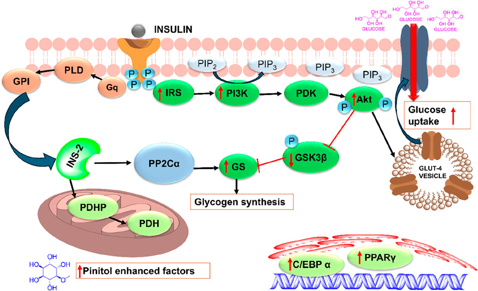
Figure 5. Plausible mechanism of action of pinitol as an insulin sensitizer (Sánchez-Hidalgo et al., 2021). C/EBPα, CCAAT/enhancer binding protein α; GLUT-4, glucose transporter 4; GPI, glycosyl phosphatidylinositol; GSK-3β, glycogen synthase kinase 3β; IR, insulin receptor; INS-2, insulin second messenger; PDH, pyruvate dehydrogenase; PDHP, pyruvate dehydrogenase phosphatase; PI3K, phosphoinositide 3 kinase; PDK-1, phosphoinositide-dependent kinase 1; PP2Cα, phosphoprotein phosphatase 2C α; PKB/Akt, protein kinase B/Akt; PPARγ, peroxisome proliferator-activated receptor γ; PLD, phospholipase D.
Kim and colleagues conducted a study on the wound-healing properties of pinitol in lipopolysaccharide-induced human dermal fibroblasts and streptozotocin-induced diabetic rat models with foot wounds and found that pinitol reduced oxidative stress by enhancing the nuclear translocation of Nrf2 and markedly upregulating antioxidant enzymes like HO-1, SOD1, SOD2, and catalase (Kim et al., 2024). Further, pinitol-induced Nrf2 overexpression suppressed the IκBα/NF-κB signaling pathway and reduced inflammatory cytokines such IL-6, IL-1β, and IL-8. Pinitol also improved collagen deposition, reduced matrix metalloproteinase (MMP) activity, and restored mitochondrial energy metabolism. These results suggested that in wound infection models, pinitol-mediated Nrf2 overexpression enhanced wound-healing effects.
4.5 Carotenoids
Astaxanthin is a dark reddish carotenoid obtained from Haematococcus pluvialis has been very widely used food supplement and is known for its potent antioxidant activity. It has been reported to correct metabolic dysregulation in different diseases, such as diabetes and age-related disorders, and is used as an oral supplement in these disorders (Wang F. et al., 2025). Poor solubility and stability are major hurdles for its development as a therapeutic option for use in diabetic wounds (Zhan et al., 2025). Recently, astaxanthin has been successfully used for diabetic wound healing by loading it into a bilayer nanofibrous membrane made of chitosan and polyvinyl alcohol, enriched with MXene and ZnO nanoparticles, respectively. The membrane, produced using coaxial electrospinning, demonstrated enhanced wound healing by scavenging ROS and reducing inflammation (Zhan et al., 2025). In another study, Astaxanthin was loaded in a nanoemulsion composed of α-tocopherol stabilized with κ-carrageenan. Transdermal administration of nanoemulsions is reported to enhance glycemic control and enhance wound healing and closure by reducing oxidative stress in the wounds (Shanmugapriya et al., 2019). In another study, astaxanthin was stabilized by loading it into chitosan-coated nanocarriers, which were further incorporated into Carbopol gel to enhance wound healing. This effect was mediated by enhanced neovascularization, accelerated epithelialization and collagen deposition, and reduced infiltration of inflammatory cells in the wound (Barari et al., 2025).
4.6 New phytochemicals and extracts for diabetic wound healing
Centella asiatica extract-loaded hydrogel, which can be easily printed on various surfaces, has been reported to be effective in treating chronic wounds, as the hydrogel ensures complete contact with the wound tissue and reduces inflammation and oxidative stress (Wang X. et al., 2024). In another study, ethanolic flaxseed extract-loaded nanofibrous scaffolds demonstrated enhanced wound closure, along with antimicrobial effects against a wide spectrum of bacteria (Abdelaziz et al., 2025). Self-assembled hydrogels composed of poorly soluble asiaticoside and saponins from Panax ginseng were able to enhance wound healing mediated by reduction of IL-6 and enhancing production of VEGF at the wound site, leading to improve collagen fiber organization (Huang H. et al., 2025). Hao et al. have reported a self-assembled hydrogel composed of mangiferin for diabetic wound healing, mediated by the reduction of intracellular ROS, modulation of inflammation, and enhancement of collagen deposition and angiogenesis to promote wound contraction and healing (Hao et al., 2024).
5 Current nanotherapeutic approaches for diabetic wound healing
The complications associated with impaired chronic wounds require approaches to decrease the infection rate, accelerate wound closure, reduce scar impressions, and overcome the limitations of existing wound therapies (Vijayakumar et al., 2019). The biocompatibility of phytochemicals makes them good candidates for therapeutic negotiators. However, they have several limitations, including poor biopharmaceutical and pharmacokinetic properties, such as poor solubility in water, rapid metabolism, and short half-lives, which limit their clinical value.
Hence, the nanomaterials employed to improve clinical efficacy with plant-based therapeutic agents have proven to be the most effective approach to overcome pharmacokinetic and biopharmaceutical hindrances (Dewanjee et al., 2020). Nanotechnology-based nanomaterials are a unique and diverse approach that utilizes materials with sizes ranging from 1 to 100 nm to enhance wound repair and reduce complications (Hamdan et al., 2017). Additionally, nanostructures have incomparable properties because of their high surface area-to-volume ratios, and some of them have antibacterial properties (Wang et al., 2022). NPs assembled or loaded with phytoconstituents promote functionalization and reduce the probability of side effects (Dewanjee et al., 2020). A nanoscale particle, for example, enhances penetration into the wound site more effectively and allows it to interact with the biological target. Thus, nanoparticles can deliver therapeutics in a sustained and controlled manner, resulting in accelerated healing (Hamdan et al., 2017). Furthermore, they enhance therapeutic efficacy, prolong release, improve patient compliance by reducing the dose frequency, increase bioavailability, increase site selection, and reduce other unwanted biopharmaceutical attributes (Sharma et al., 2016).
Hence, the commercialization and patenting of nanopolyphenols has increased significantly. Over the last 3–4 years, hundreds of patents related to nanopolyphenols have been published (Rambaran, 2022). A steady increase in the number of patents filed with herbal nanoformulations has been recorded in recent decades because of their advantages in overcoming the drawbacks faced by conventional delivery systems (Rambaran, 2022). Curcumin, quercetin, carotenoids, paclitaxel, and silymarin nanoparticles are the most filed herbal-based nanoformulation patents. Along with nanoemulsion, nanodispersion, and nanoencapsulation of Withania somnifera and emulsified nanoparticles of Arbutin (Jadhav et al., 2014; Jadhav et al., 2017).
Here, we highlight the most recent nanotherapeutic approach in which Wound Healing therapeutics are endowed via phytoconstituents-based advances.
5.1 Metallic and metallic oxide nanomaterials for diabetic wound healing
5.1.1 Silver nanoparticles (Ag NPs)
Silver is a well-known antibacterial agent generally used to treat burns and wound infections. Changes in bacterial cell walls, genetic material, and disruption of respiratory enzyme pathways are the causes of wound infections. Reactive oxygen species and inflammatory cytokines are released when bacteria form biofilms in chronic wounds, which are encased in a protective extracellular polymeric matrix. This results in persistent inflammation, prevents re-epithelialization, and triggers apoptosis.
Although silver has well-known antibacterial and anti-inflammatory properties, using too much of the metal might be harmful (Singla et al., 2017a). Hence, for efficient healing, it is necessary to determine the ideal and safe concentration of silver to employ in dressings.
With silver, Wound Healing is faster and more effective owing to its superior effectiveness toward multidrug-resistant and biofilm-forming bacteria. Generally, its salts are extensively used to treat chronic wounds and burns infected with microorganisms living within biofilms (Costerton et al., 1999; Hamdan et al., 2017). Despite its proven antimicrobial properties, the application of silver alone can sometimes cause cytotoxicity and oxidative burst induction, and cannot maintain a stable concentration, easily causing local aggregation and adverse reactions (Prasath and Palaniappan, 2019). This problem was counteracted by synthesizing AgNPs with high surface-to-volume ratios at low concentrations. A variety of silver nanostructure shapes and sizes have been investigated, and their antibacterial properties have been demonstrated in different ways (Adhya et al., 2014). Owing to their nanosized and increased surface area and AgNPs exhibit antibacterial properties. These physicochemical properties allow them to penetrate bacterial cells, disrupt membranes, and cause intracellular damage, therefore inhibiting bacterial growth (Kumar et al., 2018). Besides, AgNPs accelerate Wound Healing through the proliferation and migration of keratinocytes, which makes them a viable option for treating diabetic ulcers. Tian et al. explored the wound-healing activity of AgNPs in a rat model and reported rapid healing. Their studies have provided a positive direction for this novel approach (Tian et al., 2007). Additionally, plant-based synthesized AgNPs were also found to be potent wound healers (Table 2). In this context, Manikandan et al. biosynthesized AgNPs from Caulerpa Scalpelliformis extract and reported their biomedical application in diabetic cutaneous wounds (Manikandan et al., 2019).
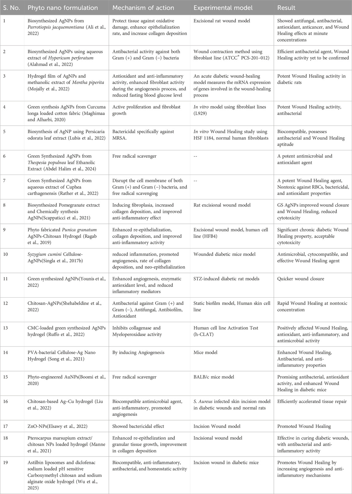
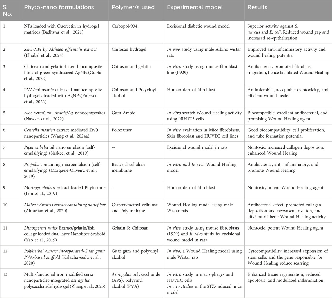
Table 2. List of the most recently developed metallic and non-metallic nanoparticles for diabetic Wound Healing.
Younis and colleagues examined the ability of cyanobacterial species to produce AgNPs and the wound-healing characteristics of the developed nanoparticles in diabetic animals (Younis et al., 2022). The cyanobacterium biosynthesized AgNPs were found spherical with a diameter range of 10–35 nm. The formed AgNPs displayed a decrease in epithelialization period, augmented collagen and the wound closure percentage, hydroxyproline, and hexosamine contents, which enhanced angiogenesis factors (HIF-1α, TGF-β1, and VEGF) in excision wound models. Furthermore, AgNPs exaggerated superoxide dismutase (SOD), catalase (CAT), and glutathione peroxidase (GPx) activities, and glutathione (GSH) and nitric oxide content and reduced malondialdehyde (MDA) levels. Topical applications of AgNPs reduced the inflammatory mediators, including IL-6, IL-1β, TNF-α, and NF-κB.
Furthermore, an injectable thiolated chitosan hydrogel loaded with NO donors and silver nanoparticles (AgNPs) was developed for successful diabetic wound treatment (Huang Z. J. et al., 2025). From which NO was released stably and sustainably responsive to reactive oxygen species (ROS) at the wound site. In the end, this combined strategy facilitates the reconstruction of epithelial structures at the wound site, thereby offering a promising solution for diabetic chronic wound healing. It accomplished effective antibacterial action, biofilm prevention, inflammation suppression, vascular repair, and better local blood circulation.
Shelar et al., green-synthesized AgNPs utilising aqueous extracts of Tagetes erecta (Marigold) and Portulaca oleracea (Purslane) (Shelar et al., 2025). To enhance their antibacterial and wound-healing properties, they were further combined with a polyherbal gel formulation. In comparison to 60% (untreated) and 95% (povidone iodine), the polyherbal gel, which contained extracts of Ficus racemosa, Emblica officinalis, Curcuma longa, Carica papaya, Terminalia bellerica, Acacia catechu, and Aloe vera, achieved 85% wound healing in diabetic rats by day 16. Combinations of AgNP and antibiotics achieved 90% healing. There was no change in HbA1c levels, suggesting glucose-independent repair. These results suggest that AgNP-polyherbal formulations, which increase antibacterial activity and assure tissue regeneration, may be a viable and successful treatment for DFU.
5.1.2 Gold nanoparticles (AuNPs)
The metal precursor used to make gold nanoparticles (AuNPs) is thermostable, making it extremely stable and non-biodegradable. As bulk gold has been proven to be non-toxic and bio-inert, it is utilized in medicine.
A second widely used nanomaterial is gold nanoparticles, which are employed for tissue regeneration, angiogenesis, Wound Healing, gene transfer, cancer cell imaging, biosensors, and targeted drug delivery (Elahi et al., 2018; Bai et al., 2020). Due to their biocompatible nature, surface reactivity, antioxidant, and surface plasmon resonance, AuNPs are significant components for their use in therapeutic and diagnostic applications (Sibuyi et al., 2021). AuNPs have been shown to have diverse therapeutic effects against several infectious, metabolic, and chronic diseases (Sibuyi et al., 2021). To aid in Wound Healing, AuNPs prevent bacteria from ROS formation and act as antioxidants. Thus, AuNPs promote Wound Healing and collagen regeneration through their antimicrobial and antioxidative properties. Similarly, the anti-inflammatory and antiangiogenic properties of AuNPs encourage the release of proteins that are vital for wound healing (Lau et al., 2017; Vijayakumar et al., 2019). Wound Healing (Lau et al., 2017; Vijayakumar et al., 2019). Ponnanikajamideen et al. synthesized AuNPs with the leaf extract of Chamaecostus cuspidatus, also known as the insulin plant (Ponnanikajamideen et al., 2019). They investigated and reported that AuNPs act as dose-dependent free radical scavengers. Additionally, they observed that blood glucose and insulin levels were restored in the Wound Healing activity mice (Ponnanikajamideen et al., 2019) model. However, to achieve better activity of AuNPs, they must be incorporated with other biomolecules, as compared to silver, gold nanomaterials alone do not possess much more antimicrobial activity. Also, gelatin, collagen, and chitosan can easily be crosslinked for better activity (Akturk et al., 2016). However, AuNPs can suppress the growth of multidrug-resistant pathogens by binding to the DNA of bacteria, blocking their double helix from unwinding during replication, and inhibiting ATP synthase enzyme activity, thereby killing bacteria (Mihai et al., 2019). Likewise, Boomi et al. prepared AuNPs using an aqueous extract of Acalypha indica, and their studies observed that AuNPs are efficient antibacterial, antioxidant, and promising Wound Healing agents (Boomi et al., 2020). The antibacterial potential of AuNPs could be due to as they enter into bacterial cells and alter their membrane potential, and the energy metabolism of bacteria is inhibited as a result (Wang et al., 2018; Wang et al., 2018). Besides, AuNPs act as sensitizers and inhibit lipid peroxidation to prevent ROS formation and reduce inflammation, thus promoting Wound Healing. Some studies have found that AuNPs can significantly accelerate Wound Healing and reduce scarring when combined with other antibacterial drugs, either synthetic or plant-based (Wang et al., 2022).
Recently, Ye and colleagues developed co-delivery of hemoglobin-resveratrol (Hb-RES) nanoparticles and morphologically switchable Au nanowires in microneedles for synergistic diabetic wound healing therapy (Ye et al., 2025). In a diabetic mouse model induced by streptozotocin (STZ), the microneedle progressively degraded, and the Hb-RES nanoparticles synergistically worked to reduce hypoxia, scavenge reactive oxygen species, and prevent macrophage differentiation into pro-inflammatory M1 phenotypes. Au nanowires constantly catalyze glucose during this procedure in the presence of inherent glucose oxidase activity. Consequently, this research offers new perceptions on the long-term control of blood glucose levels during synergistic diabetic wound healing.
Besides, Au-cluster-modified Prussian blue (PB) nanospheres (PB-Au) as antibacterial nanoplatforms for diabetic wound healing were developed (Ren et al., 2025). The prepared PB-Au showed tunable peroxidase (POD)-like activity and supported both photostability and catalytic stability, along with accelerating diabetic wound healing. The PB-Au enzyme exhibited the bacterial biofilm destruction of almost 86%, and the rate of bacterial eradication was more than 95%. According to Western blot (WB) results, PB-Au amplified the expression of platelet endothelial cell adhesion molecule-1 (CD3-1) and vascular endothelial growth factor (VEGF) by around 1.4 and 1.3 times, respectively.
5.1.3 Copper nanoparticles (CuNPs)
Copper can cause apoptotic cell death and diminution in metalloproteinase gene activity, which reduces the inflammatory reactions and paces up wound healing (Zhou et al., 2015; Feng et al., 2019). Besides, when copper nanoparticles are utilized to treat wounds, in hypertrophic and keloid scars, high levels of TGF-B gene expression decline, while IFN-Y gene expression increases in response (Sandoval et al., 2022).
Furthermore, E. coli, P. aeruginosa, S. aureus, and other multidrug-resistant bacteria and fungi that are frequently detected in diabetic ulcer infections are significantly inhibited by CuNPs (Zheng et al., 2024). To kill bacteria, CuNPs release Cu2+, which solidifies the bacterial enzyme’s structure and function (Chatterjee et al., 2014). Kumar and colleagues biosynthesized Cissus arnotiana extract containing CuNPs and reported them as a potential antibacterial agent, primarily against Gram-negative bacteria (Rajeshkumar et al., 2019). Also, copper can stimulate angiogenesis in wounds by promoting VEGF production and help to boost immunity by producing interleukin-2, which increases immunity (Bauer et al., 2005; Bauer et al., 2005). Although copper is effective against harmful microbes, its toxicity to cells is dose-dependent, which should be considered when designing formulations (Tiwari et al., 2014). Hence, precise dose optimization is crucial for therapeutic safety. Moreover, oxidation and cluster formation are also potential issues; therefore, stabilizers (e.g., chitosan) should be used to improve the stability of CuNPs (Vijayakumar et al., 2019). Zangeneh et al., synthesized CuNPs with the leaf extract of Fulcaria vulgaris and performed various in vitro and in vivo studies, including Wound Healing studies in rats via an excision wound model; their results confirmed that green-synthesized CuNPs were found efficient as antifungal, antibacterial, antioxidant and cutaneous Wound Healing agents (Zangeneh et al., 2019).
Besides, a dual drug-delivery micro/nanofibrous core-shell system of polycaprolactone/sodium sulfated alginate-polyvinyl alcohol (PCL/SSA-PVA), engineered by the emulsion electrospinning method, was used to enhance the sustained delivery of copper oxide nanoparticles (CuO NP) (Alizadeh et al., 2024). The CuO NP (0.8%w/w) scaffold discloses the maximum tube formation in HUVEC cells and upregulates the pro-angiogenesis genes (VEGFA and bFGF) expression with no cytotoxicity effects. This study strongly recommends the 0.8%w/w CuO NP-loaded PCL/SSA-PVA as an outstanding diabetic wound dressing with significantly enhanced angiogenesis and wound healing.
Also, copper carbonate nanoparticles (NPs) of an average size of 55 ± 16 nm showed a crystalline structure, and antibacterial tests confirmed enhanced inhibition zones against Pseudomonas spp., S. aureus, and other bacterial strains (Aslam et al., 2025). The largest zone of inhibition (18.5 ± 1.05 mm) was observed at 12 mg/mL for Pseudomonas spp. In wound healing activity in diabetic mice, remarks revealed a complete wound closure in NPs treated mice by day 14 as compared to the control group (96.10% wound closure). Hence, these results advocate their potential in biomedical applications, particularly for treating diabetes and bacterial infections.
5.1.4 Metal oxide nanoparticles
Zinc oxide, titanium oxide, cerium oxide, yttrium oxide, and other metal oxide nanoparticles have gained interest in medical applications because they contain important minerals for humans, and even minimum amounts of these elements often exhibit strong therapeutic activity. Owing to their biocompatibility and several therapeutic applications, including antimelanoma, antidiabetic, antibacterial, and anti-inflammatory properties, which have potential for Wound Healing, zinc oxide nanoparticles (ZnO NPs) are promising drug delivery carriers (Ezhilarasu et al., 2020). Furthermore, ZnO NPs are effective Wound Healing agents because they are highly resistant to bacteria and adhere to the wound site for longer periods, thereby stimulating healing. Since ZnO NPs possess antibacterial properties, they can be used in nanocomposites for Wound Healing and skin infection treatment by either promoting the migration of keratinocytes or disrupting the bacterial cell membrane. There are still several drawbacks to the use of ZnO NPs in Wound Healing, including their intrinsic toxicity, which requires further investigation. However, when ZnO NPs are combined with other polymers to form hydrogels, which are further infused with adipose stem cells, they exhibit optimal antimicrobial activity with minimal toxicity, also enhancing wound healing potential, and making them ideal for use in wound dressings or other formulations (Ramzan et al., 2023).
Bai and Jarubula proposed a green and eco-friendly method for the preparation of ZnO NPs using the leaf extract of Nigella sativa plant (Bai and Jarubula, 2023). The synthesized ZnO NPs were characterized with DLS and FESEM and displayed polydisperse types of ZnO NPs with an average particle size of 45 nm. Additionally, antidiabetic studies exhibited the recovery of insulin, glycogen, and blood glucose levels in diabetic mice treated with ZnO NPs. Further, they suggested that this work unlocked the opportunities for future studies in the advancement of new drugs for use in diabetic wound care during sports training. Furthermore, a novel silver-zinc oxide-eugenol (Ag + ZnO + EU) nanocomposite was synthesized to improve antimicrobial activity and promote wound healing (Nagaiah et al., 2025). Nanocomposite confirmed effective antimicrobial efficacy against wound-associated pathogens, comprising standard and clinical isolates of Pseudomonas aeruginosa, Staphylococcus aureus, and Candida albicans. In vitro scratch assays utilizing human keratinocyte cells established that the nanocomposite significantly augmented wound closure (with near-complete healing observed within 24 h), presented enhanced cell migration, and tissue regeneration. Moreover, in an in vitro assay, the nanocomposite exhibited potential antidiabetic properties by enhancing glucose uptake (up to 97.21%). Subsequently, the potential of Gliricidia sepium (Jacq.) Kunth. ex. Walp. Leaves zinc oxide nanoparticles hydrogel (GSL ZnONPs HG) for diabetic wound healing was studied (Wafaey et al., 2025). GSL ZnONPs HG reduced apoptosis, improved tissue regeneration, and controlled inflammation in diabetic wounds as confirmed by wound closure and morphology analysis. A substantial decrease in vascular cell adhesion molecule-1 (VCAM-1) and advanced glycation end products levels (AGEs), and a noteworthy rise in interleukin-10 (IL-10) and platelet-derived growth factor concentrations (PDGF) were observed. Hence, this study advocated the potential of GSL ZnONPs HG as a hopeful approach to augment diabetic wound healing.
Similarly, titanium oxide nanoparticles (TiO2 NPs), can also be employed in vivo and in vitro to accelerate the healing of wounds (Nosrati and Heydari, 2025). TiO2 NPs show promising biological functionality, encompassing anti-inflammatory, antioxidant, and antimicrobial properties, making them desirable for wound healing (Ziental et al., 2020; Ikram et al., 2021). Besides, TiO2 NPs can be altered through procedures, for instance, coating, doping, or surface functionalization, which can improve their biological properties (Diana and Mathew, 2022). Moreover, dressings and scaffolds integrating TiO2 NPs have been revealed to have a significant improvement in the wound healing process (Ghosal et al., 2019; Ismail et al., 2019; Elekhtiar et al., 2025). Also, Altememy and colleagues studied the healing impact of calcium alginate scaffold-loaded TiO2 NPs using origanum vulgare L., carvacrol, hypericum perforatum L., and hypericin on staphylococcus aureus-infected ulcers in diabetic rats. They concluded that because of their antibacterial and anti-inflammatory properties, the medicinal plants Origanum vulgare and Hypericum perforatum - particularly their active components hypericin and carvacrol - reduce inflammation and the microbial load on wounds in diabetic rats, ultimately leading to wound regeneration (Altememy et al., 2022).
In addition, cerium oxide nanoparticles (CeO2) NPs are known for their activity as free radical scavengers. Furthermore, due to the thermal stability, favorable mechanical properties, exceptional oxygen storage capacity, and high retention rate of conjugated enzymes, the utilization of CeO2 NPs exhibits incredible potential in wound healing (Chen et al., 2024). Ahmad et al. produced CeO2 nanoparticles using Abelmoschus esculentus extract, which showed effective antioxidant, antibacterial, and Wound Healing effects (Ahmed et al., 2021). A novel nano-based wound dressing containing chitosan nanoparticles encapsulated with green synthesized cerium oxide nanoparticles using Thymus vulgaris extract (CeO2-CSNPs) (Kamalipooya et al., 2024). The electrospun PCL/cellulose acetate-based nanofiber was prepared, and CeO2-CSNPs were integrated on the PCL/CA membrane by electrospraying. The in vivo diabetic wound healing experiment revealed that PCL/CA/CeO2-CSNPs nanofibers can significantly increase the repair rate of diabetic wounds by up to 95.47% after 15 days.
The next metal oxide is yttrium oxide nanoparticles (Y2O3 NPs), which are nontoxic to neutrophils and macrophages. In addition, these NPs are mostly considered for the treatment of diabetic ulcers since they require the highest free energy compared with other metal oxides. CeO2 NPs and Y2O3 NPs act by protecting against oxidative stress damage because of their antioxidant properties. These nanoparticles reduce the production of ROS and prevent apoptosis (Vijayakumar et al., 2019).
5.2 Nonmetallic nanomaterials for diabetic wound healing
Among all the nonmetallic nanomaterials, carbon-based nanomaterials have been demonstrated tremendously for their potential application in diabetic wounds. They are proposed as curing agents and have several uses in nanomedicine, such as for tissue regeneration, bioimaging, and controlled drug delivery (Naskar and Kim, 2020). Recent studies reported the antibacterial and antifungal properties of graphene oxide nanosheets, making them effective against wound infections (Hamdan et al., 2017). Black phosphorous (BP) is also an emerging nanomaterial used in diabetic Wound Healing. Ouyang et al. demonstrated a study on BP-based in situ sprayed pain relief gel for treating diabetes wounds and provided an experimental-based application of BP in diabetic ulcer treatment. Xu et al. proposed the use of epigallocatechin gallate-modified black phosphorus quantum dots (EGCG-BPQDs@H) and demonstrated that they are promising multifunctional nanoplatforms for healing MRSA (methicillin-resistant Staphylococcus aureus)-infected burn wounds in diabetic patients (Xu et al., 2021). Naturally occurring polymers such as chitosan, alginate, and hyaluronic acid are also known for their antibacterial and rapid Wound Healing properties. Ribeiro et al. prepared insulin-containing chitosan NPs, and their report revealed that both blank and insulin-containing chitosan NPs were responsible for wound maturation (Ribeiro et al., 2020).
PLE-AgNPs can be synthesized efficiently by eliminating the need for hazardous reducing and capping agents. Additionally, PLE-AgNPs exhibit significant antioxidant and cell migration potential without cell cytotoxicity, indicating potential wound-healing properties (Sharma et al., 2024).
Compared with untreated cells, glucose uptake in 3T3-L1 cells was increased, glucose spikes were reduced, and Wound Healing in treated cells was significantly promoted. In 3T3/L cells, the AgNPs also demonstrated remarkable potential in accelerating Wound Healing, achieving 92% closure of wounds after 48 h of incubation (Majeed et al., 2024).
The synthesis of drugs based on medicinal or combined Co-ZnO NPs with greater targeted activity, synthesized from C. officinalis flowers, may lead to opportunities for the discovery of a less expensive and more beneficial therapy for Wound Healing (Aydin Acar et al., 2024).
Silver nanoparticles (AgNPs) were synthesized via a green method involving cucumber pulp extract. Ointment prepared with green synthesized AgNPs effectively healed wounds within 15 days while also exhibiting antibacterial and antioxidant properties (Iqbal et al., 2024).
Compared with the control group, the A. O-ZnO-NP group presented reduced downregulation of IL-6, IL-1β, and TNF-α and increased IL-10 levels, confirming the improved anti-inflammatory effect of the self-assembly method. An in vivo study and histopathological analysis revealed the superiority of the nanoparticles in reducing signs of inflammation and wound incisions in a rat model (Elhabal et al., 2024). Rutin-NPs have the potential to enhance the wound-healing process by attenuating oxidative stress, as evidenced by the restoration of GSH, CAT, and SOD antioxidants and decreased MDA production mediated by Nrf2 activation (Naseeb et al., 2024). The RW-AuNPs were found stable in the test solutions and showed no cytotoxicity to the KMST-6 cells for up to 72 h. AuNPs synthesized from Pinotage and Cabernet Sauvignon enhanced the proliferation of KMST-6 cells and showed potential as Wound Healing agents (Mgijima et al., 2024). Folic acid-decorated nanoparticle loaded with chitosan-gelatin hydrogel was prepared by researchers to overcome issues like poor bioavailability for topical applications. The structural characterization of the nanoparticles and the final gel was performed using TEM and SEM studies, respectively. These studies revealed that the nanoparticles enhanced Wound Healing, as demonstrated by cell migration assays, indicating that they could facilitate Wound Healing by promoting epithelialization (Bhardwaj and Jangde, 2024).
5.3 Nanomaterials used as carriers of therapeutic agents in wound healing
Nowadays, therapeutic drugs are delivered to specific locations and aid in Wound Healing processes using nanocarriers. Most chronic diabetic wounds that do not heal are infected with biofilm-forming microorganisms. Because bacteria produce a complex extracellular matrix that decreases the therapeutic response of conventional drug formulations, a conventional antibiotic therapy may not be enough to deliver the drug to the infected site (Naskar and Kim, 2020). Consequently, a method to functionalize drug-loaded nanocarriers to make them more effective in penetrating the biofilm matrix and target bacteria within it has garnered a lot of interest.
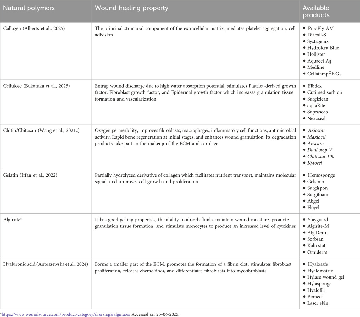
The potential advantages of nanocarriers include improved drug therapy and the capacity to modify a drug’s pharmacodynamic and pharmacokinetic characteristics holistically without altering its molecular structure. In addition to targeted delivery, they also release drugs at an optimal concentration in a controlled manner and protect against enzymatic degradation (Sivadasan et al., 2021). Among the most extensively employed nanocarrier systems for drug delivery are polymeric lipid hybrid nanoparticles, liposomes, and peptide nanostructures (Table 3). These have surfaced as a cutting-edge method for delivering drugs that exhibit antibiofilm properties. Their capacity to encapsulate and deliver both hydrophilic and lipophilic drugs concurrently, along with their simple design and preparation, biocompatibility, and biodegradability, has facilitated their use as drug delivery carriers in a range of therapies, including Wound Healing, to accurately deliver at the target site (Wilhelm Romero et al., 2021).
5.4 Nanomaterial-based scaffolds for diabetic wounds
Designing and developing scaffolds loaded with various therapeutic agents is a significant approach for diabetic wound therapy and is commonly employed in wound care. It involves the use of either natural or synthetic biomaterials, for instance, composites and hybrids. They not only allow adequate air and moisture permeability but also provide proper cell migration and proliferation, along with protection from external contamination and microbial invasion. Polymeric biomaterials with integral properties of Wound Healing have been reported and are the most promising choice for wound care management (Ijaola et al., 2022). Many microparticulate and nanoparticle-based systems, hydrogels, and fibrous scaffolds derived from nanotechnology have demonstrated great potential as wound-healing materials (Negut et al., 2020).
A scaffold is comprised of various polymers and is typically a short-lived framework. Numerous studies have demonstrated the use of biodegradable polymers in the making of 3D-bioprinted scaffolds for tissue engineering, including both natural (for example, collagen, chitosan, and gelatin) and synthetic (for instance, polycaprolactone, poly (lactic-co-glycolic acid), and polyethylene glycol) compounds. Synthetic polymers often exhibit more robust mechanical properties than natural polymers, even though natural polymers are usually extremely biocompatible. The most suitable scaffold for tissue regeneration is the original matrix of the target tissue in the original tissues, which performs numerous tasks, has a complex composition, and plays a vital role in determining the physiological identity of the tissue. Therefore, scaffolding is currently attempting to mimic the functions of the original extracellular matrix to the greatest extent possible (George and Pragna, 2022).
Wang et al. developed hyaluronic acid and natural silk fibroin-based scaffolds, and their results indicated better cytocompatibility, proliferation, and differentiation as well as avert scar formation (Wang Q. et al., 2021). Furthermore, Fu et al. prepared a composite scaffold of curcumin and poly (ε-caprolactone)-PEG-poly (ε-caprolactone), and the resulting scaffold enhanced Wound Healing (Fu et al., 2014). Natrajan et al. developed a biodurable porous scaffold of collagen using tannic acid as a cross-linking agent through a casting technique, and their results indicated increased wound closure and Wound Healing rates (Natarajan et al., 2013).
Besides, Metwally et al. developed a bioinspired 3D-printed scaffold embedding DDAB-nano ZnO/nanofibrous microspheres for regenerative diabetic Wound Healing (Metwally et al., 2023). Multiple assessments observed that the treatment of Staphylococcus aureus-infected full-thickness diabetic wounds in rats showed the superiority of DZ-MS@scaffold. The scaffold showed 95% wound-closure, effective regulation of healing-associated biomarkers, infection suppression, together with regeneration of skin structure in 14 days.
Additionally, Ma and colleagues produced a novel multifunctional self-assembled nanocellulose-based scaffold for the healing of diabetic wounds (Ma et al., 2025). In this, a nanocellulose-based smart scaffold with silk fibroin-loaded cerium oxide was developed for the treatment of diabetic wounds. Smart scaffold dressing displays excellent porosity, water retention, water absorption, controlled degradability, air permeability, and antioxidant properties. In vitro experiments demonstrated antibacterial activity against both Gram-positive (S. aureus) and Gram-negative (E. coli) bacteria. The in vivo results displayed that smart scaffold dressing can reduce inflammation at the wound site of diabetic mice and promote collagen deposition, angiogenesis, and re-epithelialization during Wound Healing in diabetic mice, demonstrating promising biocompatibility and biodegradability.
5.5 Natural polymers based dressings for diabetic wound healing
The wound dressing market currently offers a variety of products, including antimicrobial ointments, creams, gels, etc., combined with natural biodegradable proteins and polymers such as cellulose, chitosan, collagen, hyaluronic acid, silicon, and gelatin. These naturally occurring polysaccharides have been widely used in the production of different products for wound management, as they bio-mimic and recreate the native extracellular matrix to a great extent. In addition, most of these compounds possess intrinsic anti-inflammatory and antibacterial properties. A list of various natural polymers that can be employed in the treatment of diabetic Wound Healing is represented in Table 4.
6 Translation of therapeutic approaches for diabetic wounds
6.1 Patents for diabetic wound healing
Several patent studies related to phytopharmaceutical agents have been reported, but to the best of our knowledge, no studies related to nanoherbal formulations are currently available. The Patent US10206886B2, Lipid nanoparticles for Wound Healing using epidermal growth factor, and EP2895209B1, improved Wound Healing compositions comprising microspheres of polystyrene, the only nanoformulations for Wound Healing that have been patented to date. However, the pipeline for phyto/nano therapeutics plays a vital role. Nanobased topical medicines are safe and easy to use in the clinic when they are derived from phytochemical nanoformulations for the treatment of diabetic wounds. Some of the recently patented herbal formulations for treating wounds are listed in Table 5.
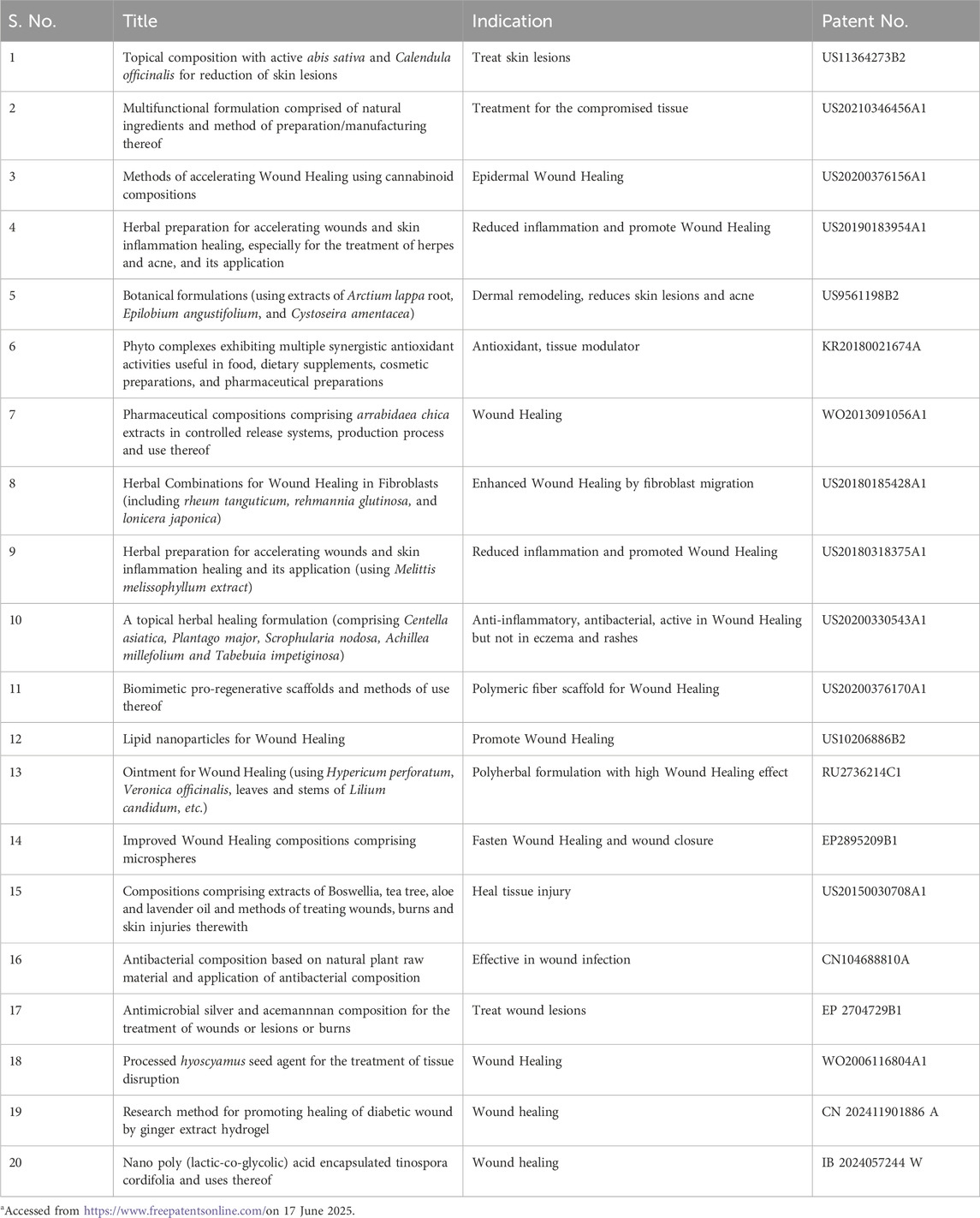
Table 5. List of currently patented formulations or phytoconstituents effective in treating diabetic wounds.
6.2 Clinical trials related to diabetic wound healing
Although Wound Healing treatments have improved in recent years, no single treatment works for all types of wounds. Therefore, scientists are always searching for novel medications and dressings that can work on injured tissue to promote recovery. Many phytochemical-based therapies, for instance, nanoparticles loaded with curcumin, Centella asiatica, berberine, Plectranthus amboinicus, honey, etc., have been investigated in multiple clinical trials, despite the difficulty of conducting clinical trials with nano-based phyto-formulations. The list of ongoing/completed clinical trials using nanoparticles or nano-herbal formulations to treat diabetic wound ulcers is presented in Table 6.
7 Conclusion
Over the past few years, diabetic wound management research has grown exponentially, as evident by the experimental research available in databases, but its clinical translation remains very poor, as discussed earlier. A common consequence of diabetes is impaired Wound Healing, which can have devastating consequences for patients suffering from the disease. As the cases of chronic diabetic wounds have become a major burden in recent times. This has led the USFDA to launch special programs focusing on the accelerated development of therapies for Diabetic Wounds. Even after these efforts, the therapy for Diabetic wounds is a limited success. Various studies suggest that diabetes affects Wound Healing by different mechanisms. A variety of approaches have been evaluated but with limited success, including stem cells, growth factors, cytokine modulators, antimicrobial and anti-inflammatory drugs, matrix metalloproteinase regulators, angiogenic stimulators, extracellular matrix promoters, and several phytopharmaceuticals. The ability to extricate the extent of healing can be improved with better delivery systems and clinical methodologies. Hence, researchers must work on the latest developments in the design of novel carriers and gain a deeper understanding of the basic concepts involved in the successful translation of existing approaches in clinical settings. The management of compromised diabetic wounds can be treated better by combining approaches. Therefore, the application of nanotechnology and herbal therapeutic agents can overcome obstacles associated with conventional delivery systems and diabetic wound complications simultaneously.
Author contributions
PY: Writing – original draft, Data curation. MS: Visualization, Conceptualization, Methodology, Writing – original draft. RV: Validation, Writing – review and editing, Funding acquisition. PS: Conceptualization, Validation, Investigation, Supervision, Writing – review and editing.
Funding
The author(s) declare that no financial support was received for the research and/or publication of this article.
Conflict of interest
The authors declare that the research was conducted in the absence of any commercial or financial relationships that could be construed as a potential conflict of interest.
Generative AI statement
The author(s) declare that no Generative AI was used in the creation of this manuscript.
Publisher’s note
All claims expressed in this article are solely those of the authors and do not necessarily represent those of their affiliated organizations, or those of the publisher, the editors and the reviewers. Any product that may be evaluated in this article, or claim that may be made by its manufacturer, is not guaranteed or endorsed by the publisher.
References
Abdelaziz, A. G., Nageh, H., Abdalla, M. S., Abdo, S. M., Amer, A. A., Loutfy, S. A., et al. (2025). Enhanced wound healing with flaxseed extract-loaded polyvinyl alcohol nanofibrous scaffolds: phytochemical composition, antioxidant activity, and antimicrobial properties. J. Sci. Adv. Mater. Devices 10, 100862. doi:10.1016/J.JSAMD.2025.100862
Abdel Halim, M. B., Eid, H. H., El Deeb, K. S., Metwally, G. F., Masoud, M. A., Ahmed-Farid, O. A., et al. (2024). The study of wound healing activity of Thespesia populnea L. bark, an approach for accelerating healing through nanoparticles and isolation of main active constituents. BMC Complement. Med. Ther. 24, 85–16. doi:10.1186/s12906-024-04343-2
Abdul-Rahman, T., Awuah, W. A., Mikhailova, T., Kalmanovich, J., Mehta, A., Ng, J. C., et al. (2024). Antioxidant, anti-inflammatory and epigenetic potential of curcumin in Alzheimer’s disease. BioFactors 50, 693–708. doi:10.1002/biof.2039
Adamczak, A., Ożarowski, M., and Karpiński, T. M. (2020). Curcumin, a natural antimicrobial agent with strain-specific activity. Pharmaceuticals 13, 153. doi:10.3390/ph13070153
Adhya, A., Bain, J., Ray, O., Hazra, A., Adhikari, S., Dutta, G., et al. (2014). Healing of burn wounds by topical treatment: a randomized controlled comparison between silver sulfadiazine and nano-crystalline silver. J. Basic Clin. Pharm. 6, 29–34. doi:10.4103/0976-0105.145776
Ahmad, M., Sultana, M., Raina, R., Pankaj, N. K., Verma, P. K., and Prawez, S. (2017). Hypoglycemic, hypolipidemic, and wound healing potential of quercetin in streptozotocin-induced diabetic rats. Pharmacogn. Mag. 13, S633–S639. doi:10.4103/pm.pm_108_17
Ahmed, H. E., Iqbal, Y., Aziz, M. H., Atif, M., Batool, Z., Hanif, A., et al. (2021). Green synthesis of CeO2 nanoparticles from the Abelmoschus esculentus extract: evaluation of antioxidant, anticancer, antibacterial, and wound-healing activities. Molecules 26, 4659. doi:10.3390/molecules26154659
Ahmed, O. M., Mohamed, T., Moustafa, H., Hamdy, H., Ahmed, R. R., and Aboud, E. (2018). Quercetin and low level laser therapy promote wound healing process in diabetic rats via structural reorganization and modulatory effects on inflammation and oxidative stress. Biomed. Pharmacother. 101, 58–73. doi:10.1016/j.biopha.2018.02.040
Akbik, D., Ghadiri, M., Chrzanowski, W., and Rohanizadeh, R. (2014). Curcumin as a wound healing agent. Life Sci. 116, 1–7. doi:10.1016/J.LFS.2014.08.016
Akturk, O., Kismet, K., Yasti, A. C., Kuru, S., Duymus, M. E., Kaya, F., et al. (2016). Collagen/Gold nanoparticle nanocomposites: a potential skin wound healing biomaterial. J. Biomater. Appl. 31, 283–301. doi:10.1177/0885328216644536
Alahmad, A., Al-Zereini, W. A., Hijazin, T. J., Al-Madanat, O. Y., Alghoraibi, I., Al-Qaralleh, O., et al. (2022). Green synthesis of silver nanoparticles using Hypericum perforatum L. aqueous extract with the evaluation of its antibacterial activity against clinical and food pathogens. Pharmaceutics 14, 1104. doi:10.3390/pharmaceutics14051104
Alberts, A., Bratu, A. G., Niculescu, A. G., and Grumezescu, A. M. (2025). Collagen-based wound dressings: innovations, mechanisms, and clinical applications. Gels 11, 271. doi:10.3390/GELS11040271
Ali, S., Sulaiman, S., Khan, A., Khan, M. R., and Khan, R. (2022). Green synthesized silver nanoparticles (AgNPs) from Parrotiopsis jacquemontiana (decne) rehder leaf extract and its biological activities. Microsc. Res. Tech. 85, 28–43. doi:10.1002/jemt.23882
Alishahi, M., Xiao, R., Kreismanis, M., Chowdhury, R., Aboelkheir, M., Lopez, S., et al. (2024). Antibacterial, anti-inflammatory, and antioxidant cotton-based wound dressing coated with chitosan/cyclodextrin–quercetin inclusion complex nanofibers. ACS Appl. Bio Mater 7, 5662–5678. doi:10.1021/acsabm.4c00751
Alizadeh, S., Samadikuchaksaraei, A., Jafari, D., Orive, G., Dolatshahi-Pirouz, A., Pezeshki-Modaress, M., et al. (2024). Enhancing diabetic wound healing through improved angiogenesis: the role of emulsion-based core-shell micro/nanofibrous scaffold with sustained CuO nanoparticle delivery. Small 20, 2309164. doi:10.1002/smll.202309164
Almasian, A., Najafi, F., Eftekhari, M., Ardekani, M. R. S., Sharifzadeh, M., and Khanavi, M. (2020). Polyurethane/Carboxymethylcellulose nanofibers containing Malva sylvestris extract for healing diabetic wounds: preparation, characterization, in vitro and in vivo studies. Mater. Sci. Eng. C 114, 111039. doi:10.1016/j.msec.2020.111039
Almuhanna, Y., Alshalani, A., AlSudais, H., Alanazi, F., Alissa, M., Asad, M., et al. (2024). Antibacterial, antibiofilm, and wound healing activities of rutin and quercetin and their interaction with gentamicin on excision wounds in diabetic mice. Biol. (Basel) 13, 676. doi:10.3390/biology13090676
Altememy, D., Darvishi, M., Shokri, S., and Abbaszadeh, S. (2022). The restorative effect of titanium dioxide nanoparticles synthesized with Origanum vulgare l., carvacrol, Hypericum perforatum l., and hypericin loaded in calcium alginate scaffold on staphylococcus aureus-infected ulcers in diabetic rats. Adv. Anim. Vet. Sci. 10, 2285–2293. doi:10.17582/journal.aavs/2022/10.11.2285.2293
Amato, G., Grimaudo, M. A., Alvarez-Lorenzo, C., Concheiro, A., Carbone, C., Bonaccorso, A., et al. (2021). Hyaluronan/poly-l-lysine/berberine nanogels for impaired wound healing. Pharmaceutics 13, 34–11. doi:10.3390/pharmaceutics13010034
Ambasta, R. K., Gupta, R., Kumar, D., Bhattacharya, S., Sarkar, A., and Kumar, P. (2019). Can luteolin be a therapeutic molecule for both Colon cancer and diabetes? Brief. Funct. Genomics 18, 230–239. doi:10.1093/BFGP/ELY036
Antoszewska, M., Sokolewicz, E. M., and Barańska-Rybak, W. (2024). Wide use of hyaluronic acid in the process of wound healing—A rapid review. Sci. Pharm. 92, 23. doi:10.3390/SCIPHARM92020023
Armstrong, D. G., Orgill, D. P., Galiano, R., Glat, P. M., Didomenico, L., Sopko, N. A., et al. (2023). A multicenter, randomized controlled clinical trial evaluating the effects of a novel autologous heterogeneous skin construct in the treatment of wagner one diabetic foot ulcers: final analysis. Int. Wound J. 20, 4083–4096. doi:10.1111/IWJ.14301
Aslam, M. W., Sabri, S., Umar, A., Khan, M. S., Abbas, M. Y., Khan, M. U., et al. (2025). Exploring the antibiotic potential of copper carbonate nanoparticles, wound healing, and glucose-lowering effects in diabetic albino mice. Biochem. Biophys. Res. Commun. 754, 151527. doi:10.1016/j.bbrc.2025.151527
Aydin Acar, C., Gencer, M. A., Pehlivanoglu, S., Yesilot, S., and Donmez, S. (2024). Green and eco-friendly biosynthesis of zinc oxide nanoparticles using Calendula officinalis flower extract: wound healing potential and antioxidant activity. Int. Wound J. 21, e14413. doi:10.1111/iwj.14413
Badhwar, R., Mangla, B., Neupane, Y. R., Khanna, K., and Popli, H. (2021). Quercetin loaded silver nanoparticles in hydrogel matrices for diabetic wound healing. Nanotechnology 32, 505102. doi:10.1088/1361-6528/ac2536
Bai, Q., Han, K., Dong, K., Zheng, C., Zhang, Y., Long, Q., et al. (2020). Potential applications of nanomaterials and technology for diabetic wound healing. Int. J. Nanomedicine 15, 9717–9743. doi:10.2147/IJN.S276001
Bai, X., and Jarubula, R. (2023). Development of novel green synthesized zinc oxide nanoparticles with antibacterial activity and effect on diabetic wound healing process of excisional skin wounds in nursing care during sports training. Inorg. Chem. Commun. 150, 110453. doi:10.1016/j.inoche.2023.110453
Barakat, M., Dipietro, L. A., and Chen, L. (2021). Limited treatment options for diabetic wounds: barriers to clinical translation despite therapeutic success in murine models. Adv. Wound Care (New Rochelle) 10, 436–460. doi:10.1089/WOUND.2020.1254
Barari, F., Maghsoudian, S., Alinezhad, V., Ahmadi, S. M., Amiri, F. T., Fatahi, Y., et al. (2025). Effective topical delivery of astaxanthin via optimized chitosan-coated nanostructured lipid carriers: a promising strategy for enhanced wound healing and tissue regeneration. J. Drug Deliv. Sci. Technol. 110, 107052. doi:10.1016/J.JDDST.2025.107052
Barku, V. (2018). Wound healing: contributions from medicinal plants and their phytoconstituents. Annu. Res. Rev. Biol. 26, 1–14. doi:10.9734/arrb/2018/41301
Bauer, S. M., Bauer, R. J., and Velazquez, O. C. (2005). Angiogenesis, vasculogenesis, and induction of healing in chronic wounds. Vasc. Endovasc. Surg. 39, 293–306. doi:10.1177/153857440503900401
Baveloni, F. G., Meneguin, A. B., Sábio, R. M., de Camargo, B. A. F., Trevisan, D. P. V., Duarte, J. L., et al. (2025). Antimicrobial effect of silver nanoparticles as a potential healing treatment for wounds contaminated with Staphylococcus aureus in wistar rats. J. Drug Deliv. Sci. Technol. 103, 106445. doi:10.1016/j.jddst.2024.106445
Bhardwaj, H., and Jangde, R. K. (2024). Development and characterization of ferulic acid-loaded chitosan nanoparticle embedded-hydrogel for diabetic wound delivery. Eur. J. Pharm. Biopharm. 201, 114371. doi:10.1016/j.ejpb.2024.114371
Boomi, P., Ganesan, R., Poorani, G. P., Jegatheeswaran, S., Balakumar, C., Prabu, H. G., et al. (2020). Phyto-engineered gold nanoparticles (AuNPs) with potential antibacterial, antioxidant, and wound healing activities under in vitro and in vivo conditions. Int. J. Nanomedicine 15, 7553–7568. doi:10.2147/IJN.S257499
Bukatuka, C. F., Mbituyimana, B., Xiao, L., Qaed Ahmed, A. A., Qi, F., Adhikari, M., et al. (2025). Recent trends in the application of cellulose-based hemostatic and wound healing dressings. J. Funct. Biomaterials 16, 151. doi:10.3390/JFB16050151
Chappidi, S., Ankireddy, S. R., Sree, C. G., Rayalcheruvu, U., and Buddolla, V. (2024). Enhancing diabetic rat wound healing through chitosan-mediated nano-scaffolds loaded with quercetin-silver complex. Mater Lett. 361, 136167. doi:10.1016/j.matlet.2024.136167
Chatterjee, A. K., Chakraborty, R., and Basu, T. (2014). Mechanism of antibacterial activity of copper nanoparticles. Nanotechnology 25, 135101. doi:10.1088/0957-4484/25/13/135101
Chen, H., Cheng, Y., Tian, J., Yang, P., Zhang, X., Chen, Y., et al. (2020). Dissolved oxygen from microalgae-gel patch promotes chronic wound healing in diabetes. Sci. Adv. 6, eaba4311. doi:10.1126/sciadv.aba4311
Chen, H., Guo, L., Wicks, J., Ling, C., Zhao, X., Yan, Y., et al. (2016). Quickly promoting angiogenesis by using a DFO-Loaded photo-crosslinked gelatin hydrogel for diabetic skin regeneration. J. Mater Chem. B 4, 3770–3781. doi:10.1039/c6tb00065g
Chen, L. Y., Cheng, H. L., Kuan, Y. H., Liang, T. J., Chao, Y. Y., and Lin, H. C. (2021). Therapeutic potential of luteolin on impaired wound healing in streptozotocin-induced rats. Biomedicines 9, 761. doi:10.3390/BIOMEDICINES9070761
Chen, S., Wang, Y., Bao, S., Yao, L., Fu, X., Yu, Y., et al. (2024). Cerium oxide nanoparticles in wound care: a review of mechanisms and therapeutic applications. Front. Bioeng. Biotechnol. 12, 1404651. doi:10.3389/fbioe.2024.1404651
Christou, C., Poulli, E., Yiannopoulos, S., and Agapiou, A. (2019). GC–MS analysis of D-pinitol in carob: syrup and fruit (flesh and seed). J. Chromatogr. B 1116, 60–64. doi:10.1016/j.jchromb.2019.04.008
Clark, R. A. F. (1994). “Wound repair. Overview and general considerations,” in The molecular and cellular biology of wound repair. Editors R. A. F. Clark,, and P. M. Henson (Plenum Press).
Costerton, J. W., Stewart, P. S., and Greenberg, E. P. (1999). Bacterial biofilms: a common cause of persistent infections. Science 284, 1318–1322. doi:10.1126/science.284.5418.1318
Dehghani, S., Dalirfardouei, R., Jafari Najaf Abadi, M. H., Ebrahimi Nik, M., Jaafari, M. R., and Mahdipour, E. (2020). Topical application of curcumin regulates the angiogenesis in diabetic-impaired cutaneous wound. Cell Biochem. Funct. 38, 558–566. doi:10.1002/cbf.3500
Dewanjee, S., Chakraborty, P., Mukherjee, B., and de Feo, V. (2020). Plant-based antidiabetic nanoformulations: the emerging paradigm for effective therapy. Int. J. Mol. Sci. 21, 2217. doi:10.3390/ijms21062217
Diana, E. J., and Mathew, T. V. (2022). Synthesis and characterization of surface-modified ultrafine titanium dioxide nanoparticles with an antioxidant functionalized biopolymer as a therapeutic agent: anticancer and antimicrobial evaluation. Colloids Surf. B Biointerfaces 220, 112949. doi:10.1016/j.colsurfb.2022.112949
Elahi, N., Kamali, M., and Baghersad, M. H. (2018). Recent biomedical applications of gold nanoparticles: a review. Talanta 184, 537–556. doi:10.1016/j.talanta.2018.02.088
Elekhtiar, S. A., Gazia, M. M. A., Osman, A., Abd-Elsalam, M. M., El-Kemary, N. M., Elksass, S., et al. (2025). A novel skin-like patch based on 3D hydrogel nanocomposite of Polydopamine/TiO2 nanoparticles and Ag quantum dots accelerates diabetic wound healing compared to stem cell therapy. J. Tissue Viability 34, 100850. doi:10.1016/j.jtv.2024.12.014
Elhabal, S. F., Abdelaal, N., Al-Zuhairy, S. A. K. S., Elrefai, M. F. M., Hamdan, A. M. E., Khalifa, M. M., et al. (2024). Green synthesis of zinc oxide nanoparticles from Althaea officinalis flower extract coated with chitosan for potential healing effects on diabetic wounds by inhibiting TNF-α and IL-6/IL-1β signaling pathways. Int. J. Nanomedicine 19, 3045–3070. doi:10.2147/IJN.S455270
Elsawy, H., Sedky, A., Abou Taleb, M. F., and El-Newehy, M. H. (2022). Antidiabetic wound dressing materials based on cellulosic fabrics loaded with zinc oxide nanoparticles synthesized by solid-state method. Polym. (Basel) 14, 2168. doi:10.3390/polym14112168
Elsayed, N. A., Aleppo, G., Aroda, V. R., Bannuru, R. R., Brown, F. M., Bruemmer, D., et al. (2023). 12. Retinopathy, neuropathy, and foot care: standards of care in Diabetes—2023. Diabetes Care 46, S203–S215. doi:10.2337/DC23-S012
Ezhilarasu, H., Vishalli, D., Dheen, S. T., Bay, B. H., and Kumar Srinivasan, D. (2020). Nanoparticle-based therapeutic approach for diabetic wound healing. Nanomaterials 10, 1234–29. doi:10.3390/nano10061234
Fan, X., Huang, J., Zhang, W., Su, Z., Li, J., Wu, Z., et al. (2024). A multifunctional, tough, stretchable, and transparent curcumin hydrogel with potent antimicrobial, antioxidative, anti-inflammatory, and angiogenesis capabilities for diabetic wound healing. ACS Appl. Mater Interfaces 16, 9749–9767. doi:10.1021/acsami.3c16837
Fathi, S., Agharloo, S., Falahatzadeh, M., Bahraminavid, S., Homayooni, A., Faghfouri, A. H., et al. (2024). Effect of curcumin supplementation on symptoms of anxiety: a systematic review and meta-analysis of randomized controlled trials. Clin. Nutr. ESPEN 62, 253–259. doi:10.1016/j.clnesp.2024.05.017
Feldman, E. L., Callaghan, B. C., Pop-Busui, R., Zochodne, D. W., Wright, D. E., Bennett, D. L., et al. (2019). Diabetic neuropathy. Nat. Rev. Dis. Prim. 5 (1), 41–18. doi:10.1038/s41572-019-0092-1
Feng, X., Xu, W., Li, Z., Song, W., Ding, J., and Chen, X. (2019). Immunomodulatory nanosystems. Adv. Sci. 6, 1900101. doi:10.1002/advs.201900101
Fitridge, R., Service, E., Jeffcoate, W., Boyko, E. J., Game, F., Cowled, P., et al. (2024). Causes, prevention, and management of diabetes-related foot ulcers. thelancet.Com. 12, 472–482. doi:10.1016/S2213-8587(24)00110-4
Fu, J., Huang, J., Lin, M., Xie, T., and You, T. (2020). Quercetin promotes diabetic wound healing via switching macrophages from M1 to M2 polarization. J. Surg. Res. 246, 213–223. doi:10.1016/j.jss.2019.09.011
Fu, S., Meng, X., Fan, J., Yang, L., Wen, Q., Ye, S., et al. (2014). Acceleration of dermal wound healing by using electrospun curcumin-loaded poly (ε-caprolactone)-poly (ethylene glycol)-poly (ε-caprolactone) fibrous mats. J. Biomed. Mater Res. B Appl. Biomater. 102, 533–542. doi:10.1002/jbm.b.33032
Gao, Y., Zhang, M., Wu, T., Xu, M., Cai, H., and Zhang, Z. (2015). Effects of D-pinitol on insulin resistance through the PI3K/Akt signaling pathway in type 2 diabetes mellitus rats. J. Agric. Food Chem. 63, 6019–6026. doi:10.1021/acs.jafc.5b01238
Genchi, G., Lauria, G., Catalano, A., Carocci, A., and Sinicropi, M. S. (2024). Neuroprotective effects of curcumin in neurodegenerative diseases. Foods 13, 1774. doi:10.3390/foods13111774
George, K. S., and Pragna, K. M. (2022). Chitosan-halloysite nano-composite for scaffolds for tissue engineering. Mater. Res. Found. 125, 103–123. doi:10.21741/9781644901915-5
Ghosal, K., Agatemor, C., Špitálsky, Z., Thomas, S., and Kny, E. (2019). Electrospinning tissue engineering and wound dressing scaffolds from polymer-titanium dioxide nanocomposites. Chem. Eng. J. 358, 1262–1278. doi:10.1016/j.cej.2018.10.117
González-Mauraza, N. H., León-González, A. J., Espartero, J. L., Gallego-Fernández, J. B., Sánchez-Hidalgo, M., and Martin-Cordero, C. (2016). Isolation and quantification of pinitol, a bioactive cyclitol, in retama spp. Nat. Prod. Commun. 11, 1934578X1601100321. doi:10.1177/1934578x1601100321
Greenhalgh, D. G. (2003). Wound healing and diabetes mellitus. Clin. Plast. Surg. 30, 37–45. doi:10.1016/s0094-1298(02)00066-4
Gupta, I., Kumar, A., Bhatt, A. N., Sapra, S., and Gandhi, S. (2022). Green synthesis-mediated silver nanoparticles based biocomposite films for wound healing application. J. Inorg. Organomet. Polym. Mater 32, 2994–3011. doi:10.1007/s10904-022-02333-w
Hamdan, S., Pastar, I., Drakulich, S., Dikici, E., Tomic-Canic, M., Deo, S., et al. (2017). Nanotechnology-driven therapeutic interventions in wound healing: potential uses and applications. ACS Cent. Sci. 3, 163–175. doi:10.1021/acscentsci.6b00371
Hao, M., Wei, S., Su, S., Tang, Z., and Wang, Y. (2024). A multifunctional hydrogel fabricated by direct self-assembly of natural herbal small molecule mangiferin for treating diabetic wounds. ACS Appl. Mater Interfaces 16, 24221–24234. doi:10.1021/ACSAMI.4C01265
Hasan, A. M. W., Al Hasan, M. S., Mizan, M., Miah, M. S., Uddin, M. B., Mia, E., et al. (2025). Quercetin promises anticancer activity through PI3K-AKT-mTOR pathway: a literature review. Pharmacol. Research-Natural Prod. 7, 100206. doi:10.1016/j.prenap.2025.100206
Hatahet, T., Morille, M., Hommoss, A., Devoisselle, J. M., Müller, R. H., and Bégu, S. (2016). Quercetin topical application, from conventional dosage forms to nanodosage forms. Eur. J. Pharm. Biopharm. 108, 41–53. doi:10.1016/j.ejpb.2016.08.011
Holt, R. I. G., Cockram, C. S., Ma, R. C. W., and Luk, A. O. Y. (2024). Diabetes and infection: review of the epidemiology, mechanisms and principles of treatment. Diabetologia 67, 1168–1180. doi:10.1007/S00125-024-06102-X
Houreld, N. N. (2014). Shedding light on a new treatment for diabetic wound healing: a review on phototherapy. Sci. World J. 2014, 398412. doi:10.1155/2014/398412
Huang, H., Chen, Y., Hu, J., Guo, X., Zhou, S., Yang, Q., et al. (2024). Quercetin and its derivatives for wound healing in rats/mice: evidence from animal studies and insight into molecular mechanisms. Int. Wound J. 21, e14389. doi:10.1111/iwj.14389
Huang, H., Yang, X., Qin, X., Shen, Y., Luo, Y., Yang, L., et al. (2025a). Co-assembled supramolecular hydrogel of asiaticoside and Panax notoginseng saponins for enhanced wound healing. Eur. J. Pharm. Biopharm. 207, 114617. doi:10.1016/J.EJPB.2024.114617
Huang, Z.-J., Ye, M.-N., Peng, X.-H., Gui, P., Cheng, F., and Wang, G.-H. (2025b). Thiolated chitosan hydrogel combining nitric oxide and silver nanoparticles for the effective treatment of diabetic wound healing. Int. J. Biol. Macromol. 311, 143730. doi:10.1016/j.ijbiomac.2025.143730
Hussain, Y., Khan, H., Alotaibi, G., Khan, F., Alam, W., Aschner, M., et al. (2022). How curcumin targets inflammatory mediators in diabetes: therapeutic insights and possible solutions. Molecules 27, 4058. doi:10.3390/molecules27134058
Ijaola, A. O., Akamo, D. O., Damiri, F., Akisin, C. J., Bamidele, E. A., Ajiboye, E. G., et al. (2022). Polymeric biomaterials for wound healing applications: a comprehensive review. J. Biomater. Sci. Polym. Ed. 33, 1998–2050. doi:10.1080/09205063.2022.2088528
Ikram, M., Javed, B., Hassan, S. W. U., Satti, S. H., Sarwer, A., Raja, N. I., et al. (2021). Therapeutic potential of biogenic titanium dioxide nanoparticles: a review on mechanistic approaches. Nanomedicine 16, 1429–1446. doi:10.2217/nnm-2021-0020
Iqbal, R., Asghar, A., Habib, A., Ali, S., Zahra, S., Hussain, M. I., et al. (2024). Therapeutic potential of green synthesized silver nanoparticles for promoting wound-healing process in diabetic mice. Biol. Trace Elem. Res. 202, 5545–5555. doi:10.1007/s12011-024-04094-8
Irfan, N. I., Mohd Zubir, A. Z., Suwandi, A., Haris, M. S., Jaswir, I., and Lestari, W. (2022). Gelatin-based hemostatic agents for medical and dental application at a glance: a narrative literature review. Saudi Dent. J. 34, 699–707. doi:10.1016/J.SDENTJ.2022.11.007
Ismail, N. A., Amin, K. A. M., Majid, F. A. A., and Razali, M. H. (2019). Gellan gum incorporating titanium dioxide nanoparticles biofilm as wound dressing: physicochemical, mechanical, antibacterial properties and wound healing studies. Mater. Sci. Eng. C 103, 109770. doi:10.1016/j.msec.2019.109770
Jadhav, N. R., Nadaf, S. J., Lohar, D. A., Ghagare, P. S., and Powar, T. A. (2017). Phytochemicals formulated as nanoparticles: inventions, recent patents and future prospects. Recent Pat. Drug Deliv. Formul. 11, 173–186. doi:10.2174/1872211311666171120102531
Jadhav, N. R., Powar, T., Shinde, S., and Nadaf, S. (2014). Herbal nanoparticles: a patent review. Asian J. Pharm. (AJP) 8, 58. doi:10.4103/0973-8398.134101
Jakubczyk, K., Drużga, A., Katarzyna, J., and Skonieczna-Żydecka, K. (2020). Antioxidant potential of curcumin—A meta-analysis of randomized clinical trials. Antioxidants 9, 1092. doi:10.3390/antiox9111092
Jangde, R., Srivastava, S., Singh, M. R., and Singh, D. (2018). In vitro and in vivo characterization of quercetin loaded multiphase hydrogel for wound healing application. Int. J. Biol. Macromol. 115, 1211–1217. doi:10.1016/j.ijbiomac.2018.05.010
Jonidi Shariatzadeh, F., Currie, S., Logsetty, S., Spiwak, R., and Liu, S. (2025). Enhancing wound healing and minimizing scarring: a comprehensive review of nanofiber technology in wound dressings. Prog. Mater Sci. 147, 101350. doi:10.1016/J.PMATSCI.2024.101350
Kalachaveedu, M., Jenifer, P., Pandian, R., and Arumugam, G. (2020). Fabrication and characterization of herbal drug enriched guar galactomannan based nanofibrous mats seeded with GMSC’s for wound healing applications. Int. J. Biol. Macromol. 148, 737–749. doi:10.1016/j.ijbiomac.2020.01.188
Kalan, L. R., Meisel, J. S., Loesche, M. A., Horwinski, J., Soaita, I., Chen, X., et al. (2019). Strain-and species-level variation in the microbiome of diabetic wounds is associated with clinical outcomes and therapeutic efficacy. Cell Host Microbe 25, 641–655. doi:10.1016/j.chom.2019.03.006
Kamalipooya, S., Fahimirad, S., Abtahi, H., Golmohammadi, M., Satari, M., Dadashpour, M., et al. (2024). Diabetic wound healing function of PCL/Cellulose acetate nanofiber engineered with chitosan/cerium oxide nanoparticles. Int. J. Pharm. 653, 123880. doi:10.1016/j.ijpharm.2024.123880
Kang, H. J., Kumar, S., D’Elia, A., Dash, B., Nanda, V., Hsia, H. C., et al. (2021). Self-assembled elastin-like polypeptide fusion protein coacervates as competitive inhibitors of advanced glycation end-products enhance diabetic wound healing. J. Control. Release 333, 176–187. doi:10.1016/J.JCONREL.2021.03.032
Karma, H. M., Michael, Y., Alex, O., Darren, L., Wangchuk, C. P., Yeshi, K., et al. (2025). Australian tropical medicinal plants and their phytochemicals with wound healing and antidiabetic properties. Phytochem. Rev. 2025, 1–43. doi:10.1007/S11101-025-10132-7
Khatoon, F., Narula, A. K., and Sehgal, P. (2024). Efficacy of collagen based biomaterials in diabetic foot ulcer wound healing. Eur. Polym. J. 217, 113345. doi:10.1016/J.EURPOLYMJ.2024.113345
Khattak, S., Ullah, I., Sohail, M., Akbar, M. U., Rauf, M. A., Ullah, S., et al. (2025). Endogenous/Exogenous stimuli-responsive smart hydrogels for diabetic wound healing. Aggregate 6, e688. doi:10.1002/agt2.688
Khayatan, D., Razavi, S. M., Arab, Z. N., Hosseini, Y., Niknejad, A., Momtaz, S., et al. (2024). Superoxide dismutase: a key target for the neuroprotective effects of curcumin. Mol. Cell Biochem. 479, 693–705. doi:10.1007/s11010-023-04757-5
Kim, J., Go, M. Y., Jeon, C. Y., Shin, J. U., Kim, M., Lim, H. W., et al. (2024). Pinitol improves diabetic foot ulcers in streptozotocin-induced diabetes rats through upregulation of Nrf2/HO-1 signaling. Antioxidants 14, 15. doi:10.3390/antiox14010015
Krzyszczyk, P., Schloss, R., Palmer, A., and Berthiaume, F. (2018). The role of macrophages in acute and chronic wound healing and interventions to promote pro-wound healing phenotypes. Front. Physiol. 9, 419. doi:10.3389/FPHYS.2018.00419
Kumar, S. S. D., Rajendran, N. K., Houreld, N. N., and Abrahamse, H. (2018). Recent advances on silver nanoparticle and biopolymer-based biomaterials for wound healing applications. Int. J. Biol. Macromol. 115, 165–175. doi:10.1016/j.ijbiomac.2018.04.003
Kurkela, O., Nevalainen, J., Arffman, M., Lahtela, J., and Forma, L. (2022). Foot-related diabetes complications: care pathways, patient profiles and costs. BMC Health Serv. Res. 22, 559. doi:10.1186/s12913-022-07853-2
Lahuta, L. B., Szablińska, J., Ciak, M., and Górecki, R. J. (2018). The occurrence and accumulation of d-pinitol in fenugreek (trigonella foenum graecum L.). Acta Physiol. Plant 40, 155. doi:10.1007/s11738-018-2734-4
Lau, P., Bidin, N., Islam, S., Shukri, W. N. B. W. M., Zakaria, N., Musa, N., et al. (2017). Influence of gold nanoparticles on wound healing treatment in rat model: photobiomodulation therapy. Lasers Surg. Med. 49, 380–386. doi:10.1002/lsm.22614
Lee, B.-H., Lee, C.-C., and Wu, S.-C. (2014). Ice plant (Mesembryanthemum crystallinum) improves hyperglycaemia and memory impairments in a wistar rat model of streptozotocin-induced diabetes. J. Sci. Food Agric. 94, 2266–2273. doi:10.1002/jsfa.6552
Lee, E., Lim, Y., Kwon, S. W., and Kwon, O. (2019). Pinitol consumption improves liver health status by reducing oxidative stress and fatty acid accumulation in subjects with non-alcoholic fatty liver disease: a randomized, double-blind, placebo-controlled trial. J. Nutr. Biochem. 68, 33–41. doi:10.1016/j.jnutbio.2019.03.006
Lee, G. B., Kim, Y., Lee, K. E., Vinayagam, R., Singh, M., and Kang, S. G. (2024). Anti-inflammatory effects of quercetin, rutin, and Troxerutin result from the inhibition of NO production and the reduction of COX-2 levels in RAW 264.7 cells treated with LPS. Appl. Biochem. Biotechnol. 196, 8431–8452. doi:10.1007/s12010-024-05003-4
Li, N., Lu, X., Yang, Y., Ning, S., Tian, Y., Zhou, M., et al. (2024). Calcium peroxide-based hydrogel patch with sustainable oxygenation for diabetic wound healing. Adv. Healthc. Mater 13, 2303314. doi:10.1002/adhm.202303314
Li, Y., Su, L., Zhang, Y., Liu, Y., Huang, F., Ren, Y., et al. (2022a). A guanosine-quadruplex hydrogel as Cascade reaction container consuming endogenous glucose for infected wound treatment—A study in diabetic mice. Adv. Sci. 9, 2103485. doi:10.1002/advs.202103485
Li, Y., Yao, J., Han, C., Yang, J., Chaudhry, M. T., Wang, S., et al. (2016). Quercetin, inflammation and immunity. Nutrients 8, 167. doi:10.3390/nu8030167
Li, Y., Zhao, S., der Merwe, L. V., Dai, W., and Lin, C. (2022b). Efficacy of curcumin for wound repair in diabetic rats/mice: a systematic review and meta-analysis of preclinical studies. Curr. Pharm. Des. 28, 187–197. doi:10.2174/1381612827666210617122026
Lim, A. W., Ng, P. Y., Chieng, N., and Ng, S. F. (2019). Moringa oleifera leaf extract–loaded phytophospholipid complex for potential application as wound dressing. J. Drug Deliv. Sci. Technol. 54, 101329. doi:10.1016/j.jddst.2019.101329
Lin, T. H., Tan, T. W., Tsai, T. H., Chen, C. C., Hsieh, T. F., Lee, S. S., et al. (2013). D-pinitol inhibits prostate cancer metastasis through inhibition of αVβ3 integrin by modulating FAK, c-Src and NF-κB pathways. Int. J. Mol. Sci. 14, 9790–9802. doi:10.3390/ijms14059790
Lipsky, B. A., Kim, P. J., Murphy, B., McKernan, P. A., Armstrong, D. G., and Baker, B. H. J. (2024). Topical pravibismane as adjunctive therapy for moderate or severe diabetic foot infections: a phase 1b randomized, multicenter, double-blind, placebo-controlled trial. Int. Wound J. 21, e14817. doi:10.1111/IWJ.14817
Liu, X., Zhou, S., Cai, B., Wang, Y., Deng, D., and Wang, X. (2022). An injectable and self-healing hydrogel with antibacterial and angiogenic properties for diabetic wound healing. Biomater. Sci. 10, 3480–3492. doi:10.1039/d2bm00224h
Lubis, F. A., Malek, N. A. N. N., Sani, N. S., and Jemon, K. (2022). Biogenic synthesis of silver nanoparticles using Persicaria odorata leaf extract: antibacterial, cytocompatibility, and in vitro wound healing evaluation. Particuology 70, 10–19. doi:10.1016/j.partic.2022.01.001
Ma, G., Fu, L., Wang, H., Yin, W., He, P., Shi, Z., et al. (2025). A novel multifunctional self-assembled nanocellulose based scaffold for the healing of diabetic wounds. Carbohydr. Polym. 361, 123643. doi:10.1016/j.carbpol.2025.123643
Maghimaa, M., and Alharbi, S. A. (2020). Green synthesis of silver nanoparticles from Curcuma longa L. and coating on the cotton fabrics for antimicrobial applications and wound healing activity. J. Photochem Photobiol. B 204, 111806. doi:10.1016/J.JPHOTOBIOL.2020.111806
Majeed, S., Abidin, N. B. Z., Muthukumarasamy, R., Danish, M., Mahmad, A., Ibrahim, M. N. M., et al. (2024). Wound healing and antidiabetic properties of green synthesized silver nanoparticles in 3T3-L1 mouse embryo fibroblast cells through 2-NBDG expression. Inorg. Chem. Commun. 159, 111692. doi:10.1016/j.inoche.2023.111692
Manikandan, R., Anjali, R., Beulaja, M., Prabhu, N. M., Koodalingam, A., Saiprasad, G., et al. (2019). Synthesis, characterization, anti-proliferative and wound healing activities of silver nanoparticles synthesized from Caulerpa scalpelliformis. Process Biochem. 79, 135–141. doi:10.1016/J.PROCBIO.2019.01.013
Manne, A. A., Arigela, B., Giduturi, A. K., Komaravolu, R. K., Mangamuri, U., and Poda, S. (2021). Pterocarpus marsupium roxburgh heartwood extract/chitosan nanoparticles loaded hydrogel as an innovative wound healing agent in the diabetic rat model. Mater Today Commun. 26, 101916. doi:10.1016/j.mtcomm.2020.101916
Maradana, M. R., Thomas, R., and O’Sullivan, B. J. (2013). Targeted delivery of curcumin for treating type 2 diabetes. Mol. Nutr. Food Res. 57, 1550–1556. doi:10.1002/mnfr.201200791
Marquele-Oliveira, F., da Silva Barud, H., Torres, E. C., Machado, R. T. A., Caetano, G. F., Leite, M. N., et al. (2019). Development, characterization and pre-clinical trials of an innovative wound healing dressing based on propolis (EPP-AF®)-containing self-microemulsifying formulation incorporated in biocellulose membranes. Int. J. Biol. Macromol. 136, 570–578. doi:10.1016/j.ijbiomac.2019.05.135
McDermott, K., Fang, M., Boulton, A. J. M., Selvin, E., and Hicks, C. W. (2023). Etiology, epidemiology, and disparities in the burden of diabetic foot ulcers. Diabetes Care 46, 209–221. doi:10.2337/DCI22-0043
Memarzia, A., Khazdair, M. R., Behrouz, S., Gholamnezhad, Z., Jafarnezhad, M., Saadat, S., et al. (2021). Experimental and clinical reports on anti-inflammatory, antioxidant, and immunomodulatory effects of Curcuma longa and curcumin, an updated and comprehensive review. BioFactors 47, 311–350. doi:10.1002/biof.1716
Metwally, W. M., El-Habashy, S. E., El-Hosseiny, L. S., Essawy, M. M., Eltaher, H. M., and El-Khordagui, L. K. (2023). Bioinspired 3D-printed scaffold embedding DDAB-Nano ZnO/nanofibrous microspheres for regenerative diabetic wound healing. Biofabrication 16, 015001. doi:10.1088/1758-5090/acfd60
Mgijima, T., Sibuyi, N. R. S., Fadaka, A. O., Meyer, S., Madiehe, A. M., Meyer, M., et al. (2024). Wound healing effects of biogenic gold nanoparticles synthesized using red wine extracts. Artif. Cells Nanomed Biotechnol. 52, 399–410. doi:10.1080/21691401.2024.2383583
Mihai, M. M., Dima, M. B., Dima, B., and Holban, A. M. (2019). Nanomaterials for wound healing and infection control. Materials 12, 2176. doi:10.3390/ma12132176
Mohammadi, E., Behnam, B., Mohammadinejad, R., Guest, P. C., Simental-Mendía, L. E., and Sahebkar, A. (2021). “Antidiabetic properties of curcumin: insights on new mechanisms,” in Studies on biomarkers and new targets in aging research in Iran: focus on turmeric and curcumin. Editor P. C. Guest (Springer Nature), 151–164.
Mohanty, C., and Sahoo, S. K. (2017). Curcumin and its topical formulations for wound healing applications. Drug Discov. Today 22, 1582–1592. doi:10.1016/j.drudis.2017.07.001
Mohsin, F., Javaid, S., Tariq, M., and Mustafa, M. (2024). Molecular immunological mechanisms of impaired wound healing in diabetic foot ulcers (DFU), current therapeutic strategies and future directions. Int. Immunopharmacol. 139, 112713. doi:10.1016/j.intimp.2024.112713
Mojally, M., Sharmin, E., Alhindi, Y., Obaid, N. A., Almaimani, R., Althubiti, M., et al. (2022). Hydrogel films of methanolic Mentha piperita extract and silver nanoparticles enhance wound healing in rats with diabetes type I. J. Taibah Univ. Sci. 16, 308–316. doi:10.1080/16583655.2022.2054607
Monteiro-Alfredo, T., and Matafome, P. (2022). Gut metabolism of sugars: formation of glycotoxins and their intestinal absorption. Diabetology 3, 596–605. doi:10.3390/diabetology3040045
Monteiro-Soares, M., and Santos, J. V. (2023). IDF diabetes atlas report on diabetes foot-related complications—2022.
Nagaiah, H. P., Samsudeen, M. B., Augustus, A. R., and Shunmugiah, K. P. (2025). In vitro evaluation of silver-zinc oxide-eugenol nanocomposite for enhanced antimicrobial and wound healing applications in diabetic conditions. Discov. Nano 20, 14. doi:10.1186/s11671-025-04183-0
Naseeb, M., Albajri, E., Almasaudi, A., Alamri, T., Niyazi, H. A., Aljaouni, S., et al. (2024). Rutin promotes wound healing by inhibiting oxidative stress and inflammation in metformin-controlled diabetes in rats. ACS Omega 9, 32394–32406. doi:10.1021/acsomega.3c05595
Naskar, A., and Kim, K. (2020). Recent advances in nanomaterial-based wound-healing therapeutics. Pharmaceutics 12, 499. doi:10.3390/pharmaceutics12060499
Natarajan, V., Krithica, N., Madhan, B., and Sehgal, P. K. (2013). Preparation and properties of tannic acid cross-linked collagen scaffold and its application in wound healing. J. Biomed. Mater Res. B Appl. Biomater. 101, 560–567. doi:10.1002/jbm.b.32856
Naveen, K. V., Saravanakumar, K., Sathiyaseelan, A., and Wang, M.-H. (2022). Eco-friendly synthesis and characterization of aloe Vera/gum arabic/Silver nanocomposites and their antibacterial, antibiofilm, and wound healing properties. Colloid Interface Sci. Commun. 46, 100566. doi:10.1016/j.colcom.2021.100566
Neag, M. A., Mocan, A., Echeverría, J., Pop, R. M., Bocsan, C. I., Crişan, G., et al. (2018). Berberine: botanical occurrence, traditional uses, extraction methods, and relevance in cardiovascular, metabolic, hepatic, and renal disorders. Front. Pharmacol. 9, 557. doi:10.3389/fphar.2018.00557
Negut, I., Dorcioman, G., and Grumezescu, V. (2020). Scaffolds for wound healing applications. Polym. (Basel) 12, 2010. doi:10.3390/polym12092010
Ng, Q. X., Koh, S. S. H., Chan, H. W., and Ho, C. Y. X. (2017). Clinical use of curcumin in depression: a meta-analysis. J. Am. Med. Dir. Assoc. 18, 503–508. doi:10.1016/j.jamda.2016.12.071
Nosrati, H., and Heydari, M. (2025). Titanium dioxide nanoparticles: a promising candidate for wound healing applications. Burns Trauma 13, tkae069. doi:10.1093/burnst/tkae069
Oguntibeju, O. O. (2019). Medicinal plants and their effects on diabetic wound healing. Vet. World 12, 653–663. doi:10.14202/vetworld.2019.653-663
Oyebode, O. A., Jere, S. W., and Houreld, N. N. (2023). Current therapeutic modalities for the management of chronic diabetic wounds of the foot. J. Diabetes Res. 2023, 1359537. doi:10.1155/2023/1359537
Özay, Y., Güzel, S., Yumrutaş, Ö., Pehlivanoğlu, B., Erdoğdu, İ. H., Yildirim, Z., et al. (2019). Wound healing effect of kaempferol in diabetic and nondiabetic rats. J. Surg. Res. 233, 284–296. doi:10.1016/j.jss.2018.08.009
Panda, D. S., Eid, H. M., Elkomy, M. H., Khames, A., Hassan, R. M., Abo El-Ela, F. I., et al. (2021). Berberine encapsulated lecithin–chitosan nanoparticles as innovative wound healing agent in type ii diabetes. Pharmaceutics 13, 1197. doi:10.3390/pharmaceutics13081197
Pang, B., Zhao, L. H., Zhou, Q., Zhao, T. Y., Wang, H., Gu, C. J., et al. (2015). Application of berberine on treating type 2 diabetes mellitus. Int. J. Endocrinol. 2015, 905749. doi:10.1155/2015/905749
Patel, S., Srivastava, S., Singh, M. R., and Singh, D. (2019). Mechanistic insight into diabetic wounds: pathogenesis, molecular targets and treatment strategies to pace wound healing. Biomed. Pharmacother. 112, 108615. doi:10.1016/j.biopha.2019.108615
Ponnanikajamideen, M., Rajeshkumar, S., Vanaja, M., and Annadurai, G. (2019). In vivo type 2 diabetes and wound-healing effects of antioxidant gold nanoparticles synthesized using the insulin plant Chamaecostus cuspidatus in albino rats. Can. J. Diabetes 43, 82–89. doi:10.1016/j.jcjd.2018.05.006
Popescu, I., Constantin, M., Pelin, I. M., Suflet, D. M., Ichim, D. L., Daraba, O. M., et al. (2022). Eco-friendly synthesized PVA/Chitosan/Oxalic acid nanocomposite hydrogels embedding silver nanoparticles as antibacterial materials. Gels 8, 268. doi:10.3390/gels8050268
Pranantyo, D., Yeo, C. K., Wu, Y., Fan, C., Xu, X., Yip, Y. S., et al. (2024). Hydrogel dressings with intrinsic antibiofilm and antioxidative dual functionalities accelerate infected diabetic wound healing. Nat. Commun. 15 (1), 954–19. doi:10.1038/s41467-024-44968-y
Prasath, S., and Palaniappan, K. (2019). Is using nanosilver mattresses/pillows safe? A review of potential health implications of silver nanoparticles on human health. Environ. Geochem Health 41, 2295–2313. doi:10.1007/s10653-019-00240-7
A. Price, J. E. Grey, G. K. Patel, and K. G. Harding (2021). ABC of wound healing: wound assessment. second (John Wiley and Sons).
Prompers, L., Huijberts, M., Apelqvist, J., Jude, E., Piaggesi, A., Bakker, K., et al. (2007). High prevalence of ischaemia, infection and serious comorbidity in patients with diabetic foot disease in Europe. Baseline results from the eurodiale study. Diabetologia 50, 18–25. doi:10.1007/S00125-006-0491-1
Qi, W., Qi, W., Xiong, D., and Long, M. (2022). Quercetin: its antioxidant mechanism, antibacterial properties and potential application in prevention and control of toxipathy. Molecules 27, 6545. doi:10.3390/molecules27196545
Qiang, L., Sample, A., Liu, H., Wu, X., and He, Y. Y. (2017). Epidermal SIRT1 regulates inflammation, cell migration, and wound healing. Sci. Rep. 7, 14110. doi:10.1038/s41598-017-14371-3
Quispe, C., Herrera-Bravo, J., Javed, Z., Khan, K., Raza, S., Gulsunoglu-Konuskan, Z., et al. (2022). Therapeutic applications of curcumin in diabetes: a review and perspective. Biomed. Res. Int. 2022, 1375892. doi:10.1155/2022/1375892
Ragab, T. I. M., Nada, A. A., Ali, E. A., Soliman, A. A. F., Emam, M., el Raey, M. A., et al. (2019). Soft hydrogel based on modified chitosan containing P. granatum peel extract and its nano-forms: multiparticulate study on chronic wounds treatment. Int. J. Biol. Macromol. 135, 407–421. doi:10.1016/j.ijbiomac.2019.05.156
Rajeshkumar, S., Menon, S., Kumar, S. V., Tambuwala, M. M., Bakshi, H. A., Mehta, M., et al. (2019). Antibacterial and antioxidant potential of biosynthesized copper nanoparticles mediated through cissus arnotiana plant extract. J. Photochem Photobiol. B 197, 111531. doi:10.1016/j.jphotobiol.2019.111531
Rambaran, T. F. (2022). A patent review of polyphenol nano-formulations and their commercialization. Trends Food Sci. Technol. 120, 111–122. doi:10.1016/j.tifs.2022.01.011
Ramzan, I., Bashir, M., Saeed, A., Khan, B. S., Shaik, M. R., Khan, M., et al. (2023). Evaluation of photocatalytic, antioxidant, and antibacterial efficacy of almond oil capped zinc oxide nanoparticles. Materials. 16 (14), 5011.
Rather, M. A., Deori, P. J., Gupta, K., Daimary, N., Deka, D., Qureshi, A., et al. (2022). Ecofriendly phytofabrication of silver nanoparticles using aqueous extract of Cuphea carthagenensis and their antioxidant potential and antibacterial activity against clinically important human pathogens. Chemosphere 300, 134497. doi:10.1016/j.chemosphere.2022.134497
Razdan, K., Garcia-Lara, J., Sinha, V. R., and Singh, K. K. (2022). Pharmaceutical strategies for the treatment of bacterial biofilms in chronic wounds. Drug Discov. Today 27, 2137–2150. doi:10.1016/J.DRUDIS.2022.04.020
Ren, X., Hu, Y., Sang, Z., Li, Y., Mei, X., and Chen, Z. (2025). Preparation of Au-modified metal organic framework nanozyme with tunable catalytic activity used for diabetic wound healing. J. Colloid Interface Sci. 687, 643–658. doi:10.1016/j.jcis.2025.02.104
Rengarajan, T., Nandakumar, N., and Balasubramanian, M. P. (2012). d-Pinitol a low-molecular cyclitol prevents 7, 12-Dimethylbenz [a] anthracene induced experimental breast cancer through regulating anti-apoptotic protein Bcl-2, mitochondrial and carbohydrate key metabolizing enzymes. Biomed. and Prev. Nutr. 2, 25–30. doi:10.1016/j.bionut.2011.11.001
Ribeiro, M. C., Correa, V. L. R., da Silva, F. K. L., Casas, A. A., das Chagas, A. de L., de Oliveira, L. P., et al. (2020). Wound healing treatment using insulin within polymeric nanoparticles in the diabetes animal model. Eur. J. Pharm. Sci. 150, 105330. doi:10.1016/j.ejps.2020.105330
Rocha, S., Santos, I., Corvo, M. L., Fernandes, E., and Freitas, M. (2025). The potential effect of polyphenols in emerging pharmacological liver targets for glucose regulation and insulin resistance: a review. Food Funct. 16, 5231–5277. doi:10.1039/D4FO06329E
Rodriguez, B. A. T., and Johnson, A. D. (2020). Platelet measurements and type 2 diabetes: investigations in two population-based cohorts. Front. Cardiovasc Med. 7, 118. doi:10.3389/fcvm.2020.00118
Ruffo, M., Parisi, O. I., Dattilo, M., Patitucci, F., Malivindi, R., Pezzi, V., et al. (2022). Synthesis and evaluation of wound healing properties of hydro-diab hydrogel loaded with green-synthetized AGNPS: in vitro and in ex vivo studies. Drug Deliv. Transl. Res. 12, 1881–1894. doi:10.1007/s13346-022-01121-w
Rybka, M., Mazurek, Ł., and Konop, M. (2022). Beneficial effect of wound dressings containing silver and silver nanoparticles in wound Healing—From experimental studies to clinical practice. Life 13, 69. doi:10.3390/life13010069
Salehi, B., Machin, L., Monzote, L., Sharifi-Rad, J., Ezzat, S. M., Salem, M. A., et al. (2020). Therapeutic potential of quercetin: New insights and perspectives for human health. ACS Omega 5, 11849–11872. doi:10.1021/acsomega.0c01818
Sánchez-Hidalgo, M., León-González, A. J., Gálvez-Peralta, M., González-Mauraza, N. H., and Martin-Cordero, C. (2021). D-Pinitol: a cyclitol with versatile biological and pharmacological activities. Phytochem. Rev. 20, 211–224. doi:10.1007/s11101-020-09677-6
Sandoval, C., Rios, G., Sepulveda, N., Salvo, J., Souza-Mello, V., and Farias, J. (2022). Effectiveness of copper nanoparticles in wound healing process using in vivo and in vitro studies: a systematic review. Pharmaceutics 14, 1838. doi:10.3390/pharmaceutics14091838
Sari, B. R., Yesilot, S., Ozmen, O., and Aydin Acar, C. (2025). Superior in vivo wound-healing activity of biosynthesized silver nanoparticles with Nepeta cataria (catnip) on excision wound model in rat. Biol. Trace Elem. Res. 203, 1502–1517. doi:10.1007/s12011-024-04268-4
Sarmah, S., Das, S., and Roy, A. S. (2020). Protective actions of bioactive flavonoids chrysin and luteolin on the glyoxal induced formation of advanced glycation end products and aggregation of human serum albumin: in vitro and molecular docking analysis. Int. J. Biol. Macromol. 165, 2275–2285. doi:10.1016/J.IJBIOMAC.2020.10.023
Scappaticci, R. A. F., Berretta, A. A., Torres, E. C., Buszinski, A. F. M., Fernandes, G. L., dos Reis, T. F., et al. (2021). Green and chemical silver nanoparticles and pomegranate formulations to heal infected wounds in diabetic rats. Antibiotics 10, 1343. doi:10.3390/antibiotics10111343
Shah, A., and Amini-Nik, S. (2017). The role of phytochemicals in the inflammatory phase of wound healing. Int. J. Mol. Sci. 18, 1068. doi:10.3390/ijms18051068
Shakeel, F., Alam, P., Anwer, M. K., Alanazi, S. A., Alsarra, I. A., and Alqarni, M. H. (2019). Wound healing evaluation of self-nanoemulsifying drug delivery system containing Piper cubeba essential oil. 3 Biotech. 9, 82–89. doi:10.1007/s13205-019-1630-y
Shanmugapriya, K., Kim, H., and Kang, H. W. (2019). A new alternative insight of nanoemulsion conjugated with κ-carrageenan for wound healing study in diabetic mice: in vitro and in vivo evaluation. Eur. J. Pharm. Sci. 133, 236–250. doi:10.1016/J.EJPS.2019.04.006
Sharma, A. R., Sharma, G., Nath, S., and Lee, S.-S. (2024). Screening the phytochemicals in perilla leaves and phytosynthesis of bioactive silver nanoparticles for potential antioxidant and wound-healing application. Green Process. Synthesis 13, 20240050. doi:10.1515/gps-2024-0050
Sharma, M., Sharma, R., and Jain, D. K. (2016). Nanotechnology based approaches for enhancing oral bioavailability of poorly water soluble antihypertensive drugs. Sci. (Cairo) 2016, 8525679. doi:10.1155/2016/8525679
Shehabeldine, A. M., Salem, S. S., Ali, O. M., Abd-Elsalam, K. A., Elkady, F. M., and Hashem, A. H. (2022). Multifunctional silver nanoparticles based on chitosan: antibacterial, antibiofilm, antifungal, antioxidant, and wound-healing activities. J. Fungi 8, 612. doi:10.3390/jof8060612
Shelar, K., Salve, P. S., Qutub, M., Tammewar, S., Tatode, A. A., and Hussain, U. M. (2025). Advanced bioinspired silver nanoparticles integrated into polyherbal gel for enhanced diabetic foot ulcer regeneration. Biol. Trace Elem. Res., 1–21. doi:10.1007/s12011-025-04666-2
Shi, G. J., Li, Y., Cao, Q. H., Wu, H. X., Tang, X. Y., Gao, X. H., et al. (2019). In vitro and in vivo evidence that quercetin protects against diabetes and its complications: a systematic review of the literature. Biomed. Pharmacother. 109, 1085–1099. doi:10.1016/j.biopha.2018.10.130
Shoham, Y., Krieger, Y., Tamir, E., Silberstein, E., Bogdanov-Berezovsky, A., Haik, J., et al. (2018). Bromelain-based enzymatic debridement of chronic wounds: a preliminary report. Int. Wound J. 15, 769–775. doi:10.1111/iwj.12925
Sibuyi, N. R. S., Moabelo, K. L., Fadaka, A. O., Meyer, S., Onani, M. O., Madiehe, A. M., et al. (2021). Multifunctional gold nanoparticles for improved diagnostic and therapeutic applications: a review. Nanoscale Res. Lett. 16, 174–27. doi:10.1186/s11671-021-03632-w
Singh, A., Shadangi, S., Gupta, P. K., and Rana, S. (2025). Type 2 diabetes mellitus: a comprehensive review of pathophysiology, comorbidities, and emerging therapies. Compr. Physiol. 15, e70003. doi:10.1002/cph4.70003
Singla, R., Soni, S., Patial, V., Kulurkar, P. M., Kumari, A., Padwad, Y. S., et al. (2017a). Cytocompatible anti-microbial dressings of Syzygium cumini cellulose nanocrystals decorated with silver nanoparticles accelerate acute and diabetic wound healing. Sci. Rep. 7, 10457–13. doi:10.1038/s41598-017-08897-9
Singla, R., Soni, S., Patial, V., Kulurkar, P. M., Kumari, A., Padwad, Y. S., et al. (2017b). In vivo diabetic wound healing potential of nanobiocomposites containing bamboo cellulose nanocrystals impregnated with silver nanoparticles. Int. J. Biol. Macromol. 105, 45–55. doi:10.1016/j.ijbiomac.2017.06.109
Sivadasan, D., Sultan, M. H., Madkhali, O., Almoshari, Y., and Thangavel, N. (2021). Polymeric lipid hybrid nanoparticles (PLNs) as emerging drug delivery platform—A comprehensive review of their properties, preparation methods, and therapeutic applications. Pharmaceutics 13, 1291. doi:10.3390/pharmaceutics13081291
Song, S., Liu, Z., Abubaker, M. A., Ding, L., Zhang, J., Yang, S., et al. (2021). Antibacterial polyvinyl alcohol/bacterial cellulose/nano-silver hydrogels that effectively promote wound healing. Mater. Sci. Eng. C 126, 112171. doi:10.1016/j.msec.2021.112171
Spampinato, S. F., Caruso, G. I., de Pasquale, R., Sortino, M. A., and Merlo, S. (2020). The treatment of impaired wound healing in diabetes: looking among old drugs. Pharmaceuticals 13, 60. doi:10.3390/ph13040060
Sultana, A., Borgohain, R., Rayaji, A., Saha, D., and Das, B. K. (2025). Promising phytoconstituents in diabetes-related wounds: mechanistic insights and implications. Curr. Diabetes Rev. 21, E270224227477. doi:10.2174/0115733998279112240129074457
Sun, M., Xie, Q., Cai, X., Liu, Z., Wang, Y., Dong, X., et al. (2020a). Preparation and characterization of epigallocatechin gallate, ascorbic acid, gelatin, chitosan nanoparticles and their beneficial effect on wound healing of diabetic mice. Int. J. Biol. Macromol. 148, 777–784. doi:10.1016/j.ijbiomac.2020.01.198
Sun, X., Wang, X., Zhao, Z., Chen, J., Li, C., and Zhao, G. (2020b). Paeoniflorin accelerates foot wound healing in diabetic rats though activating the Nrf2 pathway. Acta Histochem. 122, 151649. doi:10.1016/j.acthis.2020.151649
Sun, X., Wang, X., Zhao, Z., Chen, J., Li, C., and Zhao, G. (2021). Paeoniflorin inhibited nod-like receptor protein-3 inflammasome and NF-κB-mediated inflammatory reactions in diabetic foot ulcer by inhibiting the chemokine receptor CXCR2. Drug Dev. Res. 82, 404–411. doi:10.1002/ddr.21763
Teoh, S. L., and Das, S. (2018). Phytochemicals and their effective role in the treatment of diabetes mellitus: a short review. Phytochem. Rev. 17, 1111–1128. doi:10.1007/s11101-018-9575-z
Tetik, N., and Yüksel, E. (2014). Ultrasound-assisted extraction of d-pinitol from carob pods using response surface methodology. Ultrason. Sonochem 21, 860–865. doi:10.1016/j.ultsonch.2013.09.008
Thangapazham, R. L., Sharad, S., and Maheshwari, R. K. (2016). Phytochemicals in wound healing. Adv. Wound Care (New Rochelle) 5, 230–241. doi:10.1089/wound.2013.0505
Tian, J., Wong, K. K. Y., Ho, C., Lok, C., Yu, W., Che, C., et al. (2007). Topical delivery of silver nanoparticles promotes wound healing. ChemMedChem Chem. Enabling Drug Discov. 2, 129–136. doi:10.1002/cmdc.200600171
Tiwari, M., Narayanan, K., Thakar, M. B., Jagani, H. V., and Venkata Rao, J. (2014). Biosynthesis and wound healing activity of copper nanoparticles. IET Nanobiotechnol 8, 230–237. doi:10.1049/iet-nbt.2013.0052
Tombulturk, F. K., Soydas, T., and Kanigur-Sultuybek, G. (2024). Topical metformin accelerates wound healing by promoting collagen synthesis and inhibiting apoptosis in a diabetic wound model. Int. Wound J. 21, e14345. doi:10.1111/IWJ.14345
Ullah, A., Munir, S., Badshah, S. L., Khan, N., Ghani, L., Poulson, B. G., et al. (2020). Important flavonoids and their role as a therapeutic agent. Molecules. 25, 5243. doi:10.3390/MOLECULES25225243
van de Water, L., Varney, S., and Tomasek, J. J. (2013). Mechanoregulation of the myofibroblast in wound contraction, scarring, and fibrosis: opportunities for new therapeutic intervention. Adv. Wound Care (New Rochelle) 2, 122–141. doi:10.1089/wound.2012.0393
Vessal, M., Hemmati, M., and Vasei, M. (2003). Antidiabetic effects of quercetin in streptozocin-induced diabetic rats. Comp. Biochem. Physiology Part C Toxicol. and Pharmacol. 135, 357–364. doi:10.1016/s1532-0456(03)00140-6
Vijayakumar, V., Samal, S. K., Mohanty, S., and Nayak, S. K. (2019). Recent advancements in biopolymer and metal nanoparticle-based materials in diabetic wound healing management. Int. J. Biol. Macromol. 122, 137–148. doi:10.1016/j.ijbiomac.2018.10.120
Wafaey, A. A., El-Hawary, S. S., El Raey, M. A., Abdelrahman, S. S., Ali, A. M., Montaser, A. S., et al. (2025). Gliricidia sepium (jacq.) kunth. ex. Walp. Leaves-derived biogenic nanohydrogel accelerates diabetic wound healing in rats over 21 days. Burns 51, 107368. doi:10.1016/j.burns.2024.107368
Wang, F., Zeng, J., Lin, L., Wang, X., Zhang, L., and Tao, N. (2025a). Co-delivery of astaxanthin using positive synergistic effect from biomaterials: from structural design to functional regulation. Food Chem. 470, 142731. doi:10.1016/J.FOODCHEM.2024.142731
Wang, F., Zhang, W., Li, H., Chen, X., Feng, S., and Mei, Z. (2022). How effective are nano-based dressings in diabetic wound healing? A comprehensive review of literature. Int. J. Nanomedicine 17, 2097–2119. doi:10.2147/IJN.S361282
Wang, H., Xu, Z., Zhao, M., Liu, G., and Wu, J. (2021a). Advances of hydrogel dressings in diabetic wounds. Biomater. Sci. 9, 1530–1546. doi:10.1039/d0bm01747g
Wang, L., Yang, Y., Han, W., and Ding, H. (2024a). Novel design and development of Centella Asiatica extract - loaded poloxamer/ZnO nanocomposite wound closure material to improve anti-bacterial action and enhanced wound healing efficacy in diabetic foot ulcer. Regen. Ther. 27, 92–103. doi:10.1016/J.RETH.2024.03.006
Wang, Q., Zhou, S., Wang, L., You, R., Yan, S., Zhang, Q., et al. (2021b). Bioactive silk fibroin scaffold with nanoarchitecture for wound healing. Compos B Eng. 224, 109165. doi:10.1016/j.compositesb.2021.109165
Wang, S., Cao, M., Xu, S., Shi, J., Mao, X., Yao, X., et al. (2020). Luteolin alters macrophage polarization to inhibit inflammation. Inflammation 43, 95–108. doi:10.1007/S10753-019-01099-7
Wang, S., Yan, C., Zhang, X., Shi, D., Chi, L., Luo, G., et al. (2018). Antimicrobial peptide modification enhances the gene delivery and bactericidal efficiency of gold nanoparticles for accelerating diabetic wound healing. Biomater. Sci. 6, 2757–2772. doi:10.1039/c8bm00807h
Wang, X., Huo, H., Cao, L., Zhong, Y., Gong, J., Lin, Z., et al. (2025b). Curcumin-release antibacterial dressings with antioxidation and anti-inflammatory function for diabetic wound healing and glucose monitoring. J. Control. Release 378, 153–169. doi:10.1016/j.jconrel.2024.12.012
Wang, X., Zhang, Y., Song, A., Wang, H., Wu, Y., Chang, W., et al. (2024b). A printable hydrogel loaded with medicinal plant extract for promoting wound healing. Adv. Healthc. Mater 13, 2303017. doi:10.1002/ADHM.202303017
Wang, X.-F., Li, M.-L., Fang, Q.-Q., Zhao, W.-Y., Lou, D., Hu, Y.-Y., et al. (2021c). Flexible electrical stimulation device with chitosan-vaseline® dressing accelerates wound healing in diabetes. Bioact. Mater 6, 230–243. doi:10.1016/j.bioactmat.2020.08.003
Wang, Y., Chu, T., Wan, R., Niu, W., Bian, Y., and Li, J. (2024c). Quercetin ameliorates atherosclerosis by inhibiting inflammation of vascular endothelial cells via Piezo1 channels. Phytomedicine 132, 155865. doi:10.1016/j.phymed.2024.155865
WHO (2024). Diabetes. Available online at: https://www.who.int/news-room/fact-sheets/detail/diabetes (Accessed March 13, 2025).
Wilhelm Romero, K., Quirós, M. I., Vargas Huertas, F., Vega-Baudrit, J. R., Navarro-Hoyos, M., and Araya-Sibaja, A. M. (2021). Design of hybrid polymeric-lipid nanoparticles using curcumin as a model: preparation, characterization, and in vitro evaluation of demethoxycurcumin and bisdemethoxycurcumin-loaded nanoparticles. Polym. (Basel) 13, 4207. doi:10.3390/polym13234207
Wu, X., He, W., Mu, X., Liu, Y., Deng, J., Liu, Y., et al. (2022). Macrophage polarization in diabetic wound healing. Burns Trauma 10, tkac051. doi:10.1093/BURNST/TKAC051
Wu, X., Wang, Y., Liu, X., Ding, Q., Zhang, S., Wang, Y., et al. (2025). Carboxymethyl chitosan and sodium alginate oxide pH-sensitive dual-release hydrogel for diabetes wound healing: the combination of astilbin liposomes and diclofenac sodium. Carbohydr. Polym. 349, 122960. doi:10.1016/J.CARBPOL.2024.122960
Xiang, L., Wang, Y., Liu, S., Ying, L., Zhang, K., Liang, N., et al. (2024). Quercetin attenuates KLF4-Mediated phenotypic switch of VSMCs to macrophage-like cells in atherosclerosis: a critical role for the JAK2/STAT3 pathway. Int. J. Mol. Sci. 25, 7755. doi:10.3390/ijms25147755
Xiao, Y., Ding, T., Fang, H., Lin, J., Chen, L., Ma, D., et al. (2024). Innovative bio-based hydrogel microspheres micro-cage for neutrophil extracellular traps scavenging in diabetic wound healing. Adv. Sci. 11, 2401195. doi:10.1002/ADVS.202401195
Xing, L., Chen, B., Qin, Y., Li, X., Zhou, S., Yuan, K., et al. (2024). The role of neuropeptides in cutaneous wound healing: a focus on mechanisms and neuropeptide-derived treatments. Front. Bioeng. Biotechnol. 12, 1494865. doi:10.3389/fbioe.2024.1494865
Xu, S., Chang, L., Hu, Y., Zhao, X., Huang, S., Chen, Z., et al. (2021). Tea polyphenol modified, photothermal responsive and ROS generative Black phosphorus quantum dots as nanoplatforms for promoting MRSA infected wounds healing in diabetic rats. J. Nanobiotechnology 19, 362–20. doi:10.1186/s12951-021-01106-w
Yadav, E., Neupane, N. P., Otuechere, C. A., Yadav, J. P., Bhat, M. A., Al-Omar, M. A., et al. (2025). Cutaneous wound-healing activity of quercetin-functionalized bimetallic nanoparticles. Chem. Biodivers. 22, e202401551. doi:10.1002/cbdv.202401551
Yan, L., Vaghari-Tabari, M., Malakoti, F., Moein, S., Qujeq, D., Yousefi, B., et al. (2022). Quercetin: an effective polyphenol in alleviating diabetes and diabetic complications. Crit. Rev. Food Sci. Nutr. 63, 9163–9186. doi:10.1080/10408398.2022.2067825
Yao, C.-H., Chen, K.-Y., Chen, Y.-S., Li, S.-J., and Huang, C.-H. (2019). Lithospermi radix extract-containing bilayer nanofiber scaffold for promoting wound healing in a rat model. Mater. Sci. Eng. C 96, 850–858. doi:10.1016/j.msec.2018.11.053
Yao, H., Sun, J., Wei, J., Zhang, X., Chen, B., and Lin, Y. (2020). Kaempferol protects blood vessels from damage induced by oxidative stress and inflammation in association with the Nrf2/HO-1 signaling pathway. Front. Pharmacol. 11, 1118. doi:10.3389/fphar.2020.01118
Ye, P., Yang, Y., Liu, M., Meng, J., Zhao, J., Zhao, J., et al. (2025). Co-Delivery of Morphologically Switchable Au Nanowire and Hemoglobin-Resveratrol Nanoparticles in the Microneedle for Diabetic Wound Healing Therapy. Adv. Mater. 37, 2419430. doi:10.1002/adma.202419430
Younis, N. S., Mohamed, M. E., and El Semary, N. A. (2022). Green synthesis of silver nanoparticles by the cyanobacteria synechocystis sp.: characterization, antimicrobial and diabetic wound-healing actions. Mar. Drugs 20, 56. doi:10.3390/md20010056
Yuan, M., Liu, K., Jiang, T., Li, S., Chen, J., Wu, Z., et al. (2022). GelMA/PEGDA microneedles patch loaded with HUVECs-derived exosomes and tazarotene promote diabetic wound healing. J. Nanobiotechnology 20, 147. doi:10.1186/s12951-022-01354-4
Zangeneh, M. M., Ghaneialvar, H., Akbaribazm, M., Ghanimatdan, M., Abbasi, N., Goorani, S., et al. (2019). Novel synthesis of Falcaria vulgaris leaf extract conjugated copper nanoparticles with potent cytotoxicity, antioxidant, antifungal, antibacterial, and cutaneous wound healing activities under in vitro and in vivo condition. J. Photochem Photobiol. B 197, 111556. doi:10.1016/j.jphotobiol.2019.111556
Zawani, M., and Fauzi, M. B. (2021). Epigallocatechin gallate: the emerging wound healing potential of multifunctional biomaterials for future precision medicine treatment strategies. Polym. (Basel) 13, 3656. doi:10.3390/polym13213656
Zeng, J., Xu, H., Fan, P., Xie, J., He, J., Yu, J., et al. (2020). Kaempferol blocks neutrophil extracellular traps formation and reduces tumour metastasis by inhibiting ROS-PAD4 pathway. J. Cell Mol. Med. 24, 7590–7599. doi:10.1111/jcmm.15394
Zhan, Y., Sun, H., Zhang, Z., Chen, X., Xu, Z., He, Y., et al. (2025). Chitosan and polyvinyl alcohol-based bilayer electrospun nanofibrous membrane incorporated with astaxanthin promotes diabetic wound healing by addressing multiple factors. Int. J. Biol. Macromol. 311, 143921. doi:10.1016/J.IJBIOMAC.2025.143921
Zhang, C., Wu, W., Xin, X., Li, X., and Liu, D. (2019). Extract of ice plant (Mesembryanthemum crystallinum) ameliorates hyperglycemia and modulates the gut microbiota composition in type 2 diabetic goto-kakizaki rats. Food Funct. 10, 3252–3261. doi:10.1039/c9fo00119k
Zhang, P., He, L., Zhang, J., Mei, X., Zhang, Y., Tian, H., et al. (2020). Preparation of novel berberine nano-colloids for improving wound healing of diabetic rats by acting Sirt1/NF-κB pathway. Colloids Surf. B Biointerfaces 187, 110647. doi:10.1016/j.colsurfb.2019.110647
Zhang, T., Zhu, Q., Shao, Y., Wang, K., and Wu, Y. (2017). Paeoniflorin prevents TLR2/4-mediated inflammation in type 2 diabetic nephropathy. Biosci. Trends 11, 308–318. doi:10.5582/bst.2017.01104
Zhang, X., Wen, S., Liu, Q., Cai, W., Ning, K., Liu, H., et al. (2025). Multi-functional nanozyme-integrated astragalus polysaccharide hydrogel for targeted phased therapy in diabetic wound healing. Nano Today 62, 102739. doi:10.1016/J.NANTOD.2025.102739
Zhao, J., Ling, L., Zhu, W., Ying, T., Yu, T., Sun, M., et al. (2023). M1/M2 re-polarization of kaempferol biomimetic NPs in anti-inflammatory therapy of atherosclerosis. J. Control. Release 353, 1068–1083. doi:10.1016/j.jconrel.2022.12.041
Zhao, X., Pei, D., Yang, Y., Xu, K., Yu, J., Zhang, Y., et al. (2021). Green tea derivative driven smart hydrogels with desired functions for chronic diabetic wound treatment. Adv. Funct. Mater 31, 2009442. doi:10.1002/adfm.202009442
Zheng, K., Zhao, Z., Lin, N., Wu, Y., Xu, Y., and Zhang, W. (2017). Protective effect of pinitol against inflammatory mediators of rheumatoid arthritis via inhibition of protein tyrosine phosphatase non-receptor type 22 (PTPN22). Med. Sci. Monit. 23, 1923–1932. doi:10.12659/msm.903357
Zheng, Q., Chen, C., Liu, Y., Gao, J., Li, L., Yin, C., et al. (2024). Metal nanoparticles: advanced and promising technology in diabetic wound therapy. Int. J. Nanomedicine 19, 965–992. doi:10.2147/IJN.S434693
Zhou, M., Li, J., Liang, S., Sood, A. K., Liang, D., and Li, C. (2015). CuS nanodots with ultrahigh efficient renal clearance for positron emission tomography imaging and image-guided photothermal therapy. ACS Nano 9, 7085–7096. doi:10.1021/acsnano.5b02635
Zhou, R., Xiang, C., Cao, G., Xu, H., Zhang, Y., Yang, H., et al. (2021). Berberine accelerated wound healing by restoring TrxR1/JNK in diabetes. Clin. Sci. 135, 613–627. doi:10.1042/CS20201145
Keywords: antioxidants, hyperglycemia, phytopharmaceuticals, nanotechnology, topical therapy
Citation: Yadav PS, Singh M, Vinayagam R and Shukla P (2025) Therapies and delivery systems for diabetic wound care: current insights and future directions. Front. Pharmacol. 16:1628252. doi: 10.3389/fphar.2025.1628252
Received: 14 May 2025; Accepted: 08 July 2025;
Published: 22 July 2025.
Edited by:
Marios Spanakis, University of Crete, GreeceReviewed by:
Kushneet Kaur Sodhi, University of Delhi, IndiaSammar Elhabal, Modern University for Information and Technology, Egypt
Copyright © 2025 Yadav, Singh, Vinayagam and Shukla. This is an open-access article distributed under the terms of the Creative Commons Attribution License (CC BY). The use, distribution or reproduction in other forums is permitted, provided the original author(s) and the copyright owner(s) are credited and that the original publication in this journal is cited, in accordance with accepted academic practice. No use, distribution or reproduction is permitted which does not comply with these terms.
*Correspondence: Prashant Shukla, cHJhc2h1a2xhQG91dGxvb2suY29t, UHJhc2hhbnQuc2h1a2xhQGRkbi51cGVzLmFjLmlu; Ramachandran Vinayagam, cmFtYmlvODVAZ21haWwuY29t
†These authors have contributed equally to this work
 Preeti Singh Yadav1†
 Mahendra Singh
 Ramachandran Vinayagam
 Prashant Shukla
