- 1Yueyang Hospital of Integrated Traditional Chinese and Western Medicine, Shanghai University of Traditional Chinese Medicine, Shanghai, China
- 2Clinical Research Institute of Integrative Medicine, Shanghai Academy of Traditional Chinese Medicine, Shanghai, China
- 3Laboratory of Clinical and Molecular Pharmacology, Yueyang Hospital of Integrated Traditional Chinese and Western Medicine, Shanghai University of Traditional Chinese Medicine, Shanghai, China
Background: Ginsenoside Rb1 (Rb1) exerts pharmacological effects in attenuating the progression of atherosclerosis. However, whether the anti-atherosclerotic effects of Rb1 involve suppressing neutrophil extracellular traps (NETs)-induced endothelial activation and whether N6-methyadenosine (m6A) RNA modification is mechanistically implicated in this process remain unknown.
Methods: High fat diet (HFD)-fed Apoe−/− mice were subjected to histological, immunohistological and molecular biological analyses. Moreover, NETs-induced endothelial activation and m6A RNA methylation were assessed in human aortic endothelial cells (HAECs).
Results: Rb1 mitigated atherosclerotic lesions and endothelial activation in vivo. Rb1 diminished adhesion of neutrophils and monocytes to NETs-stimulated HAECs, offset NETs-upregulated endothelial expression of ICAM1, VCAM1, SELE and SELP, and counteracted NETs-induced endothelial barrier impairment in vitro. NETs exposure significantly decreased the level of m6A RNA methylation and increased the expression of demethylase fat mass and obesity-associated protein (FTO) in HAECs, whereas Rb1 treatment enhanced m6A RNA methylation and reduced FTO expression in the NETs-stimulated HAECs. Overexpression of FTO abrogated the protective effects of Rb1 against NETs-induced endothelial activation in HAECs. Furthermore, Fto overexpression in endothelial cells partially abolished Rb1-confered attenuation of atherosclerotic pathologies and the aortic expression of Vcam1 in HFD-fed Apoe−/− mice.
Conclusion: The work here demonstrates that Rb1 mitigates atherosclerosis in part by suppressing NETs-induced endothelial activation. Mechanistically, the pharmacological effects of Rb1 in attenuating NETs-induced endothelial activation are in part mediated by modulating FTO-mediated m6A RNA demethylation in endothelial cells.
1 Introduction
Atherosclerosis is the major pathological basis of cardiovascular disorders. Mechanistically, disruption of the functional integrity of the arterial endothelium proceeds aberrant interplay between leukocytes and activated endothelial cells, which initiates a cascade of events leading to formation of atherosclerotic plaques (Gimbrone et al., 2016; Boulanger, 2016). Thus, the pivotal roles of the activated endothelium in coordinating the inflammatory responses occurring in the arterial walls call for therapies targeting endothelial activation, which holds potential for improving the clinical outcome in atherosclerosis management.
The pro-atherogenic roles of abnormally activated neutrophils have been gaining increased attention. Neutrophils are the predominant leukocytes that roll along the endothelium during atherosclerosis (Eriksson et al., 2001). The neutrophil presence is consistently noted in atherosclerotic lesions (Trillo, 1982; van Leeuwen et al., 2008; Yvan-Charvet et al., 2008; Drechsler et al., 2010). Available evidence also supports that neutrophil extracellular traps (NETs) released from activated neutrophils promote atherogenesis (Josefs et al., 2020; Knight et al., 2014). NETs primarily contain extracellularly released decondensed chromatin and participate in neutrophils’ orthodox function of bacterial killing (Brinkmann et al., 2004). Accumulating evidence underscores the important implications of NETs in sterile inflammation that contributes to the pathophysiological changes of a broad range of non-infectious disorders including atherosclerosis (Knight et al., 2014; Döring et al., 2012; Dörin et al., 2012; Warnatsch et al., 2015; Jorch and Kubes, 2017). Of note, NETs can cause direct damage to the endothelial cells (Gupta et al., 2010). Therefore, alleviating NETs-induced endothelial damage is of therapeutic potential in mitigating atherosclerosis.
N6-methyadenosine (m6A) RNA modification exerts crucial impact on RNA metabolism, thereby modulating fundamental cellular processes by regulating expression levels of multiple functionally related gene targets (He and He, 2021). Decreased level of m6A RNA modification has been noted in both early phase and advanced atherosclerotic plaques (Quiles-Jiménez et al., 2020). Overexpression of the gene encoding fat mass and obesity-associated protein (FTO), a demethylase that reverses m6A RNA modification, promotes endothelial activation; whereas FTO knockdown mitigates endothelial activation (Mo et al., 2022). Thus, the mechanistic implications of FTO-mediated m6A RNA demethylation in endothelial activation offer a new angle to explore the pharmacological mechanisms of potential anti-atherosclerotic therapies that modulate endothelial activation.
Panax ginseng C.A.Mey. Has a broad application in the clinical management of atherosclerotic cardiovascular diseases (Kim, 2012). Ginsenosides, the major metabolites of P. ginseng C.A.Mey., are equipped with anti-atherosclerotic effects (Duan et al., 2017; Fan et al., 2012). Ginsenoside Rb1 (Rb1), one of the most abundant ginsenosides, attenuates atherosclerosis through inhibiting proinflammatory activation of macrophages and endothelial cells (Zhou et al., 2018; Qiao et al., 2017; Zhang et al., 2018; Zhou et al., 2017). However, whether the anti-atherosclerotic effects of Rb1 implicate mitigating NETs-induced endothelial activation is unclear. Additionally, it remains unknown whether Rb1 is pharmacologically active at modulating FTO-mediated m6A demethylation in endothelial activation in the presence of NETs. To address these questions, we evaluated the pharmacological impact of Rb1 on NETs-induced endothelial activation in the pathological context of atherosclerosis. Furthermore, we also investigated FTO-mediated m6A RNA demethylation to understand the mechanisms contributing to the pharmacological effects of Rb1 against NETs-induced endothelial activation in atherosclerosis.
2 Materials and methods
2.1 Reagents
Rb1 (purity >98.0%, B21050) was obtained from Shanghai Yuanye Biotechnology Co., Ltd. (China). Endothelial cell culture medium (ECM) was product from ScienCell Research Laboratories (United States). RPMI1640 medium and Opti-MEM™ I reduced serum medium were products from Thermo Fisher Scientific (United States). Evans blue dye was ordered from Solarbio (China). Phorbol 12-myristate 13-acetate (PMA) was ordered from Sigma-Aldrich (United States).
2.2 Animals and treatments
Apoe−/− male mice (C57BL/6J background) were supplied by the Vital River Laboratory Animal Technology Co., Ltd. (Beijing, China). The animals were accommodated in standardized laboratory conditions in the Animal Resource Center at Yueyang Hospital of Integrated Traditional Chinese and Western Medicine. For experiments without Fto overexpression in endothelial cells, Apoe−/− mice (8-week old) on the diet containing 20% fat and 1.5% cholesterol (high fat diet, HFD) (Shanghai SLAC Laboratory Animal Co. Ltd., Shanghai, China) received treatment of saline (n = 10), 15 mg/kg Rb1 (Rb1-L) (n = 10) or 60 mg/kg Rb1 (Rb1-H) (n = 10) in a volume of 100 μL through intraperitoneal injection for 8 weeks. For the experiments involving Fto overexpression in endothelial cells in vivo, adeno-associated virus serotype 9 (AAV9) carrying Fto or AAV9 alone was intravenously delivered to 4-week-old HFD-fed Apoe−/− mice at the dose of 2 × 10¹¹ vg per mouse. The experimental groups included the mice transduced with the AAV9 (n = 10), Rb1-treated mice transduced with AAV9 (n = 10) and Rb1-treated mice transduced with AAV9 carrying Fto (n = 10). Daily treatment of Rb1 was given at the dose of 60 mg/kg for 4 weeks and AAV9 controls received saline treatment. In some cases, C57BL/6J mice on standard chow diet were analyzed as indicated or subjected to bone marrow isolation and neutrophil enrichment. The experiments were approved by the Institutional Animal Care and Use Committee at Yueyang Hospital of Integrated Traditional Chinese and Western Medicine, Shanghai University of Traditional Chinese Medicine (Approval Numbers: YYLAC-2022-149 and YYLAC-2022-168).
2.3 Histological examination and immunohistochemistry
Hematoxylin and eosin (HE) staining, Oil red O staining, and immunohistochemistry were performed on cryosections (8 μm thick) made from aortic roots after fixation in 4% paraformaldehyde. Antibodies used in this work included rabbit anti-CD68 antibody (ab125212, Abcam, United States) (1:500), rabbit anti-VCAM1 antibody (ab134047, Abcam, United States) (1:250) and the donkey anti-rabbit IgG H&L secondary antibody (Alexa Fluor 488) (ab150073, Abcam, United States) (1:1000). The aortic sections were also counter-stained with 4-6-diamidino-2-phenylindole (DAPI) (10236276001, Roche, Switzerland). A light microscopy (DM2000, Leica, Germany) was adopted to acquire micrographs after HE and Oil red O staining. A fluorescent microscope (DM6000B, Leica, Germany) was utilized to record the fluorescent images after immunohistochemical examinations. Image analysis was conducted using Image-Pro Plus 6.0.
2.4 Neutrophil isolation and NETs preparation
Bone marrow cells were processed to enrich neutrophils using the Neutrophil Isolation Kit (Solarbio, China). Briefly, red blood cells were lysed before isolation of neutrophils. Isolated neutrophils were cultured in 10-cm cell culture dishes at 5 × 10⁵ cells/mL, followed by exposure to PMA (100 nM) for 4 h. Afterward, the cell culture dishes were thoroughly washed and NETs were collected by vigorous agitation, followed by 5-min centrifugation at 300 × g. The supernatants were collected and subjected to 20-min centrifugation at 12,000 × g at 4 C. After resuspension, the DNA content was estimated using a BioSpec-nano Spectrophotometer (Shimadzu, Japan).
2.5 Culture and treatment of endothelial cells
Human aortic endothelial cells (HAECs) (Shanghai Institute of Quicell Biotechnology, China) were cultured in ECM supplemented with reagents from the endothelial cell growth kit (ATCC, United States). HAECs from 2 to 10 passages were used for the experiments. For the immunofluorescence assay, HAECs were plated in 24-wells at 1 × 10⁵ cells/well. For gene expression, dot blotting and Western blotting analyses, HAECs were seeded in 6-well plates at 5 × 10⁵ cells/well. After incubation with saline or Rb1 (100 μM) for 30 min, the cells were exposed to NETs at 1 μg/mL. For the experiments involving FTO overexpression, HAECs were plated in 24-well plates at 1 × 10⁵ cells/well. Transfection of FTO-overexpressing pcDNA3.1(+) plasmid or pcDNA3.1(+) empty vector was performed using lipofectamine™ 2000 reagent (Invitrogen, United States). Subsequently, 30-min Rb1 (100 μM) or saline incubation was performed, which was followed by 1 μg/mL NETs exposure for 12 h for gene expression analysis or 24 h for adhesion and endothelial barrier function assessments. The dose of Rb1 was selected based on the results from a preliminary study testing the effective dose of Rb1 on neutrophil adhesion to NETs-stimulated endothelial cells, which showed that 100 μM was the optimal dose for inhibiting neutrophil-endothelial cell adhesion in the presence of NETs.
2.6 Real-time quantitative polymerase chain reaction (qPCR)
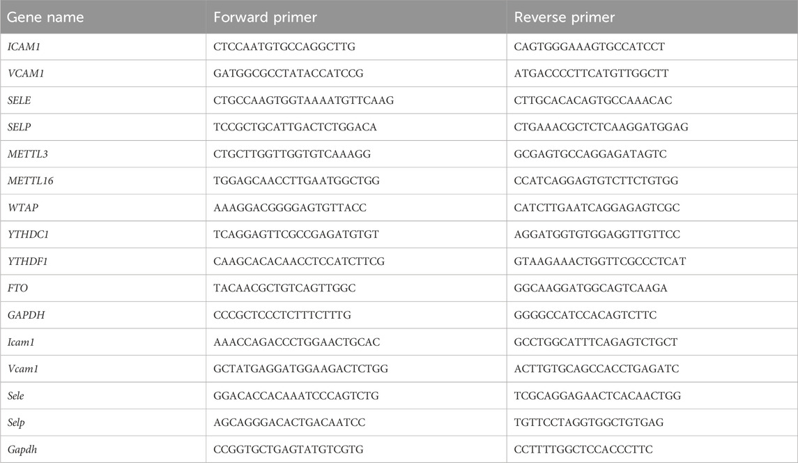
TRIzol reagent (Invitrogen, United States) was used to enrich total RNA from HAECs or mouse aortas. The PrimeScript RT Master Mix (TaKaRa, Japan) was then applied to synthesize cDNA. The LightCycler 480 SYBR Green I Master (Roche, Germany) was used to set up PCR reactions that were analyzed on a LightCycler 480 II system (Roche, United States). The primer sequences were listed in Table 1. Human GAPDH or mouse Gapdh was examined as internal controls. The fold change analysis of the gene expression was performed according to 2-[Ct (specific gene)−Ct (GAPDH or Gapdh)].
2.7 Cell adhesion assay
HAECs were seeded in 24-well plates at 1 × 10⁵ cells/well. Subsequently, after 30-min treatment of 100 μM Rb1 or saline, the cells were exposed to 1 μg/mL NETs for 24 h. For neutrophil adhesion assay, isolated neutrophils were loaded on the monolayer of HAECs at 2 × 10⁴ per well. After 30-min co-culture, the non-adherent neutrophils were rinsed off. The remaining neutrophils were then stained using anti-Ly6G antibody (127601, BioLegend, United States) (1:1000). For monocyte adhesion assay, THP-1 cells (National Collection of Authenticated Cell Cultures, China) were labeled with CellTracker™ CM-Dil (Invitrogen, United States) (30 min, 37°C). The labeled cells were subsequently loaded on the monolayer of HAECs at 2 × 10⁴ cells/well. After the 30-min co-culture and washing procedures, the fluorescence of the cell tracker was captured using a fluorescence microscope (DMI6000, Leica, Germany) to assess the adherence of THP-1 cells to HAECs. Image-Pro Plus 6.0 was utilized for image analysis.
2.8 Endothelial barrier function assays
Twenty-four-well transwell inserts were used for the seeding of HAECs (1 × 10⁵ cells/well). For transendothelial electrical resistance (TEER) assay, 30-min vehicle or Rb1 (100 μM) incubation was followed by 1 μg/mL NETs exposure for 6, 12 and 24 h. A Millicell ERS-2 voltohmmeter (EMD Millipore, Billerica, MA) was used to evaluate TEER. The value was recorded when stable and sustained TEER was established. For Evans blue dye extravasation assay, after 30-min treatment of Rb1 (100 μM) or vehicle, NETs (1 μg/mL) were added and cells were cultured for additional 24 h. Subsequently, the transwell set was washed and the following procedures included adding 600 μL of 4% BSA solution to the lower chamber and 100 μL of 0.67 mg/mL Evans blue dye (in 4% BSA) to the upper chamber. After 1-h equilibration, the upper chambers were discarded. Evans blue dye in the lower chambers was quantified using a microplate reader (Epoch, BioTek, United States) with the absorbance wavelength set at 620 nm.
2.9 Dot blotting
Dotting blotting was conducted as detailed in our previous study (Cui et al., 2023). In brief, the miRNeasy Mini Kit (Qiagen, Germany) was used to purify total RNA from HAECs. After denaturation, the RNA was loaded on the nylon membrane (Millipore, United States). The membrane was then UV cross-linked, blocked and sequentially probed with anti-N6-methyladenosine antibody (ab151230, Abcam, United States) (1:2000) and horseradish peroxidase-conjugated goat anti-rabbit secondary antibody (W4011, Promega, United States). The signal was visualized using the Western Bright ECL reagent (Advansta, United States) and recorded using the MiniChemi system (China). Image-Pro Plus 6.0 was adopted for densitometry analysis. RNA loading was estimated by staining the membrane with methylene blue.
2.10 Western blotting
RIPA buffer (Beyotime, China) supplemented with phosphatase inhibitors and proteases inhibitors (Roche, United States) was used to prepare protein lysate. The electrophoresis was done in 10% SDS-PAGE gels. The protein was transferred to the polyvinylidene fluoride membrane (Millipore, United States). The membrane was probed with rabbit anti-FTO primary antibody (ab124892, Abcam, United States) (1:1000) or mouse anti-β-actin primary antibody (66009, Proteintech, China) (1:3000) at 4°C overnight, followed by secondary antibody incubation using horseradish peroxidase-conjugated goat anti-rabbit antibody (W4011, Promega, United States) (1:3000) or goat anti-mouse secondary antibody (W4021, Promega, United States) (1:3000). SuperSignal™ West Pico PLUS Chemiluminescent Substrate (Thermo Fisher Scientific, United States) was used to visualize the signal. Images were obtained using the MiniChemi system (China). Densitometry was assessed by Image-Pro Plus 6.0.
2.11 Statistical analysis
The data were shown as mean ± standard error of the mean (SEM). SPSS 26.0 software (Ehningen, Germany) or Graphpad Prism 9 software (San Diego, United States) was employed for statistical analysis. Normality test and homogeneity of variance were assessed by Shapiro-Wilk test and Levene’s test, respectively. Multiple comparisons were made by one-way ANOVA with Tukey’s HSD post hoc analysis. Statistically significance was defined as P < 0.05.
3 Results
3.1 Rb1 attenuates endothelial activation and atherosclerosis in vivo
Prior to investigate the direct impact of Rb1 on NETs-induced endothelial activation, we evaluated the pharmacological effects of Rb1 treatment on atherosclerosis-associated endothelial activation in the HFD-fed Apoe−/− mice in vivo. Rb1 treatment resulted in smaller area of the atherosclerotic lesion (Figures 1A,B), which was corroborated by reductions in the lipid load at lesion sites as revealed by Oil red O staining (Figures 1C,D). Furthermore, the immunopositivity of Vcam1 in the atherosclerotic plaques was remarkably decreased due to Rb1 treatment (Figures 1E,F). The vehicle-treated HFD-fed Apoe−/− mice were characterized by increased aortic expression of the genes encoding Icam1, Vcam1, E-selectin and P-selectin. However, decreased aortic expression of Icam1, Vcam1, E-selectin and P-selectin was noted as a result of Rb1 treatment (Figure 1G). These results support that Rb1 treatment mitigates endothelial activation and atherosclerosis in vivo.
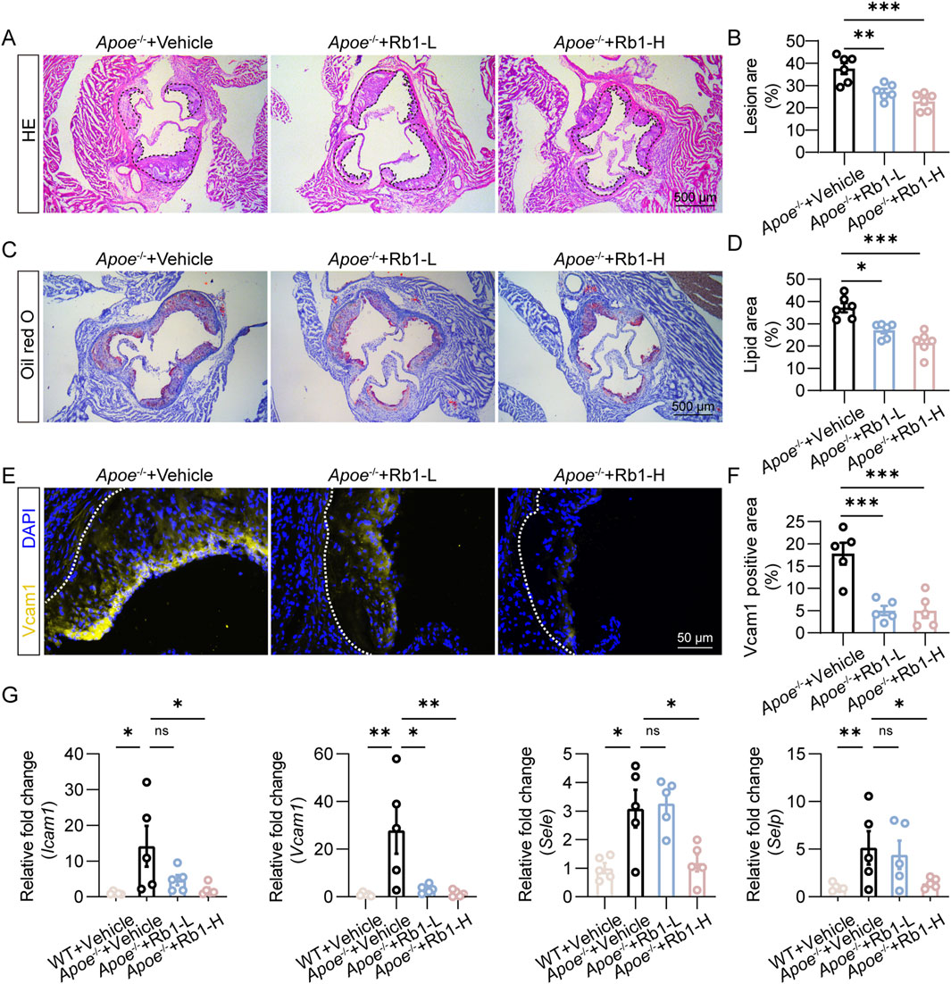
Figure 1. Rb1 mitigates atherosclerosis and endothelial activation in HFD-fed Apoe−/− mice. (A) Representative microscopic images of HE-stained aortic root cross-sections. Scale bar, 500 μm. (B) The area of the atherosclerotic lesion normalized against the lumen area (%) (n = 6 per group). (C) Oil red O positivity in the aortic root cross-sections. Scale bar, 500 μm. (D) Quantification of the plaque lipid load (n = 6 per group). (E) Vcam1 immunopositivity (in yellow) associated with the atherosclerotic lesions. DAPI highlighted the nuclei (in blue). Scale bar, 50 μm. (F) Quantification of Vcam1 immunopositivity in the atherosclerotic lesions (n = 5 per group). (G) The aortic expression of Icam1, Vcam1, Sele and Selp. Relative fold change was plotted against the wild type controls (n = 5 per group). *P < 0.05, **P < 0.01, ***P < 0.001, ns, not significant.
3.2 Rb1 mitigates NETs-induced aberrant activation of endothelial cells
Next, we evaluated the pharmacological effect of Rb1 on NETs-stimulated endothelial activation in vitro. Activated endothelial cells are characterized by augmented expression of genes that encode adhesion molecules, which facilitate the capture of leukocytes, an essential step for the subsequent transendothelial leukocyte migration. Consistent with this notion, NETs exposure led to increased expression of ICAM1, VCAM1, SELE and SELP in endothelial cells. In contrast, much lower expression of ICAM1, VCAM1, SELE and SELP was found in the Rb1-treated cells (Figure 2A). In agreement with these results, augmented adhesion of neutrophils occurred as a result of NETs stimulation. However, much lower level of neutrophil adhesion was noted in the NETs-stimulated endothelial cells treated with Rb1 (Figures 2B,C). Similar observations were also made when THP-1 monocytes were loaded on the endothelial cells (Figures 2D,E). Additionally, prolonged stimulation of NETs decreased the TEER value, whereas much higher TEER value was documented in the Rb1-treated NETs-stimulated cells (Figure 2F). Similar observations were made when extravasation of Evans blue dye across the endothelial cells was measured (Figure 2G). Taken together, these results indicate that Rb1 mitigates NETs-induced aberrant activation of endothelial cells.
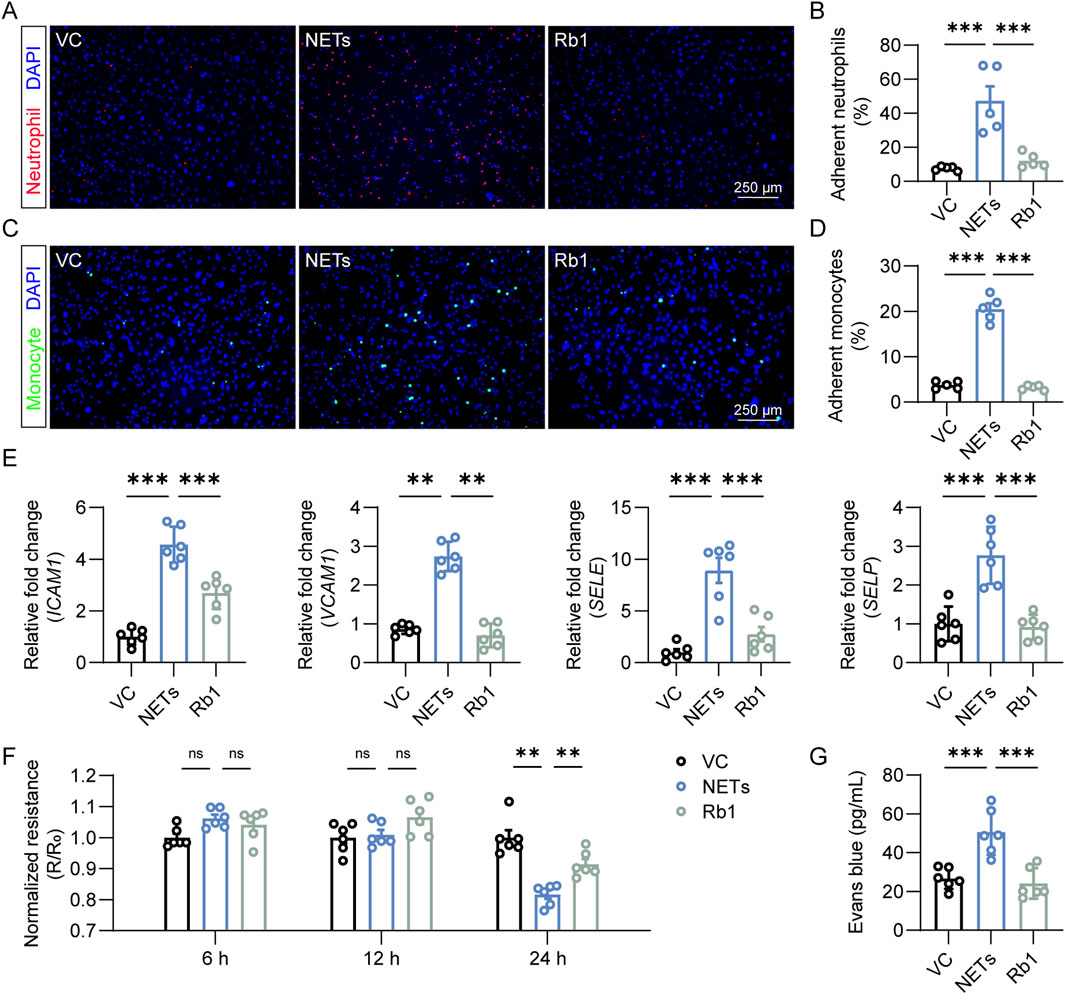
Figure 2. Rb1 inhibits NETs-stimulated activation of endothelial cells. (A) Adhesion of neutrophils (in red) to HAECs (nuclei were counterstained with DAPI, in blue). Scale bar, 250 μm. (B) Proportion of neutrophils adhering to HAECs (n = 5 per group). (C) Adhesion of THP-1 monocytes (in green) to HAECs (nuclei were counterstained with DAPI, in blue). Scale bar, 250 μm. (D) Proportion of monocytes adhering to endothelial cells (n = 5 per group). (E) The expression of ICAM1, VCAM1, SELE and SELP in HAECs. Relative fold change was plotted against VC (n = 6 per group). (F) TEER assay. Resistance (R) was normalized against the starting resistance (R0) (n = 6 per group). (G) Evans blue extravasation assay. The amount of Evans blue dye detected in the lower chamber of the transwell (n = 6 per group). **P < 0.01, ***P < 0.001, ns, not significant.
3.3 Rb1 treatment augments m6A RNA modification in NETs-stimulated endothelial cells
Furthermore, m6A RNA methylation was evaluated to probe the mechanistic implications of m6A RNA modification in NETs-induced endothelial activation. NETs stimulation led to overt reductions in m6A RNA methylation in endothelial cells. On the contrary, significantly higher level of m6A RNA methylation was observed in the NETs-stimulated cells treated with Rb1 (Figures 3A,B). To understand the mechanisms underlying altered m6A RNA methylation in NETs-stimulated endothelial cells, the expression of RNA methyltransferases such as METTL3, METTL16 and WTAP, readers of m6A on RNA such as YTHDC1 and YTHDF1 as well as RNA demethylase FTO was assessed. Increased expression of WTAP, YTHDC1 and FTO was observed in the NETs-stimulated endothelial cells (Figure 3C). Given that NETs stimulation led to lower level of m6A RNA modification in endothelial cells (Figures 3A,B), we focused on FTO in the context of NETs-induced endothelial activation. As shown in Figure 3D, in contrast to NETs-induced upregulation of FTO expression, Rb1 treatment led to significantly lower endothelial expression of FTO in the presence of NETs. Consistently, the protein level of FTO was increased in response to NETs stimulation, whereas significantly decreased level of FTO protein was noted in the Rb1-treated cells (Figures 3E,F). Collectively, these results suggest the possibility that Rb1 may increase the level of m6A RNA methylation by decreasing the expression of FTO in NETs-stimulated endothelial cells, an effect contributing to Rb1-conferred attenuation of NETs-induced endothelial activation.
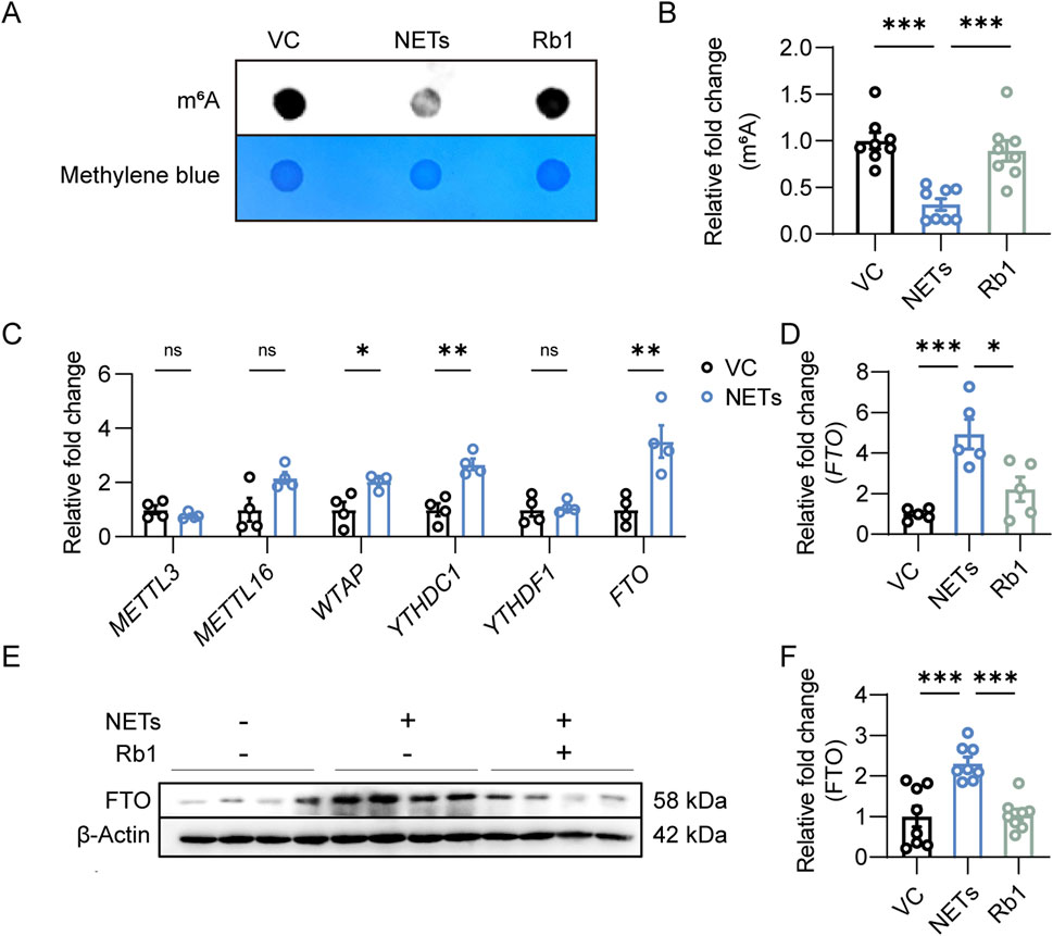
Figure 3. Rb1 increases m6A RNA methylation and decreases the expression of FTO in the NETs-stimulated endothelial cells. (A) Dot blotting analysis of m6A RNA methylation in HAECs. (B) Relative fold change in the level of m6A RNA modification (n = 8 per group). (C) Real-time qPCR analysis of the expression of METTL3, METTL16, WTAP, YTHDC1, YTHDF1 and FTO in HAECs. Relative fold change was plotted against VC (n = 4 per group). (D) The expression of FTO in HAECs. Relative fold change was plotted against VC (n = 5 per group). (E) Western blotting analysis of the protein expression of FTO in HAECs. (F) Relative fold change was plotted against VC (n = 8 per group). *P < 0.05, **P < 0.01, ***P < 0.001, ns, not significant.
3.4 Rb1 suppresses NETs-induced endothelial activation in part by lowering the expression of FTO in endothelial cells
To address the possibility that FTO is mechanistically implicated in Rb1-conferred protection against NETs-induced endothelial activation, the adhesion of neutrophils or monocytes to endothelial cells was assessed after transfecting HACEs with empty vector or FTO-overexpressing plasmid. As shown in Figures 4A,B, attenuated neutrophil-endothelial adhesion was consistently observed in the Rb1-treated NETs-exposed cells. However, FTO overexpression significantly diminished the beneficial effects of Rb1 against NETs-enhanced neutrophil adhesion to the endothelial cells. Similar observations were made when adhesion of THP-1 monocytes to endothelial cells was examined (Figures 4C,D). In agreement, FTO overexpression partially abolished the inhibitory effects of Rb1 on NETs-induced expression of ICAM1, VCAM1 and SELE (Figure 4E). Furthermore, FTO overexpression decreased TEER values and increased Evans blue dye extravasation in the Rb1-treated NETs-stimulated HAECs (Figures 4F,G). Altogether, the results here validate the hypothesis that Rb1 protects against NETs-stimulated endothelial activation in part by decreasing the expression FTO in endothelial cells.
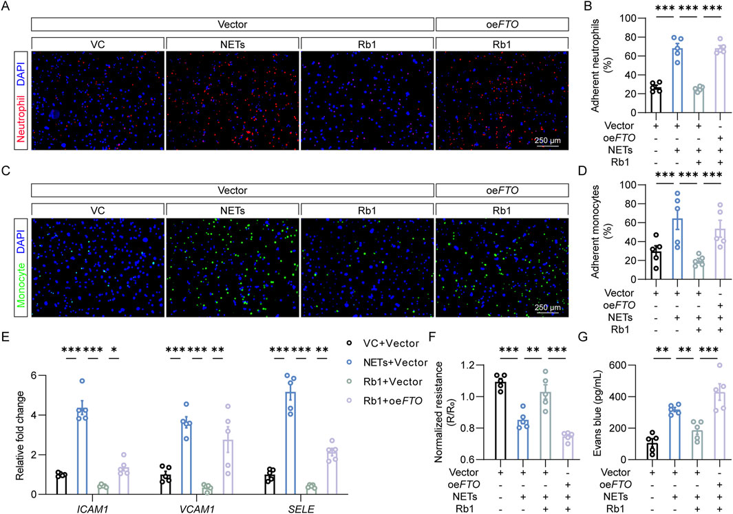
Figure 4. FTO overexpression abolishes the effects of Rb1 against NETs-stimulated endothelial activation. (A) Adhesion of neutrophils (in red) to HAECs (DAPI counterstaining of the nuclei, in blue). Scale bar, 250 μm. (B) Proportion of neutrophils adhering to HAECs (n = 5 per group). (C) Adhesion of monocytes (in green) to HAECs (DAPI counterstaining of the nuclei, in blue). Scale bar, 250 μm. (D) Proportion of monocytes adhering to endothelial cells (n = 5 per group). (E) The expression of ICAM1, VCAM1 and SELE in HAECs. Relative fold change was plotted against VC (n = 5 per group). (F) TEER in HAECs. Resistance (R) was normalized against the starting resistance (R0) (n = 5 per group). (G) Evans blue dye extravasation assay. The amount of Evans blue dye detected in the lower chamber of the transwell (n = 5 per group). *P < 0.05, **P < 0.01, ***P < 0.001.
3.5 Fto overexpression in endothelial cells abolishes the anti-atherosclerotic effects of Rb1 in vivo
To further address the mechanistic implications of FTO in Rb1-conferred protection against aberrant endothelial activation in the pathological context of atherosclerosis in vivo, AAV9-mediated overexpression of Fto was targeted to the endothelial cells via a Tie2 promoter and delivered to the HFD-fed Apoe−/− mice in conjunction with Rb1 treatment. As shown in Figures 5A,B, smaller atherosclerotic lesions were consistently observed in the Rb1-treated HFD-fed Apoe−/− mice transduced with AAV9 control, whereas significantly increased lesion size was readily detected in the Rb1-treated HFD-fed Apoe−/− mice transduced with Fto-overexpressing AAV9. Meanwhile, increased lipid load (Figures 5C,D) and macrophage burden (Figures 5E,F) were noted in the Rb1-treated HFD-fed Apoe−/− mice transduced with Fto-overexpressing AAV9. Lastly, the in situ expression of Vcam1 in the atherosclerotic plaques was significantly higher in the Rb1-treated HFD-fed Apoe−/− mice transduced with Fto-overexpressing AAV9 (Figures 5G,H). These results indicate that Fto overexpression in endothelial cells abolishes the endothelial protective and anti-atherosclerotic effects of Rb1 in vivo.
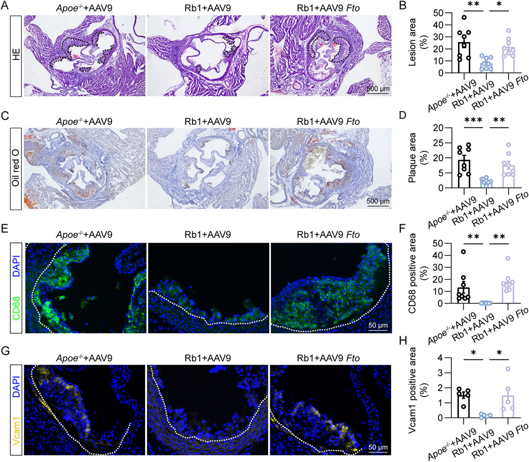
Figure 5. Fto overexpression offsets Rb1-conferred protection against atherosclerosis and endothelial activation in HFD-fed Apoe−/− mice. (A) Representative microscopic images of HE-stained aortic root cross-sections. Scale bar, 500 μm. (B) The area of the atherosclerotic lesion normalized against the lumen area (%) (n = 8 per group). (C) Oil red O positivity in the atherosclerotic plaques. Scale bar, 500 μm. (D) Quantification of the Oil red O-positive lipid load in the plaques (n = 8 per group). (E) CD68 immunopositivity (in green) and DAPI counterstaining of the nuclei (in blue) in the plaques. Scale bar, 50 μm. (F) Quantification of CD68 immunopositivity in the atherosclerotic lesions (n = 8 per group). (G) Vcam1 immunopositivity (in yellow) and DAPI counterstaining of the nuclei (in blue) in the plaques. Scale bar, 50 μm. (H) Quantification of Vcam1 immunopositivity (n = 5 per group). *P < 0.05, **P < 0.01, ***P < 0.001.
4 Discussion
Atherosclerosis is essentially an inflammatory disorder in part driven by the interplay between vascular endothelium and immune cells. Accumulating evidence supports the mechanistic contributions of neutrophil-derived NETs in the pathogenesis of atherosclerosis. Here we demonstrate that the anti-atherosclerotic effects of Rb1 involve mitigating NETs-stimulated endothelial activation, which is in part mediated by offsetting FTO-mediated m6A demethylation in endothelial cells (Figure 6).
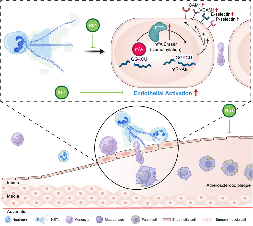
Figure 6. A schematic illustration of the pharmacological effects of Rb1 against NETs-induced endothelial activation in atherosclerosis. Our work here reveals that Rb1 mitigates NETs-stimulated aberrant activation of endothelial cells. Furthermore, Rb1 counteracts NETs-triggered dysregulation of FTO and m6A methylation in endothelial cells, which may in part contribute to the pharmacological effects of Rb1 against atherosclerosis.
First, our current study reveals that Rb1 mitigates NETs-stimulated aberrant activation of endothelial cells. Atherosclerosis is initiated by insults to the vascular endothelium. The ensuing endothelial activation promotes aberrant leukocyte-endothelium interactions that occur through the course of atherosclerosis. This process is tightly regulated by adhesion molecules (e.g., ICAM, VCAM, E-selectin and P-selectin) that undergo upregulated expression during atherosclerosis (Richardson et al., 1994; Poston et al., 1992; Johnson-Tidey et al., 1994; Nakashima et al., 1998; Li et al., 1993). NETs cause direct damage to endothelial cells (Gupta et al., 2010; Villanueva et al., 2011). Consistent with this notion, our study uncovers that NETs upregulate the expression of the genes encoding vascular adhesion molecules and enhances the adhesion of neutrophils and monocytes to endothelial cells, supporting the direct implications of NETs in endothelial activation. Furthermore, our findings uncover that Rb1 mitigates NETs-induced expression of pro-atherogenic adhesion molecules (ICAM1, VCAM1, SELE, and SELP) as well as the adhesion of neutrophils and monocytes to endothelial cells. ICAM1 mediates firm adhesion of leukocytes to the endothelial cells that undergo activation (Collins et al., 2000). VCAM1 preferentially mediates the recruitment of mononuclear leukocytes (Nakashima et al., 1998; Cybulsky and Gimbrone, 1991; Sakai et al., 1997). The selectins determine the tethering, rolling and adhesion of all types of leukocytes that interact with the activated endothelium (Collins et al., 2000; Dong et al., 1998). Therefore, the cellular and molecule evidence of Rb1-conferred protection against NETs-stimulated aberrant interaction between neutrophils/monocytes and endothelial cells provides new insights into the mechanisms underlying the anti-atherosclerotic effects of Rb1. Notably, the NETs-modulating and endothelial protective effects have also been demonstrated in a study elucidating the anti-atherosclerotic mechanisms of a traditional Chinese medicine formula comprising Prunus persica (L.) Batsch, Carthamus tinctorius L., Angelica sinensis (Oliv.) Diels, Conioselinum anthriscoides ‘Chuanxiong’, Paeonia lactiflora Pall., Rehmannia glutinosa (Gaertn.) Libosch. ex DC., Astragalus mongholicus Bunge, Cinnamomum verum J.Presl, and Epimedium sagittatum (Siebold & Zucc.) Maxim. (Shao et al., 2024). These findings, in conjunction with our current data, provide experimental evidence supporting the concept that polyherbal preparations, botanical drugs, and plant metabolites warrant further investigation for their potential pharmacological effects in mitigating atherosclerotic progression through modulation of NETs-induced endothelial activation.
Secondly, our findings reveal that FTO-mediated m6A RNA demethylation is in part implicated in the pharmacological effects of Rb1 against NETs-induced endothelial activation in atherosclerosis. The level of m6A RNA methylation is decreased in the human atherosclerotic plaques (Quiles-Jiménez et al., 2020). Importantly, FTO expression can be induced by oxidized LDL (ox-LDL). Overexpression of FTO results in increased expression of ICAM1 and VCAM1. On the contrary, FTO knockdown attenuates ox-LDL-induced or TNFα-triggered expression of ICAM1 and VCAM1 in endothelial cells, leading to reduced adhesion of monocytes to activated endothelial cells (Mo et al., 2022). FTO-mediated RNA demethylation also drives diabetes-associated endothelial activation (Zhou et al., 2023). These findings support the notion that FTO plays an important role in endothelial activation. Moreover, NETs-m6A crosstalk has been documented in the pathological context of lung cancer, pulmonary injury, and glioma (Xu et al., 2025; Zhang et al., 2022; Dai et al., 2024). Our results here further demonstrate that m6A RNA methylation is markedly reduced in the NETs-stimulated endothelial cells, which is in part explained by significantly increased expression of FTO. Therefore, our findings not only corroborate the known implications of FTO in endothelial activation and NETs-m6A crosstalk, but also add new mechanistic understanding of endothelial activation by linking NETs to FTO-regulated m6A modification in endothelial activation. Furthermore, Rb1 treatment results in higher levels of m6A RNA methylation and decreased expression of FTO in the NETs-stimulated endothelial cells. Forced expression of FTO in part cancels out Rb1-conferred protection against NETs-induced endothelial activation in vitro. Most importantly, Fto overexpression partially offsets the anti-atherosclerotic effects of Rb1 in the HFD-fed Apoe−/− mice in vivo. Therefore, the molecular, cellular and in vivo evidence here jointly indicate that Rb1 increases the level of m6A RNA methylation in the NETs-stimulated endothelial cells in part by decreasing the expression of FTO, which contributes to the effects of Rb1 at attenuating endothelial activation and progression of atherosclerotic lesions. However, the in-depth mechanisms underlying the regulated expression of FTO in response to Rb1 treatment are to be further illustrated in the future. Meanwhile, further studies are necessary to identify specific downstream m6A targets that are modified by Rb1 treatment in the NETs-stimulated endothelial cells in the context of atherosclerosis.
Moreover, it is worth noting that our findings here are in line with the research advances on the context-dependent impact of plant metabolites on m6A RNA modification. To name a few, rhein has been uncovered to be a substrate competitive inhibitor of FTO and this FTO inhibitory activity of rhein contributes to its anti-cancer effects (Chen et al., 2012; Yang et al., 2021). Baicalin has recently been revealed to mitigate male reproductive toxicity by restoring the expression of m6A reader YTHDC2 (Wen et al., 2025). Baicalin has also been reported to alleviate lactate-induced acidification in tumor microenvironment by decreasing the level of m6A reader IGF2BP3 in oral squamous cell carcinoma cells (Cui et al., 2025). Our previous work has also shown that the expression of FTO can be increased by cinnamic acid in cardiomyocytes, which in part accounts for cinnamic acid-conferred protection against pressure overload-induced left ventricular hypertrophy (Cui et al., 2023). Therefore, our current findings add to the growing body of evidence that supports the pharmacological potentials of plant metabolites on m6A RNA modification.
In conclusion, the present study reveals that Rb1 mitigates NETs-induced aberrant endothelial activation in atherosclerosis. Furthermore, the newly identified pharmacological effects of Rb1 against NETs-induced endothelial activation in part involve augmenting the level of m6A RNA methylation by suppressing FTO expression in endothelial cells. The experimental evidence presented in this work thus provides new mechanistic insights into the anti-atherosclerotic effects of Rb1.
Data availability statement
The original contributions presented in the study are included in the article/supplementary material, further inquiries can be directed to the corresponding authors.
Ethics statement
The animal study was approved by The Institutional Animal Care and Use Committee at Yueyang Hospital of Integrated Traditional Chinese and Western Medicine, Shanghai University of Traditional Chinese Medicine. The study was conducted in accordance with the local legislation and institutional requirements.
Author contributions
ZY: Investigation, Writing – original draft, Data curation, Methodology, Visualization, Formal Analysis, Validation. MX: Methodology, Writing – original draft, Resources, Funding acquisition. XT: Investigation, Writing – original draft, Methodology. PW: Methodology, Investigation, Writing – original draft. JC: Methodology, Writing – original draft. YC: Writing – review and editing, Writing – original draft, Resources, Conceptualization, Project administration, Supervision. TZ: Resources, Project administration, Funding acquisition, Writing – original draft, Supervision, Conceptualization, Writing – review and editing.
Funding
The author(s) declare that financial support was received for the research and/or publication of this article. This work was funded by National Natural Science Foundation of China (82074049 to TZ and 81903835 to MX), Shanghai University of Traditional Chinese Medicine Xinglin Talents Program (Shanghai University of Traditional Chinese Medicine Human Resource 202023, MX) and R&D Projects from KPC Pharmaceuticals (KY111.40.202).
Conflict of interest
The authors declare that the research was conducted in the absence of any commercial or financial relationships that could be construed as a potential conflict of interest.
Generative AI statement
The author(s) declare that no Generative AI was used in the creation of this manuscript.
Publisher’s note
All claims expressed in this article are solely those of the authors and do not necessarily represent those of their affiliated organizations, or those of the publisher, the editors and the reviewers. Any product that may be evaluated in this article, or claim that may be made by its manufacturer, is not guaranteed or endorsed by the publisher.
References
Boulanger, C. M. (2016). Endothelium. Arterioscler. Thromb. Vasc. Biol. 36, e26–e31. doi:10.1161/ATVBAHA.116.306940
Brinkmann, V., Reichard, U., Goosmann, C., Fauler, B., Uhlemann, Y., Weiss, D. S., et al. (2004). Neutrophil extracellular traps kill bacteria. Science 303, 1532–1535. doi:10.1126/science.1092385
Chen, B., Ye, F., Yu, L., Jia, G., Huang, X., Zhang, X., et al. (2012). Development of cell-active N6-methyladenosine RNA demethylase FTO inhibitor. J. Am. Chem. Soc. 134, 17963–17971. doi:10.1021/ja3064149
Collins, R. G., Velji, R., Guevara, N. V., Hicks, M. J., Chan, L., and Beaudet, A. L. (2000). P-Selectin or intercellular adhesion molecule (ICAM)-1 deficiency substantially protects against atherosclerosis in apolipoprotein E-deficient mice. J. Exp. Med. 191, 189–194. doi:10.1084/jem.191.1.189
Cui, Y., Liu, J., Wang, X., Wu, Y., Chang, Y., Hu, X., et al. (2025). Baicalin attenuates the immune escape of oral squamous cell carcinoma by reducing lactate accumulation in tumor microenvironment. J. Adv. Res. doi:10.1016/j.jare.2025.01.021
Cui, Y., Wang, P., Li, M., Wang, Y., Tang, X., Cui, J., et al. (2023). Cinnamic acid mitigates left ventricular hypertrophy and heart failure in part through modulating FTO-dependent N(6)-methyladenosine RNA modification in cardiomyocytes. Biomed. Pharmacother. 165, 115168. doi:10.1016/j.biopha.2023.115168
Cybulsky, M. I., and Gimbrone, M. A. (1991). Endothelial expression of a mononuclear leukocyte adhesion molecule during atherogenesis. Science 251, 788–791. doi:10.1126/science.1990440
Dai, W., Tian, R., Yu, L., Bian, S., Chen, Y., Yin, B., et al. (2024). Overcoming therapeutic resistance in oncolytic herpes virotherapy by targeting IGF2BP3-induced NETosis in malignant glioma. Nat. Commun. 15, 131. doi:10.1038/s41467-023-44576-2
Dong, Z. M., Chapman, S. M., Brown, A. A., Frenette, P. S., Hynes, R. O., and Wagner, D. D. (1998). The combined role of P- and E-selectins in atherosclerosis. J. Clin. Invest. 102, 145–152. doi:10.1172/JCI3001
Döring, Y., Drechsler, M., Wantha, S., Kemmerich, K., Lievens, D., Vijayan, S., et al. (2012). Lack of neutrophil-derived CRAMP reduces atherosclerosis in mice. Circ. Res. 110, 1052–1056. doi:10.1161/CIRCRESAHA.112.265868
Döring, Y., Manthey, H. D., Drechsler, M., Lievens, D., Megens, R. T., Soehnlein, O., et al. (2012). Auto-antigenic protein-DNA complexes stimulate plasmacytoid dendritic cells to promote atherosclerosis. Circulation 125, 1673–1683. doi:10.1161/CIRCULATIONAHA.111.046755
Drechsler, M., Megens, R. T., van Zandvoort, M., Weber, C., and Soehnlein, O. (2010). Hyperlipidemia-triggered neutrophilia promotes early atherosclerosis. Circulation 122, 1837–1845. doi:10.1161/CIRCULATIONAHA.110.961714
Duan, L., Xiong, X., Hu, J., Liu, Y., Li, J., and Wang, J. (2017). Panax notoginseng saponins for treating coronary artery disease: a functional and mechanistic overview. Front. Pharmacol. 8, 702. doi:10.3389/fphar.2017.00702
Eriksson, E. E., Xie, X., Werr, J., Thoren, P., and Lindbom, L. (2001). Direct viewing of atherosclerosis in vivo: plaque invasion by leukocytes is initiated by the endothelial selectins. FASEB J. 15, 1149–1157. doi:10.1096/fj.00-0537com
Fan, J. S., Liu, D. N., Huang, G., Xu, Z. Z., Jia, Y., Zhang, H. G., et al. (2012). Panax notoginseng saponins attenuate atherosclerosis via reciprocal regulation of lipid metabolism and inflammation by inducing liver X receptor alpha expression. J. Ethnopharmacol. 142, 732–738. doi:10.1016/j.jep.2012.05.053
Gimbrone, M. A., and García-Cardeña, G. (2016). Endothelial cell dysfunction and the pathobiology of atherosclerosis. Circ. Res. 118, 620–636. doi:10.1161/CIRCRESAHA.115.306301
Gupta, A. K., Joshi, M. B., Philippova, M., Erne, P., Hasler, P., Hahn, S., et al. (2010). Activated endothelial cells induce neutrophil extracellular traps and are susceptible to NETosis-mediated cell death. FEBS Lett. 584, 3193–3197. doi:10.1016/j.febslet.2010.06.006
He, P. C., and He, C. (2021). m(6) A RNA methylation: from mechanisms to therapeutic potential. Embo J. 40, e105977. doi:10.15252/embj.2020105977
Johnson-Tidey, R. R., McGregor, J. L., Taylor, P. R., and Poston, R. N. (1994). Increase in the adhesion molecule P-selectin in endothelium overlying atherosclerotic plaques. Coexpression with intercellular adhesion molecule-1. Am. J. Pathol. 144, 952–961.
Jorch, S. K., and Kubes, P. (2017). An emerging role for neutrophil extracellular traps in noninfectious disease. Nat. Med. 23, 279–287. doi:10.1038/nm.4294
Josefs, T., Barrett, T. J., Brown, E. J., Quezada, A., Wu, X., Voisin, M., et al. (2020). Neutrophil extracellular traps promote macrophage inflammation and impair atherosclerosis resolution in diabetic mice. JCI Insight 5, e134796. doi:10.1172/jci.insight.134796
Kim, J. H. (2012). Cardiovascular diseases and panax ginseng: a review on molecular mechanisms and medical applications. J. Ginseng Res. 36, 16–26. doi:10.5142/jgr.2012.36.1.16
Knight, J. S., Luo, W., O'Dell, A. A., Yalavarthi, S., Zhao, W., Subramanian, V., et al. (2014). Peptidylarginine deiminase inhibition reduces vascular damage and modulates innate immune responses in murine models of atherosclerosis. Circ. Res. 114, 947–956. doi:10.1161/CIRCRESAHA.114.303312
Li, H., Cybulsky, M. I., Gimbrone, M. A., and Libby, P. (1993). An atherogenic diet rapidly induces VCAM-1, a cytokine-regulatable mononuclear leukocyte adhesion molecule, in rabbit aortic endothelium. Arterioscler. Thromb. 13, 197–204. doi:10.1161/01.atv.13.2.197
Mo, W., Chen, Z., Zhang, X., Dai, G., Ma, D., Pan, J., et al. (2022). N6-Methyladenosine demethylase FTO (fat mass and obesity-associated protein) as a novel mediator of statin effects in human endothelial cells. Arterioscler. Thromb. Vasc. Biol. 42, 644–658. doi:10.1161/ATVBAHA.121.317295
Nakashima, Y., Raines, E. W., Plump, A. S., Breslow, J. L., and Ross, R. (1998). Upregulation of VCAM-1 and ICAM-1 at atherosclerosis-prone sites on the endothelium in the ApoE-deficient mouse. Arterioscler. Thromb. Vasc. Biol. 18, 842–851. doi:10.1161/01.atv.18.5.842
Poston, R. N., Haskard, D. O., Coucher, J. R., Gall, N. P., and Johnson-Tidey, R. R. (1992). Expression of intercellular adhesion molecule-1 in atherosclerotic plaques. Am. J. Pathol. 140, 665–673.
Qiao, L., Zhang, X., Liu, M., Liu, X., Dong, M., Cheng, J., et al. (2017). Ginsenoside Rb1 enhances atherosclerotic plaque stability by improving autophagy and lipid metabolism in macrophage foam cells. Front. Pharmacol. 8, 727. doi:10.3389/fphar.2017.00727
Quiles-Jiménez, A., Gregersen, I., Mittelstedt Leal de Sousa, M., Abbas, A., Kong, X. Y., Alseth, I., et al. (2020). N6-methyladenosine in RNA of atherosclerotic plaques: an epitranscriptomic signature of human carotid atherosclerosis. Biochem. Biophys. Res. Commun. 533, 631–637. doi:10.1016/j.bbrc.2020.09.057
Richardson, M., Hadcock, S. J., DeReske, M., and Cybulsky, M. I. (1994). Increased expression in vivo of VCAM-1 and E-selectin by the aortic endothelium of normolipemic and hyperlipemic diabetic rabbits. Arterioscler. Thromb. 14, 760–769. doi:10.1161/01.atv.14.5.760
Sakai, A., Kume, N., Nishi, E., Tanoue, K., Miyasaka, M., and Kita, T. (1997). P-selectin and vascular cell adhesion molecule-1 are focally expressed in aortas of hypercholesterolemic rabbits before intimal accumulation of macrophages and T lymphocytes. Arterioscler. Thromb. Vasc. Biol. 17, 310–316. doi:10.1161/01.atv.17.2.310
Shao, C. L., Meng, W. T., Wang, Y. C., Liu, J. J., Ning, K., Hou, X. X., et al. (2024). Regulating NETs contributes to a novel antiatherogenic effect of MTHSWD via inhibiting endothelial injury and apoptosis. Int. Immunopharmacol. 143, 113368. doi:10.1016/j.intimp.2024.113368
Trillo, A. A. (1982). The cell population of aortic fatty streaks in African green monkeys with special reference to granulocytic cells. An ultrastructural study. Atherosclerosis 43, 259–275. doi:10.1016/0021-9150(82)90027-2
van Leeuwen, M., Gijbels, M. J., Duijvestijn, A., Smook, M., van de Gaar, M. J., Heeringa, P., et al. (2008). Accumulation of myeloperoxidase-positive neutrophils in atherosclerotic lesions in LDLR-/- mice. Arterioscler. Thromb. Vasc. Biol. 28, 84–89. doi:10.1161/ATVBAHA.107.154807
Villanueva, E., Yalavarthi, S., Berthier, C. C., Hodgin, J. B., Khandpur, R., Lin, A. M., et al. (2011). Netting neutrophils induce endothelial damage, infiltrate tissues, and expose immunostimulatory molecules in systemic lupus erythematosus. J. Immunol. 187, 538–552. doi:10.4049/jimmunol.1100450
Warnatsch, A., Ioannou, M., Wang, Q., and Papayannopoulos, V. (2015). Inflammation. Neutrophil extracellular traps license macrophages for cytokine production in atherosclerosis. Science 349, 316–320. doi:10.1126/science.aaa8064
Wen, Y., Pan, T., Liu, L., Wang, J., Yang, K., Fu, Z., et al. (2025). Baicalin targets YTHDC2 and alleviates Male reproductive toxicity caused by co-exposure to nanoplastics and manganese through m(6)A-dependent pathway. J. Nanobiotechnology 23, 450. doi:10.1186/s12951-025-03535-3
Xu, L., Kong, Y., Li, K., Li, J., Xu, F., Xu, Y., et al. (2025). Neutrophil extracellular traps promote growth of lung adenocarcinoma by mediating the stability of m6A-mediated SLC2A3 mRNA-induced ferroptosis resistance and CD8(+) T cell inhibition. Clin. Transl. Med. 15, e70192. doi:10.1002/ctm2.70192
Yang, B., Wang, J. Q., Tan, Y., Yuan, R., Chen, Z. S., and Zou, C. (2021). RNA methylation and cancer treatment. Pharmacol. Res. 174, 105937. doi:10.1016/j.phrs.2021.105937
Yvan-Charvet, L., Welch, C., Pagler, T. A., Ranalletta, M., Lamkanfi, M., Han, S., et al. (2008). Increased inflammatory gene expression in ABC transporter-deficient macrophages: free cholesterol accumulation, increased signaling via toll-like receptors, and neutrophil infiltration of atherosclerotic lesions. Circulation 118, 1837–1847. doi:10.1161/CIRCULATIONAHA.108.793869
Zhang, H., Liu, J., Zhou, Y., Qu, M., Wang, Y., Guo, K., et al. (2022). Neutrophil extracellular traps mediate m(6)A modification and regulates sepsis-associated acute lung injury by activating ferroptosis in alveolar epithelial cells. Int. J. Biol. Sci. 18, 3337–3357. doi:10.7150/ijbs.69141
Zhang, X., Liu, M. H., Qiao, L., Zhang, X. Y., Liu, X. L., Dong, M., et al. (2018). Ginsenoside Rb1 enhances atherosclerotic plaque stability by skewing macrophages to the M2 phenotype. J. Cell. Mol. Med. 22, 409–416. doi:10.1111/jcmm.13329
Zhou, C., She, X., Gu, C., Hu, Y., Ma, M., Qiu, Q., et al. (2023). FTO fuels diabetes-induced vascular endothelial dysfunction associated with inflammation by erasing m6A methylation of TNIP1. J. Clin. Invest. 133, e160517. doi:10.1172/JCI160517
Zhou, P., Lu, S., Luo, Y., Wang, S., Yang, K., Zhai, Y., et al. (2017). Attenuation of TNF-α-Induced inflammatory injury in endothelial cells by ginsenoside Rb1 via inhibiting NF-κB, JNK and p38 signaling pathways. Front. Pharmacol. 8, 464. doi:10.3389/fphar.2017.00464
Keywords: Ginsenoside Rb1, atherosclerosis, endothelial activation, neutrophil extracellular traps, fat mass and obesity-associated protein
Citation: Yang Z, Xiong M, Tang X, Wang P, Cui J, Chen Y and Zhang T (2025) Ginsenoside Rb1 mitigates atherosclerosis in part through modulating FTO-mediated m6A RNA modification in NETs-induced endothelial activation. Front. Pharmacol. 16:1631076. doi: 10.3389/fphar.2025.1631076
Received: 19 May 2025; Accepted: 14 July 2025;
Published: 23 July 2025.
Edited by:
Juei-Tang Cheng, Chang Jung Christian University, TaiwanReviewed by:
Changle Shao, Shanghai University of Traditional Chinese Medicine, ChinaHui Zhao, Beijing Fengtai Hospital, China
Copyright © 2025 Yang, Xiong, Tang, Wang, Cui, Chen and Zhang. This is an open-access article distributed under the terms of the Creative Commons Attribution License (CC BY). The use, distribution or reproduction in other forums is permitted, provided the original author(s) and the copyright owner(s) are credited and that the original publication in this journal is cited, in accordance with accepted academic practice. No use, distribution or reproduction is permitted which does not comply with these terms.
*Correspondence: Yu Chen, Y2hlbnl1QHNodXRjbS5lZHUuY24=; Teng Zhang, emhhbmd0ZW5nMjA4OUBzaHV0Y20uZWR1LmNu
 Zhenni Yang
 Minqi Xiong1,2
 Peiwei Wang
 Yu Chen