- 1Department of Psychology, The Fifth Affiliated Hospital, Sun Yat-sen University, Zhuhai, China
- 2The School of Biomedical Engineering, Sun Yat-Sen University, Shenzhen, China
- 3Department of Child Psychiatry and Rehabilitation, Shenzhen Maternity and Child Healthcare Hospital, Southern Medical University, Shenzhen, Guangdong, China
Objective: Given the lack of consensus regarding the optimal EEG paradigm for identifying bipolar depression (BD), this study sought to systematically evaluate the efficacy of three classic EEG paradigms—eyes open, eyes closed, and free viewing—in diagnosing BD.
Methods: EEGs were collected from 28 individuals diagnosed with BD and 42 healthy controls(HCs) across three experimental conditions: eyes closed, eyes open, and free viewing. Sociodemographic data and neuropsychological testing were also collected. This research investigated notable variations in brain functional connectivity between the two groups across paradigms, the correlation of features with neuropsychological assessments, and classification outcomes.
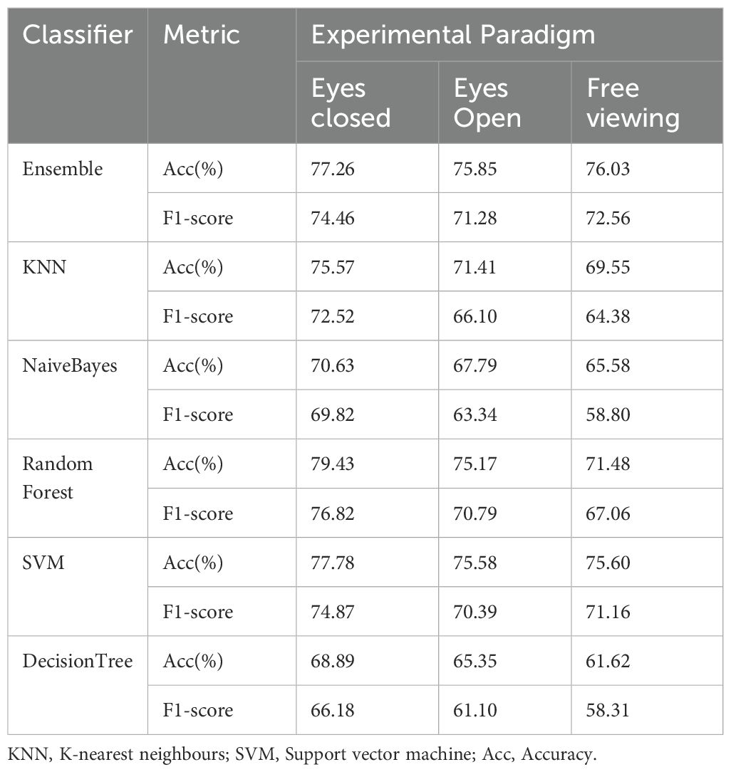
Results: The results demonstrated that under the eyes-closed paradigm, significant differences in the Phase Lag Index (PLI) were consistently observed across the δ, θ, β, and γ frequency bands. This paradigm also featured the highest number of electrodes significantly correlated with cognitive scales. Furthermore, the eyes-closed condition achieved the highest accuracy in bipolar depression recognition, with the Random Forest classifier yielding the highest accuracy of 79.43% and an F1 score of 76.82%. These findings underscore the eyes closed paradigm as a superior, straightforward EEG experimental approach for the diagnosis of bipolar depression.
Conclusions: This study indicates that the eyes closed experimental paradigm more effectively demonstrates the electrophysiological disparities between patients with BD and HCs, in comparison to the eyes open paradigm and the action observation-based free viewing paradigms, as determined through the analysis of various outcome metrics.
1 Introduction
Bipolar disorder is a severe mood disorder, primarily characterized by extreme fluctuations in mood states, alternating episodes of depression and (hypo)mania (1). The initial onset of bipolar disorder is typically depressive, with depressive episodes lasting significantly longer throughout the course of the illness than manic or hypomanic episodes. The severity of depressive symptoms surpassing mania typically manifests during the developmental stages of children and adolescents, imparting adverse effects on education and vocational prospects (2). Early onset is associated with a diminished prognosis. Furthermore, acute episodes precipitate profound cognitive and psychosocial functional impairments, significantly disrupting attention, cognitive flexibility, executive function, and working memory (3, 4). The prevalence of bipolar disorder has been rising with the evolution of modern society, along with its high disability rate and increased risk of suicide (5), thereby exerting a significant social burden (6). Individuals with bipolar disorder frequently receive incorrect diagnoses, are overdiagnosed, or are only diagnosed several years after disease onset, leading to a worse disease prognosis (3, 7). Therefore, early identification of bipolar disorders is crucial.
Recent investigations have increasingly concentrated on the neural biomarkers of bipolar disorder, which have the potential for precise and timely diagnosis (8). Among the various non-invasive monitoring methods, electroencephalographic (EEG) captures macroscopic temporal dynamics of brain activity by measuring scalp electrical potentials, thereby being considered a suitable tool (9). EEG-derived datasets are increasingly utilized in bipolar disorder research (7). Studies on biomarkers of bipolar disorder have primarily focused on aspects such as the P300 wave, functional connectivity, and oscillation frequency bands, with a growing interest in the application of machine-learning techniques (10–13). Despite adopting various experimental paradigms over the past decade, including resting-state and task-related measurements during emotional or cognitive tasks (14). There is no consensus on the optimal experimental paradigm for exploring EEG biomarkers in bipolar disorder and healthy controls (HCs) (9, 15). Current mainstream experimental paradigms include eyes closed, eyes open, and free viewing protocols, with the latter encompassing the free viewing of images and videos, as well as cognitive tasks (16–18). This study employed a video-based free viewing paradigm.
Research using the eyes closed paradigm revealed decreased alpha (α) power and increased delta (δ) and theta (θ) activity in the EEG profiles of initial bipolar disorder episodes relative to HCs (19). Some studies have found that bipolar disorder exhibits higher power across all frequencies, including the beta (β) and gamma (γ) bands than HCs. Patients with bipolar disorder show greater coherence in the α band within the parietal-temporal and central parietal regions, while interhemispheric coherence in the δ band of the frontal regions is lower (20). Similarly, another study found higher coherence in the frontal and occipital cortex, particularly in the frontal regions, compared to HCs (21). Studies employing an eyes open experimental paradigm have observed disruptions in frontal lobe slow-wave oscillations, with increased activity in the δ, θ, and α bands (22). Research has also found increased α-wave power in the central-frontal and right parietal regions in bipolar disorder (23). However, another study found slowed α-wave activity during depressive episodes in adolescents with bipolar disorder, which has been associated with decreased cognitive function (24). Within free viewing paradigms, the saccadic eye movement approach suggests γ coherence as a potential marker for cognitive dysfunction in manic phases (25). Furthermore, EEG responses to emotional facial expressions have been instrumental in distinguishing unipolar from bipolar depression (BD), highlighting the diagnostic utility of EEG activity (15).
This study explored the contributions of three mainstream experimental paradigms to electroencephalographic biomarkers of bipolar disorders. We employed video-based observations to investigate the free viewing paradigm. This approach is predicated on the premise that central brain regions exhibit a sensorimotor α-rhythm during eyes open states or the observation and execution of actions. This rhythm has a frequency range of 8–13 Hz and is a variant of the α-rhythm (26, 27). Numerous studies suggest that this sensorimotor α-rhythm may serve as an indicator that may aid in the discovery of the pathophysiological mechanisms of bipolar disorder (28; S. C. 29). A study on patients with bipolar disorder found that the bipolar disorder group exhibited lower levels of sensorimotor α-rhythm suppression compared to HCs, possibly indicating social cognitive difficulties. Kim et al. (28) examined the neural activity of euthymic bipolar disorder patients in a new virtual reality social cognitive task. They found that bipolar disorder participants exhibited slower reaction times to the emotional component of the task (despite comparable accuracy), particularly in the inferior frontal cortex, pre-motor cortex, and insula. Based on this, we chose a free viewing paradigm for video observation, with participants watching videos of goal-directed actions. Research suggests that observing videos of hand movements can enhance brain activity patterns (30). We selected videos from a dynamic action stimuli database (31) and clipped actions familiar to Chinese participants to create the free viewing videos used in our study.
Numerous studies have identified considerable variations in the results of biomarkers for bipolar disorder, which may be attributed to applying different experimental paradigms (4, 9, 32, 33). The extant data are insufficient for deducing trends or computing consistency scores and are often poorly validated (34). Consequently, this study has selected three distinct paradigms, conducting a comprehensive analysis of various electroencephalographic signals to investigate the differential diagnostic capabilities of each paradigm.
2 Methods and materials
Before starting the study, ethical approval was obtained from the Ethics Committee of the Fifth Affiliated Hospital of Sun Yat-sen University.
2.1 Participants
Participants with bipolar disorder were recruited from the psychiatric inpatient service and diagnosed using DSM-5 criteria by two independent and experienced psychiatrists, one of whom was the patient’s treating psychiatrist. All participants in the patient group were assessed to be in a depressive state at the time of evaluation, with none meeting the criteria for a current manic, hypomanic, or mixed episode. All patients with bipolar disorder were diagnosed with a bipolar depressive episode. The HC group was recruited using oral advocacy and promotional posters. Both groups underwent semi-structured interviews to assess their medical history. All participants completed a demographic questionnaire, trail making test (TMT), digit span test (DST), and symbol digit modalities test (SDMT). Additionally, symptoms of depression and mania were assessed using the Hamilton depression scale-24 items (HAMD-24; 35)and the young mania rating scale (YMRS) (36).
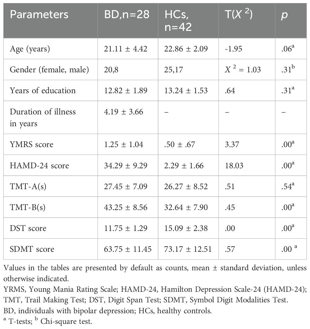
Exclusion criteria included reported comorbid intellectual disability and other psychiatric disorders, recent electroconvulsive therapy in the past six months, recent substance abuse or dependence in the past six months, and severe physical or organic diseases, such as cardiovascular or major orthopedic diseases, severe developmental disorders, and neurological diseases. This study included 70 participants; 28 were diagnosed with BD, and 42 were HCs. The demographic and clinical data of the participants are summarized in Table 1.
2.2 Experimental design and procedure
2.2.1 Neuropsychological testing
On the same morning of EEG acquisition, neurocognitive testing was conducted. These tests were completed by 28 BD patients and 42 HCs. A battery of neuropsychological tests was used to assess the following cognitive domains: (a) The Trail Making Test (TMT) is a neuropsychological test used to assess executive functions (EFs) (37). The test consists of two parts (A and B). TMT-A is typically considered a measure of visual search and processing speed, where participants must sequentially connect the numbers 1 to 25 as quickly as possible. The score is the time taken to complete the task (in seconds), with shorter times indicating better performance (38) (39). TMT-B is considered to more broadly assess psychological flexibility and executive functions (40–42). (b) The digit span test (DST) evaluates working memory and cognitive flexibility. It includes forward and backward tasks. The forward digit span test comprises a series of digits ranging from 2 to 12, which the examiner recites at a rate of one digit per second, and participants must repeat in the same order. The backward digit span test comprises digits ranging from 2 to 10, recited forward by the examiner, and participants must recite them backward. The score is the total sum of the last level of digits passed (43). (c) The symbol digit modalities test (SDMT) is an advanced form of the digit-symbol test that assesses attention, visual scanning, and motor speed. Individuals must identify nine symbols corresponding to the digits 1 to 9 and practice writing the correct number beneath each symbol. Then, they manually fill the blanks with the corresponding number under each symbol. Participants are given 90 s to complete the written test. The score was calculated by totaling the correct answers for each section (44).
2.2.2 EEG acquisition and electrophysiological recording
During EEG acquisition, participants sat quietly in a comfortable armchair and sequentially completed the following three conditions: 1) eyes closed, 2) eyes open, and 3) free viewing. eyes closed Participants sat in the chair for 3 min in the eyes closed condition. The instruction was, ‘Please close your eyes, relax, and do not think about anything.’ In the eyes open condition, participants sat in the chair for 3 min with the instruction, ‘Please open your eyes, relax, try to minimize blinking, and do not think about anything’. Under the free viewing condition, participants sat in the chair for 3 min and 20s with the instruction ‘Please view every action in the video, but do not make any behavioral movements’. The duration of the free viewing videos was 3 min and 20 s.
The EEG signals were recorded using a 32-channel system (Nicolet Monitor, USA) with an electrode cap, and recording was performed according to the 10–20 international system. The reference electrodes were placed midway between CPz and AFz. The recorded signals were digitized at a sampling frequency of 250 Hz, with impedance for all electrodes less than 10 kohm. EEG data were preprocessed and analyzed using MATLAB (R2022a; Mathworks, Natick, MA, USA) and EEGLAB v14.0. Initially, the EEG signals were re-referenced using average reference montage. Subsequently, bandpass filtering from 1 to 49 Hz and notch filtering at 50 Hz were applied to remove high-frequency and power-line noise contamination. Independent component analysis (ICA) was used to remove eye movement and blink artifacts. Next, EEG signals from each channel were segmented into non-overlapping epochs of 6 s. Power spectral density (PSD) and differential entropy (DE) features were computed for all segments across different frequency bands. Additionally, a Butterworth IIR bandpass filter was applied separately to data from all channels to obtain δ (1–4 Hz) and β (12–30 Hz) waves (zero-phase shift). Subsequently, the Hilbert transformation was used to obtain phase and amplitude information for each channel, from which debiased phase-amplitude cross-frequency coupling (dPAC) and amplitude-amplitude coupling (AAC) were calculated.
2.2.3 Phase Lag Index Functional Connectivity
The phase lag index (PLI), a measure of phase synchronization, was used to quantify the functional connectivity between each pair of EEG channels (45, 46):
where stands for the phase difference between channel a and b at frequency f and time t of trial m, M stands for the number of trials, and sgn indicates the sign (–1 for negative values, +1 for positive values, and 0 for zero values).
In this study, PLI values were calculated for three experimental paradigms using the aforementioned equation. PLI values were averaged across δ (1–4 Hz), θ (4–8 Hz), α (8–12 Hz), β (12–30 Hz), and γ (> 30 Hz) frequency bands (47) and within a time window of 0.1 to 11 s to derive a weighted 19×19 functional connectivity matrix, serving as a candidate feature for each participant. PLI was calculated for 19 electrodes representing brain regions (FP1, FP2, F3, F4, F7, F8, FZ, T7, T8, P7, P8, C3, C4, CZ, P3, P4, PZ, O1, and O2). The PLI represents the connection strength between electrode pairs, ranging between 0 and 1, with higher values indicating stronger nonzero phase locking (48).
2.2.4 Features and correlations
In this study, EEG signals were decomposed into functionally distinct frequency bands (δ, θ, α, β, and γ). PSD for each band, DE for each band, as well as δ dPAC and AAC of δ and β bands, were computed. Twelve features were extracted in this study. To ascertain the relationship between EEG signal and cognitive function, Spearman correlation analysis was conducted to examine the correlations between TMT-A, TMT-B, DST, and SDMT scores and 12 significant features under the three experimental paradigm conditions. Correlations were deemed significant at p< 0.05.
2.2.5 Classification
Based on these features, we employed six classifiers, including ensemble, K-nearest neighbours (KNN), Naive Bayes, Random Forest, support vector machine (SVM), and decision tree (DT), to evaluate the BD identification performance under the eyes closed, eyes open, and free viewing experimental paradigms. The effectiveness of BD identification was measured using accuracy and F1 score. In this study, the selected classifiers are classical machine learning classifiers, extensively applied in depression recognition research (49).
● Ensemble, an ensemble learning algorithm, uses AdaBoost M1 to train weak classifiers (usually decision tree stumps or single-layer decision trees). This method combines their results by weighting them to construct a more robust classifier. The basic learner parameters were set to decision trees, with the number of basic learners fixed at 100 (50, 51).
● K-nearest neighbours(KNN) computed the distance between a test sample and all training samples, typically using the Euclidean distance metric. The prediction of the test sample’s category was determined by a majority vote among the K closest training samples (52). The parameter K of KNN was fixed constant 5 (53).
● Naive Baye is a simple yet effective classifier based on Bayes’ theorem. It calculated the class conditional and prior probabilities, deriving the posterior probabilities. The class with the highest posterior probability was selected as the prediction (54).
● Random Forest mitigates the risk of overfitting by integrating multiple decision trees. It performs well with high-dimensional and large datasets, requiring minimal parameter tuning (55).
● The SVM was introduced by (56). Its basic idea is to classify samples by finding the hyperplane with the largest distance between samples. The regularization parameter C of the Linear-SVM was empirically set to 0.25 (57).
● DT is a supervised learning classifier based on a tree structure. They construct recursive decision rules from training samples and predict the categories of test samples according to these trained rules (58).
2.3 Statistical analysis
Statistical analysis was performed using SPSS (v25.0) and MATLAB. Continuous variables were presented as mean ± standard deviation, and categorical variables were denoted as numbers. T-tests and Pearson’s chi-square tests were utilized to compare the baseline characteristics between the two groups. The connectivity strength between the two groups was compared using nonparametric Wilcoxon signed-rank tests with False Discovery Rate (FDR) correction to control for multiple comparison issues. Uncorrected p-values as ‘p,’ and FDR corrected p-values were presented as ‘pFDR.’ The independent samples t-test was employed to assess the significance of differences in features between BD and HC groups (utilizing the MATLAB function ttest2). The MATLAB ttest2 function facilitated this comparison, allowing for a robust assessment of the significance level. Additionally, correlation analysis was conducted to investigate the relationship between EEG features and cognitive assessment scores, with Spearman’s correlation analysis (implemented using the MATLAB function corr), providing a reliable measure of the strength and direction of the relationship between the variables of interest. p< 0.05 was considered a statistically significant difference.
3 Results
3.1 Demographic characteristics
Table 1 depicts the demographic and clinical characteristics of the study sample. There were no statistically significant differences between the BD group and the HCs regarding age, gender, or years of education. The YMRS scores of BD and HC groups were< 5 points. The TMT-A scores did not differ significantly between the BD group and the HCs. However, TMT-B, DST, and SDMT scores differed statistically significantly.
3.2 Brain functional connectivity
PLI was computed for all electrode pairs in the δ, θ, α, β, and γ bands under the three experimental paradigms. Figures 1a, b illustrate the group averaged PLI functional connectivity patterns in the δ, θ, α, β, and γ frequency bands for BD patients and HCs under the eyes closed experimental paradigm condition. We observed significant differences in the functional connectivity between the two groups across all electrode pairs in the δ, θ, β, and γ frequency bands (PFDR< 0.05), the BD group demonstrating increased functional connectivity compared to HCs. A qualitative observation in the δ frequency band revealed that BD patients exhibited stronger long-range connections between the frontal and occipital cortical regions compared to HCs (PFDR< 0.05). In the θ frequency band, BD patients showed more short-range connections between the parietal and occipital cortical regions and fewer long-range connections between the frontal and the parietal/occipital cortical regions compared to HCs (PFDR< 0.05). In the β frequency band, there were fewer long-range connections between the frontal and parietal/occipital cortical regions in BD patients relative to HCs (PFDR< 0.05). In the γ frequency band, BD patients displayed stronger long-range connections between the frontal cortical regions and the visual cortex, and short-range connections across hemispheres than HCs (PFDR< 0.05). No significant effects of functional connectivity between the two groups were observed in the α frequency band (PFDR > 0.05).
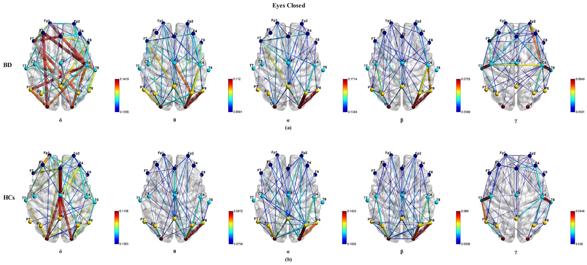
Figure 1. The visualization of the group average PLI functional connectivity for BD (a) and HCs (b) under the eyes closed experimental paradigm across five frequency bands (δ, θ, α, β, and γ), with a sparsity of 0.3.
Figure 2 illustrates the group average PLI functional connectivity patterns for BD patients (A) and HCs (B) under the eyes open paradigm condition. However, under the eyes open paradigm condition, when comparing the group average PLI across the δ, θ, α, β, and γ frequency bands, no significant differences were found between any pairs of electrode connections (PFDR > 0.05).
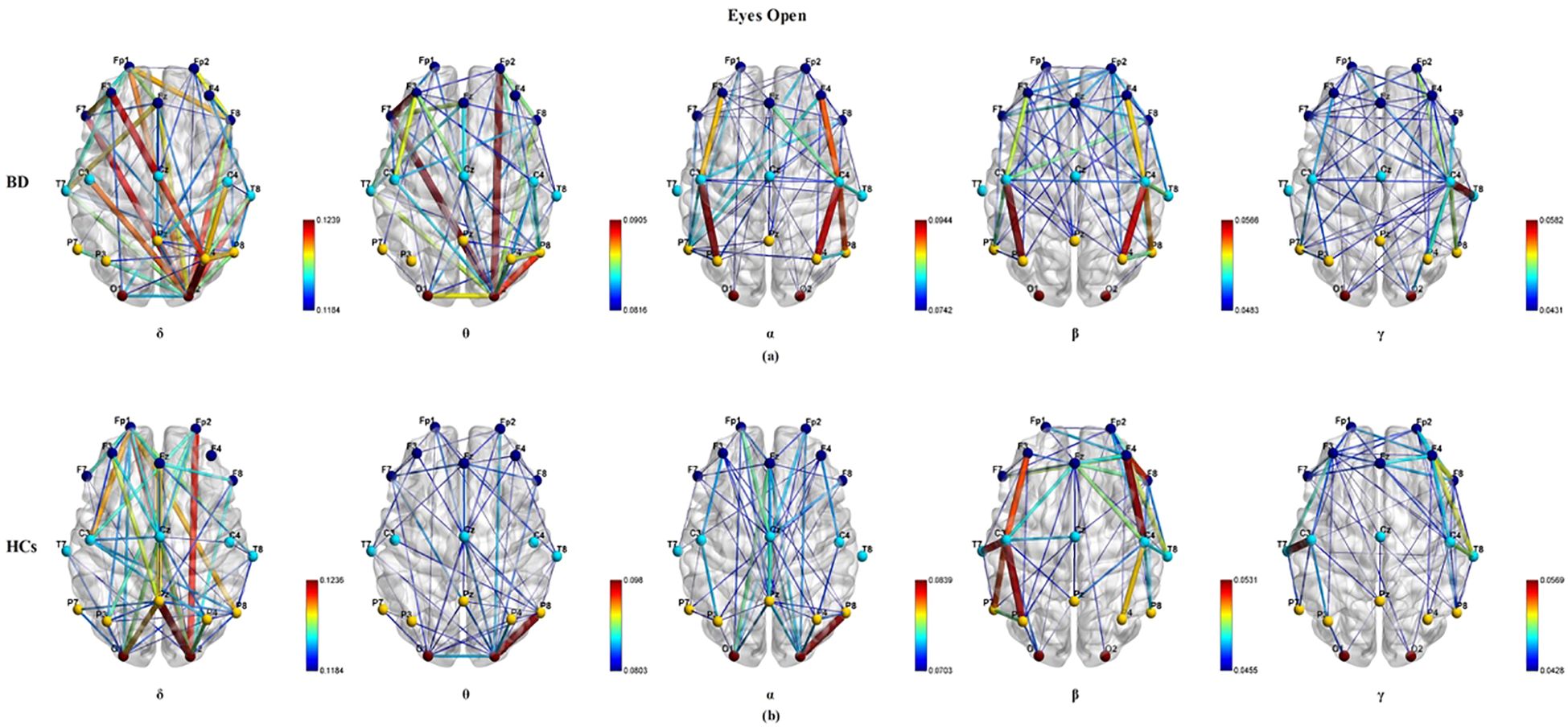
Figure 2. The visualization of the group average PLI functional connectivity for BD (a) and HCs (b) under the eyes open experimental paradigm across five frequency bands (δ, θ, α, β, and γ), with a sparsity of 0.3.
Figure 3 illustrates the group average PLI functional connectivity patterns for BD patients (a) and HCs (b) under the free viewing paradigm condition. In δ frequency band, BD patients exhibited fewer and weaker long-range connections between the frontal cortical regions and parietal/occipital cortical regions than HCs (PFDR< 0.05). In the θ frequency band, BD patients showed fewer and weaker long-range and short-range connections than HCs (PFDR< 0.05). Functional connectivity between the two groups had no significant effects on the α, β, and γ frequency bands (PFDR > 0.05). Within the three classical EEG experimental paradigms, the eyes closed paradigm exhibited significantly distinct PLI functional connectivity patterns across a broader range of frequency bands when comparing BD patients with HCs.
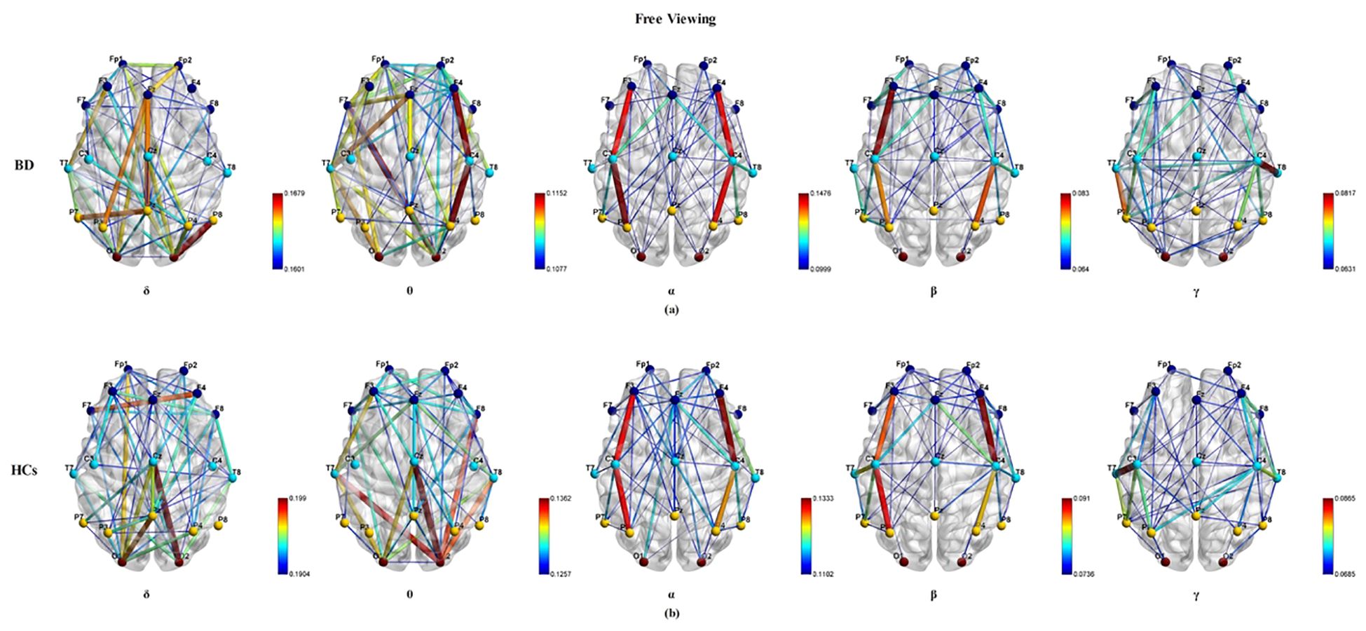
Figure 3. The visualization of the group average PLI functional connectivity for BD (a) and HCs (b) under the free viewing experimental paradigm across five frequency bands (δ, θ, α, β, and γ), with a sparsity of 0.3.
3.3 Significance analysis of features
Significant differences in the δ and β frequency bands were observed across all three classical experimental paradigms. Under the eyes closed paradigm, significant distinctions were primarily concentrated in the δ band (frontal, parietal, and occipital lobes), β band (parietal lobe), DE-δ (frontal, parietal, occipital, and temporal lobes), and DE-β (parietal lobe). In the eyes open paradigm, the features showing significant differences between BD patients and HCs were more dispersed across the δ, θ, α, β, and γ frequency bands. Within the free viewing paradigm, the significant distinctions were relatively scattered in the δ (frontal, parietal, and occipital lobes), θ (occipital lobe), α (occipital lobe), and β (parietal lobe) frequency bands. All three paradigms revealed notable differences in the δ band (frontal, parietal, and occipital lobes) between BD patients and HCs. However, the choice of paradigm influenced the observed outcomes in θ, α, β, γ, DE-θ, DE-α, DE-β, DE-γ, AAC, and dPAC features (Table 2).
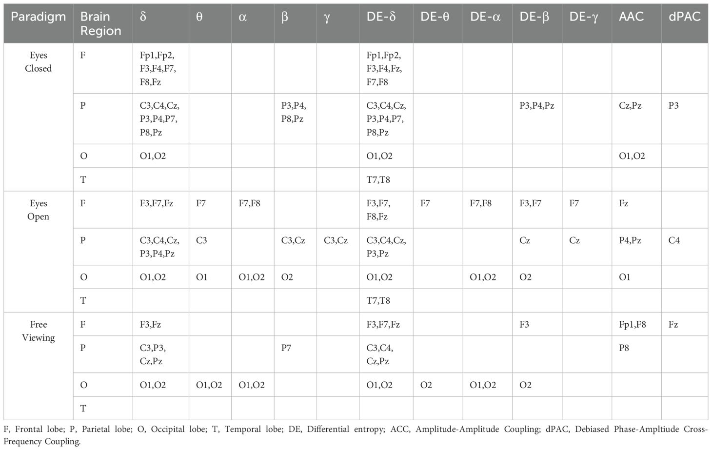
Table 2. The electrodes significantly differ across the 12 features under the three experimental paradigms.
3.4 Correlation with cognitive function
TMT-A scores exhibited more positive correlations with θ and α PSD, DE-θ, and DE-α features across three experimental paradigms. TMT-B scores showed more positive correlations with δ and θ PSD and DE- δ, and DE-θ features across three experimental paradigms. SDMT scores displayed negative correlations with δ and θ PSD, DE-δ, and DE-θ features across three experimental paradigms. DST scores exhibited negative correlations with δ PSD and DE-δ features across three experimental paradigms (Figure 4).
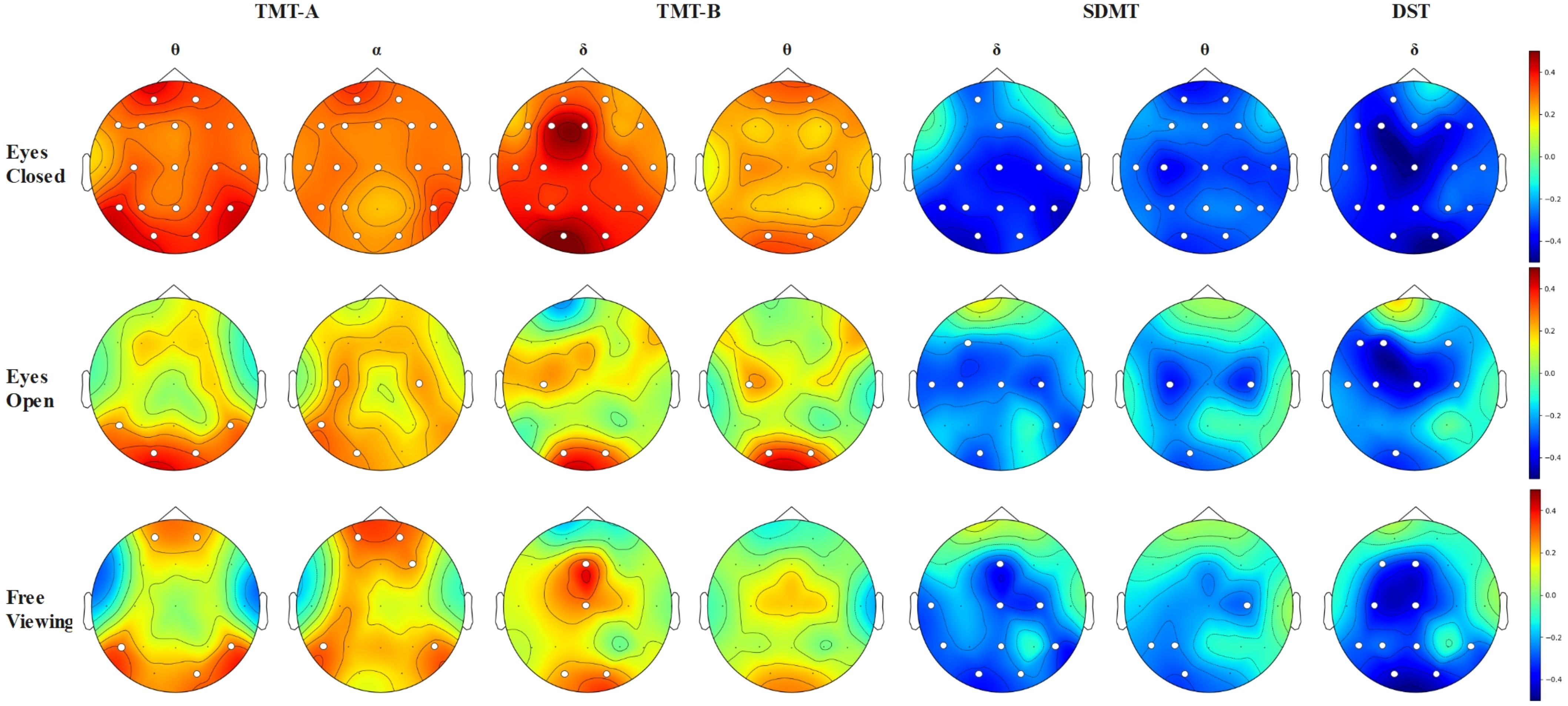
Figure 4. Topographical maps visualize the significant correlations between TMT-A, TMT-B, SDMT, and DST scores and features across different EEG frequency bands under various experimental paradigms. The white dots represent the electrode positions with significant differences.
We also observed differences in the number of significant features correlated with cognitive scores across the three experimental paradigms, with the highest number observed in the eyes closed paradigm and slightly fewer in the eyes open paradigm than in the free viewing paradigm. More brain regions showed significant correlations between EEG features and cognitive scores in the eyes closed paradigm compared to the eyes open or free viewing paradigms.
3.5 Classification
Table 3 presents the results of BD recognition using six classifiers under three different experimental paradigms. Compared to the eyes open and free viewing paradigms, the eyes closed paradigm achieved the highest accuracy and F1 score in identifying BD. Under the eyes closed paradigm, the random forest classifier yielded the highest recognition accuracy of 79.43%, with F1 score of 76.82%. For the eyes open paradigm, the ensemble learning classifier demonstrated the best BD identification performance, achieving a recognition accuracy of 75.85% and F1 score of 71.28%. Similarly, the ensemble learning classifier achieved the best BD identification performance under the free viewing paradigm, with a recognition accuracy of 76.03% and F1 score of 72.6%.
4 Discussion
To our knowledge, this study represents the first attempt to recognize bipolar disorder by analyzing EEG signals using different experimental paradigms. We investigated the brain electrical activity of two groups of patients, BD and HCs subjects, under the conditions of eyes closed, eyes open, and free viewing experimental paradigms. Our research findings suggest that the eyes closed experimental paradigm may offer the highest discriminative power regarding brain functional connectivity analysis, cognitive function correlation analysis, and classification performance. There was no clear distinction between the free viewing and eyes open experimental paradigms regarding their superiority, which could be attributed to the specific free viewing paradigm adopted in our study.
First, in the eyes closed paradigm, the BD group exhibited more significant PLI frequency bands than the HCs group, indicating more pronounced differences in EEG signals under this condition. The BD group exhibited a higher group average PLI in the δ, θ, β, and γ frequency bands than the HCs under the eyes closed condition. A study has found increased coherence in the frontal and occipital cortices, particularly in the frontal regions, presenting more diffuse long-range brain connections than HCs (21), aligning with our findings. Another study revealed that patients with depression have enhanced PLI during sleep (59). This increased neural synchrony pattern may reflect network activity in a resting state. Excessive resting-state EEG activity has been described in bipolar affective disorder and may be related to higher depression levels (60). In the eyes open experimental paradigm, no significant differences were observed in the PLI across all frequency bands between the two groups. A study investigating the recovery of brain function impairments in young bipolar disorder patients during a stable emotional period revealed that under an eyes open experimental paradigm, these patients exhibited heightened activity across all frequencies (δ, θ, α, and β), suggesting deficits in visual-spatial processing (61). Similarly, another study aimed at distinguishing between female attention deficit hyperactivity disorder and female bipolar disorder patients found that the absolute θ power was higher in the bipolar disorder group than in the HCs group under an eyes open paradigm. However, no increase in absolute θ power was noted in the bipolar disorder group during a connect-the-dots task (62). These results differ from ours, potentially due to differences in study subjects and control groups, necessitating further research. There were fewer and weaker δ and θ connections than HCs under a free viewing paradigm. Previous studies using a stimulus-task paradigm have shown that at the onset of the stimulus, HCs had a higher PLI than bipolar disorder patients in the θ and α bands, with the PLI of bipolar disorder patients increasing later. However, connectivity in the β and γ bands only showed insignificant changes, indicating faster brain responses in HCs than in bipolar disorder patients (63). Another study using a self-referential memory (SRM) paradigm found that during self and other referential processing, HCs activated more brain connectivity regions than bipolar disorder patients and HCs had a higher PLI than bipolar disorder patients at the onset of the stimulus, suggesting that bipolar disorder patients ‘s self-cognition is impeded in the transmission and integration of interhemispheric information (48). Different free viewing paradigm contents might result in variations in functional connectivity across different frequencies. However, regardless of the paradigm, these consistently indicate bipolar disorder patients’ difficulties in attention, memory, and the integration of other brain resources. Our study revealed that the eyes closed paradigm could discern more functional connectivity differences between the two groups, whereas the free viewing experimental paradigm demonstrated fewer differences than those observed in the eyes closed paradigm between the groups across frequency bands.
Second, we conducted a significance analysis of the 12 extracted features and found that significant differences in the δ and β frequency bands were consistently observed across all three classical experimental paradigms. In the eyes-closed paradigm, notable differences were primarily observed in the δ band (frontal, parietal, and occipital lobes), β band (parietal lobe), DE-δ (frontal, parietal, occipital, and temporal lobes), and DE-β (parietal lobe). A study discovered that δ-β cross-frequency coupling may reflect the neural projection of the physiopathology of bipolar disorder; significant increases in dPAC and AAC were found in FP 2, with a linear correlation between dPAC in F3 and Mood Disorder Questionnaire scores were observed. Initially, P3’s dPAC was only related to Hamilton Depression Rating Scale scores (64). Our eyes closed experimental paradigm revealed significant differences in P3’s dPAC, consistent with these findings. Other studies also identified changes in the power of δ, β, and θ waves (65) Our analysis of 12 extensively used EEG features revealed that the selection of experimental paradigm affects the results of the significance analysis for observed features. The majority of EEG features (θ, α, β, γ, DE-θ, DE-α, DE-β, DE-γ, AAC, and dPAC) demonstrated variability in differences across the paradigms. And the brain regions exhibiting significant feature differences across different paradigms. Thus, the selection of a consistent and efficient experimental paradigm is crucial for researchers in exploring biomarkers for bipolar depression.
Third, this study found thatθ and α frequency bands positively correlated with scores on the TMT-A test.δ and θ frequency bands show positive correlations with TMT-B test scores and negative correlations with DST test scores.δ frequency band also negatively correlated with SDMT test scores. A study on the spontaneous α activity and visually evoked α responses in patients with bipolar disorder revealed a reduced spontaneous EEG α activity and a marked decrease in evoked α responses. These findings may be associated with the cognitive deficits characteristic of bipolar disorder (66). Prior research supports increased δ synchronization, such as heightened EEG δ activity in bipolar disorder than HCs, confirming the potential for poorer cognitive flexibility and executive function in patients with BD (67, 68). δ oscillations are crucial for cognitive functions related to focused attention, signal detection, recognition, and decision-making, and diminished δ responses may be a common feature of cognitive dysfunction in neuropsychiatric disorders (69) (70). Similarly, this study found that diminished δ responses were negatively correlated with the DST and SDMT scores. Another study indicated that reducing activity in the central θ band might help diminish cognitive control and maladaptive behavioral responses in patients with BD. The changes in the θ band observed in this study are consistent with these findings (C. M. 71).
Finally, we employed multiple widely used classical machine learning classifiers to assess the performance of three experimental paradigms in BD identification tasks. A review indicates that most studies on bipolar disorder have utilized classical machine learning models, such as SVM, Random Forest, and KNN, with 24 studies reporting accuracies ranging from 64% to 98% for machine learning models in BD identification tasks (72). The classifiers encompassed these mainstream machine learning models to ensure the reliability and comparability of our research findings. We found that all six classifiers achieved optimal classification performance under the eyes closed experimental paradigm, indicating the superiority of the eyes closed paradigm over the eyes open and free viewing paradigms (based on action observations) in BD recognition tasks. Moreover, the random forest classifier under the eyes closed paradigm exhibited the best classification performance, with a recognition accuracy of 79.43% and an F1 score of 76.82%.
5 Limitation
This study’s primary limitations were the small sample size and the fact that not all patients were medication-free. Another significant limitation is that our free viewing paradigm did not employ an emotional paradigm involving fear, anger, sadness, or other paradigms, which may have led to different results. Future research may explore the content of the free viewing paradigm in more detail.
6 Conclusion
Our study systematically analyzed the performance differences between the eyes closed, eyes open, and free viewing experimental paradigms in the BD identification task. We found that the eyes closed paradigm exhibited significant advantages over the eyes open and free viewing paradigms regarding feature significance, cognitive relevance, and classifier recognition accuracy. However, there was no significant differences between the eyes open and free viewing paradigms, possibly due to the use of the action observation-based free viewing paradigm. Other paradigms based on emotion induction and cognitive function deserve further exploration. Our findings suggest that the eyes closed paradigm, a simple and feasible experimental paradigm, holds greater clinical significance in EEG analysis for BD identification than the eyes open and action observation-based free viewing paradigms. Future research should investigate additional experimental paradigms designed based on emotional bias and cognitive function decline characteristics, providing a more diverse range of experimental paradigm references for BD identification studies.
Data availability statement
The original contributions presented in the study are included in the article/supplementary material. Further inquiries can be directed to the corresponding authors.
Ethics statement
The studies involving humans were approved by The Ethics Committee of the Fifth Affiliated Hospital of Sun Yat-sen University. The studies were conducted in accordance with the local legislation and institutional requirements. The participants provided their written informed consent to participate in this study.
Author contributions
CY: Investigation, Methodology, Software, Writing – original draft. YP: Formal Analysis, Methodology, Writing – review & editing. WJW: Data curation, Investigation, Writing – review & editing. YH: Data curation, Writing – review & editing. NT: Investigation, Writing – review & editing. HW: Resources, Writing – review & editing. SLW: Funding acquisition, Supervision, Writing – review & editing.
Funding
The author(s) declare that financial support was received for the research and/or publication of this article. The work was supported by General Logistics Department, People’s Liberation Army (BWS19J012).
Acknowledgments
We sincerely thank all participants for their voluntary participation. We thank the doctors of The Fifth Affiliated Hospital of Sun Yat-sen University for their assistance. Finally, we thank our research colleagues for their help in this research and BrainNet Viewer toolbox (http://www.nitrc.org/project/bnv/).
Conflict of interest
The authors declare that the research was conducted in the absence of any commercial or financial relationships that could be construed as a potential conflict of interest.
Generative AI statement
The author(s) declare that no Generative AI was used in the creation of this manuscript.
Publisher’s note
All claims expressed in this article are solely those of the authors and do not necessarily represent those of their affiliated organizations, or those of the publisher, the editors and the reviewers. Any product that may be evaluated in this article, or claim that may be made by its manufacturer, is not guaranteed or endorsed by the publisher.
References
1. Bauer MS. Bipolar disorder. Ann Internal Med. (2022) 175:ITC97–ITC112. doi: 10.7326/AITC202207190. American College of Physicians.
2. Merikangas KR, Jin R, He JP, Kessler RC, Lee S, Sampson NA, et al. Prevalence and correlates of bipolar spectrum disorder in the World Mental Health Survey Initiative. Arch Gen Psychiatry. (2011) 68:241–51. doi: 10.1001/archgenpsychiatry.2011.12
3. Miskowiak KW, Burdick KE, Martinez-Aran A, Bonnin CM, Bowie CR, Carvalho AF, et al. Assessing and addressing cognitive impairment in bipolar disorder: the International Society for Bipolar Disorders Targeting Cognition Task Force recommendations for clinicians. In: Bipolar Disorders, vol. 20. Malden, MA: Blackwell Publishing Inc (2018). p. 184–94. doi: 10.1111/bdi.12595
4. Kupferschmidt DA and Zakzanis KK. Toward a functional neuroanatomical signature of bipolar disorder: Quantitative evidence from the neuroimaging literature. Psychiatry Res Neuroimaging. (2011) 193:71–9. doi: 10.1016/j.pscychresns.2011.02.011
5. GBD 2019 Mental Disorders Collaborators. Global, regional, and national burden of 12 mental disorders in 204 countries and territories 1990–2019: a systematic analysis for the Global Burden of Disease Study 2019. Lancet Psychiatry. (2022) 9:137–50. doi: 10.1016/S2215-0366(21)00395-3
6. Cloutier M, Greene M, Guerin A, Touya M, and Wu E. The economic burden of bipolar I disorder in the United States in 2015. J Affect Disord. (2018) 226:45–51. doi: 10.1016/j.jad.2017.09.011
7. Vellante F, Ferri F, Baroni G, Croce P, Migliorati D, Pettoruso M, et al. Euthymic bipolar disorder patients and EEG microstates: a neural signature of their abnormal self experience? J Affect Disord. (2020) 272:326–34. doi: 10.1016/j.jad.2020.03.175
8. Frangou S. Neuroimaging markers of risk, disease expression, and resilience to bipolar disorder. Curr Psychiatry Rep. (2019) 21:52. doi: 10.1007/s11920-019-1039-7
9. Yasin S, Hussain SA, Aslan S, Raza I, Muzammel M, and Othmani A. EEG based Major Depressive disorder and Bipolar disorder detection using Neural Networks:A review. In: Computer Methods and Programs in Biomedicine, vol. 202. Shannon, Ireland: Elsevier Ireland Ltd (2021). doi: 10.1016/j.cmpb.2021.106007
10. O’Gorman RL, Poil SS, Brandeis D, Klaver P, Bollmann S, Ghisleni C, et al. Coupling between resting cerebral perfusion and EEG. Brain Topography. (2013) 26:442–57. doi: 10.1007/s10548-012-0265-7
11. Wada M, Kurose S, Miyazaki T, Nakajima S, Masuda F, Mimura Y, et al. The P300 event-related potential in bipolar disorder: A systematic review and meta-analysis. J Affect Disord. (2019) 256:234–49. doi: 10.1016/j.jad.2019.06.010. Elsevier B.V.
12. Maggioni E, Bianchi AM, Altamura AC, Soares JC, and Brambilla P. The putative role of neuronal network synchronization as a potential biomarker for bipolar disorder: A review of EEG studies. J Affect Disord. (2017) 212:167–70. doi: 10.1016/j.jad.2016.12.045
13. Mateo-Sotos J, Torres AM, Santos JL, Quevedo O, and Basar C. A machine learning-based method to identify bipolar disorder patients. Circuits Systems Signal Process. (2022) 41:2244–65. doi: 10.1007/s00034-021-01889-1
14. Abi-Dargham A, Moeller SJ, Ali F, DeLorenzo C, Domschke K, Horga G, et al. Candidate biomarkers in psychiatric disorders: state of the field. In: World Psychiatry, vol. 22. Hoboken, NJ: John Wiley and Sons Inc (2023). p. 236–62. doi: 10.1002/wps.21078
15. Koller-Schlaud K, Ströhle A, Bärwolf E, Behr J, and Rentzsch J. EEG frontal asymmetry and theta power in unipolar and bipolar depression. J Affect Disord. (2020) 276:501–10. doi: 10.1016/j.jad.2020.07.011
16. Erguzel TT, Sayar GH, and Tarhan N. Artificial intelligence approach to classify unipolar and bipolar depressive disorders. Neural Computing Appl. (2016) 27:1607–16. doi: 10.1007/s00521-015-1959-z
17. Li X, La R, Wang Y, Niu J, Zeng S, Sun S, et al. EEG-based mild depression recognition using convolutional neural network. Med Biol Eng Computing. (2019) 57:1341–52. doi: 10.1007/s11517-019-01959-2
18. Zhu J, Wei S, Xie X, Yang C, Li Y, Li X, et al. Content-based multiple evidence fusion on EEG and eye movements for mild depression recognition. Comput Methods Programs Biomed. (2022) 226:107100. doi: 10.1016/j.cmpb.2022.107100
19. Clementz B, Sponheim S, Lacono W, and Beiser M. Resting EEG in first-episode schizophrenia patients, bipolar psychosis patients, and their first-degree relatives. Psychophysiology. (1994) 31:486–94. doi: 10.1111/j.1469-8986.1994.tb01052.x
20. Kam JWY, Bolbecker AR, O’Donnell BF, Hetrick WP, and Brenner CA. Resting state EEG power and coherence abnormalities in bipolar disorder and schizophrenia. J Psychiatr Res. (2013) 47:1893–901. doi: 10.1016/j.jpsychires.2013.09.009
21. Barttfeld P, Petroni A, Báez S, Urquina H, Sigman M, Cetkovich M, et al. Functional connectivity and temporal variability of brain connections in adults with attention deficit/hyperactivity disorder and bipolar disorder. Neuropsychobiology. (2014) 69:65–75. doi: 10.1159/000356964
22. Narayanan B, O’Neil K, Berwise C, Stevens MC, Calhoun VD, Clementz BA, et al. Resting state electroencephalogram oscillatory abnormalities in schizophrenia and psychotic bipolar patients and their relatives from the bipolar and schizophrenia network on intermediate phenotypes study. Biol Psychiatry. (2014) 76:456–65. doi: 10.1016/j.biopsych.2013.12.008
23. Ghanizadeh A-Z, Al M, Moeini M, Khaleghi A, and Mohammadi MR. Characteristics of alpha band frequency in adolescents with bipolar II disorder: A resting-state QEEG study. Iranian J Psychiatry. (2015) 10(1):8–12.
24. Khaleghi A, Mohammadi MR, Moeini M, Zarafshan H, and Fadaei Fooladi M. Abnormalities of alpha activity in frontocentral region of the brain as a biomarker to diagnose adolescents with bipolar disorder. Clin EEG Neurosci. (2019) 50:311–8. doi: 10.1177/1550059418824824
25. Velasques B, Bittencourt J, Diniz C, Teixeira S, Basile LF, Inácio Salles J, et al. Changes in saccadic eye movement (SEM) and quantitative EEG parameter in bipolar patients. J Affect Disord. (2013) 145:378–85. doi: 10.1016/j.jad.2012.04.049
26. Fox NA, Bakermans-Kranenburg MJ, Yoo KH, Bowman LC, Cannon EN, Vanderwert RE, et al. Assessing human mirror activity with EEG mu rhythm: A meta-analysis. psychol Bull. (2016) 142:291–313. doi: 10.1037/bul0000031
27. Lin NH, Liu CH, Lee P, Guo LY, Sung JL, Yen CW, et al. Backward walking induces significantly larger upper-mu-rhythm suppression effects than forward walking does. Sensors (Switzerland). (2020) 20:1–18. doi: 10.3390/s20247250
28. Kim E, Jung YC, Ku J, Kim JJ, Lee H, Kim SY, et al. Reduced activation in the mirror neuron system during a virtual social cognition task in euthymic bipolar disorder. Prog Neuropsychopharmacol Biol Psychiatry. (2009) 33:1409–16. doi: 10.1016/j.pnpbp.2009.07.019
29. Andrews SC, Enticott PG, Hoy KE, Thomson RH, and Fitzgerald PB. Reduced mu suppression and altered motor resonance in euthymic bipolar disorder: Evidence for a dysfunctional mirror system? Soc Neurosci. (2016) 11:60–71. doi: 10.1080/17470919.2015.1029140
30. Cuellar ME and del Toro CM. Time-frequency analysis of Mu rhythm activity during picture and video action naming tasks. Brain Sci. (2017) 7(9):114. doi: 10.3390/brainsci7090114
31. Umla-Runge K, Zimmer HD, Fu X, and Wang L. An action video clip database rated for familiarity in China and Germany. Behav Res Methods. (2012) 44:946–53. doi: 10.3758/s13428-012-0189-x
32. Painold A, Faber PL, Milz P, Reininghaus EZ, Holl AK, Letmaier M, et al. Brain electrical source imaging in manic and depressive episodes of bipolar disorder. Bipolar Disord. (2014) 16:690–702. doi: 10.1111/bdi.12198
33. Shehzad Z, Kelly AMC, Reiss PT, Gee DG, Gotimer K, Uddin LQ, et al. The resting brain: Unconstrained yet reliable. Cereb Cortex. (2009) 19:2209–29. doi: 10.1093/cercor/bhn256
34. Newson JJ and Thiagarajan TC. EEG frequency bands in psychiatric disorders: A review of resting state studies. Front Hum Neurosci. (2019) 12:521. doi: 10.3389/fnhum.2018.00521. Frontiers Media S.A.
35. Hamilton M. A rating scale for depression. J Neurol Neurosurg Psychiat. (1960) 23:56–62. doi: 10.1037/t04100-000
36. Young RC, Biggs JT, Ziegler VE, and Meyer DA. A rating scale for mania: reliability, validity and sensitivity. Brit J Psychiat. (1978) 133(5):429–35. doi: 10.1192/bjp.133.5.429
37. Linari I, Juantorena GE, Ibáñez A, Petroni A, and Kamienkowski JE. Unveiling Trail Making Test: visual and manual trajectories indexing multiple executive processes. Sci Rep. (2022) 12(1):14265. doi: 10.1038/s41598-022-16431-9
38. Sánchez-Cubillo I, Periáñez JA, Adrover-Roig D, Rodríguez-Sánchez JM, Ríos-Lago M, Tirapu J, et al. Construct validity of the Trail Making Test: Role of task-switching, working memory, inhibition/interference control, and visuomotor abilities. J Int Neuropsychol Soc. (2009) 15:438–50. doi: 10.1017/S1355617709090626. Cambridge University Press.
39. Misdraji EL and Gass CS. The Trail Making Test and its neurobehavioral components. J Clin Exp Neuropsychol. (2010) 32:159–63. doi: 10.1080/13803390902881942
40. Arbuthnott K and Frank J. Trail making test, part B as a measure of executive control: validation using a set-switching paradigm. J Clin Exp Neuropsychol. (2000) 22:518–28. doi: 10.1076/1380-3395(200008)22:4;1-0;FT518
41. Kortte KB, Horner MD, and Windham WK. The trail making test, Part B: Cognitive flexibility or ability to maintain set? Appl Neuropsychol. (2002) 9:106–9. doi: 10.1207/S15324826AN0902_5
42. Tombaugh TN. Trail Making Test A and B: Normative data stratified by age and education. Arch Clin Neuropsychol. (2004) 19:203–14. doi: 10.1016/S0887-6177(03)00039-8
43. Simard M and van Reekum R. Memory assessment in studies of cognition-enhancing drugs for alzheimer??s disease. Drugs Aging. (1999) 14:197–230. doi: 10.2165/00002512-199914030-00004
44. Sheridan LK, Fitzgerald HE, Adams KM, Nigg JT, Martel MM, Puttler LI, et al. Normative Symbol Digit Modalities Test performance in a community-based sample. Arch Clin Neuropsychol. (2006) 21:23–8. doi: 10.1016/j.acn.2005.07.003
45. Stam CJ, Nolte G, and Daffertshofer A. Phase lag index: Assessment of functional connectivity from multi channel EEG and MEG with diminished bias from common sources. Hum Brain Mapp. (2007) 28:1178–93. doi: 10.1002/hbm.20346
46. Zhao Z, Li J, Niu Y, Wang C, Zhao J, Yuan Q, et al. Classification of schizophrenia by combination of brain effective and functional connectivity. Front Neurosci. (2021) 15:651439. doi: 10.3389/fnins.2021.651439
47. Canolty RT and Knight RT. The functional role of cross-frequency coupling. Trends Cogn Sci. (2010) 14:506–15. doi: 10.1016/j.tics.2010.09.001
48. Zhang J, Liu T, Shi Z, Tan S, Suo D, Dai C, et al. Impaired self-referential cognitive processing in bipolar disorder: A functional connectivity analysis. Front Aging Neurosci. (2022) 14:754600. doi: 10.3389/fnagi.2022.754600
49. Khosla A, Khandnor P, and Chand T. Automated diagnosis of depression from EEG signals using traditional and deep learning approaches: A comparative analysis. In: Biocybernetics and Biomedical Engineering, vol. 42. Amsterdam, Netherlands: Elsevier B.V (2022). p. 108–42. doi: 10.1016/j.bbe.2021.12.005
50. All KM and Pazzani MJ. Error Reduction through Learning Multiple Descriptions, Vol. 24. Netherlands: Kluwer Academic Publishers, Boston. (1996) 24:173–202.
51. Freund Y and Schapire RE. A decision-theoretic generalization of on-line learning and an application to boosting, vol. 55 San Diego, CA, United States: Journal of Computer and System Sciences. (1997) p. 119–39.
52. Altman NS. An introduction to kernel and nearest-neighbor nonparametric regression. In: Source: The American Statistician, vol. 46. (1992) p. 175–85.
53. Zheng WL, Zhu JY, and Lu BL. Identifying stable patterns over time for emotion recognition from eeg. IEEE Trans Affect Computing. (2019) 10:417–29. doi: 10.1109/TAFFC.2017.2712143
54. Domingos P and Pazzani M. On the Optimality of the Simple Bayesian Classifier under Zero-One Loss Vol. 29. Dordrecht, Netherlands: Kluwer Academic Publishers (1997).
55. Martin D. Buhmann Justus-Liebig University GG. Encyclopedia of machine learning and data mining. In: Encyclopedia of Machine Learning and Data Mining. Springer, US (2017). doi: 10.1007/978-1-4899-7687-1
56. Cortes C, Vapnik V, and Saitta L. Support-vector networks editor. In: Machine Leaming, vol. 20. Dordrecht, Netherlands: Kluwer Academic Publishers (1995).
57. Miranda-Correa JA, Abadi MK, Sebe N, and Patras I. AMIGOS: A dataset for affect, personality and mood research on individuals and groups. IEEE Trans Affect Computing. (2021) 12:479–93. doi: 10.1109/TAFFC.2018.2884461
58. Wu X, Kumar V, Ross QJ, Ghosh J, Yang Q, Motoda H, et al. Top 10 algorithms in data mining. Knowledge Inf Syst. (2008) 14:1–37. doi: 10.1007/s10115-007-0114-2
59. Lian J, Song Y, Zhang Y, Guo X, Wen J, and Luo Y. Characterization of specific spatial functional connectivity difference in depression during sleep. J Neurosci Res. (2021) 99:3021–34. doi: 10.1002/jnr.24947
60. Pomarol-Clotet E, Moro N, Sarró S, Goikolea JM, Vieta E, Amann B, et al. Failure of de-activation in the medial frontal cortex in mania: Evidence for default mode network dysfunction in the disorder. World J Biol Psychiatry. (2012) 13:616–26. doi: 10.3109/15622975.2011.573808
61. El-Badri SM, Ashton CH, Moore PB, Marsh VR, and Ferrier IN. Electrophysiological and cognitive function in young euthymic patients with bipolar affective disorder. Bipolar Disord. (2001) 3:79–87. doi: 10.1034/j.1399-5618.2001.030206.x
62. Rommel AS, Kitsune GL, Michelini G, Hosang GM, Asherson P, McLoughlin G, et al. Commonalities in EEG spectral power abnormalities between women with ADHD and women with bipolar disorder during rest and cognitive performance. Brain Topography. (2016) 29:856–66. doi: 10.1007/s10548-016-0508-0
63. Özerdem A, Güntekin B, Atagün I, Turp B, and Başar E. Reduced long distance gamma (28–48 Hz) coherence in euthymic patients with bipolar disorder. J Affect Disord. (2011) 132:325–32. doi: 10.1016/j.jad.2011.02.028
64. Kesebir S, Demirer RM, and Tarhan N. CFC delta-beta is related with mixed features and response to treatment in bipolar II depression. Heliyon. (2019) 5(6):e01898. doi: 10.1016/j.heliyon.2019.e01898
65. Howells FM, Ives-Deliperi VL, Horn NR, and Stein DJ. Mindfulness based cognitive therapy improves frontal control in bipolar disorder: A pilot EEG study. BMC Psychiatry. (2012) 12:15. doi: 10.1186/1471-244X-12-15
66. Basar E, Güntekin B, Atagün I, Turp Gölbas B, Tülay E, and Özerdem A. Brain’s alpha activity is highly reduced in euthymic bipolar disorder patients. Cogn Neurodynamics. (2012) 6:11–20. doi: 10.1007/s11571-011-9172-y
67. Chen SS, Tu PC, Su TP, Hsieh JC, Lin YC, and Chen LF. Impaired frontal synchronization of spontaneous magnetoencephalographic activity in patients with bipolar disorder. Neurosci Lett. (2008) 445:174–8. doi: 10.1016/j.neulet.2008.08.080
68. Howells FM, Temmingh HS, Hsieh JH, Van Dijen AV, Baldwin DS, and Stein DJ. Electroencephalographic delta/alpha frequency activity differentiates psychotic disorders: A study of schizophrenia, bipolar disorder and methamphetamine-induced psychotic disorder. Trans Psychiatry. (2018) 8(1):75. doi: 10.1038/s41398-018-0105-y. Nature Publishing Group.
69. Başar E and Güntekin B. Review of delta, theta, alpha, beta, and gamma response oscillations in neuropsychiatric disorders. Suppl Clin Neurophysiol. (2013) 62:303–41. doi: 10.1016/B978-0-7020-5307-8.00019-3
70. Schmiedt-Fehr C and Basar-Eroglu C. Event-related delta and theta brain oscillations reflect age-related changes in both a general and a specific neuronal inhibitory mechanism. Clin Neurophysiol. (2011) 122:1156–67. doi: 10.1016/j.clinph.2010.10.045
71. Andrews CM, Menkes MW, Suzuki T, Lasagna CA, Chun J, O’Donnell L, et al. Reduced theta-band neural oscillatory activity during affective cognitive control in bipolar I disorder. J Psychiatr Res. (2023) 158:27–35. doi: 10.1016/j.jpsychires.2022.12.012
Keywords: bipolar depression (BD), electroencephalogram (EEG), experimental paradigms, phase lag index (PLI), neuropsychological assessments, classification
Citation: Yang C, Pi Y, Wang W, Huang Y, Tang N, Wang H and Wen S (2025) Evaluating the efficacy of three classical EEG paradigms in the discrimination of bipolar depression. Front. Psychiatry 16:1545132. doi: 10.3389/fpsyt.2025.1545132
Received: 14 December 2024; Accepted: 28 April 2025;
Published: 29 May 2025.
Edited by:
Heinz Grunze, Psychiatrie Schwäbisch Hall, GermanyReviewed by:
Cristiane Salum, Federal University of ABC, BrazilAnnarita Vignapiano, Department of Mental Health, Italy
Copyright © 2025 Yang, Pi, Wang, Huang, Tang, Wang and Wen. This is an open-access article distributed under the terms of the Creative Commons Attribution License (CC BY). The use, distribution or reproduction in other forums is permitted, provided the original author(s) and the copyright owner(s) are credited and that the original publication in this journal is cited, in accordance with accepted academic practice. No use, distribution or reproduction is permitted which does not comply with these terms.
*Correspondence: Hong Wang, d2FuZ2gzOTNAbWFpbC5zeXN1LmVkdS5jbg==; Shenglin Wen, d2Vuc2hsQG1haWwuc3lzdS5lZHUuY24=
†These authors have contributed equally to this work
 Chen Yang
 Yao Pi
 Weijie Wang1
 Shenglin Wen