- 1Department of Maxillofacial and Oral Surgery, College of Dentistry, University of Bilad Alrafidain, Baqubah, Iraq
- 2Department of Maxillofacial and Oral Surgery, College of Dentistry, University of Sulaimani, Sulaymaniyah, Iraq
- 3Associate Dental Surgeon, Nationwide Health Care, NHS, Sheffield, United Kingdom
- 4Academic Unit of Restorative Dentistry, School of Clinical Dentistry, Claremont Crescent, Sheffield, United Kingdom
- 5Department of Conservative Dentistry, College of Dentistry, University of Sulaimani, Sulaymaniyah, Iraq
- 6Medical Laboratory Department, College of Health and Medical Technology, Sulaimani Polytechnic University, Sulaymaniyah, Iraq
- 7Department of Periodontics, College of Dentistry, University of Sulaimani, Sulaymaniyah, Iraq
Background: Implant stability plays a key role in establishing implant osseointegration and is a crucial factor in determining the appropriate timing for functional loading with a fixed prosthesis. This study aims to investigate the primary and secondary stability of a one-piece compressive conometric implant system in the posterior upper and lower jaws.
Materials and methods: A total of 46 compressive one-piece dental implants (DIs) from ROOTT Trade Company were placed in the upper and lower jaws. Ready-made conometric TEC (titanium) with a prosthesis was fixed to the abutment. Implant stability was measured using an implant stability tester (AnyCheck device; NeoBiotech, Republic of Korea) by resonance frequency analysis (RFA) at facial, oral, mesial, distal, and occlusal surfaces. These measures were collected at baseline immediately after implant insertion, after 3 months, and at 6 months. The Kruskal–Wallis test was used to compare between the time points.
Results: In total, 14 patients (8 men, 6 women; mean age 61.08 years; age range 35–79 years) were included. The median baseline RFA values show statistically significant differences between maxillary [55, interquartile range (IQR): 51–61] and mandibular (64, IQR: 60–67) as well as between men (59, IQR: 53–64) and women (67, IQR: 64–69). The occlusal surface shows the highest RFA compared to other surfaces. DI stability significantly increases in the maxilla and men after 6 months compared to the baseline (p = 0.0006 and 0.0001, respectively).
Conclusions: This study suggests that the one-piece compressive conometric implant system demonstrates reliable primary and secondary stability in the posterior upper and lower jaws, with a notable improvement after 6 months.
Introduction
Primary dental implant (DI) stability plays a major role in establishing implant osseointegration and is considered an important factor in determining the appropriate timing for functional loading with a fixed prosthesis. There are different methods to determine implant stability, such as clinically examining DI mobility using a manual instrument, insertion torque value (ITV), using an electronic instrument, and radiograph evaluation of the bone around the implant; however, researchers have found that these techniques are not sensitive enough. Thus, quantitative methods to measure DI stability have been introduced, such as the use of resonance frequency analysis (RFA) (1, 2). The RFA device (Osstell ISO; Integration Diagnostics AB, Gothenburg, Sweden) can measure DI stability with more valuable information, and its range of 1–99 with a value greater than 70 is considered to indicate higher DI stability (3–5). The damping capacity analysis (DCA) device (Periotest; Medizintectechnik Gulden, Modautal, Germany) is another non-invasive method of measuring DI stability (6). Recently, AnyCheck (Neobiotech, Seoul, Republic of Korea) was introduced as a modified DCA device to measure DI stability (again in the range of 1–99). DI stability values of 1–59 were considered low stability, values of 60–64 indicated moderate stability, and values above 65 indicated high stability (7).
Primary DI stability is achieved at the time of placement and is a mechanical phenomenon influenced by the implant design, placement technique, and the quality and quantity of the local bone. In contrast, secondary DI stability results from bone formation at the implant–tissue interface and within the surrounding bone (7, 8). In addition, the timing of DI loading—whether immediate or delayed—can impact secondary DI stability. Studies have shown that applying different approaches to immediate loading can lead to survival rates comparable with those of conventionally delayed loading (9).
Various types of prostheses are commonly retained to DIs, including removable options with precision attachments and fixed restorations retained either by cement or screws. However, both cement- and screw-retained restorations have disadvantages, including screw loosening and residual cement causing mucosal trauma (10). Conometric retention offers an alternative approach, relying on frictional contact and the elastic deformation of the coping to secure the prosthesis to the implant abutment rather than using cement or screws (11, 12).
One-piece implants with compressive threads, introduced by ROOTT Implant Company, are designed for multiple-unit restorations and can support immediate loading when adequate bone is available. Studies comparing the stability of one-piece and two-piece DIs have shown that both systems are clinically effective and offer consistent, long-term results (13). Recently, the compressive DI has been developed with conometric titanium caps (TECs) available in various sizes and lengths. However, the primary and secondary DI stability of this system has yet to be examined. Notably, the DI with TEC addresses common issues such as screw loosening and mucosal trauma caused by residual cement. Therefore, this study aimed to compare the primary and secondary stability of the compressive conometric DI (ROOTT compressive with conometric cap) in the posterior upper and lower jaws and to determine the optimal timing for functional loading.
Materials and method
This clinical trial was registered at the Clinical Trial Registry of the U.S. National Library of Medicine (registration no. NCT06800508) under the protocol title Primary and Secondary Stability of a One-Piece Compressive Implant System (University of Sulaimani Unique Protocol ID: 227/24). All patients enrolled in the study were already seeking DIs to replace missing teeth. Participation was voluntary, and all patients provided written informed consent after reading the patient information sheet. Each participant was followed up at 3 and 6 months after implantation. Recruitment took place between January 2024 and March 2025 at a private practice in Sulaimani City, Iraq, conducted by expert clinicians in implant and fixed prosthodontics. The inclusion criteria were as follows: missing teeth for more than 2 months, a minimum 10 mm of alveolar bone height, and no pathological findings at the implant site. Patients were excluded if they met any of the following criteria: systemic diseases that could impair osseointegration (e.g. uncontrolled diabetes), radiation therapy to the head or neck region within the past 12 months, smoking more than 10 cigarettes per day, pregnancy or lactation, prior bisphosphonate therapy, history of bone grafting or socket preservation at the implant site, or active periodontal infection.
Implant placement and functional loading protocol placement
Preoperative cone-beam computed tomography was performed for all patients to assess the anatomical features of the implant sites (Figure 1). Elastomeric impressions were taken of both maxillary and mandibular arches, and study casts were fabricated using dental stone. Then, an acrylic surgical guide with a metal sleeve was fabricated to localize the implant insertion site in the jawbone without a flap.
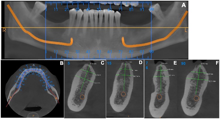
Figure 1. Cone-beam computed tomography images for patients included in the study. (A) An overview of the alveolar bone level by orthopantomography. (B) Axial view for the implant sites. (C–F) Sagittal view for measuring the alveolar bone level.
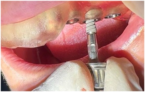
After administration of infiltration local anesthesia, the surgical guide was positioned on the jaw and secured in place. A drilling sequence was performed according to the manufacturer's instructions to prepare a hole for implant placement (Figure 2). A compressive one-piece implant (fixture and abutment) from ROOTT Company, with a length of 10–12 mm, was inserted using a torque wrench set to 35 N/cm (14). Patients were excluded if the insertion torque was below 35 N/cm or if any loss of socket wall integrity, such as fenestration, was observed. A readymade conometric TEC (titanium) was then tapped onto the implant abutment using a small hammer until fully set. Each conometric TEC was selected, fitted, and marked on the abutment.
To take an impression, a transfer cup was placed on each abutment and tied to it with soft wire to prevent it moving from its position. A one-step elastomeric heavy and light body impression was then taken, and the analog was inserted into the transfer cup. The impression and marked econometric TEC were sent to the technician for construction of the ceramic fused-to-metal bridge. After receiving the bridges and marking each conometric TEC separately within 3 days with an occlusal hole to localize the abutment, the conometric TECs were returned to the corresponding abutments marked previously. The bridges were cemented with glass-ionomer luting cement onto the retained conometric TECs fixed to the DI abutment. After the cement set, the bridge was removed from the oral cavity together with the conometric TEC as one piece using a special screwdriver inserted through the hole in the bridge, which pushed the bridge in the opposite direction to the abutment without damaging the implant. The conometric TEC, removed along with the bridge as a single unit, was cleaned by removing excess cement, polished, and reinserted with light tapping (Figure 3).
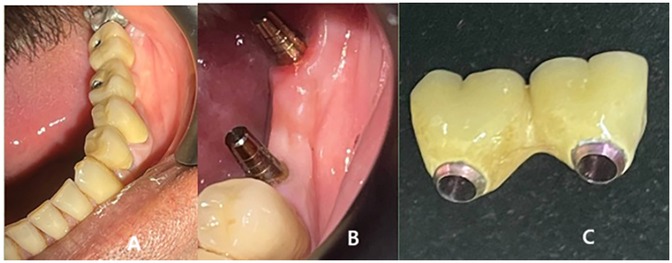
Figure 3. (A) After bridge cementation, (B) Removal of the bridge every time to measure the stability of the implant, and (C) The removed bridge.
Dental implant stability
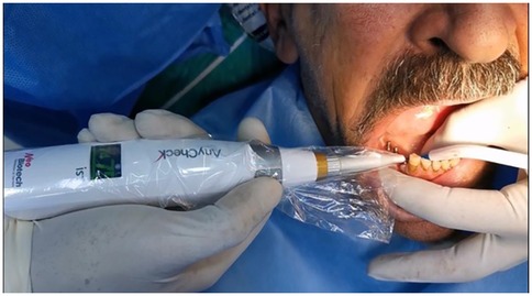
DI stability was measured as RFA using an implant stability tester (AnyCheck device; Neo Biotech, Republic of Korea). Each measurement was repeated six times, following the company’s instructions that the metal rod of the AnyCheck should maintain a contact angle with the abutment within the range 0°–30° upward while measuring (Figure 4).
The first measurement (T1) was taken immediately after implant insertion and DI stability measurements were repeated after 3 months (T2) and 6 months (T3) for all five directions [buccal (B), lingual L), mesial (M), distal (D), and occlusal (O)] after removing the bridge with the driver as mentioned earlier. The bridge was then reinserted using pressure on the corresponding abutment.
Statistical analysis
Data were assessed for normality using the Shapiro–Wilk test. The data were presented as median and interquartile range (IQR) of 25% and 75%. The Mann–Whitney U-test and Kruskal–Wallis test were used to compare the study's time points (T1, T2, and T3). The level of significance was set at p < 0.05 for the comparison of the study’s time points. All calculations were performed using SPSS software package version 26 (IBM Corp., Armonk, NY, USA).
Results
A total of 14 patients (8 men, 6 women; mean age 61.08 years; age range 35–79 years) were included in the study. Out of the 54 implants inserted, only 46 DIs, placed in the upper and lower jaws, were included in the final analysis (Figure 5). The other eight DIs were excluded from the study due to loss of integrity in the socket walls, such as fenestration and dehiscence of the supported bone and torque less than 35 N/cm. Further, participants not adhering to the study's time points were excluded.
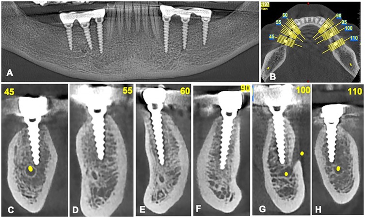
Figure 5. Cone-beam computed tomography images for patients after implant insertion. (A) An overview of the dental implants by orthopantomography. (B) Axial view for the implant placed. (C–H) Sagittal view of dental implants.
The median baseline RFA values shows statistically significant differences between maxillary (55, IQR: 51–61) and mandibular (64, IQR: 60–67) as well as between men (59, IQR: 53–64) and women (67, IQR: 64–69) (Figure 6).
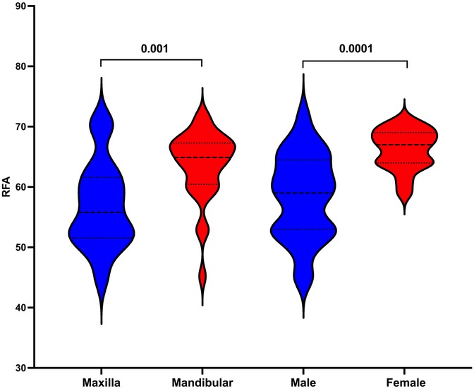
Figure 6. Comparison of baseline resonance frequency analysis between maxilla and mandibular as well as between men and women.
Of the examined surfaces, the occlusal surface showed the highest RFA compared to other surfaces for both maxillary and mandibular DIs. Meanwhile, as mentioned earlier, the RFA in the maxilla was observed to be lower than its mandibular counterparts at the baseline time point. The RFA increased at the 3- and 6-month time points for the facial, oral, and occlusal surfaces, and these increases were statistically significant at the 6-month time point (p < 0.05). However, the increase in RFA for the mandibular DIs was not statistically significant (Table 1).

Table 1. Comparison of dental implant stability for the examined surfaces at the study's time points in maxillary and mandibular jaws.
Similarly, the RFA values increased at the 3- and 6-month time points compared to baseline for all examined surfaces in men. These increases at 6 months were statistically significant for all surfaces except the mesial surface (p < 0.05). In women, however, this increase in RFA was found not to be statistically significant at either the 3-month or 6-month time point when compared to baseline (Table 2).

Table 2. Comparison of dental implant stability for the examined surfaces at the study's time points in men and women.
In addition, average RFA values of all DIs were compared across the study's time points. The results showed a statistically significant increase in mean RFA values for DIs in the maxilla at 6 months (median 65, IQR: 63–67) compared to baseline (median 55, IQR: 51–61, P = 0.0006) and 3 months (median 59, IQR: 56–62; p = 0.02). Although mean RFA values in the mandible increased at both 3 months (median 65, IQR: 56–69) and 6 months (median 68, IQR: 62–71), these changes were not statistically significant compared to baseline (median 64, IQR: 60–67; p > 0.05). Furthermore, no statistically significant difference was found in DI stability between the maxilla and mandible at the 6-month time point (p = 0.39) (Figure 7).
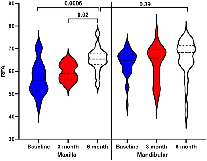
Figure 7. Comparison dental implant stability in the maxilla and mandibular at the study's time points.
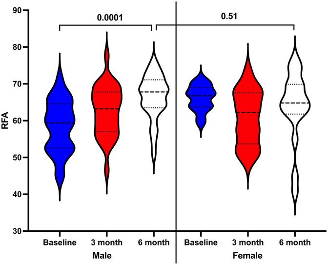
On the other hand, despite increases in mean RFA values at both 3 months (median 63, IQR: 57–67) and 6 months (median 67, IQR: 63–71) in men compared to baseline (median 59, IQR: 52–64), only the 6-month RFA value showed a statistically significant increase (p = 0.0001). However, no statistically significant differences were found in women when comparing the 3-month (median 62, IQR: 53–67) and 6-month (median 64, IQR: 61–69) RFA values to baseline (median 66, IQR: 63–69; p > 0.05). Lastly, comparison of DI stability at 6 months between men and women revealed no statistically significant difference (p = 0.51) (Figure 8).
Discussion
This study was conducted to investigate the effect of immediate functional loading on implant stability using a compressive one-piece dental implant retained by a conometric bridge to the fixture as an alternative to cemented and screw type. The study was based on the premise that previous results on immediate functional loading of DIs have been inconclusive (3, 13, 15–17). Furthermore, it is important to highlight that the compressive one-piece implant with immediate functional leading was introduced recently and has yet to be examined for its primary and secondary stability over time. The compressive one-piece DI system is specially designed to provide sufficient primary stability and is used in clinical situations involving horizontal alveolar ridge atrophy.
On the other hand, the conometric method applied in this study is an alternative to the cemented type, relying mainly on friction between the abutment and crown. The activation process occurs through applying a suitable insertion force for the conometric, which provides retentive performance equal to that of a cemented system without compromising mechanical functionality (11). Conometric techniques help overcome problems associated with cemented restoration, such as residual cement under the restoration leading to gingival inflammation and damage to the restoration during removal for any reason. This easy removal of the conometric-retained bridge without damaging the fixture or abutment enabled us to measure the stability of the DI separately and accurately at the study's time points.
Measuring DI stability before making a final decision for functional loading influences the DI's success and prognosis. Although both qualitative and quantitative methods are used to assess DI stability, a quantitative method was selected in this study to achieve greater precision (6, 18). It is well acknowledged that primary stability in a compression dental implant system depends on a combination of factors, including bone quality, implant design, insertion torque, placement technique, and bone–implant contact during insertion. Each factor contributes to the mechanical anchorage of the implant, which is essential for ensuring successful osseointegration in the early stages after placement (19–23).
Generally, the results of the current study showed that DI stability increases up to 6 months, likely related to osseointegration occurring over this period (24). Significantly higher stability was detected in the mandible compared to the maxilla, which may be related to bone type (type II in the mandible and type IV in the maxilla). Indeed, bone type has been proven to affect DI stability (25). It is important to note that in the current study, DI stability in the maxilla improved significantly after 6 months, with RFA comparable to that of the mandible. It has been reported that compared with delayed loading, immediate loading is associated with a higher incidence of implant failure (13). However, the immediate implant-supported conometric retention system provides fixed retention for a complete prosthesis with four implants and a 100% survival rate over 2 years.
Although our results showed higher DI stability in women compared to men, other studies found no influence of sex on DI stability (26). This can be explained by the fact the primary stability of a compressive DI system significantly relies upon the force applied during implant insertion, which creates a compression of the surrounding bone and hence increases the DI and bone surface area (27, 28). Interestingly, DI stability in men increased at the 6-month time point, matching the increase seen in women at the same time, supporting previous reports that sex does not affect secondary DI stability (29, 30).
This study has some limitations, such as a short follow-up and a lack of comparison with delayed loading or non-compressive implant systems. Nonetheless, to the best of our knowledge, it is the first study to investigate the stability of the compressive DI system with immediate loading using a conometric retainer.
Conclusions
The results of the study indicate the reliability of both the primary and secondary stability of the one-piece compressive conometric implant system in the posterior upper and lower jaws after 6 months. In addition, secondary DI stability improved over the 6-month period. Further studies with a longer follow-up and larger sample size are needed to confirm these findings.
Data availability statement
The raw data supporting the conclusions of this article will be made available by the authors upon reasonable request.
Ethics statement
The studies involving humans were approved by the Ethics Committee of College of Dentistry, University of Sulaimani. The studies were conducted in accordance with the local legislation and institutional requirements. The participants provided their written informed consent to participate in this study. Written informed consent was obtained from the individual(s) for the publication of any potentially identifiable images or data included in this article.
Author contributions
TA-H: Methodology, Writing – original draft, Conceptualization, Investigation, Resources. BE-D: Data curation, Resources, Validation, Methodology, Writing – original draft. AA-Z: Methodology, Conceptualization, Writing – review & editing, Project administration, Formal analysis. SG: Writing – review & editing, Supervision, Formal analysis, Validation, Investigation.
Funding
The author(s) declare that no financial support was received for the research and/or publication of this article.
Acknowledgments
Thanks to Zahawi Dental Lab for performing all prosthodontic tasks.
Conflict of interest
The authors declare that the research was conducted in the absence of any commercial or financial relationships that could be construed as a potential conflict of interest.
Generative AI statement
The author(s) declare that no Generative AI was used in the creation of this manuscript.
Publisher's note
All claims expressed in this article are solely those of the authors and do not necessarily represent those of their affiliated organizations, or those of the publisher, the editors and the reviewers. Any product that may be evaluated in this article, or claim that may be made by its manufacturer, is not guaranteed or endorsed by the publisher.
References
1. Molly L. Bone density and primary stability in implant therapy. Clin Oral Implants Res. (2006) 17:124–35. doi: 10.1111/j.1600-0501.2006.01356.x
2. Jar C, Archibald A, Gibson M, Westover L. An analytical model to measure dental implant stability with the Advanced System for Implant Stability Testing (ASIST). J Mech Behav Biomed Mater. (2024) 150:106238. doi: 10.1016/j.jmbbm.2023.106238
3. Sarfaraz H, Johri S, Sucheta P, Rao S. Study to assess the relationship between insertion torque value and implant stability quotient and its influence on timing of functional implant loading. J Indian Prosthodont Soc. (2018) 18:139–46. doi: 10.4103/jips.jips_203_17
4. Khan MW, Inayat N, Zafar MS, Zaigham AM. A resonance frequency analysis to investigate the impact of implant size on primary and secondary stability. Pak J Med Sci. (2024) 40:1261–6. doi: 10.12669/pjms.40.6.8213
5. Olmedo-Gaya M-V, Romero-Olid M-N, Ocaña-Peinado FM, Vallecillo-Rivas M, Vallecillo C, Reyes-Botella C. Influence of different surgical techniques on primary implant stability in the posterior maxilla: a randomized controlled clinical trial. Clin Oral Investig. (2023) 27:3499–508. doi: 10.1007/s00784-023-04962-y
6. Lee J, Pyo S-W, Cho H-J, An J-S, Lee J-H, Koo K-T, et al. Comparison of implant stability measurements between a resonance frequency analysis device and a modified damping capacity analysis device: an in vitro study. J Periodontal Implant Sci. (2020) 50:56–66. doi: 10.5051/jpis.2020.50.1.56
7. Atsumi M, Park SH, Wang HL. Methods used to assess implant stability: current status. Int J Oral Maxillofac Implants. (2007) 22:743–54.17974108
8. Monje A, Ravidà A, Wang HL, Helms JA, Brunski JB. Relationship between primary/mechanical and secondary/biological implant stability. Int J Oral Maxillofac Implants. (2019) 34:s7–s23. doi: 10.11607/jomi.19suppl.g1
9. Nkenke E, Fenner M. Indications for immediate loading of implants and implant success. Clin Oral Implants Res. (2006) 17(Suppl 2):19–34. doi: 10.1111/j.1600-0501.2006.01348.x
10. Chee W, Jivraj S. Screw versus cemented implant supported restorations. Br Dent J. (2006) 201:501–7. doi: 10.1038/sj.bdj.4814157
11. Bressan E, Lops D, Tomasi C, Ricci S, Stocchero M, Carniel EL. Experimental and computational investigation of Morse taper conometric system reliability for the definition of fixed connections between dental implants and prostheses. Proc Inst Mech Eng H. (2014) 228:674–81. doi: 10.1177/0954411914545556
12. Degidi M, Nardi D, Sighinolfi G, Degidi D. The conometric concept for the definitive rehabilitation of a single posterior implant by using a conical indexed abutment: a technique. J Prosthet Dent. (2020) 123:576–9. doi: 10.1016/j.prosdent.2019.02.020
13. Chen J, Cai M, Yang J, Aldhohrah T, Wang Y. Immediate versus early or conventional loading dental implants with fixed prostheses: a systematic review and meta-analysis of randomized controlled clinical trials. J Prosthet Dent. (2019) 122:516–36. doi: 10.1016/j.prosdent.2019.05.013
14. Venkatakrishnan C, Bhuminathan S, Chandran CR. Dental implant insertion torque and bone density–short review. Biomed Pharmacol J. (2017) 10:1305–9. doi: 10.13005/bpj/1234
15. Daher FI, Abi-Aad HL, Dimassi HI, Baba NZ, Majzoub ZA. Factors affecting implant stability quotients at immediately and conventionally loaded implants in the posterior maxilla: a split-mouth randomized controlled trial. J Prosthodont. (2021) 30:590–603. doi: 10.1111/jopr.13296
16. Gjelvold B, Kisch J, Chrcanovic BR. A randomized clinical trial comparing immediate loading and delayed loading of single-tooth implants: 5-year results. J Clin Med. (2021) 10:1–9. doi: 10.3390/jcm10051077
17. Pardal-Peláez B, Flores-Fraile J, Pardal-Refoyo JL, Montero J. Implant loss and crestal bone loss in immediate versus delayed load in edentulous mandibles: a systematic review and meta-analysis. J Prosthet Dent. (2021) 125:437–44. doi: 10.1016/j.prosdent.2020.01.032
18. Chang WJ, Lee SY, Wu CC, Lin CT, Abiko Y, Yamamichi N, et al. A newly designed resonance frequency analysis device for dental implant stability detection. Dent Mater J. (2007) 26:665–71. doi: 10.4012/dmj.26.665
19. Rues S, Schmitter M, Kappel S, Sonntag R, Kretzer JP, Nadorf J. Effect of bone quality and quantity on the primary stability of dental implants in a simulated bicortical placement. Clin Oral Investig. (2021) 25:1265–72. doi: 10.1007/s00784-020-03432-z
20. Heimes D, Becker P, Pabst A, Smeets R, Kraus A, Hartmann A, et al. How does dental implant macrogeometry affect primary implant stability? A narrative review. Int J Implant Dent. (2023) 9:20. doi: 10.1186/s40729-023-00485-z
21. do Vale Souza JP, de Moraes Melo Neto CL, Piacenza LT, Freitas da Silva EV, de Melo Moreno AL, Penitente PA, et al. Relation between insertion torque and implant stability quotient: a clinical study. Eur J Dent. (2021) 15:618–23. doi: 10.1055/s-0041-1725575
22. Romero-Serrano M, Romero-Ruiz MM, Herrero-Climent M, Rios-Carrasco B, Gil-Mur J. Correlation between implant surface roughness and implant stability: a systematic review. Dent J (Basel). (2024) 12:1–18. doi: 10.3390/dj12090276
23. Karl M, Grobecker-Karl T. Effect of bone quality, implant design, and surgical technique on primary implant stability. Quintessence Int. (2018) 22:189–98. doi: 10.3290/j.qi.a39745
24. Huang Y-C, Huang Y-C, Ding S-J. Primary stability of implant placement and loading related to dental implant materials and designs: a literature review. J Dent Sci. (2023) 18:1467–76. doi: 10.1016/j.jds.2023.06.010
25. Yoon HG, Heo SJ, Koak JY, Kim SK, Lee SY. Effect of bone quality and implant surgical technique on implant stability quotient (ISQ) value. J Adv Prosthodont. (2011) 3:10–5. doi: 10.4047/jap.2011.3.1.10
26. Shiffler K, Lee D, Rowan M, Aghaloo T, Pi-Anfruns J, Moy PK. Effect of length, diameter, intraoral location on implant stability. Oral Surg Oral Med Oral Pathol Oral Radiol. (2016) 122:e193–8. doi: 10.1016/j.oooo.2016.06.016
27. O’Sullivan D, Sennerby L, Meredith N. Influence of implant taper on the primary and secondary stability of osseointegrated titanium implants. Clin Oral Implants Res. (2004) 15:474–80. doi: 10.1111/j.1600-0501.2004.01041.x
28. Chong L, Khocht A, Suzuki JB, Gaughan J. Effect of implant design on initial stability of tapered implants. J Oral Implantol. (2009) 35:130–5. doi: 10.1563/1548-1336-35.3.130
29. Vollmer A, Saravi B, Lang G, Adolphs N, Hazard D, Giers V, et al. Factors influencing primary and secondary implant stability—a retrospective cohort study with 582 implants in 272 patients. Appl Sci. (2020) 10:8084. doi: 10.3390/app10228084
Keywords: compressive implant, conometric, one-piece implant, primary stability, secondary stability
Citation: Al-Hity TR, El-Dhuwaib B, Al-Zahawi AR and Gul SS (2025) Comparison of primary and secondary stability of a one-piece compressive conometric implant system in the posterior upper and lower jaws. Front. Oral Health 6:1625593. doi: 10.3389/froh.2025.1625593
Received: 9 May 2025; Accepted: 7 July 2025;
Published: 25 July 2025.
Edited by:
Francisco Correia, University of Porto, PortugalReviewed by:
Albraa Alolayan, Taibah University, Saudi ArabiaNipun Dhalla, Manav Rachna International Institute of Research and Studies (MRIIRS), India
Copyright: © 2025 Al-Hity, El-Dhuwaib, Al-Zahawi and Gul. This is an open-access article distributed under the terms of the Creative Commons Attribution License (CC BY). The use, distribution or reproduction in other forums is permitted, provided the original author(s) and the copyright owner(s) are credited and that the original publication in this journal is cited, in accordance with accepted academic practice. No use, distribution or reproduction is permitted which does not comply with these terms.
*Correspondence: Sarhang Sarwat Gul, c2FyaGFuZy5oYW1hQHVpbnZzdWwuZWR1Lmlx; c2FyaGFuZy5oYW1hQHNwdS5lZHUuaXE=
 Thabat R. Al-Hity1,2,3
 Sarhang Sarwat Gul