Abstract
MicroRNA (miRNA or miR)-based approaches to interrupt the transmission of mosquito-borne diseases have been explored since 2005. A review of these studies and areas in which to proceed is needed. In this review, significant progress is reviewed at the level of individual miRNAs, and miRNA diversification and relevant confounders are described in detail. Current miRNA studies in mosquitoes include four steps, namely, identifying miRNAs, validating miRNA-pathogen interactions, exploring action mechanisms, and performing preapplication investigations. Notably, regarding the Plasmodium parasite, mosquito miRNAs generally bind to mosquito immunity- or development-related mRNAs, indirectly regulating Plasmodium infection; However, regarding arboviruses, mosquito miRNAs can bind to the viral genome, directly modifying viral replication. Thus, during explorations of miRNA-based approaches, researchers need select an ideal miRNA for investigation based on the mosquito species, tissue, and mosquito-borne pathogen of interest. Additionally, strategies for miRNA-based approaches differ for arboviruses and protozoan parasites.
Background
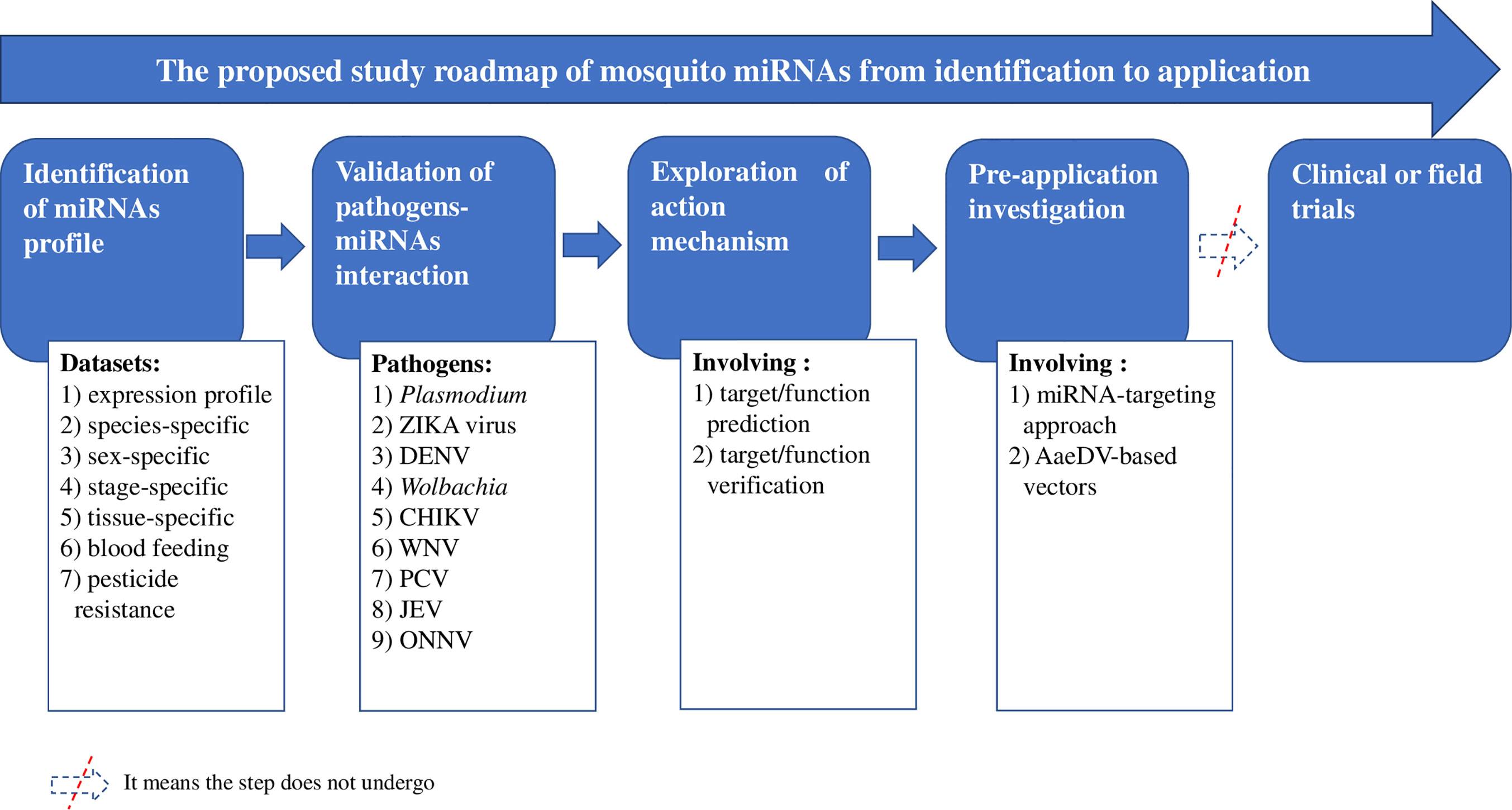
Insecticide-based interventions [e.g., long-lasting insecticide-treated bed nets (LLINs) and indoor residual spraying (IRS)] are important components of integrated mosquito management programs designed to block the transmission of mosquito-borne diseases. The insecticides used in these interventions exert strong selection pressure on resistance and lead to the evolution and spread of mosquito resistance, representing a major concern in mosquito-borne disease control programs (Xu et al., 2014). The World Health Organization (WHO) claims that innovative vector control tools are urgently needed (World Health Organization, 2012). MicroRNAs (miRNAs) are single-stranded, conserved, and small endogenous noncoding RNAs that have important regulatory functions at the posttranscriptional level in diverse organisms (; ). The regulation of miRNAs is indispensable for various processes, including apoptosis, development, differentiation, viral infection, and so on (; ). More importantly, the functions of miRNAs can be explored and utilized. For instance, miR-15, miR-16, miR-34, and Let-7 have been patented and approved for cancer diagnosis or treatment (Mishra et al., 2016). Given their characteristics and functions, miRNAs represent one possibility for establishing a new tool, namely, miRNA-based approaches. Thus, numerous studies on mosquito miRNAs have been performed since 2005 (Wang et al., 2005) with the ultimate goal of utilizing miRNA-based approaches for disease control (Liu et al., 2017; ). The development of a mosquito miRNA-based approach is presumed to always follow the research roadmap of identifying mosquito miRNAs, observing miRNA-pathogen interaction, exploring action mechanisms, performing preapplication investigations and conducting clinical or field trials. Advances at each step of the roadmap need to be outlined to provide important insight into potential applications.
The Mosquito miRNA Databases Analyzed
Publications focused on investigating mosquito global miRNA profiles were selected for extraction of information, including the authors, publication year, study materials, methods, miRNA names, canonical sequences, and so on. Then, this information was supplemented during the review of the other included papers. Approximately 1635 mature or predicted miRNAs were collected. Of the 1635 miRNAs, 853 (52.17%) were limited to identification and lacked any additional study information. The remaining 782 (47.83%) were further investigated; thus, they were tracked by their annotated names for study development.
Overall, the miRNAs of 24 mosquito species, including 2 species of Aedes (Li et al., 2009; ; Gu et al., 2013; ; Hu et al., 2015; Maharaj et al., 2015; Liu et al., 2015; Liu Y. X. et al., 2016; ; Su et al., 2017; Zhang et al., 2017), 2 species of Culex (Skalsky et al., 2010; Hong et al., 2014), and 20 species of Anopheles mosquitoes (Wang et al., 2005; ; Mead and Tu, 2008; ; Liu et al., 2014; ; Liu et al., 2017; ; ; ), were studied. The study materials involved almost every possible type, including mosquito genome sequences obtained from websites; whole mosquitoes at all developmental stages; mosquitoes at different blood feeding states, ages, pathogen infection statuses, diapause statuses or insecticide resistance states; mosquito tissues except for legs; and even mosquito cell nuclei and cytoplasm (Tables 1–6).
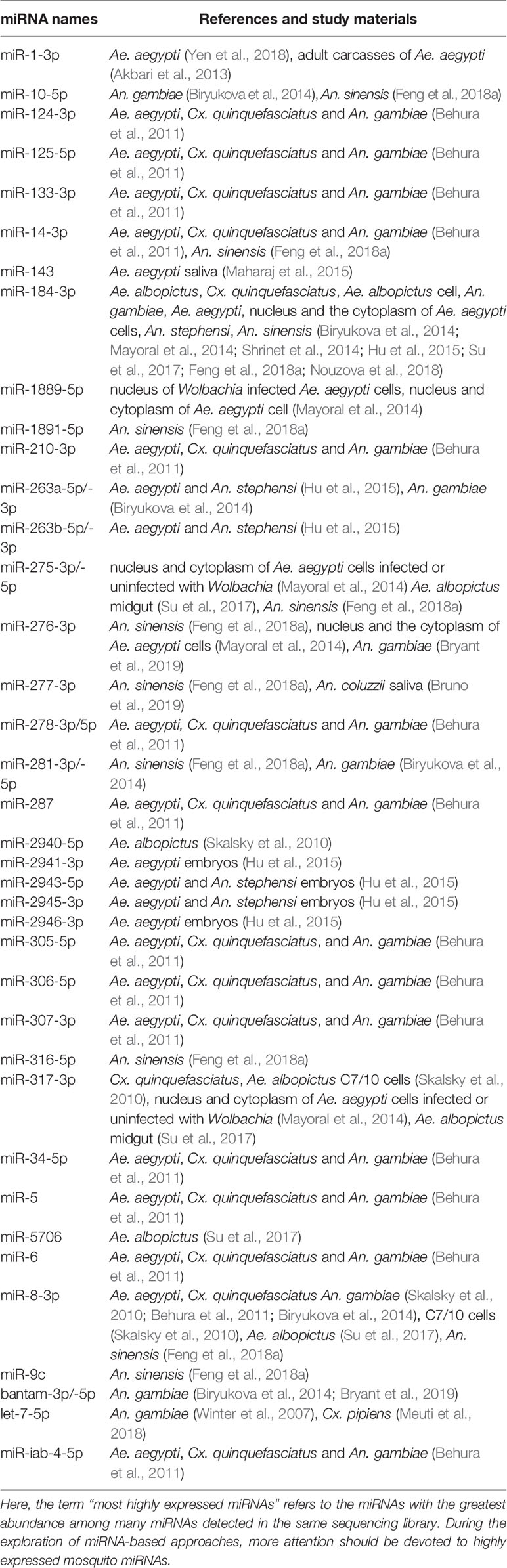
Table 1
| miRNA names | References and study materials |
|---|---|
| miR-1-3p | Ae. aegypti (Yen et al., 2018), adult carcasses of Ae. aegypti () |
| miR-10-5p | An. gambiae (), An. sinensis () |
| miR-124-3p | Ae. aegypti, Cx. quinquefasciatus and An. gambiae () |
| miR-125-5p | Ae. aegypti, Cx. quinquefasciatus and An. gambiae () |
| miR-133-3p | Ae. aegypti, Cx. quinquefasciatus and An. gambiae () |
| miR-14-3p | Ae. aegypti, Cx. quinquefasciatus and An. gambiae (), An. sinensis () |
| miR-143 | Ae. aegypti saliva (Maharaj et al., 2015) |
| miR-184-3p | Ae. albopictus, Cx. quinquefasciatus, Ae. albopictus cell, An. gambiae, Ae. aegypti, nucleus and the cytoplasm of Ae. aegypti cells, An. stephensi, An. sinensis (; Mayoral et al., 2014; Shrinet et al., 2014; Hu et al., 2015; Su et al., 2017; ; Nouzova et al., 2018) |
| miR-1889-5p | nucleus of Wolbachia infected Ae. aegypti cells, nucleus and cytoplasm of Ae. aegypti cell (Mayoral et al., 2014) |
| miR-1891-5p | An. sinensis () |
| miR-210-3p | Ae. aegypti, Cx. quinquefasciatus and An. gambiae () |
| miR-263a-5p/-3p | Ae. aegypti and An. stephensi (Hu et al., 2015), An. gambiae () |
| miR-263b-5p/-3p | Ae. aegypti and An. stephensi (Hu et al., 2015) |
| miR-275-3p/-5p | nucleus and cytoplasm of Ae. aegypti cells infected or uninfected with Wolbachia (Mayoral et al., 2014) Ae. albopictus midgut (Su et al., 2017), An. sinensis () |
| miR-276-3p | An. sinensis (), nucleus and the cytoplasm of Ae. aegypti cells (Mayoral et al., 2014), An. gambiae () |
| miR-277-3p | An. sinensis (), An. coluzzii saliva () |
| miR-278-3p/5p | Ae. aegypti, Cx. quinquefasciatus and An. gambiae () |
| miR-281-3p/-5p | An. sinensis (), An. gambiae () |
| miR-287 | Ae. aegypti, Cx. quinquefasciatus and An. gambiae () |
| miR-2940-5p | Ae. albopictus (Skalsky et al., 2010) |
| miR-2941-3p | Ae. aegypti embryos (Hu et al., 2015) |
| miR-2943-5p | Ae. aegypti and An. stephensi embryos (Hu et al., 2015) |
| miR-2945-3p | Ae. aegypti and An. stephensi embryos (Hu et al., 2015) |
| miR-2946-3p | Ae. aegypti embryos (Hu et al., 2015) |
| miR-305-5p | Ae. aegypti, Cx. quinquefasciatus, and An. gambiae () |
| miR-306-5p | Ae. aegypti, Cx. quinquefasciatus, and An. gambiae () |
| miR-307-3p | Ae. aegypti, Cx. quinquefasciatus, and An. gambiae () |
| miR-316-5p | An. sinensis () |
| miR-317-3p | Cx. quinquefasciatus, Ae. albopictus C7/10 cells (Skalsky et al., 2010), nucleus and cytoplasm of Ae. aegypti cells infected or uninfected with Wolbachia (Mayoral et al., 2014), Ae. albopictus midgut (Su et al., 2017) |
| miR-34-5p | Ae. aegypti, Cx. quinquefasciatus and An. gambiae () |
| miR-5 | Ae. aegypti, Cx. quinquefasciatus and An. gambiae () |
| miR-5706 | Ae. albopictus (Su et al., 2017) |
| miR-6 | Ae. aegypti, Cx. quinquefasciatus and An. gambiae () |
| miR-8-3p | Ae. aegypti, Cx. quinquefasciatus An. gambiae (Skalsky et al., 2010; ; ), C7/10 cells (Skalsky et al., 2010), Ae. albopictus (Su et al., 2017), An. sinensis () |
| miR-9c | An. sinensis () |
| bantam-3p/-5p | An. gambiae (; ) |
| let-7-5p | An. gambiae (Winter et al., 2007), Cx. pipiens (Meuti et al., 2018) |
| miR-iab-4-5p | Ae. aegypti, Cx. quinquefasciatus and An. gambiae () |
The generally most highly expressed miRNAs in mosquitoes.
Here, the term “most highly expressed miRNAs” refers to the miRNAs with the greatest abundance among many miRNAs detected in the same sequencing library. During the exploration of miRNA-based approaches, more attention should be devoted to highly expressed mosquito miRNAs.
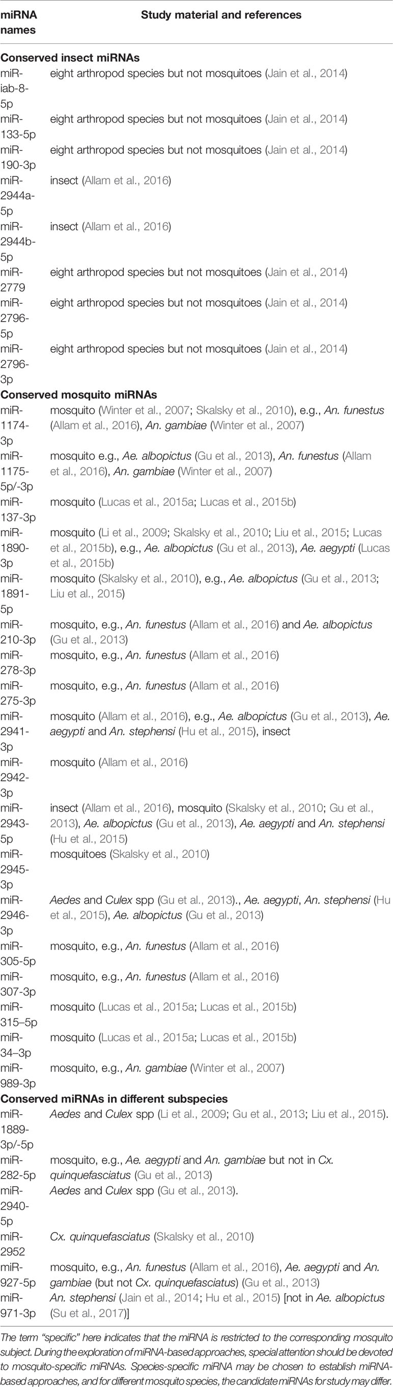
Table 2
| miRNA names | Study material and references |
|---|---|
| Conserved insect miRNAs | |
| miR-iab-8-5p | eight arthropod species but not mosquitoes (Jain et al., 2014) |
| miR-133-5p | eight arthropod species but not mosquitoes (Jain et al., 2014) |
| miR-190-3p | eight arthropod species but not mosquitoes (Jain et al., 2014) |
| miR-2944a-5p | insect () |
| miR-2944b-5p | insect () |
| miR-2779 | eight arthropod species but not mosquitoes (Jain et al., 2014) |
| miR-2796-5p | eight arthropod species but not mosquitoes (Jain et al., 2014) |
| miR-2796-3p | eight arthropod species but not mosquitoes (Jain et al., 2014) |
| Conserved mosquito miRNAs | |
| miR-1174-3p | mosquito (Winter et al., 2007; Skalsky et al., 2010), e.g., An. funestus (), An. gambiae (Winter et al., 2007) |
| miR-1175-5p/-3p | mosquito e.g., Ae. albopictus (Gu et al., 2013), An. funestus (), An. gambiae (Winter et al., 2007) |
| miR-137-3p | mosquito (Lucas et al., 2015a; Lucas et al., 2015b) |
| miR-1890-3p | mosquito (Li et al., 2009; Skalsky et al., 2010; Liu et al., 2015; Lucas et al., 2015b), e.g., Ae. albopictus (Gu et al., 2013), Ae. aegypti (Lucas et al., 2015b) |
| miR-1891-5p | mosquito (Skalsky et al., 2010), e.g., Ae. albopictus (Gu et al., 2013; Liu et al., 2015) |
| miR-210-3p | mosquito, e.g., An. funestus () and Ae. albopictus (Gu et al., 2013) |
| miR-278-3p | mosquito, e.g., An. funestus () |
| miR-275-3p | mosquito, e.g., An. funestus () |
| miR-2941-3p | mosquito (), e.g., Ae. albopictus (Gu et al., 2013), Ae. aegypti and An. stephensi (Hu et al., 2015), insect |
| miR-2942-3p | mosquito () |
| miR-2943-5p | insect (), mosquito (Skalsky et al., 2010; Gu et al., 2013), Ae. albopictus (Gu et al., 2013), Ae. aegypti and An. stephensi (Hu et al., 2015) |
| miR-2945-3p | mosquitoes (Skalsky et al., 2010) |
| miR-2946-3p | Aedes and Culex spp (Gu et al., 2013)., Ae. aegypti, An. stephensi (Hu et al., 2015), Ae. albopictus (Gu et al., 2013) |
| miR-305-5p | mosquito, e.g., An. funestus () |
| miR-307-3p | mosquito, e.g., An. funestus () |
| miR-315–5p | mosquito (Lucas et al., 2015a; Lucas et al., 2015b) |
| miR-34–3p | mosquito (Lucas et al., 2015a; Lucas et al., 2015b) |
| miR-989-3p | mosquito, e.g., An. gambiae (Winter et al., 2007) |
| Conserved miRNAs in different subspecies | |
| miR-1889-3p/-5p | Aedes and Culex spp (Li et al., 2009; Gu et al., 2013; Liu et al., 2015). |
| miR-282-5p | mosquito, e.g., Ae. aegypti and An. gambiae but not in Cx. quinquefasciatus (Gu et al., 2013) |
| miR-2940-5p | Aedes and Culex spp (Gu et al., 2013). |
| miR-2952 | Cx. quinquefasciatus (Skalsky et al., 2010) |
| miR-927-5p | mosquito, e.g., An. funestus (), Ae. aegypti and An. gambiae (but not Cx. quinquefasciatus) (Gu et al., 2013) |
| miR-971-3p | An. stephensi (Jain et al., 2014; Hu et al., 2015) [not in Ae. albopictus (Su et al., 2017)] |
Species-specific miRNAs in mosquitoes.
The term “specific” here indicates that the miRNA is restricted to the corresponding mosquito subject. During the exploration of miRNA-based approaches, special attention should be devoted to mosquito-specific miRNAs. Species-specific miRNA may be chosen to establish miRNA-based approaches, and for different mosquito species, the candidate miRNAs for study may differ.
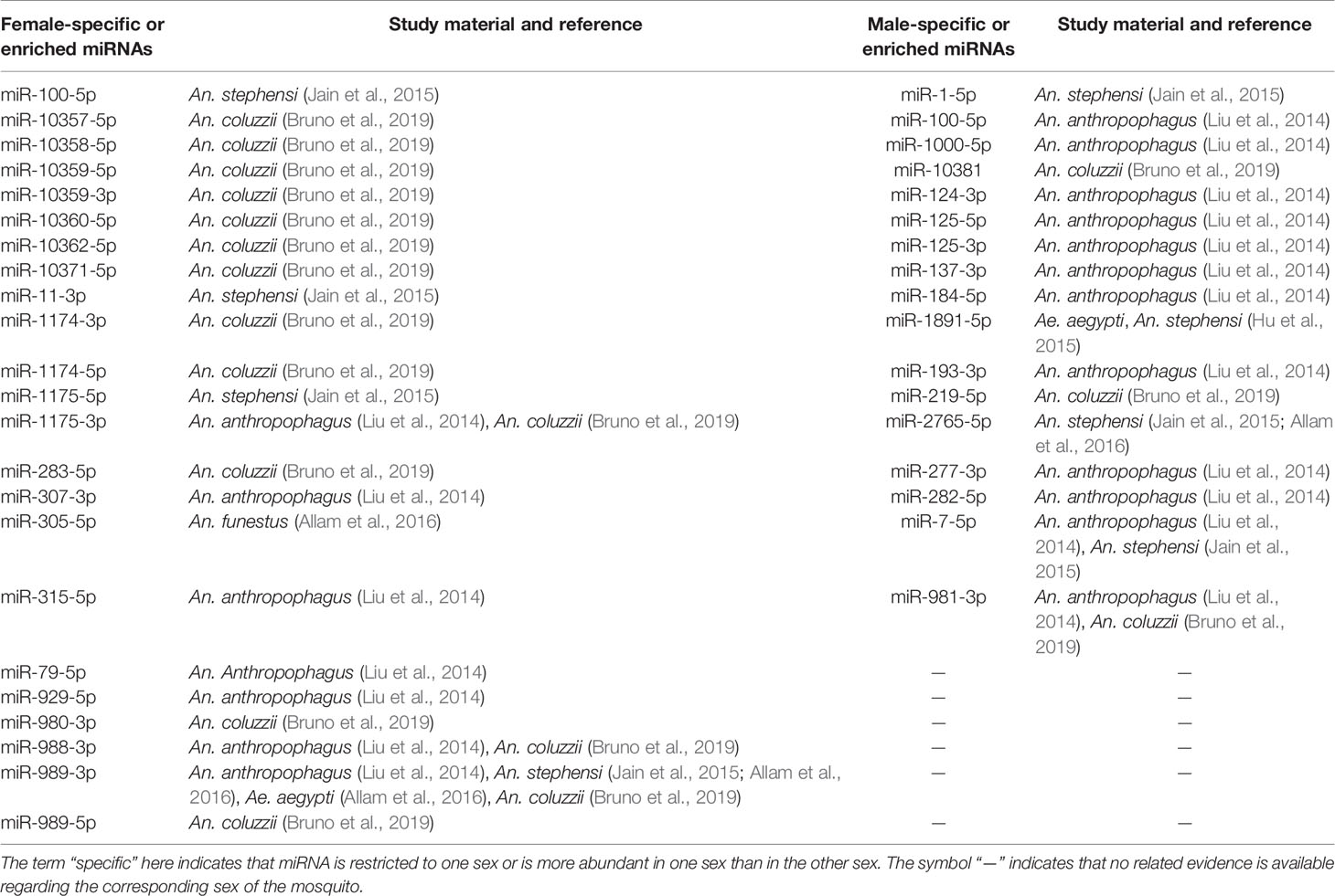
Table 3
| Female-specific or enriched miRNAs | Study material and reference | Male-specific or enriched miRNAs | Study material and reference |
|---|---|---|---|
| miR-100-5p | An. stephensi (Jain et al., 2015) | miR-1-5p | An. stephensi (Jain et al., 2015) |
| miR-10357-5p | An. coluzzii () | miR-100-5p | An. anthropophagus (Liu et al., 2014) |
| miR-10358-5p | An. coluzzii () | miR-1000-5p | An. anthropophagus (Liu et al., 2014) |
| miR-10359-5p | An. coluzzii () | miR-10381 | An. coluzzii () |
| miR-10359-3p | An. coluzzii () | miR-124-3p | An. anthropophagus (Liu et al., 2014) |
| miR-10360-5p | An. coluzzii () | miR-125-5p | An. anthropophagus (Liu et al., 2014) |
| miR-10362-5p | An. coluzzii () | miR-125-3p | An. anthropophagus (Liu et al., 2014) |
| miR-10371-5p | An. coluzzii () | miR-137-3p | An. anthropophagus (Liu et al., 2014) |
| miR-11-3p | An. stephensi (Jain et al., 2015) | miR-184-5p | An. anthropophagus (Liu et al., 2014) |
| miR-1174-3p | An. coluzzii () | miR-1891-5p | Ae. aegypti, An. stephensi (Hu et al., 2015) |
| miR-1174-5p | An. coluzzii () | miR-193-3p | An. anthropophagus (Liu et al., 2014) |
| miR-1175-5p | An. stephensi (Jain et al., 2015) | miR-219-5p | An. coluzzii () |
| miR-1175-3p | An. anthropophagus (Liu et al., 2014), An. coluzzii () | miR-2765-5p | An. stephensi (Jain et al., 2015; ) |
| miR-283-5p | An. coluzzii () | miR-277-3p | An. anthropophagus (Liu et al., 2014) |
| miR-307-3p | An. anthropophagus (Liu et al., 2014) | miR-282-5p | An. anthropophagus (Liu et al., 2014) |
| miR-305-5p | An. funestus () | miR-7-5p | An. anthropophagus (Liu et al., 2014), An. stephensi (Jain et al., 2015) |
| miR-315-5p | An. anthropophagus (Liu et al., 2014) | miR-981-3p | An. anthropophagus (Liu et al., 2014), An. coluzzii () |
| miR-79-5p | An. Anthropophagus (Liu et al., 2014) | — | — |
| miR-929-5p | An. anthropophagus (Liu et al., 2014) | — | — |
| miR-980-3p | An. coluzzii () | — | — |
| miR-988-3p | An. anthropophagus (Liu et al., 2014), An. coluzzii () | — | — |
| miR-989-3p | An. anthropophagus (Liu et al., 2014), An. stephensi (Jain et al., 2015; ), Ae. aegypti (), An. coluzzii () | — | — |
| miR-989-5p | An. coluzzii () | — | — |
Sex-specific miRNAs in adult mosquitoes.

The term “specific” here indicates that miRNA is restricted to one sex or is more abundant in one sex than in the other sex. The symbol “—” indicates that no related evidence is available regarding the corresponding sex of the mosquito.
Table 4
| miRNA names | Study materials and expression levels in certain stages or statuses* | |||||||||
|---|---|---|---|---|---|---|---|---|---|---|
| Egg& | Larva& | Pupa& | Adult& | Blood feeding# | Other stages$ | |||||
| miR-1-3p | — | — | An. stephensi (Jain et al., 2015) | — | Ae. aegypti fat body 24 h PBM (Zhang et al., 2017), Ae. aegypti (Hussain et al., 2013), An. stephensi (Jain et al., 2014), An. anthropophagus midgut (Liu et al., 2017) | Ae. albopictus nondiapause pharate larva (), Cx. pipiens DR-strain (Hong et al., 2014) | ||||
| miR-1-5p | — | — | An. stephensi (Jain et al., 2015) | An. stephensi (Jain et al., 2015) | — | — | ||||
| miR-10-3p | — | — | — | — | An. gambiae (Fu et al., 2017) | — | ||||
| miR-10-5p | An. sinensis () | — | — | An. stephensi (Jain et al., 2015) | — | — | ||||
| miR-100-5p | — | — | — | An. stephensi (Jain et al., 2015) | Ae. aegypti at 12 h PBM (), An. anthropophagus midguts (Liu et al., 2017) | Cx. pipiens DR strain (Hong et al., 2014) | ||||
| miR-1000-5p | — | — | — | — | An. gambiae (), An. anthropophagus midguts (Liu et al., 2017), Ae. aegypti fat body at 48 h PBM (Zhang et al., 2017) | — | ||||
| miR-11-3p | — | — | — | An. stephensi (Jain et al., 2015), An. sinensis () | Ae. aegypti midgut (Li et al., 2009), An. stephensi (Jain et al., 2014), An. anthropophagus (Liu et al., 2017), Ae. aegypti fat body 72 h PBM (Zhang et al., 2017) | Cx. pipiens DR strain (Hong et al., 2014) | ||||
| miR-11-5p | — | — | — | — | Ae. aegypti fat body at 48 h PBM (Zhang et al., 2017) | — | ||||
| miR-1174-5p | — | — | — | — | — | Cx. pipiens DR strain (Hong et al., 2014) | ||||
| miR-1174-3p | — | An. sinensis () An. stephensi (Jain et al., 2015) | — | — | An. gambiae midguts (Winter et al., 2007), Ae. aegypti and An. gambiae (Jain et al., 2014), Ae. aegypti fat body at 48 h PBM (Zhang et al., 2017), An. gambiae (Fu et al., 2017), An. anthropophagus midguts (Liu et al., 2017), Ae. albopictus midguts (Su et al., 2017) | — | ||||
| miR-1175-5p | — | An. funestus () An. sinensis () | — | An. sinensis () | Ae. albopictus midguts (Su et al., 2017) | Cx. pipiens DR strain (Hong et al., 2014) | ||||
| miR-1175-3p | — | An. sinensis () | — | An. sinensis () | An. gambiae midgut (Winter et al., 2007), Aedes spp. (Jain et al., 2014),Ae. albopictus midgut (Su et al., 2017) | — | ||||
| miR-12-5p | — | — | — | — | leftover of An. gambiae (Winter et al., 2007), An. anthropophagus midguts (Liu et al., 2017), Ae. albopictus midguts (Su et al., 2017) | — | ||||
| miR-124-3p | An. sinensis () | — | — | — | — | old Cx. pipiens (Meuti et al., 2018) | ||||
| miR-125-5p | — | An. funestus () | — | — | Ae. aegypti 12 h PBM (), An. anthropophagus midguts (Liu et al., 2017), Ae. aegypti fat body at 72 h PBM (Zhang et al., 2017) | Cx. pipiens DR strain (Hong et al., 2014) | ||||
| miR-127 | — | — | — | — | — | Cx. pipiens DR strain (Hong et al., 2014) | ||||
| miR-13-3p | — | — | — | — | An. anthropophagus midguts (Liu et al., 2017) | Cx. pipiens DR strain (Hong et al., 2014) | ||||
| miR-13-5p | — | — | — | — | Ae. aegypti fat body at 48 h PBM (Zhang et al., 2017) | |||||
| miR-133-3p | An. funestus () | An. funestus () | An. stephensi (Jain et al., 2015) | — | Ae. aegypti fat body at 48 h PBM (Zhang et al., 2017) | Cx. pipiens DR strain (Hong et al., 2014) | ||||
| miR-133-5p | — | — | An. stephensi (Jain et al., 2015) | An. stephensi (Jain et al., 2015) | — | — | ||||
| miR-137-3p | — | — | — | — | An. anthropophagus midguts (Liu et al., 2017) | — | ||||
| miR-137-5p | An. funestus () | An. funestus () | — | — | — | — | ||||
| miR-14-5p | — | — | — | — | — | Ae. albopictus nondiapause pharate larva () | ||||
| miR-14-3p | An. funestus () | An. funestus () | — | — | Ae. aegypti (Hussain et al., 2013), An. anthropophagus midguts (Liu et al., 2017) | old or DR Cx. pipiens (Hong et al., 2014; Meuti et al., 2018) | ||||
| miR-1767 | — | — | — | — | midguts of Ae. albopictus (Su et al., 2017) | — | ||||
| miR-184-3p | An. funestus () | — | — | — | Ae. aegypti (Li et al., 2009), Ae. aegypti fat body at 72 h PBM (Zhang et al., 2017), fat body of Ae. aegypti and An. gambiae (Fu et al., 2017), An. anthropophagus midguts (Liu et al., 2017), Ae. albopictus midguts (Su et al., 2017), Aedes aegypti (Nouzova et al., 2018) | — | ||||
| miR-1889-5p | — | — | — | — | Ae. aegypti fat body at 36 h PBM (Zhang et al., 2017) | — | ||||
| miR-1890-3p | — | — | Ae. aegypti, An. stephensi (Hu et al., 2015; Jain et al., 2015; ; ) | — | Ae. aegypti fat body at 36 h PBM (Zhang et al., 2017) | Ae. albopictus diapause oocyte (), Cx. pipiens DR strain (Hong et al., 2014) | ||||
| miR-1891-5p | — | — | An. sinensis () | An. stephensi (Jain et al., 2015) | — | Cx. pipiens DR strain (Hong et al., 2014) | ||||
| miR-190-5p | — | — | Ae. aegypti midgut (Li et al., 2009), An. Stephensi (Jain et al., 2014) | Cx. pipiens DR strain (Hong et al., 2014) | ||||||
| miR-190-3p | — | — | An. stephensi (Jain et al., 2015) | An. stephensi (Jain et al., 2015) | An. Stephensi (Jain et al., 2014) | — | ||||
| miR-193-5p | — | — | Ae. albopictus midguts (Su et al., 2017) | — | ||||||
| miR-193-3p | — | — | An. stephensi (Jain et al., 2015) An. funestus () An. sinensis () | — | — | — | ||||
| miR-1951 | — | — | — | — | Ae. albopictus midguts (Su et al., 2019) | — | ||||
| miR-210-3p | An. stephensi (Mead and Tu, 2008) | An. stephensi (Mead and Tu, 2008; Jain et al., 2015) | Ae. aegypti fat body at 48 h PBM (Zhang et al., 2017) | Cx. pipiens DR strain (Hong et al., 2014) | ||||||
| miR-219-5p | — | — | — | An. stephensi (Jain et al., 2015) | — | — | ||||
| miR-2491-3p | — | — | An. sinensis () | — | — | — | ||||
| miR-252-5p | — | — | — | — | Ae. aegypti fat body at 6 h PBM (Zhang et al., 2017) | Cx. pipiens DR strain (Hong et al., 2014) | ||||
| miR-252-3p | — | — | — | — | Ae. aegypti fat body at 6 h PBM (Zhang et al., 2017) | |||||
| miR-263a-5p | — | — | — | — | Cx. pipiens DR strain (Hong et al., 2014) | |||||
| miR-275-3p | — | — | — | — | An. stephensi (Jain et al., 2014), An. anthropophagus midguts (Liu et al., 2017), Ae. aegypti fat body at 48h PBM (Zhang et al., 2017), An. gambiae (Lampe and Levashina, 2018), Ae.albopictus midgut (Su et al., 2017) | old Cx. pipiens (Meuti et al., 2018), Cx. pipiens DR-strain (Hong et al., 2014) | ||||
| miR-275-5p | — | — | — | — | Ae. aegypti fat body at 24 h PBM (Zhang et al., 2017) | |||||
| miR-276-3p | — | — | — | — | An.anthropophagus midgut (Liu et al., 2017), Ae.aegypti fat body at 24h PBM (Zhang et al., 2017) | Cx. pipiens DR strain (Hong et al., 2014) | ||||
| miR-276-5p | — | — | — | — | Ae. aegypti fat body at 24 h PBM (Zhang et al., 2017), An. gambiae midgut (Lampe and Levashina, 2018), An. gambiae head (Lampe and Levashina, 2018) | |||||
| miR-2765-5p | — | — | — | An. stephensi (Jain et al., 2015) An. funestus () | — | — | ||||
| miR-277-3p | An. funestus () | An. stephensi (Jain et al., 2015) | An. stephensi (Jain et al., 2015) | An. anthropophagus midguts (Liu et al., 2017) | old or DR Cx. pipiens (Hong et al., 2014; Meuti et al., 2018) | |||||
| miR-2779 | — | — | — | — | — | |||||
| miR-278-3p | An. funestus () | Ae.aegypti fat body at 48h PBM (Zhang et al., 2017), An.anthropophagus midgut (Liu et al., 2017) | Cx. pipiens DR strain (Hong et al., 2014) | |||||||
| miR-278-5p | — | — | — | — | Ae. aegypti fat body at 6 h PBM (Zhang et al., 2017), fat body of Ae. aegypti (Fu et al., 2017) | |||||
| miR-279-3p | An. gambiae () | An. gambiae () | — | — | Ae.aegypti fat body at 24h PBM (Zhang et al., 2017), An.anthropophagus midgut (Liu et al., 2017) | Cx. pipiens DR strain (Hong et al., 2014) | ||||
| miR-2796-3p | An. funestus () | — | — | — | — | — | ||||
| miR-281-3p | An. sinensis () | An. sinensis () | An. stephensi (Jain et al., 2014), Ae. aegypti fat body at 48 h PBM (Zhang et al., 2017) | Cx. pipiens DR strain (Hong et al., 2014) | ||||||
| miR-281-5p | An. gambiae () | An. gambiae () | An. sinensis () | An. stephensi (Jain et al., 2014), Ae. aegypti fat body at 48 h PBM (Zhang et al., 2017), Ae. aegypti (Li et al., 2009), Ae. albopictus midgut (Su et al., 2017) | ||||||
| miR-282-5p | — | — | An. stephensi (Jain et al., 2015) | — | Ae. albopictus diapause pharate larva () | |||||
| miR-283-5p | Ae. albopictus () | — | Ae. albopictus midguts (Su et al., 2017) | |||||||
| miR-2840 | — | — | — | — | — | Cx. pipiens DR strain (Hong et al., 2014) | ||||
| miR-285-3p | — | — | An. stephensi (Jain et al., 2015) | — | — | Cx. pipiens DR strain (Hong et al., 2014) | ||||
| miR-286-3p | — | — | An. stephensi (Jain et al., 2015) | fat body of Ae. aegypti (Fu et al., 2017) | ||||||
| miR-2940-3p | — | — | — | — | Ae. aegypti fat body at 36 h PBM (Zhang et al., 2017) | |||||
| miR-2940-5p | — | — | — | — | Ae. aegypti (Hussain et al., 2013) | |||||
| miR-2941-3p | — | — | — | — | Ae. aegypti fat body at 48 h PBM (Zhang et al., 2017), Ae. albopictus midguts (Su et al., 2017) | Cx. pipiens DR strain (Hong et al., 2014) | ||||
| miR-2942-3p | — | Ae. albopictus (Puthiyakunnon et al., 2013) | — | — | — | Ae. albopictus nondiapause pharate larva (), Cx. pipiens DR strain (Hong et al., 2014) | ||||
| miR-2943-5p | An.anthropophagus (Liu et al., 2014) An. sinensis () | — | — | — | — | — | ||||
| miR-2944a-5p | — | — | — | — | An. gambiae (Fu et al., 2017) | — | ||||
| miR-2945-3p | — | — | — | — | Ae. aegypti fat body at 36 h PBM (Zhang et al., 2017) | — | ||||
| miR-2946-3p | — | — | — | — | Ae. aegypti fat body at 48 h PBM (Zhang et al., 2017) | — | ||||
| miR-2951-5p | — | — | — | — | midgut of Ae. albopictus (Su et al., 2017) | Cx. pipiens DR strain (Hong et al., 2014) | ||||
| miR-2952 | — | — | — | — | — | Cx. pipiens DR strain (Hong et al., 2014) | ||||
| miR-2981 | — | — | — | — | — | Cx. pipiens DR strain (Hong et al., 2014) | ||||
| miR-2c-3p | — | — | — | — | — | Cx. pipiens DR strain (Hong et al., 2014; Guo et al., 2017) | ||||
| miR-309a-3p | An. funestus (Hu et al., 2015; ) Ae. aegypti, An. stephensi (Hu et al., 2015) | An. stephensi (Jain et al., 2015) | — | old Cx. pipiens (Meuti et al., 2018), Cx. pipiens DR strain (Hong et al., 2014) | ||||||
| miR-305-5p | — | An. funestus () | An. funestus () | An. funestus () | Ae. aegypti (), An. Stephensi (Jain et al., 2014), Ae.aegypti fat body at 48 h PBM (Zhang et al., 2017), An. gambiae midgut (Lampe and Levashina, 2018), An.anthropophagus midgut (Liu et al., 2017) | Cx. pipiens DR strain (Hong et al., 2014) | ||||
| miR-305-3p | — | — | — | — | Ae. aegypti fat body at 24 h PBM (Zhang et al., 2017) | nondiapausing Cx. pipiens (Meuti et al., 2018) | ||||
| miR-306-5p | — | — | — | — | Ae. aegypti (Li et al., 2009), An. stephensi (Jain et al., 2014), Ae. aegypti fat body at 72 h PBM (Zhang et al., 2017), An. gambiae ovary (Nouzova et al., 2018), An.anthropophagus midgut (Liu et al., 2017) | Cx. pipiens DR strain (Hong et al., 2014) | ||||
| miR-306-3p | — | — | — | — | Ae. aegypti fat body at 72 h PBM (Zhang et al., 2017) | — | ||||
| miR-307-3p | — | — | — | — | An. anthropophagus midguts (Liu et al., 2017) | Cx. pipiens DR strain (Hong et al., 2014) | ||||
| miR-308-3p | — | — | — | — | An. gambiae (), Ae. aegypti fat body at 24h PBM (Zhang et al., 2017) | — | ||||
| miR-308-5p | — | — | — | — | Ae. aegypti fat body at 72 h PBM (Zhang et al., 2017) | — | ||||
| miR-315-5p | — | — | An. stephensi (Jain et al., 2015) | midgut of An. anthropophagus (Liu et al., 2017) | — | |||||
| miR-315-3p | — | — | — | — | midgut of An. anthropophagus (Liu et al., 2017) | — | ||||
| miR-316-5p | An. funestus () | An. funestus () | — | — | Ae. aegypti fat body at 48 h PBM (Zhang et al., 2017) | Cx. pipiens DR strain (Hong et al., 2014) | ||||
| miR-317-3p | — | An. stephensi (Jain et al., 2015) An. funestus () An. sinensis () | An. stephensi (Jain et al., 2015) An. funestus () An. sinensis () | An. gambiae midguts (Winter et al., 2007), Ae. aegypti midguts (Li et al., 2009; Su et al., 2017), Ae. aegypti (Hussain et al., 2013; Nouzova et al., 2018), Ae. aegypti fat body at 36 h PBM (Zhang et al., 2017), Ae. albopictus midguts (Su et al., 2017) | Cx. pipiens DR strain (Hong et al., 2014) | |||||
| miR-33-5p | — | — | — | — | — | Cx. pipiens DR strain (Hong et al., 2014) | ||||
| miR-34-3p | — | — | — | — | Ae. aegypti fat body at 48 h PBM (Zhang et al., 2017) | — | ||||
| miR-34-5p | — | An. stephensi (Jain et al., 2015) An. funestus () An. sinensis () | — | An. stephensi (Jain et al., 2015) An. funestus () An. sinensis () | Ae. aegypti midgut (Li et al., 2009; Su et al., 2017), Ae. aegypti fat body at 24 h PBM (Zhang et al., 2017), An. anthropophagus midguts (Liu et al., 2017), Ae. albopictus midguts (Su et al., 2017) | — | ||||
| miR-375-3p | — | An. stephensi (Jain et al., 2015) | Ae. aegypti (Nouzova et al., 2018) | An. stephensi (Jain et al., 2015) | Ae. aegypti (Hussain et al., 2013), Ae. albopictus midguts (Su et al., 2017), Ae. aegypti fat body at 48 h PBM (Zhang et al., 2017), Cx. pipiens (Meuti et al., 2018) | nondiapausing Cx. pipiens (Meuti et al., 2018) Cx. pipiens DR strain (Hong et al., 2014) | ||||
| miR-375-5p | — | — | — | — | — | Cx. pipiens DR strain (Hong et al., 2014) | ||||
| miR-3809-3p | — | — | — | — | Ae. albopictus midguts (Su et al., 2017) | old Cx. pipiens (Meuti et al., 2018) | ||||
| miR-3809-5p | — | — | — | — | Ae. albopictus midguts (Su et al., 2017) | — | ||||
| miR-424-3p | — | — | — | — | Ae. albopictus midguts (Su et al., 2017) | — | ||||
| miR-4448 | — | — | — | — | Ae. albopictus midguts (Su et al., 2017) | Cx. pipiens DR strain (Hong et al., 2014) | ||||
| miR-4728-5p | — | — | — | — | Ae. albopictus midguts (Su et al., 2017) | — | ||||
| miR-493-3p | — | — | — | — | — | Cx. pipiens DR strain (Hong et al., 2014) | ||||
| miR-4968-3p | — | An. sinensis () | — | — | — | — | ||||
| miR-622 | — | — | — | — | Ae. albopictus midguts (Su et al., 2017) | — | ||||
| miR-7-5p | — | An. stephensi (Jain et al., 2015) | — | — | An. gambiae () | Cx. pipiens DR strain (Hong et al., 2014) | ||||
| miR-71-3p | — | — | — | — | Ae. aegypti fat body at 48 h PBM (Zhang et al., 2017), An. anthropophagus midguts (Liu et al., 2017), Ae. albopictus midguts (Su et al., 2017) | Cx. pipiens DR strain (Hong et al., 2014) | ||||
| miR-79-3p | — | — | — | — | Ae. aegypti fat body at 6 h PBM (Zhang et al., 2017) | Cx. pipiens DR strain (Hong et al., 2014) | ||||
| miR-79-5p | — | — | — | — | An. anthropophagus midguts (Liu et al., 2017) | |||||
| miR-8-3p | — | An. funestus () | Ae. aegypti () | — | An. anthropophagus midguts (Liu et al., 2017), Ae. aegypti fat body at 24 h PBM (Zhang et al., 2017), Ae. aegypti midguts (Li et al., 2009), Ae. aegypti (), Ae.albopictus (Su et al., 2017) | old Cx. pipiens (Meuti et al., 2018), Cx. pipiens DR strain (Hong et al., 2014) | ||||
| miR-87-3p | An. funestus () | — | An. funestus () | — | Ae. aegypti fat body at 48 h PBM (Zhang et al., 2017) | Cx. pipiens DR strain (Hong et al., 2014) | ||||
| miR-927-5p | — | — | — | An. stephensi (Jain et al., 2015) | Ae. aegypti fat body at 48 h PBM (Zhang et al., 2017) | — | ||||
| miR-927-3p | An. funestus () | An. funestus () | — | An. stephensi (Jain et al., 2015) | — | — | ||||
| miR-929-3p | — | — | — | — | An. stephensi (Jain et al., 2014) | — | ||||
| miR-929-5p | — | — | — | An. stephensi (Jain et al., 2015), An. funestus () | Ae. aegypti fat body at 48 h PBM (Zhang et al., 2017) | — | ||||
| miR-92a-3p | — | An. funestus () | — | — | — | — | ||||
| miR-932-5p | — | — | — | — | An. anthropophagus midguts (Liu et al., 2017), An. Stephensi (Jain et al., 2014) | Cx. pipiens DR strain (Hong et al., 2014) | ||||
| miR-956-3p | — | — | — | — | An. gambiae (), Ae.albopictus and Ae. aegypti (Su et al., 2017) | |||||
| miR-957-3p | — | — | — | — | An.anthropophagus midguts (Liu et al., 2017), Ae.aegypti fat body at 48h PBM (Zhang et al., 2017) | Ae. albopictus diapause oocyte (), Cx. pipiens DR strain (Hong et al., 2014) | ||||
| miR-965-3p | — | — | — | An. stephensi (Jain et al., 2015) | — | Cx. pipiens DR strain (Hong et al., 2014) | ||||
| miR-970-3p | An. funestus () | An. funestus () | — | — | Ae. aegypti fat body at 36h PBM (Zhang et al., 2017), An.anthropophagus midgut (Liu et al., 2017) | Cx. pipiens DR strain (Hong et al., 2014) | ||||
| miR-976-5p | — | — | — | — | Ae. albopictus and Ae. aegypti (Su et al., 2017) | — | ||||
| miR-980-3p | — | — | — | An. stephensi (Jain et al., 2015) | — | — | ||||
| miR-981-3p | — | — | — | — | An. anthropophagus gut (Liu et al., 2017), Ae.aegypti fat body at 48h PBM (Zhang et al., 2017) | Cx. pipiens DR strain (Hong et al., 2014) | ||||
| miR-988-5p | — | — | An. stephensi (Jain et al., 2015) | An. stephensi (Jain et al., 2015) | Ae. aegypti fat body at 72 h PBM (Zhang et al., 2017) | — | ||||
| miR-988-3p | — | — | — | — | An. gambiae (Fu et al., 2017) | — | ||||
| miR-989-3p | — | — | — | An. stephensi (Jain et al., 2015) | Ae. aegypti (Li et al., 2009), An. stephensi (Jain et al., 2014), An. anthropophagus midguts (Liu et al., 2017), Ae. aegypti fat body at 24 h PBM (Zhang et al., 2017), Ae. albopictus (Su et al., 2017), An. gambiae ovaries (Lampe and Levashina, 2018) | Cx. pipiens DR strain (Hong et al., 2014) | ||||
| miR-993-5p | — | — | — | An. stephensi (Jain et al., 2015) | — | — | ||||
| miR-993-3p | An. funestus () | An. funestus () | — | — | Ae. aegypti fat body at 36 h PBM (Zhang et al., 2017) | Cx. pipiens DR strain (Hong et al., 2014) | ||||
| miR-996-3p | An. funestus () | An. stephensi (Jain et al., 2015) An. funestus () | — | An. stephensi (Jain et al., 2015) | Ae. aegypti fat body at 72 h PBM (Zhang et al., 2017), An. anthropophagus midgut (Liu et al., 2017) | — | ||||
| miR-996-5p | — | — | — | — | An. anthropophagus midgut (Liu et al., 2017) | — | ||||
| miR-988-3p | — | — | — | — | Ae. aegypti (Li et al., 2009), Ae. aegypti fat body at 72 h PBM (Zhang et al., 2017) | |||||
| miR-998-3p | An. funestus () | — | An. stephensi (Jain et al., 2015) | — | Ae. albopictus (Su et al., 2017) | Cx. pipiens DR strain (Hong et al., 2014) | ||||
| miR-999-3p | — | — | — | — | Ae. aegypti fat body at 24 h PBM (Zhang et al., 2017) | Cx. pipiens DR strain (Hong et al., 2014) | ||||
| miR-9a-5p | — | — | — | — | — | Cx. pipiens DR strain (Hong et al., 2014) | ||||
| miR-iab-4-3p | — | — | An. stephensi (Jain et al., 2015) | — | — | |||||
| miR-iab-4-5p | — | — | — | — | Ae. aegypti fat body at 72 h PBM (Zhang et al., 2017) | |||||
| bantam-3p | — | — | An. stephensi (Jain et al., 2015) | — | Ae. aegypti fat body at 6h PBM (Zhang et al., 2017), An.anthropophagus midgut (Liu et al., 2017) | Ae.albopictus oocyte () | ||||
| bantam-5p | — | — | Ae. aegypti () | — | Ae. aegypti (Hussain et al., 2013) | — | ||||
| let-7-5p | An. funestus (Mead and Tu, 2008; ) | An. funestus () | Ae. albopictus (Gu et al., 2013), An. stephensi (Jain et al., 2015) | — | Ae. aegypti gut at 12 h PBM (), Ae. aegypti fat body at 6 h PBM (Zhang et al., 2017), An. anthropophagus midguts (Liu et al., 2017), Ae. albopictus (Su et al., 2017) | — | ||||
Stage- or status-specific miRNAs.
*The “study materials” are written in two styles, namely bold and nonbold, which indicates that the miRNAs are upregulated and downregulated, respectively. “—”, no evidence of upregulation or downregulation is available. &The term “specific” here indicates that miRNAs are upregulated or downregulated in one mosquito development stage when compared with the others. #The term “specific” here indicates that the miRNA is upregulated or downregulated in blood-feeding mosquitoes compared with non-blood-feeding mosquitoes, in one study (Zhang et al., 2017), the comparisons were conducted at the time points 72 post eclosion, 6, 12, 24, 36, 48 and 72h post blood meal (PBM). $The term “specific” here indicates that the miRNA is upregulated or downregulated in one group compared with the opposite group. DR, deltamethrin-resistant.
Table 5
| miRNA names | Study materials and enriched tissues, organs, or cell compartments | |||||||
|---|---|---|---|---|---|---|---|---|
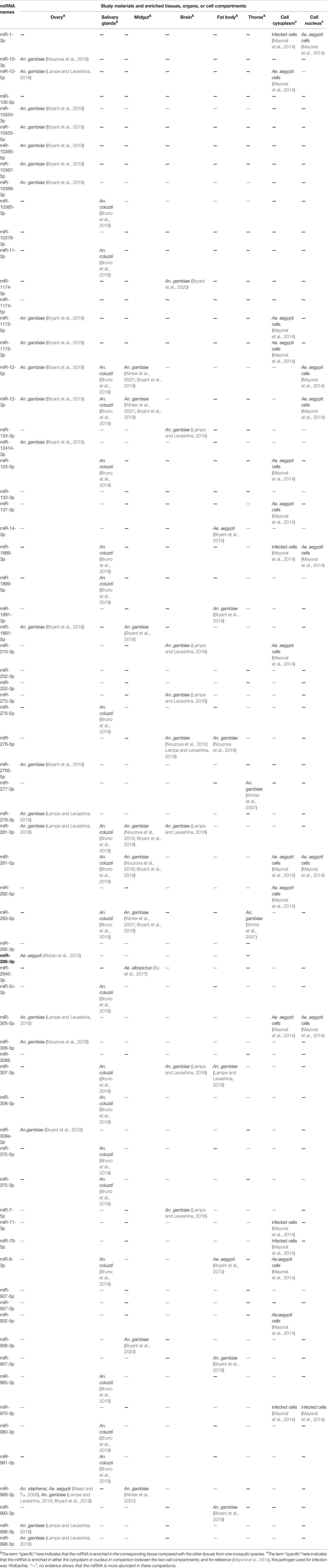
| Ovary& | Salivary glands& | Midgut& | Brain& | Fat body& | Thorax& | Cell cytoplasm# | Cell nucleus# | |
| miR-1-3p | — | — | — | — | — | — | infected cells (Mayoral et al., 2014) | Ae. aegypti cells (Mayoral et al., 2014) |
| miR-10-3p | An. gambiae (Nouzova et al., 2018) | — | — | — | — | — | — | — |
| miR-10-5p | An. gambiae (Lampe and Levashina, 2018) | — | — | — | — | — | Ae. aegypti cells (Mayoral et al., 2014) | — |
| miR-100-5p | — | — | — | — | — | — | — | — |
| miR-10355-3p | An. gambiae () | — | — | — | — | — | — | — |
| miR-10355-5p | An. gambiae () | — | — | — | — | — | — | — |
| miR-10365-5p | An. gambiae () | — | — | — | — | — | — | — |
| miR-10367-5p | An. gambiae () | — | — | — | — | — | — | — |
| miR-10368-3p | An. gambiae () | — | — | — | — | — | — | — |
| miR-10365-3p | — | An. coluzzii () | — | — | — | — | — | — |
| miR-10376-3p | — | — | — | — | — | — | — | — |
| miR-11-3p | — | An. coluzzii () | — | — | — | — | — | — |
| miR-1174-3p | — | — | — | An. gambiae () | — | — | — | — |
| miR-1174-5p | — | — | — | — | — | — | — | — |
| miR-1175-5p | An. gambiae () | — | — | — | — | — | Ae. aegypti cells (Mayoral et al., 2014) | — |
| miR-1175-3p | An. gambiae () | — | — | — | — | — | Ae. aegypti cells (Mayoral et al., 2014) | — |
| miR-12-5p | An. gambiae () | An. coluzzii () | An. gambiae (Winter et al., 2007; ) | — | — | — | — | Ae. aegypti cells (Mayoral et al., 2014) |
| miR-12-3p | An. gambiae () | An. coluzzii () | An. gambiae (Winter et al., 2007; ) | — | — | — | — | Ae. aegypti cells (Mayoral et al., 2014) |
| miR-124-3p | — | — | — | An. gambiae (Lampe and Levashina, 2018) | — | — | — | — |
| miR-12414-3p | An. gambiae () | — | — | — | — | — | — | — |
| miR-125-5p | An. coluzzii () | — | — | — | — | Ae. aegypti cells (Mayoral et al., 2014) | — | |
| miR-133-3p | — | — | — | — | — | — | — | — |
| miR-137-3p | — | — | — | — | — | — | Ae. aegypti cells (Mayoral et al., 2014) | — |
| miR-14-3p | — | — | — | — | Ae. aegypti () | — | — | — |
| miR-1889-3p | — | An. coluzzii () | — | — | — | — | infected cells (Mayoral et al., 2014) | Ae. aegypti cells (Mayoral et al., 2014) |
| miR-1889-5p | — | An. coluzzii () | — | — | — | — | — | — |
| miR-1891-3p | — | — | — | — | An. gambiae () | — | — | — |
| miR-1891-5p | An. gambiae () | — | An. gambiae () | — | — | — | — | — |
| miR-210-3p | — | — | — | An. gambiae (Lampe and Levashina, 2018) | Ae. aegypti cells (Mayoral et al., 2014) | |||
| miR-252-5p | — | — | — | — | — | — | — | — |
| miR-252-3p | — | — | — | — | — | — | — | — |
| miR-275-3p | — | — | — | An. gambiae (Lampe and Levashina, 2018) | — | — | — | — |
| miR-275-5p | — | An. coluzzii () | — | — | — | — | — | — |
| miR-276-5p | — | — | — | An. gambiae (Nouzova et al., 2018; Lampe and Levashina, 2018) | An. gambiae (Nouzova et al., 2018) | — | — | — |
| miR-2765-5p | An. gambiae () | — | — | — | — | — | — | — |
| miR-277-3p | — | — | — | — | — | An. gambiae (Winter et al., 2007) | — | — |
| miR-279-3p | An. gambiae (Lampe and Levashina, 2018) | — | — | — | — | — | — | — |
| miR-281-3p | An. gambiae (Lampe and Levashina, 2018) | An. coluzzii () | An. gambiae (Nouzova et al., 2018; ) | An. gambiae (Lampe and Levashina, 2018) | — | — | — | — |
| miR-281-5p | An. coluzzii () | An. gambiae (Nouzova et al., 2018; ) | — | — | — | Ae. aegypti cells (Mayoral et al., 2014) | Ae. aegypti cells (Mayoral et al., 2014) | |
| miR-282-5p | — | — | — | — | — | — | Ae. aegypti cells (Mayoral et al., 2014) | |
| miR-283-5p | — | An. coluzzii () | An. gambiae (Winter et al., 2007; ) | — | — | An. gambiae (Winter et al., 2007) | — | — |
| miR-285-3p | — | — | — | — | — | — | ||
| miR-286-3p | Ae. aegypti () | — | — | — | — | — | ||
| miR-2945-3p | — | — | Ae. albopictus (Su et al., 2017) | — | — | — | — | — |
| miR-2c-3p | — | An. coluzzii () | — | — | — | — | — | — |
| miR-305-5p | An. gambiae (Lampe and Levashina, 2018) | — | — | — | — | — | Ae. aegypti cells (Mayoral et al., 2014) | Ae. aegypti cells (Mayoral et al., 2014) |
| miR-306-5p | An. gambiae (Nouzova et al., 2018) | — | — | — | — | — | — | — |
| miR-3069 | — | — | — | — | — | — | — | — |
| miR-307-3p | — | An. coluzzii () | An. gambiae (Lampe and Levashina, 2018) | An. gambiae (Lampe and Levashina, 2018) | — | — | — | |
| miR-308-3p | — | An. coluzzii () | — | — | — | — | — | — |
| miR-309a-3p | An.gambiae () | — | — | — | — | — | — | — |
| miR-375-5p | — | An. coluzzii () | — | — | — | — | — | — |
| miR-375-3p | — | An. coluzzii () | — | — | — | — | — | — |
| miR-7-5p | — | — | — | An. gambiae (Lampe and Levashina, 2018) | — | — | — | — |
| miR-71-3p | — | — | — | — | — | — | infected cells (Mayoral et al., 2014) | — |
| miR-79-5p | — | — | — | — | — | — | infected cells (Mayoral et al., 2014) | — |
| miR-8-3p | — | An. coluzzii () | — | — | Ae. aegypti () | — | Ae.aegypti cells (Mayoral et al., 2014) | |
| miR-927-5p | — | — | — | — | — | — | — | — |
| miR-927-3p | — | — | — | — | — | — | — | — |
| miR-932-5p | — | — | — | — | — | — | Ae.aegypti cells (Mayoral et al., 2014) | |
| miR-956-3p | — | — | An. gambiae () | — | — | — | — | — |
| miR-957-5p | — | — | — | — | An. gambiae () | — | — | — |
| miR-965-3p | — | An. coluzzii () | — | — | — | — | — | — |
| miR-970-3p | — | — | — | — | — | — | infected cells (Mayoral et al., 2014) | infected cells (Mayoral et al., 2014) |
| miR-980-3p | — | An. coluzzii () | — | — | — | — | — | — |
| miR-981-3p | — | An. coluzzii () | — | — | — | — | — | — |
| miR-989-3p | An. stephensi, Ae. aegypti (Mead and Tu, 2008), An. gambiae (Lampe and Levashina, 2018; ) | — | An. gambiae (Winter et al., 2007) | — | — | — | — | — |
| miR-993-3p | — | — | — | — | An. gambiae () | — | — | — |
| miR-998-3p | An. gambiae (Lampe and Levashina, 2018) | — | — | — | — | — | — | — |
| miR-998-5p | An. gambiae (Lampe and Levashina, 2018) | — | — | — | — | — | — | — |
Tissue-, organ- or cell compartment- specific miRNAs.

&The term “specific” here indicates that the miRNA is enriched in the corresponding tissue compared with the other tissues from one mosquito species. #The term “specific” here indicates that the miRNA is enriched in either the cytoplasm or nucleus in comparison between the two cell compartments; and for reference (Mayoral et al., 2014), the pathogen used for infection was Wolbachia. “—”, no evidence shows that the miRNA is more abundant in these comparisons.
Table 6
| miRNA names | Study materials* and changes in expression levels upon pathogen infection | ||||||
|---|---|---|---|---|---|---|---|
| CHIKV infection | Plasmodium infection | DENV infection | Wolbachia infection | ZIKA infection | WNV infection | BTV infection | |
| miR-1-3p | Ae. albopictus cells (Shrinet et al., 2014) | An. anthropophagus midgut (Liu et al., 2017), An. stephensi (Jain et al., 2014) | Ae. albopictus midgut (Su et al., 2017), Ae. albopictus (Liu et al., 2015) | Ae. aegypti cell cytoplasm (Mayoral et al., 2014), Ae. aegypti cell nucleus (Mayoral et al., 2014) | Ae. aegypti (Saldana et al., 2017) | — | — |
| miR-1-5p | — | — | — | — | Ae. aegypti (Saldana et al., 2017) | — | — |
| miR-10-5p | Ae.aegypti saliva, Ae. albopictus saliva (Maharaj et al., 2015), Ae. aegypti () | An. anthropophagus midguts (Liu et al., 2017), An. stephensi (Jain et al., 2014) | Ae. aegypti (Liu et al., 2015; ), C6/36 cells () | Ae. aegypti cell nucleus (Mayoral et al., 2014) | — | — | — |
| miR-100-5p | Ae. albopictus cells (Shrinet et al., 2014), Ae. aegypti saliva, Ae. albopictus saliva (Maharaj et al., 2015), Ae. aegypti (Saldana et al., 2017) | An. anthropophagus midguts (Liu et al., 2017), An. stephensi (Jain et al., 2014) | Ae. albopictus (Yan et al., 2014) | Ae. aegypti cell cytoplasm and nucleus (Mayoral et al., 2014) | — | — | — |
| miR-1000-5p | Ae.aegypti saliva (Maharaj et al., 2015), Ae. albopictus cells (Shrinet et al., 2014) | An. anthropophagus midguts (Liu et al., 2017), An. stephensi (Jain et al., 2014) | Ae. albopictus midgut (Su et al., 2017), Ae. aegypti () | Ae. aegypti cell cytoplasm and nucleus (Mayoral et al., 2014) | — | — | — |
| miR-109 | Ae. aegypti saliva (Maharaj et al., 2015) | — | — | — | — | — | — |
| miR-11-3p | Ae. albopictus cells (Shrinet et al., 2014), Ae. aegypti saliva (Maharaj et al., 2015) | An. anthropophagus midguts (Liu et al., 2017), An. stephensi (Jain et al., 2014) | — | Ae. aegypti cell cytoplasm (Mayoral et al., 2014) | — | — | — |
| miR-11-5p | Ae. albopictus cells (Shrinet et al., 2014) | — | — | Ae. aegypti cell nucleus (Mayoral et al., 2014) | — | — | — |
| miR-115 | Ae. aegypti saliva (Maharaj et al., 2015) | — | — | — | — | — | — |
| miR-117 | Ae. aegypti saliva (Maharaj et al., 2015) | — | — | — | — | — | — |
| miR-1174-3p | Ae. aegypti saliva (Maharaj et al., 2015) | An. stephensi (Jain et al., 2014), An. gambiae (Winter et al., 2007) | Ae. albopictus midgut (Su et al., 2017) | Ae. aegypti cell cytoplasm (Mayoral et al., 2014) | — | — | — |
| miR-1175-5p | — | An. stephensi (Jain et al., 2014) | Ae. albopictus midgut (Su et al., 2017) | Ae. aegypti cell nucleus (Mayoral et al., 2014), Ae. aegypti cell cytoplasm (Mayoral et al., 2014) | — | — | — |
| miR-1175-3p | Ae. albopictus cells (Shrinet et al., 2014), Ae. aegypti saliva (Maharaj et al., 2015) | An. anthropophagus midguts (Liu et al., 2017), An. gambiae (Winter et al., 2007), An.stephensi (Jain et al., 2014) | Ae. albopictus midgut (Su et al., 2017), Ae. aegypti () | Ae. aegypti cell cytoplasm (Mayoral et al., 2014) | — | — | — |
| miR-11900 | Ae. aegypti cells (Miesen et al., 2016; Zhang et al., 2017) | — | — | — | — | ||
| miR-12-5p | Ae. aegypti saliva (Maharaj et al., 2015), Ae. albopictus cells (Shrinet et al., 2014) | An. anthropophagus midguts (Liu et al., 2017), An. stephensi (Jain et al., 2014) | Ae. albopictus midguts (Su et al., 2017) | Ae. aegypti cells (Osei-Amo et al., 2012) | — | — | — |
| miR-12-3p | — | — | — | Ae. aegypti cell nucleus and cytoplasm (Mayoral et al., 2014) | — | — | — |
| miR-124-3p | — | An. stephensi (Jain et al., 2014) | Ae. aegypti (), C6/36 cells () | Ae. aegypti cell nucleus and cytoplasm (Mayoral et al., 2014) | — | — | — |
| miR-1247 | Ae. aegypti saliva (Maharaj et al., 2015) | — | — | — | — | — | — |
| miR-125-5p | Ae. albopictus cells (Shrinet et al., 2014), Ae. aegypti saliva, Ae. albopictus saliva (Maharaj et al., 2015) | An. anthropophagus midguts (Liu et al., 2017), An. stephensi (Jain et al., 2014) | — | Ae. aegypti (Hussain et al., 2013), Ae. aegypti cell cytoplasm and nucleus (Mayoral et al., 2014) | — | — | — |
| miR-125-3p | Ae. albopictus cells (Shrinet et al., 2014) | An. stephensi (Jain et al., 2014) | — | — | — | — | — |
| miR-127 | Ae. albopictus saliva (Maharaj et al., 2015) | — | — | — | — | — | |
| miR-13-3p | Ae. aegypti saliva (Maharaj et al., 2015), C6/36 cells (Shrinet et al., 2014), Ae. albopictus saliva (Maharaj et al., 2015) | An. anthropophagus midgut (Liu et al., 2017), An. stephensi (Jain et al., 2014) | — | — | — | — | Ae. albopictus cells (Xing et al., 2016) |
| miR-13-5p | Ae. albopictus cells (Shrinet et al., 2014) | An. stephensi (Jain et al., 2014) | — | — | — | — | |
| miR-133-3p | Ae. aegypti saliva, Ae. albopictus saliva (Maharaj et al., 2015), Ae. albopictus cells (Shrinet et al., 2014) | An. stephensi (Jain et al., 2014), An. anthropophagus midgut (Liu et al., 2017) | — | — | — | — | Ae. albopictus cells (Xing et al., 2016) |
| miR-133-5p | An. stephensi (Jain et al., 2014) | — | — | — | — | — | |
| miR-137-3p | Ae.albopictus saliva (Maharaj et al., 2015), Ae. albopictus cells (Shrinet et al., 2014) | An. stephensi iBF at 42h (Jain et al., 2014), An. anthropophagus midguts (Liu et al., 2017), An. stephensi iBF at 5d (Jain et al., 2014) | — | — | — | — | — |
| miR-14-3p | Ae. aegypti saliva, Ae. albopictus saliva (Maharaj et al., 2015), Ae. albopictus cells (Shrinet et al., 2014) | An. stephensi (Jain et al., 2014), An. anthropophagus midguts (Liu et al., 2017) | — | Ae. aegypti cell cytoplasm (Mayoral et al., 2014) | — | — | — |
| miR-143 | Ae. aegypti saliva, Ae. albopictus saliva (Maharaj et al., 2015) | — | — | — | — | — | — |
| miR-15-3p | — | — | HEK293 and HeLa cells (Smith et al., 2017) | — | HEK293 and HeLa cells (Smith et al., 2017) | HEK293 and HeLa cells (Smith et al., 2017) | — |
| miR-157 | Ae. albopictus saliva (Maharaj et al., 2015) | — | — | — | — | — | — |
| miR-1571 | Ae. aegypti saliva (Maharaj et al., 2015) | — | — | — | — | — | — |
| miR-1767 | — | Ae. albopictus midgut (Su et al., 2017; Su et al., 2019) | — | — | — | — | |
| miR-184-3p | Ae. albopictus saliva (Maharaj et al., 2015), C6/36 cells (), Ae. aegypti saliva (Maharaj et al., 2015) | An. anthropophagus midguts (Liu et al., 2017), An. stephensi (Jain et al., 2014) | Ae. albopictus (Liu et al., 2015), Ae. albopictus midguts (Su et al., 2017) | — | — | — | — |
| miR-1889-3p | — | — | Ae. albopictus and C6/36 cells (Yan et al., 2014), Ae. albopictus (Liu et al., 2015) | Ae. aegypti cell cytoplasm (Mayoral et al., 2014) | — | — | — |
| miR-1889-5p | Ae. aegypti saliva (Maharaj et al., 2015) | — | — | Ae. aegypti cell cytoplasm (Mayoral et al., 2014) | — | — | Ae. albopictus cells (Xing et al., 2016) |
| miR-1890-3p | Ae. aegypti saliva (Maharaj et al., 2015), Ae. albopictus cells (Shrinet et al., 2014) | An. anthropophagus midguts (Liu et al., 2017), An. stephensi (Jain et al., 2014) | — | Ae. aegypti cell cytoplasm (Mayoral et al., 2014) | — | — | — |
| miR-1891-5p | Ae. aegypti saliva, Ae. albopictus saliva (Maharaj et al., 2015), Ae. albopictus cells (Shrinet et al., 2014) | An. stephensi (Jain et al., 2014) | Ae. albopictus midgut (Su et al., 2017), C6/36 cells, Ae. albopictus (Yan et al., 2014) | Ae. aegypti (Hussain et al., 2011) | — | — | — |
| miR-190-5p | Ae. albopictus cells (Shrinet et al., 2014) | An. stephensi (Jain et al., 2014) | C6/36 cells () | Ae. aegypti cell cytoplasm (Mayoral et al., 2014) | — | — | — |
| miR-190-3p | — | An. stephensi (Jain et al., 2014) | — | — | — | — | — |
| miR-193-5p | — | — | Ae. albopictus (Su et al., 2019) | — | — | — | — |
| miR-193-3p | Ae. albopictus cells (Shrinet et al., 2014) | — | — | — | — | — | — |
| miR-1951 | — | Ae. albopictus midgut (Su et al., 2019) | — | — | — | — | |
| miR-210-3p | Ae. aegypti saliva (Maharaj et al., 2015), Ae. albopictus saliva (Maharaj et al., 2015) | An. stephensi (Jain et al., 2014), An. anthropophagus midguts (Liu et al., 2017) | C6/36 cells () | Ae. aegypti (Hussain et al., 2011), C6/36 cells cytoplasm (Mayoral et al., 2014) | — | — | — |
| miR-210-5p | Ae. albopictus cells (Shrinet et al., 2014) | — | Ae. aegypti () | — | — | — | — |
| miR-214 | Ae.aegypti saliva (Maharaj et al., 2015), Ae. albopictus saliva (Maharaj et al., 2015) | — | — | — | — | — | — |
| miR-219-5p | Ae. albopictus cells (Shrinet et al., 2014) | — | — | Ae. aegypti cell cytoplasm (Mayoral et al., 2014) | — | — | — |
| miR-229 | Ae. aegypti saliva (Maharaj et al., 2015) | — | — | — | — | — | — |
| miR-23 | Ae. aegypti saliva (Maharaj et al., 2015) | — | — | — | — | — | — |
| miR-2308 | Ae. albopictus saliva (Maharaj et al., 2015) | — | — | — | — | — | — |
| miR-242 | Ae. aegypti saliva (Maharaj et al., 2015) | — | — | — | — | — | — |
| miR-249 | Ae. albopictus saliva, Ae. aegypti saliva (Maharaj et al., 2015) | — | — | — | — | — | — |
| miR-252-5p | Ae. aegypti saliva (Maharaj et al., 2015), Ae. albopictus saliva (Maharaj et al., 2015) | An. stephensi (Jain et al., 2014), An. anthropophagus midgut (Liu et al., 2017) | C6/36 cells (Yan et al., 2014), C6/36 cells () | Ae. aegypti (Hussain et al., 2011), Ae. aegypti cell cytoplasm (Mayoral et al., 2014) | — | — | — |
| miR-252-3p | — | — | Ae. albopictus cells (Shrinet et al., 2014) | — | — | — | — |
| miR-263a-3p | — | — | C6/36 cells () | — | — | — | — |
| miR-263a-5p | — | — | — | — | Ae. aegypti (Saldana et al., 2017) | — | — |
| miR-275-3p | Ae. albopictus cells (Shrinet et al., 2014), Ae. aegypti saliva, Ae.albopictus saliva (Maharaj et al., 2015) | An. stephensi (Jain et al., 2014), An. anthropophagus midgut (Liu et al., 2017) | Ae. albopictus (Liu et al., 2015), C7/10 cells, Ae. albopictus midgut (Su et al., 2017) | — | — | — | — |
| miR-275-5p | Ae. albopictus cells (Shrinet et al., 2014) | — | — | — | — | — | — |
| miR-276-3p | Ae. aegypti saliva, Ae. albopictus saliva (Maharaj et al., 2015) | An. anthropophagus midguts (Liu et al., 2017), An. stephensi (Jain et al., 2014) | Ae. albopictus midgut (Su et al., 2017), Ae. aegypti (), Ae. albopictus (Liu et al., 2015) | Ae. aegypti cell cytoplasm (Mayoral et al., 2014) | — | — | — |
| miR-276-5p | — | An. stephensi (Jain et al., 2014) | Ae. albopictus (Liu et al., 2015), Ae. albopictus midguts (Su et al., 2019), Ae. aegypti (), Ae. albopictus midgut (Su et al., 2017) | — | — | — | Ae. albopictus cells (Xing et al., 2016) |
| miR-2765-5p | Ae. albopictus cells (Shrinet et al., 2014) | — | — | Ae. aegypti cell nucleus (Mayoral et al., 2014) | — | — | — |
| miR-277-3p | Ae. albopictus cells (Shrinet et al., 2014), Ae. aegypti saliva, Ae. albopictus saliva (Maharaj et al., 2015) | An. anthropophagus midguts (Liu et al., 2017), An. stephensi (Jain et al., 2014) | Ae. albopictus (Liu et al., 2015) | Ae. aegypti (Hussain et al., 2011), Ae. aegypti cell nucleus (Mayoral et al., 2014) | — | — | — |
| miR-278-3p | Ae. albopictus cells (Shrinet et al., 2014) | An. anthropophagus midguts (Liu et al., 2017), An. stephensi (Jain et al., 2014) | — | Ae. aegypti cell cytoplasm (Mayoral et al., 2014) | — | — | Ae. albopictus cells (Xing et al., 2016) |
| miR-278-5p | Ae. albopictus cells (Shrinet et al., 2014), Ae. aegypti () | — | — | Ae. aegypti cell cytoplasm (Mayoral et al., 2014) | — | — | — |
| miR-279-3p | Ae.aegypti saliva (Maharaj et al., 2015), Ae. albopictus cells (Shrinet et al., 2014), Ae. albopictus saliva (Maharaj et al., 2015) | An. anthropophagus midgut (Liu et al., 2017), An. stephensi (Jain et al., 2014) | — | — | — | — | — |
| miR-2796-5p | Ae. albopictus cells (Shrinet et al., 2014) | An. stephensi (Jain et al., 2014) | Ae. albopictus (Liu et al., 2015) | — | — | — | — |
| miR-2779 | Ae. aegypti () | — | — | — | — | — | — |
| miR-28 | Ae. albopictus saliva (Maharaj et al., 2015) | — | — | — | — | — | — |
| miR-281-3p | — | An. stephensi (Jain et al., 2014) | Ae. albopictus (Zhou et al., 2014) | Ae. aegypti (Hussain et al., 2011), Ae. aegypti cell cytoplasm (Mayoral et al., 2014) | — | — | — |
| miR-281-5p | Ae. albopictus saliva (Maharaj et al., 2015), Ae. aegypti saliva (Maharaj et al., 2015) | An. stephensi (Jain et al., 2014) | Ae. albopictus (Liu et al., 2015; Su et al., 2017) | Ae. aegypti cell nucleus (Mayoral et al., 2014) | — | — | — |
| miR-282-5p | — | — | Ae. albopictus (Su et al., 2017) | Ae. aegypti cell nucleus (Mayoral et al., 2014) | — | — | — |
| miR-283-5p | Ae. albopicts midgut (Maharaj et al., 2015), Ae. albopictus cells (Shrinet et al., 2014) | An. anthropophagus midguts (Liu et al., 2017) | — | — | — | — | — |
| miR-285-3p | Ae. aegypti saliva (Maharaj et al., 2015), Ae. albopic- tus saliva (Maharaj et al., 2015), Ae. aegypti () | An. stephensi (Jain et al., 2014) | — | Ae. aegypti cell cytoplasm (Mayoral et al., 2014) | — | — | Ae. albopictus cells (Xing et al., 2016) |
| miR-286b-5p | — | — | C6/36 cells () | — | — | — | — |
| miR-286a-3p | — | — | C6/36 cells () | — | — | — | — |
| miR-2940-3p | — | — | Ae. albopictus (Liu et al., 2015) | — | Ae. aegypti (Saldana et al., 2017) | — | — |
| miR-2940-5p | Ae. albopictus cells (Shrinet et al., 2014), Ae. aegypti saliva (Maharaj et al., 2015) | An. gambiae () | Ae. albopictus (Liu et al., 2015) | Ae. aegypti and Aedes aegypti cells (Hussain et al., 2011; Zhang et al., 2013; Mayoral et al., 2014; ) | — | C6/36 cells (Slonchak et al., 2014) | — |
| miR-2941-3p | Ae. albopictus cells (Shrinet et al., 2014) | — | Ae. albopictus (Su et al., 2019), Ae. albopictus (Liu et al., 2015), Ae. albopictus midgut (Liu et al., 2016) | Ae. aegypti (Hussain et al., 2011), Aedes aegypti cell nucleus and cytoplasm (Mayoral et al., 2014) | Ae. aegypti (Saldana et al., 2017) | — | — |
| miR-2943-5p | — | — | Ae. albopictus midgut (Liu et al., 2016) | Ae. aegypti (Hussain et al., 2011) | — | — | — |
| miR-2944a-5p | Ae. albopictus cells (Shrinet et al., 2014) | An. stephensi (Shrinet et al., 2014) | — | — | — | — | — |
| miR-2945-3p | Ae. aegypti saliva (Maharaj et al., 2015), Ae. albopictus cells (Shrinet et al., 2014) | An. stephensi (Jain et al., 2014) | C6/36 cells (Yan et al., 2014), Ae. albopictus (Liu et al., 2015), Ae. aegypti () | Ae. aegypti (Hussain et al., 2011), Ae. aegypti cells (Mayoral et al., 2014) | — | — | — |
| miR-2946-3p | Ae. aegypti saliva (Maharaj et al., 2015) | — | — | Ae. aegypti cells (Mayoral et al., 2014) | Ae. aegypti (Saldana et al., 2017) | — | Ae. albopictus cells (Xing et al., 2016) |
| miR-2951-5p | Ae. aegypti () | — | — | — | — | — | — |
| miR-2a-3p | — | — | C6/36 cells () | — | — | — | — |
| miR-3 | Ae. aegypti saliva (Maharaj et al., 2015) | — | — | — | — | — | — |
| miR-305-5p | Ae. aegypti saliva (Maharaj et al., 2015), Ae. albopictus cell (Shrinet et al., 2014), Ae.albopictus saliva (Maharaj et al., 2015) | An. gambiae midguts (), An. anthropophagus midguts (Liu et al., 2017), An. stephensi (Jain et al., 2014) | Ae. aegypti () | Ae. aegypti cells (Mayoral et al., 2014) | — | — | — |
| miR-305-3p | — | An. stephensi (Jain et al., 2014) | — | — | — | — | — |
| miR-306-5p | Ae. aegypti saliva (Maharaj et al., 2015), Ae. albopictus cells (Shrinet et al., 2014) | An. stephensi (Jain et al., 2014), An. anthropophagus midguts (Liu et al., 2017) | Ae. albopictus (Liu et al., 2015) | Ae. aegypti cell cytoplasm (Mayoral et al., 2014), Ae. aegypti cell nucleus (Mayoral et al., 2014) | Ae. aegypti (Saldana et al., 2017) | — | — |
| miR-3069 | Ae. aegypti saliva (Maharaj et al., 2015) | — | — | — | — | — | — |
| miR-307-3p | Ae. aegypti saliva (Maharaj et al., 2015) | — | — | — | — | — | — |
| miR-308-3p | — | An. anthropophagus midguts (Liu et al., 2017), An. stephensi (Jain et al., 2014) | Ae. aegypti () | — | Ae. aegypti (Saldana et al., 2017) | — | Ae. albopictus cells (Xing et al., 2016) |
| miR-308-5p | C6/36 cells (Shrinet et al., 2014), Ae. aegypti saliva (Maharaj et al., 2015) | — | — | Ae. aegypti (Hussain et al., 2011), Ae. aegypti cell nucleus (Mayoral et al., 2014) | Ae. aegypti (Saldana et al., 2017) | — | — |
| miR-309a-3p | Ae. aegypti saliva, Ae. albopictus saliva (Maharaj et al., 2015) | An. anthropophagus midgut (Liu et al., 2017), An. stephensi (Jain et al., 2014) | — | — | — | — | — |
| miR-31-5p | Ae. aegypti saliva (Maharaj et al., 2015) | An. anthropophagus midguts (Liu et al., 2017) | — | — | — | — | — |
| miR-315-5p | Ae. aegypti saliva, Ae. albopictus saliva (Maharaj et al., 2015) | An. stephensi (Jain et al., 2014) | — | — | — | — | — |
| miR-315-3p | An. anthropophagus midguts (Liu et al., 2017) | — | — | — | — | — | |
| miR-317-3p | Ae. aegypti saliva (Maharaj et al., 2015), Ae. aegypti (), Ae. albopictus saliva (Maharaj et al., 2015) | An. gambiae (), An. stephensi (Jain et al., 2014), An. anthropophagus midguts (Liu et al., 2017) | Ae. albopictus (Liu et al., 2015), Ae. albopictus midgut (Su et al., 2017) | Ae. aegypti (Hussain et al., 2011), Ae. aegypti cell cytoplasm (Mayoral et al., 2014) | — | — | — |
| miR-317-5p | Ae. albopictus cells (Shrinet et al., 2014) | — | — | — | — | — | — |
| miR-320 | Ae. aegypti saliva (Maharaj et al., 2015) | — | — | — | — | — | — |
| miR-33-5p | Ae. aegypti saliva (Maharaj et al., 2015), Ae. albopictus cell (Shrinet et al., 2014) | — | Ae. albopictus (Yan et al., 2014) | Ae. aegypti cell cytoplasm (Mayoral et al., 2014) | — | HEK293 cells (Slonchak et al., 2015) | — |
| miR-3368-5p | — | — | Ae. aegypti () | — | — | — | — |
| miR-34-3p | Ae. aegypti cells (Shrinet et al., 2014) | — | — | Ae. aegypti cell cytoplasm (Mayoral et al., 2014), Ae. aegypti cell nucleus (Mayoral et al., 2014) | — | — | — |
| miR-34-5p | Ae. aegypti saliva (Maharaj et al., 2015), Ae. aegypti (), Ae. aegypti cells (Shrinet et al., 2014), Ae. albopictus saliva (Maharaj et al., 2015) | An. anthropophagus midguts (Liu et al., 2017), An. gambiae (Winter et al., 2007), An. stephensi (Jain et al., 2014) | Ae. aegypti (), Ae. albopictus midgut (Su et al., 2017), Ae. albopictus (Su et al., 2019) | Ae. aegypti (Hussain et al., 2011), Ae. aegypti cell nucleus (Mayoral et al., 2014) | — | — | Ae. albopictus cells (Xing et al., 2016) |
| miR-341 | Ae. aegypti saliva (Maharaj et al., 2015) | — | — | — | — | — | — |
| miR-3722-5p | — | — | Ae. aegypti () | — | — | — | — |
| miR-375-3p | Ae. albopictus cells (Shrinet et al., 2014), Ae. aegypti saliva (Maharaj et al., 2015) | An. anthropophagus midguts (Liu et al., 2017), An. stephensi (Jain et al., 2014) | Ae. albopictus midgut (Su et al., 2017) | — | Ae. aegypti (Saldana et al., 2017) | — | — |
| miR-359 | Ae. aegypti saliva (Maharaj et al., 2015) | — | — | — | — | — | — |
| miR-360 | Ae. aegypti saliva (Maharaj et al., 2015) | — | — | — | — | — | — |
| miR-40 | Ae. aegypti saliva (Maharaj et al., 2015) | — | — | — | — | — | — |
| miR-402 | Ae. aegypti saliva (Maharaj et al., 2015) | — | — | — | — | — | — |
| miR-408 | Ae. aegypti saliva (Maharaj et al., 2015) | — | — | — | — | — | — |
| miR-4110-5p | — | — | Ae. albopictus midgut (Su et al., 2019) | — | — | — | — |
| miR-424-3p | — | — | Ae. albopictus midgut (Su et al., 2019) | — | — | — | — |
| miR-4275-5p | — | — | Ae. aegypti () | — | — | — | — |
| miR-4448 | — | — | Ae. albopictus midgut (Su et al., 2017), Ae. albopictus midgut (Su et al., 2019) | — | — | — | — |
| miR-446 | Ae. aegypti saliva (Maharaj et al., 2015) | — | — | — | — | — | |
| miR-4682 | Ae. aegypti saliva (Maharaj et al., 2015) | — | — | — | — | — | |
| miR-47 | Ae. aegypti saliva (Maharaj et al., 2015) | — | — | — | — | — | |
| miR-4728-5p | — | Ae. albopictus midgut (Su et al., 2017) | — | — | — | — | |
| miR-5 | Ae. aegypti saliva, Ae. albopictus saliva (Maharaj et al., 2015) | — | — | — | — | — | |
| miR-5108-5p | — | Ae. aegypti () | — | — | — | — | |
| miR-5119-5p | — | Ae. aegypti () | — | — | — | — | |
| miR-5706 | — | Ae. albopictus midgut (Su et al., 2019) | — | — | — | — | |
| miR-576 | Ae. aegypti saliva (Maharaj et al., 2015) | — | — | — | — | — | — |
| miR-6 | Ae. aegypti saliva (Maharaj et al., 2015) | — | — | — | — | — | — |
| miR-62 | Ae. albopictus saliva (Maharaj et al., 2015) | — | — | — | — | — | — |
| miR-620 | Ae. aegypti saliva (Maharaj et al., 2015) | — | — | — | — | — | — |
| miR-622 | — | Ae. albopictus midgut (Su et al., 2017; Su et al., 2019) | — | — | — | — | |
| miR-69 | Ae. aegypti saliva (Maharaj et al., 2015) | — | — | — | — | — | — |
| miR-7-5p | C6/36 cells (Shrinet et al., 2014) | An. stephensi (Jain et al., 2014) | — | Ae. aegypti cell nucleus (Mayoral et al., 2014) | — | — | Ae. albopictus cells (Xing et al., 2016) |
| miR-71-5p | Ae. aegypti () | An. stephensi (Jain et al., 2014) | — | Ae. aegypti (Saldana et al., 2017) | — | — | |
| miR-71-3p | Ae. aegypti saliva (Maharaj et al., 2015), C6/36 cells (Shrinet et al., 2014), Ae. albopictus saliva (Maharaj et al., 2015) | An. stephensi (Jain et al., 2014), An. anthropophagus midguts (Liu et al., 2017) | Ae. albopictus midgut (Su et al., 2017) | Ae. aegypti cell cytoplasm (Mayoral et al., 2014) | — | — | — |
| miR-778 | Ae. aegypti and Ae. albopictus midgut (Maharaj et al., 2015) | — | — | — | — | — | — |
| miR-79-5p | — | — | — | Ae. aegypti cell cytoplasm (Mayoral et al., 2014) | — | — | — |
| miR-8-3p | Ae. albopictus saliva (Maharaj et al., 2015), Ae. albopictus cells (Shrinet et al., 2014) | An. stephensi (Jain et al., 2015; Guo et al., 2017) | Ae. albopictus (Liu et al., 2015), C7/10 cells, Ae. albopictus midgut (Su et al., 2017) | Ae. aegypti (Hussain et al., 2011), Ae. aegypti cell cytoplasm (Mayoral et al., 2014) | — | — | — |
| miR-8-5p | Ae. albopictus cells (Shrinet et al., 2014) | An. stephensi (Jain et al., 2014) | Ae. albopictus (Liu et al., 2015) | Ae. aegypti cell cytoplasm (Mayoral et al., 2014) | — | — | — |
| miR-80 | Ae.aegypti saliva (Maharaj et al., 2015) | — | — | — | — | — | — |
| miR-87-3p | Ae. aegypti saliva (Maharaj et al., 2015), Ae. albopictus cells (Shrinet et al., 2014) | An. anthropophagus midgut (Liu et al., 2017) | Ae. albopictus midgut (Su et al., 2017), C6/36 cells () | — | — | — | — |
| miR-87-5p | — | — | Ae. albopictus midgut (Su et al., 2019) | — | — | — | — |
| miR-927-5p | Ae. albopictus cell (Shrinet et al., 2014), Ae. aegypti midgut, Ae. albopictus midgut (Maharaj et al., 2015) | An. stephensi (Jain et al., 2014) | Ae. albopictus (Liu et al., 2015), C6/36 cells (; ) | — | — | — | — |
| miR-927-3p | — | An. stephensi (Jain et al., 2014) | Ae. albopictus midgut (Su et al., 2017) | — | — | — | Ae. albopictus cells (Xing et al., 2016) |
| miR-929-5p | — | An. stephensi (Jain et al., 2014) | — | — | — | — | — |
| miR-92b-3p | — | — | C6/36 cells () | — | — | — | — |
| miR-92a-3p | — | An. gambiae () | — | — | — | Cx.Quinquefa- sciatus (Skalsky et al., 2010) | — |
| miR-932-3p | — | — | mosquito cell nucleus (Mayoral et al., 2014) | — | — | — | |
| miR-932-5p | Ae. aegypti saliva (Maharaj et al., 2015), C6/36 cells (Shrinet et al., 2014), Ae. albopictus saliva (Maharaj et al., 2015) | An. stephensi (Jain et al., 2014) | Ae. aegypti () | Ae. aegypti cell cytoplasm (Mayoral et al., 2014) | — | — | — |
| miR-956-3p | — | — | Ae. albopictus midgut (Su et al., 2017) | — | — | — | — |
| miR-957-3p | Ae. aegypti saliva, Ae. albopictus saliva (Maharaj et al., 2015) | An. stephensi (Jain et al., 2014), An. anthropophagus midgut (Liu et al., 2017) | Ae. albopictus (Liu et al., 2015) | — | — | Cx.Quinquefa- sciatus (Skalsky et al., 2010) | — |
| miR-965-3p | — | — | — | Ae. aegypti cell nucleus and cytoplasm (Mayoral et al., 2014) | — | — | — |
| miR-970-3p | Ae. aegypti saliva (Maharaj et al., 2015) | An. stephensi (Jain et al., 2014), An. anthropophagus midguts (Liu et al., 2017) | C6/36 cells () | — | — | — | — |
| miR-976-5p | Ae. albopictus midgut (Su et al., 2019) | — | — | — | — | ||
| miR-980-3p | Ae.aegypti saliva (Maharaj et al., 2015), Ae. albopictus cells (Shrinet et al., 2014) | An. stephensi (Jain et al., 2014), An. anthropophagus midgut (Liu et al., 2017) | — | — | Ae. aegypti (Saldana et al., 2017) | Cx.quinquefa- sciatus (Skalsky et al., 2010) | Ae. albopictus cells (Xing et al., 2016) |
| miR-980-5p | Ae. albopictus cells (Shrinet et al., 2014) | — | — | — | — | — | — |
| miR-981-3p | — | An. anthropophagus midguts (Liu et al., 2017), An. stephensi (Jain et al., 2014) | — | — | — | — | — |
| miR-988-5p | Ae. albopictus cells (Shrinet et al., 2014) | Ae. albopictus midgut (Su et al., 2017) | — | — | — | — | |
| miR-988-3p | Ae. albopictus cells (Shrinet et al., 2014) | An. stephensi (Jain et al., 2014) | — | Ae. aegypti (Hussain et al., 2011) | — | — | — |
| miR-989-3p | Ae. aegypti () | An. gambiae midgut (Winter et al., 2007; ), An. gambiae leftover (Winter et al., 2007) | Ae. albopictus midgut (Su et al., 2019), Ae. albopictus midguts (Liu et al., 2016), Ae. albopictus (Su et al., 2017) | Ae. aegypti (Hussain et al., 2011), Ae. aegypti cell nucleus and cytoplasm (Mayoral et al., 2014) | Ae. aegypti (Saldana et al., 2017) | Cx.quinquef- asciatus (Skalsky et al., 2010) | — |
| miR-993-5p | — | An. stephensi (Jain et al., 2014) | — | — | — | — | — |
| miR-993-3p | — | An. stephensi (Jain et al., 2014) | Ae. albopictus midgut (Liu et al., 2015) | — | — | — | — |
| miR-996-3p | Ae. albopictus saliva (Maharaj et al., 2015), C6/36 cells (Shrinet et al., 2014), Ae. aegypti saliva (Maharaj et al., 2015) | An. stephensi (Jain et al., 2014) | — | Ae. aegypti cell cytoplasm (Mayoral et al., 2014) | — | — | — |
| miR-996-5p | — | An. anthropophagus midgut (Liu et al., 2017) | — | — | — | — | — |
| miR-988-5p | — | — | — | Ae. aegypti cell nucleus (Mayoral et al., 2014) | — | — | — |
| miR-988-3p | — | — | — | Ae. aegypti cell cytoplasm (Mayoral et al., 2014) | — | — | — |
| miR-998-3p | Ae. albopictus cells (Shrinet et al., 2014), Ae. aegypti saliva (Maharaj et al., 2015), Ae. aegypti () | An. anthropophagus midguts (Liu et al., 2017), An. stephensi (Jain et al., 2014) | Ae. albopictus midgut (Su et al., 2017) | — | — | — | |
| miR-998-5p | Ae. albopictus cells (Shrinet et al., 2014) | — | — | — | — | — | — |
| miR-999-3p | Ae. albopictus cells (Shrinet et al., 2014), Ae. aegypti saliva, Ae. albopictus saliva (Maharaj et al., 2015) | An. stephensi (Jain et al., 2014) | Ae. aegypti () | — | — | — | — |
| miR-9a-5p | — | — | C6/36 cells () | — | — | — | — |
| miR-iab-4-5p | — | An. anthropophagus midguts (Liu et al., 2017) | — | — | — | — | — |
| miR-iab-8-5p | Ae. albopictus cells (Shrinet et al., 2014) | — | — | — | — | — | — |
| bantam-3p | Ae. aegypti saliva (Maharaj et al., 2015), C6/36 cells (Shrinet et al., 2014), Ae. albopictus saliva (Maharaj et al., 2015) | An. anthropophagus midguts (Liu et al., 2017), An. stephensi (Jain et al., 2014) | Ae. aegypti () | Ae. aegypti cell nucleus (Mayoral et al., 2014) | — | — | — |
| bantam-5p | — | An. stephensi (Jain et al., 2014) | Ae. aegypti (), Ae. albopictus (Liu et al., 2015) | Ae. aegypti cell nucleus (Mayoral et al., 2014), Ae. aegypti cell cytoplasm (Mayoral et al., 2014) | — | — | — |
| let-7-5p | Ae. albopictus cells (Shrinet et al., 2014), Ae. albopictus saliva (Maharaj et al., 2015) | An. stephensi (Jain et al., 2014), An. anthropophagus midguts (Liu et al., 2017) | Ae. albopictus, C6/36 cells (Yan et al., 2014), Ae. aegypti (), Ae. albopictus (Liu et al., 2015; Su et al., 2017) | Ae. aegypti cells (Hussain et al., 2011), nucleus, cytoplasm of cells (Mayoral et al., 2014) | — | — | — |
Alterations in miRNA abundance in response to infection with different pathogens in various mosquito samples.
*The “study materials” are written in two styles, namely bold and nonbold, which means that the miRNAs are upregulated or downregulated upon infection with the corresponding microbes or pathogens infection, respectively. “—”, no related data on upregulation or downregulation is available.
Steps of the Research Roadmap
Progress in studying individual miRNAs with annotated names in the database was tracked (Tables 1–7 and S1), and an overview of study advances is provided in Figure 1. The exploration of miRNA-based approaches proceeded through the following four steps along the proposed research roadmap: identifying mosquito miRNAs (Tables 1–5); validating pathogen- miRNA interactions (Tables 6 and S1); exploring the mechanism of action, which refers mainly to target prediction and verification (Tables 7 and S1); and performing preapplication investigations (Liu P. et al., 2016). These steps involved the 20 items listed in Figure 1, for example, interactions between miRNAs and Plasmodium, dengue virus (DENV), Zika virus (ZIKA), Chikungunya virus (CHIKV), Wolbachia, West Nile virus (WNV), Palm Creek virus (PCV), Japanese Encephalitis virus (JEV), and o’nyong’nyong virus (ONNV).
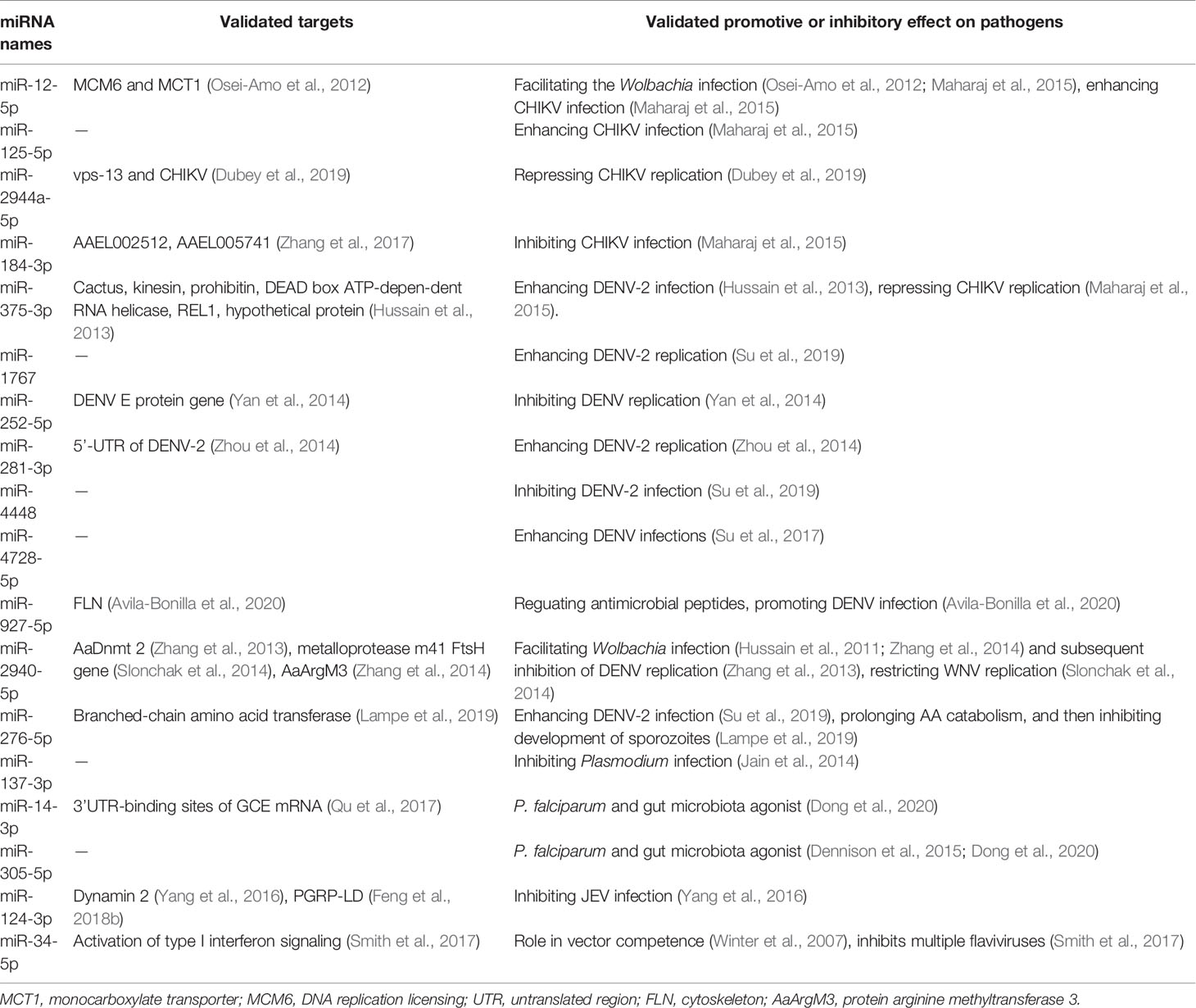
Table 7
| miRNA names | Validated targets | Validated promotive or inhibitory effect on pathogens |
|---|---|---|
| miR-12-5p | MCM6 and MCT1 (Osei-Amo et al., 2012) | Facilitating the Wolbachia infection (Osei-Amo et al., 2012; Maharaj et al., 2015), enhancing CHIKV infection (Maharaj et al., 2015) |
| miR-125-5p | — | Enhancing CHIKV infection (Maharaj et al., 2015) |
| miR-2944a-5p | vps-13 and CHIKV () | Repressing CHIKV replication () |
| miR-184-3p | AAEL002512, AAEL005741 (Zhang et al., 2017) | Inhibiting CHIKV infection (Maharaj et al., 2015) |
| miR-375-3p | Cactus, kinesin, prohibitin, DEAD box ATP-depen-dent RNA helicase, REL1, hypothetical protein (Hussain et al., 2013) | Enhancing DENV-2 infection (Hussain et al., 2013), repressing CHIKV replication (Maharaj et al., 2015). |
| miR-1767 | — | Enhancing DENV-2 replication (Su et al., 2019) |
| miR-252-5p | DENV E protein gene (Yan et al., 2014) | Inhibiting DENV replication (Yan et al., 2014) |
| miR-281-3p | 5’-UTR of DENV-2 (Zhou et al., 2014) | Enhancing DENV-2 replication (Zhou et al., 2014) |
| miR-4448 | — | Inhibiting DENV-2 infection (Su et al., 2019) |
| miR-4728-5p | — | Enhancing DENV infections (Su et al., 2017) |
| miR-927-5p | FLN () | Reguating antimicrobial peptides, promoting DENV infection () |
| miR-2940-5p | AaDnmt 2 (Zhang et al., 2013), metalloprotease m41 FtsH gene (Slonchak et al., 2014), AaArgM3 (Zhang et al., 2014) | Facilitating Wolbachia infection (Hussain et al., 2011; Zhang et al., 2014) and subsequent inhibition of DENV replication (Zhang et al., 2013), restricting WNV replication (Slonchak et al., 2014) |
| miR-276-5p | Branched-chain amino acid transferase (Lampe et al., 2019) | Enhancing DENV-2 infection (Su et al., 2019), prolonging AA catabolism, and then inhibiting development of sporozoites (Lampe et al., 2019) |
| miR-137-3p | — | Inhibiting Plasmodium infection (Jain et al., 2014) |
| miR-14-3p | 3’UTR-binding sites of GCE mRNA (Qu et al., 2017) | P. falciparum and gut microbiota agonist () |
| miR-305-5p | — | P. falciparum and gut microbiota agonist (; ) |
| miR-124-3p | Dynamin 2 (Yang et al., 2016), PGRP-LD () | Inhibiting JEV infection (Yang et al., 2016) |
| miR-34-5p | Activation of type I interferon signaling (Smith et al., 2017) | Role in vector competence (Winter et al., 2007), inhibits multiple flaviviruses (Smith et al., 2017) |
miRNAs with validated targets that exert promotive or inhibitory effects on pathogens.
MCT1, monocarboxylate transporter; MCM6, DNA replication licensing; UTR, untranslated region; FLN, cytoskeleton; AaArgM3, protein arginine methyltransferase 3.
Figure 1
However, no clinical or field trial has been reported, indicating that the miRNA-based approach may have encountered a bottleneck of application in mosquito-borne disease prevention and control, although several attempts to establish application models have been conducted (Heiss et al., 2011; Tsetsarkin et al., 2015; Tsetsarkin et al., 2016a; Tsetsarkin et al., 2016b). The details of significant progress achieved at each step are reviewed below.
Advances at Each Step of the Research Roadmap
Identification of miRNA Profiles in Mosquitoes
The first step in the research roadmap is to understand mosquito miRNA profiles. Currently, studies of mosquito miRNA profiles focus mainly on mosquito miRNA identification (Mead and Tu, 2008; Skalsky et al., 2010; Gu et al., 2013; Jain et al., 2014; ; ; Hu et al., 2015; ; Su et al., 2017; ; ) with the detection of spatial and temporal expression patterns (Jain et al., 2014; Yen et al., 2018), the functional arm between the 5’ and 3’ ends (Skalsky et al., 2010; ; ), the production of miRNAs with varying lengths and sequences (i.e., isomiRs) (Skalsky et al., 2010; ; ; Nouzova et al., 2018), and miRNA clusters (). At this step in the roadmap, variations in the spatial and temporal expression of miRNAs have been observed by analyzing several factors. (i) Mosquito species: For example, miR-282-5p was found to be conserved in Ae. aegypti and An. gambiae but not in Cx. quinquefasciatus (Gu et al., 2013) (Table 2). Additionally, miR-1175-3p expression has been found to display opposite trends (upregulated or downregulated by blood feeding) in Aedes albopictus and Anopheles gambiae, and the corresponding molecular mechanisms may also be different in each species (Winter et al., 2007; Su et al., 2017) (Table 4). (ii) Sexes: For example, miR-989-5p was found to be restricted in female Anopheles coluzzii () (Table 3). (iii) Developmental stages: Notably, miR-1-3p was found to be enriched in the pupa of female Anopheles stephensi (Jain et al., 2015) compared to the larvae and adult (Table 4). (iv) Blood feeding and insecticide resistance statuses: The expression of miR-999-3p was found to be downregulated in deltamethrin-resistant Culex pipiens (Hong et al., 2014) (Table 4). (v) Tissues: For example, miR-998-5p was found to be specifically expressed in the ovary of An. gambiae (Lampe and Levashina, 2018) (Table 5), and miR-8-3p was found to be particularly enriched in the salivary glands in An. coluzzii () but in the fat body in Aedes aegypti () (Table 5). Moreover, in addition to these specificities, miRNAs may even exhibit cellular cytoplasm- or nucleus- specificity (Mayoral et al., 2014) (Table 5).
More examples based on individual miRNAs are noted in Tables 2–5. Overall, the expression levels of miRNAs are regulated by complicated factors, including mosquito species, sexes, developmental stages, tissues or organs, aging, blood feeding, and so on (Tables 2–5). In addition to differences in expression levels, the preferred or functional arm also varies among these factors in terms of the change in 5p/3p ratio or even dominant arm shifts (Skalsky et al., 2010; ; ). For example, the 5p/3p ratios of miR-956-3p and miR-219-5p are significantly reduced by blood feeding (). Moreover, isomiR production based on acylation, uridylation, adenine and uracil extension/addition can be induced by blood feeding and insecticide resistance (Skalsky et al., 2010; ).
miRNA-Pathogen Interactions in the Mosquitoes
The second step in the research roadmap always begins with an observation of statistical correlations between miRNA regulation and pathogen infection in mosquitoes. The pathogens primarily include Plasmodium, DENV, CHIKV, Wolbachia, Zika virus, WNV, JEV, PCV and ONNV (Table 6). The miRNA abundance may vary upon pathogen infection in mosquitoes according to differences in the studied material, e.g., miR-10-5p is upregulated in CHIKV-infected Ae. aegypti (); conversely, it is downregulated in DENV-infected Ae. aegypti (Liu et al., 2015; ) (Table 6). More importantly, a few miRNAs were found to exhibit similar regulation patterns in different independent studies, and the repeatability of these results makes them more reliable (Table 6), as described in the following examples below. The miR-10-5p, -125-5p, -143, -275-3p, -277-3p, -308-5p, and -927-5p have been consistently shown to be upregulated upon CHIKV infection (Table 6). Downregulation of miR-133-3p, -14-3p, -252-5p, -275-3p, miR-306-3p, -71-3p, -957-3p, -970-3p, -980-3p, or let-7-5p has been observed upon Plasmodium infection (Table 6). Upregulation of miR-1767, -34-5p, or -622 and downregulation of miR-1-3p, -275-3p, -317-3p, -4448, -8-3p, or bantam-5p have been detected upon DENV infection. Upregulation of miR-125-5p, -252-5p, -277-3p, -281-3p, -2940-5p, -2941-3p, -308-5p, or let-7-5p and downregulation of miR-210-3p, -2945-3p, or -989-3p have been observed upon Wolbachia infection. Notably, miR-2940-5p, -375-3p, -87-3p, -988-5p, and -999-3p are consistently regulated by CHIKV and DENV, which may provide insight into coregulation by these two pathogens and the subsequent codevelopment of miRNA-based approaches for transmission control. More specifically, miR-2940-5p is inversely regulated by DENV and Wolbachia, consistent with the results that Wolbachia uses miRNA-2940-5p to inhibit DENV infection in Ae. aegypti (Hussain et al., 2011; Zhang et al., 2013) (Table 6).
As in the first study step in the roadmap, in addition to differences in expression levels, changes in 5p/3p ratio, dominant arm shifts, and isomiR production can be modified by pathogen infection ().
JEV, PCV and ONNV pathogens do not appear in the summary presented in Table 6. Except for a study showing that miR-124 inhibits JEV replication in PK15 porcine kidney epithelial cells (Yang et al., 2016), no report that has indicated that miRNAs are statistically correlated with JEV infection in mosquito or mosquito cells. PCV and ONNV infection exert remarkably limited effects on the mosquito miRNA profile; therefore, miRNAs may not play an important role in the interaction of PCV with Ae. aegypti (Lee et al., 2017) or ONNV with Anopheles coluzzii (). Thus, researchers are currently unable to select a miRNA as an ideal candidate to establish a miRNA-based approach for the control of three mosquito-borne diseases.
After statistical correlations between miRNA alterations and pathogen infection being observed, their causal relationship should be confirmed (Tables 7 and S1). Overexpression or suppression of a miRNA is the most widely used approach to study causality (Jones-Rhoades et al., 2006). Eighteen miRNAs have been validated to exert promotive or inhibitory effects on CHIKV (Maharaj et al., 2015; ), DENV (Hussain et al., 2013; Zhang et al., 2013; Yan et al., 2014; Zhou et al., 2014; Su et al., 2017; Su et al., 2019; ), WNV (Slonchak et al., 2014), Plasmodium (Jain et al., 2014; ; Lampe et al., 2019; ), Wolbachia (Hussain et al., 2011; Zhang et al., 2014), or JEV (Yang et al., 2016) infections via these types of experiments (Table 7). Notably, miR-2940-5p restricts the replication of both WNV and DENV in mosquitoes (Zhang et al., 2013; Slonchak et al., 2014), and miR-375-3p exerts the opposite effect on DENV-2 and CHIKV (Hussain et al., 2013; Maharaj et al., 2015). It is easy to find that the upregulating and downregulating miRNAs in response to pathogen infection co-exist in the mosquito (Table 7). No miRNA has been reported to induce multi-antipathogen effects on the two kinds of flaviviruses and Plasmodium protozoans. A more detailed description of the progress achieved by studies examining miRNA-pathogen interactions in mosquitoes is presented in Table S1.
Exploration of the Mechanism of Action
Research on the mechanism of action mostly focuses on the prediction and verification of miRNA targets or functions through genetic disruption methods. Bioinformatic analysis tools, such as TargetScan, PITA and RNAhybrid, are always used for target prediction. The verification methods usually include the transfection of miRNA-specific antagomirs into mosquito cells, miRNA mimic/inhibitor microinjection in mosquitoes, real-time quantitative polymerase chain reaction (qRT-PCR), or luciferase assays (Liu B. et al., 2016; Ma et al., 2017; Nouzova et al., 2018; Yen et al., 2019; ; Fu et al., 2020). The most recent method applied to elucidate the targets and biological functions of mosquito miRNAs is high-throughput sequencing of covalent ligation of endogenous Argonaute-bound RNAs isolated by crosslinking and immunoprecipitation (CLEAR-CLIP). In this assay, the miRNA and its target mRNA are joined in the purified RNA-induced silencing complex (RISC) complex to form one chimeric molecule. Analysis of the chimeric miRNA-target molecule among the RNA molecules associated with Argonaute (AGOs) proteins facilitates the systematic identification of miRNA-target interactions ().
The miRNAs noted in bold in Table S1 have been confirmed to contribute to blood digestion (; Jain et al., 2014), egg development (; Puthiyakunnon et al., 2013; Jain et al., 2014; Lucas et al., 2015a; Zhang et al., 2017), ovary development (Ling et al., 2017), larval eclosion (Puthiyakunnon et al., 2013; ), reproduction (Zhang et al., 2016; Fu et al., 2017), the stability and nuclear translocation of AGO1 (Hussain et al., 2013), lipid accumulation (), metabolism (Ling et al., 2017), host-pathogen interactions (Yan et al., 2014; Lucas et al., 2015b; ; Yen et al., 2019), and insecticide resistance (Hong et al., 2014) (Table S1).
In the canonical mechanism of action of miRNAs, mature miRNAs guide the RISC to the 3’ untranslated regions (UTRs) of target mRNAs via complementary base pair interactions, thus regulating the expression of target genes (Hammond et al., 2000; Lee et al., 2002; Lee et al., 2003). Most miRNA-target (mRNA) interactions are consistent with the canonical action mechanism; however, exceptions have been identified for miRNA-virus interactions (e.g., DENV and CHIKV) in terms of the target type or regulatory outcome. First, during arbovirus infection, mosquito miRNAs can directly bind to the 3’-UTR of the viral genome (not necessary an mRNA), regulating virus replication (Yan et al., 2014; Lucas et al., 2015b; ; Yen et al., 2019), which differs from the canonical mechanism of action. However, the mechanisms underlying mosquito miRNA-Plasmodium interactions are always consistent with the canonical mechanism of action, namely, miRNAs generally bind to mosquito immunity- or development-related mRNAs, indirectly regulating pathogen infection (Jain et al., 2014; ; ) (Table S1). However, the exact mechanism of translational or viral repression remains unclear (Winter et al., 2007). Second, miRNAs always negatively regulate their targets by inducing mRNA cleavage (Yekta et al., 2004) or degradation (), or by repressing translation (); however, positive regulation by miRNAs is repeatedly observed in mosquitoes (Hussain et al., 2013; Zhou et al., 2014; Maharaj et al., 2015; Su et al., 2019). In addition to repressing gene expression, miRNAs can also induce the expression of genes with complementary promoter sequences, switching these genes from repressed to activated (Hussain et al., 2013; Zhou et al., 2014; Maharaj et al., 2015; Su et al., 2019).
More interestingly, the miRNA-target interaction may involve a complex network. A network was observed among clusters of miR-2-3p, miR-13-3p, miR-71-5p, CYP9J35 (a target of miR-2-3p and -13-3p), and CYP325BG3 (a target of miR-71-5p) in insecticide resistant Cx. pipiens (Hong et al., 2014).
Moreover, infection with one pathogen affects coinfection with another pathogen in mosquitoes, especially for Wolbachia or engineered mosquito densoviruses (MDVs), which can modify host miRNA profiles or use a specific host miRNA to manipulate pathogen invasion in mosquitoes (Osei-Amo et al., 2012; Maharaj et al., 2015; Liu P. et al., 2016).
Preapplication Investigation
Of the 1635 putative or mature miRNAs reported in mosquitoes, only a few have advanced to the step of preapplication investigations, and the names of these miRNAs are italicized in Table S1. The first attempt to establish an application is to exploit the vector specificity and stability of MDVs, which are restricted to mosquitoes. Anti-miRNA sponges targeting endogenous let-7-5p and miR-210-3p were introduced into MDVs in Ae. aegypti (noted as AaeDV-based vectors in Figure 1), and both sponges downregulated the expression levels of these miRNAs. According to the study, this recombinant vector is useful to purposefully inhibit or promote the expression of endogenous miRNAs and subsequently regulate pathogen infection in mosquitoes (Liu P. et al., 2016). This study is similar to a study research that used a plasmid construction technique to express artificial miRNAs that inhibit JEV (Wu et al., 2011) and DENV (Xie et al., 2013) in vitro or impede mosquito reproduction and embryonic development ().
Given the specificity of miRNA-virus interactions in which mosquito miRNAs can directly inhibit the virus via complementary base pair interactions, methods to introduce sequences complementary to mosquito miRNAs (noted as miRNA-targeting approaches in Figure 1) into arboviruses have been established (Heiss et al., 2011; Tsetsarkin et al., 2015; Tsetsarkin et al., 2016a; Tsetsarkin et al., 2016b). The introduction of a single copy of a miRNA target sequence into the DENV genome was shown to lead to the reduction of DENV 4 replication in vivo and in vitro (Heiss et al., 2011; Tsetsarkin et al., 2015), consistent with the results of a similar study with another pathogen, JEV (Yen et al., 2013). More interestingly, multiple insertions of heterologous target sequences of different miRNAs into the virus were shown to increase virus attenuation, whereas the insertion of two or three copies of homologous sequence (the same miRNAs) into the virus did not increase virus attenuation (Tsetsarkin et al., 2016a; Tsetsarkin et al., 2016b).
The two preapplication investigations indicate the possible application of miRNA-based approaches, e.g., 1) expressing a miRNA inhibitor in vector mosquitoes by establishing genetically modified mosquitoes, subsequently reducing the fitness between mosquitoes and pathogens and interrupting the transmission of mosquito-borne pathogens (Heiss et al., 2011; Tsetsarkin et al., 2015; Tsetsarkin et al., 2016a; Tsetsarkin et al., 2016b); and 2) inserting specific miRNA target sequences into the flavivirus genome, resulting in selective tissue-specific attenuation and nonhuman-range restriction of live attenuated vaccine viruses (Tsetsarkin et al., 2016a; Tsetsarkin et al., 2016b).
Concluding Remarks
Currently, miRNA-based approaches employ four steps that address 20 aspects as listed in Figure 1. These exploratory studies are limited because of the bottleneck at the preapplication investigation step (Figure 1) and require further advances towards field or clinical applications. Twenty-four mosquito species have been analyzed for miRNA-related studies. The study materials have ranged widely, from entire mosquitoes to the cytoplasm or nucleus of mosquito cells, from eggs to adult mosquitoes, or from sugar-fed to pathogen-infected mosquitoes (Tables 1–6).
The expression of miRNAs is regulated by complex factors, including mosquito species, sex, developmental stage, tissue or organ, age, blood feeding status, pathogen infection status and pathogen type (Tables 2–7). Thus, miRNA expression levels detected in entire mosquitoes may lead to biased results, and for one arbovirus, some miRNAs may promote infection in mosquitoes, while for another arbovirus, miRNAs may inhibit infection (Table 7). Thus, during the exploration of miRNA-based approaches for the interruption of mosquito-borne disease transmission, an irrational approach is to commonly define a miRNA as solely inhibiting or promoting pathogen infection in mosquitoes, when the actual effects of a miRNA depend on those complex factors. Most importantly, the results presented here indicate that the selection of a candidate miRNA according to unique conditions or objectives during miRNA-based approach development is crucial. The current statuses of individual miRNAs presented in Tables 1–7 and S1 provide guidance for selection.
As described above, the main variations in miRNAs attributed to the mosquito species or infecting pathogen include changes in the expression level, isomiR production, or 5p/3p ratio or even a shift in the dominant arm. In our opinion, these variations in miRNAs might collectively or individually affect the formulation of miRNA-target RISC complexes, and subsequently influence the fitness between the mosquito and pathogen (Winter et al., 2007). And for mosquito miRNA-arbovirus interactions, the targets of miRNAs can be RNA genomes of arboviruses, which are always mRNA obeying the canonical action mechanism. These viewpoints are presented in Figure 2.
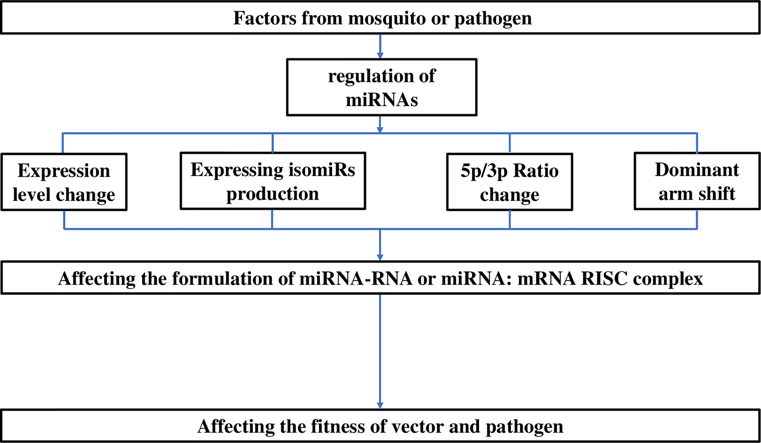
Figure 2
Moreover, although the canonical action mechanism of miRNAs always results in repression, the mosquito miRNA-target interaction can lead to two possible forms of regulation, namely, repression or enhancement of pathogen infection in mosquitoes. Both upregulation and downregulation of miRNAs in response to pathogen infection widely coexist in the mosquito, subsequently promoting and inhibiting pathogen infection, respectively. In our opinion, these findings suggest that inhibitory and inducing miRNA expressions are essential to balance the miRNA-pathogen interaction, maintaining persistent infection and preventing considerable harm to the mosquito (Figure 3).
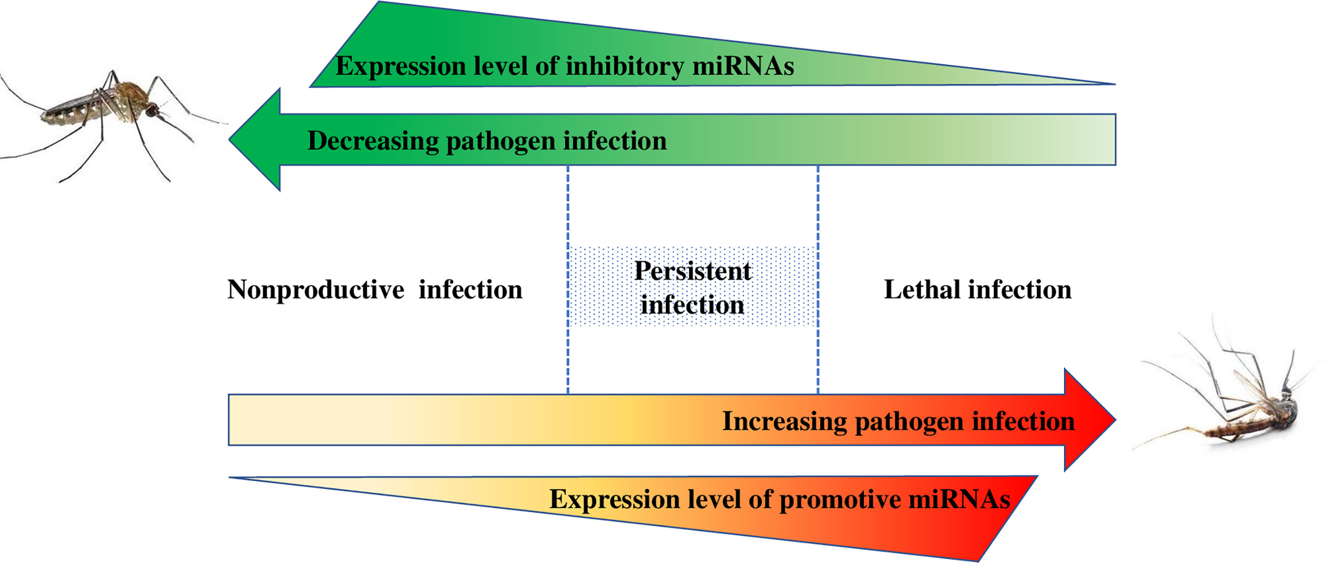
Figure 3
Currently, the antiviral effects of mosquito miRNAs on pathogens in combination with genetic engineering and molecular biology techniques may allow the use of these miRNA-based approaches as new tools to interrupt the transmission of mosquito-borne diseases. In this review, the significant progress achieved at the level of individual miRNAs facilitates the selection of an abundant, specific and effective mosquito miRNA (see Tables 1–7 and S1) that can be referenced for further research with different and specific objectives to increase the pace of development of applications and overcome the bottleneck (Tsetsarkin et al., 2015). More importantly, mosquito miRNAs can directly bind to the arbovirus genome, modifying viral replication. However, regarding the Plasmodium parasite, mosquito miRNAs generally bind to mosquito immunity or development-related mRNAs, indirectly regulating Plasmodium infection. Hence, the strategies for miRNA-based approaches differ for arboviruses and protozoan parasites.
Funding
BZ received a grant from the National Science and Technology Major Program of China (No. 2018ZX10734-404). This project was financially supported by Ministry of Science and Technology of the People’s Republic of China. The funder had no role in study design, data collection and analysis, decision to publish, or preparation of the manuscript.
Statements
Author contributions
Conceptualization and formal analysis: T-LX. Data curation: T-LX, Y-WS, and X-YF.
Supervision: BZ and X-NZ. Writing-original draft: T-LX. Writing-review & editing: BZ and X-NZ. All authors contributed to the article and approved the submitted version.
Acknowledgments
The ideas presented here were developed during the completion of research projects funded by the Belt and Road Young Scientist Research Exchange Program (No. 17430741900).
Conflict of interest
The authors declare that the research was conducted in the absence of any commercial or financial relationships that could be construed as a potential conflict of interest.
Supplementary material
The Supplementary Material for this article can be found online at: https://www.frontiersin.org/articles/10.3389/fcimb.2021.665444/full#supplementary-material
References
1
AkbariO. S.AntoshechkinI.AmrheinH.WilliamsB.DiloretoR.SandlerJ.et al. (2013). The Developmental Transcriptome of the Mosquito Aedes Aegypti, an Invasive Species and Major Arbovirus Vector. G33, 1493–1509. doi: 10.1534/g3.113.006742
2
AllamM.SpillingsB. L.AbdallaH.MapiyeD.KoekemoerL. L.ChristoffelsA. (2016). Identification and Characterization of microRNAs Expressed in the African Malaria Vector Anopheles Funestus Life Stages Using High Throughput Sequencing. Malaria J.15, 542. doi: 10.1186/s12936-016-1591-0
3
AsadS.HussainM.HugoL.Osei-AmoS.ZhangG.WattersonD.et al. (2018). Suppression of the Pelo Protein by Wolbachia and Its Effect on Dengue Virus in Aedes Aegypti. PloS Negl. Trop. Dis.12, e0006405. doi: 10.1371/journal.pntd.0006405
4
Avila-BonillaR. G.Yocupicio-MonroyM.MarchatL. A.De Nova-OcampoM. A.Del AngelR. M.Salas-BenitoJ. S. (2017). Analysis of the miRNA Profile in C6/36 Cells Persistently Infected With Dengue Virus Type 2. Virus Res.232, 139–151. doi: 10.1016/j.virusres.2017.03.005
5
Avila-BonillaR. G.Yocupicio-MonroyM.MarchatL. A.Perez-IshiwaraD. G.Cerecedo-MercadoD. A.Del AngelR. M.et al. (2020). miR-927 Has Pro-Viral Effects During Acute and Persistent Infection With Dengue Virus Type 2 in C6/36 Mosquito Cells. J. Gen. Virol. 101, 8. doi: 10.1099/jgv.0.001441
6
BartelD. P. (2004). MicroRNAs: Genomics, Biogenesis, Mechanism, and Function. Cell116, 281–297. doi: 10.1016/S0092-8674(04)00045-5
7
BartelD. P. (2009). MicroRNAs: Target Recognition and Regulatory Functions. Cell136, 215–233. doi: 10.1016/j.cell.2009.01.002
8
BatzZ. A.GoffA. C.ArmbrusterP. A. (2017). MicroRNAs Are Differentially Abundant During Aedes Albopictus Diapause Maintenance But Not Diapause Induction. Insect Mol. Biol.26, 721–733. doi: 10.1111/imb.12332
9
BehuraS. K.HaugenM.FlanneryE.SarroJ.TessierC. R.SeversonD. W.et al. (2011). Comparative Genomic Analysis of Drosophila Melanogaster and Vector Mosquito Developmental Genes. PloS One6, e21504. doi: 10.1371/journal.pone.0021504
10
BiedlerJ. K.QiY.PledgerD.MaciasV. M.JamesA. A.TuZ. (2014). Maternal Germline-Specific Genes in the Asian Malaria Mosquito Anopheles Stephensi: Characterization and Application for Disease Control. G35, 157–166. doi: 10.1534/g3.114.015578
11
BiryukovaI.YeT.LevashinaE. (2014). Transcriptome-Wide Analysis of microRNA Expression in the Malaria Mosquito Anopheles Gambiae. BMC Genomics15, 557. doi: 10.1186/1471-2164-15-557
12
BrunoA.AlessioC.CarmineF.FrancescoS.VladimirB.MarcoD. L.et al. (2019). MicroRNAs From Saliva of Anopheline Mosquitoes Mimic Human Endogenous miRNAs and may Contribute to Vector-Hostpathogen Interactions. Sci. Rep.9, 2955. doi: 10.1038/s41598-019-39880-1
13
BryantB.MacdonaldW.RaikhelA. S. (2010). microRNA miR-275 Is Indispensable for Blood Digestion and Egg Development in the Mosquito Aedes Aegypti. Proc. Natl. Acad. Sci. U. S. A.107, 22391–22398. doi: 10.1073/pnas.1016230107
14
BryantW. B.MillsM. K.OlsonB. J.MichelK. (2019). Small RNA-Seq Analysis Reveals Mirna Expression Dynamics Across Tissues in the Malaria Vector, Anopheles Gambiae. G39, 1507–1517. doi: 10.1534/g3.119.400104
15
BryantW. B.RayS.MillsM. K. (2020). Global Analysis of Small non-Coding RNA Populations Across Tissues in the Malaria Vector, Anopheles Gambiae. Insects11, 7. doi: 10.3390/insects11070406
16
CampbellC. L.HarrisonT.HessA. M.EbelG. D. (2014). MicroRNA Levels Are Modulated in Aedes Aegypti After Exposure to Dengue-2. Insect Mol. Biol.23, 132–139. doi: 10.1111/imb.12070
17
CarissimoG.PainA.BeldaE.VernickK. D. (2018). Highly Focused Transcriptional Response of Anopheles Coluzzii to O’nyong Nyong Arbovirus During the Primary Midgut Infection. BMC Genomics19, 526. doi: 10.1186/s12864-018-4918-0
18
CastellanoL.RizziE.KrellJ.Di CristinaM.GaliziR.MoriA.et al. (2015). The Germline of the Malaria Mosquito Produces Abundant miRNAs, endo-siRNAs, piRNAs and 29-Nt Small RNAs. BMC Genomics16, 100. doi: 10.1186/s12864-015-1257-2
19
ChatterjeeR.ChaudhuriK. (2006). An Approach for the Identification of microRNA With an Application to Anopheles Gambiae. Acta Biochim. Polonica53, 303–309. doi: 10.18388/abp.2006_3343
20
DennisonN. J.BenMarzouk-HidalgoO. J.DimopoulosG. (2015). MicroRNA-Regulation of Anopheles Gambiae Immunity to Plasmodium Falciparum Infection and Midgut Microbiota. Dev. Comp. Immunol.49, 170–178. doi: 10.1016/j.dci.2014.10.016
21
DongS.FuX.DongY.SimoesM. L.ZhuJ.DimopoulosG. (2020). Broad Spectrum Immunomodulatory Effects of Anopheles Gambiae microRNAs and Their Use for Transgenic Suppression of Plasmodium. PloS Pathog.16, e1008453. doi: 10.1371/journal.ppat.1008453
22
DritsouV.DeligianniE.DialynasE.AllenJ.PoulakakisN.LouisC.et al. (2014). Non-Coding RNA Gene Families in the Genomes of Anopheline Mosquitoes. BMC Genomics15, 1038. doi: 10.1186/1471-2164-15-1038
23
DubeyS. K.ShrinetJ.JainJ.AliS.SunilS. (2017). Aedes Aegypti microRNA miR-2b Regulates Ubiquitin-Related Modifier to Control Chikungunya Virus Replication. Sci. Rep.7, 17666. doi: 10.1038/s41598-017-18043-0
24
DubeyS. K.ShrinetJ.SunilS. (2019). Aedes Aegypti microRNA, miR-2944b-5p Interacts With 3’UTR of Chikungunya Virus and Cellular Target vps-13 to Regulate Viral Replication. PloS Negl. Trop. Dis.13, e0007429. doi: 10.1371/journal.pntd.0007429
25
EichhornS. W.GuoH.McGearyS. E.Rodriguez-MiasR. A.ShinC.BaekD.et al. (2014). mRNA Destabilization Is the Dominant Effect of Mammalian microRNAs by the Time Substantial Repression Ensues. Mol. Cell56, 104–115. doi: 10.1016/j.molcel.2014.08.028
26
EtebariK.Osei-AmoS.BlombergS. P.AsgariS. (2015). Dengue Virus Infection Alters Post-Transcriptional Modification of microRNAs in the Mosquito Vector Aedes Aegypti. Sci. Rep.5, 15968. doi: 10.1038/srep15968
27
FabianM. R.SonenbergN. (2012). The Mechanics of miRNA-Mediated Gene Silencing: A Look Under the Hood of Mirisc. Nat. Struct. Mol. Biol.19, 586–593. doi: 10.1038/nsmb.2296
28
FengX.WuJ.ZhouS.WangJ.HuW. (2018b). Characterization and Potential Role of microRNA in the Chinese Dominant Malaria Mosquito Anopheles Sinensis (Diptera: Culicidae) Throughout Four Different Life Stages. Cell Bioscience8, 29. doi: 10.1186/s13578-018-0227-1
29
FengX.ZhouX.ZhouS.WangJ.HuW. (2018a). Analysis of microRNA Profile of Anopheles Sinensis by Deep Sequencing and Bioinformatic Approaches. Parasites Vectors11, 172. doi: 10.1186/s13071-018-2734-7
30
FuX.DimopoulosG.ZhuJ. (2017). Association of microRNAs With Argonaute Proteins in the Malaria Mosquito Anopheles Gambiae After Blood Ingestion. Sci. Rep.7, 6493. doi: 10.1038/s41598-017-07013-1
31
FuX.LiuP.DimopoulosG.ZhuJ. (2020). Dynamic miRNA-mRNA Interactions Coordinate Gene Expression in Adult Anopheles Gambiae. PloS Genet.16, e1008765. doi: 10.1371/journal.pgen.1008765
32
GuJ.HuW.WuJ.ZhengP.ChenM.JamesA. A.et al. (2013). miRNA Genes of an Invasive Vector Mosquito, Aedes Albopictus. PloS One8, e67638. doi: 10.1371/journal.pone.0067638
33
GuoQ.HuangY.ZouF.LiuB.TianM.YeW.et al. (2017). The Role of miR-2~13~71 Cluster in Resistance to Deltamethrin in Culex Pipiens Pallens. Insect Biochem. Mol. Biol.84, 15–22. doi: 10.1016/j.ibmb.2017.03.006
34
HammondS. M.BernsteinE.BeachD.HannonG. J. (2000). An RNA-directed Nuclease Mediates Post-Transcriptional Gene Silencing in Drosophila Cells. Nature404, 293–296. doi: 10.1038/35005107
35
HeissB. L.MaximovaO. A.PletnevA. G. (2011). Insertion of microRNA Targets Into the Flavivirus Genome Alters Its Highly Neurovirulent Phenotype. J. Virol.85, 1464–1472. doi: 10.1128/JVI.02091-10
36
HongS.GuoQ.WangW.HuS.FangF.LvY.et al. (2014). Identification of Differentially Expressed microRNAs in Culex Pipiens and Their Potential Roles in Pyrethroid Resistance. Insect Biochem. Mol. Biol.55, 39–50. doi: 10.1016/j.ibmb.2014.10.007
37
HuW.CriscioneF.LiangS.TuZ. (2015). MicroRNAs of Two Medically Important Mosquito Species: Aedes Aegypti and Anopheles Stephensi. Insect Mol. Biol.24, 240–252. doi: 10.1111/imb.12152
38
HussainM.FrentiuF. D.MoreiraL. A.O’NeillS. L.AsgariS. (2011). Wolbachia Uses Host microRNAs to Manipulate Host Gene Expression and Facilitate Colonization of the Dengue Vector Aedes Aegypti. Proc. Natl. Acad. Sci. U. S. A.108, 9250–9255. doi: 10.1073/pnas.1105469108
39
HussainM.WalkerT.O’NeillS. L.AsgariS. (2013). Blood Meal Induced microRNA Regulates Development and Immune Associated Genes in the Dengue Mosquito Vector, Aedes Aegypti. Insect Biochem. Mol. Biol.43, 146–152. doi: 10.1016/j.ibmb.2012.11.005
40
JainS.RanaV.ShrinetJ.SharmaA.TridibesA.SunilS.et al. (2014). Blood Feeding and Plasmodium Infection Alters the miRNome of Anopheles Stephensi. PloS One9, e98402. doi: 10.1371/journal.pone.0098402
41
JainS.RanaV.TridibesA.SunilS.BhatnagarR. K. (2015). Dynamic Expression of miRNAs Across Immature and Adult Stages of the Malaria Mosquito Anopheles Stephensi. Parasites Vectors8, 179. doi: 10.1186/s13071-015-0772-y
42
Jones-RhoadesM. W.BartelD. P.BartelB. (2006). MicroRNAs and Their Regulatory Roles in Plants. Annu. Rev. Plant Biol.57, 19–53. doi: 10.1146/annurev.arplant.57.032905.105218
43
LampeL.JentzschM.KierszniowskaS.LevashinaE. A. (2019). Metabolic Balancing by miR-276 Shapes the Mosquito Reproductive Cycle and Plasmodium Falciparum Development. Nat. Commun.10, 5634. doi: 10.1038/s41467-019-13627-y
44
LampeL.LevashinaE. A. (2018). Microrna Tissue Atlas of the Malaria Mosquito Anopheles Gambiae. G38, 185–193. doi: 10.1534/g3.117.300170
45
LeeY.AhnC.HanJ.ChoiH.KimJ.YimJ.et al. (2003). The Nuclear RNase III Drosha Initiates microRNA Processing. Nature425, 415–419. doi: 10.1038/nature01957
46
LeeM.EtebariK.Hall-MendelinS.van den HurkA. F.Hobson-PetersJ.VatipallyS.et al. (2017). Understanding the Role of microRNAs in the Interaction of Aedes Aegypti Mosquitoes With an Insect-Specific Flavivirus. J. Gen. Virol.98, 1892–1903. doi: 10.1099/jgv.0.000832
47
LeeY.JeonK.LeeJ. T.KimS.KimV. N. (2002). MicroRNA Maturation: Stepwise Processing and Subcellular Localization. EMBO J.21, 4663–4670. doi: 10.1093/emboj/cdf476
48
LiS.MeadE. A.LiangS.TuZ. (2009). Direct Sequencing and Expression Analysis of a Large Number of miRNAs in Aedes Aegypti and a Multi-Species Survey of Novel Mosquito Mirnas. BMC Genomics10, 581. doi: 10.1186/1471-2164-10-581
49
LingL.KokozaV. A.ZhangC.AksoyE.RaikhelA. S. (2017). MicroRNA-277 Targets Insulin-Like Peptides 7 and 8 to Control Lipid Metabolism and Reproduction in Aedes Aegypti Mosquitoes. Proc. Natl. Acad. Sci. U. S. A.114, E8017–E8024. doi: 10.1073/pnas.1710970114
50
LiuW.HaoZ.HuangL.ChenL.WeiQ.CaiL.et al. (2017). Comparative Expression Profile of microRNAs in Anopheles Anthropophagus Midgut After Blood-Feeding and Plasmodium Infection. Parasites Vectors10, 86. doi: 10.1186/s13071-017-2027-6
51
LiuW.HuangH.XingC.LiC.TanF.LiangS. (2014). Identification and Characterization of the Expression Profile of microRNAs in Anopheles Anthropophagus. Parasites Vectors7, 159. doi: 10.1186/1756-3305-7-159
52
LiuP.LiX.GuJ.DongY.LiuY.SanthoshP.et al. (2016). Development of Non-Defective Recombinant Densovirus Vectors for microRNA Delivery in the Invasive Vector Mosquito, Aedes Albopictus. Sci. Rep.6, 20979. doi: 10.1038/srep20979
53
LiuY. X.LiF. X.LiuZ. Z.JiaZ. R.ZhouY. H.ZhangH.et al. (2016). Integrated Analysis of miRNAs and Transcriptomes in Aedes Albopictus Midgut Reveals the Differential Expression Profiles of Immune-Related Genes During Dengue Virus Serotype-2 Infection. Insect Sci.23, 377–385. doi: 10.1111/1744-7917.12339
54
LiuB.TianM.GuoQ.MaL.ZhouD.ShenB.et al. (2016). Mir-932 Regulates Pyrethroid Resistance in Culex Pipiens Pallens (Diptera: Culicidae). J. Med. Entomology53, 1205–1210. doi: 10.1093/jme/tjw083
55
LiuY.ZhouY.WuJ.ZhengP.LiY.ZhengX.et al. (2015). The Expression Profile of Aedes Albopictus miRNAs Is Altered by Dengue Virus Serotype-2 Infection. Cell Bioscience5, 16. doi: 10.1186/s13578-015-0009-y
56
LucasK. J.RoyS.HaJ.GervaiseA. L.KokozaV. A.RaikhelA. S. (2015a). MicroRNA-8 Targets the Wingless Signaling Pathway in the Female Mosquito Fat Body to Regulate Reproductive Processes. Proc. Natl. Acad. Sci. U. S. A.112, 1440–1445. doi: 10.1073/pnas.1424408112
57
LucasK. J.ZhaoB.RoyS.GervaiseA. L.RaikhelA. S. (2015b). Mosquito-Specific microRNA-1890 Targets the Juvenile Hormone-Regulated Serine Protease JHA15 in the Female Mosquito Gut. RNA Biol.12, 1383–1390. doi: 10.1080/15476286.2015.1101525
58
MaharajP. D.WidenS. G.HuangJ.WoodT. G.ThangamaniS. (2015). Discovery of Mosquito Saliva microRNAs During CHIKV Infection. PloS Negl. Trop. Dis.9, e0003386. doi: 10.1371/journal.pntd.0003386
59
MaK.LiX.HuH.ZhouD.SunY.MaL.et al. (2017). Pyrethroid-Resistance Is Modulated by miR-92a by Targeting CpCPR4 in Culex Pipiens Pallens. Comp. Biochem. Physiol. Part B Biochem. Mol. Biol.203, 20–24. doi: 10.1016/j.cbpb.2016.09.002
60
ayoralJ. G.EtebariK.HussainM.KhromykhA. A.AsgariS. (2014). Wolbachia Infection Modifies the Profile, Shuttling and Structure of microRNAs in a Mosquito Cell Line. PloS One9, e96107. doi: 10.1371/journal.pone.0096107
61
MeadE. A.TuZ. (2008). Cloning, Characterization, and Expression of microRNAs From the Asian Malaria Mosquito, Anopheles Stephensi. BMC Genomics9, 244. doi: 10.1186/1471-2164-9-244
62
MeutiM. E.Bautista-JimenezR.ReynoldsJ. A. (2018). Evidence That microRNAs Are Part of the Molecular Toolkit Regulating Adult Reproductive Diapause in the Mosquito, Culex Pipiens. PloS One13, e0203015. doi: 10.1371/journal.pone.0203015
63
MiesenP.IvensA.BuckA. H.van RijR. P. (2016). Small RNA Profiling in Dengue Virus 2-Infected Aedes Mosquito Cells Reveals Viral piRNAs and Novel Host Mirnas. PloS Negl. Trop. Dis.10, e0004452. doi: 10.1371/journal.pntd.0004452
64
MishraS.YadavT.RaniV. (2016). Exploring miRNA Based Approaches in Cancer Diagnostics and Therapeutics. Crit. Rev. Oncol. Hematol.98, 12–23. doi: 10.1016/j.critrevonc.2015.10.003
65
NouzovaM.EtebariK.NoriegaF. G.AsgariS. (2018). A Comparative Analysis of Corpora Allata-Corpora Cardiaca microRNA Repertoires Revealed Significant Changes During Mosquito Metamorphosis. Insect Biochem. Mol. Biol.96, 10–18. doi: 10.1016/j.ibmb.2018.03.007
66
Osei-AmoS.HussainM.O’NeillS. L.AsgariS. (2012). Wolbachia-Induced aae-miR-12 miRNA Negatively Regulates the Expression of MCT1 and MCM6 Genes in Wolbachia-infected Mosquito Cell Line. PloS One7, e50049. doi: 10.1371/journal.pone.0050049
67
PuthiyakunnonS.YaoY.LiY.GuJ.PengH.ChenX. (2013). Functional Characterization of Three MicroRNAs of the Asian Tiger Mosquito, Aedes Albopictus. Parasites Vectors6, 230. doi: 10.1186/1756-3305-6-230
68
QuZ.BendenaW. G.NongW.SiggensK. W.NoriegaF. G.KaiZ. P.et al. (2017). MicroRNAs Regulate the Sesquiterpenoid Hormonal Pathway in Drosophila and Other Arthropods. Proc. Biol. Sci.284, 1869. doi: 10.1098/rspb.2017.1827
69
SaldanaM. A.EtebariK.HartC. E.WidenS. G.WoodT. G.ThangamaniS.et al. (2017). Zika Virus Alters the microRNA Expression Profile and Elicits an RNAi Response in Aedes Aegypti Mosquitoes. PloS Negl. Trop. Dis.11, e0005760. doi: 10.1371/journal.pntd.0005760
70
ShrinetJ.JainS.JainJ.BhatnagarR. K.SunilS. (2014). Next Generation Sequencing Reveals Regulation of Distinct Aedes microRNAs During Chikungunya Virus Development. PloS Negl. Trop. Dis.8, e2616. doi: 10.1371/journal.pntd.0002616
71
SkalskyR. L.VanlandinghamD. L.ScholleF.HiggsS.CullenB. R. (2010). Identification of microRNAs Expressed in Two Mosquito Vectors, Aedes Albopictus and Culex Quinquefasciatus. BMC Genomics11, 119. doi: 10.1186/1471-2164-11-119
72
SlonchakA.HussainM.TorresS.AsgariS.KhromykhA. A. (2014). Expression of Mosquito Microrna Aae-miR-2940-5p Is Downregulated in Response to West Nile Virus Infection to Restrict Viral Replication. J. Virol.88, 8457–8467. doi: 10.1128/JVI.00317-14
73
SlonchakA.ShannonR. P.PaliG.KhromykhA. A. (2015). Human MicroRNA Mir-532-5p Exhibits Antiviral Activity Against West Nile Virus Via Suppression of Host Genes SESTD1 and TAB3 Required for Virus Replication. J. Virol.90, 2388–2402. doi: 10.1128/JVI.02608-15
74
SmithJ. L.JengS.McWeeneyS. K.HirschA. J. (2017). A MicroRNA Screen Identifies the Wnt Signaling Pathway as a Regulator of the Interferon Response During Flavivirus Infection. J. Virol.91, 8. doi: 10.1128/JVI.02388-16
75
SuJ.LiC.ZhangY.YanT.ZhuX.ZhaoM.et al. (2017). Identification of microRNAs Expressed in the Midgut of Aedes Albopictus During Dengue Infection. Parasites Vectors10, 63. doi: 10.1186/s13071-017-1966-2
76
SuJ.WangG.LiC.XingD.YanT.ZhuX.et al. (2019). Screening for Differentially Expressed miRNAs in Aedes Albopictus (Diptera: Culicidae) Exposed to DENV-2 and Their Effect on Replication of DENV-2 in C6/36 Cells. Parasites Vectors12, 44. doi: 10.1186/s13071-018-3261-2
77
TsetsarkinK. A.LiuG.KenneyH.Bustos-ArriagaJ.HansonC. T.WhiteheadS. S.et al. (2015). Dual miRNA Targeting Restricts Host Range and Attenuates Neurovirulence of Flaviviruses. PloS Pathog.11, e1004852. doi: 10.1371/journal.ppat.1004852
78
TsetsarkinK. A.LiuG.KenneyH.HermanceM.ThangamaniS.PletnevA. G. (2016a). Concurrent micro-RNA Mediated Silencing of Tick-Borne Flavivirus Replication in Tick Vector and in the Brain of Vertebrate Host. Sci. Rep.6, 33088. doi: 10.1038/srep33088
79
TsetsarkinK. A.LiuG.ShenK.PletnevA. G. (2016b). Kissing-Loop Interaction Between 5’ and 3’ Ends of Tick-Borne Langat Virus Genome ‘Bridges the Gap’ Between Mosquito- and Tick-Borne Flaviviruses in Mechanisms of Viral RNA Cyclization: Applications for Virus Attenuation and Vaccine Development. Nucleic Acids Res.44, 3330–3350. doi: 10.1093/nar/gkw061
80
WangX.ZhangJ.LiF.GuJ.HeT.ZhangX.et al. (2005). MicroRNA Identification Based on Sequence and Structure Alignment. Bioinformatics21, 3610–3614. doi: 10.1093/bioinformatics/bti562
81
World Health Organization (2012). Global strategy for dengue prevention and control 2012-2020. WHO. Available at: https://apps.who.int/iris/handle/10665/75303.
82
WinterF.EdayeS.HuttenhoferA.BrunelC. (2007). Anopheles Gambiae miRNAs as Actors of Defence Reaction Against Plasmodium Invasion. Nucleic Acids Res.35, 6953–6962. doi: 10.1093/nar/gkm686
83
WuZ.XueY.WangB.DuJ.JinQ. (2011). Broad-Spectrum Antiviral Activity of RNA Interference Against Four Genotypes of Japanese Encephalitis Virus Based on Single microRNA Polycistrons. PloS One6, e26304. doi: 10.1371/journal.pone.0026304
84
XieP. W.XieY.ZhangX. J.HuangH.HeL. N.WangX. J.et al. (2013). Inhibition of Dengue Virus 2 Replication by Artificial Micrornas Targeting the Conserved Regions. Nucleic Acid Ther.23, 244–252. doi: 10.1089/nat.2012.0405
85
XingS.DuJ.GaoS.TianZ.ZhengY.LiuG.et al. (2016). Analysis of the miRNA Expression Profile in an Aedes Albopictus Cell Line in Response to Bluetongue Virus Infection. Infection Genet. Evolution: J. Mol. Epidemiol. Evolutionary Genet. Infect. Dis.39, 74–84. doi: 10.1016/j.meegid.2016.01.012
86
XuT.ZhongD.TangL.ChangX.FuF.YanG.et al. (2014). Anopheles Sinensis Mosquito Insecticide Resistance: Comparison of Three Mosquito Sample Collection and Preparation Methods and Mosquito Age in Resistance Measurements. Parasites Vectors7, 54. doi: 10.1186/1756-3305-7-54
87
YangS.PeiY.LiX.ZhaoS.ZhuM.ZhaoA. (2016). miR-124 Attenuates Japanese Encephalitis Virus Replication by Targeting DNM2. Virol. J.13, 105. doi: 10.1186/s12985-016-0562-y
88
YanH.ZhouY.LiuY.DengY.ChenX. (2014). miR-252 of the Asian Tiger Mosquito Aedes Albopictus Regulates Dengue Virus Replication by Suppressing the Expression of the Dengue Virus Envelope Protein. J. Med. Virol.86, 1428–1436. doi: 10.1002/jmv.23815
89
YektaS.ShihI. H.BartelD. P. (2004). MicroRNA-directed Cleavage of HOXB8 Mrna. Science304, 594–596. doi: 10.1126/science.1097434
90
YenP. S.ChenC. H.SreenuV.KohlA.FaillouxA. B. (2019). Assessing the Potential Interactions Between Cellular miRNA and Arboviral Genomic RNA in the Yellow Fever Mosquito, Aedes Aegypti. Viruses11, 6. doi: 10.3390/v11060540
91
YenP. S.JamesA.LiJ. C.ChenC. H.FaillouxA. B. (2018). Synthetic miRNAs Induce Dual Arboviral-Resistance Phenotypes in the Vector Mosquito Aedes Aegypti. Commun. Biol.1, 11. doi: 10.1038/s42003-017-0011-5
92
YenL. C.LinY. L.SungH. H.LiaoJ. T.TsaoC. H.SuC. M.et al. (2013). Neurovirulent Flavivirus Can be Attenuated in Mice by Incorporation of Neuron-Specific microRNA Recognition Elements Into Viral Genome. Vaccine31, 5915–5922. doi: 10.1016/j.vaccine.2011.09.102
93
ZhangX.AksoyE.GirkeT.RaikhelA. S.KarginovF. V. (2017). Transcriptome-Wide microRNA and Target Dynamics in the Fat Body During the Gonadotrophic Cycle of Aedes Aegypti. Proc. Natl. Acad. Sci. U. S. A.114, E1895–E1903. doi: 10.1073/pnas.1701474114
94
ZhangG.HussainM.AsgariS. (2014). Regulation of Arginine Methyltransferase 3 by a Wolbachia-Induced microRNA in Aedes Aegypti and Its Effect on Wolbachia and Dengue Virus Replication. Insect Biochem. Mol. Biol.53, 81–88. doi: 10.1016/j.ibmb.2014.08.003
95
ZhangG.HussainM.O’NeillS. L.AsgariS. (2013). Wolbachia Uses a Host microRNA to Regulate Transcripts of a Methyltransferase, Contributing to Dengue Virus Inhibition in Aedes Aegypti. Proc. Natl. Acad. Sci. U. S. A.110, 10276–10281. doi: 10.1073/pnas.1303603110
96
ZhangY.ZhaoB.RoyS.SahaT. T.KokozaV. A.LiM.et al. (2016). microRNA-309 Targets the Homeobox Gene SIX4 and Controls Ovarian Development in the Mosquito Aedes Aegypti. Proc. Natl. Acad. Sci. U. S. A.113, E4828–E4836. doi: 10.1073/pnas.1609792113
97
ZhouY.LiuY.YanH.LiY.ZhangH.XuJ.et al. (2014). miR-281, an Abundant Midgut-Specific miRNA of the Vector Mosquito Aedes Albopictus Enhances Dengue Virus Replication. Parasites Vectors7, 488. doi: 10.1186/s13071-014-0488-4
Summary
Keywords
miRNAs, mosquito, Plasmodium, interruption, miRNA-based approach, mosquito-borne diseases
Citation
Xu T-L, Sun Y-W, Feng X-Y, Zhou X-N and Zheng B (2021) Development of miRNA-Based Approaches to Explore the Interruption of Mosquito-Borne Disease Transmission. Front. Cell. Infect. Microbiol. 11:665444. doi: 10.3389/fcimb.2021.665444
Received
08 February 2021
Accepted
02 June 2021
Published
21 June 2021
Volume
11 - 2021
Edited by
Lubin Jiang, Institut Pasteur of Shanghai (CAS), China
Reviewed by
Fabrizio Lombardo, Sapienza University of Rome, Italy; Maria Luisa Simões, Johns Hopkins University, United States
Updates
Copyright
© 2021 Xu, Sun, Feng, Zhou and Zheng.
This is an open-access article distributed under the terms of the Creative Commons Attribution License (CC BY). The use, distribution or reproduction in other forums is permitted, provided the original author(s) and the copyright owner(s) are credited and that the original publication in this journal is cited, in accordance with accepted academic practice. No use, distribution or reproduction is permitted which does not comply with these terms.
*Correspondence: Xiao-Nong Zhou, zhouxn1@chinacdc.cn; Bin Zheng, cdcipdzhengbin@126.com
This article was submitted to Parasite and Host, a section of the journal Frontiers in Cellular and Infection Microbiology
Disclaimer
All claims expressed in this article are solely those of the authors and do not necessarily represent those of their affiliated organizations, or those of the publisher, the editors and the reviewers. Any product that may be evaluated in this article or claim that may be made by its manufacturer is not guaranteed or endorsed by the publisher.