 Laura Murcia-Flores1
 María Pilar Pecci-Lloret
 Francisco Javier Rodríguez-Lozano
- 1Department of Health Sciences, Catholic University San Antonio of Murcia, Murcia, Spain
- 2Dermatology, Stomatology, Radiology and Physical Medicine, Hospital Morales Meseguer, Medicine School, University of Murcia, Murcia – Biomedical Research Institute (IMIB), Murcia, Spain
The oral cavity serves as the gateway to the human organism, hosting a diverse community of microorganisms that coexist in a state of symbiosis. Disruption of this balance leads to oral dysbiosis, a condition associated with infections and oral pathologies, which may contribute to the etiopathogenesis of systemic disorders such as Parkinson’s disease, a neurodegenerative movement disorder characterized by resting tremor, rigidity, and bradykinesia. While oral dysbiosis is recognized as a risk factor and an aggravating element for Parkinson’s disease, it is not regarded as a direct cause. This systematic review aims to synthesize existing research exploring the potential relationship between oral dysbiosis and the development of Parkinson’s disease. Following a comprehensive analysis, 12 studies were selected, comprising 11 case-control studies and one observational analytical study. These studies investigated the composition of oral microbiota in different sample groups, revealing a higher abundance of pathogenic oral bacteria in individuals diagnosed with Parkinson’s disease. The findings suggest that oral dysbiosis may influence both the onset of Parkinson’s disease and the progression of symptoms such as cognitive decline. These results pave the way for future research, particularly regarding alterations in oral microbiota as potential biomarkers for early diagnosis and disease monitoring.
Systematic review registration: https://www.crd.york.ac.uk/prospero/, identifier CRD42024540056.
1 Introduction
The oral cavity, often described as the gateway to the body, hosts a diverse microbiota that plays a crucial role in maintaining overall health (Nicholson and Landry, 2022). This complex environment, comprising habitats like the tongue, teeth, soft and hard palates, and cheeks, provides the ideal conditions—characterized by moisture and warmth—for the coexistence of approximately 500 to 700 different microbial species. Under healthy conditions, this microbiota maintains a symbiotic relationship with the human host, contributing to the defense against pathogenic invasions and supporting both oral and systemic health (Asakawa et al., 2024; Kunath et al., 2024).
In a balanced oral ecosystem, commensal species such as Streptococcus mitis, Gemella, Granulicatella, and Veillonella dominate. Streptococcus mitis, in particular, is prevalent across various oral habitats and plays a significant role in forming biofilms on tooth surfaces through its interaction with salivary proteins like α-amylase (Wade, 2013). This biofilm, when in equilibrium, functions as a protective barrier against external pathogens. However, disturbances in this delicate balance—whether through changes in salivary flow, dietary habits, medication intake, or aging—can lead to dysbiosis. In oral dysbiosis, there is an increase in pathogenic species and a decrease in beneficial species (Song et al., 2022). This shift in the microbial composition often results in the proliferation of pathogenic species such as Porphyromonas gingivalis and Streptococcus mutans, which are associated with oral diseases like periodontitis, caries, and endodontic infections (Asakawa et al., 2024; Chung et al., 2024).
Recent studies have highlighted the potential link between oral dysbiosis and systemic conditions, particularly neurodegenerative diseases such as Parkinson’s disease (PD) (Nicholson and Landry, 2022). This relationship is increasingly evident as research uncovers distinct differences in the oral microbiota of patients with Parkinson’s compared to healthy individuals. The pathogenesis of Parkinson’s, a disorder characterized by motor dysfunction and attributed to the degeneration of dopaminergic neurons in the substantia nigra, may be influenced by factors such as chronic inflammation and microbial infections (Tao et al., 2024). Notably, the presence of pathogens like Porphyromonas gingivalis, known for its role in periodontal disease, has been implicated in promoting systemic inflammation and potentially contributing to neurodegenerative processes (Olsen et al., 2020; Goyal et al., 2023; Li et al., 2024), this is due to its ability to secrete gingipains, which degrade essential neuronal proteins, contributing to the disruption of the blood-brain barrier (Nonaka et al., 2022; Li et al., 2024). Therefore, it is believed that the oral microbiota may influence neuroinflammation (Ranjan et al., 2018), in addition to being closely related to the gut microbiota, which also influences this type of disease (Alam et al., 2024; Costa et al., 2024).
Given the emerging evidence linking oral microbiota and neurodegenerative diseases, this systematic review aims to explore the association between oral dysbiosis and if it contributes to the development and progression of Parkinson’s disease by influencing neuroinflammatory and neurodegenerative pathways. Understanding this connection could pave the way for new preventative and therapeutic strategies, highlighting the importance of oral health in managing systemic conditions. Furthermore, this review seeks to bridge the gap in current research by focusing on the oral microbiome, an area that, despite its significance, has received less attention compared to the gut microbiome in the context of neurodegenerative diseases.
2 Methods
This systematic review was conducted in accordance with the PRISMA 2020 guidelines, which stand for “Preferred Reporting Items for Systematic Reviews and Meta-Analyses” (Liberati et al., 2009). Additionally, the review was registered with the PROSPERO database (CRD42024540056) to ensure transparency and avoid duplication of similar reviews (Schiavo, 2019). Additionally, the PCO model (Santos et al., 2007)was used to formulate the following research question:
Is there an association between oral dysbiosis and Parkinson’s disease? (P: Patients/animals with Parkinson’s disease; C: Healthy patients; O: oral pathogens involved in Parkinson’s disease).
The search strategy, study selection, data extraction, and quality assessment (including the risk of bias evaluation) were conducted independently by two investigators (A.S.G. and F.J.R.L.). Any disagreements or uncertainties during the process were resolved through consultation with a third investigator (L.M.F.).
2.1 Search strategy
The search terms used to retrieve articles were derived from the MeSH (Medical Subject Heading) thesaurus. The terms related to oral microbiota included: “oral bacterium,” “microorganisms,” “periodontal pathogens,” “oral pathogens,” while those associated with the disease of interest were: “Parkinson disease” and “Parkinson.” Boolean operators (“AND” “OR,” and “NOT”) were employed to link these terms effectively. The search was conducted in February 2024.
2.2 Inclusion and exclusion criteria
The inclusion and exclusion criteria are presented in Table 1 and were established based on the research question and study objectives.
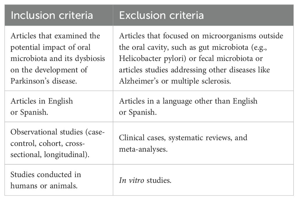
Table 1. Inclusion and exclusion criteria.
2.3 Study selection
The references retrieved from the search were imported into the citation management software EndNote (Clarivate Analytics, London, United Kingdom) to identify and eliminate any duplicates. An initial screening was conducted by examining the titles, followed by a review of the abstracts according to the predefined inclusion and exclusion criteria. Articles that satisfied these criteria were then subjected to a full-text review to determine their eligibility for inclusion in the qualitative synthesis.
2.4 Study data
For the bibliometric analysis, the following information was recorded for each article: author and year of publication, journal, and country of publication. Additionally, a table was created to summarize the following data: author, year of publication, study design, sample size and group, type of oral microbiota, method of microbiota extraction, and the association between Parkinson’s disease and oral dysbiosis, if any.
2.5 Quality assessment
The quality of the articles included in this systematic review was evaluated using a modified version of the STROBE (Strengthening the Reporting of Observational Studies in Epidemiology) checklist (von Elm et al., 2008) (Table 2). The checklist is primarily applied to cohort studies, case-control studies, and cross-sectional studies, and it assesses various aspects of study quality across 10 key points, such as participants, variables, data sources, and statistical methods. The risk of bias in each study was categorized as low, moderate, or high based on the number of checklist points fulfilled: low risk (8–11 points), moderate risk (5–7 points), and high risk (1–4 points). Each checklist item was marked with a tick if the requirement was met or with a cross if it was not.
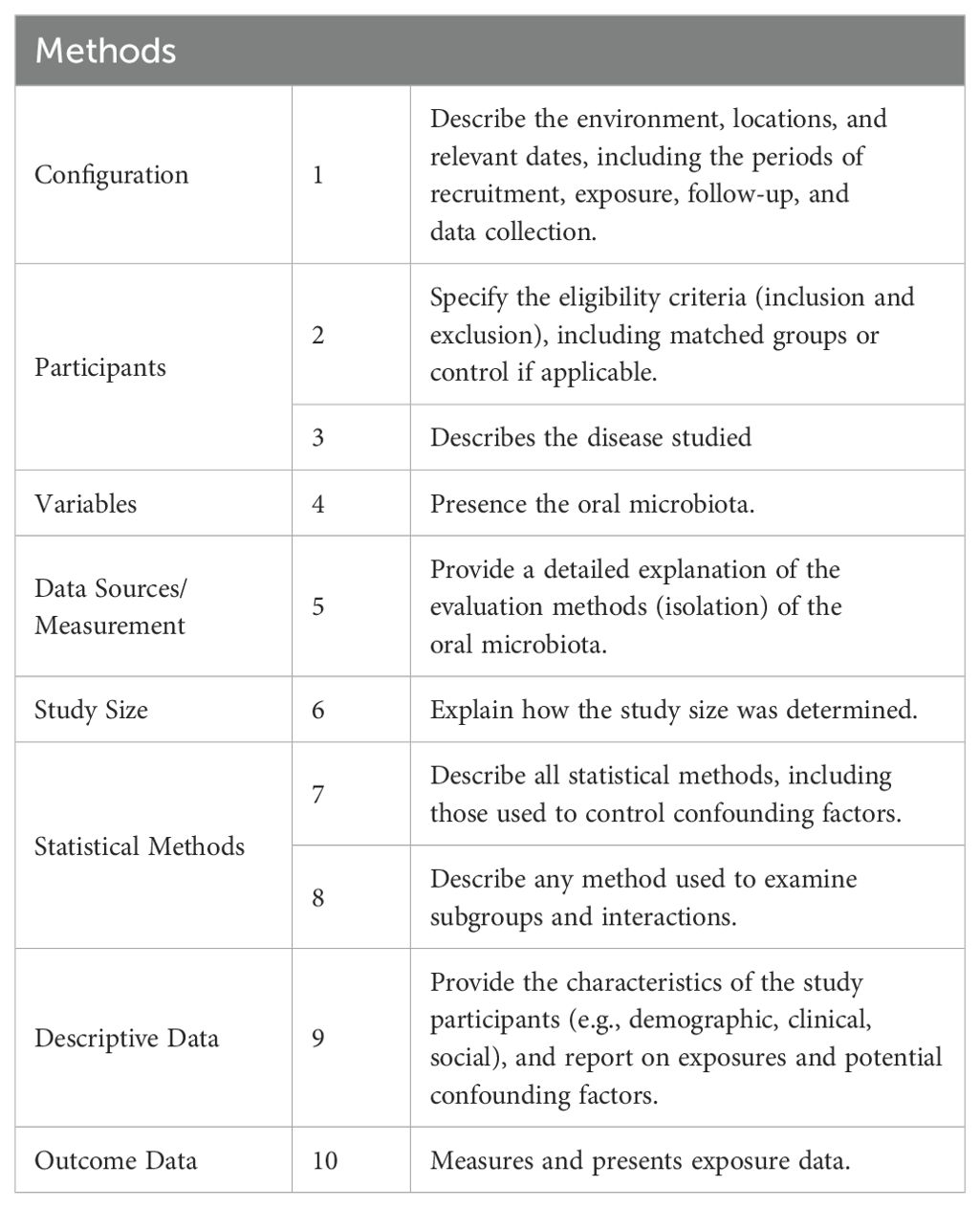
Table 2. List of criteria used to evaluate the quality of observational studies based on an adapted version of the STROBE guidelines.
3 Results
3.1 Study selection and flow diagram
Following a comprehensive and meticulous database search, a total of 849 references were identified: 199 from PubMed, 494 from Scopus, and 156 from Web of Science. The results obtained from each database are summarized in Table 3. These references were imported into the Endnote, where 280 duplicates, including undetected ones, were removed. The remaining 558 articles underwent screening based on their titles and abstracts, leading to the exclusion of 530 articles. This left 28 articles for full-text review. Upon further examination, 7 articles were excluded for not addressing oral microbiota, 2 were excluded due to being published in Chinese, and 7 were excluded for being systematic reviews. Consequently, 12 articles were selected for inclusion in this review (Figure 1).
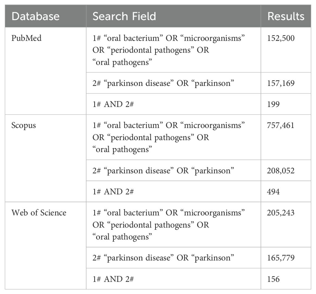
Table 3. Results obtained from each database.
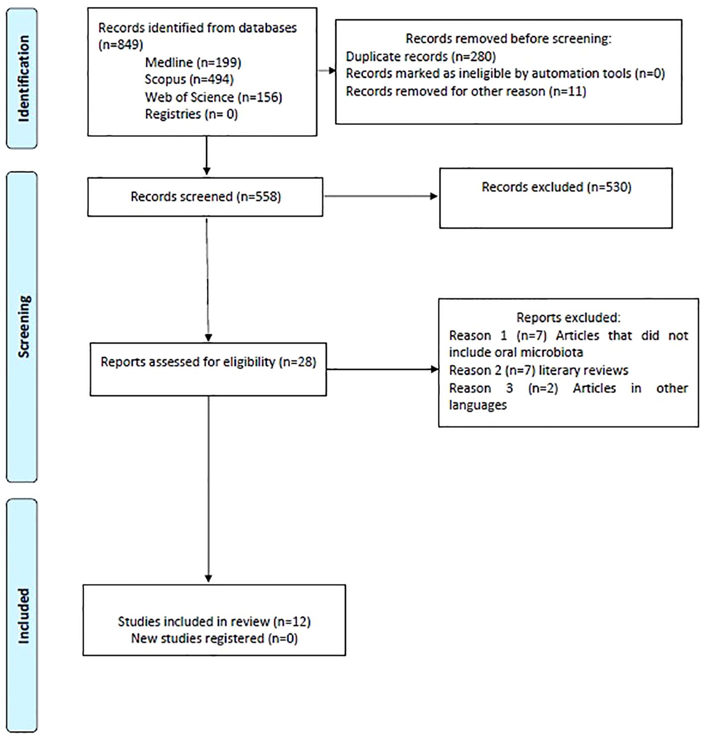
Figure 1. Flow diagram.
3.2 Characteristics of the studies
3.2.1 Bibliometric analysis
The distribution of the selected articles by year of publication is shown in Figure 2, where we can observe that the year with the highest prevalence of studies was 2023 with three studies, followed by 2022 and 2021 with two studies each. The distribution by country in Figure 3, with Turkey, the USA, and China being the countries with the highest number of articles, with two each. Finally,and by journal in Figure 4, with all journals having a single publication.
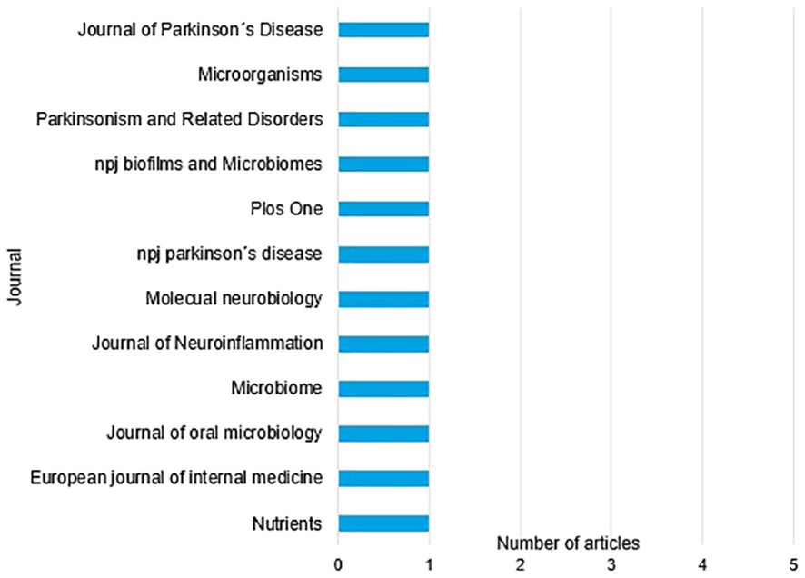
Figure 2. Organization of articles by year of publication.
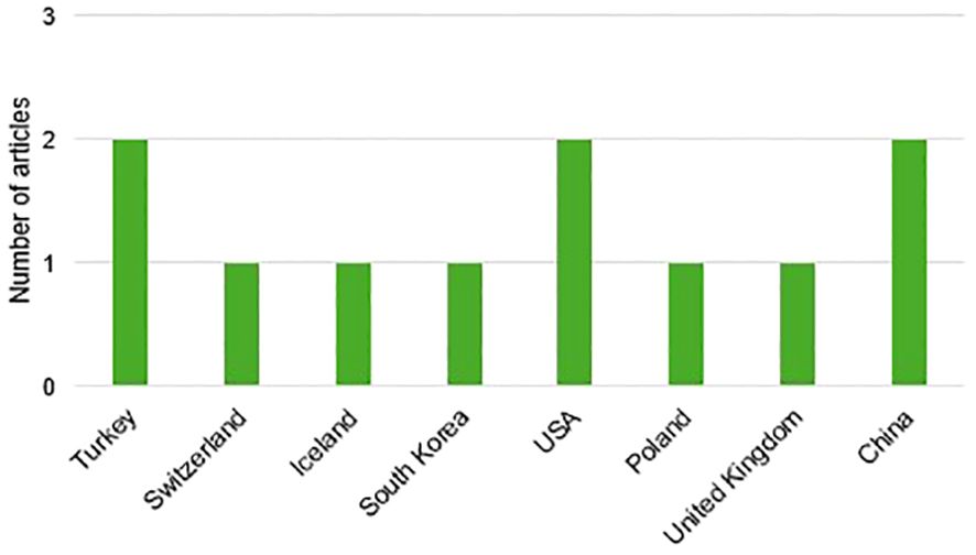
Figure 3. Organization of articles by country of publication.
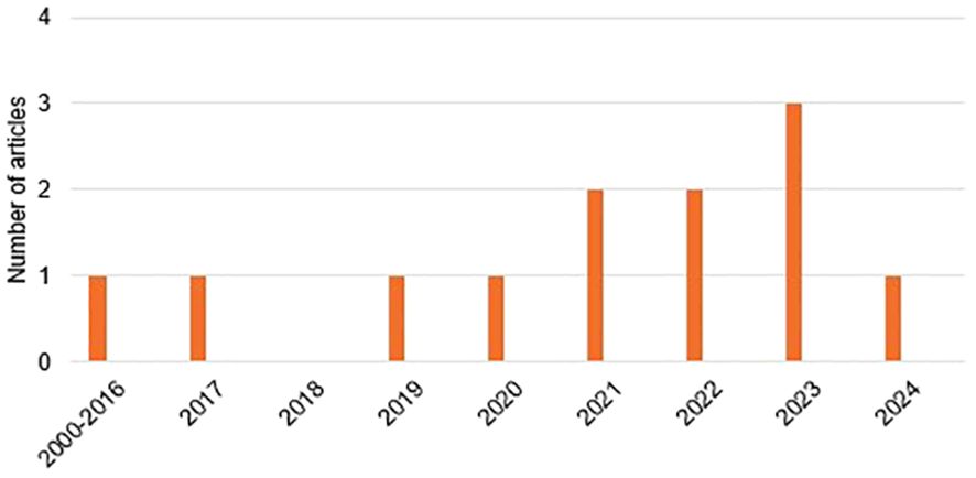
Figure 4. Organization of articles by journal of publication.
3.2.2 Study design
All the selected articles are case-control studies, where healthy subjects are compared with those diagnosed with Parkinson’s disease, except for the oldest study conducted by Gosney et al. (Gosney et al., 2003), in which all subjects have Parkinson’s disease. This particular study is an observational analytical study. Additionally, it is important to note that, among the selected studies, all were conducted on human subjects, except for two studies that were performed on mice.
3.2.3 Oral microbiota
As shown in Table 4, the genera with the highest abundance among Parkinson’s patients are, firstly, the genus Streptobacillaceae, cited in six studies, with specific mention of bacteria such as S. mutans and S. anginosus, which are significantly relevant to the subject under discussion. Some studies also refer to other types, such as S. sanguinis or S. sobrinus, although these are of lesser relevance (Zapala et al., 2022). The families Lactobacillaceae and Prevotellaceae are also frequently cited, appearing in the results of five of the analyzed studies. The latter is noted for an abundance of bacteria such as P. intermedia, P. histicola, and P. melaninogenica. Additionally, three studies highlight the genus Veillonellaceae (Pereira et al., 2017; Fleury et al., 2021; Bai et al., 2023). Notably, however, only two studies identified P. gingivalis as the primary pathogen (Feng et al., 2020; Li et al., 2024). The remaining genera have been identified as abundant in the oral microbiota of these patients in only one study.
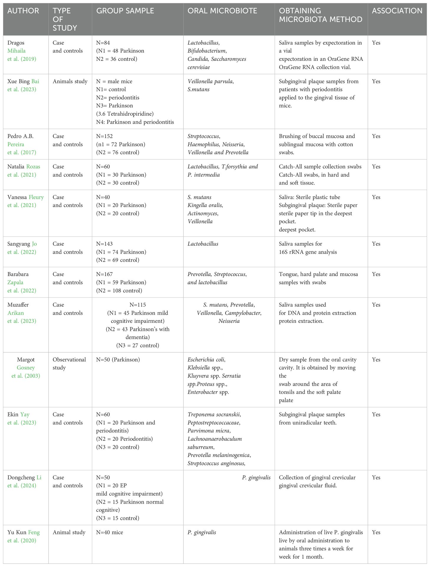
Table 4. Results: main characteristics of the studies included.
3.3 Quality analysis
The analysis shows a moderate to low risk of bias, with 8 articles rated as low risk and 4 as moderate risk. Notably, no studies were found to have a high risk of bias (Table 5). Key findings include that all articles fulfilled item 5 (data sources and measurements), item 1 (study design), and item 3 (participant description). Additionally, all studies provided exposure data (item 10) and clearly defined diagnostic criteria (item 4). However, only one article, by Zapała et al (Zapala et al., 2022), met item 6 (sample size calculation), and just 5 articles detailed the statistical methods, including subgroups and interactions. One article, by Gosney et al (Gosney et al., 2003), had the highest risk of bias, scoring 5 points, due to the absence of inclusion/exclusion criteria, sample size details, and statistical analysis with subgroups, along with a lack of participant characteristic descriptions.
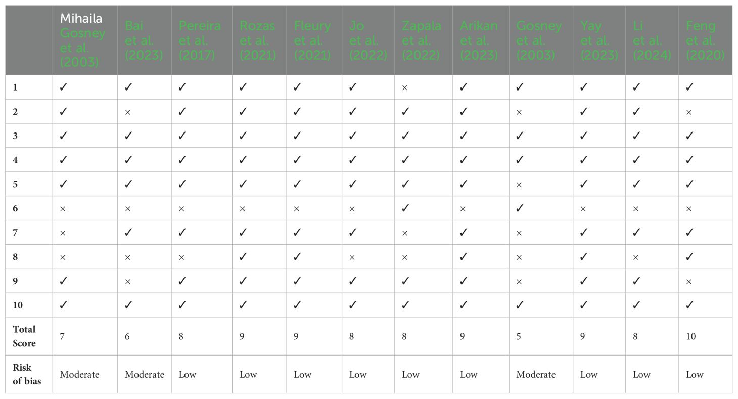
Table 5. Results of the quality assessment conducted with an adapted version of the STROBE guidelines.
4 Discussion
As the global population continues to age, the prevalence of Parkinson’s disease (PD) is rising, particularly among the elderly, with cognitive impairment recognized as a critical non-motor symptom (Mosaddad et al., 2023). PD is the second most prevalent neurological disorder, affecting approximately 1% of individuals over 60 and between 4% and 5% of those over 80. The incidence is 1.5 times higher in males than females. Despite genetic variations being implicated in less than 10% of Parkinson’s cases, environmental risk factors have emerged as an area of significant research interest (Zapala et al., 2022). The complex relationships between aging, genetic predispositions, and environmental factors are thought to underlie the still-unclear etiology of PD. Emerging evidence suggests that certain pathogenic bacteria within the oral microbiome may contribute to cognitive decline and the progression of Parkinson’s disease (Rozas et al., 2021; Tao et al., 2024).
Studies have demonstrated that the oral microbiota plays a crucial role in both local and systemic health, interacting with a variety of oral and systemic diseases. In older adults, physiological changes can disrupt the balance between pathogenic and commensal bacteria, potentially increasing pathogenic gene expression and disease susceptibility (Sampaio-Maia et al., 2016; Kozak and Pawlik, 2023). The common occurrence of tooth loss in this age group further disrupts the oral microbial environment by removing habitats essential for certain bacteria, such as subgingival sites and tooth surfaces. Additionally, dentures alters the oral microbiome by impacting bacterial quantity and diversity. The frequent occurrence of oral diseases in older adults suggests that these age-associated changes in the oral microbiome may play a role in the development of such conditions. This hypothesis is supported by research linking age-related microbial shifts (Preza et al., 2009; Liu et al., 2020) to the onset of systemic diseases prevalent among the aging population (Soliman et al., 2022).
The relationship between the oral cavity and the brain has garnered increasing attention, particularly regarding the oral-gut-brain axis and its role in neurological diseases (Laugisch et al., 2024). Previous research has demonstrated that gut microbiota can communicate with the brain via the gut-brain axis, influencing neurodevelopmental processes. The gut and oral microbiota are susceptible to various host-related factors, which can, in turn, make the brain more vulnerable to dysfunction (Ma et al., 2024). While research specifically addressing oral-brain communication is still limited, emerging hypotheses suggest that the oral microbiota-brain axis could impact neural activity, similar to the way aroma release and perception are influenced (Chen et al., 2023).
Unlike gut microbiota, the oral microbiota is distinct and can enter the gastrointestinal tract through saliva, thereby altering the intestinal microbial community (Qian et al., 2023). This translocation can trigger inflammation-related changes and modulate the immune system (Socala et al., 2021). The movement of oral bacteria to the gut is a characteristic feature of certain diseases, and the transmission of oral microbes via fecal matter can significantly shape the gastrointestinal microbiome. Moreover, studies suggest that the oral cavity may act as a reservoir for intestinal pathogens, which have the potential to activate the intestinal immune system and contribute to chronic inflammation (Goyal et al., 2023).
Numerous hypotheses have been proposed regarding the bacterial family Lactobacillaceae in relation to Parkinson’s disease. Jo et al. (2022) suggest that L. reuteri, present in the mouth and gut, may increase the release of α-synuclein in the enteric nervous system, potentially leading to gut colonization by pathogens. Similarly, Mihaila et al. (2019) found that this pathogen is significantly elevated in individuals with Parkinson’s disease and is positively correlated with slowed movement. On the other hand, Zapala et al. (2022) highlighted that, although the genus Prevotellaceae is a common resident in the oral cavity, it can occasionally become pathogenic, causing intestinal inflammation and contributing to systemic inflammation, which is implicated in various diseases, including Parkinson’s. They identified P. histicola, P. melaninogenica, and P. gingivalis as the most abundant bacteria in these patients. However, these findings differ from other studies, such as Pereira et al (Pereira et al., 2017), who reported a greater diversity of bacteria including Streptococcus, Neisseria, Haemophilus, and Veillonella, and Rozas et al (Rozas et al., 2021), who identified Lactobacillus, T. forsythia, and P. intermedia as more prevalent. Despite differences, these studies concur that members of the Prevotellaceae family increased in the oral cavity of Parkinson’s patients, in contrast to a decrease in this genus in the gut microbiota, which appears to be possibly linked to the oral hygiene of this type of patients (Pereira et al., 2017). This pattern suggests that opportunistic pathogens within this species may be present due to hygiene deficiencies associated with motor and non-motor symptoms (Pereira et al., 2017). Nonetheless, the results regarding the abundance of this genus do not yet provide precise conclusions, and further studies are necessary to confirm the hypothesis that these bacteria could serve as biomarkers, we would need better-designed, standardized studies with proper statistical analysis (Zapala et al., 2022).
Several studies have explored the association between periodontal disease and Parkinson’s disease, focusing on the role of systemic inflammation and the resulting neuroinflammation caused by periodontitis in the development of Parkinson’s. This process is thought to involve changes in the permeability of the blood-brain barrier, microglial activation, and the entry of pathogens into the brain. Li et al. (2024) propose that the pathogenesis of cognitive impairment in neurodegenerative diseases is not entirely clear, but note that patients with such impairments often exhibit oral dysbiosis. They identify P. gingivalis as a potential key factor in cognitive decline, particularly due to its lysine-gingipain enzyme. Various studies suggest two possible mechanisms by which P. gingivalis could contribute to cognitive deterioration: first, by entering the bloodstream from periodontal pockets, triggering systemic inflammation and β-amyloid production, which disrupts the blood-brain barrier, allowing lipopolysaccharides and gingipain to penetrate the brain and cause neuroinflammation; second, by entering the gut, inducing dysbiosis, inflammation, and abnormal accumulation of α-synuclein, which then spreads to the brain via the vagus nerve (Feng et al., 2020; Laugisch et al., 2024; Li et al., 2024).
Alterations in the oral microbiome have been extensively studied in Alzheimer’s disease, another neurodegenerative disorder, where oral dysbiosis—primarily due to periodontal disease—has been shown to significantly contribute to the formation of β-amyloid deposits in the brain, a major etiopathogenic hallmark of the disease (Sureda et al., 2020; Wan and Fan, 2023). Lipopolysaccharides and other byproducts from periodontitis elevate cerebral pro-inflammatory mediators (IL-1β, TNF-α, IL-6), which promote amyloid accumulation, leading to neurodegeneration and Alzheimer’s disease (Sureda et al., 2020). Similarly, oral dysbiosis is implicated in other systemic diseases, including cancer (Tuominen and Rautava, 2021), cardiovascular pathologies (Tonelli et al., 2023), aspiration pneumonia (Pathak et al., 2021), and diabetes (Xiao et al., 2017). In cancer, bacteria such as P. gingivalis and F. nucleatum exhibit carcinogenic properties by stimulating cell proliferation, facilitating cellular invasion, and inciting chronic inflammation (Tuominen and Rautava, 2021). In cardiovascular disease, oral dysbiosis influences disease progression through mechanisms such as biofilm formation, platelet aggregation, endothelial dysfunction, and systemic inflammation, with bacteria like A. actinomycetemcomitans, P. gingivalis, S. mutans, and T. denticola detected in aortic aneurysms and heart valves, and the first two also present in atherosclerotic lesions (Tonelli et al., 2023). Although diabetes mellitus may alter the composition and diversity of the oral microbiota, its precise impact remains controversial (Xiao et al., 2017).
The primary and significant limitation of this systematic review is the limited number of articles published on the impact of oral microbiota on Parkinson’s disease. Most of the available research focuses on gut microbiota or Alzheimer’s disease, which has been more extensively studied. Additionally, many studies were duplicated due to the search being conducted across multiple databases. Articles that were not in Spanish or English were excluded, as well as those that were systematic or narrative reviews. Another limitation found is the significant heterogeneity in the methodology of the selected studies, as well as in sample size and inclusion criteria, making direct comparison of results more complex. Additionally, the case-control selection allows for an association to be established but not a true causal relationship. Therefore, longitudinal studies are needed to evaluate changes in the composition of the oral microbiota over time and their impact on disease progression.
According to the results of this review, oral dysbiosis appears to play a significant role in the etiopathogenesis of Parkinson’s disease, as well as in the symptom progression, due to the abundance of certain oral bacteria that can negatively impact both the oral and systemic microbiome. This highlights the need for continued research into the oral microbiota as a potential non-invasive biomarker, which could help predict Parkinson’s risk and cognitive decline progression.
5 Conclusion
Given the results obtained, which highlight the relationship between the oral microbiota and Parkinson’s disease, it becomes evident how undervalued oral health is in the context of neurodegenerative diseases. Future longitudinal studies are needed to analyze changes in the oral microbiota during the early stages of the disease and to monitor their progression over time. Dental treatment aimed at preventing alterations in the microbiota could serve as a complementary strategy for patients at risk of developing neurodegenerative disorders.
Data availability statement
The original contributions presented in the study are included in the article/supplementary material. Further inquiries can be directed to corresponding author.
Author contributions
LM-F: Conceptualization, Methodology, Writing – review & editing. AS-G: Conceptualization, Methodology, Writing – original draft. MP-L: Conceptualization, Methodology, Writing – review & editing. FR-L: Conceptualization, Methodology, Writing – original draft.
Funding
The author(s) declare that no financial support was received for the research and/or publication of this article.
Conflict of interest
The authors declare that the research was conducted in the absence of any commercial or financial relationships that could be construed as a potential conflict of interest.
Generative AI statement
The author(s) declare that no Generative AI was used in the creation of this manuscript.
Publisher’s note
All claims expressed in this article are solely those of the authors and do not necessarily represent those of their affiliated organizations, or those of the publisher, the editors and the reviewers. Any product that may be evaluated in this article, or claim that may be made by its manufacturer, is not guaranteed or endorsed by the publisher.
References
Alam, M., Abbas, K., Mustafa, M., Usmani, N., Habib, S. (2024). Microbiome-based therapies for Parkinson’s disease. Front. Nutr. 11, 1496616. doi: 10.3389/fnut.2024.1496616
Arikan, M., Demir, T. K., Yildiz, Z., Nalbantoglu, O. U., Korkmaz, N. D., Yilmaz, N. H., et al. (2023). Metaproteogenomic analysis of saliva samples from Parkinson’s disease patients with cognitive impairment. NPJ Biofilms Microb. 90 (8), 86. doi: 10.1038/s41522-023-00452-x
Asakawa, M., Kageyama, S., Said, H. S., Ma, J., Suma, S., Furuta, M., et al. (2024). Association of oral fungal profiles with health status and bacterial composition in elderly adults receiving community support and home care service. Appl. Environ. Microbiol. 90 (8), e0085724. doi: 10.1128/aem.00857-24
Bai, X.-B., Xu, S., Zhou, L.-J., Meng, X.-Q., Li, Y.-L., Chen, Y.-L., et al. (2023). Oral pathogens exacerbate Parkinson’s disease by promoting Th1 cell infiltration in mice. Microbiome 11 (1), 254. doi: 10.1186/s40168-023-01685-w
Chen, Y., Jin, Y., Li, K., Qiu, H., Jiang, Z., Zhu, J., et al. (2023). Is there an association between Parkinson’s disease and periodontitis? A systematic review and meta-analysis. J. Parkinsons Dis. 13, 1107–1125. doi: 10.3233/JPD-230059
Chung, Y. L., Lee, J. J., Chien, H. H., Chang, M. C., Jeng, J. H. (2024). Interplay between diabetes mellitus and periodontal/pulpal-periapical diseases. J. Dent. Sci. 19, 1338–1347. doi: 10.1016/j.jds.2024.03.021
Costa, C., Correia-de-Sá, T., Araujo, R., Barbosa, F., Burnet, P. W. J., Ferreira-Gomes, J., et al. (2024). The oral-gut microbiota relationship in healthy humans: identifying shared bacteria between environments and age groups. Front. Microbiol. 15, 1475159. doi: 10.3389/fmicb.2024.1475159
Feng, Y. K., Wu, Q. L., Peng, Y. W., Liang, F. Y., You, H. J., Feng, Y. W., et al. (2020). Oral P. gingivalis impairs gut permeability and mediates immune responses associated with neurodegeneration in LRRK2 R1441G mice. J. Neuroinflamm. 17, 347. doi: 10.1186/s12974-020-02027-5
Fleury, V., Zekeridou, A., Lazarevic, V., Gaia, N., Giannopoulou, C., Genton, L., et al. (2021). Oral dysbiosis and inflammation in Parkinson’s disease. J. Parkinsons Dis. 11, 619–631. doi: 10.3233/JPD-202459
Gosney, M., Punekar, S., Playfer, J. R., Bilsborrow, P. K., Martin, M. V. (2003). The incidence of oral Gram-negative bacteria in patients with Parkinson’s disease. Eur. J. Intern Med. 14, 484–487. doi: 10.1016/j.ejim.2003.09.009
Goyal, L., Gupta, S., Perambudhuru, Y. (2023). Association between periodontitis and cognitive impairment in adults. Evid Based Dent. 24, 123–124. doi: 10.1038/s41432-023-00915-2
Jo, S., Kang, W., Hwang, Y. S., Lee, S. H., Park, K. W., Kim, M. S., et al. (2022). Oral and gut dysbiosis leads to functional alterations in Parkinson’s disease. NPJ Parkinsons Dis. 8, 87. doi: 10.1038/s41531-022-00351-6
Kozak, M., Pawlik, A. (2023). The role of the oral microbiome in the development of diseases. Int. J. Mol. Sci. 24 (6), 5231. doi: 10.3390/ijms24065231
Kunath, B. J., De Rudder, C., Laczny, C. C., Letellier, E., Wilmes, P. (2024). The oral-gut microbiome axis in health and disease. Nat. Rev. Microbiol. 22 (12), 791–805. doi: 10.1038/s41579-024-01075-5
Laugisch, O., Ruppert-Jungck, M. C., Auschill, T. M., Eick, S., Sculean, A., Heumann, C., et al. (2024). Glucose-6-Phosphatase-Dehydrogenase activity as modulative association between Parkinson’s disease and periodontitis. Front. Cell Infect. Microbiol. 14, 1298546. doi: 10.3389/fcimb.2024.1298546
Li, D., Ren, T., Li, H., Huang, M., Chen, J., He, Q., et al. (2024). Oral microbiota and porphyromonas gingivalis kgp genotypes altered in Parkinson’s disease with mild cognitive impairment. Mol. Neurobiol. 61 (11), 8631–8639. doi: 10.1007/s12035-024-04119-2
Li, F., Ma, C., Lei, S., Pan, Y., Lin, L., Pan, C., et al. (2024). Gingipains may be one of the key virulence factors of Porphyromonas gingivalis to impair cognition and enhance blood-brain barrier permeability: An animal study. J. Clin. Periodontol. 51, 818–839. doi: 10.1111/jcpe.13966
Liberati, A., Altman, D. G., Tetzlaff, J., Mulrow, C., Gøtzsche, P. C., Ioannidis, J. P., et al. (2009). The PRISMA statement for reporting systematic reviews and meta-analyses of studies that evaluate health care interventions: explanation and elaboration. J. Clin. Epidemiol. 62, e1–34. doi: 10.1016/j.jclinepi.2009.06.006
Liu, S., Wang, Y., Zhao, L., Sun, X., Feng, Q. (2020). Microbiome succession with increasing age in three oral sites. Aging (Albany NY). 12, 7874–7907. doi: 10.18632/aging.103108
Ma, X. Z., Chen, L. L., Qu, L., Li, H., Wang, J., Song, N., et al. (2024). Gut microbiota-induced CXCL1 elevation triggers early neuroinflammation in the substantia nigra of Parkinsonian mice. Acta Pharmacol. Sin. 45, 52–65. doi: 10.1038/s41401-023-01147-x
Mihaila, D., Donegan, J., Barns, S., LaRocca, D., Du, Q., Zheng, D., et al. (2019). The oral microbiome of early stage Parkinson’s disease and its relationship with functional measures of motor and non-motor function. PloS One 14, e0218252. doi: 10.1371/journal.pone.0218252
Mosaddad, S. A., Mahootchi, P., Safari, S., Rahimi, H., Aghili, S. S. (2023). Interactions between systemic diseases and oral microbiota shifts in the aging community: A narrative review. J. Basic Microbiol. 63, 831–854. doi: 10.1002/jobm.202300141
Nicholson, J. S., Landry, K. S. (2022). Oral dysbiosis and neurodegenerative diseases: correlations and potential causations. Microorganisms 10 (7), 1326. doi: 10.3390/microorganisms10071326
Nonaka, S., Kadowaki, T., Nakanishi, H. (2022). Secreted gingipains from Porphyromonas gingivalis increase permeability in human cerebral microvascular endothelial cells through intracellular degradation of tight junction proteins. Neurochem. Int. 154, 105282. doi: 10.1016/j.neuint.2022.105282
Olsen, I., Kell, D. B., Pretorius, E. (2020). Is Porphyromonas gingivalis involved in Parkinson’s disease? Eur. J. Clin. Microbiol. Infect. Dis. 39, 2013–2018. doi: 10.1007/s10096-020-03944-2
Pathak, J. L., Yan, Y., Zhang, Q., Wang, L., Ge, L. (2021). The role of oral microbiome in respiratory health and diseases. Respir. Med. 185, 106475. doi: 10.1016/j.rmed.2021.106475
Pereira, P. A. B., Aho, V. T. E., Paulin, L., Pekkonen, E., Auvinen, P., Scheperjans, F. (2017). Oral and nasal microbiota in Parkinson’s disease. Parkinsonism Relat. Disord. 38, 61–67. doi: 10.1016/j.parkreldis.2017.02.026
Preza, D., Olsen, I., Willumsen, T., Boches, S. K., Cotton, S. L., Grinde, B., et al. (2009). Microarray analysis of the microflora of root caries in elderly. Eur. J. Clin. Microbiol. Infect. Dis. 28, 509–517. doi: 10.1007/s10096-008-0662-8
Qian, J., Lu, J., Cheng, S., Zou, X., Tao, Q., Wang, M., et al. (2023). Periodontitis salivary microbiota exacerbates colitis-induced anxiety-like behavior via gut microbiota. NPJ Biofilms Microb. 9, 93. doi: 10.1038/s41522-023-00462-9
Ranjan, R., Abhinay, A., Mishra, M. (2018). Can oral microbial infections be a risk factor for neurodegeneration? A review of the literature. Neurol. India. 66, 344–351. doi: 10.4103/0028-3886.227315
Rozas, N. S., Tribble, G. D., Jeter, C. B. (2021). Oral factors that impact the oral microbiota in Parkinson’s disease. Microorganisms 9 (8), 161. doi: 10.3390/microorganisms9081616
Sampaio-Maia, B., Caldas, I. M., Pereira, M. L., Perez-Mongiovi, D., Araujo, R. (2016). The oral microbiome in health and its implication in oral and systemic diseases. Adv. Appl. Microbiol. 97, 171–210. doi: 10.1016/bs.aambs.2016.08.002
Santos, C., Pimenta, C., Nobre, M. R. C. (2007). The PICO strategy for the research question construction and evidence search. Rev. Latino-Americana Enfermagem. 15, 508–511. doi: 10.1590/S0104-11692007000300023
Schiavo, J. H. (2019). PROSPERO: an international register of systematic review protocols. Med. Ref. Serv. Quarter. 38, 171–180. doi: 10.1080/02763869.2019.1588072
Socala, K., Doboszewska, U., Szopa, A., Serefko, A., Wlodarczyk, M., Zielinska, A., et al. (2021). The role of microbiota-gut-brain axis in neuropsychiatric and neurological disorders. Pharmacol. Res. 172, 105840. doi: 10.1016/j.phrs.2021.105840
Soliman, A. I., LaMonte, M. J., Hovey, K. M., McSkimming, D. I., Andrews, C. A., Diaz, P. I., et al. (2022). Relationship between the subgingival microbiome and menopausal hormone therapy use: The Buffalo OsteoPerio study. J. Periodontol. 93, 1635–1648. doi: 10.1002/JPER.22-0027
Song, X., Greiner-Tollersrud, O. K., Zhou, H. (2022). Oral microbiota variation: A risk factor for development and poor prognosis of esophageal cancer. Dig. Dis. Sci. 67, 3543–3556. doi: 10.1007/s10620-021-07245-2
Sureda, A., Daglia, M., Arguelles Castilla, S., Sanadgol, N., Fazel Nabavi, S., Khan, H., et al. (2020). Oral microbiota and Alzheimer’s disease: Do all roads lead to Rome? Pharmacol. Res. 151, 104582. doi: 10.1016/j.phrs.2019.104582
Tao, K., Yuan, Y., Xie, Q., Dong, Z. (2024). Relationship between human oral microbiome dysbiosis and neuropsychiatric diseases: An updated overview. Behav. Brain Res. 471, 115111. doi: 10.1016/j.bbr.2024.115111
Tonelli, A., Lumngwena, E. N., Ntusi, N. A. B. (2023). The oral microbiome in the pathophysiology of cardiovascular disease. Nat. Rev. Cardiol. 20, 386–403. doi: 10.1038/s41569-022-00825-3
Tuominen, H., Rautava, J. (2021). Oral microbiota and cancer development. Pathobiology 88, 116–126. doi: 10.1159/000510979
von Elm, E., Altman, D. G., Egger, M., Pocock, S. J., Gøtzsche, P. C., Vandenbroucke, J. P. (2008). The Strengthening the Reporting of Observational Studies in Epidemiology (STROBE) statement: guidelines for reporting observational studies. J. Clin. Epidemiol. 61, 344–349. doi: 10.1016/j.jclinepi.2007.11.008
Wade, W. G. (2013). The oral microbiome in health and disease. Pharmacol. Res. 69, 137–143. doi: 10.1016/j.phrs.2012.11.006
Wan, J., Fan, H. (2023). Oral microbiome and Alzheimer’s disease. Microorganisms 11 (10), 2550. doi: 10.3390/microorganisms11102550
Xiao, E., Mattos, M., Vieira, G. H. A., Chen, S., Correa, J. D., Wu, Y., et al. (2017). Diabetes enhances IL-17 expression and alters the oral microbiome to increase its pathogenicity. Cell Host Microbe 22, 120–8 e4. doi: 10.1016/j.chom.2017.06.014
Yay, E., Yilmaz, M., Toygar, H., Balci, N., Alvarez Rivas, C., Bolluk Kilic, B., et al. (2023). Parkinson’s disease alters the composition of subgingival microbiome. J. Microbiol. 15, 2250650. doi: 10.1080/20002297.2023.2250650
Keywords: Parkinson, disease, oral dysbiosis, neurodegenerative, systematic review
Citation: Murcia-Flores L, Sánchez-García A, Pecci-Lloret MP and Rodríguez-Lozano FJ (2025) Association between oral dysbiosis and Parkinson’s disease: a systematic review. Front. Cell. Infect. Microbiol. 15:1564362. doi: 10.3389/fcimb.2025.1564362
Received: 21 January 2025; Accepted: 14 April 2025;
Published: 13 May 2025.
Edited by:
Soumyadev Sarkar, Arizona State University, United StatesReviewed by:
Maria Gonzalez, University of the Andes, ChileSaima Khatoon, Jamia Hamdard University, India
Mudassir Alam, Aligarh Muslim University, India
Copyright © 2025 Murcia-Flores, Sánchez-García, Pecci-Lloret and Rodríguez-Lozano. This is an open-access article distributed under the terms of the Creative Commons Attribution License (CC BY). The use, distribution or reproduction in other forums is permitted, provided the original author(s) and the copyright owner(s) are credited and that the original publication in this journal is cited, in accordance with accepted academic practice. No use, distribution or reproduction is permitted which does not comply with these terms.
*Correspondence: María Pilar Pecci-Lloret, bWFyaWFwaWxhci5wZWNjaUB1bS5lcw==