 Joana Moreira da Silva1,2
 Juliana Menezes1,2,3
 Laura Fernandes1,2
 Cátia Marques3,4,5
 Sofia Santos Costa6
 Dorina Timofte7
 Andreia J. Amaral1,2,8
 Constança Pomba1,2,5*
- 1CIISA - Centre for Interdisciplinary Research in Animal Health, Faculty of Veterinary Medicine, University of Lisbon, Lisbon, Portugal
- 2AL4AnimalS - Associate Laboratory for Animal and Veterinary Sciences, Lisbon, Portugal
- 3IMVET- Research in Veterinary Medicine, Faculty of Veterinary Medicine, Lusófona University- Lisbon University Centre, Lisbon, Portugal
- 4Animal and Veterinary Research Center (CECAV), Lusófona University, University Centre of Lisbon, Lisbon, Portugal
- 5Genevet™, Veterinary Molecular Diagnostic Laboratory, Carnaxide, Portugal
- 6Global Health and Tropical Medicine, GHTM, Associate Laboratory in Translation and Innovation Towards Global Health, Associated Laboratory in Translation and Innovation Towards Global Health (LA-REAL), Instituto de Higiene e Medicina Tropical, IHMT, Universidade Nova de Lisboa, Lisbon, Portugal
- 7Institute of Infection, Veterinary and Ecological Sciences, School of Veterinary Science, Department of Veterinary Anatomy, Physiology and Pathology, University of Liverpool, Leahurst, United Kingdom
- 8Science and Technology School, University of Évora, Évora, Portugal
Introduction: Intensive medical care provided in companion animal practices carries the potential risk of selecting and disseminating multidrug-resistant organisms (MDROs). However, data on infection, prevention and control standards specific to small animal veterinary practices (SAVPs) remains limited. The goal of our work was to evaluate the environmental contamination and staff carriage by MDROs in veterinary practices across Portugal.
Methods: Fourteen SAVPs were enrolled. Environmental samples were collected from critical areas such as operating room, wards and pre-operative area. Veterinary team members voluntarily gave nasal, hand and rectal swabs. All samples were screened for the presence of, including extended-spectrum β-lactamases (ESBL)- and carbapenemase-producing Gram-negative bacteria and methicillin-resistant Staphylococcus spp. (MRS). Whole-genome sequencing was performed for carbapenem resistant strains.
Results: Environmental evaluation by surface swabs revealed that 6.5% (n=32/490) were contaminated with multidrug-resistant Gram-negative bacteria. OXA-23-producing Acinetobacter spp. (n=5) and IMP-8-producing Pseudomonas juntendi (n=2) strains were described on different locations of different SAVPs. Moreover, Stenotrophomonas maltophilia (n=12) and Pseudomonas aeruginosa (n=3) strains were also found on multiple surfaces of different SAVPs. Three human samples (two rectal, one hand) had carbapenem-resistant P. aeruginosa strains by OprD mutations, while S. maltophilia strains were recovered from four samples (two rectal, two hands). One nasal swab was positive for carbapenem-resistant Klebsiella pneumoniae ST11. Only one SAVP surface was positive for the newly typed for methicillin-resistant Staphylococcus aureus (MRSA) ST9220-II. MRSA nasal carriage was found in 14% of samples (n=9/64), with an equal prevalence of ST22-IV and ST8-VI. As for hand samples, MRSA was present in 10.7% (n=4/38), with a predominance of ST8-VI.
Discussion: These emerging data indicate that SAVPs may significantly contribute to the dissemination of MDROs. To address this, rigorous infection, prevention and control (IPC) measures should be implemented, alongside educational workshops directed to all veterinary staff as well as to veterinary and nursing students.
1 Introduction
In recent years, antimicrobial resistance has led to approximately 1.27 million global deaths (Murray et al., 2022). The rise in antimicrobial resistance is driven by multiple mechanisms, notably the acquisition of resistance genes, such as mecA or blaCTX-M-15 and blaKPC-like, through plasmids (Dulon et al., 2011; Mathers et al., 2015). Key culprits include Escherichia coli, Staphylococcus aureus, Klebsiella pneumoniae, Acinetobacter baumannii and Pseudomonas aeruginosa. These microorganisms are linked to community- and to healthcare-associated infections, posing challenges due to extended treatment periods, increased mortality and higher economic costs. The European Centre for Disease Prevention and Control (ECDC) reported that 4.3 million patients in Europe experienced at least one healthcare-associated infections in 2022-2023 (European Centre for Disease Prevention and Control, 2024). The World Health Organization updated the list of medically important antibiotics, classifying which antibiotics can be used in veterinary medicine, following the prescribing cascade. As expected, last-line antibiotics, such as carbapenems and oxazolidinones, are only to be used in human medicine (European Centre for Disease Prevention and Control, 2024), while 3rd generation cephalosporins, high-priority critically important antimicrobials, are authorized for both human and veterinary medicine (Volkov, 2024). This classification is aligned with European Medicines Agency guidelines, released in 2019 (EMA, 2019).
In the last years, the bond between humans and companion animals has significantly evolved, with animals now being regarded as integral members of the family. This shift has led to an increase not only in the number of veterinary practices, as well as in rapid advancements in small animal care and animals’ lifespan, pressuring veterinarians towards higher standards of healthcare (Eurodev, 2024). Although carbapenems are not approved for veterinary use (EMA, 2019), carbapenem-resistant Enterobacterales, Acinetobacter spp. and P. aeruginosa are sporadically but consistently reported in companion animals in Europe (Gentilini et al., 2018; Moreira da Silva et al., 2024b). Furthermore, cases of multidrug-resistant organisms (MDROs) such extended-spectrum β-lactamases (ESBL) - producing Enterobacterales and methicillin-resistant Staphylococcus spp. (MRS) sharing between humans and companion animals have been reported (Sing et al., 2008; Nienhoff et al., 2009; Grönthal et al., 2018; Hemeg, 2021; Menezes et al., 2022; Menezes et al., 2023), showing the need for a One Health and integrative approach to mitigate the dissemination of MDROs.
The implementation of infection, prevention and control (IPC) programs has long been recognized as a cornerstone to minimize the spread of MDROs within human hospital environments. Not only do they protect hospital staff and patients, IPC also helps to minimize the spillover of resistant bacteria into the community (World Health Organization, 2023). In veterinary medicine, antimicrobial stewardship and IPC programs are rising concepts; notwithstanding, a lack of studies characterizing veterinary IPC programs or which MDROs are circulating within small animal veterinary practices (SAVP) still exists. According to ECDC (European Centre for Disease Prevention and Control, 2016), only genomic-level investigations, such as Whole Genome Sequencing (WGS), can provide the resolution needed to track the distribution of resistance genes across hosts, time, and space. In this way, this study aimed to assess the standard cleanliness of high-touch surfaces in SAVP and to characterize the environmental contamination and staff transient carriage by relevant bacterial pathogens, particularly carbapenem-resistant Gram-negative bacteria and MRS, together with determining bacterial transmission between humans, companion animals, and hospital surfaces through WGS. The outcomes of this study clarified how effective standard IPC protocols in SAVPs are, as well as elucidate on the role SAVPs have in the dissemination of MDROs within a One Health context.
2 Materials and methods
2.1 Hospitals and clinics characteristics
Fourteen SAVP were enrolled between March 2021 and November 2023, and coded from A to N. SAVP-A to SAVP-G were hospitals and SAVP-H to SAVP-N were clinics. According to the Portuguese legislation, it is mandatory for hospitals to have emergency care available 24/7, as opposed to clinics. SAVPs were located in different regions of Portugal. A previously published self-assessment form was adapted and provided to each SAVP covering different aspects of each facility’s functioning characteristics and IPC practices (Schmidt et al., 2020). After the results evaluation, all SAVP received a written report of the findings and suggestions for improvement.
2.2 On-site collection
Contact plates (Plate Count Agar, 28.26 cm²) and surface swabs (TS/5–42 with 10 mL neutralizing buffer, TSC Ltd.) were used to sample critical surfaces in the practice environment. Surface swab samples were collected from a defined area of 100 cm² using a sterile template frame. Both methods were applied to flat surfaces, while only surface swabs were used for irregular surfaces. Samples were taken from the locations as they were found, without any previous cleaning and disinfecting procedure. Surfaces that had been immediately used before sampling were excluded to avoid biased results.
Critical areas for sampling were defined as high-touch and high-rotation surfaces within the practice. Due to their importance for veterinary activities and potential to act as transmission hubs, three locations were always sampled: operating room, wards and pre-operative area. Other locations such as examination rooms and treatment areas were included in the study depending on availability at the time of sampling (if the areas were not in use and were cleaned) and the individual concerns pertaining to each SAVP. Supplementary Tables 1, 2 describe the sampling techniques and the surfaces considered in each SAVP.
The disinfection protocols and products in use were defined by each SAVP, and as such, these varied across practices. This information was recorded in the general SAVP self-assessment form previously mentioned.
2.3 Human sampling
Sampling was voluntary and written informed consent was obtained. Three swabs (one per nostril and one for both hands) were collected from each member of the working force per SAVP (89 veterinary doctors, 32 nurses, 35 technicians and 13 administrative staff, n=169). Rectal swabs were sampled from 30 people (17.7%, n= 30/169). To reduce potential bias, hand swabs were taken randomly during daily procedures, to avoid unusual hygiene prior to collection. All samples were carefully coded and placed in a cooler until processing. Ethical approval for the study was obtained (CEBEA011/2021) and performed in accordance to relevant guidelines.
2.4 Sample analysis
Contact plates were incubated at 37°C and colony forming units (CFU) were counted at 24 and 48 hours of incubation. Following previously established efficacy criteria for aerobic colony count (ACC), a growth >2.5 CFU/cm2 indicated failure of the cleaning protocol (Mulvey et al., 2011).
For surface swabs, a cleaning criterion of >1 CFU/cm2 was applied (Dancer, 2004) for direct plating growth in non-selective media Brain Heart Agar (Biokar Diagnostics, France) after 24 and 48 hours incubation at 37°C. Following an overnight enrichment step on Brain Heart Infusion broth (Biokar Diagnostics, France) at 37°C, samples were plated onto MacConkey agar supplemented with 1.5 mg/mL cefotaxime or 1.5 mg/mL of meropenem (Thermofisher Scientific, United States) (to select for ESBL-producing or carbapenem-resistant bacteria, respectively), CHROMagar™ Acinetobacter supplemented with CHROMagar™ MDR Selective (CR102, Chromagar, Japan), and Brilliance™ MRSA 2 agar (Thermofisher Scientific, United States).
One randomly selected nasal swab was placed overnight on buffered peptone water (Biokar Diagnostics, France) at 37°C, and then plated onto MacConkey agar plates supplemented with 1.5 mg/mL cefotaxime or 1.5 mg/mL meropenem, and on CHROMagar™ Acinetobacter supplemented with CHROMagar™ MDR Selective. The other nasal swab was placed overnight on sodium chloride supplemented with 13% tryptone soy broth at 37°C and then plated onto Mannitol Salt Agar (Biokar Diagnostics, France) and Brilliance™ MRSA 2 agar (Thermofisher Scientific, United States).
Rectal and hand swabs were incubated overnight at 37°C in peptone water, followed by plating onto the non-selective and selective media described above.
In all cases, up to three isolates with similar phenotypical appearance were isolated for further characterization. Microdilution antibiotic susceptibility testing was performed for Gram-negative bacteria isolated from MacConkey agar with cefotaxime and meropenem supplementation following EUCAST guidelines (European Committee on Antimicrobial Susceptibility Testing, 2024), to further confirm their resistant phenotype.
2.5 Resistance genes detection
DNA was extracted from pure cultures using a boiling extraction method (Dashti et al., 2009). Multiplex PCRs followed by Sanger sequencing were performed as previously described for the detection of β-lactamase genes in Gram-negative bacteria, including ESBL and carbapenemase genes (Menezes et al., 2022). Gram-negative bacteria species identification was performed by sequencing 16S rRNA (Srinivasan et al., 2015).
Staphylococci species identification and mecA gene detection was performed as previously described (Couto et al., 2015; Rodrigues et al., 2018). MLST and SCCmec identification (Kondo et al., 2007) was performed for methicillin-resistant Staphylococcus aureus (MRSA), and one new sequence type (ST) was assigned in accordance to PubMLST (Jolley et al., 2018).
2.6 Whole-genome sequencing analysis
One representative resistant strain from each surface was selected for WGS. Genomic DNA was extracted from RNase-treated lysates via NZY Tissue gDNA Isolation kit (NZYTech, Portugal). All libraries for WGS were prepared using TruSeq DNA PCR-Free preparation kit (Illumina, United States). DNA sequencing was performed using Illumina NovaSeq platform with 2×150 bp paired-end reads. De novo assembled genomes were obtained using a previously described pipeline (Menezes et al., 2022). ResFinder 4.1 (available at the Centre of Genomic Epidemiology – https://www.genomicepidemiology.org/) and CARD database (available at https://card.mcmaster.ca/home (Alcock et al., 2023),) were used for the screening of the novel generated assemblies for identification of antimicrobial resistance genes. Single-nucleotide polymorphism (SNP) analysis was conducted for each bacterial species using Parsnp v1.2 for multiple sequence alignment of the generated assemblies plus a reference genome. De novo assemblies were submitted to NCBI under the bioprojects accession numbers PRJNA1131754 and PRJNA1000421 (OXA-23-producing Acinetobacter spp.). Newly identified STs were assigned and submitted to the PubMLST database.
3 Results
3.1 Surface hygiene evaluation by direct culture
According to contact plate evaluation, 28% of the flat surfaces (n=74/264) failed the cleaning efficacy assessment when interpreted with the criteria of >2.5 CFU/cm2, in accordance to Mulvey et al., 2011 (Mulvey et al., 2011) (Supplementary File 1). On the other hand, flat and irregular surface evaluation using swabs revealed that 17.7% of surfaces (n=87/490) failed the criteria established by Dancer 2004 of >1 CFU/cm2 (Dancer, 2004) (Supplementary File 1). Contact plates and swab results were discordant in 27.6% (n=72/264) of flat surfaces. Considering both evaluation methods, flat surfaces cleaning efficacy failure increased to 36.8% (n=96/261). All SAVP had at least one unclean surface (Supplementary File 1) with frequencies varying from 3.3% (n=1/30) in SAVP-C to 72% (n=18/25) in SAVP-G.
Surfaces that frequently failed cleaning efficacy assessment included weight scales (60.9%, n=14/23), sinks and taps (44.1%, n=15/34), animal cages (40.5%, n=17/42), shearing blades (41.7%, n=10/24) and keyboards (30.3%, n=10/33).
The average results on the self-assessment form were 37/60 points (25–51 points). The highest scoring practice was SAVP-G; yet, SAVP-G showed the highest level of environmental contamination in the different areas of the hospital, with 72% of surfaces (n=18/25) failing the cleaning efficacy assessment (Supplementary File 1).
3.2 Surface contamination with Gram-negative bacteria
Considering all surfaces, 6.5% (n=32/490) had positive growth in the selective culture media for Gram-negative bacteria, namely, Acinetobacter spp. (3.1%, n=15/490), Stenotrophomonas spp. (2.5%, n=12/490), Pseudomonas spp. (1.0%, n=5/490), Enterobacter spp. (0.4%, n=2/490), Escherichia spp. (0.2%, n=1/490), Klebsiella spp. (0.2%, n=1/490), and Leclercia spp. (0.2%, n=1/490). Table 1 summarizes all the different bacterial species found on each SAVP.
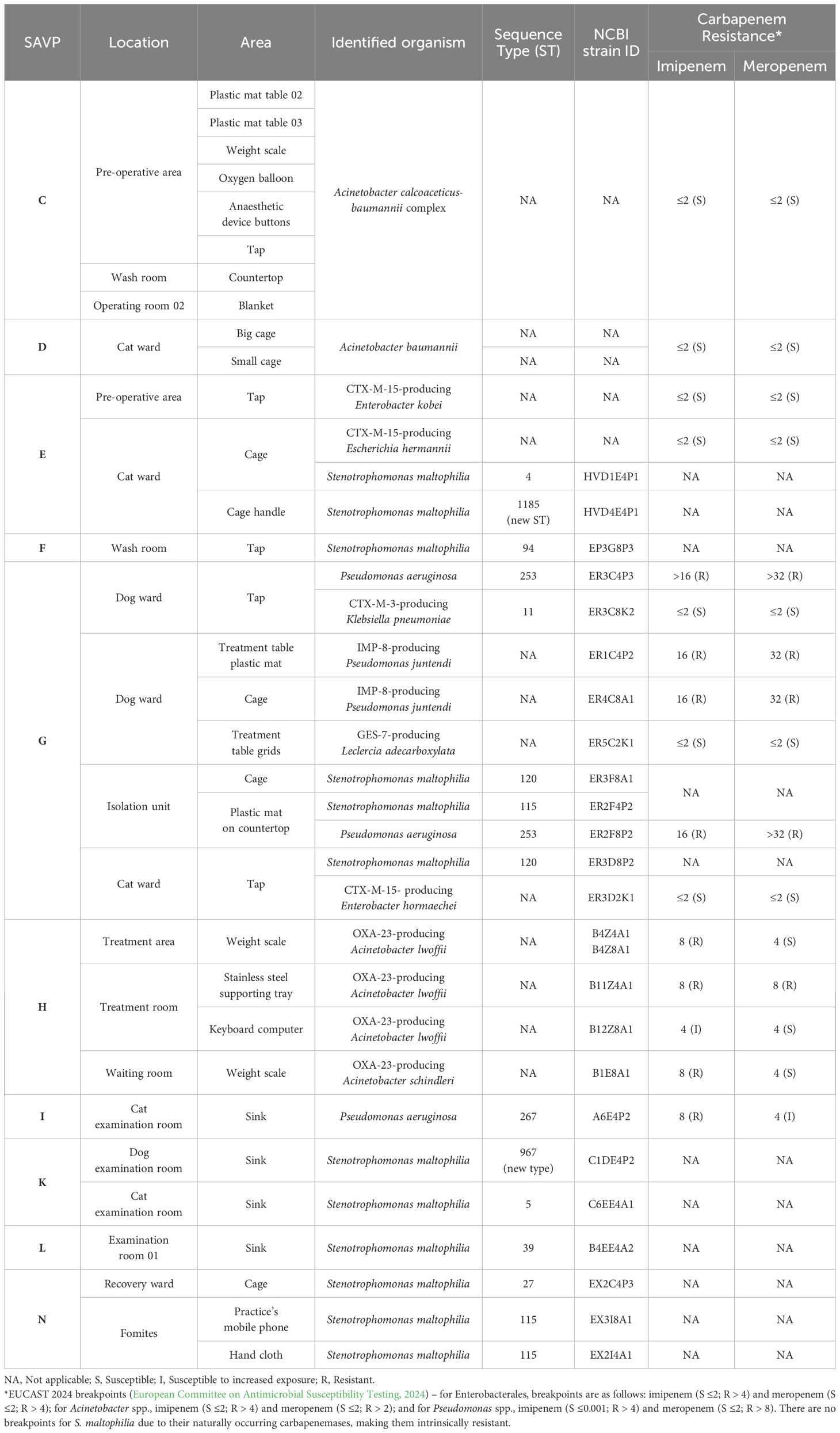
Table 1. Gram-negative bacteria identified upon environmental evaluation of SAVPs A-N.
Carbapenem-resistant non-fermenting Gram-negative bacteria were also recovered on 58.9% (n=20/32) of contaminated surfaces. Overall, 57.1% (n=8/14) of SAVP had at least one positive surface for carbapenem-resistant Gram-negative bacteria – three hospitals and five clinics (Table 1).
Considering veterinary hospitals, Stenotrophomonas maltophilia, which is intrinsically resistant to multiple classes of antibiotics, including carbapenems (due to intrinsic carbapenemases L1 and L2 (Brooke, 2021)), was identified on two surfaces of the cat ward of SAVP-E, and on the tap of the washing area of SAVP-F. In SAVP-G, P. aeruginosa and S. maltophilia were present on different surfaces (Table 1). Additionally, carbapenem-resistant Pseudomonas juntendi was found on a plastic mat on the countertop and inside one dog cage (Table 1).
Regarding veterinary clinics, carbapenem-resistant Acinetobacter spp. was found on four surfaces of SAVP-H. Carbapenem-resistant P. aeruginosa (A6E4P2) was found on the cat examination room sink of SAVP-I. S. maltophilia were found on the examination room sinks of SAVP-K) and SAVPL. For SAVP-N, it was present, on the post-surgery cage the practice’s mobile phone and a hand cloth (Table 1).
All S. maltophilia were susceptible to trimethoprim/sulfamethoxazole (MIC < 0,001 mg/L). All carbapenem-resistant isolates were characterized using WGS.
3.2.1 Whole-genome sequence analysis of carbapenem-resistant Gram-negative bacteria from surfaces
The two S. maltophilia isolated from SAVP-E belonged to different STs, namely, ST4 in a cat cage (HVD1E4P1), and the newly assigned ST1185 on a cage handle from the same cat ward (HVD4E4P1). The S. maltophilia from SAVP-F belonged to ST94 (EP3G8P3).
On SAVP-H, all the carbapenem-resistant Acinetobacter spp. (n=5) harbored blaOXA-23 carbapenemase gene in the same plasmid across different sub-species, as previously described by our group (Moreira da Silva et al., 2024a).
The two P. aeruginosa strains from SAVP-G belonged to ST253 and were unrelated (single nucleotide polymorphism (SNP) difference > 14, Supplementary Table 4) (Schürch et al., 2018). Both strains were carbapenem-resistant, having the same mutations on OprD porin channel, and on nalC and mexR genes (MexAB-OprM efflux pump repressor and regulator, respectively) (Table 2). Both P. juntendi were IMP-8-producers (ER1C4P2 and ER4C8A1) and were considered unrelated by using the SNP relatedness cut-off of P. aeruginosa (SNP difference >14, Supplementary Table 5), as no cut-of has been proposed for this sub-species (Schürch et al., 2018). The blaIMP-8 carbapenemase gene was possibly chromosomally inserted as no plasmids were detected (Aytan-Aktug et al., 2022).
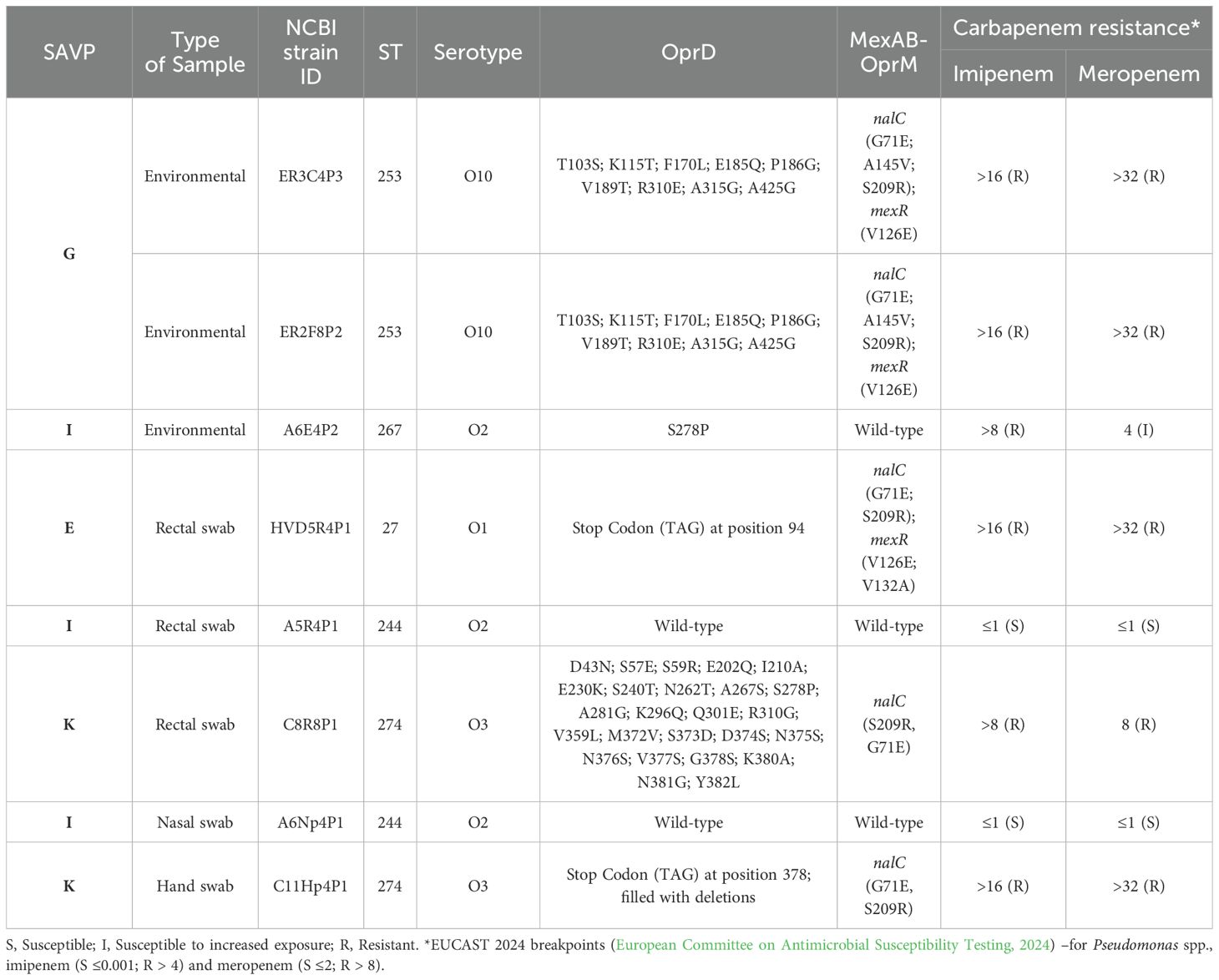
Table 2. Mutations on OprD porin channel and MexAB-OprM efflux pump of P. aeruginosa strains (n=8).
Finally, one S. maltophilia ST115 strain (ER2F4P2) and two ST120 strains (ER3F8A1 and ER3D8P2) were identified. Interestingly, the two ST120 were closely related (5 SNP difference) showing its likely dissemination between the isolation unit and cat ward of SAVP-G (Supplementary Table 6).
SAVP-I, SAVP-K and SAVP-L are veterinary clinics from the same business group (i.e. some staff alternate between veterinary practices); however, none of the isolated strains were shared between practices. The SAVP-I carbapenem-resistant P. aeruginosa ST267 strain (A6E4P2) had one mutation on OprD porin channel and a wild-type MexAb-OprM efflux pump (Table 2). As for S. maltophilia strains, multiple STs were identified, namely ST5 (C6EE4A1) and a newly assigned ST967 (C1DE8A3) in SAVP-K; and ST39 strain (B4EE4A2) in SAVP-L.
Lastly, on SAVP-N, there was one S. maltophilia ST27 strain (EX2C4P3) and two ST115 strain (EX3I8A1 and EX2I4A1; 10 SNP difference).
3.3 Carbapenem-resistant Gram-negative bacteria transient carriage by veterinary staff
Diverse and normal microbial flora was observed across the 30 fecal samples available. Carbapenem-resistant non-fermenting Gram-negative bacteria were isolated from 16.7% (n=5/30) participants, including P. aeruginosa (n=3) and S. maltophilia (n=2).
The three P. aeruginosa strains were isolated from participants of different SAVP, namely a ST27 (HVD5R4P1) in one veterinarian from SAVP-E; a ST244 (A5R4P1) in one veterinarian from SAVP-I; and a ST274 (C8R8P1) in one nurse from SAVP-K.
P. aeruginosa ST244 strain was carbapenem-susceptible and was found to have a wild-type OprD porin channel and MexAB-OprM efflux pump. P. aeruginosa ST27 strain had an early stop codon on oprD together with mutations on nalC and mexR; and P. aeruginosa ST274 had several mutations on oprD, thus explaining their carbapenem-resistant phenotype (Table 2).
One S. maltophilia ST317 strain (B3R4P1) was detected in a nurse from SAVP-L, and one S. maltophilia ST84 strain (C10R8A1) in a technician from SAVP-K, both susceptible to trimethoprim/sulfamethoxazole.
Gram-negatives were rarely isolated from nasal swabs (1.2%, n=2/169) using selective media. One veterinarian from SAVP-G was positive for an imipenem-resistant CTX-M-3-producing K. pneumoniae ST11 (R11Np4K1; MIC= 8 mg/mL), albeit susceptible to meropenem, showing a 15% truncated Omp36K in the WGS analysis (Supplementary Figure 1). One veterinarian from SAVP-I was positive for a carbapenem-susceptible P. aeruginosa ST244 (A6Np4P1) that was related to the ST244 (A5R4P1, SNP difference ≤ 3, Supplementary Table 3) isolated from the rectal swab of a distinct veterinarian from the same veterinary practice.
Only 1.8% (n=3/169) hand swabs were positive for Gram-negative bacteria. One P. aeruginosa ST274 strain (C11Hp4P1) was found on a technician from SAVP-K, showing an early stop codon on oprD, leading to failure in of the protein expression; and mutations on nalC (Table 2). In SAVP-N, two related S. maltophilia ST115 (6 SNP difference, Supplementary Table 7) were detected on the hands of a veterinarian (X4Hp4P2) and a technician (X3Hp4P1). These were closely related to the strains present in the fomites (≤ 10 SNPs difference, Supplementary Table 7), showing likely dissemination across the practice fomites and staff.
3.4 Surface contamination with methicillin-resistant Staphylococcus spp.
Nearly five percent (n=23/489) of surfaces were contaminated with methicillin-resistant Staphylococcus spp. (MRS). Two veterinary hospitals (SAVP-A; and SAVP-C) and one clinic (SAVP-N) had surfaces contaminated with MR Staphylococcus pseudintermedius (MRSP) (n=4) (Table 3). Overall, coagulase-negative MRS isolates predominated (78.3%, n=18/23), especially MR Staphylococcus epidermidis (72%, n=13/18) (Table 3). One newly typed ST9220 (clonal complex, or CC, 5) MRSA, harboring SCCmecII, was isolated from a cage at the cat ward of hospital SAVP-E.
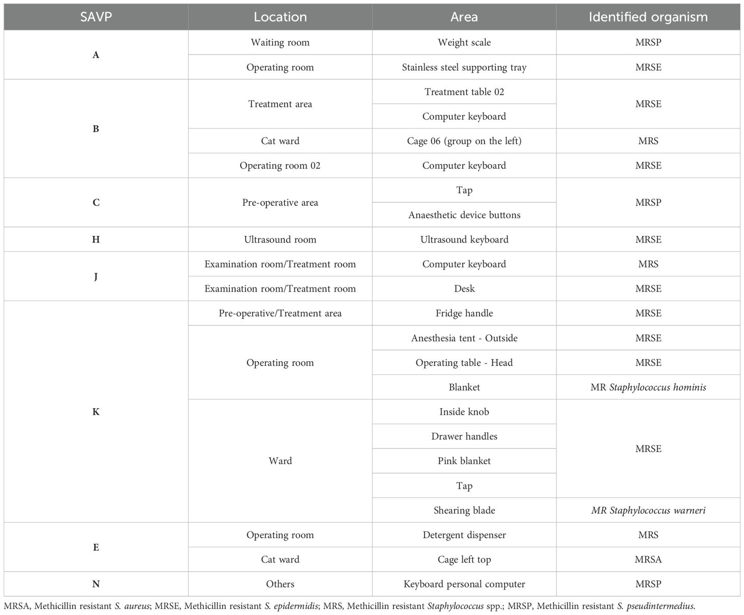
Table 3. Methicillin resistant Staphylococcus spp. on environmental evaluation of SAVPs A-N.
3.5 Methicillin-resistant Staphylococcus spp. transient carriage by staff
A total of 169 nasal swabs were collected, of which 38% were positive for MRS (n=64/169). Amongst these, 14% were MRSA (n=9/64). Further characterization identified them as belonging to different CCs: ST22-IV (also known as EMRSA-15) (n=2) (SAVP-A, n=1; SAVP-B, n=1) and ST974-IV (SAVP-I, n=1) from CC22; ST8-VI (n=3) (SAVP-B, n=2; SAVP-L, n=1) from CC8; ST30-V (SAVP-C, n=1) from CC30; ST9220-II (SAVP-E, n=1) and ST125-IV (SAVP-F, n=1) from CC5. MRSP was not detected.
Considering hand swabs, 22.4% (n=38/169) were positive for MRS, and again, none was identified as MRSP. MLST and SSCmec cassette characterization of MRSA (10.5%, n=4/38) yielded the following classifications: ST8-VI (n=2) (SAVP-I, n=1; SAVP-L, n=1) from CC8, ST9220-II (SAVP-E, n=1) from CC5 and non-typable ST from CC45 (SAVP-B, n=1) – it was not possible to type this strain of MRSA, albeit it carried SCCmecV.
Both MRSA ST8-VI strains belong to different clinics, despite being part of the same business group, with these members rotating between the different practices.
Interestingly, the new MRSA ST9220-II was only identified on SAVP-E in one surface, one nasal swab from a nurse, and one hand swab from a technician, pointing to an ongoing dissemination within this veterinary hospital.
4 Discussion
This study depicts the environmental contamination and veterinary staff carriage by resistant bacteria towards medically important antimicrobials within veterinary clinics and hospitals in Portugal. It was found that 6.5% (n=32/490) surfaces analyzed were positive for MDROs, amongst which were detected carbapenem-resistant bacteria, namely OXA-23-producing Acinetobacter spp., (n=5) and IMP-8 P. juntendi (n=2). Veterinary carriage analysis revealed that 38% and 22.4% were positive for MRS in their nasal cavities and hand swabs, respectively. For rectal swabs, only 16.7% yielded carbapenem-resistant bacteria carriage.
Notably, carbapenem-resistant Gram-negative bacteria (Acinetobacter spp., Pseudomonas spp. and S. maltophilia) and MRSA, belonging to epidemiologically relevant clones such as EMRSA-15, were found in several high-touch surfaces and fomites of SAVPs. The SAVP-G is a particularly interesting case, since, despite having a high self-assessment score about ongoing IPC procedures, it showed the highest level of environmental contamination, together with the isolation of the highest diversity of resistant bacteria. These results highlight that inadequate/insufficient IPC programs or the lack of compliance to them, may facilitate environmental contamination by MDROs, increasing the chance of their dissemination within SAVP and into the environment and community (e.g., staff, tutors, animal patients).
The hygiene evaluation of high-touch surfaces was conducted by two distinct methods, using contact plates and/or surface swabs. The observed discrepancies between results could be attributed to the design of the contact plates, which support the growth of all types of microorganisms (such as fungi and yeast), while surface swabs are loaded with a universal neutralizing liquid buffer specifically geared towards bacterial growth. Conversely, swabs can be used to evaluate irregular surfaces, supporting the use of both techniques for a more complete evaluation of environmental contamination. Considering both methods, 96 surfaces failed the cleaning efficacy assessment (Dancer, 2004). Coincidently, most of these were high-rotation surfaces such as weight scales, sinks and taps, animal cages and computer keyboards.
The worldwide ongoing rising in carbapenem-resistance is worrisome, as these are last-line antibiotics (EMA, 2019). In this study carbapenem-resistant bacteria and possible dissemination events within some SAPV were detected. On SAVP-H, OXA-23-producing Acinetobacter spp. strains were found on different surfaces of the same room, indicating a possible transfer (previously published (Moreira da Silva et al., 2024a)).
The detection of various clones of carbapenem-resistant P. aeruginosa strains on high-touch surfaces is worrisome. Between 2022-2023, 7.9% of healthcare-associated infections reported in Europe in the human healthcare setting were caused by P. aeruginosa, of which 29.7% were resistant to carbapenems (European Centre for Disease Prevention and Control, 2024). P. aeruginosa ST244, ST27 and ST253 clonal lineages are commonly associated with outbreaks and nosocomial infections in humans (del Barrio-Tofiño et al., 2020). A previous study conducted in a veterinary teaching hospital reported the presence of P. aeruginosa ST244 on sinks (Soonthornsit et al., 2023), while P. aeruginosa ST27 has higher affinity towards cystic-fibrosis patients (Weimann et al., 2024). There aren’t any guidelines that support rectal sampling on healthy healthcare-workers in human medicine, as studies have shown that in low prevalence settings of MDROs, the transient carriage by healthcare-workers will also be low (Bassyouni et al., 2015; Decker et al., 2018). Since in this study’s context, the reality of MDROs carriage by healthcare-workers is unknown, we decided to evaluate it. The low percentage of rectal samples positive for carbapenem-resistant bacteria (n=5/30, 16.7%) of veterinary healthcare-workers aligned with what is already known in human medicine as they work in a low-exposure setting to MDROs.
Strains reported on our study were from healthy human samples (P. aeruginosa ST244 from one nasal and one rectal swab from unrelated individuals; carbapenem-resistant P. aeruginosa ST27 from one rectal swab). Although SAVP-I and SAVP-K belong to the same business group, it was interesting to perceive that each SAVP had its own associated clone shared between staff members, namely, P. aeruginosa ST244 on SAVP-I, and carbapenem-resistant P. aeruginosa ST274 on SAVP-K (rectal swab and hand swab from unrelated individuals). Although none of these clones were recovered from the environment, their sharing among team members points to their potential for dissemination. These results highlight the importance of screening veterinary staff as part of an effective IPC protocol to identify potential carriers, thereby preventing transmission to patients, staff, and the community. Moreover, a carbapenem-resistant P. aeruginosa ST253 was present on a plastic mat on a countertop inside an isolation unit (SAVP-G), an area with specific cleaning and disinfection protocols (Weese, 2004), and although it is not expected for this area to be completely sterile, potentially nosocomial microorganisms should not be present.
Genetically unrelated IMP-8-producing P. juntendi strains were present on the inside of an empty cage and on the plastic cover of the treatment table in the dog ward of SAVP-G. To the best of our knowledge, this is the first description of IMP-8-producing P. juntendi. So far, only one description of carbapenem-resistant P. juntendi, harboring an IMP-1, has been made in a Chinese human patient (Zheng et al., 2022). This data highlights the capacity of this species to acquire and disseminate resistance genetic elements, making it a pathogen to be considered in epidemiological surveillance schemes.
S. maltophilia is an opportunistic pathogen with intrinsic resistance to many antibiotics, including carbapenem, posing major challenges in clinical settings (Mojica et al., 2022). In our study, multiple clones (including the new ST967 and ST1185) of S. maltophilia were described in the environment of various SAVPs, as well as colonizing staff members. These bacteria are ubiquitous in the environment, yet they have also been associated with nosocomial and community-acquired infections (Gröschel et al., 2020; Mojica et al., 2022; European Centre for Disease Prevention and Control, 2024) The relatedness between environmental and pathogenic S. maltophilia strains has been described, indicating that the environment may be a source of human contamination –including from sinks and taps (Mojica et al., 2022). As expected, the majority of the contaminated surfaces in our study were water-related. The spread of S. maltophilia through fomites ultimately causing human infection has also been described (Gideskog et al., 2020). The occurrence of S. maltophilia ST115 strains on the handcloth, the practice’s mobile phone and on the hands of two staff members of SAVP-N demonstrate that such objects likely acted as fomite and contributed to dissemination of this clone (Gideskog et al., 2020). Yet, in veterinary medicine, reports of infections caused by this agent are rare. Nonetheless, S. maltophilia ST115 clonal lineage has been associated with infections in cats (Shimizu et al., 2021), underscoring its disease-causing capability and the importance of closely monitoring it.
In Europe and according to the 2022–2023 ECDC report, 23.7% of S. aureus causing healthcare-associated infections were MRSA, showing a 6.3% decrease from the previous report (European Centre for Disease Prevention and Control, 2024). EMRSA-15 is a major clone found in hospitals and in the community in Portugal (Tavares et al., 2013). This epidemic clone has also been described on clinical strains from pets (Couto et al., 2015; Costa et al., 2022), with studies showing that working in close contact with companion animals is a risk factor for MRSA carriage (Weiß et al., 2013; Bal et al., 2016; Feßler et al., 2018; Rodrigues et al., 2018). The newly described MRSA ST9220-II, belonging to CC5, was found on SAVP-E. The presence on a surface as well as on a nasal swab from a nurse and a hand swab from a technician suggests that this clone might be spreading within this veterinary practice, possibly through contaminated surfaces.
A Portuguese study identified 61% of nasal carriage of MRS among veterinary professionals (Rodrigues et al., 2018), which is higher than what was found in the present study. However, this same study reported 14% carriage of MRSA, comparable to our findings, with EMRSA-15 also being the most prevalent clone (Rodrigues et al., 2018). The frequent colonization by MRS and MRSA reported in these two studies showcases that veterinary healthcare providers may contribute to the transmission cycle of these pathogens into the community. The frequency of MRSA recovered from hand swabs was lower. It is known that hand hygiene is a pivotal measure in IPC programs, with studies showing that improvements in the healthcare workers’ hand hygiene protocols cause a direct decrease in the carriage of MRSA (Marimuthu et al., 2014).
Overall, the detection of MRS and carbapenem-resistant Gram-negative bacteria on high-touch surfaces in SAVPs underscores the need for strict IPC procedures. These measures are essential not only to protect patients but also to address the ongoing antimicrobial resistance crisis.
5 Conclusion
The current study depicts varying levels of environmental contamination and staff carriage of carbapenem-resistant Gram-negative and MRS strains in SAVP across Portugal. These findings question the effectiveness of ongoing IPC protocols and highlight the risk of environment/staff cross-contamination through high-touch surfaces. This data suggests that SAVP may play an active role in the spread of priority pathogens resistant to medically important antimicrobials, emphasizing the need for targeted educational workshops for veterinary healthcare students and professionals. In the long run, implementing and monitoring evidence-based IPC protocols and staff training should be mandatory to ensure strict compliance in SAVP.
Data availability statement
The datasets presented in this study can be found in online repositories. The names of the repository/repositories and accession number(s) can be found below: https://www.ncbi.nlm.nih.gov/, PRJNA1131754 https://www.ncbi.nlm.nih.gov/, PRJNA1000421.
Ethics statement
The studies involving humans were approved by the Comissão de ética e bem-estar animal (CEBEA) of Veterinary Faculty of the University of Lisbon - CEBEA011/2021. The studies were conducted in accordance with the local legislation and institutional requirements. The participants provided their written informed consent to participate in this study.
Author contributions
JMS: Conceptualization, Data curation, Investigation, Writing – original draft, Writing – review & editing. JM: Investigation, Writing – review & editing. LF: Investigation, Writing – review & editing. CM: Writing – original draft, Writing – review & editing. SC: Writing – review & editing. DT: Writing – review & editing. AA: Writing – review & editing. CP: Conceptualization, Supervision, Writing – review & editing.
Funding
The author(s) declare that financial support was received for the research and/or publication of this article. This research was funded by the Portuguese Foundation for Science and Technology (FCT), under projects UIDB/00276/2020 (CIISA) and LA/P/0059/2020 (AL4AnimalS) and GHTM through FCT (UID/04413/2020) and LA-REAL -LA/P/0117/2020. This study was funded by FCT project 2022.08669.PTDC. -VetCare. JMS, JM and LF were supported by a Fundação para a Ciência e Tecnologia (FCT) PhD fellowship (2020.06540.BD; 2020.07562.BD; UI/BD/153070/2022, respectively).
Acknowledgments
The authors would like to thank all the members of the small animal veterinary practices participating in the project.
Conflict of interest
The authors declare that the research was conducted in the absence of any commercial or financial relationships that could be construed as a potential conflict of interest.
Generative AI statement
The author(s) declare that no Generative AI was used in the creation of this manuscript.
Publisher’s note
All claims expressed in this article are solely those of the authors and do not necessarily represent those of their affiliated organizations, or those of the publisher, the editors and the reviewers. Any product that may be evaluated in this article, or claim that may be made by its manufacturer, is not guaranteed or endorsed by the publisher.
Supplementary material
The Supplementary Material for this article can be found online at: https://www.frontiersin.org/articles/10.3389/fcimb.2025.1582411/full#supplementary-material
References
Alcock, B. P., Huynh, W., Chalil, R., Smith, K. W., Raphenya, A. R., Wlodarski, M. A., et al. (2023). CARD 2023: expanded curation, support for machine learning, and resistome prediction at the Comprehensive Antibiotic Resistance Database. Nucleic Acids Res. 51, D690–D699. doi: 10.1093/nar/gkac920
Aytan-Aktug, D., Grigorjev, V., Szarvas, J., Clausen, P. T. L. C., Munk, P., Nguyen, M., et al. (2022). SourceFinder: a machine-learning-based tool for identification of chromosomal, plasmid, and bacteriophage sequences from assemblies. Microbiol Spectr. 10. doi: 10.1128/spectrum.02641-22
Bal, A. M., Coombs, G. W., Holden, M. T. G., Lindsay, J. A., Nimmo, G. R., Tattevin, P., et al. (2016). Genomic insights into the emergence and spread of international clones of healthcare-, community- and livestock-associated meticillin-resistant Staphylococcus aureus: Blurring of the traditional definitions. J. Global Antimicrobial Resistance. Elsevier Ltd; 6, 95–101. doi: 10.1016/j.jgar.2016.04.004
Bassyouni, R. H., Gaber, S. N., Wegdan, A. A. (2015). Fecal carriage of extended-spectrum β-lactamase- and AmpC- producing escherichia coli among healthcare workers. J. Infect. Dev. Ctries. 9, 304–308. doi: 10.3855/jidc.5633
Brooke, J. S. (2021). Advances in the microbiology of stenotrophomonas maltophilia. Clin. Microbiol. Rev. 34. doi: 10.1128/CMR.00030-19
Costa, S. S., Ribeiro, R., Serrano, M., Oliveira, K., Ferreira, C., Leal, M., et al. (2022). Staphylococcus aureus causing skin and soft tissue infections in companion animals: antimicrobial resistance profiles and clonal lineages. Antibiotics. 11, 599. doi: 10.3390/antibiotics11050599
Couto, N., Belas, A., Kadlec, K., Schwarz, S., Pomba, C. (2015). Clonal diversity, virulence patterns and antimicrobial and biocide susceptibility among human, animal and environmental MRSA in Portugal. J. Antimicrobial Chemotherapy. 70, 2483–2487. doi: 10.1093/jac/dkv141
Dancer, S. J. (2004). How do we assess hospital cleaning? A proposal for microbiological standards for surface hygiene in hospitals. J. Hosp. Infection. 56, 10–15. doi: 10.1016/j.jhin.2003.09.017
Dashti, A. A., Jadaon, M. M., Abdulsamad, A. M., Dashti, H. M. (2009). Heat treatment of bacteria: A simple method of DNA extraction for molecular techniques. Kuwait Med. J. 41, 117–122. Available online at: https://applications.emro.who.int/imemrf/kmj_2009_41_2_117.pdf.
Decker, B. K., Lau, A. F., Dekker, J. P., Spalding, C. D., Sinaii, N., Conlan, S., et al. (2018). Healthcare personnel intestinal colonization with multidrug-resistant organisms. Clin. Microbiol. Infection. 24, 82.e1–82.e4. doi: 10.1016/j.cmi.2017.05.010
del Barrio-Tofiño, E., López-Causapé, C., Oliver, A. (2020). Pseudomonas aeruginosa epidemic high-risk clones and their association with horizontally-acquired β-lactamases: 2020 update. Int. J. Antimicrob Agents 56. doi: 10.1016/j.ijantimicag.2020.106196
Dulon, M., Haamann, F., Peters, C., Schablon, A., Nienhaus, A. (2011). Mrsa prevalence in european healthcare settings: A review. BMC Infect. Dis. 11. doi: 10.1186/1471-2334-11-138
EMA (2019). Categorisation of antibiotics for use in animals for prudent and responsible use (European Medicines Agency), 1–73. Available at: https://www.ema.europa.eu/en/documents/report/categorisation-antibiotics-european-union-answer-request-european-commission-updating-scientific_en.pdf (Accessed July 30, 2024).
Eurodev (2024). Exploring the veterinary industry in europe. Available online at: https://www.eurodev.com/blog/veterinary-industry-in-europe-trends (Accessed Jun 14, 2024).
European Centre for Disease Prevention and Control (2016). Expert opinion on whole genome sequencing for public health surveillance Strategy to harness whole genome sequencing to strengthen EU outbreak investigations and public health surveillance. Available online at: www.ecdc.europa.eu (Accessed April 12, 2025).
European Centre for Disease Prevention and Control (2024). Point prevalence survey of healthcare-associated infections and antimicrobial use in European acute care hospitals 2022-2023. Available online at: www.ecdc.europa.eu (Accessed July 30, 2024).
European Committee on Antimicrobial Susceptibility Testing. (2024). “The European Committee on Antimicrobial Susceptibility Testing,” in Breakpoint tables for interpretation of MICs and zone diameters. Version 14.0, vol. 2024.
Feßler, A. T., Schuenemann, R., Kadlec, K., Hensel, V., Brombach, J., Murugaiyan, J., et al. (2018). Methicillin-resistant Staphylococcus aureus (MRSA) and methicillin-resistant Staphylococcus pseudintermedius (MRSP) among employees and in the environment of a small animal hospital. Vet Microbiol. 221, 153–158. doi: 10.1016/j.vetmic.2018.06.001
Gentilini, F., Turba, M. E., Pasquali, F., Mion, D., Romagnoli, N., Zambon, E., et al. (2018). Hospitalized pets as a source of carbapenem-resistance. Front. Microbiol 9, 1-9. doi: 10.3389/fmicb.2018.02872
Gideskog, M., Welander, J., Melhus, Å. (2020). Cluster of S. maltophilia among patients with respiratory tract infections at an intensive care unit. Infection Prev. Practice. 2. doi: 10.1016/j.infpip.2020.100097
Grönthal, T., Österblad, M., Eklund, M., Jalava, J., Nykäsenoja, S., Pekkanen, K., et al. (2018). Sharing more than friendship – transmission of NDM-5 ST167 and CTX-M-9 ST69 Escherichia coli between dogs and humans in a family, Finland, 2015. Eurosurveillance 23. doi: 10.2807/1560-7917.ES.2018.23.27.1700497
Gröschel, M. I., Meehan, C. J., Barilar, I., Diricks, M., Gonzaga, A., Steglich, M., et al. (2020). The phylogenetic landscape and nosocomial spread of the multidrug-resistant opportunist Stenotrophomonas maltophilia. Nat. Commun. 11. doi: 10.1038/s41467-020-15123-0
Hemeg, H. A. (2021). Determination of phylogenetic relationships among methicillin-resistant Staphylococcus aureus recovered from infected humans and Companion Animals. Saudi J. Biol. Sci. 28, 2098–2101. doi: 10.1016/j.sjbs.2021.01.017
Jolley, K. A., Bray, J. E., Maiden, M. C. J. (2018). Open-access bacterial population genomics: BIGSdb software, the PubMLST.org website and their applications. Wellcome Open Res. 3. doi: 10.12688/wellcomeopenres.14826.1
Kondo, Y., Ito, T., Ma, X. X., Watanabe, S., Kreiswirth, B. N., Etienne, J., et al. (2007). Combination of multiplex PCRs for staphylococcal cassette chromosome mec type assignment: Rapid identification system for mec, ccr, and major differences in junkyard regions. Antimicrob Agents Chemother. 51, 264–274. doi: 10.1128/AAC.00165-06
Marimuthu, K., Pittet, D., Harbarth, S. (2014). The effect of improved hand hygiene on nosocomial MRSA control. Antimicrobial Resistance Infection Control 3. doi: 10.1186/2047-2994-3-34
Mathers, A. J., Peirano, G., Pitout, J. D. D. (2015). The role of epidemic resistance plasmids and international high- risk clones in the spread of multidrug-resistant Enterobacteriaceae. Clin. Microbiol Rev. 28, 565–591. doi: 10.1128/CMR.00116-14
Menezes, J., Frosini, S. M., Belas, A., Marques, C., da Silva, J. M., Amaral, A. J., et al. (2023). Longitudinal study of ESBL/AmpC-producing Enterobacterales strains sharing between cohabiting healthy companion animals and humans in Portugal and in the United Kingdom. Eur. J. Clin. Microbiol. Infect. Diseases. 42, 1011–1024. doi: 10.1007/s10096-023-04629-2
Menezes, J., Moreira da Silva, J., Frosini, S. M., Loeffler, A., Weese, S., Perreten, V., et al. (2022). mcr-1 colistin resistance gene sharing between Escherichia coli from cohabiting dogs and humans, Lisbon, Portugal, 2018 to 2020. Eurosurveillance 27. doi: 10.2807/1560-7917.ES.2022.27.44.2101144
Mojica, M. F., Humphries, R., Lipuma, J. J., Mathers, A. J., Rao, G. G., Shelburne, S. A., et al. (2022). Clinical challenges treating Stenotrophomonas maltophilia infections: An update. JAC-Antimicrobial Resistance 4. doi: 10.1093/jacamr/dlac040
Moreira da Silva, J., Menezes, J., Fernandes, L., Marques, C., Costa, S. S., Timofte, D., et al. (2024a). Dynamics of blaOXA-23 gene transmission in Acinetobacter spp. from contaminated veterinary environmental surfaces: an emerging One Health threat? J. Hosp. Infection. 146, 116–124. doi: 10.1016/j.jhin.2024.02.001
Moreira da Silva, J., Menezes, J., Fernandes, L., Santos Costa, S., Amaral, A., Pomba, C. (2024b). Carbapenemase-producing Enterobacterales strains causing infections in companion animals—Portugal. Microbiol Spectr.doi: 10.1128/spectrum.03416-23
Mulvey, D., Redding, P., Robertson, C., Woodall, C., Kingsmore, P., Bedwell, D., et al. (2011). Finding a benchmark for monitoring hospital cleanliness. J. Hosp. Infection 77, 25–30. doi: 10.1016/j.jhin.2010.08.006
Murray, C. J., Ikuta, K. S., Sharara, F., Swetschinski, L., Robles Aguilar, G., Gray, A., et al. (2022). Global burden of bacterial antimicrobial resistance in 2019: a systematic analysis. Lancet 399, 629–655. doi: 10.1016/S0140-6736(21)02724-0
Nienhoff, U., Kadlec, K., Chaberny, I. F., Verspohl, J., Gerlach, G. F., Schwarz, S., et al. (2009). Transmission of methicillin-resistant Staphylococcus aureus strains between humans and dogs: Two case reports. J. Antimicrobial Chemotherapy. 64, 660–662. doi: 10.1093/jac/dkp243
Rodrigues, A. C., Belas, A., Marques, C., Cruz, L., Gama, L. T., Pomba, C. (2018). Risk factors for nasal colonization by methicillin-resistant staphylococci in healthy humans in professional daily contact with companion animals in Portugal. Microb Drug Resist. 24, 434–446. doi: 10.1089/mdr.2017.0063
Schmidt, J. S., Kuster, S. P., Nigg, A., Dazio, V., Brilhante, M., Rohrbach, H., et al. (2020). Poor infection prevention and control standards are associated with environmental contamination with carbapenemase-producing Enterobacterales and other multidrug-resistant bacteria in Swiss companion animal clinics. Antimicrob Resist. Infect. Control. 9, 1–13. doi: 10.1186/s13756-020-00742-5
Schürch, A. C., Arredondo-Alonso, S., Willems, R. J. L., Goering, R. V. (2018). Whole genome sequencing options for bacterial strain typing and epidemiologic analysis based on single nucleotide polymorphism versus gene-by-gene–based approaches. Clin. Microbiol. Infection 24, 350–354. doi: 10.1016/j.cmi.2017.12.016
Sing, A., Tuschak, C., Hörmansdorfer, S. (2008). Methicillin-resistant Staphylococcus aureus in a family and its pet cat. N. Engl. J. Med. 358 (11), 1200–1201. doi: 10.1056/NEJMc0706805
Shimizu, T., Tsuyuki, Y., Shimoike, K., Iyori, K., Miyamoto, T., Harada, K. (2021). Antimicrobial resistance and multilocus sequence types of Stenotrophomonas maltophilia isolated from dogs and cats in Japan. J. Med. Microbiol 70. doi: 10.1099/jmm.0.001344
Soonthornsit, J., Pimwaraluck, K., Kongmuang, N., Pratya, P., Phumthanakorn, N. (2023). Molecular epidemiology of antimicrobial-resistant Pseudomonas aeruginosa in a veterinary teaching hospital environment. Vet Res. Commun. 47, 73–86. doi: 10.1007/s11259-022-09929-0
Srinivasan, R., Karaoz, U., Volegova, M., MacKichan, J., Kato-Maeda, M., Miller, S., et al. (2015). Use of 16S rRNA gene for identification of a broad range of clinically relevant bacterial pathogens. PloS One 10, 1–22. doi: 10.1371/journal.pone.0117617
Tavares, A., Miragaia, M., Rolo, J., Coelho, C., De Lencastre, H. (2013). High prevalence of hospital-associated methicillin-resistant Staphylococcus aureus in the community in Portugal: Evidence for the blurring of community-hospital boundaries. Eur. J. Clin. Microbiol. Infect. Diseases. 32, 1269–1283. doi: 10.1007/s10096-013-1872-2
Volkov, S. (2024). WHO List of Medically Important Antimicrobials A risk management tool for mitigating antimicrobial resistance due to non-human use. Available online at: https://iris.who.int/ (Accessed August 1, 2024).
Weese, J. S. (2004). “Barrier precautions, isolation protocols, and personal hygiene in veterinary hospitals,” in Veterinary clinics of north america - equine practice 20 (3), 543–559. doi: 10.1016/j.cveq.2004.07.006
Weiß, S., Kadlec, K., Feßler, A. T., Schwarz, S. (2013). Identification and characterization of methicillin-resistant Staphylococcus aureus, Staphylococcus epidermidis, Staphylococcus haemolyticus and Staphylococcus pettenkoferi from a small animal clinic. Vet Microbiol 167, 680–685. doi: 10.1016/j.vetmic.2013.07.036
Weimann, A., Dinan, A. M., Ruis, C., Bernut, A., Pont, S., Brown, K., et al. (2024). Evolution and host-specific adaptation of Pseudomonas aeruginosa. Sci. (1979) 385. doi: 10.1126/science.adi0908
World Health Organization. (2023). Global strategy on infection prevention and control. Geneva: World Health Organization; 2023. Licence: CC BY-NC-SA 3.0 IGO.
Keywords: small animal veterinary practices, multidrug-resistant organisms, environmental contamination IPC, carbapenem resistance, MRSA, veterinary staff carriage
Citation: Moreira da Silva J, Menezes J, Fernandes L, Marques C, Costa SS, Timofte D, Amaral AJ and Pomba C (2025) Evaluation of multidrug-resistant bacteria and their molecular mechanisms found in small animal veterinary practices in Portugal. Front. Cell. Infect. Microbiol. 15:1582411. doi: 10.3389/fcimb.2025.1582411
Received: 24 February 2025; Accepted: 10 April 2025;
Published: 05 May 2025.
Edited by:
Mattia Pirolo, University of Copenhagen, DenmarkReviewed by:
Chamara De Silva Benthotage, Southern Cross University, AustraliaSalome N. Seiffert, Zentrum für Labormedizin (ZLM), Switzerland
Copyright © 2025 Moreira da Silva, Menezes, Fernandes, Marques, Costa, Timofte, Amaral and Pomba. This is an open-access article distributed under the terms of the Creative Commons Attribution License (CC BY). The use, distribution or reproduction in other forums is permitted, provided the original author(s) and the copyright owner(s) are credited and that the original publication in this journal is cited, in accordance with accepted academic practice. No use, distribution or reproduction is permitted which does not comply with these terms.
*Correspondence: Constança Pomba, Y3BvbWJhQGZtdi51bGlzYm9hLnB0; amNzaWx2YUBlZ2FzbW9uaXouZWR1LnB0