 Michelle Cristina Erckmann1
 Aline Almeida2
 Diogo Dominguini3Daniela Becker2Josiane Khun Rutz4Dachamir Hotza5,6Abhishek Parolia7Vanessa Valgas Dos Santos1Michael Ramos Nunes5,8Cleonice Gonçalves Da Rosa1
 Anelise Viapiana Masiero1,7*
- 1Multi-User Laboratory, Graduate Program in Environment and Health, Planalto Catarinense University, Lages, SC, Brazil
- 2Laboratory of Plasmas, Films, and Surfaces, Santa Catarina State University (UDESC), Joinville, SC, Brazil
- 3Laboratory of Experimental Pathophysiology, Graduate Program in Health Sciences, University of Extreme South of Santa Catarina (UNESC), Criciúma, Brazil
- 4Center of Biomaterials Development and Control, Faculty of Dentistry, Federal University of Pelotas, Pelotas, Rio Grande do Sul, Brazil
- 5Graduate Program in Chemical Engineering (PosENQ), Federal University of Santa Catarina (UFSC), Florianópolis, SC, Brazil
- 6Department of Chemical and Food Engineering (EQA), Federal University of Santa Catarina (UFSC), Florianópolis, SC, Brazil
- 7Department of Endodontics, College of Dentistry and Dental Clinics, University of Iowa, Iowa City, IA, United States
- 8Federal Institute of Santa Catarina, Lages, SC, Brazil
Introduction: Dental caries is a multifactorial disease with high prevalence, particularly in vulnerable populations, where Streptococcus mutans contributes to lesion progression via acid production and biofilm formation. Minimally invasive strategies, such as photodynamic therapy (PDT) combined with advanced delivery systems, offer promising alternatives for caries management.
Methods: Zein-based nanocapsules loaded with curcumin (Nano-curcumin) were synthesized via nanoprecipitation and characterized for encapsulation efficiency, particle size, polydispersity, zeta potential, morphology, and curcumin release. Biocompatibility was assessed using rabbit oral mucosal cells via MTT and trypan blue assays. Antimicrobial efficacy was tested in vitro on primary dentin slices contaminated with S. mutans across four groups: Nano-curcumin, Nano-curcumin + PDT, diode laser, and untreated control. Colony-forming units (CFU) were quantified after treatment. Statistical analysis was performed using ANOVA and Tukey’s test (p < 0.05).
Results: Nano-curcumin demonstrated high encapsulation efficiency (~100%), spherical morphology, low polydispersity (0.108), and favorable colloidal stability, with sustained curcumin release over 24 hours. Cytotoxicity assays showed >50% cell viability at 100 μg·mL⁻¹ and ~80% at intermediate concentrations (50–75 µg·mL-¹). Both curcumin nanocapsules and their photosensitized versions significantly reduced S. mutans CFU compared to controls (p < 0.05), with PDT-enhanced nanocapsules showing the greatest reduction, though not statistically different from non-photosensitized nanocapsules.
Discussion: Curcumin-loaded zein nanocapsules are biocompatible and effective against S. mutans, with controlled release properties. Photodynamic activation further enhances antimicrobial activity, supporting their potential as a minimally invasive approach for managing carious lesions, particularly in pediatric dentistry. This strategy integrates a natural photosensitizer with a biodegradable polymeric matrix, providing a safe and innovative alternative for caries control.
1 Introduction
Dental caries continues to be a major public health issue, particularly among vulnerable populations. This is largely due to frequent carbohydrate consumption, increased acidity, and disruption of the oral microbiota (Takahashi and Nyvad, 2016). This acidic environment promotes pathogenic biofilm formation and enamel demineralization, with Streptococcus. mutans playing a central role. This bacterium efficiently metabolizes sugars, producing acid while thriving in low-pH conditions. It also contributes, to biofilm stability by synthesizing an extracellular polysaccharide matrix which enhances resistance to antimicrobial agents (de Oliveira et al., 2019; Pourhajibagher et al., 2019).
Although conventional treatments focus on removing caries and restoring teeth they often fall short in achieving long-term disease control. Primary teeth are especially susceptible to rapid caries progression due to their thinner dentin, larger pulp chambers, and increased permeability (Nehete et al., 2014). To address these challenges, minimally invasive techniques such as selective caries removal (SCR), stepwise caries removal (SWR), and the Hall Technique have been developed. These methods aim to preserve tooth vitality while minimizing patient discomfort (Aïem et al., 2020; Innes et al., 2011; Machiulskiene et al., 2020).
In addition to preserving tooth structure, controlling residual bacteria is essential to prevent pulp inflammation and recurrent lesion (Diniz et al., 2015). Photodynamic therapy (PDT) has emerged as a promising antimicrobial strategy, utilizing light-activated photosensitizers to selectively eliminate cariogenic bacteria (Wilson and Patterson, 2008). Among these, curcumin stands out as a photosensitizer due to its antibacterial (Carolina Alves et al., 2019), antifungal (Zorofchian Moghadamtousi et al., 2014), antineoplastic (Ghaffari et al., 2020), anti-inflammatory (Zhi et al., 2021) and antioxidant properties (Kamwilaisak et al., 2022).
When used in PDT, curcumin exhibits high cytotoxicity against pathogenic microorganisms, particularly against Gram-positive bacteria (Adamczak et al., 2020). These properties make it a promising candidate for the development of new antimicrobial therapies (Hosseinpour-Nader et al., 2022). Nanotechnology further enhances curcumin’s therapeutic potential by improving its stability, bioavailability, and antimicrobial efficacy (Hosseinpour-Nader et al., 2022). Incorporating nanoparticles into dental materials has also been shown to enhance their mechanical and biological properties (Andreatta et al., 2023; Batista et al., 2024; da Rosa et al., 2022; Masiero et al., 2024; Narciso et al., 2019; Parizzi et al., 2025).
In this context, targeted strategies against S. mutans including the use of nanoparticles to modulate the cariogenic microbiome have shown encouraging results (Sayed et al., 2020). However, despite growing interest in PDT and the the known antimicrobial potential of curcumin, few studies have explored the combined use of curcumin-loaded nanostructures and PDT in primary dentin, which differs morphological and histological characteristics compared to permanent teeth. Moreover, limited research has assessed the biocompatibility of such systems in healthy oral tissues, particularly in pediatric settings.
To address these gaps, the present study aimed to synthesize and characterize zein-based nanocapsules loaded with curcumin, evaluate their biocompatibility with oral mucosal cells, and investigate their their in vitro antimicrobial efficacy on primary dentin contaminated with S. mutans, both with and without photodynamic activation. This innovative approach combines a natural photosensitizers with a biodegradable polymeric matrix, offering a minimally invasive and potentially safer alternative for the treatment of carious lesions in children.
2 Material and methods
This study was approved by the Research Ethics Committee (CAAE No. 6.246.02).
2.1 Materials
The materials used in this study included curcumin, zein, poloxamer 407, and Dulbecco’s Modified Eagle Medium (DMEM), all from Sigma-Aldrich (Saint Louis, MO, USA). The culture media used comprised Mueller-Hinton agar and Tryptic Soy agar (Himedia, Thane, India), along with Brain and Heart Infusion Agar (BHI) (Merck, Darmstadt, Germany). The bacterial strain employed was S. mutans ATCC 25175. All other reagents were also obtained from Sigma-Aldrich.
2.2 Methods
2.2.1 Synthesis and physicochemical characterization of zein nanocapsules loaded with curcumin
Curcumin-loaded zein nanocapsules (Nano-curcumin) were synthesized using the nanoprecipitation method in triplicate (n=3), following the methodology described by (Gonçalves da Rosa et al., 2020; Suzuki et al., 2016). To prepare the organic phase zein (20 mg mL-1) in 6.67 mL of 85% ethanol. Once fully solubilized, 134 μL of an alcoholic curcumin solution (1.5 mg mL-1) was added. The organic phase was then poured into 20 mL of an aqueous phase containing the surfactant Pluronic (0.8% v/v) under constant agitation at 10,000 rpm using a homogenizer IKA T25 homogenizer (IKA, Wilmington, NC,USA) for 3 minutes.
Nanocapsule formation occurred via nanoprecipitation upon contact between the two phases. The resulting suspension was stirred under a fume hood with magnetic agitation to ensure complete evaporation of the organic solvent. A control formulation without curcumin (Nano-curcumin-free) was prepared using the same procedure.
To confirm nanoencapsulation, the encapsulation efficiency (EE) of the curcumin-loaded zein nanocapsules was evaluated in triplicate (n=3) following to the methodology described by (Gonçalves da Rosa et al., 2020). EE was determined using a centrifugal ultrafiltration method, as outlined by Parizzi et al. (2025). Samples were centrifuged using Amicon Ultra centrifugal filters with a 30 kDa Ultracel membrane at 6,000 rpm for 30 minutes, allowing non-encapsulated curcumin to pass through the membrane.
The free curcumin in the supernatant was quantified using UV-Vis spectroscopy (Spectrostar Nano, BMG Labtech, Weston Parkway Suite, NC, USA) at a wavelength of approximately 430 nm. The molar concentration of curcumin was calculated based on a calibration curve prepared with an alcoholic curcumin solution.
Encapsulation efficiency (EE) was calculated using Equation 1:
To confirm curcumin encapsulation, UV-Vis spectrophotometry was performed using a Spectrostar Nano scanning spectrophotometer (BMG Labtech, Weston Parkway Suite, NC, USA). Measurements were taken across a wavelength range of 200 to 600 nm, with a resolution of 1 nm. The absorbance peak (λmax) of free curcumin was determined after dilution in absolute ethanol, while nanocapsule suspensions were diluted in ultrapure water prior to analysis.
Physicochemical characterization included the assessment of particle size (nm), polydispersity index (PDI), and zeta potential (mV), using dynamic light scattering (DLS) with a Zetasizer Advance (Malvern Panalytical, Worcestershire, UK). Samples of Nano-curcumin and control formulations were diluted in Milli-Q® water and analyzed at 25 °C, with a scattering angle of 173°, in triplicate (n = 3), using electrophoretic cells.
Nanocapsule morphology was examined using transmission electron microscopy (TEM) JEOL JEM 2100 (Tokyo, Japan) operating at 70 kV. Solutions containing curcumin-loaded zein nanocapsules and control samples were diluted in ultrapure Milli-Q water. Approximately 5 µL of each sample was deposited onto carbon-coated copper grids (200 mesh). After air drying at room temperature, the grids were observed under the microscope.
The curcumin release assay was conducted using a citrate–phosphate buffer at pH 7.0, as described by Parizzi et al. (2025). For each experiment, 1 mL of the nanoparticle dispersion was placed into a dialysis membrane (pore size: 25 Å; molecular weight cut-off: 12,000–16,000 Da) and immersed in 100 mL of buffer under continuous stirring. Samples of the external medium were collected at regular intervals from 1 to 8 hours, with additional aliquots taken at 12 and 24 hours. The amount of curcumin released was quantified by UV-Vis spectrophotometry using the Spectrostar Nano (BMG Labtech, Weston Parkway Suite, NC, USA) at 425 nm. Concentrations were determined using a calibration curve constructed with curcumin standards.
2.2.2 Cytotoxicity and cell viability assay
The cytotoxicity and cell viability of curcumin-loaded zein nanocapsules (Nano-curcumin) were evaluated using surface mucosal cells derived from rabbits. The cells were cultured in high-glucose Dulbecco’s Modified Eagle Medium (DMEM) supplemented with 10% fetal bovine serum, 100 U/mL penicillin, and 100 mg/mL streptomycin. Cultures were maintained in a humidified atmosphere at 37 °C with 5% CO2 and 95% air until confluence was reached.
Once confluent,the cells were seeded at a density of 10,000 cells per well in 96-well plates. A single dose of nanocapsules was added at concentrations of 25, 50, 75, and 100 μg·mL-1.
Following treatment, the samples were irradiated using a low-power diode laser InGaAlP (DMC-Therapy, Sao Carlos, SP, Brazil) at a wavelength of 660 nm, continuous emission, a power output of 100 mW, and a total energy of 9 joules over 90 seconds (Knorst et al., 2019).The laser tip was positioned 10 mm above the wells, and irradiation was applied alternately to sets of four wells to ensure proper spacing between the light source and avoid overlapping light exposure.
Cell viability was assessed using the MTT assay (0.5 mg·mL-1) and trypan blue exclusion (TBE). For the MTT assay, 20 μL of a stock MTT solution (5 mg·mL-1) was added to each well and incubated for 4 hours. After incubation, cells were dissolved in DMSO, and optical density was measured at 490 nm in equipment Spectrostar Nano (BMG Labtech, Weston Parkway Suite NC, USA)The percentage of viable cells relative to the control was calculated based on the absorbance values, considering the ratiobetween treated cells (Abs. sample) and the absorbance of the cell-free culture medium (Abs blank), as indicated in Equation 2.
2.2.3 In vitro antimicrobial evaluation of zein nanocapsules loaded with curcumin on primary tooth dentin contaminated with Streptococcus mutans
2.2.3.1 Sample preparation
Forty mandibular and maxillary primary molars free of caries, restoration and with no visible cracks or fractures were collected for this study. The teeth were donated by patients prior consent from their legal guardians. After collection, the specimens were washed to remove impurities, sterilized in an autoclave, and stored in distilled water until sectioning. For the cutting procedure, each tooth was mounted on an acrylic plate using low-melting-point plasticized wax and sectioned using a precision cutting machine (Isomet 1000, Buehler, Coventry, UK). Sections of approximately 1 mm thick were obtained using a 0.4 mm diamond disc (Buehler, Lake Bluff, IL, USA) operating at 300 rpm. Based on the inclusion criteria a final sample of 28 slices was selected. These slices were then sterilized again by autoclaving before being used in the contamination procedure.
2.2.3.2 Bacterial culture and contamination procedure
To evaluate the antimicrobial activity of the nanoparticles, lyophilized S. mutans (ATCC 25175) strains were rehydrated according to the manufacturer’s instructions and incubated anaerobically in Tryptone Soy Broth (TSB) at 37°C for 48 hours. Following incubation, the samples were plated on solid Blood Agar using the streak plate method to obtain isolated colonies. From these colonies, a bacterial suspension equivalent to 1.5 × 10⁸ cells·mL-¹ was prepared using the 0.5 McFarland scale. The 1 mm dentin slices were then incubated in 990 μL of TSB medium supplemented with 10 μL of the S. mutans bacterial suspension and maintained under anaerobic conditions at 37°C for 48 hours.
2.2.3.3 Experimental groups and treatments
After the incubation period, the dentin slices were removed from the bacterial suspension, transferred to 1 mL of saline solution, and immediately divided into the following experimental groups (n = 7):
● Group 1: Contaminated Dentin + NanoCurcumin (Dent-NanoCurcumin)
● Group 2: Contaminated Dentin + NanoCurcumin + Photodynamic Therapy (Dent-NanoCurc-PDT)
● Group 3: Contaminated Dentin + Diode Laser
● Group 4: Contaminated Dentin (Dent) – Control
Groups 1 and 2 were incubated with 1 mL of their respective nanocapsule dispersion (containing 7.5 µg/mL of curcumin) for 4 hours at room temperature, while Groups 3 and 4 were incubated with saline solution under the same conditions. Subsequently, Groups 2 and 3 were treated with a low-power InGaAlP diode laser (DMC-Therapy, São Carlos, SP, Brazil) at a wavelength of 660 nm, in continuous emission mode, with a power output of 100 mW and a total energy of 9 joules applied over 90 seconds (Parizzi et al., 2025; Knorst et al., 2019).
2.2.3.4 Microbiological Analysis
Following the treatments, the dentin slices were transferred to 1 mL of saline solution and immediately incubated in TSB broth for 30 minutes. To assess antimicrobial activity, 10 μL of the broth was placed at the center of a sterile Petri dish, over which Mueller-Hinton agar was poured. After solidification, the plates were incubated anaerobically at 37°C for 48 hours. Colony-forming units (CFU) were then counted, and the results were expressed as CFU·mL-¹ (Fernandes et al., 2022).
2.2.3.5 Data analysis
Results were expressed as means and standard deviations from triplicate measurements. Statistical analysis was performed using analysis of variance (ANOVA), followed by Tukey’s test for multiple comparisons, with a significance level of 5%. Data were analyzed using STATISTICA 7 software.
3 Results
3.1 Physicochemical characterization of nanocapsules
The encapsulation efficiency of curcumin in zein matrices was close to 100%. Figure 1 shows the absorbance spectra of free curcumin and nanocurcumin, obtained by UV-Vis spectroscopy. Free curcumin exhibited a well-defined absorbance peak at approximately 425 nm. In contrast, the nanocurcumin spectrum displayed an altered profile, with the absence of this characteristic peak and increased absorbance in the UV region.
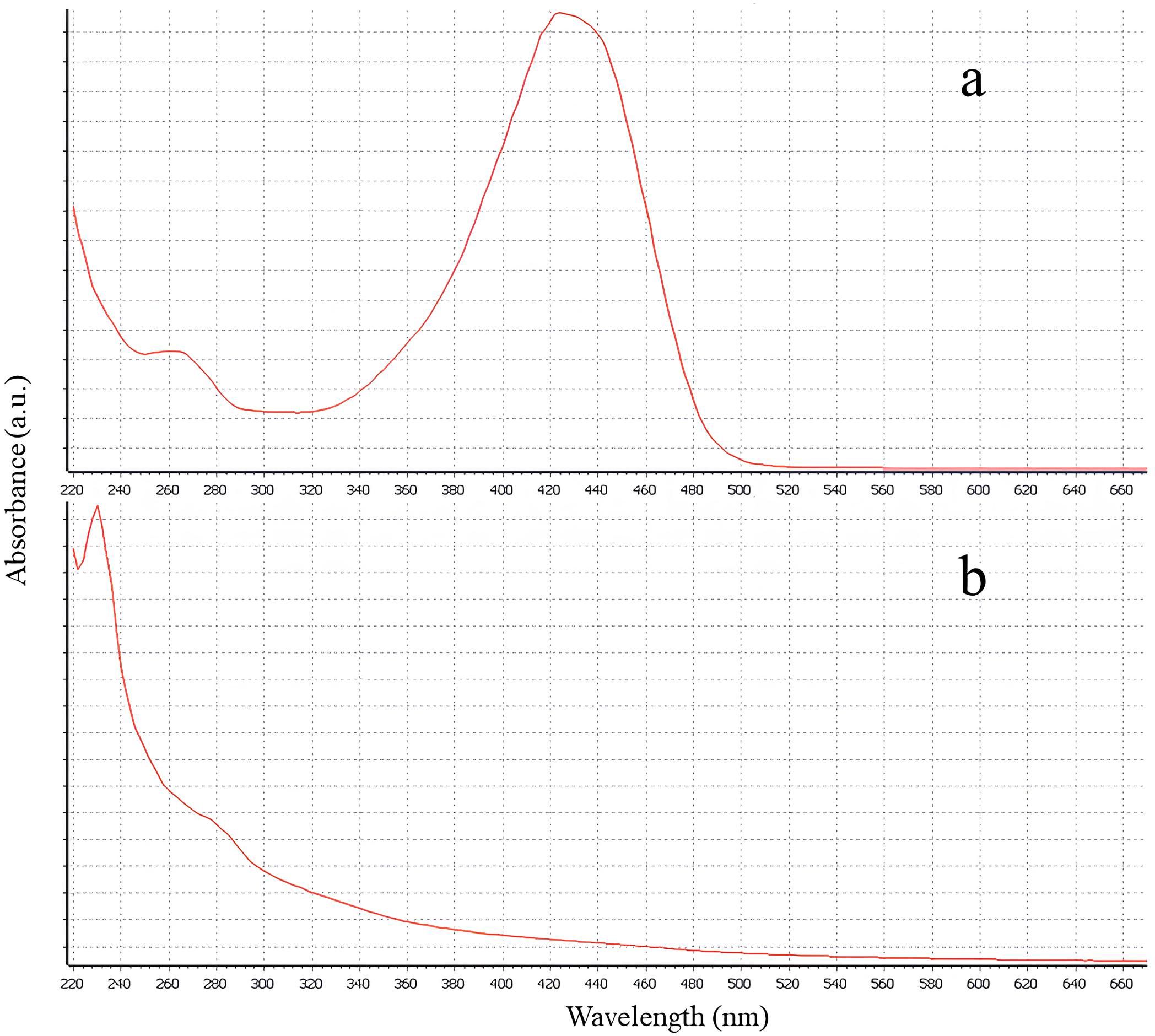
Figure 1. UV-Vis scan spectra: (a) free curcumin; (b) nanocurcumin.
Table 1 presents the measurements of average particle size, polydispersity index (PDI), and zeta potential for nano-curcumin and nano-curcumin-free samples. Nano-curcumin particles exhibited a larger average size (139.4 ± 1.0 nm) compared to the nano-curcumin-free particles (128.5 ± 0.7 nm), with a statistically significant difference (Figure 2). The PDI values indicate that nano-curcumin had a lower polydispersity index (0.108 ± 0.07) whereas nano-curcumin-free showed a significantly higher PDI (0.271 ± 0.03). Regarding zeta potential, nano-curcumin exhibited a lower value (10.9 ± 0.5 mV) compared to nano-curcumin-free (40.0 ± 2.8 mV), also with a significant difference.
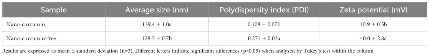
Table 1. Average particle size, polydispersity index (PDI), and zeta potential.
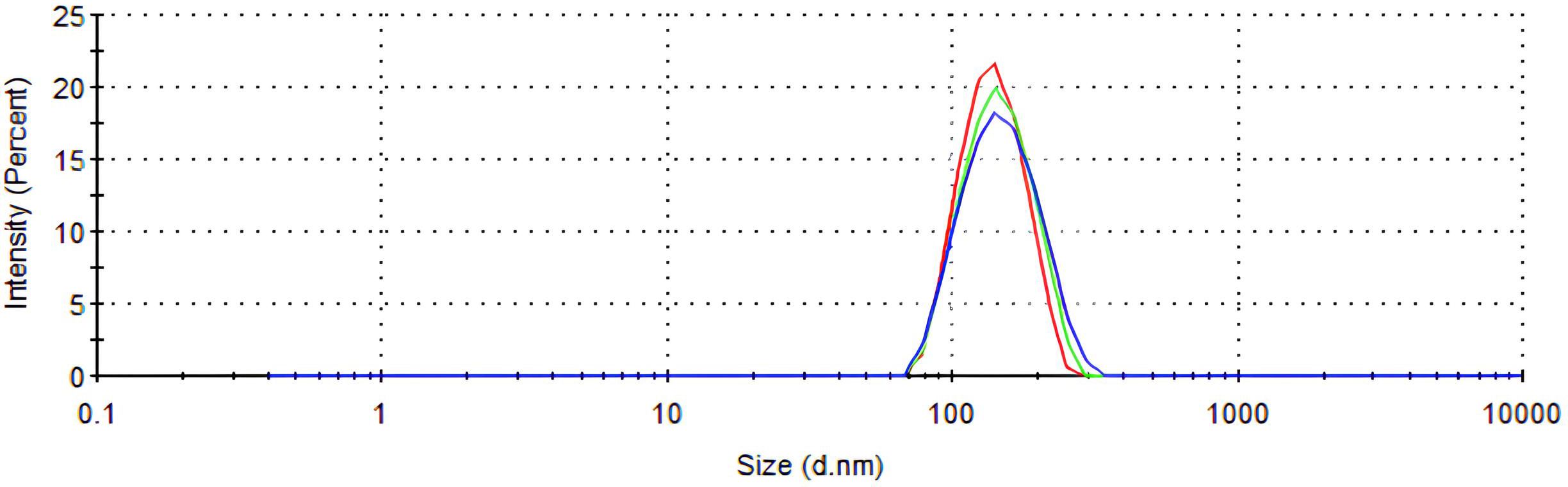
Figure 2. Particle size distribution of the Nano-curcumin sample.
The transmission electron microscopy (TEM) analysis of zein nanocapsules loaded with curcumin revealed key morphological characteristics. TEM micrographs indicated that the nanocapsules exhibited a spherical shape (Figure 3).
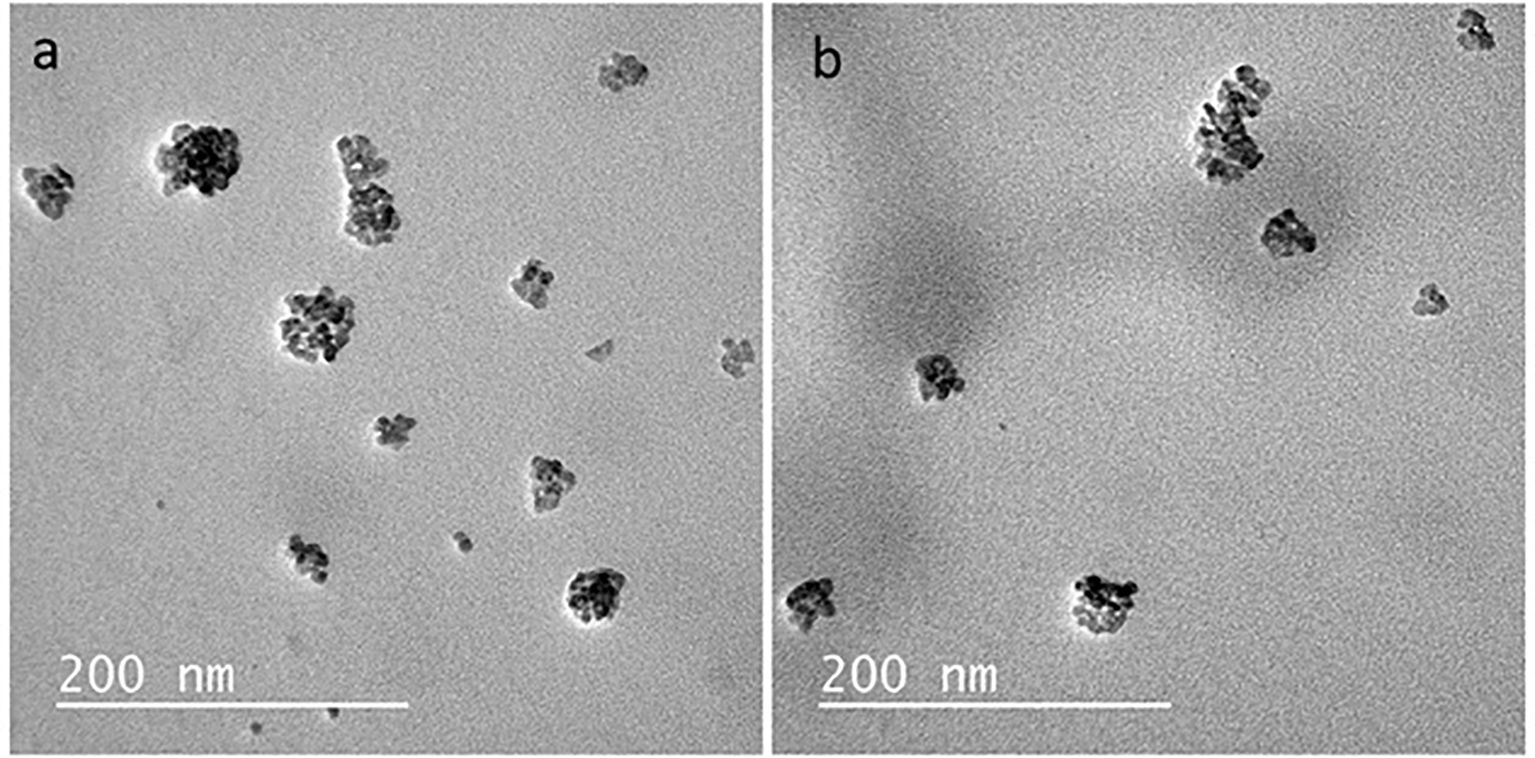
Figure 3. TEM micrographs of the nanocapsules: (a) Nano-curcumin-free (b) Nano-curcumin.
Spectrophotometric analysis showed that nano-curcumin exhibited a sustained release in buffered aqueous medium over a 24-hour period. The release profile indicates a gradual increase in curcumin concentration in the external medium, with a more pronounced release during the first 8 hours and a tendency toward stabilization after 12 hours, as shown in Figure 4.
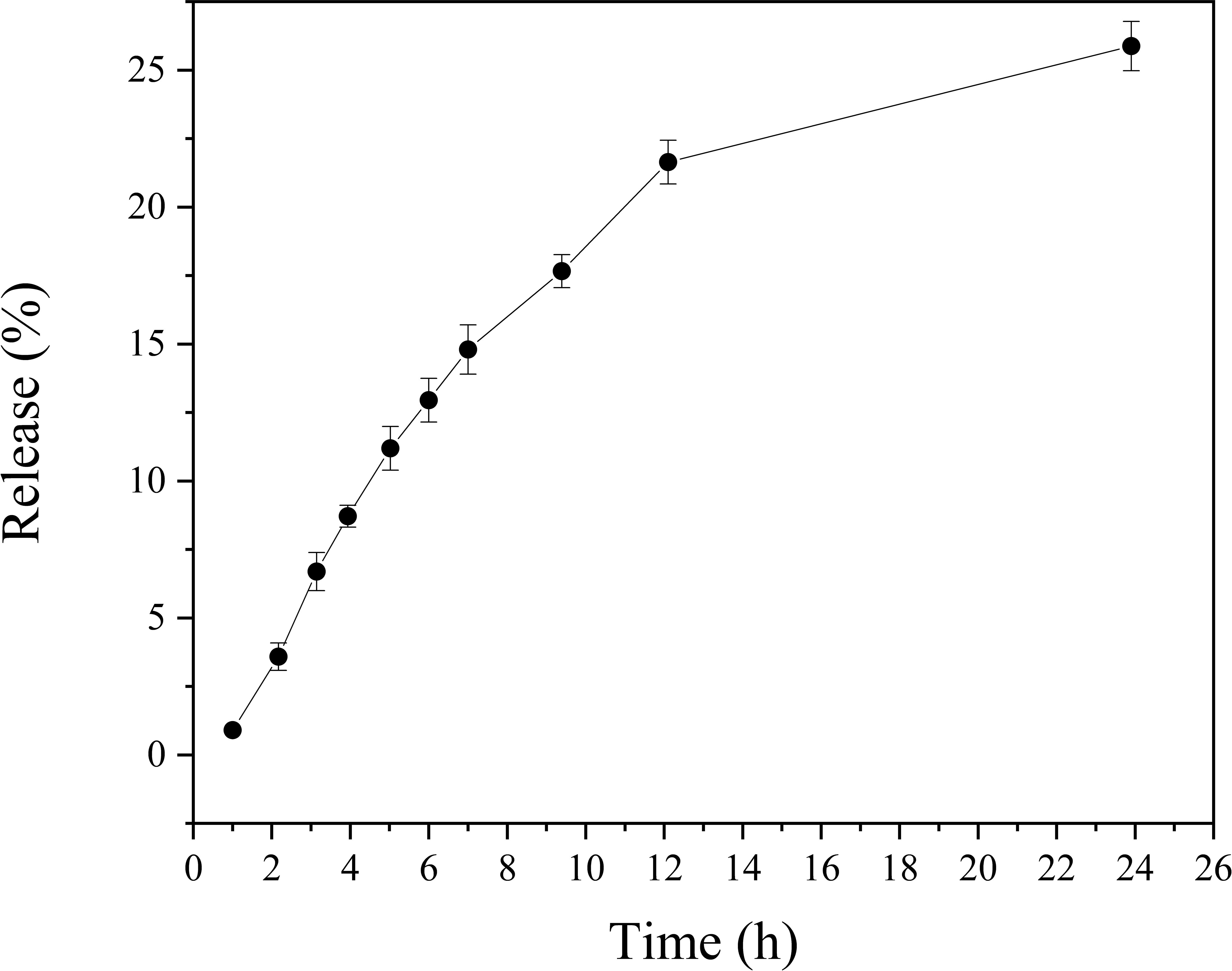
Figure 4. Release of nano-curcumin in citrate–phosphate buffer (pH 7.0) over 24 hours.
3.2 Cytotoxicity and cell viability assay
At a 100 μg·mL-1 nanocapsule concentration, the cell survival rate exceeded 50μg·mL-1 while concentrations of 50 μg·mL-1 and 75μg·mL-1 resulted in cell survival rates approaching 80% (Figure 5).
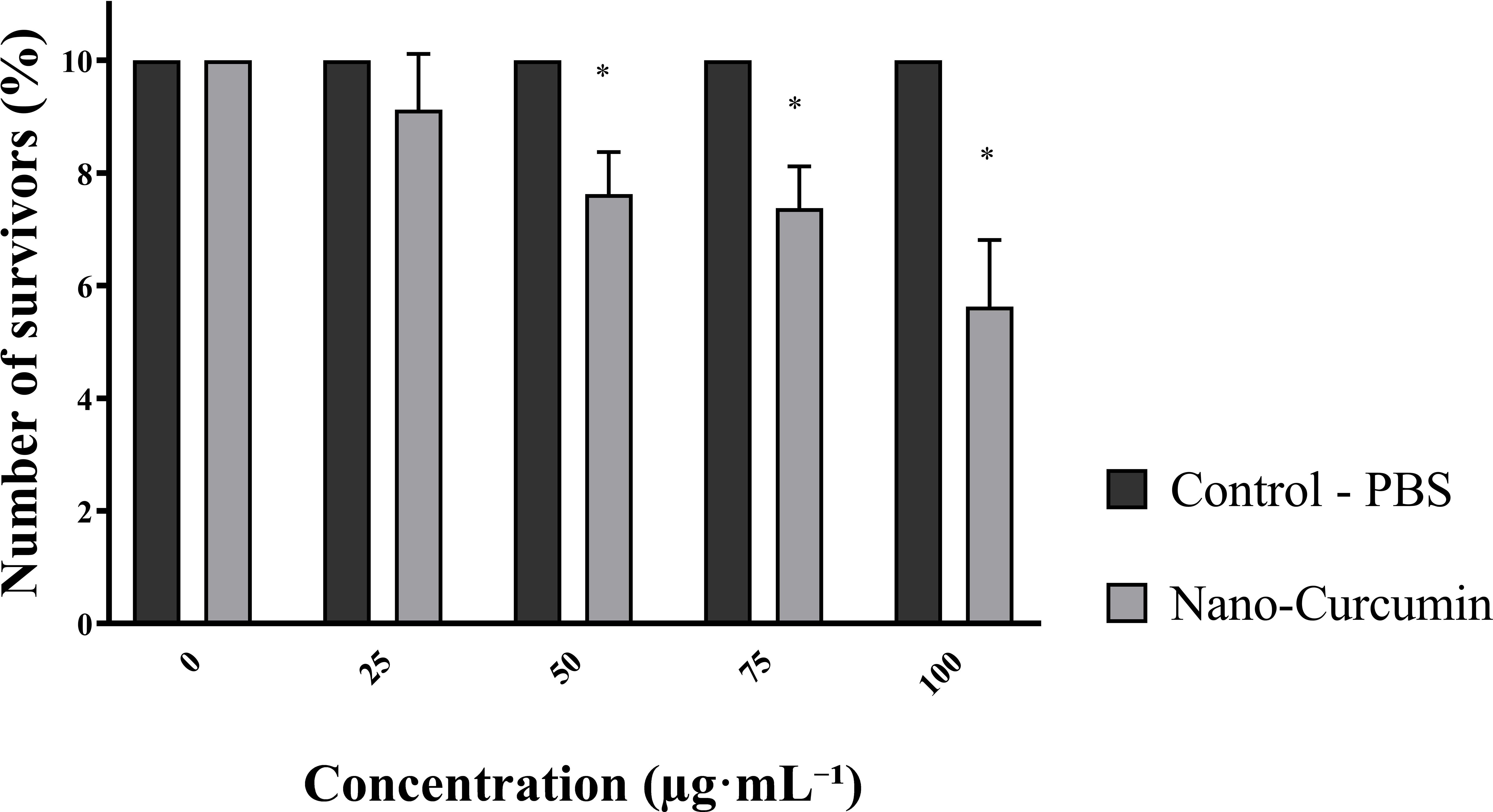
Figure 5. Survival of oral mucosal cells in response to different concentrations of zein nanocapsules loaded with curcumin. *The p-values: p < 0.05 indicate a significant difference according to Tukey’s test.
Zein nanocapsules loaded with curcumin at a 100 μg·mL-1 concentration (7.5 µg·mL-¹ of curcumin), indicating that half of the healthy oral mucosal cells exposed to this concentration did not survive (Figure 6).
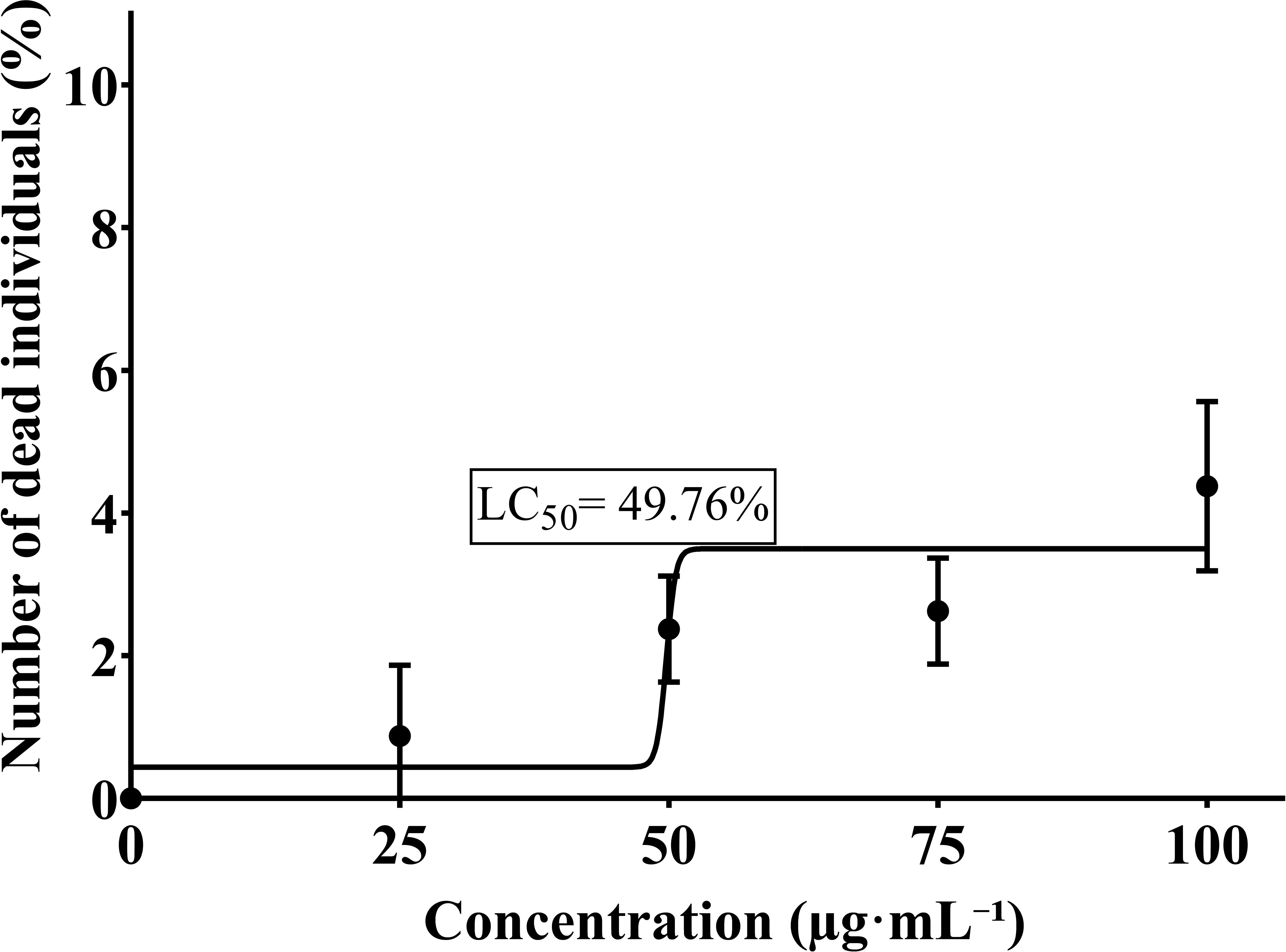
Figure 6. Median lethal concentration (LC50) of zein nanocapsules loaded with curcumin.
3.3 Microbiological analysis
The results of the microbiological analysis of dentin contaminated with S. mutans are presented in Figure 7. Both curcumin nanocapsules and their photosensitized versions significantly reduced S. mutans CFU/mL compared to untreated controls (p<0.05). However, the group treated with photosensitized curcumin nanocapsules exhibited the lowest CFU/mL count, this reduction was not statistically different from that observed in the non-photosensitized curcumin nanocapsules group.
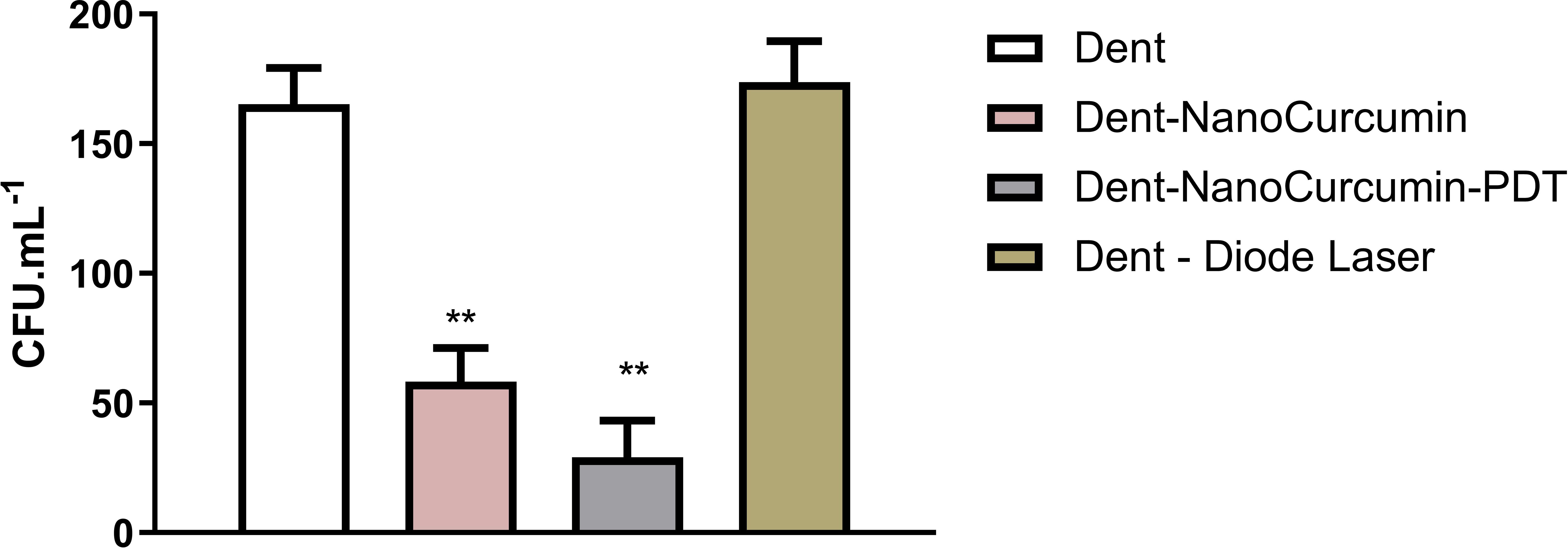
Figure 7. Comparison of antimicrobial effectiveness against s. mutans between the tested groups. **The p-values: p < 0.05 indicate a significant difference according to Tukey’s test.
4 Discussion
The high encapsulation efficiency (~100%) of curcumin in zein matrices observed in this study aligns with previous reports highlighting the effectiveness of this biopolymer as a carrier for bioactive compounds (Da Rosa et al., 2020). Given curcumin’s poor solubility and susceptibility to degradation (Hu et al., 2024), encapsulation within a zein matrix offers significant advantages including enhanced stability, protection against oxidation, and shielding from adverse interactions that could compromise its biological activity in the oral environment (Choi et al., 2016). This high retention capacity is particularly advantageous for controlled-release systems, where sustained bioavailability at the target site—such as infected dentin—is essential for therapeutic efficacy.
UV-Vis spectroscopy revealed notable changes in the absorbance profile of curcumin following encapsulation. Free curcumin exhibited a distinct peak at approximately 425 nm, corresponding to π→π* electronic transitions of the β-diketone conjugated system in its enolic form, typically observed in organic solvents (Urošević et al., 2022). In the nanoencapsulated form, this peak was significantly diminished or absent, with a relative increase in absorbance at wavelengths below 400 nm (Wu et al., 2023). These spectral changes suggest that curcumin was incorporated into the hydrophobic regions of the polymeric matrix, resulting in conformational restriction and reduced interaction with the dispersion medium.
These modifications are likely due to non-covalent interactions between curcumin and zein. Zein contains hydrophobic segments that facilitate molecular entrapment and enable additional intermolecular interactions, such as hydrogen bonding and Van der Waals forces (Chen et al., 2015). Furthermore, π–π stacking between the aromatic rings of curcumin and zein contributes to the structural stabilization of the nanoparticles (Ding et al., 2023; Liu et al., 2023).
In addition to confirming encapsulation, comparative studies have shown that encapsulated curcumin undergoes significantly less degradation under UV radiation. Literature indicates that free curcumin degrades by more than 60% after 30 minutes of exposure, whereas the encapsulated form shows less than 10% degradation under the same conditions (Wu et al., 2023). Encapsulation also promotes the dispersion of curcumin in an amorphous state, enhancing its solubility in aqueous media and absorption in the gastrointestinal tract, potentially improving oral bioavailability (Liu et al., 2023).
Thus, the attenuation of the 425 nm peak and the altered absorbance pattern observed in nano-curcumin reflect structural constraints imposed by the zein matrix, resulting in increased protection against degradation and greater potential for use in nutraceutical and pharmaceutical formulations.
The observed increase in nanoparticle size following curcumin incorporation suggests strong physicochemical interactions between zein and curcumin during nanoprecipitation process. This contributes to greater structural stability and reduced aggregation, attributed to the hydrophobic nature of curcumin (Da Rosa et al., 2015). Additionally, the lower polydispersity index (PDI) observed in curcumin-loaded formulations compared to those without the compound indicates a more uniform size distribution - an important factor for optimizing bioavailability and ensuring effective dentin penetrationthereby enhancing antimicrobial action against cariogenic microorganisms (Danaei et al., 2018).
Zeta potential results further support these findings. The lower surface charge observed in nano-curcumin formulations suggests potential alterations in colloidal stability due to interactions with zein. While higher zeta potential values typically indicate greater electrostatic stability (Nunes et al., 2022), the reduction observed here may increase the likelihood of aggregation over time, emphasizing the need for appropriate stabilization or storage strategies to preserve therapeutic efficacy (Danaei et al., 2018).
These findings underscore the potential of nano-curcumin formulations for targeted antimicrobial therapy in dentistry, particularly in minimally invasive approaches for caries management. By ensuring high encapsulation efficiency, enhanced stability, and controlled release, this formulation presents a promising alternative for dental applications. Future studies should explore its long-term stability and in vivo performance to validate clinical applicability and optimize formulation parameters.
The sustained release observed is consistent with the release mechanisms commonly associated with zein-based polymeric systems. Due to its hydrophobic and compact structure, zein acts as a physical barrier to drug diffusion, promoting a passive and controlled release profile (Oh and Flanagan, 2010; Gonzalez-Valdivieso et al., 2021). Hydrophobic interactions and potential hydrogen bonding between curcumin and the zein matrix further contribute to compound retention, limiting its rapid diffusion into the external medium (Lenzuni et al., 2023; Zhang et al., 2023).
Curcumin’s inherently low aqueous solubility (<0.001 mg/mL) further restricts its release in buffered aqueous environments. Even after encapsulation, this intrinsic property limits immediate availability, reinforcing the gradual release profile observed (Karthikeyan et al., 2020; Omidian et al., 2023). The combination of poor solubility and the hydrophobic nature of zein results in a controlled release without burst effects.
The release of bioactive compounds from zein-based systems typically follows anomalous kinetics, often fitting the Korsmeyer–Peppas model. This suggests that both diffusion and slow matrix relaxation or degradation contribute to the release mechanism (Zhang et al., 2021; Lin et al., 2024). These processes occur simultaneously, with diffusion predominating in the initial phase and matrix reorganization influencing release behavior at later stages.
Systems with these characteristics are highly desirable for topical formulations targeting the oral cavity. Prolonged release on surfaces such as the oral mucosa, gingiva, or dentin enhances the local retention of curcumin, thereby amplifying its pharmacological activity. Curcumin is well known for its anti-inflammatory, antioxidant, and antimicrobial properties, with demonstrated benefits in the treatment of gingivitis, periodontitis, and chronic oral lesions (Inchingolo et al., 2024). Moreover, controlled release reduces the frequency of reapplication and contributes to a superior safety profile by avoiding local concentration peaks. These findings confirm that zein is an effective carrier system for the controlled release of hydrophobic compounds like curcumin, representing a promising strategy for minimally invasive therapies in oral healthcare.
The results of this study confirm the biocompatibility of zein nanocapsules loaded with curcumin for oral mucosal cells—an essential requirement for safe use in dental applications. Minimizing cytotoxicity is Inchingly critical to ensure that formulation does not harm surrounding healthy tissues while maintaining its therapeutic effects (Shahi et al., 2019). In this study, at the highest tested concentration of nanocapsules (100 μg·mL-¹, equivalent to 7.5 μg·mL-¹ of curcumin), cell survival remained above 50%. At concentrations of 50 and 75 μg·mL-¹, viability approached 80%. Although a viability threshold above 50% is generally acceptable, these results suggest that even at full concentration, the formulation does not induce critical toxicity. This is particularly relevant for applications in the oral cavity, where direct mucosal exposure requires safe and well-tolerated materials.
The observed LC50 value (49.75 µg·mL-¹) indicates that 50% of the exposed cells did not survive at the highest tested concentration. LC50 is a standard toxicological parameter representing concentration at which half of the cells are affected, serving as a key indicator of formulation safety (Idrees and Kujan, 2023). The nanometric scale of the zein nanocapsules may have enhanced their penetration and interaction with oral mucosal cells, increasing curcumin’s local bioavailability. While this property is advantageous for targeted antimicrobial action, it also underscores the importance of carefully optimizing dosage to mitigate toxicity risks and maintain a favorable therapeutic index.
These findings are consistent with previous studies on the Inchingly biocompatibility of curcumin when encapsulated in nanocarriers. Meng et al. (2023) demonstrated that starch-based nanocapsules loaded with curcumin exhibited low toxicity toward healthy cells while effectively inhibiting tumor cells, suggesting that encapsulation plays an important role in modulating curcumin’s biological interactions. Similarly, Minhaco et al. (2023) reported over 80% cell viability at 325 μg/mL using PLGA-curcumin nanoparticles in oral cells, supporting their biomedical potential.
While reducing zein-curcumin concentrations may lower cytotoxicity, maintaining antimicrobial efficacy is key. Curcumin nanostructures remain effective at low doses; for example, 6.25 µg/mL of curcumin nanocrystals inhibited P. gingivalis growth. The 7.5 µg/mL used in this study supports the efficacy of nano-curcumin at low concentrations (Maleki Dizaj et al., 2022).
These results reinforce the promise of zein-based curcumin nanocapsules for safe, effective dental applications—especially in pediatric care, where non-invasive, biocompatible treatments are preferred.
Photodynamic therapy (PDT) with photosensitizers (PS) is well-documented for treating oral pathogens, including S. mutans (Cusicanqui Méndez et al., 2019; Hosseinpour-Nader et al., 2022; Manoil et al., 2014; Parizzi et al., 2025; Silvestre et al., 2023). Upon irradiation, the PS generates reactive oxygen species (ROS), primarily singlet oxygen, which promotes oxidative damage to microbial DNA, organelles, and cell membranes, ultimately leading to bacterial death (Taraszkiewicz et al., 2013).
Curcumin, a natural polyphenol derived from plants, displays broad-spectrum antibacterial activity due to its unique molecular structure and antioxidant properties. It disrupts quorum sensing, impairs biofilm formation, reduces virulence factor expression, and prevents bacterial adhesion to host cell receptors. When activated, curcumin acts as a photosensitizer, generating phototoxic effects that inhibit bacterial growth and enhance the efficacy of other antimicrobials through synergistic interactions (Zheng et al., 2020).
By producing reactive oxygen species (ROS) such as hydrogen peroxide, superoxide, and singlet oxygen, curcumin damages bacterial cell structures by oxidizing membranes, proteins, and nucleic acids, leading to cell death. Gram-positive bacteria are particularly susceptible due to their permeable peptidoglycan-rich cell walls, which facilitate photosensitizer entry (Ghate et al., 2019).
Additionally, curcumin induces intracellular damage, affecting DNA and proteins, disrupting biofilm adhesion, and downregulating virulence genes involved in pathogenicity. While its effect on outer membranes is limited, photodynamic inactivation (PDI) with curcumin causes significant internal damage and cytoplasmic leakage. The superior antimicrobial effect of light-activated curcumin highlights its potential for targeting biofilm-associated infections and oral pathogens such as Streptococcus mutans (Ghate et al., 2015; Hu et al., 2018; Huang et al., 2020; Pereira et al., 2014; Tonon et al., 2015).
Microbiological analysis confirmed that both conventional and photosensitized curcumin nanocapsules significantly reduced S. mutans CFU/mL in dentin. Although the photosensitized group showed the lowest count, the difference was not statistically significant.
This may be due to the strong inherent antimicrobial activity of nano-curcumin, suboptimal PDT parameters (e.g., light dose, wavelength, pre-irradiation time), or limited light penetration into biofilms. Pre-irradiation time is crucial for PS penetration (de Oliveira et al., 2019), especially in mature biofilms, which are more resistant due to their dense extracellular matrix (Silvestre et al., 2023; Taraszkiewicz et al., 2013).
In this study, extended pre-irradiation was used to assess whether PDT efficacy could be enhanced or if nano-curcumin alone was sufficient. Less structured biofilms may respond well to nano-curcumin without PDT, as the formulation improves curcumin delivery and release.
Curcumin’s poor solubility and rapid degradation limit its therapeutic use, but nanoencapsulation improves stability, lowers the MIC, and enhances antimicrobial efficacy (Pourhajibagher et al., 2018).
Studies support combining PDT with nano-curcumin. Pourhajibagher et al. (2022) found that a 5% nano-curcumin cavity liner with PDT inhibited S. mutans for 60 days. Araújo et al. (2017) required 5 g·L-¹ for photoactivation effects on S. mutans and L. acidophilus. In contrast, this study used only 0.0075 g·L-¹, yet showed efficacy—suggesting nanoencapsulation allows lower doses and reduces side effects like dental staining.
PDT also improves restoration outcomes.Clinical studies show its use in selective caries removal (SCR) reduces microbial load and enhances restoration success (Alves et al., 2019; Borges et al., 2010; Melo et al., 2015; Steiner-Oliveira et al., 2015). Long-term data confirm no compromise in restoration integrity after 6–12 months (Alves et al., 2019).
Combining nano-curcumin with PDT promotes remineralization by enhancing calcium and phosphate penetration into demineralized dentin (Koo et al., 2013; Wilson and Patterson, 2008; Zaleh et al., 2022). This approach aligns with minimally invasive dentistry, especially in pediatrics, by preserving primary teeth, reducing discomfort, and shortening treatment time. However, PDT is less effective in deeper dentin due to light scattering and absorption (Koo et al., 2013; Zanin et al., 2005), highlighting the need for personalized treatment based on lesion depth.
Despite promising results, limitations exist. In vitro conditions don’t fully replicate the oral environment, where saliva, mechanical forces, and microbial diversity affect outcomes. While effective against S. mutans, further studies should assess polymicrobial biofilms. Curcumin’s limited dentin penetration may reduce efficacy in deep lesions, emphasizing the need to optimize nanoencapsulation for better bioavailability.
Tooth staining is another concern. Although some in vitro studies report no significant discoloration (Araujo et al., 2023), these are based on simplified models. PDT with nano-curcumin may be a viable alternative to silver diamine fluoride, especially in posterior teeth where esthetics are less critical (Araujo et al., 2023).
Long-term in vivo studies are needed to assess safety, biocompatibility, and clinical effectiveness. Future research should refine PDT parameters—pre-irradiation time, light dose, and treatment frequency—and explore nano-curcumin’s role in remineralization and its interaction with restorative materials.
5 Conclusions
This study synthesized and characterized curcumin-loaded zein nanocapsules with high encapsulation efficiency, spherical morphology, low polydispersity, and good colloidal stability. Cytotoxicity assays showed oral mucosal cell viability above 50% at high concentrations, supporting safety for topical use. Curcumin nanocapsules significantly reduced S. mutans on primary dentin. Although PDT further reduced bacterial load, the difference was not statistically significant, indicating the nanocarrier alone enhances antimicrobial efficacy via improved penetration and sustained release. With the limitation of this study, these results support nano-curcumin as a safe, effective, and minimally invasive strategy for caries management in primary teeth, aligning with conservative pediatric dental practices.
Data availability statement
The original contributions presented in the study are included in the article/supplementary material. Further inquiries can be directed to the corresponding author.
Ethics statement
The studies involving humans were approved by Research Ethics Committee on Human Subjects of the University of Planalto Catarinense. The studies were conducted in accordance with the local legislation and institutional requirements. Written informed consent for participation in this study was provided by the participants’ legal guardians/next of kin. The animal study was approved by Ethics Committee on the Use of Animals – CEUA/UNESC. The study was conducted in accordance with the local legislation and institutional requirements.
Author contributions
ME: Formal Analysis, Methodology, Data curation, Validation, Conceptualization, Writing – original draft, Writing – review & editing, Investigation. AA: Formal Analysis, Methodology, Writing – review & editing, Data curation, Writing – original draft. DD: Writing – original draft, Writing – review & editing, Formal Analysis, Data curation, Methodology. DB: Formal Analysis, Writing – original draft, Data curation, Methodology, Writing – review & editing. JR: Data curation, Writing – original draft, Formal Analysis, Methodology, Writing – review & editing. DH: Formal Analysis, Methodology, Data curation, Writing – review & editing, Writing – original draft. AP: Conceptualization, Writing – review & editing, Formal Analysis, Writing – original draft. VS: Data curation, Formal Analysis, Methodology, Conceptualization, Writing – original draft, Writing – review & editing, Investigation. MN: Validation, Writing – review & editing, Conceptualization, Writing – original draft, Investigation, Formal Analysis, Methodology, Data curation. CR: Writing – original draft, Visualization, Formal Analysis, Project administration, Funding acquisition, Conceptualization, Methodology, Data curation, Resources, Supervision, Writing – review & editing, Validation, Investigation. AM: Conceptualization, Methodology, Visualization, Data curation, Supervision, Validation, Investigation, Funding acquisition, Writing – review & editing, Resources, Project administration, Writing – original draft, Formal Analysis.
Funding
The author(s) declare financial support was received for the research and/or publication of this article. This research was funded by Fundação de Amparo à Pesquisa e Inovação do Estado Santa Catarina (FAPESC) grant numbers FAPESC 12/2020 TO2021TR1482, FAPESC 12/2020 TO2021TR001430, FAPESC 15/2021 TO2021TR001220, FAPESC 54/2022 TO2023TR000648, FAPESC 54/2022 TO2023TR000883, FAPESC 54/2022 TO2023TR000884, FAPESC 15/2023 TO2023TR001418, FAPESC 15/2023 TO2023TR001518, FAPESC n◦ 18/2024, FAPESC n◦ 20/2024, and FAPESC n◦ 61/2024. This research was also funded by the Conselho Nacional de Desenvolvimento Científico e Tecnológico (CNPq) n◦ 69/2022-PIBPG and the Coordenação de Aperfeiçoamento de Pessoal de Nível Superior (CAPES)—Programa de Desenvolvimento da Pós-Graduação (PDPG).
Conflict of interest
The authors declare that the research was conducted in the absence of any commercial or financial relationships that could be construed as a potential conflict of interest.
Generative AI statement
The author(s) declare that Generative AI was used in the creation of this manuscript. This manuscript was entirely developed without the use of artificial intelligence tools such as ChatGPT during the research, data analysis, and initial writing phases. ChatGPT was employed only at the final stage of manuscript preparation to enhance the clarity, fluency, and readability of the English language, without altering the scientific content or the authors’ original ideas.
Any alternative text (alt text) provided alongside figures in this article has been generated by Frontiers with the support of artificial intelligence and reasonable efforts have been made to ensure accuracy, including review by the authors wherever possible. If you identify any issues, please contact us.
Publisher’s note
All claims expressed in this article are solely those of the authors and do not necessarily represent those of their affiliated organizations, or those of the publisher, the editors and the reviewers. Any product that may be evaluated in this article, or claim that may be made by its manufacturer, is not guaranteed or endorsed by the publisher.
References
Adamczak, A., Ożarowski, M., and Karpiński, T. M. (2020). Curcumin, a natural antimicrobial agent with strain-specific activity. Pharmaceuticals 13, 153. doi: 10.3390/ph13070153
Aïem, E., Joseph, C., Garcia, A., Smaïl-Faugeron, V., and Muller-Bolla, M. (2020). Caries removal strategies for deep carious lesions in primary teeth: Systematic review. Int. J. Paediatr. Dent. 30, 392–404. doi: 10.1111/ipd.12616
Alves, L. V. G. L., Curylofo-Zotti, F. A., Borsatto, M. C., Salvador, S. L., de, S., Valério, R. A., et al. (2019). Influence of antimicrobial photodynamic therapy in carious lesion. Randomized split-mouth clinical trial in primary molars. Photodiagnosis Photodyn. Ther. 26, 124–130. doi: 10.1016/j.pdpdt.2019.02.018
Andreatta, G. B., Marcon, R. B., Da Rosa, C. G., and Masiero, A. V. (2023). Propriedades biológicas e físico-químicas de cimentos endodônticos nanoparticulados. Cuad. Educ. Desarro. 15, 16209–16228. doi: 10.55905/cuadv15n12-059
Araujo, L. P., Marchesin, A. R., Gobbo, L. B., da Rosa, W. L. D. O., de Jesus Soares, A., de Almeida, J. F. A., et al. (2023). Tooth color change after photodynamic therapy in endodontics: A systematic review. Photodiagnosis Photodyn. Ther. 42, 103626. doi: 10.1016/j.pdpdt.2023.103626
Araújo, N. C., de Menezes, R. F., Carneiro, V. S. M., Dos Santos-Neto, A. P., Fontana, C. R., Bagnato, V. S., et al (2017). Photodynamic inactivation of cariogenic pathogens using curcumin as photosensitizer. Photomed. Laser Surg. 35, 259–263. doi: 10.1089/pho.2016.4156
Batista, D. G., Sganzerla, W. G., da Silva, L. R., Vieira, Y. G. S., Almeida, A. R., Dominguini, D., et al. (2024). Antimicrobial and cytotoxic potential of eucalyptus essential oil-based nanoemulsions for mouthwashes application. Antibiotics 13, 942. doi: 10.3390/antibiotics13100942
Borges, F. M. C., de-Melo, M. a. S., Lima, J. M. P., Zanin, I. C. J., Rodrigues, L. K. A., and Nobre-dos-Santos, M. (2010). Evaluation of the effect of photodynamic antimicrobial therapy in dentin caries: a pilot in vivo study, in: Lasers in Dentistry XVI. Presented at the Lasers in Dentistry XVI, SPIE, pp. 63–71. doi: 10.1117/12.842339
Carolina Alves, R., Fernandes, R. P., Fonseca-Santos, B., Damiani Victorelli, F., and Chorilli, M. (2019). A critical review of the properties and analytical methods for the determination of curcumin in biological and pharmaceutical matrices. Crit. Rev. Anal. Chem. 49, 138–149. doi: 10.1080/10408347.2018.1489216
Chen, X., Zou, L.-Q., Niu, J., Liu, W., Peng, S.-F., and Liu, C.-M. (2015). The stability, sustained release and cellular antioxidant activity of curcumin nanoliposomes. Molecules 20, 14293–14311. doi: 10.3390/molecules200814293
Choi, J. E., Waddell, J. N., Lyons, K. M., and Kieser, J. A. (2016). Intraoral pH and temperature during sleep with and without mouth breathing. J. Oral Rehabil. 43, 356–363. doi: 10.1111/joor.12372
Cusicanqui Méndez, D. A., Gutierrez, E., Campos Chaves Lamarque, G., Lopes Rizzato, V., Afonso Rabelo Buzalaf, M., Andrade Moreira MaChado, M. A., et al. (2019). The effectiveness of curcumin-mediated antimicrobial photodynamic therapy depends on pre-irradiation and biofilm growth times. Photodiagnosis Photodyn. Ther. 27, 474–480. doi: 10.1016/j.pdpdt.2019.07.011
Da Rosa, C. G., De Oliveira Brisola Maciel, M. V., De Carvalho, S. M., De Melo, A. P. Z., Jummes, B., Da Silva, T., et al. (2015). Characterization and evaluation of physicochemical and antimicrobial properties of zein nanoparticles loaded with phenolics monoterpenes. Colloids Surf. A Physicochem. Eng. Asp. 481, 337–344. doi: 10.1016/j.colsurfa.2015.05.019
Da Rosa, C. G., Sganzerla, W. G., De Oliveira Brisola Maciel, M. V., De Melo, A. P. Z., Da Rosa Almeida, A., Ramos Nunes, M., et al. (2020). Development of poly (ethylene oxide) bioactive nanocomposite films functionalized with zein nanoparticles. Colloids Surf. A Physicochem. Eng. Asp. 586, 124268. doi: 10.1016/j.colsurfa.2019.124268
da Rosa, C. G., Narciso, A. M., Nunes, M. R., and Masiero, A. V. (2022). “10 - Applicability of silver nanoparticles and innovation of magnetic nanoparticles in dentistry,” in Fundamentals and Industrial Applications of Magnetic Nanoparticles, Woodhead Publishing Series in Electronic and Optical Materials. Eds. Hussain, C. M. and Patankar, K. K. (The Officers’ Mess Business Centre, United Kingdom, Cambridge, MA, United States, Kidlington, United Kingdom: Woodhead Publishing), 317–348. doi: 10.1016/B978-0-12-822819-7.00023-5
de Oliveira, A. B., Ferrisse, T. M., Marques, R. S., De Annunzio, S. R., Brighenti, F. L., and Fontana, C. R. (2019). Effect of photodynamic therapy on microorganisms responsible for dental caries: A systematic review and meta-analysis. Int. J. Mol. Sci. 20, 3585. doi: 10.3390/ijms20143585
Ding, R., Zhang, M., Zhu, Q., Qu, Y., Jia, X., and Yin, L. (2023). Curcumin loaded Zein-alginate nanogels with “core-shell” structure: formation, characterization and simulated digestion. Int. J. Biol. Macromol. 251, 126201. doi: 10.1016/j.ijbiomac.2023.126201
Danaei, M., Dehghankhold, M., Ataei, S., Hasanzadeh Davarani, F., Javanmard, R., Dokhani, A., et al. (2018). Impact of particle size and polydispersity index on the clinical applications of lipidic nanocarrier systems. Pharmaceutics 10, 57. doi: 10.3390/pharmaceutics10020057
Diniz, I. M. A., Horta, I. D., Azevedo, C. S., Elmadjian, T. R., Matos, A. B., Simionato, M. R. L., et al. (2015). Antimicrobial photodynamic therapy: a promise candidate for caries lesions treatment. Photodiagnosis Photodyn. Ther. 12, 511–518. doi: 10.1016/j.pdpdt.2015.04.006
Fernandes, F. G. L., De Moraes, F. B., De Cezare, J. A., Degasperi, G. R., Fontana, C. E., Grandizoli, D. R. P., et al. (2022). In vitro evaluation of EDTA combined with photodynamic therapy to reduce Streptococcus mutans in carious dentin. Photodiagnosis Photodyn. Ther. 37, 102718. doi: 10.1016/j.pdpdt.2022.102718
Ghaffari, S.-B., Sarrafzadeh, M.-H., Salami, M., and Khorramizadeh, M. R. (2020). A pH-sensitive delivery system based on N-succinyl chitosan-ZnO nanoparticles for improving antibacterial and anticancer activities of curcumin. Int. J. Biol. Macromol. 151, 428–440. doi: 10.1016/j.ijbiomac.2020.02.141
Ghate, V., Leong, A. L., Kumar, A., Bang, W. S., Zhou, W., and Yuk, H.-G. (2015). Enhancing the antibacterial effect of 461 and 521 nm light emitting diodes on selected foodborne pathogens in trypticase soy broth by acidic and alkaline pH conditions. Food Microbiol. 48, 49–57. doi: 10.1016/j.fm.2014.10.014
Ghate, V. S., Zhou, W., and Yuk, H.-G. (2019). Perspectives and trends in the application of photodynamic inactivation for microbiological food safety. Compr. Rev. Food Sci. Food Saf. 18, 402–424. doi: 10.1111/1541-4337.12418
Gonçalves da Rosa, C., Zapelini de Melo, A. P., Sganzerla, W. G., Machado, M. H., Nunes, M. R., Vinicius de Oliveira Brisola Maciel, M., et al. (2020). Application in situ of zein nanocapsules loaded with Origanum vulgare Linneus and Thymus vulgaris as a preservative in bread. Food Hydrocoll. 99, 105339. doi: 10.1016/j.foodhyd.2019.105339
Gonzalez-Valdivieso, J., Girotti, A., Schneider, J., and Arias, F. J. (2021). Advanced nanomedicine and cancer: Challenges and opportunities in clinical translation. Int. J. Pharm. 599, 120438. doi: 10.1016/j.ijpharm.2021.120438
Hosseinpour-Nader, A., Karimi, N., Ghafari, H.-A., and Ghorbanzadeh, R. (2022). Effect of nanomicelle curcumin-based photodynamic therapy on the dynamics of white spot lesions and virulence of Streptococcus mutans in patients undergoing fixed orthodontic treatment: A randomized double-blind clinical trial. Photodiagnosis Photodyn. Ther. 40, 103183. doi: 10.1016/j.pdpdt.2022.103183
Hu, X., Huang, Y.-Y., Wang, Y., Wang, X., and Hamblin, M. R. (2018). Antimicrobial photodynamic therapy to control clinically relevant biofilm infections. Front. Microbiol. 9. doi: 10.3389/fmicb.2018.01299
Hu, Y., Rees, N. H., Qiu, C., Wang, J., Jin, Z., Wang, R., et al. (2024). Fabrication of zein/modified cyclodextrin nanofibers for the stability enhancement and delivery of curcumin. Food Hydrocoll. 156, 110262. doi: 10.1016/j.foodhyd.2024.110262
Huang, J., Chen, B., Li, H., Zeng, Q.-H., Wang, J. J., Liu, H., et al. (2020). Enhanced antibacterial and antibiofilm functions of the curcumin-mediated photodynamic inactivation against Listeria monocytogenes. Food Control 108, 106886. doi: 10.1016/j.foodcont.2019.106886
Idrees, M. and Kujan, O. (2023). A curcumin-based oral gel has potential protective efficacy against oral mucositis: in vitro study. J. Pers. Med. 14, 1. doi: 10.3390/jpm14010001
Inchingolo, F., Inchingolo, A. D., Latini, G., Trilli, I., Ferrante, L., Nardelli, P., et al. (2024). The role of curcumin in oral health and diseases: a systematic review. Antioxidants 13, 660. doi: 10.3390/antiox13060660
Innes, N. P. T., Evans, D. J. P., and Stirrups, D. R. (2011). Sealing caries in primary molars: randomized control trial, 5-year results. J. Dent. Res. 90, 1405–1410. doi: 10.1177/0022034511422064
Kamwilaisak, K., Rittiwut, K., Jutakridsada, P., Iamamorphanth, W., Pimsawat, N., Knijnenburg, J. T. N., et al. (2022). Rheology, stability, antioxidant properties, and curcumin release of oil-in-water Pickering emulsions stabilized by rice starch nanoparticles. Int. J. Biol. Macromol. 214, 370–380. doi: 10.1016/j.ijbiomac.2022.06.032
Karthikeyan, A., Senthil, N., and Min, T. (2020). Nanocurcumin: A promising candidate for therapeutic applications. Front. Pharmacol. 11. doi: 10.3389/fphar.2020.00487
Knorst, J. K., Barriquello, G. S., Villetti, M. A., Santos, R. C. V., and Kantorski, K. Z. (2019). Antimicrobial effect of methylene blue formulations with oxygen carrier at different pHs: preliminary study. BDS 22, 39–45. doi: 10.14295/bds.2019.v22i1.1635
Koo, H., Falsetta, M. L., and Klein, M. I. (2013). The exopolysaccharide matrix: A virulence determinant of cariogenic biofilm. J. Dent. Res. 92, 1065–1073. doi: 10.1177/0022034513504218
Lenzuni, M., Bonfadini, S., Criante, L., Zorzi, F., Summa, M., Bertorelli, R., et al. (2023). Dynamic investigation of zein-based degradable and hemocompatible coatings for drug-eluting stents: a microfluidic approach. Lab. Chip 23, 1576–1592. doi: 10.1039/D3LC00012E
Lin, Z., Zhan, L., Qin, K., Li, Y., Qin, Y., Yang, L., et al. (2024). Design and characterization of a novel core–shell nano delivery system based on zein and carboxymethylated short-chain amylose for encapsulation of curcumin. Foods 13, 1837. doi: 10.3390/foods13121837
Liu, G., An, D., Li, J., and Deng, S. (2023). Zein-based nanoparticles: Preparation, characterization, and pharmaceutical application. Front. Pharmacol. 14. doi: 10.3389/fphar.2023.1120251
Machiulskiene, V., Campus, G., Carvalho, J. C., Dige, I., Ekstrand, K. R., Jablonski-Momeni, A., et al. (2020). Terminology of dental caries and dental caries management: consensus report of a workshop organized by ORCA and cariology research group of IADR. Caries Res. 54, 7–14. doi: 10.1159/000503309
Manoil, D., Filieri, A., Gameiro, C., Lange, N., Schrenzel, J., Wataha, J. C., et al. (2014). Flow cytometric assessment of Streptococcus mutans viability after exposure to blue light-activated curcumin. Photodiagnosis Photodyn. Ther. 11, 372–379. doi: 10.1016/j.pdpdt.2014.06.003
Maleki Dizaj, S., Shokrgozar, H., Yazdani, J., Memar, M. Y., Sharifi, S., and Ghavimi, M. A. (2022). Antibacterial Effects of Curcumin Nanocrystals against Porphyromonas gingivalis Isolated from Patients with Implant Failure. Clin. Pract. 12, 809–817. doi: 10.3390/clinpract12050085
Masiero, A. V., Barletta, F. B., Nunes, M. R., Sganzerla, W. G., Erckmann, M. C., and da Rosa, C. G. (2024). “Chapter 13 - Silver nanoparticles and their role in the treatment of endodontic infections,” in Silver Nanoparticles for Drug Delivery. Ed. Kesharwani, P. (London United Kingdom, San Diego CA, United States, Cambridge, MA, United States, Oxford, United Kingdom: Academic Press), 289–311. doi: 10.1016/B978-0-443-15343-3.00011-5
Melo, M. A. S., Rolim, J. P. M. L., Passos, V. F., Lima, R. A., Zanin, I. C. J., Codes, B. M., et al. (2015). Photodynamic antimicrobial chemotherapy and ultraconservative caries removal linked for management of deep caries lesions. Photodiagnosis Photodyn. Ther. 12, 581–586. doi: 10.1016/j.pdpdt.2015.09.005
Meng, Q., Zhou, L., Zhong, S., Wang, J., Wang, J., Gao, Y., et al. (2023). Stimulus-responsive starch-based nanocapsules for targeted delivery and antibacterial applications. Int. J. Biol. Macromol. 241, 124664. doi: 10.1016/j.ijbiomac.2023.124664
Minhaco, V. M. T. R., Maquera Huacho, P. M., Mancim Imbriani, M. J., Tonon, C. C., Chorilli, M., Rastelli, A. N., et al. (2023). Improving antimicrobial activity against endodontic biofilm after exposure to blue light-activated novel curcumin nanoparticle. Photodiagnosis Photodyn. Ther. 42, 103322. doi: 10.1016/j.pdpdt.2023.103322
Narciso, A. M., Paim, B., Da Rosa, C. G., Paes, J. V., Nunes, M. R., et al. (2019). Síntese verde de nanopartículas de prata para uso em biomateriais odontológicos. Revista Interdisciplinar de Estudos em Saúde, 64–73. doi: 10.33362/ries.v8i2.2124
Nehete, A. P., Singh, J. S., Mehta, R. V., Desai, R. S., Bansal, S. P., and Kakade, A. (2014). Study of the microstructure of mineralised tissues in 50 human primary teeth. Int. J. Oral. Maxillofac. Pathol. 5 (4), 12–18.
Nunes, M. R., Da Rosa, C. G., De Borba, J. R., Dos Santos, G. M., Ferreira, A. L., and Barreto, P. L. M. (2022). Zein Nanoparticles: Bioactive Compounds and Controlled Delivery. In: Jana, S. and Jana, S. (eds.), Nanoengineering of Biomaterials. (Wiley), 411–436. doi: 10.1002/9783527832095.ch13
Oh, Y. K. and Flanagan, D. R. (2010). Diffusional properties of zein membranes and matrices. Drug Dev. Ind. Pharm. 36, 497–507. doi: 10.3109/03639040903264389
Omidian, H., Wilson, R. L., and Chowdhury, S. D. (2023). Enhancing therapeutic efficacy of curcumin: advances in delivery systems and clinical applications. Gels 9, 596. doi: 10.3390/gels9080596
Parizzi, M., Almeida, A. R., Salvador, G., Dominguini, D., Fernandes, M., Becker, D., et al. (2025). Photosensitized methylene blue nanoparticles: A promising approach for the control of oral infections. Biomedicines 13, 673. doi: 10.3390/biomedicines13030673
Pereira, M. A., Faustino, M. A. F., Tomé, J. P. C., Neves, M.G.P.M.S., Tomé, A. C., Cavaleiro, J. A. S., et al. (2014). Influence of external bacterial structures on the efficiency of photodynamic inactivation by a cationic porphyrin. Photochem. Photobiol. Sci. 13, 680–690. doi: 10.1039/c3pp50408e
Pourhajibagher, M., Bahrami, R., and Bahador, A. (2022). An ex vivo evaluation of physico-mechanical and anti-biofilm properties of resin-modified glass ionomer containing ultrasound waves-activated nanoparticles against Streptococcus mutans biofilm around orthodontic bands. Photodiagnosis Photodyn. Ther. 40, 103051. doi: 10.1016/j.pdpdt.2022.103051
Pourhajibagher, M., Salehi Vaziri, A., Takzaree, N., and Ghorbanzadeh, R. (2019). Physico-mechanical and antimicrobial properties of an orthodontic adhesive containing cationic curcumin doped zinc oxide nanoparticles subjected to photodynamic therapy. Photodiagnosis Photodyn. Ther. 25, 239–246. doi: 10.1016/j.pdpdt.2019.01.002
Pourhajibagher, M., Kazemian, H., Chiniforush, N., Hosseini, N., Pourakbari, B., Azizollahi, A., et al. (2018). Exploring different photosensitizers to optimize elimination of planktonic and biofilm forms of Enterococcus faecalis from infected root canal during antimicrobial photodynamic therapy. Photodiagnosis Photodyn. Ther. 24, 206–211. doi: 10.1016/j.pdpdt.2018.09.014
Sayed, M., Hiraishi, N., Matin, K., Abdou, A., Burrow, M. F., and Tagami, J. (2020). Effect of silver-containing agents on the ultra-structural morphology of dentinal collagen. Dent. Mater. 36, 936–944. doi: 10.1016/j.dental.2020.04.028
Shahi, S., Özcan, M., Maleki Dizaj, S., Sharifi, S., Al-Haj Husain, N., Eftekhari, A., et al (2019). A review on potential toxicity of dental material and screening their biocompatibility. Toxicol. Mech. Methods 29, 368–377. doi: 10.1080/15376516.2019.1566424
Silvestre, A. L. P., Dos Santos, A. M., De Oliveira, A. B., Ferrisse, T. M., Brighenti, F. L., Meneguin, A. B., et al. (2023). Evaluation of photodynamic therapy on nanoparticles and films loaded-nanoparticles based on chitosan/alginate for curcumin delivery in oral biofilms. Int. J. Biol. Macromol. 240, 124489. doi: 10.1016/j.ijbiomac.2023.124489
Steiner-Oliveira, C., Longo, P. L., Aranha, A. C. C., Ramalho, K. M., Mayer, M. P. A., and de Paula Eduardo, C. (2015). Randomized in vivo evaluation of photodynamic antimicrobial chemotherapy on deciduous carious dentin. J. Biomed. Opt. 20, 108003. doi: 10.1117/1.JBO.20.10.108003
Suzuki, I. L., Inada, N. M., Marangoni, V. S., Corrêa, T. Q., Zucolotto, V., Kurachi, C., et al (2016). Synthesis and characterization of PLGA nanoparticles containing mixture of curcuminoids for optimization of photodynamic inactivation. In: Kessel, D. H. and Hasan, T. (eds.), SPIE Proceedings (San Francisco, California, United States), 969413. doi: 10.1117/12.2213781
Takahashi, N. and Nyvad, B. (2016). Ecological hypothesis of dentin and root caries. Caries Res. 50, 422–431. doi: 10.1159/000447309
Taraszkiewicz, A., Fila, G., Grinholc, M., and Nakonieczna, J. (2013). Innovative strategies to overcome biofilm resistance. BioMed Res. Int. 2013, 150653. doi: 10.1155/2013/150653
Tonon, C. C., Paschoal, M. A., Correia, M., Spolidório, D. M. P., Bagnato, V. S., Giusti, J. S. M., et al. (2015). Comparative effects of photodynamic therapy mediated by curcumin on standard and clinical isolate of Streptococcus mutans. J. Contemp Dent. Pract. 16, 1–6. doi: 10.5005/jp-journals-10024-1626
Urošević, M., Nikolić, L., Gajić, I., Nikolić, V., Dinić, A., and Miljković, V. (2022). Curcumin: biological activities and modern pharmaceutical forms. Antibiotics 11, 135. doi: 10.3390/antibiotics11020135
Wilson, B. C. and Patterson, M. S. (2008). The physics, biophysics and technology of photodynamic therapy. Phys. Med. Biol. 53, R61–109. doi: 10.1088/0031-9155/53/9/R01
Wu, J., Chen, J., Wei, Z., Zhu, P., Li, B., Qing, Q., et al. (2023). Fabrication, evaluation, and antioxidant properties of carrier-free curcumin nanoparticles. Molecules 28, 1298. doi: 10.3390/molecules28031298
Zaleh, A.-A., Salehi-Vaziri, A., Pourhajibagher, M., and Bahador, A. (2022). The synergistic effect of Nano-propolis and curcumin-based photodynamic therapy on remineralization of white spot lesions: An ex vivo study. Photodiagnosis Photodyn. Ther. 38, 102789. doi: 10.1016/j.pdpdt.2022.102789
Zanin, I. C. J., Gonçalves, R. B., Junior, A. B., Hope, C. K., and Pratten, J. (2005). Susceptibility of Streptococcus mutans biofilms to photodynamic therapy: an in vitro study. J. Antimicrob. Chemother. 56, 324–330. doi: 10.1093/jac/dki232
Zhang, Z., Li, X., Sang, S., Julian McClements, D., Chen, L., Long, J., et al. (2023). Preparation, properties and interaction of curcumin loaded zein/HP-β-CD nanoparticles based on electrostatic interactions by antisolvent co-precipitation. Food Chem. 403, 134344. doi: 10.1016/j.foodchem.2022.134344
Zhang, H., Van Os, W. L., Tian, X., Zu, G., Ribovski, L., Bron, R., et al. (2021). Development of curcumin-loaded zein nanoparticles for transport across the blood–brain barrier and inhibition of glioblastoma cell growth. Biomater. Sci. 9, 7092–7103. doi: 10.1039/D0BM01536A
Zheng, D., Huang, C., Huang, H., Zhao, Y., Khan, M. R. U., Zhao, H., et al (2020). Antibacterial Mechanism of Curcumin: A Review. Chem. Biodivers. 17, e2000171. doi: 10.1002/cbdv.202000171
Zhi, K., Yang, H., Shan, Z., Huang, K., Zhang, M., and Xia, X. (2021). Dual-modified starch nanospheres encapsulated with curcumin by self-assembly: Structure, physicochemical properties and anti-inflammatory activity. Int. J. Biol. Macromol. 191, 305–314. doi: 10.1016/j.ijbiomac.2021.09.117
Keywords: dental caries, curcumin, nanotechnology, photodynamic therapy, pediatric dentistry
Citation: Erckmann MC, Almeida A, Dominguini D, Becker D, Rutz JK, Hotza D, Parolia A, Dos Santos VV, Nunes MR, Da Rosa CG and Masiero AV (2025) Curcumin-photosensitized nanocapsules: biocompatibility and antimicrobial evaluation in primary tooth dentin contaminated with Streptococcus mutans. Front. Cell. Infect. Microbiol. 15:1614363. doi: 10.3389/fcimb.2025.1614363
Received: 18 April 2025; Accepted: 08 August 2025;
Published: 08 September 2025.
Edited by:
Keke Zhang, Wenzhou Medical University, ChinaReviewed by:
Xinxin Tian, Tufts University, United StatesPan Yangyang, Wenzhou Medical University, China
Copyright © 2025 Erckmann, Almeida, Dominguini, Becker, Rutz, Hotza, Parolia, Dos Santos, Nunes, Da Rosa and Masiero. This is an open-access article distributed under the terms of the Creative Commons Attribution License (CC BY). The use, distribution or reproduction in other forums is permitted, provided the original author(s) and the copyright owner(s) are credited and that the original publication in this journal is cited, in accordance with accepted academic practice. No use, distribution or reproduction is permitted which does not comply with these terms.
*Correspondence: Anelise Viapiana Masiero, YW5lbWFzaWVyb0B1bmlwbGFjbGFnZXMuZWR1LmJy