 Irina Negut1
 Camilla Mazzanti2
 Rossella Laurano3
 Gianluca Ciardelli2,3
 Simona Bronco2
 Cláudia S. Oliveira4*†
- 1Lasers Department, National Institute for Lasers, Plasma and Radiation Physics, Măgurele, Romania
- 2Institute for Chemical-Physical Processes, National Research Council (CNR-IPCF), Pisa, Italy
- 3Department of Mechanical and Aerospace Engineering, Politecnico di Torino, Turin, Italy
- 4Universidade Católica Portuguesa, CBQF - Centro de Biotecnologia e Química Fina – Laboratório Associado, Escola Superior de Biotecnologia, Porto, Portugal
Bioactive textiles have emerged as multifunctional materials to actively interact with the human skin and its microbiome. By embedding natural or synthetic bioactive compounds, such as chitosan, essential oils, plant extracts, and metallic nanoparticles, these materials aim to prevent and target infections, modulate inflammation, and promote skin homeostasis. Given the critical role of the skin microbiome in maintaining barrier integrity and immune balance, strategies that selectively inhibit pathogenic microorganisms (e.g., Staphylococcus aureus, Cutibacterium acnes) while preserving beneficial commensals like Staphylococcus epidermidis are essential to avoid dysbiosis and associated dermatological disorders. This review highlights current trends in the design and functionalization of bioactive textiles, emphasizing sustainable and biocompatible approaches that leverage natural antimicrobial compounds and green synthesis techniques. It also examines conventional evaluation pipelines primarily based on 1D microbiological assays and 2D skin models, highlighting their limitations in predicting real-world performance. Advanced in vitro models, particularly 3D reconstructed human skin platforms incorporating both pathogenic and commensal microbiota members, are presented as indispensable tools to study fabric–skin–microbe interactions under physiologically relevant conditions. These models enable accurate assessment of antimicrobial efficacy, biocompatibility, and microbiome impact, providing a bridge between in vitro and clinical outcomes. Furthermore, the potential of bioactive textiles in managing microbiome-related skin conditions, such as atopic dermatitis and acne, is discussed alongside the importance of developing microbiome-safe materials. Despite encouraging clinical evidence demonstrating pathogen reduction and symptomatic improvement, the successful translation of these materials to clinical practice needs interdisciplinary research and the adoption of advanced preclinical strategies to ensure innovative solutions for personalized skin health.
1 Introduction
The human skin microbiome is a critical determinant of skin health, acting as a dynamic interface between the body and the external environment. This complex ecosystem of bacteria, fungi, and viruses is essential for maintaining barrier function, modulating immune responses, and resisting pathogen colonization. Disruptions in this microbial equilibrium, often termed dysbiosis, are implicated in a wide range of dermatological disorders including acne, atopic dermatitis (AD), psoriasis, and chronic wounds (Byrd et al., 2018; Carmona-Cruz et al., 2022). In this context, bioactive textiles have emerged as a promising therapeutic approach for restoring microbial balance while simultaneously addressing rising concerns about antibiotic resistance and environmental sustainability (Suellen Ferro de Oliveira and Kekhasharu Tavaria, 2023).
Bioactive textiles are engineered by incorporating functional agents, such as antimicrobial, anti-inflammatory, antioxidant, or prebiotic compounds, into fibers or coatings, enabling them to exert therapeutic or protective effects upon contact with the skin (Gulati et al., 2022; Ghosh et al., 2025a) These materials are increasingly being used in dermatology, personal care, medical applications, and everyday wear, where fabrics act as a secondary skin interface. Their functionalities have been especially valuable in preventing infections, modulating inflammatory responses, and improving skin healing (Reta et al., 2024).
Recent years have witnessed an expansion in the use of sustainable, biocompatible materials in bioactive textiles. Natural agents such as chitosan, honey, aloe vera, neem, turmeric, essential oils, and plant extracts have demonstrated broad-spectrum antimicrobial activity against skin pathogens like Staphylococcus aureus, Escherichia coli, Pseudomonas aeruginosa, Cutibacterium acnes, and Candida albicans, all of which are commonly implicated in skin infections (Szadkowski et al., 2024; Oliveira et al., 2025). These materials have shown great promise in mitigating infection risks while preserving or even enhancing beneficial microbial populations, thereby fostering a healthy skin microbiome.
However, despite their benefits, the precise effects of antimicrobial textiles on the skin microbiome, especially on beneficial species like Staphylococcus epidermidis, remain underexplored. This commensal bacterium plays a vital role in skin homeostasis, immune modulation, and pathogen defense. Understanding how bioactive textiles affect both harmful and beneficial skin microbes is crucial for ensuring their safe and targeted application.
To facilitate effective evaluation of these materials, a tiered assessment strategy has been proposed, moving from basic in vitro models to more complex, biologically relevant skin models. The one-dimensional (1D) evaluation model begins with classical microbiological assays to evaluate the effects of such materials in skin pathogenic microorganisms, while the final tier includes three-dimensional (3D) advanced skin models that mimic the stratified epidermis/dermis and key microbiota members, enabling a more accurate prediction of in vivo behavior (Galvan et al., 2024).
This structured pipeline supports the development of microbiome-compatible textiles and facilitates translation into real-world applications. It enables researchers to fine-tune antimicrobial performance while preserving skin commensals and minimizing inflammation. Moreover, the integration of 3D microbiome-inclusive models ensures more robust safety and efficacy data prior to clinical or commercial deployment.
In summary, bioactive textiles represent a convergence between materials science, dermatology, microbiology, and sustainability, offering innovative solutions that extend from infection control to therapeutic skin care. Their applications range from advanced skin wounds to everyday garments, addressing both health and environmental concerns. Moving forward, the adoption of interdisciplinary approaches, particularly the transition from 1D to 3D evaluation models will be critical to ensure the development of next-generation textiles that are not only effective, safe, environmentally responsible but also skin microbiome-friendly.
2 Skin microbiota
The human skin functions as a multilayered defense system, comprising four interconnected barriers: physical, chemical, immune, and microbial (Nicolaou and Kendall, 2024) (Figure 1). The components operate synergistically to maintain skin homeostasis and prevent the onset of infections, inflammation, and even skin cancers. However, disruptions in any of these systems can compromise the integrity of the others, resulting in skin disease states (Byrd et al., 2018).
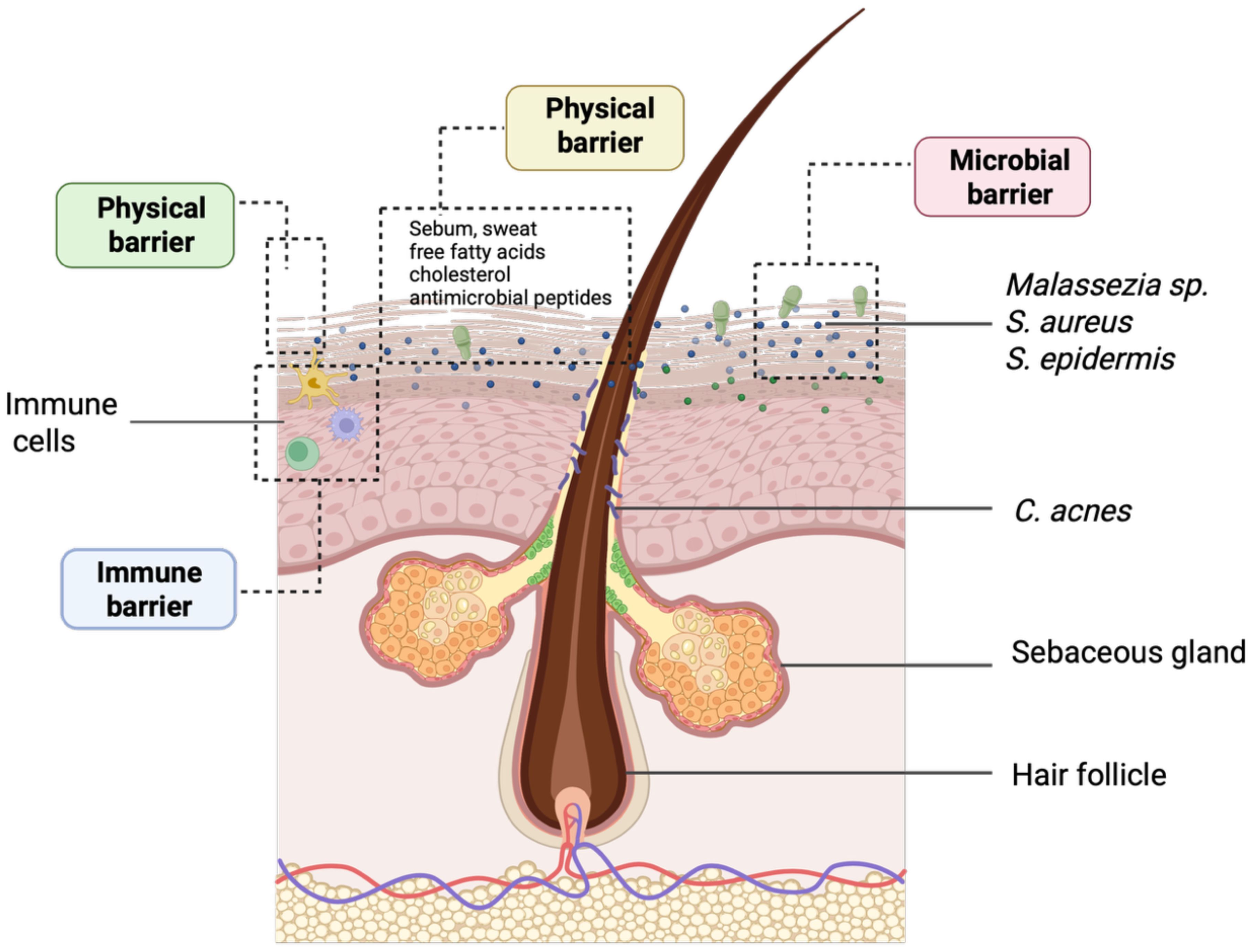
Figure 1. Representative overview of the epidermal barriers and the localization of the most important skin microbiota members. The epidermal barrier protects against pathogens, chemicals, and UV radiation while preventing excessive trans-epidermal water loss. This defense relies on physical, chemical microbiological, and immunological components, largely regulated by a complex lipid network. The image was created in https://BioRender.com.
The chemical barrier, maintained by its natural acidity and lipid composition, plays a critical role in regulating moisture retention and suppressing microbial overgrowth. Key lipid components, such as ceramides, free fatty acids, and cholesterol, produced by keratinocytes within the stratum corneum, contribute to elasticity, hydration, and defense against pathogen entry (Nicolaou and Kendall, 2024). Simultaneously, keratinocytes establish tight junctions and secrete cytokines and antimicrobial peptides, reinforcing the skin as both a physical and immunological barrier (Zhang et al., 2022). Beyond these structures, the immune defense layer comprises resident innate immune cells, including Langerhans cells and dermal dendritic cells, as well as adaptive immune cells that are recruited in response to stress or infection. Together, these elements coordinate antigen recognition, immune activation, and the development of immunological memory (Coates et al., 2018).
Next-generation sequencing has revealed that the skin microbiome is predominantly composed of Gram-positive bacteria, particularly Cutibacterium and Staphylococcus, with distribution varying by site (Prajapati et al., 2025) (Figure 1). Sebaceous areas typically harbor Cutibacterium acnes, whereas moist regions, such as the axillae and elbows, favor Staphylococcus and Corynebacterium species. Among commensals, Staphylococcus epidermidis plays a key protective role by producing antimicrobial peptides, competing with pathogens like Staphylococcus aureus, and reinforcing barrier integrity through tight junction protein induction (Koberle et al., 2022).
The fungal community, largely represented by Malassezia species, contributes to defense by secreting indoles and other metabolites that inhibit pathogenic fungi (Koberle et al., 2022).
Disruption of the skin microbial ecosystem, through hygiene practices, environmental stressors, or immune dysregulation, can shift commensals toward opportunistic behavior. For example, Cutibacterium acnes contributes to acne not by mere overgrowth but through alterations in strain composition (Koberle et al., 2022). Likewise, increased Staphylococcus aureus colonization is a hallmark of AD while dysregulated Malassezia populations are linked to seborrheic dermatitis (Kreouzi et al., 2025).
This intricate relationship between microbiota and skin barrier function underscores the potential of bioactive textiles that selectively suppress pathogens while preserving beneficial microbes as non-invasive strategies to restore microbial homeostasis.
3 Bioactive textiles: a short overview
Bioactive textiles represent a new frontier in material science, distinguished by the incorporation of biologically active components designed to provide therapeutic or protective effects beyond conventional textile properties. These textiles are engineered by incorporating active substances, such as antimicrobial agents, antioxidants, or healing compounds, into the fiber matrix or surface to interact with the user or environment in a purposeful way (Gulati et al., 2022; Szadkowski et al., 2024; Ghosh et al., 2025a). This class of functional fabrics has attracted increasing attention due to its potential in healthcare, personal care, sportswear, and sustainable fashion sectors (Korica et al., 2022; Ghosh et al., 2025b).
Among the most researched bioactivities, the antimicrobial functionality is the most challenging one. This is particularly critical in medical and hygiene contexts, where bioactive textiles serve to prevent infections, reduce pathogen transmission, or manage wound healing.
3.1 Fabrication approaches and antimicrobial activity of bioactive textiles
Bioactive textiles are engineered by incorporating functional agents into fabric substrates to endow them with antimicrobial, antioxidant, anti-inflammatory, or other biologically relevant properties. These materials are particularly important in healthcare, personal care, and hygiene applications, where textile surfaces often come in direct contact with human skin and are prone to microbial colonization.
The development of bioactive textiles involves several fabrication and functionalization methods. The most common strategy is finishing, where bioactive compounds are applied onto the fabric surface using techniques such as pad-dry-cure, exhaustion, or spray coating. In this regard, a wide range of antimicrobial agents, both synthetic and natural, have been integrated into textile materials, including triclosan, metals and their salts, quaternary ammonium compounds (QACs), organosilicons, chitosan, essential oils, and plant extracts (Stan et al., 2019; Roman et al., 2020; Hedayati et al., 2021; Julaeha et al., 2021; Mehravani et al., 2021; Garcia et al., 2022; Gulati et al., 2022; Szadkowski et al., 2023). Among these, Ag- and Zn-based salts have shown broad-spectrum antimicrobial activity against diverse microorganisms, such as Gram-positive and Gram-negative bacteria, fungi, viruses, yeasts, and algae (Gopal et al., 2021; Hedayati et al., 2021; Garcia et al., 2022). QACs have also demonstrated notable antimicrobial efficacy, although their activity spectrum is generally narrower than that of metal-based agents (Jennings et al., 2015; Jiao et al., 2017). Recently, plant-derived bioactives (e.g., extracts of neem, aloe vera, eucalyptus, clove, and turmeric) have gained attention for their sustainability and safety profiles. These agents are incorporated into textiles via eco-friendly methods like solvent-free dyeing or embedding during finishing stages (Reta et al., 2024; Oliveira et al., 2025).
Another innovative approach involves immobilization of antimicrobial agents via covalent bonding, which enhances wash durability and minimizes environmental leaching. For instance, chloroxylenol has been covalently bonded to cotton via bifunctional reactive finishes, enabling robust antimicrobial activity even after 20 washing cycles (Reta et al., 2024). Nanoencapsulation is another key method, where many bioactive compounds, (e.g., metals, dyes, essential oils, plant extracts, chitosan) are encapsulated into nanoparticles (NPs) (Roman et al., 2020; Vakayil et al., 2021; Sanchez and Fiscal Ladino, 2023). These are then deposited onto textile fibers to provide sustained release, enhanced fabric bonding, and improved biocompatibility (Costa et al., 2022).
The antimicrobial activity of bioactive textiles is influenced by several parameters, including the adhesion, survival, and proliferation of pathogens on the textile surface, the type and concentration of antimicrobial agents, and the method of their incorporation into the fabric structure. These factors significantly impact both the immediate antimicrobial efficacy and the long-term durability of the textile’s bioactivity (Sanders et al., 2021; Gulati et al., 2022).
Surface characteristics, such as wettability, porosity, and pore size, play a crucial role in bacterial adhesion. For instance, hydrophilic textile surfaces with larger pore volumes promote higher bacterial adherence compared to superhydrophobic surfaces with reduced pore sizes (Hemmatian et al., 2021). Similarly, anti-adhesive coatings, such as poly(l-lysine)-g-poly(ethylene glycol), have demonstrated an over 80% reduction in bacterial attachment on polyester substrates, maintaining their functionality even after 20 wash cycles (Schmidt-Emrich et al., 2016).
Equally important is the method of antimicrobial agent integration, such as physical adsorption, covalent bonding, or NPs embedding, each affecting release profile, durability, and biocompatibility. For example, textiles functionalized with enzymatically bonded nanoAg within dynamic polysulfide networks exhibited >99.99% antibacterial activity, along with excellent wash durability and biocompatibility (Wu et al., 2025).
3.2 Mode of action on skin microbiota
Bioactive textiles interact with skin microbiota primarily through their embedded biologically active compounds, which influence the microbial composition and activity on the skin. These materials can both support beneficial microorganisms and suppress pathogenic species, depending on their functional design and the active agents they release (Suellen Ferro de Oliveira and Kekhasharu Tavaria, 2023).
Antimicrobial action is typically achieved through the integration of metal NPs (e.g., Ag, ZnO), chitosan, plant-based compounds, or biosurfactants. These agents act by disrupting microbial membranes, interfering with metabolism, or inhibiting biofilm formation (Ghosh et al., 2025b) (Figure 2). Natural antimicrobials, such as essential oils or polyphenols, are increasingly favored due to their biodegradability and lower toxicity compared to synthetic alternatives (Ghosh et al., 2025b).
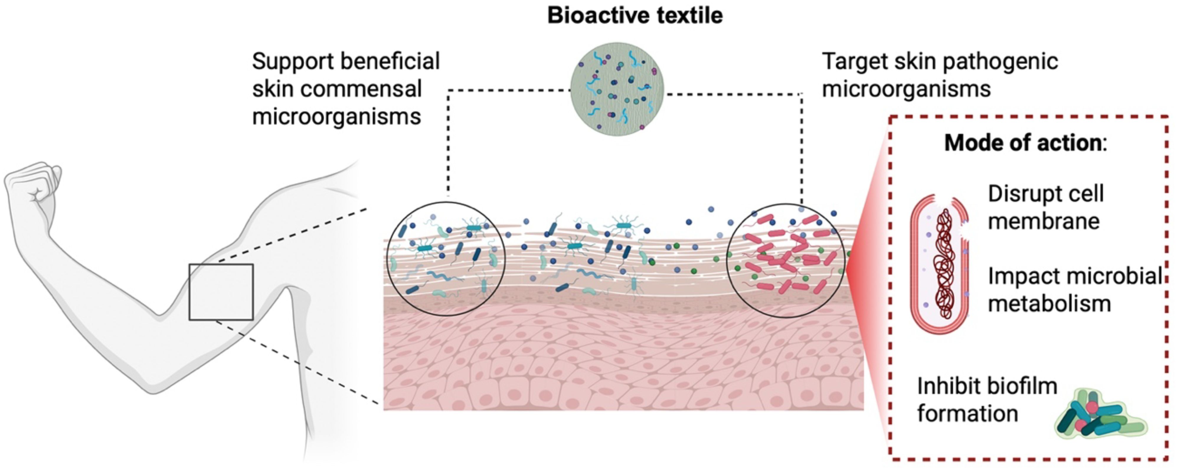
Figure 2. Impact of bioactive textiles with antimicrobial activity in commensal and pathogenic members of the human skin. The image was created in https://BioRender.com.
In parallel, sustainability concerns are driving innovation toward eco-friendly and circular materials. Researchers have demonstrated the successful use of agro-industrial waste (e.g., grape pomace, peanut skins, pomegranate peels) as sources of natural dyes and bioactives for textiles, enabling the valorization of biowaste while imparting antimicrobial and antioxidant effects (Lopes et al., 2025).
Moreover, therapeutic textiles with antimicrobial activity have shown promise in managing skin dysbiosis by integrating antimicrobial and antioxidant functions to reduce inflammation and support a healthy microbiome (de Oliveira and Tavaria, 2024). However, standardized models for microbiome safety assessment, along with comprehensive clinical studies, are still necessary to explore and validate the long-term effects of these materials.
In this line, a growing area of concern and innovation is the interaction between bioactive textiles and the skin microbiome. While many antimicrobial fabrics are effective against pathogens, they may also harm beneficial microbes such as Staphylococcus epidermidis, which play a vital role in skin homeostasis and immune regulation. This has led to increasing emphasis on developing microbiome-safe textiles that preserve or even promote commensal microbial populations while selectively targeting pathogens (Suellen Ferro de Oliveira and Kekhasharu Tavaria, 2023).
Prebiotic-infused textiles represent another approach, where materials contain substances that selectively support beneficial microbes. For example, short-chain fructo-oligosaccharides (scFOS) applied to skin models or incorporated into biomaterials have been shown to promote Staphylococcus epidermidis while inhibiting Cutibacterium acnes and Staphylococcus aureus (Le Bourgot et al., 2022). This strategy aligns with efforts to design microbiome-friendly products that enhance skin barrier function while minimizing inflammation and opportunistic infections.
Multifunctionality is also a central theme in the field. Emerging bioactive textiles are not limited to one function but often combine antimicrobial, antioxidant, anti-inflammatory, and moisture-regulating properties. For instance, cellulose-based wound dressings enriched with ascorbic acid or other antioxidants have demonstrated not only strong biocompatibility but also enhanced healing capabilities, highlighting the therapeutic potential of such textiles (Firmanda et al., 2024).
4 Evaluation strategies for antimicrobial bioactive textiles: a 1D conceptual framework
To facilitate the systematic development and assessment of antimicrobial bioactive textiles, a 1D evaluation model has been extensively used. In this context, a 1D model represents a linear, stepwise conceptual framework in which textile materials are assessed progressively through increasingly complex and biologically relevant assays. This classic 1D pipeline begins with basic antimicrobial screening and advances through mechanistic characterization, functional validation, and ultimately biological relevance.
The first stage of testing the antimicrobial activity of bioactive textiles on the 1D model involves traditional in vitro antimicrobial screening, using standardized microbiological assays designed to evaluate their direct effects on clinically relevant pathogens (Hedayati et al., 2021; Garcia et al., 2022; Mirza et al., 2023; Liu et al., 2024). These include agar diffusion (to assess inhibition zones), colony-forming unit (CFU) counting, and broth dilution techniques (Ivankovic et al., 2022). These methodologies provide both qualitative and quantitative insights, serving as a crucial step in screening the antimicrobial performance of such materials. These assays evaluate microbial growth inhibition under conditions where the microorganism is in direct contact with the textile material, thus enabling a comprehensive evaluation of the material’s inhibitory potential against common pathogens such as Staphylococcus aureus, Escherichia coli, Klebsiella pneumoniae, and Candida albicans (dos Santos et al., 2021).
Following initial screening, materials demonstrating antimicrobial activity undergo advanced mechanistic assays to further elucidate antimicrobial mechanisms. These include live/dead fluorescence microscopy to distinguish viable from non-viable cells, metabolic activity assays (e.g., 3-(4,5-dimethylthiazol-2-yl)-2,5-diphenyl tetrazolium bromid - MTT, resazurin reduction), adenosine triphosphate (ATP) quantification to assess microbial viability, and quantitative real-time PCR (qPCR) to quantify microbial DNA load. These approaches are particularly useful for evaluating the impact of bioactive textiles on biofilm formation and persistence, addressing a critical challenge in chronic skin infections and antimicrobial resistance (AMR) (dos Santos et al., 2021; Oliveira et al., 2025).
In the third stage of the pipeline, functional performance is evaluated beyond planktonic microbial inhibition. Biofilm formation assays assess whether the textile can inhibit microbial adhesion and colonization (dos Santos et al., 2021). Antioxidant properties are measured using 2,2-diphenyl-1-picrylhydrazyl (DPPH) and 2,2’-azino-bis(3-ethylbenzothiazoline-6-sulfonic acid) (ABTS) radical scavenging assays to determine the textile’s ability to neutralize reactive oxygen species (ROS) (Oliveira et al., 2025), which are involved in inflammation and delayed healing. Such multifunctionality enhances the applicability of antimicrobial textiles in clinical and daily-use settings.
However, the traditional antimicrobial evaluation typically target pathogens implicated in skin wound infections and hospital cross-infections, namely Gram-positive bacteria (e.g., Staphylococcus aureus, Staphylococcus pyogenes), Gram-negative bacteria (e.g., Escherichia coli, Pseudomonas aeruginosa, Klebsiella pneumoniae), and opportunistic fungi such as Candida albicans (Smith et al., 2020; dos Santos et al., 2021; Garcia et al., 2022; Vojnits et al., 2024). Given the increasing concern over AMR, recent studies also incorporate multi-drug resistant (MDR) strains, such as methicillin-resistant Staphylococcus aureus (MRSA), vancomycin-resistant Enterococcus (VRE), and carbapenem-resistant Pseudomonas aeruginosa, to assess the efficacy of bioactive textiles in combating persistent infections (Vojnits et al., 2024).
In this sense, many antimicrobial textiles, particularly those impregnated with Ag, copper, ZnO, antibiotics, and anti-inflammatory drugs, have been shown to reduce skin-associated pathogens in conventional in vitro antimicrobial assays (Garcia et al., 2022; Markovic et al., 2022; Liu et al., 2024). For example, cotton fabrics coated with silica NPs containing both an anti-inflammatory drug (ibuprofen) and an antibiotic (levofloxacin or norfloxacin) designed for wound healing, exhibited significant antibacterial activity against Staphylococcus aureus using standard agar diffusion assay (Liu et al., 2024). Similarly, polyamide fabrics incorporating zinc oxide NPs exhibited significant antibacterial effects against Staphylococcus aureus and Escherichia coli, supporting their potential use in medical and hygiene applications (Garcia et al., 2022).
In another example, antibacterial nanocomposite textiles were developed by coating cotton/peat blend fabrics with chitosan and Cu2O/CuO nanostructures with gallic acid serving as a reducing agent (Markovic et al., 2022). The treated fabrics showed enhanced antibacterial activity against Staphylococcus aureus and Escherichia coli. Likewise, cotton fabrics using bioactive formulations combining chitosan and thyme essential oil, along with mineral fillers like silica, ZnO, and TiO2, presented strong antimicrobial activity against various microorganisms, including Staphylococcus aureus, Escherichia coli, and B. subtilis (Szadkowski et al., 2023). In addition to their antimicrobial properties, these bioactive fabrics also demonstrated flame-retardant activity and biodegradability. According to the authors, such multifunctional bioactive textiles hold promising for advanced applications in military and medical context.
Although synthetic antimicrobial agents are known for their potent and often broad-spectrum activity, their incorporation into textile materials raises significant environmental and health concerns. Many of these compounds are linked to potential human toxicity and ecotoxicological risks, thereby posing serious sustainability challenges. For example, during laundering, these agents can leach from the textile fibers and enter aquatic ecosystems, where they may disrupt microbial communities and exert toxic effects on aquatic organisms (Bibi et al., 2024). Such unintended environmental release not only negatively impact the ecological balance but also raises concerns about the widespread use of synthetic antimicrobials in textile materials applications.
These limitations highlight the growing need for safer and more sustainable antimicrobial alternatives that are both biodegradable and biocompatible. Natural agents, including biopolymers, plant-derived compounds, and green-synthesized NPs, are emerging as promising solutions, offering antimicrobial properties while minimizing environmental and health-related risks (Costa et al., 2018; Stan et al., 2019; Mouro et al., 2020; El-Tarabily et al., 2021; Soroh et al., 2021; Choi and Park, 2023; Freitas et al., 2024; Pallottelli et al., 2024). Among these, chitosan, honey, and plant-based products, namely aloe vera, essential oils, and various plant extracts, have demonstrated broad-spectrum antimicrobial activity against pathogenic bacteria and yeasts such as Bacillus subtilis, Escherichia coli, Pseudomonas aeruginosa, Cutibacterium acnes, Staphylococcus aureus, and Candida albicans when incorporated into textile materials.
For example, cotton fabrics treated with a biosolution containing propolis and honey, with or without potassium alum as a mordant, exhibited significant antibacterial effects against Staphylococcus aureus, Bacillus subtilis, Cutibacterium acnes, and Escherichia coli (Freitas et al., 2024). In addition to their antimicrobial properties, these textiles showed potent antioxidant activity, achieving over 90% ABTS radical scavenging. These findings emphasize the potential of beehive-derived products, such as propolis and honey, as multifunctional natural agents for daily personal care applications. Their integration into reusable products like face masks not only enhances antimicrobial and antioxidant protection but also aligns with the principles of sustainability and circular product design.
Another example investigated cotton fabrics dyed with Clostridium obtusa extract in the presence of various mordants, particularly aluminum and copper (Choi and Park, 2023). These bioactive fabrics demonstrated not only strong antibacterial activity against Staphylococcus aureus and Klebsiella pneumoniae, two common pathogens frequently isolated from healthcare workers’ uniforms, but also higher antibacterial activity against the MRSA super bacteria. Based on these results, the authors propose the integration of Clostridium obtusa-dyed textiles into hospital environments, including patient garments, bed linens, and healthcare uniforms, to reduce cross-infection risks and reinforce infection control strategies.
Despite the promising antimicrobial performance of some natural agent-based bioactive textiles, a critical limitation remains: their reduced durability and diminished effectiveness after repeated laundering (Babu and Ravindra, 2015). Therefore, there is a need for further research focused on enhancing wash fastness and long-term functional stability to enable broader, real-world adoption in clinical and personal care contexts.
Bioactive textiles with anti-biofilm properties represent another growing area of interest, particularly in medical and hygiene applications. These materials are typically functionalized with antimicrobial agents capable of inhibiting the adhesion, colonization, and proliferation of biofilm-forming microorganisms, namely Staphylococcus aureus, Escherichia coli, Klebsiella pneumoniae, and Candida albicans (dos Santos et al., 2021; Mirza et al., 2023). This is especially critical in healthcare settings, where biofilms can contribute to persistent skin infections and reduce the effectiveness of conventional treatments. Among the various antimicrobial functions explored in bioactive textiles, antibiofilm activity is particularly important for skin wound applications, where preventing microbial biofilm formation is essential to promote healing and reduce the risk of chronic infection.
While existing studies have provided valuable insights into the ability of bioactive textiles to inhibit pathogenic microorganisms, as well as antibiofilm activity, their impact on the skin’s commensal microbiota remains largely unexplored (Figure 3). Limited information is available regarding the effects of such materials on Staphylococcus epidermidis, a dominant member of the human skin microbiome that plays a critical role in maintaining skin homeostasis. This knowledge gap raises concerns about the potential unintended consequences of antimicrobial textiles on Staphylococcus epidermidis and other beneficial skin microbes. Addressing this issue is essential for the development of microbiome-friendly textiles that combine effective antimicrobial performance with the preservation of skin microbial balance.
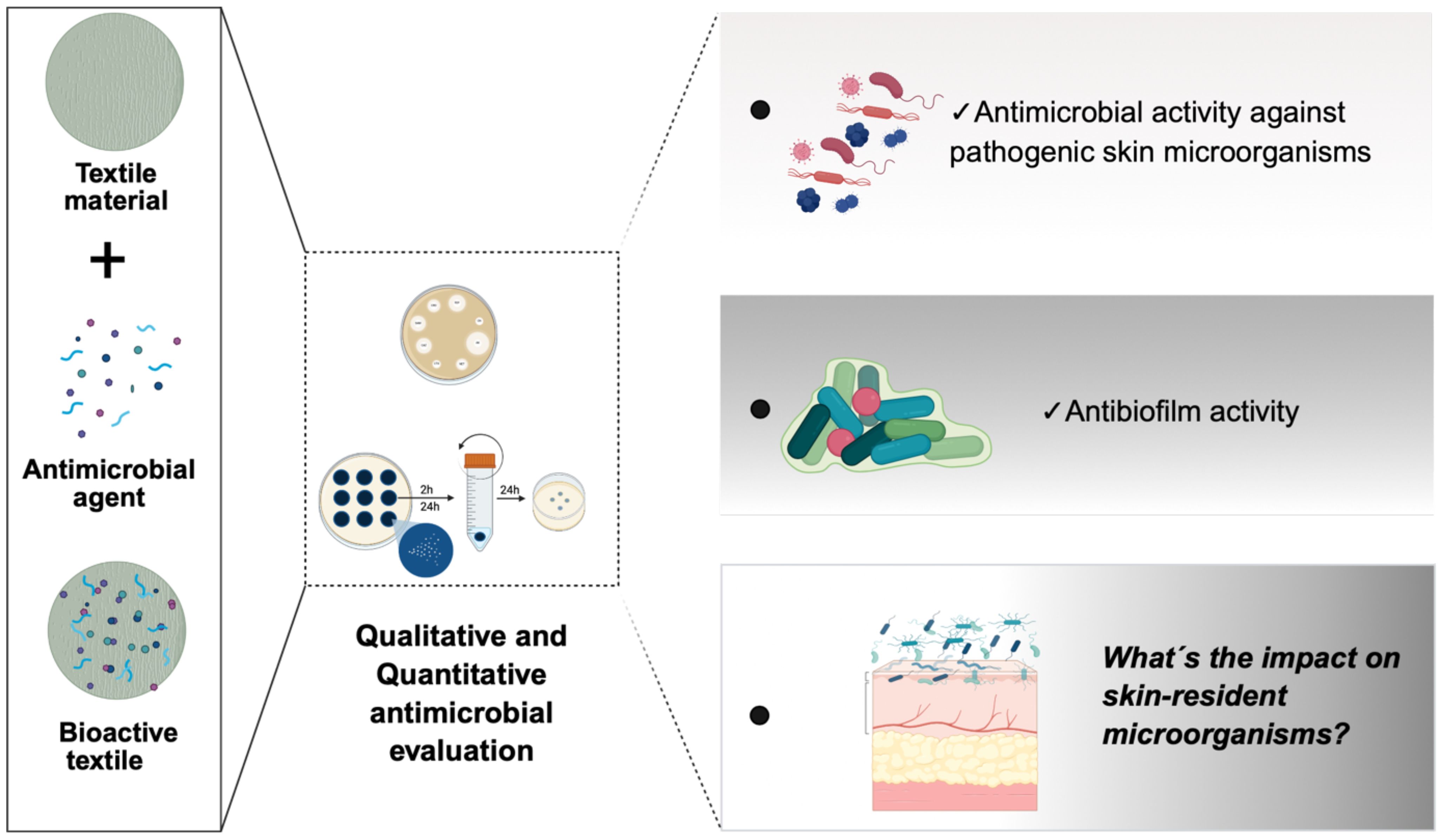
Figure 3. From design to microbiome impact - Evaluating bioactive textiles for skin applications. Left panel (design of bioactive textiles): Textile materials are functionalized with antimicrobial agents to develop bioactive textiles aimed at targeting pathogenic skin microorganisms. Center panel (assessment of antimicrobial activity): Standard antimicrobial assays are conducted to evaluate the antimicrobial properties of the bioactive textiles. These include disc diffusion, time-kill kinetics, and microdilution assays, performed under various conditions and different time points to assess their performance against microbial growth. Right panel (evaluation of biological impact): Key outcomes assessed include: i) Antimicrobial activity against pathogenic skin microorganisms; ii) Antibiofilm activity, reflecting the ability of bioactive textiles to inhibit or disrupt microbial biofilm formation; iii) Impact on skin-resident microorganisms (microbiome safety): a critical underexplored area, focusing on the preservation of beneficial commensal skin microbiota. The image was created in https://BioRender.com.
5 Transitioning bioactive textiles to 2D skin models
Following initial antimicrobial screening using 1D microbiological assays, some bioactive textiles are typically assessed using 2D cell culture models (Figure 4). 2D models have been useful to evaluate the biocompatibility of bioactive textile materials, particularly when intended for direct contact with human skin. These 2D models typically consist of monolayers of human skin keratinocytes or human dermal fibroblasts, which provide fundamental insights into cell viability, proliferation, metabolic activity, ROS, and early-stage biocompatibility (Hassan et al., 2024; Pallottelli et al., 2024; Oliveira et al., 2025).
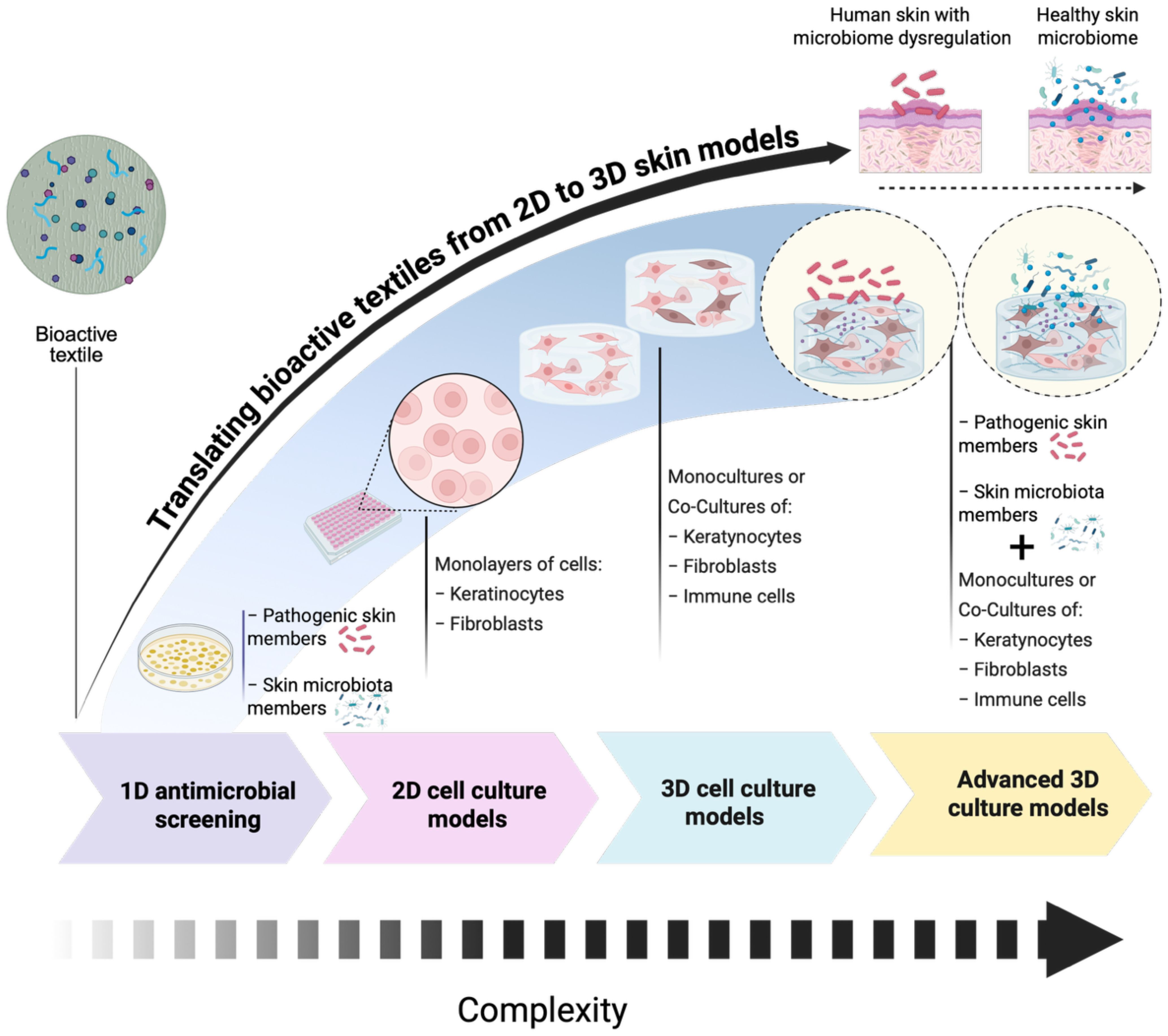
Figure 4. Schematic representation of the progression of in vitro models used to evaluate antimicrobial strategies of bioactive textile materials. The models evolve in complexity from left to right using 1D antimicrobial screening (simplified systems with pathogens in indirect or direct contact with bioactive textiles). Main limitation: These models do not include host cells, providing limited insight into host–pathogen interactions. 2D cell culture: Involves monolayers of keratinocytes and/or fibroblasts grown on flat surfaces. These models enable initial screening of host responses but lack the structural and cellular complexity of skin. 3D cell culture: Incorporates 3D organization of cells, such as fibroblasts and keratinocytes, in monocultures or co-cultures. These models better mimic the skin’s architecture and can include immune cells for enhanced relevance. Advanced 3D culture: Represents the most physiologically relevant in vitro skin models. These systems support the co-culture of keratinocytes, fibroblasts, and immune cells in a 3D matrix and can be challenged with skin-relevant pathogens (e.g., bacteria or fungi). They allow detailed study of host–microbe interactions and antimicrobial efficacy under near-physiological conditions. The image was created in https://BioRender.com.
At this stage, some studies were dedicated to the evaluation of bioactive textiles antimicrobial activity with preliminary cytotoxicity assessments (Hassan et al., 2024; Pallottelli et al., 2024). In terms of biocompatibility, most antimicrobial textiles have shown no cytotoxic or moderate cytotoxic effects when tested using either indirect or direct contact with 2D human skin cell models. For example, cotton fabric loaded with Ag NPs, combined with ketoconazole and β-cyclodextrin, exhibited potent antifungal and antibacterial activity against key skin pathogens, including Candida albicans, Aspergillus niger, Staphylococcus aureus and Escherichia coli (Hedayati et al., 2021). The cytotoxicity was evaluated via an indirect method using a monolayer of human skin fibroblasts, revealing no cytotoxic effects after 24h of incubation. According to the authors, these findings support the safety of the bioactive textile materials for skin contact and highlight its potential for dermatological and personal care applications. Likewise, cotton gauze coated with a dual layer system of Poly (vinyl alcohol) and chitosan containing extracts of Agrimonia eupatoria L. demonstrated effective inhibition of Staphylococcus aureus and Pseudomonas aeruginosa growth (Mouro et al., 2020). Cytotoxicity was assessed using a direct contact method with a human dermal fibroblast monolayer, which showed no cytotoxic effects after 1, 3, and 7 days of incubation, indicating sustained biocompatibility over time.
Although these studies demonstrate the feasibility of developing bioactive textiles with effective antimicrobial properties against well-known skin pathogens and acceptable biocompatibility, they often fail to assess the broader impact of such materials on the skin’s resident microbiota members. Specifically, there is a lack of investigation into whether these materials selectively inhibit pathogenic microorganisms or unintentionally disrupt commensal microbial populations that are essential for skin health. Furthermore, their potential to modulate the balance between pathogenic and beneficial microbes, namely in the context of skin conditions pathological conditions correlated with microbiome dysregulation remains largely unexplored.
Despite these gaps, bioactive textiles targeting the skin microbiome dysregulations, namely AD, are frequently advanced to clinical trials after only minimal cytotoxicity testing, typically limited to short-term assays on 2D cell cultures (de Oliveira and Tavaria, 2024; Hassan et al., 2024). This practice highlights a critical shortfall in preclinical validation and highlights the need for more comprehensive evaluation of their impact on the human skin microbiome, particularly through the extensive use of advanced in vitro models such as 3D reconstructed human skin, prior to clinical translation.
6 3D skin models: essential tools for evaluating the impact of bioactive textiles on the skin microbiome
Despite their widespread use, conventional 2D culture systems are unable to fully capture and recapitulate the structural and physiological complexity of human skin. This limitation hinders the comprehensive evaluation of key parameters failing to predict critical functional outcomes, including tissue penetration, barrier integrity, cell-cell, cell-microbe, cell-material interaction, microbiome impact and modulation, immune responses, wound healing capacity, and tissue regeneration. Therefore, 2D tissue models are considered unreliable pre-clinical testing tools. Indeed, to ensure the safe and effective clinical translation of bioactive materials, it is essential to first accurately replicate the native skin microenvironment, particularly its cellular composition, structural organization, and dynamic biological interactions. In this context, 3D human skin models have emerged as physiologically relevant platforms that more accurately mimic key properties of the human skin, including its architecture and functionality (Poumay and Coquette, 2007; Flaten et al., 2015; Niehues et al., 2018; Villata et al., 2024). Moreover, the bottom-up approach underpinning the design of such multi-component 3D in vitro models makes them suitable candidates for the investigation of pathological conditions and for testing the safety and efficacy of innovative biomaterials and therapies. For instance, the combination of healthy skin models with cells from the immune systems, such as pro-inflammatory macrophages, could become a reliable pathological platform for wound healing-related studies being able to mimic the prolonged inflammation and oxidative stress proper of chronic skin wounds. On the other hand, the contamination of such 3D models with specific bacterial strains in their planktonic or multi-species form leads to the development of pathological platforms suitable for the investigation of the efficacy of innovative antimicrobial therapies.
Figure 5 schematically represents the evolution of 3D in vitro skin models from the easiest skin explant models to the advanced bioengineered skin-on-a-chip platforms. Moreover, particular attention will be focused on models specifically designed for wound healing- and microbiome-related applications.
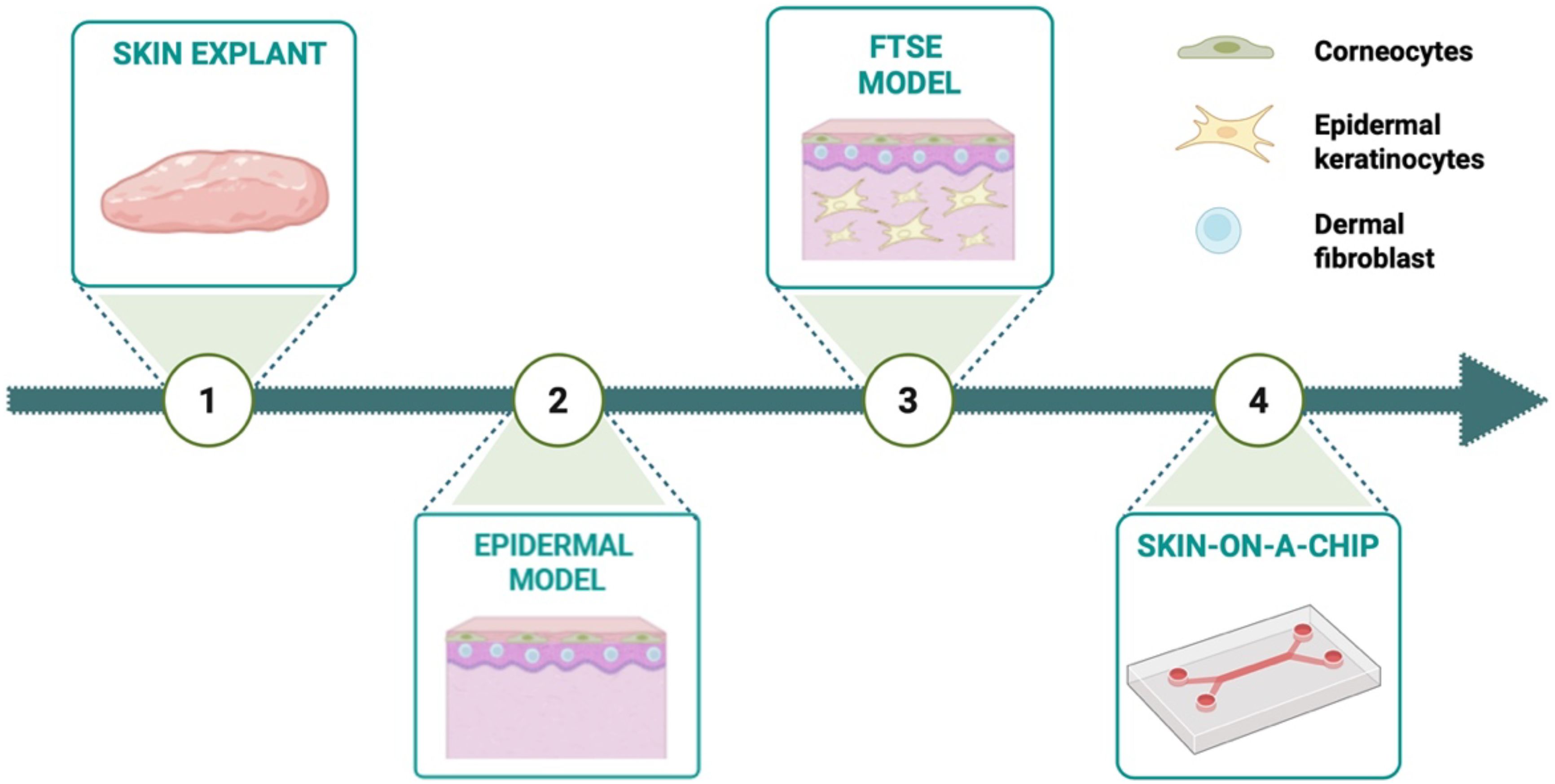
Figure 5. Evolution of 3D in vitro skin model from skin explants to skin-on-a-chip systems. The Figure was created in Biorender.
6.1 Skin explants
The skin is the largest organ of the human body, playing fundamental roles such as serving as a protective physical barrier, controlling thermoregulation, providing sensation, maintaining homeostasis, and supporting immunity. It is composed of three layers: epidermis, dermis, and subcutis, and contains different cell phenotypes (Maggiotto et al., 2025). To study its complex structure and interaction with external agents, skin explants, i.e., native tissue portion from a donor, are considered the most realistic 3D skin models.
Most commonly, skin explants come from plastic surgery or cadavers, and in both cases, proper ethical approval is needed for their use in research (Abd et al., 2016). Generally, skin explants are derived from abdominal, breast, or back skin because they are the most abundant donor sites. Two types of skin explants can be distinguished: full-thickness and partial-thickness skin explants. The former contains both epidermal and dermal layers, and it is characterized by all skin cell types, guaranteeing an accurate representation of their physiological and functional interconnections. On the other hand, it is affected by some limitations such as loss of the vascular and nervous systems. Although these disadvantages exist, full-thickness skin explants are considered physiologically relevant models for studying the mechanisms contributing to skin disease (Mathes et al., 2014). Differently, the partial-thickness skin explant is composed of the full epidermidis and a portion of the underlying dermis. However, this model captures only part of the complexity of human skin, as endothelial cells, melanocytes, and Langerhans cells are gradually lost under culture conditions over time.
Irrespective of the type of skin explants, they are considered promising tools to evaluate the efficacy and skin absorption of personal care products, as well as their effects on skin infections, metabolism, immune responses, melanogenesis, and irritation. The relevance of these models is further highlighted by several studies that have employed skin explants in various experimental contexts, as outlined below.
For instance, Eberlin et al. used the skin explant to evaluate various skin disorders (i.e., photoaging, skin barrier disorders, AD), and permeation of substances through the skin using immunofluorescence, immunoassay, and omics platforms to study the interaction between cutaneous tissue and environmental factors (Eberlin et al., 2020). Another example employed an ex vivo wound model based on full-thickness human skin to evaluate the polyvinylpyrrolidone foils and nanofiber mats as a ciprofloxacin delivery system for treating infected wounds (Rancan et al., 2019). More recently, a 3D ovine skin explant model was exploited to investigate anaerobic infection by Dichelobacter nodosus (Maboni et al., 2017). The model preserved tissue viability for up to 28 h, showed bacterial invasion of the epidermis, and triggered an inflammatory response, demonstrating its potential for studying anaerobic skin infections.
Despite skin explants being closer to the in vivo environment, they are characterized by high inter-donor variability, availability, and biological limitations. Indeed, the cutaneous absorption response is strongly influenced by the anatomical site of collection, due to stratum corneum thickness, hydration, and lipid composition (Abd et al., 2016).
6.2 Epidermal models
The human epidermis is a self-repairing barrier that separates our internal body from the external environment. Its main role is to protect the body against dehydration, nutrient loss, and physical, chemical, and biological hazards. Concerning cell phenotypes, keratinocytes are the main cells involved in the production and maintenance of the epidermal barrier. During the progressive maturation, keratinocytes change their morphology from a cuboidal shape located in the basal layer to a squamous morphology in the cornified layer. Other morphological changes include the development of a prickle-cell shape in the spinous layer and the keratohyalin granules, within the granular layer beneath the cornified barrier. Other cell types present in the epidermis are Merkel cells and Langerhans cells.
Understanding the cell population and the differentiation program of epidermal keratinocytes toward skin barrier formation, as well as the impact of external agents and drugs on the skin, requires the development of reliable epidermal models (Poumay and Coquette, 2007). However, such models partially reproduce the skin being composed of only the epidermal layer and not considering the dermal layer and immune cells. Commercially, several epidermal models are available on the market such as EpiSkin®, SkinEthic®, EpiDerm® which are useful to study skin irradiation, corrosion, penetration, UV damage, bacterial adhesion, and epidermal permeability (Flaten et al., 2015; Pupovac et al., 2018). Poumay et al. developed a fully differentiated cultured epidermis anchored on a polycarbonate filter (Poumay et al., 2004). The obtained model successfully revealed the characteristic morphology with basal, spinous, granular, and cornified epidermal layers, as confirmed by histological analysis, immunofluorescent staining and electron microscopy. Therefore, the proposed model showed promises for the investigation of cell biology, toxicological tests, and drug skin absorption mechanisms.
In fact, epidermal models represent a significant in vitro tool for understanding skin physiology, irritation, and permeability. They represent a simplified approximation of human skin structure due to the absence of the dermal layer, which limits their physiological relevance. A more realistic representation of human skin is achieved through more complex models, as described in the following sections.
6.3 Full-thickness skin equivalents
Full-thickness skin equivalents (FTSEs) are 3D in vitro models that more realistically reproduce both epidermis and dermis layers, obtained by the maturation of keratinocytes and fibroblasts, respectively. Specifically, the engineering of FTSEs encompasses (i) fibroblasts seeding followed by maturation of the dermal layer; (ii) keratinocytes seeding followed by maturation of the epidermal layer and, (iii) establishment of the air-liquid interface (ALI) to allow epidermal layer stratification and keratinization (Moon et al., 2021; Villata et al., 2024).
FTSEs can be classified in scaffold-free and scaffold-based 3D models: in the first case, skin cells are layer-by-layer seeded on a support without the use of any additional materials and allowed to deposit their own extracellular matrix (ECM) (Michel et al., 1999; Liu et al., 2013); in the latter case, cells are embedded in hydrogel-based formulations which function as an hydrated environment resembling the native ECM (Reuter et al., 2017; Casale et al., 2018). For instance, to engineer the dermis, fibroblasts are often seeded in hydrogels prepared from polymers of natural origin, such as collagen/fibrin hydrogel (Bauer et al., 2022). Concerning the epidermis, the development is more complicated due to its several distinct layers.
Based on the reconstruction method of the dermal skin layer, FTSE models can be also classified into three categories: collagen-based, de-epidermized dermis, and self-assembled skin substitutes (Moon et al., 2021). The first one involves seeding epidermal keratinocytes on dermal matrix composed of collagen and dermal fibroblasts. Collagen gel not only serves as support but also supplies nutrients and guarantees cell-to-cell and cell-to-matrix interactions. This model ensures excellent biocompatibility and cell adhesion. However, it shows limited mechanical strength and a short service life. The second model consists of decellularized human skin, used as a dermal substitute that supports keratinocyte attachment and proliferation. The last model consists of keratinocytes and fibroblasts without the addition of exogenous extracellular matrix materials. Dermal fibroblasts are cultured until they reach confluence, secreting their extracellular matrix to form a dermal sheet. Keratinocytes are then seeded on top of these stacked dermal sheets, where they undergo differentiation and cornification. Through the addition of different cell types inside FTSEs, it is possible to investigate the role of these cells in skin health and skin disease (Pupovac et al., 2018).
Mertsching et al. demonstrated the versatility of this model across various applications. Firstly, they investigated the irritating effects of different tested compounds on FTSE (Mertsching et al., 2008). Secondly, they employed the skin equivalent as an in vitro tumor model by introducing various tumor cell lines. Additionally, they utilized the model as a disease platform by culturing bacteria on the FTSE to simulate infections, and as a wound model by mechanically creating scratches on the surface.
Therefore, considering their 3D organization and composition, FTSE models are more reliable than the previously described model. However, they also present some disadvantages, such as high costs, limited suitability for long-term culture, and the absence of a vascularization system.
6.4 Skin-on-a-chip platforms
A promising alternative approach in the design of advanced 3D skin models is the integration of microfluidic systems. Conventional 3D constructs fail to accurately replicate human skin, not only because they consist of a limited number of cell types, but also due to the lack of a vascularized network (Sun et al., 2023). The absence of blood vessels limits the perfusion of nutrients and drugs, leading to results that do not accurately reflect real physiological conditions. Dynamic culture allows the recreation of a more physiologically controlled environment, defining physical and biochemical parameters as flow, forces, and chemical gradient (Risueno et al., 2021).
In this line, Mohamadi et al. demonstrated that a microfluidic skin system can better replicate a physiological environment by comparing static and dynamic culture conditions in terms of mechanical strength, water adsorption, skin morphology, gene expression, and biopsy longevity (Mohamadali et al., 2023). Regarding the architecture of microfluidic systems, two different strategies have been implemented:
i. transferred skin-on-a-chip.
ii. in situ skin-on-a-chip.
In the first approach skin fragments derived either from ex vivo human skin biopsies or in vitro generated human skin equivalents are introduced in the microfluidic chip systems and perfused by a microfluidic channel below the tissue construct (Risueno et al., 2021). In the second strategy, skin tissue models are directly generated within the chip due upon maturation of seeded cells. In this configuration the microfluidics system not only perfuses the nutrients but also supports the maturation of the tissue. These models equipped with a dynamic perfusion system, better mimic physiological conditions, enabling realistic studies of absorption, diffusion, efficacy, and potential toxicity (Sun et al., 2023).
For this reason, skin-on-a-chip platforms have found relevant applications in the pharmaceutical and cosmetic field, as they enable the evaluation of drugs or products in a controlled microenvironment (Risueno et al., 2021). For instance, Quan et al. developed a reliable tissue model designed for toxicology studies and drug testing in the context of bacteria-infected, damaged skin (Quan et al., 2022). They fabricated an interface-controlled skin-on-chip integrating cutaneous extracellular matrix and cells within a microfluidic system featuring mechanical stimulation. The combination of a perfusion system and an air–liquid interface enabled the development of an in vitro skin model with enhanced morphological maturation and improved barrier function. After the maturation of the healthy skin model, bacteria were seeded on the top establishing a pathological model. The authors treated the model with dexamethasone and polyphyllin H, which demonstrated a significant repair effect on the skin barrier.
The integration of the skin model into a microfluidic system allows for a better simulation of real physiological conditions and enables the development of an automated platform capable of simultaneously analyzing multiple samples under different experimental conditions.
Furthermore, one major advantage of this technology lies in the possibility of incorporating biosensors to monitor skin status and drug delivery in real time. In conclusion, skin-on-chip represents a powerful tool for studying the biological mechanisms, both healthy and pathological, that affect this organ.
6.5 Case study applications of 3D in vitro skin models
6.5.1 Wound healing models
Wounds refer to skin damage characterized by cuts, punctures, burns, or other conditions. They are categorized as either acute or chronic depending on their healing duration and progression (Ahmad, 2022). Engineering acute or chronic wound models represent a promising strategy for studying and understanding the wound healing pathway and preclinically testing innovative biomaterials and therapies.
In this regard, several grafts are commercially available on the market to study the healing of wounds, such as Hyalograft 3D, Apligraf and TissueTech Autograft system. On the other hand, models such as EpiDermFT™, Phenion® FT Model, and StrataTest® are used for 3D wound healing assays (Stamm et al., 2016). To create a wound model, the process begins with a skin model consisting of a dermal layer composed of fibroblasts and an epidermal layer in which keratinocytes are cultured. The epidermal layer is exposed to air to achieve a structure that closely resembles the physiological one. Once a mature skin model is developed, artificial wounds mimicking burn or excisional wounds are performed. Concerning burn wounds, Schneider et al. created a highly standardized in vitro 3D epidermal burn model derived from primary epidermal keratinocytes (Schneider et al., 2021). Healthy epidermal models were cultured for 12 days before being burned through a metal rod at room temperature. The wound model was cultured in vitro up to 14 days, then treated using drug-based therapies. Specifically, this model was exploited to study the physiological wound healing pathway up to 14 days and to test the safety of pharmacological agents. Although this approach is widely used and efficient, it is associated with high variability in terms of shape and size of resulting wounds. In this regard, Javid et al. proposed an alternative approach, using a 3D printer to create a more reliable tool for producing standardized and reproducible burn wounds characterized by controlled geometry (Tabatabaei and Javid, 2024).
Regarding excisional wounds, lesions are mechanically induced using scalpels and mashers (Stamm et al., 2016). Silva et al., 2025 developed a bioengineered skin wound model by using needles to puncture the skin structure. By modulating needle length, penetration force, and angle, this method allowed precision, and control over the depth, size, and shape of the wound (Salazar Silva et al., 2025). The proposed protocol was controlled, reproducible, standardized, versatile, and scalable for analyzing wound healing cascade and potential treatments. The onset of chronic wounds is often associated with bacterial infections.
Bioengineered wound models are commonly employed for the evaluation of antibacterial agents. In this line, a study investigated new antimicrobial therapeutics and host-pathogen interactions on mechanically wounded human skin models infected with Staphylococcus aureus and Pseudomonas aeruginosa (Wiegand et al., 2024). This research used the model to investigate the efficacy of silver-containing and polyhexamethylene biguanide-containing wound dressing. Additionally, Andersoon et al. studied the effect of antimicrobial compounds and formulations on ex vivo porcin skin model (Andersson et al., 2021). The authors proposed a biologically relevant platform which represented a bridge between the in vitro and the in vivo model. The model featured a natural ECM and realistic tissue architecture that faithfully replicated the wound bed microenvironment. Infection in the model was induced by introducing two bacterial species commonly involved in wound healing processes. Using scanning electron microscopy (SEM), the authors observed biofilm formation, closely resembling the physiological condition. This model presented itself as a valuable tool for studying the penetration efficacy of topical formulations and materials, including bioactive textiles.
Although 3D wound models represent a valid alternative to in vivo testing, they still offer only an approximate representation of physiological reality. Ideal models should accurately replicate the pathological phenotype. It would be beneficial to include in these models the hypodermal compartment, as well as a vascular system and patient-specific cells such as fibroblasts, keratinocytes, and immune cells.
6.5.2 Skin models for microbiome research
Although bacterial contaminations are one of the major concerns to be faced to prevent pathogenic skin infections, the presence of a symbiotic community composed of bacteria, fungi and viruses is essential in maintaining skin homeostasis. Specifically, such symbiotic community, referred to as skin microbiota, is similar to the gut microbiota in composition and second only for bacterial density (i.e., 104–106 bacteria/cm2) (Cundell, 2018). It is mainly composed of resident microbes and only a small percentage of transient microbes that can opportunistically increase in the presence of skin diseases. Moreover, the skin microbiota plays a key role in providing nutrients, inhibiting pathogenic growth and regulating epidermal differentiation (Smythe and Wilkinson, 2023). Therefore, understanding the role and functions of skin microbiota in extremely important to provide insights in skin pathologies and facilitating the development and clinical translation of more effective treatments, particularly the use of bioactive textile materials. However, the microbiota complexity and the dynamic interactions between the host and microbial communities are the main challenging aspects to consider in the development of realistic and reliable microbiota models for research (Smythe and Wilkinson, 2023). Moreover, the use of systems mimicking skin structure and physiology is another key feature to fully recapitulate the microbial ecosystem in living skin.
In this context, 3D skin equivalents represent promising candidates for studying host-microbiome interactions combining well-differentiated tissue layers with key skin microbiome components that closely resemble native skin tissue. For instance, Maloney et al. developed an in vitro human skin microbiome model containing both stratified host tissue and a microbial consortium composed of six different bacterial strains (Maloney et al., 2025). Specifically, the model was developed by seeding keratinocytes on transwell inserts, promoting their maturation for 7 days until the achievement of a functional ALI as assessed through Trans-Epithelial Electrical Resistance measurements and lastly, bacteria contaminated. The obtained model provided a stable microbiome for up to several days and was found to be a suitable platform to provide insights in the interactions between host and microbes during the pathogenesis of chronic skin diseases.
A similar approach for in vitro skin model design was also explored by van Drongelen and colleagues to study the relationship between filaggrin expression and epidermal colonization by Staphylococcus aureus (van Drongelen et al., 2014). Results demonstrated that reduced filaggrin expression was associated with increased bacteria colonization, a key consequence in epidermal barrier damages ascribed to pathologies such as AD.
Despite 3D in vitro skin equivalents are currently considered advanced and reliable platforms for native skin tissue mimicking, the absence of glands, blood vessels, appendages and immune cells could be a limiting issue when microbiota modeling is desired. Indeed, the skin houses microbial communities that inhabit spatially distinct regions based on the cutaneous topography (Costello et al., 2009). For instance, Cutibacterium species are mainly found in the pilosebaceous units of sebum-rich areas requiring anaerobic growth conditions (Fitz-Gibbon et al., 2013), while Staphylococcus and Corynebacterium are colonizers of warm and moist environments such as hair follicles and sweat glands (Smythe and Wilkinson, 2023).
Furthermore, 3D skin models have been further improved by adding such key components of the native skin physiology, namely vasculature and improved immunocompetences. For instance, Abaci and co-workers developed a multi-layer skin equivalent with perfusable and spatially controlled vascular networks by combining the latest microfabrication techniques and primary and induced pluripotent stem cell-derived endothelial cells (Abaci et al., 2016). Results demonstrated the successful engineering of mature epidermis and endothelial barrier, thus paving the way to model exploitation for drug screening. More recently, Kang et al. engineered an even more realistic skin model incorporating hair follicles and epidermal/papillary dermal layers relying on the potentialities of 3D bioprinting (Kang et al., 2022). The obtained constructs were characterized by a microporous network resembling the native skin extracellular matrix, promoted epidermis-dermis interactions mediated by the papillary layer and, spontaneous capability to develop hair pore structures.
6.5.3 3D skin models to pre-clinically evaluate bioactive textiles
Although current 3D skin models are indispensable and intensively used for studying the biological effects of various topically applied compounds on human skin components, their use to evaluate bioactive textiles for dermatological applications remains largely unexplored.
In fact, advancing from traditional 2D to 3D skin models offers a physiologically relevant platform to assess key parameters such as biocompatibility, antimicrobial efficacy, and host–microbe interactions. This transition enhances the predictive power of preclinical studies and strengthens the translational potential of bioactive textiles for personalized clinical approaches. In this line, Bengalli et al., 2021 developed antibacterial textiles aimed at preventing infection by incorporating metal oxide NPs, which are considered as promising antibacterial agents. To evaluate the potential toxicological effects on the skin, the authors evaluated these bioactive textiles using an in vitro reconstructed 3D epidermal model, (EpiDerm™), under conditions simulating human sweat (Bengalli et al., 2021a). The translation to 3D skin models allowed the assessment of epidermal permeability to metal ions, the relationship between metal ion release and sweat pH, and ultimately the safety of antibacterial textiles on intact skin.
A similar study evaluated the toxicity of polypyrrole NPs-coated textiles using 3D skin models (Bengalli et al., 2021b). Polypyrrole is a conductive polymer with antibacterial properties, biocompatibility, electrical conductivity, and suitability for application in hydrogels, biosensors and controlled drug delivery textiles. The safety of NPs release from textiles was assessed using the EpiDerm ™ model in combination with Balb/3T3 fibroblasts, through tests evaluating skin irritation and corrosion. The results indicated that polypyrrole particles did not induce toxic effects on skin model, supporting their preclinical safety for skin-contact applications.
Overall, these studies highlight the potential of in vitro 3D skin models as sustainable and scalable alternatives for assessing the biological impact of bioactive textiles in human skin components (Table 1). Nevertheless, only a limited number of studies have explored the use of 3D skin models as preclinical testing platforms for novel bioactive textiles, probably due to the complexity of the experimental workflow, which involves generating a fully differentiated 3D skin construct, followed by textile contact, and monitoring the model over time. Therefore, greater efforts are needed to advance these applications and expand the knowledge in this emergent area.
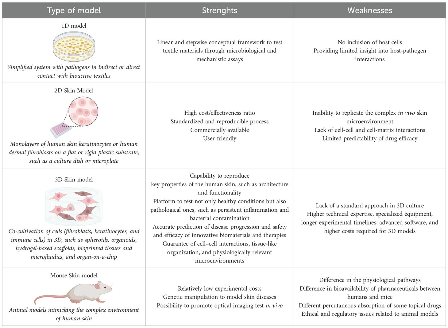
Table 1. Overview of the experimental models ranging from 1D and 2D systems to advance 3D and animal models used to assess the performance of bioactive textiles in human skin components.
7 Antimicrobial textiles in the management of skin microbiome dysregulation
The human skin microbiome, composed of a diverse community of commensal microorganisms, plays a vital role in maintaining skin barrier integrity, modulating immune responses, and providing defense against pathogens (Skowron et al., 2021). Disruption of this delicate microbial ecosystem, known as dysbiosis, has been associated with the pathogenesis of various dermatological conditions, including (chronic) wounds, AD, acne vulgaris, psoriasis, hidradenitis suppurativa, and even certain types of skin cancer (Yang et al., 2022). In this dysbiotic state, the balance between beneficial and harmful microorganisms is disturbed, often leading to a reduction in commensal microbial diversity and abundance. This shift creates an ideal environment for the overgrowth of opportunistic or pathogenic species, which can exacerbate inflammation, impair skin barrier function, and contribute to disease progression (Hou et al., 2022).
While conventional treatment strategies for skin microbiota dysregulation usually involves the use of topical or systemic antibiotics, these approaches tend to act broadly and non-selectively, disrupting both pathogenic and beneficial microbial communities (Ito and Amagai, 2022). Such indiscriminate modulation of the skin microbiome may compromise its protective functions and contribute to the emergence and spread of multidrug-resistant bacterial strains, thereby limiting long-term therapeutic efficacy.
As an alternative to conventional antibiotic-based therapies, bioactive textiles with antimicrobial properties are gaining attention as a promising strategy to target pathogenic skin microorganisms (Gauger et al., 2006; Haug et al., 2006; Juenger et al., 2006; Lopes et al., 2015; Suellen Ferro de Oliveira and Kekhasharu Tavaria, 2023; Liu et al., 2024). In fact, their therapeutic potential has been increasingly explored in clinical studies involving patients with skin disorders associated to microbiota dysregulation, where microbial imbalance contributes to disease severity (Gauger et al., 2003; Gauger et al., 2006; Haug et al., 2006; Ricci et al., 2006; Lopes et al., 2015; Schaunig and Kopera, 2017; Thomas et al., 2017; Morand and Hatami, 2019; Ragamin et al., 2021).
A good example is AD, a chronic and relapsing inflammatory skin disorder, associated with skin barrier dysfunction, transepidermal water loss, inflammation, and intense pruritus (Criado et al., 2024). A key factor in AD is the colonization and overgrowth of Staphylococcus aureus which exacerbates inflammation and difficult healing (Ogonowska et al., 2020). In this line, several studies have highlighted the therapeutic potential of antimicrobial textiles, namely silver-coated, zinc-coated, chitosan-coated, and cellulose-based fabrics, due to their ability to reduce the proliferation of Staphylococcus aureus while alleviating the clinical symptoms associated with AD (Gauger, 2006; Gauger et al., 2006; Jaros et al., 2020; Ragamin et al., 2021).
Another relevant example is acne vulgaris, a multifactorial skin disorder whose pathogenesis involves the overgrowth of Cutibacterium acnes, often associated with increased sebum production and localized inflammation. In this context, the use of antimicrobial textiles has shown therapeutic promise. A study investigating a silver-impregnated textile (AEM 5772/5; DermaSilk), known for its antibacterial and antifungal properties, demonstrated a clinically significant reduction in acne lesions on the back, even in the absence of additional treatments or lifestyle modifications (Schaunig and Kopera, 2017).
However, a significant limitation of current clinical studies, mirroring in vitro 2D and 3D models evaluations, is the insufficient assessment of how antimicrobial textiles affect commensal members of the skin microbiome. Most investigations have primarily focused on the reduction of pathogenic bacteria, particularly Staphylococcus aureus, without evaluating the broader impact on the overall microbial community or the potential role of these materials in restoring or disrupting microbiome balance.
The complex interaction between antimicrobial bioactive textiles, namely those used in clothing, and the human skin microbiome requires deep investigation to elucidate the true implications of such interventions in dermatological care. Understanding how these materials influence microbial diversity and function is essential for evaluating their effectiveness in managing microbiota-associated skin conditions.
Notably, antimicrobial textiles can be engineered to selectively inhibit pathogenic species like Staphylococcus aureus while preserving or even promoting the growth of beneficial commensals such as Staphylococcus epidermidis and Staphylococcus hominis, thereby helping to maintain the skin’s microbial homeostasis (Figure 6). Nonetheless, in-depth studies using (advanced) 3D skin models are needed to characterize the full spectrum of microbial responses and determine whether these textiles can reliably modulate dysbiosis in conditions such as AD, acne, or chronic wounds.
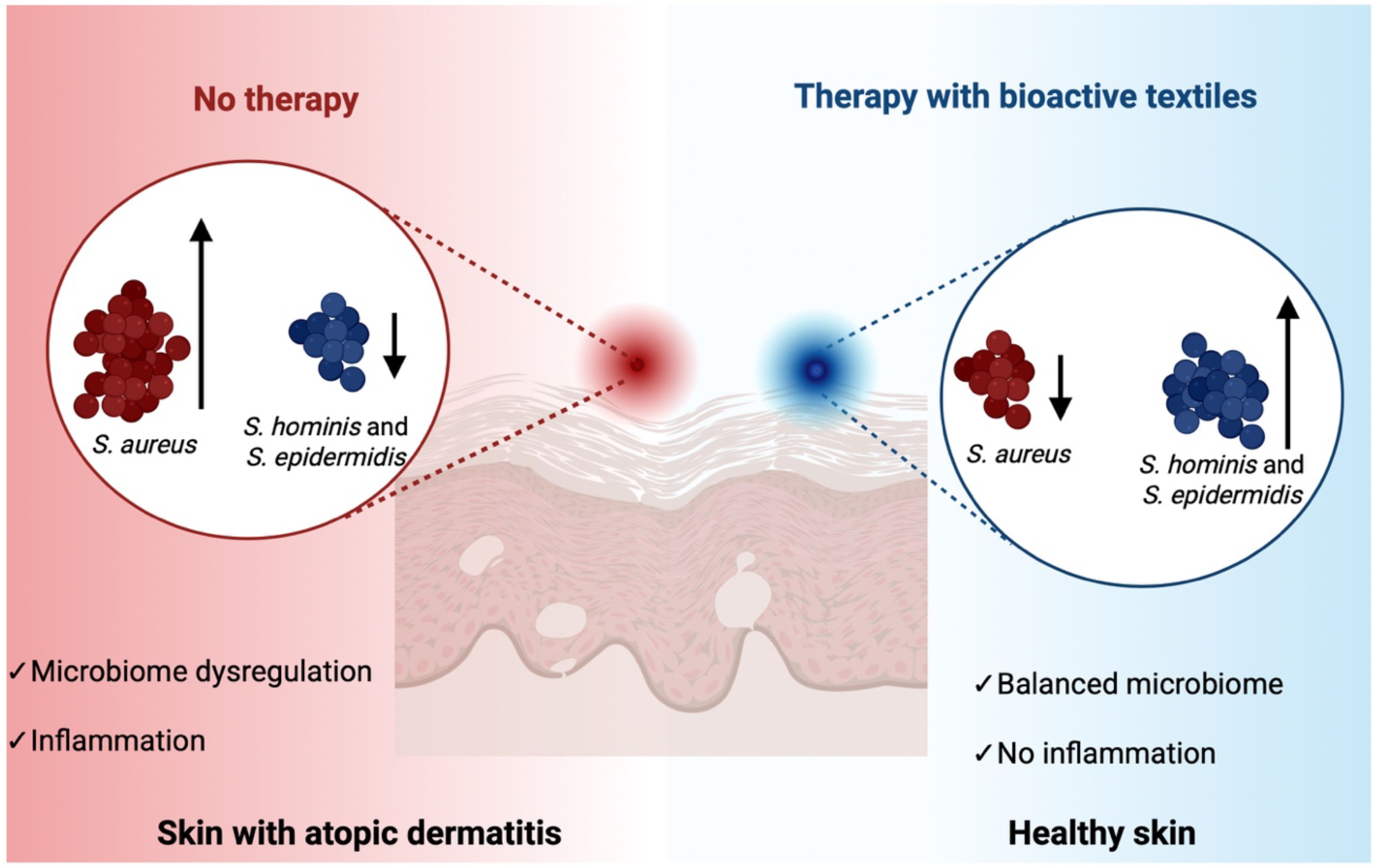
Figure 6. Impact of antimicrobial bioactive textiles on skin microbiome balance in atopic dermatitis (AD). Schematic representation comparing skin microbiome composition in the absence (left) and presence (right) of therapy with bioactive textiles. Without intervention, S. aureus predominates over commensal bacteria species such as S. hominis and S. epidermidis, contributing to microbial dysbiosis and exacerbated skin inflammation. In contrast, therapy using antimicrobial bioactive textiles may selectively suppresses S. aureus while preserving beneficial commensal bacteria, promoting microbial balance. The figure was created in Biorender.
7.1 Interactions between antimicrobial textiles and the skin immune system
Bioactive textiles with antimicrobial properties interact not only with the skin microbiome but also with the host immune system, either directly or indirectly. The skin serves as both a physical and immunological barrier, where keratinocytes, Langerhans cells, dermal dendritic cells, and resident T cells continuously sense microbial and chemical cues. By modulating microbial populations, bioactive textiles may influence the integrity of this barrier, which is critical for protection against allergens, irritants, and pathogens (Broadhead et al., 2021).
For example, the selective reduction of Staphylococcus aureus colonization has been associated with decreased epithelial damage and transepidermal water loss, thereby supporting barrier repair and reducing inflammatory flares in AD. Clinical studies have demonstrated that silver-coated garments reduce Staphylococcus aureus burden and improve disease severity in AD patients, highlighting the potential for microbiome-mediated immune modulation (Gauger et al., 2003; Gauger, 2006; Gauger et al., 2006). However, broad-spectrum antimicrobial activity may inadvertently eliminate beneficial commensals such as Staphylococcus epidermidis, which play a key role in promoting keratinocyte differentiation, stimulating antimicrobial peptide production, and regulating innate immune responses via TLR2-dependent pathways (Lai et al., 2010). Loss of such protective bacteria may compromise barrier defense and shift immune homeostasis.
Beyond microbial modulation, bioactive textiles incorporating anti-inflammatory or antioxidant compounds (e.g., chitosan, flavonoids, honey-derived products) can directly attenuate innate immune activation by reducing oxidative stress and suppressing pro-inflammatory cytokine release from keratinocytes and macrophages (Lopes et al., 2015; Jaros et al., 2020; de Oliveira and Tavaria, 2024; Freitas et al., 2024). Chitosan-functionalized textiles, for instance, exhibit both antimicrobial and anti-inflammatory properties through the suppression of pro-inflammatory citokynes (Liu et al., 2025). Conversely, uncontrolled release of metal nanoparticles or synthetic antimicrobials may provoke inflammatory responses, undermining tissue homeostasis (Sadrolvaezin et al., 2023).
The role of bioactive textiles in allergic skin responses also warrants attention. Certain textile finishes, including metals, dyes, and preservatives, have been implicated in allergic and irritant contact dermatitis, acting as haptens and triggering immune sensitization in predisposed individuals (Sadrolvaezin et al., 2023). In contrast, textiles engineered with biocompatible or soothing compounds may reduce allergen penetration, alleviate irritation, and mitigate hypersensitivity reactions (Sanchez Armengol et al., 2022). Since allergic inflammation reshapes skin physiology and microbiota composition, explore this crosstalk is essential to understand the complexity of textile–immune interactions.
Taken together, bioactive textiles have the capacity to influence chronic inflammatory responses in conditions such as acne vulgaris, psoriasis, AD, and chronic wounds, where dysbiosis and excessive immune activation are tightly linked. Mechanistic in vitro and ex vivo work have supported this microbiome–immune cross-talk, while recent advances in immunocompetent, microbiome-inclusive 3D skin models provide translational platforms to study these interactions (Lemoine et al., 2020; Holken et al., 2023). Such models, incorporating keratinocytes, immune cells (e.g., macrophages, T cells), and commensal microbes, allow assessment of whether a given textile formulation reduces pathogenic colonization without inducing sensitization or unintended immune activation. Furthermore, the mouse skin model remains a cornerstone in vivo system for investigating host–microbe interactions. Recent work using germ-free murine skin has demonstrated its value in dissecting the distinct contributions of commensals and pathogens to cutaneous immunity, providing critical insights that cannot yet be fully replicated in vitro (Uberoi et al., 2025). Such findings are particularly relevant for the evaluation of textile-based interventions, as they allow the evaluation of how bioactive fabrics influence microbial colonization, immune activation, and barrier function within a physiologically relevant context.
In summary, the interplay between antimicrobial textiles, the skin microbiome, and host immunity is complex and highly context-dependent. While some of them show promise in restoring microbial balance and attenuating inflammation, others may have the risk of disrupting commensal ecosystems or provoking adverse immune reactions. Future research should systematically evaluate these outcomes to guide the rational design of safe and effective bioactive textiles for specific dermatological applications.
8 Conclusion and future perspectives
Bioactive textiles are emerging as transformative materials in dermatological and healthcare applications, offering specific biological properties and protective functions while aligning with sustainability goals. They represent a shift from passive materials to functional interfaces capable of modulating the skin’s microbiome and barrier function. As demonstrated, bioactive compounds, from metal ions to essential oils and plant-derived polyphenols, have shown promise in reducing skin pathogenic microorganisms’ colonization. However, current evidence remains limited regarding their short and long-term effects on beneficial microorganisms like Staphylococcus epidermidis, highlighting the need for a more nuanced understanding of microbiome-targeted textile design.
One of the primary challenges lies in bridging in vitro results with in vivo outcomes. Conventional 1D and 2D skin models fall in mimicking the dynamic and complex interactions between the skin barrier, immune responses, and microbial communities. Thus, future research must focus on advanced 3D skin models that incorporate not only skin pathogenic members but also commensal microbiota members to better simulate realistic skin-textile interactions. In terms of materials science, the integration of sustainable biopolymers, nanocomposites, and bioactive herbal agents offers a promising avenue for designing eco-friendly and microbiome-compatible textiles (Dasgupta et al., 2025; Yang et al., 2025). Nonetheless, large-scale production, stability of bioactive agents, and regulatory approvals remain hurdles for clinical translation and commercialization.
Future research in bioactive textiles is moving toward a more targeted, sustainable, and technologically advanced approach to skin health management. One of the key priorities is microbiome-specific targeting, wherein textile materials are designed to selectively inhibit pathogenic microbes, such as Staphylococcus aureus or Cutibacterium acnes, while preserving or even promoting the growth of beneficial skin commensals like Staphylococcus epidermidis and Staphylococcus hominis. Achieving this balance is essential for maintaining skin homeostasis and preventing dysbiosis-related conditions. To ensure that these materials are safe and effective, microbiome-integrated testing is becoming an essential aspect of textile evaluation. Conventional antimicrobial testing often fails to account for the complex interactions between skin, fabric, and microbial communities. Therefore, advanced in vitro 3D skin models incorporating commensal microbiota, along with metagenomic sequencing tools, are being developed to assess how these textiles influence microbial diversity and composition under realistic conditions.
Another major direction involves the use of sustainable and biodegradable materials. Many bioactive textiles are now incorporating green chemistry principles, using plant-derived compounds, biosynthesized NPs, and biodegradable polymers to reduce ecological impact. This shift not only aligns with environmental sustainability goals but also addresses concerns over toxicity and bioaccumulation associated with synthetic agents.
Lastly, smart textile technologies are gaining momentum. These innovative fabrics can incorporate sensors or responsive release systems that detect skin pH, temperature, or microbial activity, and deliver therapeutic compounds in a controlled way. Such responsive textiles offer real-time adaptability to changing skin conditions, potentially enhancing treatment outcomes and user comfort. In this line, wearable systems are being designed to monitor inflammatory responses or wound conditions and adjust bioactive release accordingly (Broadhead et al., 2021). Together, these future directions highlight a multidisciplinary effort to design textiles that are microbiome-friendly, clinically effective, and environmentally responsible.
In summary, bioactive textiles offer immense promise but require interdisciplinary collaboration and innovation in skin modeling, materials science, and microbiome research to fully realize their clinical and commercial potential.
Author contributions
IN: Formal Analysis, Writing – original draft. CM: Writing – original draft. RL: Writing – original draft, Writing – review & editing. GC: Writing – review & editing. SB: Writing – review & editing. CO: Visualization, Funding acquisition, Validation, Writing – review & editing, Conceptualization, Supervision.
Funding
The author(s) declare financial support was received for the research and/or publication of this article. This work was supported by National Funds from Fundação para a Ciência e a Tecnologia (FCT) through the DETEX project (https://doi.org/10.54499/2023.12365.PEX). This work was also supported by the ATTITUDE project titled “vAlorization of wasTe products for the fabrication of sustainable patient-personalIzed paTches targeting different phases of the wound hEaling cascade”, which was financed by Fondazione Pisa (Italy) Call 2022 “Scientific and Technological Research” in macroarea of Biomedicine. IN acknowledges a grant from the Ministry of Research, Innovation and Digitalization, CCCDI—UEFISCDI, project number PN-IV-P7-7.1-PED-2024-0137, within PNCDI IV. IN also acknowledges the Romanian Ministry of Research, Innovation and Digitalization under the Romanian National Nucleu Program LAPLAS VII—contract No. 30N/2023.
Conflict of interest
The authors declare that the research was conducted in the absence of any commercial or financial relationships that could be construed as a potential conflict of interest.
The author(s) declared that they were an editorial board member of Frontiers, at the time of submission. This had no impact on the peer review process and the final decision.
Generative AI statement
The author(s) declare that no Generative AI was used in the creation of this manuscript.
Any alternative text (alt text) provided alongside figures in this article has been generated by Frontiers with the support of artificial intelligence and reasonable efforts have been made to ensure accuracy, including review by the authors wherever possible. If you identify any issues, please contact us.
Publisher’s note
All claims expressed in this article are solely those of the authors and do not necessarily represent those of their affiliated organizations, or those of the publisher, the editors and the reviewers. Any product that may be evaluated in this article, or claim that may be made by its manufacturer, is not guaranteed or endorsed by the publisher.
References
Abaci, H. E., Guo, Z., Coffman, A., Gillette, B., Lee, W. H., Sia, S. K., et al. (2016). Human skin constructs with spatially controlled vasculature using primary and iPSC-derived endothelial cells. Adv. Healthc Mater 5, 1800–1807. doi: 10.1002/adhm.201500936
Abd, E., Yousef, S. A., Pastore, M. N., Telaprolu, K., Mohammed, Y. H., Namjoshi, S., et al. (2016). Skin models for the testing of transdermal drugs. Clin. Pharmacol. 8, 163–176. doi: 10.2147/CPAA.S64788
Ahmad, N. (2022). In vitro and in vivo characterization methods for evaluation of modern wound dressings. Pharmaceutics 15. doi: 10.3390/pharmaceutics15010042
Andersson, M. A., Madsen, L. B., Schmidtchen, A., and Puthia, M. (2021). Development of an experimental ex vivo wound model to evaluate antimicrobial efficacy of topical formulations. Int. J. Mol. Sci. 22. doi: 10.3390/ijms22095045
Babu, K. M. and Ravindra, K. B. (2015). Bioactive antimicrobial agents for finishing of textiles for health care products. J. Text I 106, 706–717. doi: 10.1080/00405000.2014.936670
Bauer, M., Metzger, M., Corea, M., SChadl, B., Grillari, J., and Dungel, P. (2022). Novel 3D-printed cell culture inserts for air-liquid interface cell culture. Life (Basel) 12. doi: 10.3390/life12081216
Bengalli, R., Colantuoni, A., Perelshtein, I., Gedanken, A., Collini, M., Mantecca, P., et al. (2021a). In vitro skin toxicity of CuO and ZnO nanoparticles: Application in the safety assessment of antimicrobial coated textiles. NanoImpact 21, 100282. doi: 10.1016/j.impact.2020.100282
Bengalli, R., Fiandra, L., Vineis, C., Sanchez-Ramirez, D. O., Azoia, N. G., Varesano, A., et al. (2021b). Safety assessment of polypyrrole nanoparticles and spray-coated textiles. Nanomaterials (Basel) 11. doi: 10.3390/nano11081991
Bibi, A., Afza, G., Afzal, Z., Farid, M., Sumrra, S. H., Hanif, M. A., et al. (2024). Synthetic vs. natural antimicrobial agents for safer textiles: a comparative review. RSC Adv. 14, 30688–30706. doi: 10.1039/D4RA04519J
Broadhead, R., Craeye, L., and Callewaert, C. (2021). The future of functional clothing for an improved skin and textile microbiome relationship. Microorganisms 9. doi: 10.3390/microorganisms9061192
Byrd, A. L., Belkaid, Y., and Segre, J. A. (2018). The human skin microbiome. Nat. Rev. Microbiol. 16, 143–155. doi: 10.1038/nrmicro.2017.157
Carmona-Cruz, S., Orozco-Covarrubias, L., and Saez-de-Ocariz, M. (2022). The human skin microbiome in selected cutaneous diseases. Front. Cell Infect. Microbiol. 12, 834135. doi: 10.3389/fcimb.2022.834135
Casale, C., Imparato, G., Urciuolo, F., Rescigno, F., Scamardella, S., Escolino, M., et al. (2018). Engineering a human skin equivalent to study dermis remodelling and epidermis senescence. Vitro after UVA exposure. J. Tissue Eng. Regener. Med. 12, 1658–1669. doi: 10.1002/term.2693
Choi, N. Y. and Park, B. I. (2023). Dyeing performance and anti-superbacterial activity of cotton fabrics dyed with chamaecyparis obtusa. Molecules 28. doi: 10.3390/molecules28186497
Coates, M., Blanchard, S., and MacLeod, A. S. (2018). Innate antimicrobial immunity in the skin: A protective barrier against bacteria, viruses, and fungi. PloS Pathog. 14, e1007353. doi: 10.1371/journal.ppat.1007353
Costa, E. M., Silva, S., MaChado, M., Sousa, S. C., Tavaria, F. K., and Pintado, M. (2022). Chitosan nanoparticles as bioactive vehicles for textile dyeing: A proof of concept. Polymers-Basel 14. doi: 10.3390/polym14224821
Costa, E. M., Silva, S., Veiga, M., Tavaria, F. K., and Pintado, M. M. (2018). Chitosan’s biological activity upon skin-related microorganisms and its potential textile applications. World J. Microbiol. Biotechnol. 34, 93. doi: 10.1007/s11274-018-2471-2
Costello, E. K., Lauber, C. L., Hamady, M., Fierer, N., Gordon, J. I., and Knight, R. (2009). Bacterial community variation in human body habitats across space and time. Science 326, 1694–1697. doi: 10.1126/science.1177486
Criado, P. R., Miot, H. A., Bueno-Filho, R., Ianhez, M., Criado, R. F. J., and de Castro, C. C. S. (2024). Update on the pathogenesis of atopic dermatitis. Bras. Dermatol. 99, 895–915. doi: 10.1016/j.abd.2024.06.001
Cundell, A. M. (2018). Microbial ecology of the human skin. Microb. Ecol. 76, 113–120. doi: 10.1007/s00248-016-0789-6
Dasgupta, S., Maji, S., Dey, S., Chattopadhyay, M., Chanda, A., Acharjee, S., et al. (2025). Exploring nanocomposite materials in clinical dermatology: Innovations for treating skin diseases. Next Nanotechnology 7, 100139. doi: 10.1016/j.nxnano.2025.100139
de Oliveira, C. S. F. and Tavaria, F. K. (2024). Therapeutic textiles: A promising approach for human skin dysbiosis? Exp. Dermatol. 33, e15081. doi: 10.1111/exd.15081
dos Santos, A. C. R., Teodoro, G. R., and Sibelino, S. K. (2021). Comparative evaluation of the antimicrobial effect of electric plasma in textile between a standard strain of Klebsiella pneumoniae and a resistant clinical strain (KPC). Res. Biomed. Eng. 37, 697–707. doi: 10.1007/s42600-021-00179-8
Eberlin, S., da Silva, M. S., Facchini, G., da Silva, G. H., Pinheiro, A. L. T. A., Eberlin, S., et al. (2020). The ex vivo skin model as an alternative tool for the efficacy and safety evaluation of topical products. Atla-Altern Lab. Anim. 48, 10–22. doi: 10.1177/0261192920914193
El-Tarabily, K. A., El-Saadony, M. T., Alagawany, M., Arif, M., Batiha, G. E., Khafaga, A. F., et al. (2021). M, E. A. E.-H., Using essential oils to overcome bacterial biofilm formation and their antimicrobial resistance. Saudi J. Biol. Sci. 28, 5145–5156. doi: 10.1016/j.sjbs.2021.05.033
Firmanda, A., Mahardika, M., Fahma, F., Amelia, D., Pratama, A. W., Amalia, N., et al. (2024). Cellulose-enriched ascorbic acid for wound dressing application: Future medical textile. J. Appl. Polym Sci. 141. doi: 10.1002/app.56013
Fitz-Gibbon, S., Tomida, S., Chiu, B. H., Nguyen, L., Du, C., Liu, M., et al. (2013). Propionibacterium acnes strain populations in the human skin microbiome associated with acne. J. Invest. Dermatol. 133, 2152–2160. doi: 10.1038/jid.2013.21
Flaten, G. E., Palac, Z., Engesland, A., Filipovic-Grcic, J., Vanic, Z., and Skalko-Basnet, N. (2015). In vitro skin models as a tool in optimization of drug formulation. Eur. J. Pharm. Sci. 75, 10–24. doi: 10.1016/j.ejps.2015.02.018
Freitas, A. S., Oliveira, R., Ribeiro, A., and Almeida-Aguiar, C. (2024). Biofunctional textiles: antioxidant and antibacterial finishings of cotton with propolis and honey. Int. J. Mol. Sci. 25. doi: 10.3390/ijms25158034
Galvan, A., Pellicciari, C., and Calderan, L. (2024). Recreating human skin. In Vitro: Should Microbiota Be Taken into Account? Int. J. Mol. Sci. 25. doi: 10.3390/ijms25021165
Garcia, M. M., da Silva, B. L., Sorrechia, R., Pietro, R., and Chiavacci, L. A. (2022). Sustainable antibacterial activity of polyamide fabrics containing znO nanoparticles. ACS Appl. Bio Mater 5, 3667–3677. doi: 10.1021/acsabm.2c00104
Gauger, A. (2006). Silver-coated textiles in the therapy of atopic eczema. Curr. Probl Dermatol. 33, 152–164. doi: 10.1159/000093942
Gauger, A., Fischer, S., Mempel, M., Schaefer, T., Foelster-Holst, R., Abeck, D., et al. (2006). Efficacy and functionality of silver-coated textiles in patients with atopic eczema. J. Eur. Acad. Dermatol. Venereol 20, 534–541. doi: 10.1111/j.1468-3083.2006.01526.x
Gauger, A., Mempel, M., Schekatz, A., Schafer, T., Ring, J., and Abeck, D. (2003). Silver-coated textiles reduce Staphylococcus aureus colonization in patients with atopic eczema. Dermatology 207, 15–21. doi: 10.1159/000070935
Ghosh, J., Repon, M. R., Pranta, A. D., Rupanty, N. S., Khan, F., and Noor, T. (2025a). Bioactive component integrated textiles: A promising source of medicine and healthcare. J. Eng. Fiber Fabr 20. doi: 10.1177/15589250241308561
Ghosh, J., Rupanty, N. S., Noor, T., Asif, T. R., Islam, T., and Reukov, V. (2025b). Functional coatings for textiles: advancements in flame resistance, antimicrobial defense, and self-cleaning performance. RSC Adv. 15, 10984–11022. doi: 10.1039/D5RA01429H
Gopal, V., Nilsson-Payant, B. E., French, H., Siegers, J. Y., Yung, W. S., Hardwick, M., et al. (2021). Zinc-embedded polyamide fabrics inactivate SARS-coV-2 and influenza A virus. ACS Appl. Mater Interfaces 13, 30317–30325. doi: 10.1021/acsami.1c04412
Gulati, R., Sharma, S., and Sharma, R. K. (2022). Antimicrobial textile: recent developments and functional perspective. Polym Bull. (Berl) 79, 5747–5771. doi: 10.1007/s00289-021-03826-3
Hassan, M. M., Heins, K., and Zheng, H. (2024). Wound dressing based on silver nanoparticle embedded wool keratin electrospun nanofibers deposited on cotton fabric: preparation, characterization, antimicrobial activity, and cytocompatibility. ACS Appl. Bio Mater 7, 2164–2174. doi: 10.1021/acsabm.3c01111
Haug, S., Roll, A., Schmid-Grendelmeier, P., Johansen, P., Wuthrich, B., Kundig, T. M., et al. (2006). Coated textiles in the treatment of atopic dermatitis. Curr. Probl Dermatol. 33, 144–151. doi: 10.1159/000093941
Hedayati, N., Montazer, M., Mahmoudirad, M., and Toliyat, T. (2021). Cotton fabric incorporated with β-cyclodextrin/ketoconazole/Ag NPs generating outstanding antifungal and antibacterial performances. Cellulose 28, 8095–8113. doi: 10.1007/s10570-021-04001-7
Hemmatian, T., Lee, H., and Kim, J. (2021). Bacteria adhesion of textiles influenced by wettability and pore characteristics of fibrous substrates. Polymers (Basel) 13. doi: 10.3390/polym13020223
Holken, J. M., Friedrich, K., Merkel, M., Blasius, N., Engels, U., Buhl, T., et al. (2023). A human 3D immune competent full-thickness skin model mimicking dermal dendritic cell activation. Front. Immunol. 14, 1276151. doi: 10.3389/fimmu.2023.1276151
Hou, K., Wu, Z. X., Chen, X. Y., Wang, J. Q., Zhang, D., Xiao, C., et al. (2022). Microbiota in health and diseases. Signal Transduct Target Ther. 7, 135. doi: 10.1038/s41392-022-00974-4
Ito, Y. and Amagai, M. (2022). Controlling skin microbiome as a new bacteriotherapy for inflammatory skin diseases. Inflammation Regener. 42, 26. doi: 10.1186/s41232-022-00212-y
Ivankovic, T., Rajic, A., Ercegovic Razic, S., Rolland du Roscoat, S., and Skenderi, Z. (2022). Antibacterial properties of non-modified wool, determined and discussed in relation to ISO 20645:2004 standard. Molecules 27. doi: 10.3390/molecules27061876
Jaros, J., Wilson, C., and Shi, V. Y. (2020). Fabric selection in atopic dermatitis: an evidence-based review. Am. J. Clin. Dermatol. 21, 467–482. doi: 10.1007/s40257-020-00516-0
Jennings, M. C., Minbiole, K. P., and Wuest, W. M. (2015). Quaternary ammonium compounds: an antimicrobial mainstay and platform for innovation to address bacterial resistance. ACS Infect. Dis. 1, 288–303. doi: 10.1021/acsinfecdis.5b00047
Jiao, Y., Niu, L. N., Ma, S., Li, J., Tay, F. R., and Chen, J. H. (2017). Quaternary ammonium-based biomedical materials: State-of-the-art, toxicological aspects and antimicrobial resistance. Prog. Polym Sci. 71, 53–90. doi: 10.1016/j.progpolymsci.2017.03.001
Juenger, M., Ladwig, A., Staecker, S., Arnold, A., Kramer, A., Daeschlein, G., et al. (2006). Efficacy and safety of silver textile in the treatment of atopic dermatitis (AD). Curr. Med. Res. Opin. 22, 739–750. doi: 10.1185/030079906X99990
Julaeha, E., Puspita, S., Eddy, D. R., Wahyudi, T., Nurzaman, M., Nugraha, J., et al. (2021). Microencapsulation of lime (Citrus aurantifolia) oil for antibacterial finishing of cotton fabric. RSC Adv. 11, 1743–1749. doi: 10.1039/D0RA09314A
Kang, M. S., Kwon, M., Lee, S. H., Kim, W. H., Lee, G. W., Jo, H. J., et al. (2022). 3D printing of skin equivalents with hair follicle structures and epidermal-papillary-dermal layers using gelatin/hyaluronic acid hydrogels. Chem. Asian J. 17, e202200620. doi: 10.1002/asia.202200620
Koberle, M., Amar, Y., Holge, I. M., Kaesler, S., and Biedermann, T. (2022). Cutaneous barriers and skin immunity. Handb. Exp. Pharmacol. 268, 43–52. doi: 10.1007/164_2021_477
Korica, M. D., Kramar, A., Persin Fratnik, Z., Obradovic, B., Kuraica, M. M., Dojcinovic, B., et al. (2022). Obtaining medical textiles based on viscose and chitosan/zinc nanoparticles with improved antibacterial properties by using a dielectric barrier discharge. Polymers (Basel) 14. doi: 10.3390/polym14194152
Kreouzi, M., Theodorakis, N., Nikolaou, M., Feretzakis, G., Anastasiou, A., Kalodanis, K., et al. (2025). Skin microbiota: mediator of interactions between metabolic disorders and cutaneous health and disease. Microorganisms 13. doi: 10.3390/microorganisms13010161
Lai, Y., Cogen, A. L., Radek, K. A., Park, H. J., Macleod, D. T., Leichtle, A., et al. (2010). Activation of TLR2 by a small molecule produced by Staphylococcus epidermidis increases antimicrobial defense against bacterial skin infections. J. Invest. Dermatol. 130, 2211–2221. doi: 10.1038/jid.2010.123
Le Bourgot, C., Meunier, C., Gaio, E., Murat, V., Micheletto, M., Tedesco, E., et al. (2022). Effects of short chain fructo-oligosaccharides on selected skin bacteria. Sci. Rep. 12, 9702. doi: 10.1038/s41598-022-13093-5
Lemoine, L., Dieckmann, R., Al Dahouk, S., Vincze, S., Luch, A., and Tralau, T. (2020). Microbially competent 3D skin: a test system that reveals insight into host-microbe interactions and their potential toxicological impact. Arch. Toxicol. 94, 3487–3502. doi: 10.1007/s00204-020-02841-z
Liu, M., Guinart, A., Granados, A., Gimbert-Surinach, C., Fernandez, E., Pleixats, R., et al. (2024). Coated cotton fabrics with antibacterial and anti-inflammatory silica nanoparticles for improving wound healing. ACS Appl. Mater Interfaces 16, 14595–14604. doi: 10.1021/acsami.4c00383
Liu, Y., Luo, H., Wang, X., Takemura, A., Fang, Y. R., Jin, Y., et al. (2013). In vitro construction of scaffold-free bilayered tissue-engineered skin containing capillary networks. BioMed. Res. Int. 2013, 561410. doi: 10.1155/2013/561410
Liu, Y., Shang, J., Chen, Y., and Feng, X. (2025). Potential applications of chitosan in seborrheic dermatitis and other skin diseases: A comprehensive review. Clin. Cosmet Investig. Dermatol. 18, 533–542. doi: 10.2147/CCID.S504778
Lopes, J. D. C., Madureira, J., Margaca, F. M. A., and Cabo Verde, S. (2025). Grape pomace: A review of its bioactive phenolic compounds, health benefits, and applications. Molecules 30. doi: 10.3390/molecules30020362
Lopes, C., Soares, J., Tavaria, F., Duarte, A., Correia, O., Sokhatska, O., et al. (2015). Chitosan coated textiles may improve atopic dermatitis severity by modulating skin staphylococcal profile: A randomized controlled trial. PloS One 10, e0142844. doi: 10.1371/journal.pone.0142844
Maboni, G., Davenport, R., Sessford, K., Baiker, K., Jensen, T. K., Blanchard, A. M., et al. (2017). A novel 3D skin explant model to study anaerobic bacterial infection. Front. Cell Infect. Microbiol. 7, 404. doi: 10.3389/fcimb.2017.00404
Maggiotto, F., Dalla Valle, E., Fietta, A., Visentin, L. M., Giomo, M., and Cimetta, E. (2025). 3D bioprinting of a perfusable skin-on-chip model suitable for drug testing and wound healing studies. Mater Today Bio 33, 101974. doi: 10.1016/j.mtbio.2025.101974
Maloney, A. L., Crawford, T., Hurlbut, J., Martinez, M., Mulhern, T. J., Wiellette, E. L., et al. (2025). A multi-strain human skin microbiome model provides a testbed for disease modeling. Front. Microbiomes 4. doi: 10.3389/frmbi.2025.1473292
Markovic, D., Zille, A., Ribeiro, A. I., Mikucioniene, D., Simoncic, B., Tomsic, B., et al. (2022). Antibacterial bio-nanocomposite textile material produced from natural resources. Nanomaterials (Basel) 12. doi: 10.3390/nano12152539
Mathes, S. H., Ruffner, H., and Graf-Hausner, U. (2014). The use of skin models in drug development. Adv. Drug Delivery Rev. 69-70, 81–102. doi: 10.1016/j.addr.2013.12.006
Mehravani, B., Ribeiro, A. I., and Zille, A. (2021). Gold nanoparticles synthesis and antimicrobial effect on fibrous materials. Nanomaterials (Basel) 11. doi: 10.3390/nano11051067
Mertsching, H., Weimer, M., Kersen, S., and Brunner, H. (2008). Human skin equivalent as an alternative to animal testing. GMS Krankenhhyg Interdiszip 3, Doc11.
Michel, M., L’Heureux, N., Pouliot, R., Xu, W., Auger, F. A., and Germain, L. (1999). Characterization of a new tissue-engineered human skin equivalent with hair. In Vitro Cell Dev. Biol. Anim. 35, 318–326. doi: 10.1007/s11626-999-0081-x
Mirza, K., Naaz, F., Ahmad, T., Manzoor, N., and Sardar, M. (2023). Development of cost-effective, ecofriendly selenium nanoparticle-functionalized cotton fabric for antimicrobial and antibiofilm activity. Fermentation-Basel 9. doi: 10.3390/fermentation9010018
Mohamadali, M., Ghiaseddin, A., Irani, S., Amirkhani, M. A., and Dahmardehei, M. (2023). Design and evaluation of a skin-on-a-chip pumpless microfluidic device. Sci. Rep. 13, 8861. doi: 10.1038/s41598-023-34796-3
Moon, S., Kim, D. H., and Shin, J. U. (2021). In vitro models mimicking immune response in the skin. Yonsei Med. J. 62, 969–980. doi: 10.3349/ymj.2021.62.11.969
Morand, M. and Hatami, A. (2019). Silver-coated textiles in hidradenitis suppurativa: A case report. SAGE Open Med. Case Rep. 7, 2050313X19845212. doi: 10.1177/2050313X19845212
Mouro, C., Dunne, C. P., and Gouveia, I. C. (2020). Designing new antibacterial wound dressings: development of a dual layer cotton material coated with poly(Vinyl alcohol)_Chitosan nanofibers incorporating agrimonia eupatoria L. Extract. Molecules 26. doi: 10.3390/molecules26010083
Nicolaou, A. and Kendall, A. C. (2024). Bioactive lipids in the skin barrier mediate its functionality in health and disease. Pharmacol. Ther. 260, 108681. doi: 10.1016/j.pharmthera.2024.108681
Niehues, H., Bouwstra, J. A., El Ghalbzouri, A., Brandner, J. M., Zeeuwen, P., and van den Bogaard, E. H. (2018). 3D skin models for 3R research: The potential of 3D reconstructed skin models to study skin barrier function. Exp. Dermatol. 27, 501–511. doi: 10.1111/exd.13531
Ogonowska, P., Gilaberte, Y., Baranska-Rybak, W., and Nakonieczna, J. (2020). Colonization with staphylococcus aureus in atopic dermatitis patients: attempts to reveal the unknown. Front. Microbiol. 11, 567090. doi: 10.3389/fmicb.2020.567090
Oliveira, C. S., Costa, A., Mendanha, D., Macedo, T., Moreira, J., Oliveira, J., et al. (2025). Eucalyptus-enhanced cotton: pretreatment and bioactive coating strategies for the development of sustainable textiles with antimicrobial and antioxidant activities for skin applications. ACS Appl. Mater Interfaces 17, 35009–35022. doi: 10.1021/acsami.5c02800
Pallottelli, L., Sgargetta, D., Varfaj, I., Macchiarulo, A., Galli, F., Romani, A., et al. (2024). Natural cellulosic biofunctional textiles from onion (Allium cepa L.) skin extracts: A sustainable strategy for skin protection. Ind. Crop Prod 212. doi: 10.1016/j.indcrop.2024.118295
Poumay, Y. and Coquette, A. (2007). Modelling the human epidermis. vitro: Tools basic Appl. Res. Arch. Dermatol. Res. 298, 361–369. doi: 10.1007/s00403-006-0709-6
Poumay, Y., Dupont, F., Marcoux, S., Leclercq-Smekens, M., Herin, M., and Coquette, A. (2004). A simple reconstructed human epidermis: preparation of the culture model and utilization in. Vitro Stud. Arch. Dermatol. Res. 296, 203–211. doi: 10.1007/s00403-004-0507-y
Prajapati, S. K., Lekkala, L., Yadav, D., Jain, S., and Yadav, H. (2025). Microbiome and postbiotics in skin health. Biomedicines 13. doi: 10.3390/biomedicines13040791
Pupovac, A., Senturk, B., Griffoni, C., Maniura-Weber, K., Rottmar, M., and McArthur, S. L. (2018). Toward immunocompetent 3D skin models. Adv. Healthc Mater 7, e1701405. doi: 10.1002/adhm.201701405
Quan, Q., Weng, D., Li, X., An, Q., Yang, Y., Yu, B., et al. (2022). Analysis of drug efficacy for inflammatory skin on an organ-chip system. Front. Bioeng Biotechnol. 10, 939629. doi: 10.3389/fbioe.2022.939629
Ragamin, A., Fieten, K. B., Tupker, R. A., de Wit, J., van Mierlo, M. M. F., Jansen, M. S., et al. (2021). The effectiveness of antibacterial therapeutic clothing based on silver or chitosan as compared with non-antibacterial therapeutic clothing in patients with moderate to severe atopic dermatitis (ABC trial): study protocol for a pragmatic randomized controlled trial. Trials 22, 902. doi: 10.1186/s13063-021-05836-y
Rancan, F., Contardi, M., Jurisch, J., Blume-Peytavi, U., Vogt, A., Bayer, I. S., et al. (2019). Evaluation of drug delivery and efficacy of ciprofloxacin-loaded povidone foils and nanofiber mats in a wound-infection model based on ex vivo human skin. Pharmaceutics 11. doi: 10.3390/pharmaceutics11100527
Reta, B. A., Babu, K. M., and Tesfaye, T. (2024). Studies on healthcare and hygiene textile materials treated with natural antimicrobial bioactive agents derived from plant extracts. Aatcc J. Res. 11, 73–89. doi: 10.1177/24723444231215444
Reuter, C., Walles, H., and Groeber, F. (2017). Preparation of a three-dimensional full thickness skin equivalent. Methods Mol. Biol. 1612, 191–198. doi: 10.1007/978-1-4939-7021-6_14
Ricci, G., Patrizi, A., Mandrioli, P., Specchia, F., Medri, M., Menna, G., et al. (2006). Evaluation of the antibacterial activity of a special silk textile in the treatment of atopic dermatitis. Dermatology 213, 224–227. doi: 10.1159/000095040
Risueno, I., Valencia, L., Jorcano, J. L., and Velasco, D. (2021). Skin-on-a-chip models: General overview and future perspectives. APL Bioeng 5, 030901. doi: 10.1063/5.0046376
Roman, L. E., Gomez, E. D., Solis, J. L., and Gomez, M. M. (2020). Antibacterial cotton fabric functionalized with copper oxide nanoparticles. Molecules 25. doi: 10.3390/molecules25245802
Sadrolvaezin, A., Pezhman, A., Zare, I., Nasab, S. Z., Chamani, S., Naghizadeh, A., et al. (2023). Systemic allergic contact dermatitis to palladium, platinum, and titanium: mechanisms, clinical manifestations, prevalence, and therapeutic approaches. MedComm (2020) 4, e386. doi: 10.1002/mco2.386
Salazar Silva, C. S., Petzold, W., Hirsch, U., Schmelzer, C. E. H., and Friedmann, A. (2025). A standardized in vitro bioengineered skin for penetrating wound modeling. In Vitro Model. 4, 15–30. doi: 10.1007/s44164-025-00082-x
Sanchez, M. A. and Fiscal Ladino, J. A. (2023). Antimicrobial evaluation of silver nanoparticles using extracts of Crescentia cujete L. Braz. J. Biol. 84, e270215. doi: 10.1590/1519-6984.270215
Sanchez Armengol, E., Blanka Kerezsi, A., and Laffleur, F. (2022). Allergies caused by textiles: control, research and future perspective in the medical field. Int. Immunopharmacol 110, 109043. doi: 10.1016/j.intimp.2022.109043
Sanders, D., Grunden, A., and Dunn, R. R. (2021). A review of clothing microbiology: the history of clothing and the role of microbes in textiles. Biol. Lett. 17, 20200700. doi: 10.1098/rsbl.2020.0700
Schaunig, C. and Kopera, D. (2017). Silk textile with antimicrobial AEM5772/5 (Dermasilk): a pilot study with positive influence on acne vulgaris on the back. Int. J. Dermatol. 56, 589–591. doi: 10.1111/ijd.13541
Schmidt-Emrich, S., Stiefel, P., Rupper, P., Katzenmeier, H., Amberg, C., Maniura-Weber, K., et al. (2016). Rapid assay to assess bacterial adhesion on textiles. Materials (Basel) 9. doi: 10.3390/ma9040249
Schneider, V., Kruse, D., de Mattos, I. B., Zophel, S., Tiltmann, K. K., Reigl, A., et al. (2021). A 3D in vitro model for burn wounds: monitoring of regeneration on the epidermal level. Biomedicines 9. doi: 10.3390/biomedicines9091153
Skowron, K., Bauza-Kaszewska, J., Kraszewska, Z., Wiktorczyk-Kapischke, N., Grudlewska-Buda, K., Kwiecinska-Pirog, J., et al. (2021). Human skin microbiome: impact of intrinsic and extrinsic factors on skin microbiota. Microorganisms 9. doi: 10.3390/microorganisms9030543
Smith, R., Russo, J., Fiegel, J., and Brogden, N. (2020). Antibiotic delivery strategies to treat skin infections when innate antimicrobial defense fails. Antibiotics (Basel) 9. doi: 10.3390/antibiotics9020056
Smythe, P. and Wilkinson, H. N. (2023). The skin microbiome: current landscape and future opportunities. Int. J. Mol. Sci. 24. doi: 10.3390/ijms24043950
Soroh, A., Owen, L., Rahim, N., Masania, J., Abioye, A., Qutachi, O., et al. (2021). Microemulsification of essential oils for the development of antimicrobial and mosquito repellent functional coatings for textiles. J. Appl. Microbiol. 131, 2808–2820. doi: 10.1111/jam.15157
Stamm, A., Reimers, K., Strauß, S., Vogt, P., Scheper, T., and Pepelanova, I. (2016). In vitro wound healing assays – state of the art. BioNanoMaterials 17, 79–87. doi: 10.1515/bnm-2016-0002
Stan, M. S., Chirila, L., Popescu, A., Radulescu, D. M., Radulescu, D. E., and Dinischiotu, A. (2019). Essential oil microcapsules immobilized on textiles and certain induced effects. Materials (Basel) 12. doi: 10.3390/ma12122029
Suellen Ferro de Oliveira, C. and Kekhasharu Tavaria, F. (2023). The impact of bioactive textiles on human skin microbiota. Eur. J. Pharm. Biopharm 188, 66–77. doi: 10.1016/j.ejpb.2023.05.004
Sun, W. X., Liu, Z. J., Xu, J., Cheng, Y., Yin, R. X., Ma, L., et al. (2023). 3D skin models along with skin-on-a-chip systems: A critical review. Chin. Chem. Lett. 34. doi: 10.1016/j.cclet.2022.107819
Szadkowski, B., Piotrowska, M., Rybinski, P., and Marzec, A. (2023). Natural bioactive formulations for biodegradable cotton eco-fabrics with antimicrobial and fire-shielding properties. Int. J. Biol. Macromol 237, 124143. doi: 10.1016/j.ijbiomac.2023.124143
Szadkowski, B., Sliwka-Kaszynska, M., and Marzec, A. (2024). Bioactive and biodegradable cotton fabrics produced via synergic effect of plant extracts and essential oils in chitosan coating system. Sci. Rep. 14, 8530. doi: 10.1038/s41598-024-59105-4
Tabatabaei, F. and Javid, M. (2024). 3d-printed tool for creating standardized and high-throughput burn wounds in ex vivo human viable skin tissues. Tissue Eng. Pt A 30, S503–S504. doi: 10.1016/j.stlm.2024.100162
Thomas, K. S., Bradshaw, L. E., Sach, T. H., Cowdell, F., Batchelor, J. M., Lawton, S., et al. (2017). Randomised controlled trial of silk therapeutic garments for the management of atopic eczema in children: the CLOTHES trial. Health Technol. Assess. 21, 1–260. doi: 10.3310/hta21160
Uberoi, A., Murga-Garrido, S. M., Bhanap, P., Campbell, A. E., Knight, S. A. B., Wei, M., et al. (2025). Commensal-derived tryptophan metabolites fortify the skin barrier: Insights from a 50-species gnotobiotic model of human skin microbiome. Cell Chem. Biol. 32, 111–125.e6. doi: 10.1016/j.chembiol.2024.12.007
Vakayil, R., Muruganantham, S., Kabeerdass, N., Rajendran, M., Palve, A. M., Ramasamy, S., et al. (2021). Acorus calamus-zinc oxide nanoparticle coated cotton fabrics shows antimicrobial and cytotoxic activities against skin cancer cells. Process Biochem. 111, 1–8. doi: 10.1016/j.procbio.2021.08.024
van Drongelen, V., Haisma, E. M., Out-Luiting, J. J., Nibbering, P. H., and El Ghalbzouri, A. (2014). Reduced filaggrin expression is accompanied by increased Staphylococcus aureus colonization of epidermal skin models. Clin. Exp. Allergy 44, 1515–1524. doi: 10.1111/cea.12443
Villata, S., Baruffaldi, D., Cue Lopez, R., Paoletti, C., Bosch, P., Napione, L., et al. (2024). Broadly accessible 3D in vitro skin model as a comprehensive platform for antibacterial therapy screening. ACS Appl. Mater Interfaces 16, 70284–70296. doi: 10.1021/acsami.4c16397
Vojnits, K., Mohseni, M., Parvinzadeh Gashti, M., Nadaraja, A. V., Karimianghadim, R., Crowther, B., et al. (2024). Advancing antimicrobial textiles: A comprehensive study on combating ESKAPE pathogens and ensuring user safety. Materials (Basel) 17. doi: 10.3390/ma17020383
Wiegand, C., Fink, S., Mogrovejo, D. C., Ruhlandt, M., Wiencke, V., Eberlein, T., et al. (2024). A standardized wound infection model for antimicrobial testing of wound dressings in vitro. Int. Wound J. 21, e14811. doi: 10.1111/iwj.14811
Wu, L. L., Li, Z. R., Bao, X. M., Cheng, X. Y., Deng, C., Yu, Y. Y., et al. (2025). Enzymatic redox-mediated fabrication of textiles with multimode synergistic antimicrobial activity through embedding nanosilver in dynamic polydisulfide networks. Adv. Funct. Mater 35. doi: 10.1002/adfm.202420046
Yang, Y., Qu, L., Mijakovic, I., and Wei, Y. (2022). Advances in the human skin microbiota and its roles in cutaneous diseases. Microb. Cell Fact 21, 176. doi: 10.1186/s12934-022-01901-6
Yang, B. X., Xu, J. Y., Liu, S. Y., Wu, C. Y., Li, Y., Lv, M. Q., et al. (2025). Applications of bioactive herbal extracts in dressing materials for skin wound repair: Ingredients, mechanisms and innovations. Interd Med. doi: 10.1002/INMD.20240117
Keywords: bioactive textiles, skin microbiome, 3D skin models, antimicrobial strategies, microbiome-safe materials
Citation: Negut I, Mazzanti C, Laurano R, Ciardelli G, Bronco S and Oliveira CS (2025) From 1D microbiological assays to 3D advanced skin models: enhancing preclinical strategies to unravel the impact of bioactive textiles on the human skin microbiome. Front. Cell. Infect. Microbiol. 15:1676663. doi: 10.3389/fcimb.2025.1676663
Received: 30 July 2025; Accepted: 06 October 2025;
Published: 23 October 2025.
Edited by:
Florence Carrouel, Université Claude Bernard Lyon 1, FranceReviewed by:
Lei Dai, Chinese Academy of Sciences (CAS), ChinaMuhamad Aidilfitri Mohamad Roslan, Universiti Putra Malaysia Sarawak, Malaysia
Copyright © 2025 Negut, Mazzanti, Laurano, Ciardelli, Bronco and Oliveira. This is an open-access article distributed under the terms of the Creative Commons Attribution License (CC BY). The use, distribution or reproduction in other forums is permitted, provided the original author(s) and the copyright owner(s) are credited and that the original publication in this journal is cited, in accordance with accepted academic practice. No use, distribution or reproduction is permitted which does not comply with these terms.
*Correspondence: Cláudia S. Oliveira, Y3Nmb2xpdmVpcmFAdWNwLnB0
†ORCID: Cláudia S. Oliveira, orcid.org/0000-0002-0113-9528