 Liyan Dai1†Lichen Mao1†
 Wenhui Shi1
 Alfred Ndjekadom1
 Xiaochun Wang1
 Yuwei Liu1
 Shixing Yang1
 Likai Ji1Jianqiang Wang2*Jin Ma3*
 Wen Zhang1*
 Quan Shen1*
- 1Department of Laboratory Medicine, School of Medicine, Jiangsu University, Zhenjiang, China
- 2Intensive Care Unit, Jintan District Hospital of Traditional Chinese Medicine, Changzhou, China
- 3Department of Laboratory Medicine, The Affiliated People’s Hospital, Jiangsu University, Zhenjiang, China
Circoviruses are small viruses in the Circoviridae family that associate with a wide spectrum of disease from asymptomatic to lethal in different animal. In the current study, a novel swine-origin circovirus with unique genomic architecture was determined from both oropharyngeal swabs and fecal samples collected from febrile pigs in Jiangsu Province, China. The complete genome sequence of this novel porcine circovirus strain (named as PCV-CH-ZJ01) was determined to be 2544 nt in length, containing four major open reading frames: Rep1, 498–833 nt, 112 aa; Rep2, 1055–1546 nt, 164 aa; Cap1, 1756–2055 nt, 100 aa; Cap2, 2089–2529 nt, 147 aa. Comparative sequence analysis revealed differential conservation patterns across viral components and shared low amino acid similarity with other strains on the whole. Phylogenetic trees based the whole genome and amino acid of two Reps revealed that PCV-CH-ZJ01 belonged to circovirus and clustered with human and rhesus macaque circovirus strains, which raised the concerns of potential cross-species transmission between swine and human.
1 Introduction
Circoviruses, belonging to the family Circoviridae, are small, non-enveloped viruses characterized by the smallest known viral genomes among animal viruses, with circular single-stranded DNA genomes (approximately 2000 nucleotides [nt]) (Tischer et al., 1982; Hui et al., 2021; Wang et al., 2023; Gao et al., 2024). Generally, the viral genome exhibits an ambisense organization, harboring two large (>600 nucleotide) open reading frames (ORFs) which encode the replication-associated protein (Rep) and capsid protein (Cap) on complementary strands of a dsDNA replication intermediate formed during viral replication (Feher et al., 2023). Based on the phylogenetic analysis of Rep and Cap, members of the family are classified in one of two genera, Circovirus and Cyclovirus, according to the International Committee on Taxonomy of Viruses (ICTV) (Breitbart et al., 2017; Rosario et al., 2017).
Circoviruses exhibit extensive evolutionary adaptability, infecting diverse vertebrate hosts spanning animals (swine, cervids, bovines, canines, ursids, felids, mustelids), aquatic, and reptilian species, as well as human beings (Lorincz et al., 2011; Lorincz et al., 2012; Li et al., 2013; Bexton et al., 2015; Chiappetta et al., 2017; Nishizawa et al., 2018; Chang et al., 2020; Fisher et al., 2020; Zhang et al., 2020; Hui et al., 2021; Li et al., 2021). While infections manifest across a clinical spectrum from asymptomatic to lethal, porcine circoviruses (PCVs) represent the most consequential veterinary pathogens within this family. PCV2 and PCV3 specifically induce systemic inflammation, immunosuppression, and reproductive disorders in global swine populations, with PCV2-associated diseases alone causing huge economic losses in the pork industry. Recent metagenomic analyses have revealed some key discoveries: unprecedented viral diversity in non-porcine hosts and environmental reservoirs, and the detection of PCV in human, as well as phylogenetic evidence of cross-species transmission between avian and mammalian lineages. These findings raise public health concerns about zoonotic transmission of PCV to human.
Here we report the discovery and characterization of a novel circovirus identified through viral metagenomic analysis of porcine throat swabs and fecal samples. This emerging strain demonstrates a unique genomic architecture while maintaining significant genetic homology with known human circoviruses, and exhibits an alarmingly high prevalence rate in China swine populations.
2 Materials and methods
2.1 Sample collection
Viral screening was conducted through metagenomic analysis of 39 clinical specimens including 21 oropharyngeal swabs and 18 fecal samples collected from febrile pigs in Jiangsu Province, China during August 2022. To investigate the prevalence of the novel circovirus determined in this study in healthy pig herds, 326 fecal samples were collected in December 2023. Specimens were homogenized in phosphate-buffered saline (PBS; 1:10 w/v) and vortexed for 10 minutes. After a centrifugation at 15,000 × g for 15 minutes, the supernatants were transferred to new 1.5 ml tubes. The throat swab and fecal samples were combined into samples pools, respectively, and totally two sequencing samples pools were generated.
2.2 Viral metagenomic analysis
Viral metagenomic next-generation sequencing (NGS) was conducted as described before (Shen et al., 2022; Lu et al., 2025). Briefly, the procedure began by filtering 500 µL of each sample’s supernatant through a 0.45 µm membrane filter (Millipore, USA) to effectively remove large contaminants including eukaryotic cells and bacteria. Subsequently, the filtrates were digested with DNase I/RNase A (37°C, 1 h) to reduce the concentration of non-capsid protected nucleic acids. Following enzymatic treatment, viral nucleic acids were extracted using the QiaAmp Mini Viral RNA Extraction Kit (Qiagen, Germany) according to the manufacturer’s instruction. During the cDNA synthesis process, first-strand cDNA was generated using the SuperScript III First-Strand Synthesis System (Invitrogen, USA), and the second strand was synthesized using Klenow fragment DNA polymerase (TaKaRa, China), respectively. Thus, two independent libraries were constructed using the XT DNA Sample Preparation Kit and sequenced on the Illumina HiSeq platform with a dual-barcoding strategy.
2.3 Bioinformatics analysis
For bioinformatics analysis, the raw data produced by the HiSeq platform were processed according to the standard procedure which included debarcoding, trimming and assembling (Deng et al., 2015). Firstly, the raw 250 bp length of paired-end reads were debarcoded by vendor software from Illumina company. Reads were considered as duplicates if bases 5 to 55 were identical among them and one copy of duplicates was kept at random. Low quality tails were trimmed using Phred with the score of 10, and the clonal reads were removed. Adaptors were then removed using VecScreen with the default parameters, which is a system from NCBI for quickly identifying segments of a nucleic acid sequence that are of vector origin. The filtered clean reads were De Novo assembled by SOAPdenovo2 with a k-mer size of 63 and default parameters. The assembled contigs were aligned with a customized in-house viral proteome database using BLASTx with an E-value cutoff of 1×10⁻⁵.
2.4 PCR screening of novel circovirus strain
To determine the prevalence of the novel circovirus strain, a pair of PCR primers (Pigcir-F: 5’-GTGAAGAAAGGTAGTTTACAAGA-3’; Pigcir-R: 5’-ACTTTTACCGCATCCAGCGA-3’) were designed based on the conserved region of the circovirus genome, targeting a 491-bp amplicon. Viral DNA was extracted from plasma isolated from blood samples using the QIAamp Viral DNA Mini Kit (Qiagen, USA) following the manufacturer’s instructions. The amplification conditions were optimized as follows: initial denaturation at 94°C for 3 min, followed by 40 cycles of denaturation at 94°C for 30 sec, annealing at 55°C for 30 sec, and extension at 72°C for 90 sec.
2.5 Genome structure and phylogenetic analysis
The genome structure of this novel circovirus including potential open reading frames (ORFs) was identified using Geneious Prime and the NCBI ORFfinder tool. For phylogenetic analysis, the representative reference sequences based on ICTV were obtained from GenBank and multiple sequence alignments were carried out by MUSCLE with default parameters (Edgar, 2004; Breitbart et al., 2017). Phylogenetic trees based on complete genome, amino acid of Rep, and amino acid of Cap were subsequently constructed, repetitively, using the Maximum-likelihood (ML) method in MEGA 11, with 1000 bootstrap replicates.
3 Results
3.1 Viral metagenomic overview
The two libraries generated 90,350 and 1,776,682 clean sequence reads from oropharyngeal swab and fecal samples, respectively. Their average GC contents (GC%) were 54.0% and 45.0%, respectively. Following de novo assembly, 11,652 and 105,677 contigs were obtained for the oropharyngeal swab and fecal sample libraries. The contig N50 values were 319 for the oropharyngeal swab library and 407 for the fecal sample library.
3.2 Genome structure and comparative sequence analysis
The complete genome sequence of porcine circovirus strain named as PCV-CH-ZJ01 was determined to be 2,544 nucleotides in length, containing four major open reading frames (Figure 1). These open reading frames encode two replicase proteins (Rep1: 498–833 nucleotides, 112 amino acids; Rep2: 1055–1546 nucleotides, 164 amino acids) and two capsid proteins (Cap1: 1756–2055 nucleotides, 100 amino acids; Cap2: 2089–2529 nucleotides, 147 amino acids). In alignment with characterized porcine circovirus strains encompassing PCV1 (GenBank accession no. AY094619) and PCV2 (GenBank accession no. AY184287), a conserved octanucleotide motif (AGTATTAC) demonstrating replication origin (ori) functionality was characterized within the genomic organization of PCV-CH-ZJ01 (Figure 1B) (Cheung, 2004).

Figure 1. The unique genome structure of PCV-CH-ZJ01. (A) the four ORFs of PCV-CH-ZJ01, Rep1: 498–833 nucleotides, 112 amino acids; Rep2: 1055–1546 nucleotides, 164 amino acids; Cap1: 1756–2055 nucleotides, 100 amino acids; Cap2: 2089–2529 nucleotides, 147 amino acids. B an octanucleotide motif sequence of the Ori.
Comparative sequence analysis revealed differential conservation patterns across viral components. At the nucleotide level, no significant homology was detected for Rep1 through BLASTn screening of the GenBank database. In contrast, Rep2 demonstrated 95.0% nucleotide identity with a human circovirus strain previously identified in Amerindian fecal specimens (MG571893). Similarly, Cap1 showed no database matches, while Cap2 exhibited limited genetic relatedness with 73.7% nucleotide identity to the aforementioned Amerindian-derived circovirus reference. Protein sequence comparisons showed distinct evolutionary relationships. The Rep1 protein displayed 78.5% amino acid identity to the replication-associated protein of a U.S.-origin rhesus macaque gut circovirus (KU043439), whereas Rep2 was found to share 95.7% identity with a primate-derived circovirus Rep protein from Uganda (USL90207). Notably, Cap1 failed to produce significant BLASTp matches due to low sequence conservation, and Cap2 was further characterized by limited homology (37.5% identity) to the capsid protein of the same rhesus macaque-associated strain (KU043439).
3.3 Phylogenetic analysis and molecular prevalence of the novel porcine circovirus
Phylogenetic trees based on the whole genome and amino acid of two Reps (aa of Rep1+Rep2) revealed that PCV-CH-ZJ01 belonged to circovirus and clustered with human and rhesus macaque rhadinovirus strains (Figures 2A, B). In contrast, the phylogenetic tree based on the amino acid of the Caps protein unexpectedly grouped this novel PCV with cycloviruses (Figure 2C).
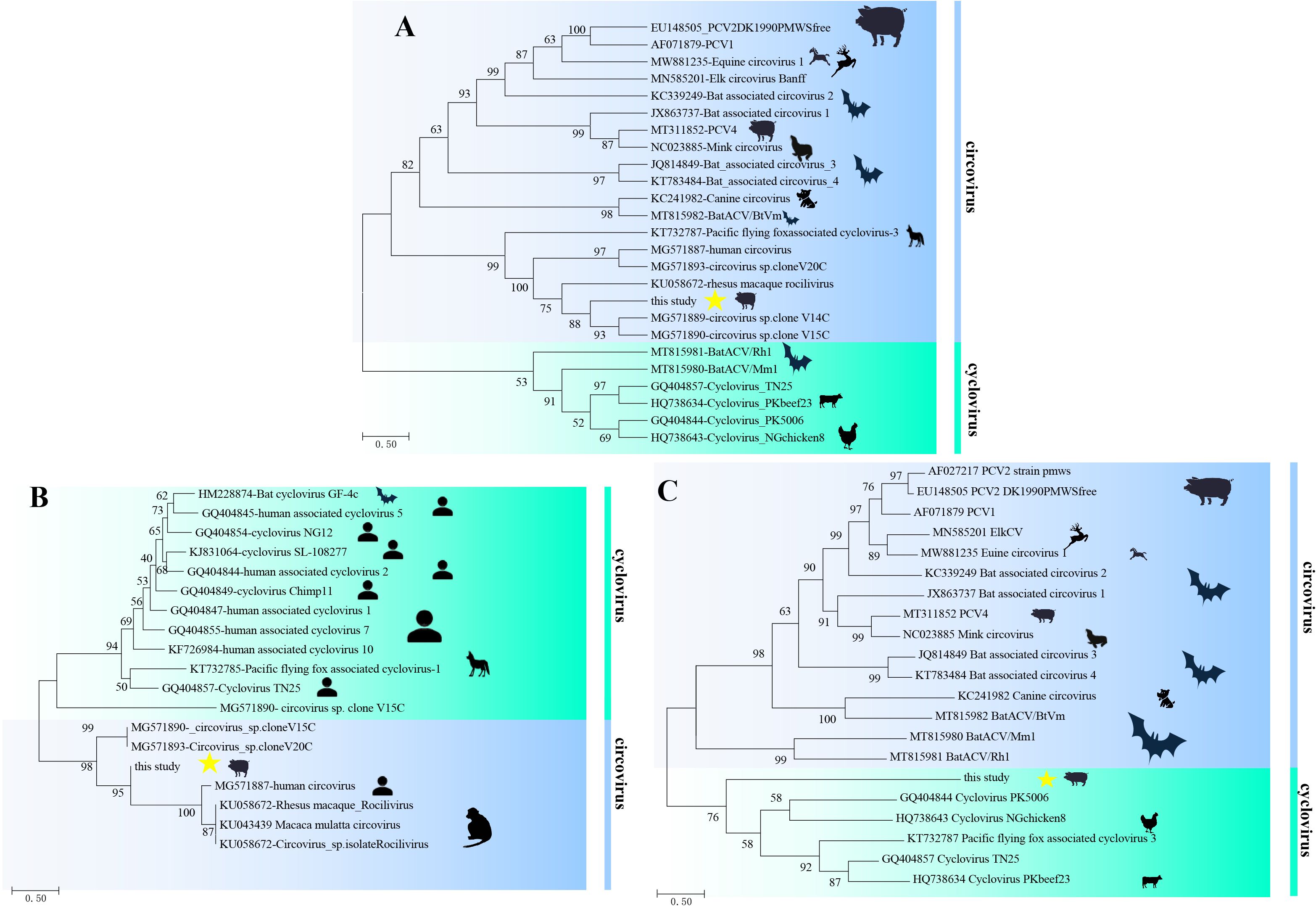
Figure 2. Phylogenetic trees of PCV-CH-ZJ01. These trees were constructed based on the complete genome nucleotide sequences (A), as well as the amino acid sequences of Rep (B) and Cap (C), using the maximum likelihood method with 1,000 bootstrap replicates.
Result of PCR screening showed that the positive rate of this novel PCV in the fecal samples was 86.2% (281/326). According to the ICTV guidelines, circoviruses classified within the same species must exhibit nucleotide similarity of the complete genome exceeding 80% and a capsid protein amino acid similarity of over 70% (Zhang et al., 2020). Based on the aforementioned data, it becomes even more evident that the virus we have identified represents a totally novel PCV.
4 Discussion
The novel porcine circovirus was characterized by a unique bipartite genomic architecture, in which dual replication-associated proteins (Rep1/Rep2) and divergent capsid proteins (Cap1/Cap2) were identified. This structural divergence was evidenced by non-collinear gene arrangements compared to canonical circoviruses, along with the complete absence of homologous sequences for Cap1 in public databases. Phylogenetic clustering was observed to differ markedly from established circovirus lineages, with distinct branching patterns being supported across whole-genome, Rep-, and Cap-based trees. The relatively high bootstrap values of the clades comprising the PCV-CH-ZJ01 strain and human or non-human primate viruses indicate that the phylogenetic analysis was credible. Notably, Rep1 was found to share 79.35% amino acid identity with a circovirus isolated from rhesus macaques, while Rep2 exhibited 98% identity to a mammalian circovirus strain (GenBank USL90207.1). These primate-anchored sequence homologies were interpreted as molecular evidence of potential cross-species transmission between swine and primates, probably facilitated by ancestral recombination events or host-jumping processes.
High molecular prevalence of the virus was detected in healthy swine populations, with a 72.3% fecal shedding rate being recorded, which suggested that PCV-CH-ZJ01 may not be a highly pathogenic strain. The genetic novelty of Caps was hypothesized to alter receptor-binding specificity, and contribute to immune evasion. Nevertheless, the conserved mammalian motifs in Rep2 and primate-linked signatures in Rep1 were considered to warrant further investigation into zoonotic spillover risks, particularly given precedents of circovirus potential cross-species transmission. The samples used for library construction in this study were collected from febrile pigs on commercial farms; nevertheless, the samples employed to investigate the prevalence of PCV-CH-ZJ01 were obtained from pig herds without obvious clinical signs. Therefore, based on the available data, it is challenging to determine whether PCV-CH-ZJ01 induces significant disease or what its potential clinical manifestations might be. In our subsequent studies, we plan to isolate this strain and conduct animal experiments to assess its pathogenicity.
Data availability statement
The datasets presented in this study can be found in online repositories. The names of the repository/repositories and accession number(s) can be found in the article/supplementary material.
Ethics statement
The animal studies were approved by Ethics Committee of Jiangsu University. The studies were conducted in accordance with the local legislation and institutional requirements. Written informed consent was obtained from the owners for the participation of their animals in this study.
Author contributions
QS: Writing – review & editing, Writing – original draft. LD: Writing – original draft. LM: Writing – review & editing, Writing – original draft. WS: Writing – review & editing, Writing – original draft. AN: Writing – review & editing, Writing – original draft. XW: Writing – original draft, Writing – review & editing. YL: Writing – review & editing, Writing – original draft. SY: Writing – review & editing, Writing – original draft. LJ: Writing – review & editing, Writing – original draft. JW: Writing – review & editing, Writing – original draft. JM: Writing – original draft, Writing – review & editing. WZ: Writing – original draft, Writing – review & editing.
Funding
The author(s) declared financial support was received for the research and/or publication of this article. This research was financially supported by the National Key Research and Development Programs of China (No. 2023YFD1801301), the Funding for Project of Qingdao Leading Talents in Entrepreneurship and Innovation (No. 202208170046) to WZ, the Major Basic Research Project of the Natural Science Foundation of the Jiangsu Higher Education Institutions (No. 22KJA320001) to QS, Changzhou Social Development Project (No. CE20225001) to JW, and Jiangsu University Medical Clinical Science and Technology Development Fund (No. JLY2021008), Guidance of Zhenjiang Science and Technology Plan (Soft Science Research) (No.YJ2024026) to JM.
Acknowledgments
The authors sincerely thank Mr. Da Tong for their assistance in sample collection.
Conflict of interest
The authors declare that the research was conducted in the absence of any commercial or financial relationships that could be construed as a potential conflict of interest.
Generative AI statement
The author(s) declare that no Generative AI was used in the creation of this manuscript.
Any alternative text (alt text) provided alongside figures in this article has been generated by Frontiers with the support of artificial intelligence and reasonable efforts have been made to ensure accuracy, including review by the authors wherever possible. If you identify any issues, please contact us.
Publisher’s note
All claims expressed in this article are solely those of the authors and do not necessarily represent those of their affiliated organizations, or those of the publisher, the editors and the reviewers. Any product that may be evaluated in this article, or claim that may be made by its manufacturer, is not guaranteed or endorsed by the publisher.
References
Bexton, S., Wiersma, L. C., Getu, S., van Run, P. R., Verjans, G. M., Schipper, D., et al. (2015). : detection of circovirus in foxes with meningoencephalitis, United Kingdom, 2009-2013. Emerging Infect. Dis. 21, 1205–1208. doi: 10.3201/eid2107.150228
Breitbart, M., Delwart, E., Rosario, K., Segales, J., Varsani, A., and Ictv Report, C. (2017). ICTV virus taxonomy profile: circoviridae. J. Gen. Virol. 98, 1997–1998. doi: 10.1099/jgv.0.000871
Chang, W. S., Li, C. X., Hall, J., Eden, J. S., Hyndman, T. H., Holmes, E. C., et al. (2020). Meta-transcriptomic discovery of a divergent circovirus and a chaphamaparvovirus in captive reptiles with proliferative respiratory syndrome. Viruses 12(10):1073. doi: 10.3390/v12101073
Cheung, A. K. (2004). Identification of an octanucleotide motif sequence essential for viral protein, DNA, and progeny virus biosynthesis at the origin of DNA replication of porcine circovirus type 2. Virology 324, 28–36. doi: 10.1016/j.virol.2004.03.037
Chiappetta, C. M., Cibulski, S. P., Lima, F. E. S., Varela, A. P. M., Amorim, D. B., Tavares, M., et al. (2017). Molecular detection of circovirus and adenovirus in feces of fur seals (Arctocephalus spp.). Ecohealth 14, 69–77. doi: 10.1007/s10393-016-1195-8
Deng, X., Naccache, S. N., Ng, T., Federman, S., Li, L., Chiu, C. Y., et al. (2015). An ensemble strategy that significantly improves de novo assembly of microbial genomes from metagenomic next-generation sequencing data. Nucleic Acids Res. 43, e46. doi: 10.1093/nar/gkv002
Edgar, R. C. (2004). MUSCLE: multiple sequence alignment with high accuracy and high throughput. Nucleic Acids Res. 32, 1792–1797. doi: 10.1093/nar/gkh340
Feher, E., Jakab, F., and Banyai, K. (2023). Mechanisms of circovirus immunosuppression and pathogenesis with a focus on porcine circovirus 2: a review. Vet. Q 43, 1–18. doi: 10.1080/01652176.2023.2234430
Fisher, M., Harrison, T. M. R., Nebroski, M., Kruczkiewicz, P., Rothenburger, J. L., Ambagala, A., et al. (2020). Discovery and comparative genomic analysis of elk circovirus (ElkCV), a novel circovirus species and the first reported from a cervid host. Sci. Rep. 10, 19548. doi: 10.1038/s41598-020-75577-6
Gao, Y. Y., Wang, Q., Li, H. W., Zhang, S., Zhao, J., Bao, D., et al. (2024). Genomic composition and pathomechanisms of porcine circoviruses: A review. Virulence 15, 2439524. doi: 10.1080/21505594.2024.2439524
Hui, A., Altan, E., Slovis, N., Fletcher, C., Deng, X., and Delwart, E. (2021). Circovirus in blood of a febrile horse with hepatitis. Viruses 13(5):944. doi: 10.3390/v13050944
Li, L., McGraw, S., Zhu, K., Leutenegger, C. M., Marks, S. L., Kubiski, S., et al. (2013). Circovirus in tissues of dogs with vasculitis and hemorrhage. Emerging Infect. Dis. 19, 534–541. doi: 10.3201/eid1904.121390
Li, H., Xiao, Y., Zhang, H., Chen, X., Lv, J., Yang, S., et al. (2021). A novel anellovirus from hospitalized neonates. Arch. Virol. 166, 2623–2625. doi: 10.1007/s00705-021-05155-2
Lorincz, M., Csagola, A., Farkas, S. L., Szekely, C., and Tuboly, T. (2011). First detection and analysis of a fish circovirus. J. Gen. Virol. 92, 1817–1821. doi: 10.1099/vir.0.031344-0
Lorincz, M., Dan, A., Lang, M., Csaba, G., Toth, A. G., Szekely, C., et al. (2012). Novel circovirus in European catfish (Silurus glanis). Arch. Virol. 157, 1173–1176. doi: 10.1007/s00705-012-1291-1
Lu, X., Zhang, Q., Wu, H. S., Ji, L. K., Wang, Y. S., Xu, J., et al. (2025). Virome characteristics of small mammals and their associated environments in pastoral areas on the Qinghai-Tibet Plateau. NPJ Biofilms Microbiomes 11(1):168. doi: 10.1038/s41522-025-00814-7
Nishizawa, T., Sugimoto, Y., Takeda, T., Kodera, Y., Hatano, Y., Takahashi, M., et al. (2018). Identification and full-genome characterization of novel circoviruses in masked palm civets (Paguma larvata). Virus Res. 258, 50–54. doi: 10.1016/j.virusres.2018.10.004
Rosario, K., Breitbart, M., Harrach, B., Segales, J., Delwart, E., Biagini, P., et al. (2017). Revisiting the taxonomy of the family Circoviridae: establishment of the genus Cyclovirus and removal of the genus Gyrovirus. Arch. Virol. 162, 1447–1463. doi: 10.1007/s00705-017-3247-y
Shen, Q., Zhuang, Z., Lu, J., Qian, L., Li, G., Kanton, A. G., et al. (2022). Genome analysis of goose-origin astroviruses causing fatal gout in shanghai, China reveals one of them belonging to a novel type is a recombinant strain. Front. Vet. Sci. 9, 878441. doi: 10.3389/fvets.2022.878441
Tischer, I., Gelderblom, H., Vettermann, W., and Koch, M. A. (1982). A very small porcine virus with circular single-stranded DNA. Nature 295, 64–66. doi: 10.1038/295064a0
Wang, Y., Sun, Y., Li, X., Chen, R., Li, W., Ji, L., et al. (2023). Molecular detection and characterization of three novel parvoviruses belonging to two different subfamilies in zoo birds. Res. Sq. 168(6):163. doi: 10.1007/s00705-023-05785-8
Keywords: circovirus, genomic structure, swine, cross-species transmission, viral metagenomics
Citation: Dai L, Mao L, Shi W, Ndjekadom A, Wang X, Liu Y, Yang S, Ji L, Wang J, Ma J, Zhang W and Shen Q (2025) A circovirus-like genomic sequence with unique architecture and phylogenetic relatedness to human-linked viral lineages. Front. Cell. Infect. Microbiol. 15:1698076. doi: 10.3389/fcimb.2025.1698076
Received: 03 September 2025; Accepted: 25 November 2025; Revised: 19 November 2025;
Published: 11 December 2025.
Edited by:
Luciane Amorim Santos, Bahiana School of Medicine and Public Health, BrazilReviewed by:
Filipe Ferreira De Almeida Rego, Gonçalo Moniz Institute (IGM), BrazilMenghang Wang, Chinese Academy of Agricultural Sciences, China
Copyright © 2025 Dai, Mao, Shi, Ndjekadom, Wang, Liu, Yang, Ji, Wang, Ma, Zhang and Shen. This is an open-access article distributed under the terms of the Creative Commons Attribution License (CC BY). The use, distribution or reproduction in other forums is permitted, provided the original author(s) and the copyright owner(s) are credited and that the original publication in this journal is cited, in accordance with accepted academic practice. No use, distribution or reproduction is permitted which does not comply with these terms.
*Correspondence: Jin Ma, NjEyMTYxMDVAcXEuY29t; Quan Shen, c2hlbnF1YW5AdWpzLmVkdS5jbg==; Wen Zhang, emhhbmd3ZW5AdWpzLmVkdS5jbg==; Jianqiang Wang, d3dqcTE5OEBzaW5hLmNvbQ==
†These authors have contributed equally to this work