ORIGINAL RESEARCH article

Front. Immunol., 08 February 2021

Sec. Primary Immunodeficiencies

Volume 11 - 2020 | https://doi.org/10.3389/fimmu.2020.619146

Clinical, Immunological, and Molecular Features of Severe Combined Immune Deficiency: A Multi-Institutional Experience From India

  • 1. Allergy Immunology Unit, Department of Pediatrics, Advanced Pediatrics Centre, Post Graduate Institute of Medical Education and Research, Chandigarh, India

  • 2. Bone Marrow Transplantation Unit, Department of Internal Medicine, Post Graduate Institute of Medical Education and Research, Chandigarh, India

  • 3. Department of Immunopathology, Post Graduate Institute of Medical Education and Research, Chandigarh, India

  • 4. Department of Immunology, Bai Jerbai Wadia Hospital for Children, Mumbai, India

  • 5. Bone Marrow Transplantation Unit, Bai Jerbai Wadia Hospital for Children, Mumbai, India

  • 6. ICMR-National Institute of Immunohematology, Mumbai, India

  • 7. Pediatric Immunology and Rheumatology, Aster CMI hospital, Bengaluru, India

  • 8. Pediatric Hemat-oncology and BMT Unit, Aster CMI Hospital, Bengaluru, India

  • 9. Anand Neuberg Diagnostic and Research Centre, Bengaluru, India

  • 10. Kanchi Kamakoti Child Trust Hospitals for Children, Chennai, India

  • 11. Institute of Child Health, Madras Medical College, Chennai, India

  • 12. Sanjay Gandhi Postgraduate Institute of Medical Sciences, Lucknow, India

  • 13. Christian Medical College, Vellore, India

  • 14. Zydus Hospitals, Ahmedabad, India

  • 15. Department of Pediatrics, Kasturba Medical College, Mangalore, India

  • 16. Apollo Children’s Hospital, Chennai, India

  • 17. Aditya Birla Memorial Hospital, Pune, India

  • 18. Department of Histopathology, Post Graduate Institute of Medical Education and Research, Chandigarh, India

  • 19. Department of Hematology, Post Graduate Institute of Medical Education and Research, Chandigarh, India

  • 20. Department of Pediatrics, National Defense Medical College, Saitama, Japan

  • 21. Department of Community Pediatrics, Perinatal and Maternal Medicine, Tokyo Medical and Dental University, Tokyo, Japan

  • 22. Department of Paediatrics and Adolescent Medicine, The University of Hong Kong, Hong Kong, Hong Kong

  • 23. Kazusa DNA Research Institute, Chiba, Japan

  • 24. Duke University Medical Center, Durham, NC, United States

Abstract

Background:

Severe Combined Immune Deficiency (SCID) is an inherited defect in lymphocyte development and function that results in life-threatening opportunistic infections in early infancy. Data on SCID from developing countries are scarce.

Objective:

To describe clinical and laboratory features of SCID diagnosed at immunology centers across India.

Methods:

A detailed case proforma in an Excel format was prepared by one of the authors (PV) and was sent to centers in India that care for patients with primary immunodeficiency diseases. We collated clinical, laboratory, and molecular details of patients with clinical profile suggestive of SCID and their outcomes. Twelve (12) centers provided necessary details which were then compiled and analyzed. Diagnosis of SCID/combined immune deficiency (CID) was based on 2018 European Society for Immunodeficiencies working definition for SCID.

Results:

We obtained data on 277 children; 254 were categorized as SCID and 23 as CID. Male-female ratio was 196:81. Median (inter-quartile range) age of onset of clinical symptoms and diagnosis was 2.5 months (1, 5) and 5 months (3.5, 8), respectively. Molecular diagnosis was obtained in 162 patients - IL2RG (36), RAG1 (26), ADA (19), RAG2 (17), JAK3 (15), DCLRE1C (13), IL7RA (9), PNP (3), RFXAP (3), CIITA (2), RFXANK (2), NHEJ1 (2), CD3E (2), CD3D (2), RFX5 (2), ZAP70 (2), STK4 (1), CORO1A (1), STIM1 (1), PRKDC (1), AK2 (1), DOCK2 (1), and SP100 (1). Only 23 children (8.3%) received hematopoietic stem cell transplantation (HSCT). Of these, 11 are doing well post-HSCT. Mortality was recorded in 210 children (75.8%).

Conclusion:

We document an exponential rise in number of cases diagnosed to have SCID over the last 10 years, probably as a result of increasing awareness and improvement in diagnostic facilities at various centers in India. We suspect that these numbers are just the tip of the iceberg. Majority of patients with SCID in India are probably not being recognized and diagnosed at present. Newborn screening for SCID is the need of the hour. Easy access to pediatric HSCT services would ensure that these patients are offered HSCT at an early age.

Introduction

Severe Combined Immune Deficiency (SCID) is an inborn error of immunity characterized by defect in T lymphocyte development and function. Children with SCID often develop life-threatening opportunistic fungal, bacterial, or viral infections in early infancy. SCID is considered a medical emergency and affected children often succumb to severe infections if diagnosis and definitive treatment are delayed. The estimated incidence of SCID is 1 in 50,000 to 100,000 live births (). Recent data also suggest an incidence of SCID as high as 1 in 3,000 live births in countries with high consanguinity rates (). However, due to lack of awareness and diagnostic facilities in developing countries, diagnosis is often missed. Hematopoietic stem cell transplantation (HSCT) is the definitive management for SCID. Early diagnosis and management are essential for successful outcomes. Several countries such as United States of America, Israel, Germany, Switzerland, Sweden, Norway, Iceland, New Zealand, and Taiwan have initiated newborn screening for SCID based on quantification of T-cell receptor excision circles (TRECs) to facilitate early diagnosis ().

Opportunistic infections in SCID are recurrent, typically start in early infancy, and result in failure to thrive. Common infection patterns seen in SCID include oral thrush, disseminated BCGosis, disseminated cytomegalovirus, and life-threatening bacterial and fungal infections. Non-infective manifestations of SCID include Omenn syndrome (OS), graft versus host reaction, autoimmunity, and hemophagocytic lymphohistiocytosis (). CD3+ T lymphocyte numbers are usually decreased in SCID (T-). However, in cases of maternal T-cell engraftment or OS, CD3+ T cell numbers can be normal or increased. The expanded T cells are autoreactive in OS, whereas, they are alloreactive in cases with transplacental-acquired maternal T-cell engraftment. T lymphocyte function and naïve T cell numbers are reduced in such cases. T- SCID can be classified based on presence or absence of B lymphocytes and natural killer cells as T-B-NK+, T-B-NK-, T-B+NK-, and T-B+NK+. Combined immunodeficiencies (CID) are also characterized by presence of opportunistic infections and immune dysregulation; however, the age of onset is little older and have a milder immunodeficiency compared to SCID ().

Until date, 58 different monogenic defects have been identified to result in immunodeficiencies affecting both cellular and humoral immunity and 18 amongst these are known to result in SCID (). Molecular defects in SCID can be broadly classified as abnormalities in VDJ recombination (RAG1, RAG2, DCLRE1C, NHEJ1, LIG4, PRKDC), abnormalities of cytokine signaling (IL2RG, JAK3, IL7RA), toxic metabolite accumulation (ADA, PNP), defective survival of hematopoietic precursors (AK2, RAC2), abnormalities of T-cell receptor and signaling (PTPRC, CD3D, CD3E, CD3Z, LAT), and abnormalities of actin cytoskeleton (CORO1A). While X-linked SCID due to defect in IL2RG is considered to be the commonest form of SCID in the US, Canada, and Europe, autosomal recessive form of SCID due to defects in RAG1/2 are the commonest forms of SCID in countries where consanguinity rates are high (–). However, after initiation of newborn screening program, defects in RAG1/RAG2 are now increasingly being identified even in countries like US and Canada where consanguinity rates are low ().

Reports of clinical data and outcomes of SCID from developing nations are scarce. Being a tropical nation with universal coverage of BCG vaccination in newborns, microbiological pattern of infections in SCID in India is expected to be different from other cohorts. Molecular spectrum is also expected to be different considering high rates of consanguinity and endogamous marriages in India (–). A recent cohort of 57 patients from Mumbai, India showed a high incidence of autosomal recessive forms of SCID with RAG1/2 defects being the commonest (). We aim to describe the clinical, immunological, and molecular features of children with SCID in this large multicentric cohort from India.

Methods

A detailed case proforma in an Excel format was prepared by one of the authors (PV) and was sent to centers that are recognized as Foundation for Primary Immunodeficiency Diseases (FPID) centers for care of primary immunodeficiencies in India. The format was also sent to tertiary-care centers that manage patients with primary immunodeficiency diseases (PIDs). Information on clinical, laboratory, and molecular details of patients with SCID and their outcomes was sought and collated. Twelve (12) centers provided details of 319 patients that were then compiled and analyzed. Fifteen (15) patients from 2 other centers with either flow-cytometry or mutation-proven SCID are not included in final analysis as data were incomplete. Twenty-three (23) children did not fulfil the criteria for clinical definition for SCID and were not included for analysis. Duplicate entries (n=4) were also noted and excluded.

Data of 277 children who had a clinical profile suggestive of SCID were taken for final analysis (Supplementary Table 1). Children were categorized as SCID/OS/CID/atypical SCID as per the European Society for Immunodeficiencies (ESID) working definition (). Three (3) patients were classified as possible SCID as they did not fulfil the complete ESID definition, however, the treating team had a high index of suspicion based on clinical and immunological features (Table 1).

Table 1

S NoAge/SexClinical featuresOrganisms isolatedAbsolute lymphocyte countImmunoglobulin profileLymphocyte subsetsMolecular defectESID Working Definition
Pt. 18 months/maleRecurrent episodes of diarrhea, failure to thrive, pneumonia, meningitisStool: Clostridium difficile toxin assay positive2.260IgG <1.64 g/L
IgA <0.36 g/L
IgM- 0.25 g/L
CD3- 0.3% (No: 6-7)
CD19- 66% (No: 1492)
CD56- 30% (No: 675)
Not doneSCID
Pt. 25 months/maleRecurrent episodes of pneumonia, diarrhea, failure to thrive, elder male sibling expired at 6 months due to severe infectionsBlood culture: Alcaligens faecalis0.410IgG <2.26 g/L
IgA <0.1 g/L
IgM<0.2 g/L
CD3- 0.15% (No: 0-1)
CD19- 0% (No: 0)
CD56- 84% (No: 345)
Not doneSCID
Pt. 36.5 months/maleRecurrent episodes of pneumonia, meningitis, hepatosplenomegaly, pancytopenia, transaminitis (HLH), 3 elder male siblings died at early infancy due to recurrent infectionsBlood culture: Pseudomonas aeruginosa
Disseminated BCGosis and angioinvasive aspergillosis in lungs in autopsy
0.940IgG<2.99 g/L
IgA- 0.49 g/L
IgM- 0.88 g/L
CD3- 0%
CD19- 86% (808)
CD56- 0.3%
IL2RGSCID
Pt. 45 months/male2 episodes of pneumonia, recurrent diarrhea, umbilical sepsis, failure to thrive, 3 elder male siblings died at early infancy due to recurrent infectionsN.A.2.050IgG- 2.64 g/L
IgA <0.46 g/L
IgM- 0.18 g/L
CD3- 0%
CD19- 96.1% (1968)
CD56- 0%
IL2RGSCID
Pt. 53 months/maleErythroderma, generalized adenopathy, diarrhea, lymphocytosis, eosinophilia (Omenn syndrome), failure to thrive, elder male sibling died due to eczema and pneumonia at 3rd monthN.A.18.540N.A.CD3- 70.94% (13,124)
CD19- 0.1%
CD56- 7% (1,295)
RAG2Omenn syndrome
Pt. 66 months/malePersistent pneumonia, oral thrush, 7 maternal uncles died at early infancy due to recurrent infectionsN.A.2.322IgG- 0.65 g/L
IgA- 0.22 g/L
IgM- 0.24 g/L
CD3- 0%
CD19- 96.75% (2,245)
CD56- 3.2% (74)
IL2RGSCID
Pt. 73 months/maleRecurrent episodes of pneumonia, diarrhea, meningitis, generalized erythroderma (incomplete Omenn), elder male sibling died at early infancy due to rash and pneumoniaN.A.1.566IgG- 2.14 g/L
IgM- 0.24 g/L
CD3- 74.79% (1167)
CD19- 0.27% (42)
CD56- 23% (360)
CD3+45RA+ 45RO-: 18.65% compared to 82% in control
Not doneOmenn syndrome
Pt. 810 months/maleRecurrent episodes of diarrhea, pneumonia, otitis media, failure to thrive, BCG site ulceration, hepatosplenomegaly, generalized adenopathy, erythroderma, eosinophilia (Omenn syndrome), 5 maternal uncles died at early infancy due to recurrent infectionsDisseminated BCGosis, disseminated Mycobacterium avium, disseminated CMV, and Aspergillus pneumonia in autopsy3.600IgG- 1.04 g/L
IgA- 0.07 g/L
IgM- 0.31 g/L
IgE- 700 U/L (Normal: 0.-6 U/L)
CD3- 95.79% (3,448)
CD19- 0.2% (7)
CD56- 1% (36)
IL2RGOmenn syndrome
Pt. 92 months/femaleRecurrent episodes of oral thrush, failure to thrive, 1 elder male sibling expired due to sepsis in early infancyN.A.0.648IgG- 2.72 g/L
IgA- 0.09 g/L
IgM- 0.41 g/L
CD3- 1.1% (7)
CD19- 0.2% (1)
CD56- 93.6% (607)
DCLRE1CSCID
Pt. 103 months/maleRecurrent episodes of pneumonia, diarrhea, rickets, nephrocalcinosis, distal renal tubular acidosis, oral thrush, failure to thriveN.A.0.896IgG- 0.88 g/L
IgA <0.06 g/L
IgM- 0.19 g/L
CD3- 1.1% (10)
CD19- 92.2% (8,26)
CD56- 6.4% (57)
IL7RASCID
Pt. 116 months/malePustulosis, hepatosplenomegaly, BCG site ulceration, transfusion-associated GVHD, elder male sibling died at 5 months due to pneumoniaDisseminated BCGosis, Blood culture: Enterobacter sp.1.462N.A.CD3- 1.25% (183)
CD19- 95% (1389)
CD56- 0.45% (6-7)
No gene variants found in IL2RG, JAK3, RAG1, RAG2SCID
Pt. 124 months/maleRecurrent episodes of pneumonia, diarrhea, failure to thrive, meningitis, oral thrush, hepatosplenomegaly, rash, eosinophilia (Omenn phenotype), one elder female sibling expired in early infancyPneumocystis jirovecii pneumonia, disseminated CMV in autopsy4.176IgG- 2.06 g/L
IgA- 0.08 g/L
IgM- 0.41 g/L
CD3- 71.6% (2,993)
CD19- 1.0% (42)
CD56- 12% (504)
CD3+45RO-45RA+: 24% as compared to 82% in control
Not doneOmenn syndrome
Pt. 136 months/malePersistent pneumonia, diarrhea, oral thrush, erythematous rash, hepatosplenomegaly (incomplete Omenn), nephrotic range proteinuria, two elder siblings (one male and other female) expired in early infancyBlood culture: Acinetobacter sp.; Pneumonia and meningitis due to Aspergillus sp. and ventriculitis due to CMV in autopsy1.404IgG- 2.46 g/L
IgA- 0.37 g/L
IgM- 1.38 g/L
CD3- 93.7% (1,312)
CD19- 0.2% (3)
CD56- 5.6% (78)
Not doneOmenn syndrome
Pt. 147 months/maleRecurrent pneumonia, failure to thrive, oral thrushEndotracheal aspirate: Klebsiella sp.; RSV pneumonia and disseminated CMV in autopsy0.156IgG- 7.86 g/L
IgA- 0.61 g/L
IgM <0.11 g/L
CD3- 45.7% (72)
CD19- 1.6% (2-3)
CD56- 21.7% (34)
PNPSCID
Pt. 156 months/maleRecurrent episodes of pneumonia, failure to thrive, 5 elder siblings died at early infancyN.A.1.391IgG <0.93 g/L
IgA <0.16 g/L
IgM <0.11 g/L
CD3- 0%
CD19- 0%
CD56- 92.2% (1,291)
RAG2SCID
Pt. 166 months/femaleRecurrent pneumonia, diarrhea, failure to thrive, hepatosplenomegalyBlood culture: Candida sp.0.785IgG- 6.33 g/L
IgA- 0.07 g/L
IgM <0.11 g/L
CD3- 0.2% (1-2)
CD19- 38.9% (312)
CD56- 52.2% (407)
IL7RASCID
Pt. 174 months/maleRecurrent pneumonia, pus discharging sinuses in neck, generalized rash (incomplete Omenn), 3 elder siblings (one female and 2 male) died in early infancyCMV PCR+, Blood culture: Enterococcus cloacae1.800IgG <0.95 g/L
IgA <0.17 g/L
IgM- 0.12 g/L
CD3- 83.3% (1,499)
CD19- 0.2% (3-4)
CD56- 14.3% (257)
CD3+45RO-45RA+: 27.5% compared to 82% in control
RAG2Omenn syndrome
Pt. 189 months/male2 episodes of pneumonia, failure to thrive, meningoencephalitis and hydrocephalus, MRI Brain: multiple tuberculomas noted over parietal and occipital area, 2 elder male siblings expired in early infancyN.A.0.612IgG <0.95 g/L
IgA <0.17 g/L
IgM <0.15 g/L
CD3- 4.2%
CD19- 0.2%
CD56- 85%
RAG1SCID
Pt. 192 months/maleRecurrent episodes of pneumonia, failure to thriveN.A.0.655IgG- 2.02 g/L
IgA <0.16 g/L
IgM <0.11 g/L
CD3- 0%
CD19- 0.13% (1)
CD56- 72% (468)
RAG1SCID
Pt. 205 months/maleGeneralized rash, alopecia, loose stools (incomplete Omenn), failure to thrive, meningitisN.A.1.372IgA <0.17 g/L
IgM <0.12 g/L
CD3- 69.6% (954)
CD19- 0.15% (2)
CD56- 10.7% (147)
DCLRE1COmenn syndrome
Pt. 215 months/maleYounger sibling of Pt. 15, recurrent episodes of pneumonia, diarrhea, failure to thriveCMV DNA PCR positive0.480IgG <0.94 g/L
IgA- 0.18 g/L
IgM <0.12 g/L
CD3- 21.7% (109)
CD19- 1% (5)
CD56- 86% (430)
RAG2SCID
Pt. 221.5 months/malePersistent pneumonia, diarrhea, elder female sibling expired at early infancyBlood culture: Candida sp.0.328IgG <2.02 g/L
IgA <0.17 g/L
CD3- 75% (248)
CD19- 8.3% (27)
CD56- 7.1% (23)
ADASCID
Pt. 235 months/maleOral thrush, pneumonia, meningitis, one elder female sibling expired due to anemia and pneumonia in early infancyDisseminated CMV and early invasive pulmonary aspergillosis in autopsy0.788IgG- 2.49 g/LCD3- 0.79% (6)
CD19- 1.02% (8)
CD56- 92.7% (744)
RAG1SCID
Pt. 242 years/maleRecurrent pneumonia, diarrhea, otitis media, failure to thrive, esophageal candidiasisN.A.8.567IgG- 5.19 g/L
IgA <0.17 g/L
IgM- 0.85 g/L
CD3- 25.76% (2,236)
CD3+CD4+- 33.5% (737)
CD3+CD8+- 50.2% (1,104)
CD19- 51.95% (4,451)
CD56- 11.6% (994)
CD3+45RA+45RO-: 31.7% compared to 74% in control
No gene variants foundCID
Pt. 254 months/maleYounger sibling of pt. 8, recurrent episodes of pneumonia and diarrhea, failure to thriveN.A.5.280IgG <2.05 g/L
IgM <0.25 g/L
CD3- 0.23% (12)
CD19- 94.6% (4,995)
CD56- 0.47% (25)
IL2RGSCID
Pt. 2610 months/maleRecurrent episodes of pneumonia, failure to thriveN.A.0.378N.A.CD3- 2.7%
CD19- 2.15%
CD56- 85.2%
DCLRE1CSCID
Pt. 272.5 months/femaleRecurrent pneumonia, otitis media, oral thrush, diarrhea, erythroderma, hepatosplenomegaly, eosinophilia (incomplete Omenn syndrome), elder female sibling expired in early infancyN.A.1.650IgG- 1.23 g/L
IgA <0.17 g/L
IgM <0.25 g/L
CD3- 7.67% (127)
CD19- 0.69% (11)
CD56- 82.7% (1,365)
CD3+45RA+45RO-: 6.42% compared to 72% of control
RAG2SCID/Omenn syndrome
Pt. 285 months/maleRecurrent episodes of pneumonia, failure to thriveBAL: Pseudomonas sp.; Blood culture: Candida sp.0.360IgG <2.05 g/L
IgA- 0.07 g/L
IgM <0.05 g/L
CD3- 2.3% (8)
CD19- 3.8% (14)
CD56- 92.2% (332)
RAG1SCID
Pt. 298 months/femalePersistent pneumonia, recurrent episodes of diarrhea, failure to thrive, chorioretinitis, hepatosplenomegalyDisseminated CMV; Blood culture: Acinetobacter baumanii1.316IgG- 4.17 g/L
IgA- 0.22 g/L
CD3- 11.3% (149)
CD19- 69.8% (921)
CD56- 1.75% (23)
JAK3SCID
Pt. 301.5 months/maleRecurrent episodes of pneumonia, diarrhea, failure to thrive, elder male sibling died in early infancyBlood culture: Acinetobacter baumanii0.204IgG- 1.96 g/L
IgM <0.25 g/L
CD3- 54% (108)
CD19- 24% (48)
CD56- 20% (40)
ADASCID
Pt. 314 years/maleRecurrent episodes of pneumonia since early infancy, failure to thriveN.A.0.116IgG- 4.73 g/L
IgA- 1.05 g/L
IgM- 1.12 g/L
CD3- 64.8% (78)
CD19- 4% (5)
CD56- 7% (8-9)
ADAAtypical SCID
Pt. 329 months/maleRecurrent episodes of pneumonia, diarrhea, failure to thriveN.A.0.154IgG- 2.27 g/L
IgA- 0.27 g/L
IgM <0.25 g/L
CD3- 44.4% (67)
CD19- 38.5% (58)
CD56- 5.7% (9)
Not doneSCID
Pt. 332 months/femaleRecurrent episodes of pneumonia, diarrhea, failure to thriveN.A.0.977IgG- 2.45 g/L
IgA- 0.23 g/L
IgM- 0.29 g/L
CD3- 32% (314)
CD19- 57% (559)
CD56- 1.2% (12)
Not doneSCID
Pt. 345 months/maleRecurrent diarrhea, failure to thrive, BCG site ulceration, pneumonia, erythroderma, eosinophilia, alopecia (Omenn syndrome)Blood culture: Enterococcus sp.; Pneumocystis jirovecii pneumonia and disseminated BCGosis in autopsy2.498IgG <2.05 g/L
IgM- 0.34 g/L
IgE- 369 kU/L (up to 7.3)
CD3- 78.01% (1,950)
CD19- 4.44% (110)
CD56- 13.12% (325)
CD3+45RA+RO-: 2.26% compared to 83.7% in control
CD3+HLA-DR+: 86.25% compared to 8.6% in control
No gene variants foundOmenn syndrome
Pt. 356 months/maleRecurrent pneumonia, failure to thrive, elder male sibling expired in early infancy due to pneumoniaN.A.0.868N.A.CD3- 3% (26)
CD19- 94% (818)
CD56- 0.4% (3)
IL2RGSCID
Pt. 361.5 months/femaleAnasarca, nephrotic range proteinuria, pneumonia, failure to thrive, erythematous rash (incomplete Omenn), elder male sibling expired in early infancyN.A.0.722IgG- 8.29 g/L
IgA- 0.75 g/L
CD3- 89%
CD3+CD4+- 8%
CD3+CD8+- 85.1%
CD19- 0.3%
CD56- 0.8%
CD3+45RA+45RO-: 30% compared to 90% in control
CD3+45RA-45RO+: 79.14% compared to 19.24% in control
CD3+HLA-DR+: 90.13% compared to 12.7% in control
ADAOmenn syndrome
Pt. 375 months/femaleRecurrent pneumonia, diarrhea, failure to thrive, BCG site ulcerationN.A.0.861IgG <2.0 g/L
IgA <0.17 g/L
CD3- 34% (292)
CD3+CD4+- 29.7% (89)
CD3+CD8+- 55.3% (165)
CD19- 45% (387)
CD56- 12.1% (103)
Not doneSCID
Pt. 385 months/maleRecurrent pneumonia, diarrhea, failure to thriveN.A.0.140N.A.CD3- 9.6% (13)
CD19- 8.7% (12)
CD56- 80% (112)
RAG1SCID
Pt. 392 months/maleRecurrent episodes of diarrhea, failure to thrive, sacral abscess, 2 elder siblings died in early infancy due to repeated infectionsBlood culture: Staphylococcus aureus; Disseminated CMV in autopsy0.06N.A.CD3- 50% (30)
CD19- 7.7% (4-5)
CD56- 34.6% (21)
ADASCID
Pt. 402.5 months/maleRecurrent pneumonia, otitis media, failure to thrive, 6 maternal uncles and 2 elder male siblings died at early infancy due to repeated infectionsN.A.1.406N.A.CD3- 0.07% (01)
CD19- 91.5% (1,598)
CD56- 1.8% (33)
Not doneSCID
Pt. 414 years/femaleEczematoid eruptions and chronic otitis media since early infancy, autoimmune hemolytic anemia, generalized adenopathyN.A.1.922IgG- 21.56 g/L
IgA- 4.77 g/L
IgM- 0.57 g/L
IgE- 933 U/L (Normal: up to 60)
CD3- 24.79% (912)
CD3+CD4+- 21.2% (193)
CD3+CD8+- 55% (500)
CD19- 42.3% (812)
CD56- 2.7% (58)
CD3+45RA+RO-: 45% compared to 76% in control
CD3+CD4+45RA+RO-: 14.9% compared to 67% in control
CD3+CD8+45RA+45RO-: 35.8% compared to 72% in control
STK4CID
Pt. 424 months/maleRecurrent pneumonia, diarrhea, failure to thrive, oral thrush, 1 maternal uncle died at 2 years due to repeated infectionsBlood culture: Moraxella sp.1.302N.A.CD3- 1.3% (17)
CD19- 85.16% (1,109)
CD56- 2.9% (37)
Not doneSCID
Pt. 432.5 months/maleChronic diarrhea, failure to thrive, esophageal candidiasis, maternal cousin (male) expired at early infancy due to pneumoniaN.A.0.415N.A.CD3- 3.8% (15)
CD19- 84% (336)
CD56- 3% (12)
IL2RGSCID
Pt. 443 months/maleRecurrent pneumonia, diarrhea, failure to thrive, erythroderma, eosinophilia, hepatosplenomegaly (maternal T cell engraftment), 1 maternal uncle died at early infancy due to pneumoniaBlood culture: Weisella confusa7.457N.A.CD3- 15.9% (1,192)
CD19- 76.4% (5,692)
CD56- 1.9% (142)
CD3+45RA+RO-: 5.43% compared to 59% in control
CD3+45RA-45RO+: 96.9% compared to 60% in control
CD3+HLA-DR+: 83.5% compared to 15.7% in control
IL2RGAtypical SCID
Pt. 454 months/maleRecurrent pneumonia, diarrhea, failure to thrive, oral thrush, BCG site ulcerationN.A.2.831IgG <0.87 g/L
IgA <0.16 g/L
CD3- 0.2% (5-6)
CD19- 97.7% (2,765)
CD56- 0.48% (13-14)
No gene variants foundSCID
Pt. 465 months/maleRecurrent fever, BCG site ulceration, hepatosplenomegaly, oral thrushDisseminated BCGosis2.086IgG <1.46 g/LCD3- 0.6% (13)
CD19- 97.8% (2,044)
CD56- 0.2% (4)
Not doneSCID
Pt. 4715 days/maleYounger sibling of pt. 31, pneumonia, recurrent diarrhea, failure to thriveBlood culture: Candida sp.0.094N.A.CD3- 42% (38)
CD19- 40% (36)
CD56- 16% (14)
ADASCID
Pt. 484 months/maleYounger sibling of pt. 27, recurrent pneumonia, diarrhea, failure to thrive, erythroderma, hepatosplenomegaly, eosinophilia (Omenn syndrome)N.A.1.896N.A.CD3- 74% (1,406)
CD19- 0.4% (8)
CD56- 22% (418)
CD3+45RA+45RO-: 16% compared to 71% in control
RAG2Omenn syndrome
Pt. 493 years/maleRecurrent sinopulmonary infections, diarrhea, failure to thrive, 1 episode of liver abscess, intra-cranial B cell lymphoma, defective T lymphocyte proliferation on stimulation with PHA.N.A.3.265IgG- 4.02 g/LCD3- 45.14% (1,467)
CD3+CD4+- 6.9% (103)
CD3+CD8+- 70.3% (1,033)
CD19- 6.83% (222)
CD56- 25.01% (816)
CD3+45RA+45RO-: 71.06% compared to 64% in control
CD3+CD4+45RA+45RO-: 3.6% compared to 72% in control
CD3+CD8+45RA+45RO-: 75.3% compared to 68% in control
CORO1AAtypical SCID
Pt. 506 months/maleRecurrent episodes of pneumonia, failure to thriveN.A.0.411IgG <0.95 g/L
IgA <0.17 g/L
IgM <0.25 g/L
CD3- 20% (80)
CD19- 73% (292)
CD56- 1.4% (5-6)
JAK3SCID
Pt. 5110 months/malePneumonia, diarrhea, failure to thrive, meningoencephalitisEndotracheal aspirate: Klebsiella pneumoniae0.810IgG <0.95 g/L
IgA <0.17 g/L
IgM <0.25 g/L
CD3- 4% (32)
CD19- 95% (760)
CD56- 1% (8)
IL2RGSCID
Pt. 523 months/maleRecurrent pneumonia, diarrhea, failure to thriveN.A.0.199N.A.CD3- 0.82% (2)
CD19- 1.17% (2-3)
CD56- 88.9% (178)
DCLRE1CSCID
Pt. 535 months/malePneumonia, failure to thrive, complicated otitis media with facial nerve palsy, transfusion-associated GVHDN.A.0.292N.A.CD3- 0.2% (0-1)
CD19- 29% (87)
CD56- 60% (180)
Not doneSCID
Pt. 543.5 years/maleSevere eczema since early infancy, pustules, otitis media, pneumonia, chest wall abscess, eosinophilia (incomplete Omenn)Pus culture- Staphylococcus aureus1.244IgG- 1.64 g/L
IgA- 1.56 g/L
IgE- 4269 kU/L (upto 32)
IgG1- 1.01 g/L
IgG2- 0.95 g/L
IgG3- 0.23 g/L
IgG4- 0.71 g/L
CD3- 60% (744)
CD3+CD4+- 17.3% (128)
CD3+CD8+- 71.5% (529)
CD19- 2.3% (28)
CD56- 15% (186)
CD3+45RA-45RO-: 36.6% compared to 65% in control
CD3+45RA-45RO+: 67% compared to 31% in control
CD3+HLA-DR+: 64.2% compared to 19.3% in control
No gene variants foundOmenn syndrome
Pt. 555 months/maleRecurrent pneumonia, diarrhea, failure to thrive, hyperferritinemia, hypofibrinogenemia, pancytopenia (HLH)ET aspirate: Klebsiella pneumoniae, Acinetobacter baumanii; PCR positivity for H1N11.547IgG- 2.32 g/L
IgA <0.2 g/L
IgM- 0.22 g/L
CD3- 1.74% (27)
CD19- 91.6% (1,426)
CD56- 5% (78)
IL2RGSCID
Pt. 566 months/femalePneumonia, failure to thrive, diarrhea, BCG site ulcerationN.A.1.098IgG- 0.54 g/L
IgA <0.2 g/L
IgM <0.17 g/L
CD3- 0% (0)
CD19- 2% (22)
CD56- 79% (869)
DCLRE1CSCID
Pt. 577 months/malePneumonia, diarrhea, failure to thrive, hepatosplenomegaly, BCG site ulcerationN.A.0.855N.A.CD3- 5.1% (43)
CD19- 77.5% (667)
CD56- 17% (146)
Not doneSCID
Pt. 5811 months/maleRecurrent pneumonia, failure to thrive, hepatosplenomegaly, generalized adenopathy, BCG site ulceration, erythematous rash (incomplete Omenn), meningitis with hydrocephalusDisseminated BCGosis, CMV DNA PCR positive, Endotracheal aspirate: Klebsiella pneumoniae1.832IgG- 7.74 g/L
IgA- 0.36 g/L
IgM- 2.42 g/L
CD3- 68% (1,244)
CD3+CD4+- 7.8% (97)
CD3+CD8+- 45.1% (558)
CD19- 5.6% (102)
CD56- 23.5% (430)
CD3+CD4+45RA-45RO+: 90.4% compared to 30.2% in control
CD3+HLA-DR+: 67.9% compared to 5.8% in control
RAG1Omenn syndrome
Pt. 595 months/maleBCG site ulceration, persistent diarrhea, generalized papular rashM. bovis0.931IgA<0.10 g/LCD3- <1%
CD19- 97% (902)
CD56- <1%
IL2RGSCID
Pt. 606 months/maleBCG site ulceration, oral thrush, septicemiaCandida sp.2.129N.ACD3- 29% (617)
CD19- 62% (1320)
CD56- 8% (170)  
No gene variants identifiedSCID
Pt.615 months/femaleBCG site ulceration, pneumonia, erythroderma, alopecia, CMV DNA PCR- positiveCMV, M. bovis1.144IgG-9.03 g/L
IgA-0.17 g/L
IgM-0.41 g/L
CD3-70.70% (806)
CD19-0.14% (2)
CD56-17.70% (202)
CD3+45RA+-12.57% compared to 86% in control
RAG2Omenn syndrome
Pt. 624 months/maleSevere pneumonia, CT chest: diffuse bilateral ground glass opacities with multifocal consolidationNil0.507IgG- <2.02 g/L
IgA- <0.17 g/L
IgM- <25 g/L
CD3- 57.23% (290)
CD19-0.05% (1)
CD56-35.08% (179)
RAG1SCID
Pt. 635 months/maleSevere pneumonia, CT chest: bilateral small random nodulesNil1.236IgG- <2.03 g/L
IgA- <0.17 g/L
IgM- <0.25 g/L
CD3- 0.28% (4)
CD19-96.20% (1193)
CD56-0.51% (6)
IL2RGSCID
Pt. 645 months/femalePersistent pneumonia- pneumothorax, oral thrushCandida sp.0.180IgG- <2.03 g/L
IgA- <0.17 g/L
IgM- 0.33 g/L
CD3-0.16% (1)
CD19-0.16% (1)
CD56-74.40% (134)
DCLRE1CSCID
Pt. 651.5 months/femaleLeft ear complicated otitis media, pneumonia, diarrheaS. aureus2.443IgG-8.04 g/L
IgA-0.75 g/L
IgM-1.38 g/L
CD3-22.87% (559)
CD19-73.60% (1776)
CD56-1.43% (34)
No gene variants identifiedSCID
Pt. 667 months/maleBCG adenitis, encephalitisM. bovis0.506IgG- <2.05 g/L
IgA- <0.17 g/L
IgM- <0.26 g/L
CD3-18.19% (93)
CD19-0.08% (1)
CD56-77.24% (394)
DCLRE1CSCID
Pt. 678 months/femaleRecurrent diarhea, failure to thrive, pneumonia, axillary adenopathyNil6.864IgG-<2.03 g/L
IgM->4 g/L
CD3-69.75% (4785)
CD3+CD4+ - 32% of CD3+ lymphocytes (1530)
CD3+CD8+ - 62% of CD3+ lymphocytes (2967)
CD3+45RA+ - 24.3% compared to 85% in healthy control
CD19-8.95% (617)
CD56-2.57% (178)
No gene variants identifiedSCID
Pt. 6810 months/malerecurrent gastroenteritis, pneumonia, DCT+ autoimmune hemolytic anemiaCMV2.200IgG-3.33 g/L
IgA- <0.17 g/L
IgM- 1.19 g/L
CD3-86.75% (1910)
CD19-0.64% (13)
CD56-6.50% (143)
CD3+CD45RA+ -38.2% compared to 79% in control 
NHEJ1Atypical SCID
Pt. 695 months/malepersistent pneumonia, absent BCG scarNil0.287IgG-3.47 g/L
IgA- 0.21 g/L
IgM-0.76 g/L
CD3-51.62% (150)
CD19-31.30% (91)
CD56-9.46% (28)
No gene variants identifiedSCID
Pt. 704 months/maleRecurrent pneumonia, diarrhoea, generalized erythematous macular rash, CMV retinitis, seizures, GVHD skin lesionsCMV4.921IgG- <1.99 g/L
IgA- <0.36 g/L
IgM- <0.25 g/L
CD3-0.54% (25)
CD19-0.54% (25)
CD56-91.74% (4512)
DCLRE1CSCID
Pt. 716 months/maleRecurrent pneumonia, otitis media, ulceration at BCG site, hepatosplenomegalyEnterococcus sp.0.816IgA-0. 56 g/LCD3-1.39% (12)
CD19-90.95% (746)
CD56-5.4% (44)
IL2RGSCID
Pt. 7211 months/maleSkin pustule and abscess, generalized erythematous macular rash, oral thrushNil1.118IgG-<0.90 g/L
IgA- <0.21 g/L
CD3-24.80% (278)
CD19-6.20% (69)
CD56-67.6% (757)
NHEJ1SCID
Pt. 733.5 months/maleRecurrent pneumonia, diarrhoea, generalized erythematous macular rashNil2.420IgG- 1.62 g/L
IgA- 0.09 g/L
IgM- 0.48 g/L
CD3-5% (121)
CD19- 47% (1137)
CD56- 42% (1016)
No gene variants identifiedSCID
Pt. 743 months/maleRecurrent pneumonia, diarrhoeaNil1.643IgG- 3.09 g/L
IgA- <0.07 g/L
CD3-87.90% (1442)
CD19-1.7% (28)
CD56-2.2% (33)
CD3+45RA+ -1.6% compared to 78% in control
ADAAtypical SCID
Pt. 753 months/maleRecurrent pneumonia, diarrhoea, failure to thriveNil2.862IgG- 2.14 g/L
IgA- <0.20 g/L
CD3-35.90% (1026)
CD19-3.11% (89)
CD56- 38% (1087)
No gene variants identifiedSCID
Pt. 7612 months/femaleRecurrent pneumonia, diarrhoea, oral thrushNil4.300IgG- 3.28 g/L
IgA- 1.46 g/L
IgM- 2.99 g/L
CD3-3.29% (142)
CD19-79.37% (3414)
CD56-9.63% (413)
No gene variants identifiedSCID
Pt. 771.5 months/femaleRecurrent pneumonia, otitis media, generalized erythematous macular rashPichia fermentans; E. coli4.720IgG- 4.27 g/L
IgA- <0.16 g/L
IgM- 0.35 g/L
CD3-49.03% (2303)
CD19-1.27% (61)
CD56-37.44% (1765)
CD3+45RA+ - 1.46% compared to 73% in control
RAG1Omenn syndrome
Pt. 786 months/maleRecurrent pneumonia, diarrhoea, generalized erythematous macular rashNil1.808IgG- <2.02 g/L
IgA- 0.20 g/L
IgM-1.71 g/L
CD3-95.65% (1732)
CD19-1.78% (32)
CD56-0.53% (9)
CD3+45RA+ - 11% compared to 86% in control
IL2RGAtypical SCID
Pt. 796.5 months/maleRecurrent pneumonia, diarrhoeaNil0.600IgG- 3.65 g/L
IgA- 0.38g/L
IgM- 0.41 g/L
CD3-29.54% (177)
CD19-41.13% (247)
CD56-18.87% (114)
No gene variants identifiedSCID
Pt. 8015 months/maleRecurrent pneumonia, diarrhoea, oral thrushKlebsiella pneumoniae, CMV7.191IgG-2.02 g/L
IgA-0.18 g/L
IgM-0.46 g/L
CD3-12.36% (892)
CD19-0.74% (53)
CD56-51.5% (3703)
CD3+45RA+ - 14.29% (decreased)
No gene variants identifiedSCID
Pt. 8142 months/femaleRecurrent pneumonia, oral thrushNil1.615IgG-21.77 g/L
IgA-1.23 g/L
IgM-1.81 g/L
CD3-37.40% (606)
CD4- 5.7%
CD8- 14.7%
CD19-22.6% (366)
CD56- 46% (743)
No gene variants identifiedSCID
Pt. 823 months/maleRecurrent pneumonia, failure to thrive, oral thrush, one elder female sibling expired due to pneumonia in early infancyNil2.492IgG-2.72 g/L
IgA-0.09 g/L
IgM-0.73 g/L
CD3-30% (747)
CD19-9.10% (227)
CD56- 41% (1021)
No gene variants identifiedSCID
Pt. 833 months/femaleRecurrent pneumonia, diarrhoea, otitis media, oral thrushNil2.608IgG-<2.02 g/L
IgA-<0.17 g/L
IgM-0.90 g/L
CD3-32.68% (853)
CD19-29.79% (783)
CD56-33.41% (872)
CD3+ 45RA+ -2.23% (decreased)
No gene variants identifiedSCID
Pt. 844 months/maleRecurrent pneumonia, diarrhoea, otitis media, ulceration at BCG siteNil0.663IgG-<2.02 g/L
IgA-<0.17 g/L
IgM-<0.25 g/L
CD3-87.66% (579)
CD19-0.05% (1)
CD56-10.22% (66)
RAG1SCID
Pt. 854 months/maleRecurrent pneumonia, severe erythroderma, developmental delayNil1.441IgG-<2.02 g/L
IgA-<0.17 g/L
CD3-0.16% (3)
CD19-94.81% (1365)
CD56-0.67% (10)
IL2RGSCID
Pt. 8615 months/maleRecurrent pneumonia, generalized eczematoid macular rash, developmental delay, myopathyNil14.84IgG-13.16 g/L
IgA-1.70 g/L
IgM<0.26 g/L
IgE- 8423 U/L
CD3-92.20% (13,683)
CD19-2.85% (416)
CD56-3.21% (475)
CD4+45RA+ - 12.17% compared to 56% in control
CD8+45RA+ - 18.6% compared to 72% in control
STIM1CID
Pt. 876 months/maleRecurrent pneumonia, extensive eczematoid rashCMV6.556IgG-13.75 g/L
IgA-0.42 g/L
IgM-1.88 g/L
IgE- 622 U/L
CD3-53.90% (3536)
CD4- 4.9% (320)
CD8- 30.2% (1968)
CD19-25.9% (1706)
CD56-5.6% (368)
No gene variants identifiedOmenn syndrome
Pt. 882.5 months/femaleRecurrent pneumonia, diarrhoea, BCG site abscessNil0.055IgG-2.33 g/L
IgA<0.17 g/L
IgM<0.25 g/L
CD3-95.58% (48)
CD19-0.07% (1)
CD56-0.79% (1)
ADASCID
Pt. 894 months/femaleRecurrent diarrhoea, otitis media, generalized erythematous macular rash, ulceration at BCG siteEnterococcus faecalis2.352IgG<2.02 g/L
IgM<0.21 g/L
CD3-68.95% (1621)
CD19-0.05% (1)
CD56-25.37% (597)
CD3+ 45RA+ -6.79% compared to 70% in control
HLA DR in CD3+ - 74.2% compared to 15% in control
RAG1Omenn syndrome
Pt. 904.5 months/maleRecurrent pneumonia,Nil0.724IgG-<0.95 g/L
IgA<0.17 g/L
CD3-2.41% (17)
CD19-0.45% (4)
CD56-90.91% (655)
DCLRE1CSCID
Pt. 914 months/maleRecurrent pneumonia, chorioretinitis, failure to thrive, 3 maternal uncles died at early infancy due to severe infectionsBlood CMV PCR positive1.760IgG < 2.7 g/L
IgA <0.4 g/L
IgM- 1.07 g/L
CD3- 1% (18)
CD19- 61% (1,098)
CD56- 1% (18)
IL2RGSCID
Pt. 924 months/maleOral thrush, pneumonia, failure to thriveN.A.0.650N.A.CD3- 0.4% (2-3)
CD19- 97% (631)
CD56- 0.4% (2-3)
IL2RGSCID
Pt. 936 months/femaleRecurrent pneumonia, CMV chorioretinitisBlood CMV PCR positive2.200IgG <2.7 g/L
IgA <0.4 g/L
IgM <0.25 g/L
CD3- 7.6% (167)
CD19- 1% (22)
CD56- 40% (880)
RAG2SCID
Pt. 944.5 months/malePersistent pneumonia, failure to thrive, elder sibling died at 6 months due to severe pneumoniaN.A.1.750IgG <1.37 g/L
IgA <0.26 g/L
IgM <0.16 g/L
CD3- 4% (70)
CD19- 47% (823)
CD56- 25% (438)
IL7RASCID
Pt. 956 months/maleRecurrent pneumonia, failure to thriveN.A.2.000IgG <1.37 g/L
IgA <0.26 g/L
IgM- 0.53 g/L
CD3- 20% (400)
CD19- 80% (1,600)
CD56- 0.1% (2)
IL2RGSCID
Pt. 963 months/femaleOral thrush, septicemiaN.A.0.720IgG- 0.8 g/L
IgA <0.26 g/L
IgM <0.18 g/L
CD3- 5% (36)
CD19- 7% (50)
CD56- 53% (382)
RAG2SCID
Pt. 975 months/maleRecurrent episodes of pneumonia, diarrhea, failure to thrive, oral thrush, BCG site ulceration, elder female sibling expired at 6 months due to recurrent infectionsN.A.0.850IgG <2.7 g/L
IgA <0.4 g/L
IgM <0.25 g/L
CD3- 5% (43)
CD19- 2.3% (20)
CD56- 46% (391)
Not doneSCID
Pt. 983 months/femaleRecurrent pneumonia, failure to thriveN.A.0.700IgG <2.7 g/L
IgA <0.4 g/L
IgM <0.25 g/L
CD3- 6.5% (46)
CD19- 3.6% (25)
CD56- 34% (238)
Not doneSCID
Pt. 993 months/femaleRecurrent pneumonia, failure to thriveN.A.0.680IgG- 0.65 g/L
IgA <0.5g/L
IgM <0.25 g/L
CD3- 2% (14)
CD19- 4% (27)
CD56- 53% (360)
DCLRE1CSCID
Pt. 1003.5 months/malePersistent pneumonia, failure to thriveN.A.4.911IgG- 0.82 g/L
IgA <0.26 g/L
IgM <0.18 g/L
CD3- 9.4% (461)
CD19- 0.4% (20)
CD56- 90% (4,410)
Not doneSCID
Pt. 1016 months/femalePersistent pneumonia, failure to thriveN.A.5.756IgG- 6.7 g/L
IgA <0.26 g/L
IgM- 0.78 g/L
CD3- 2% (116)
CD19- 41% (2,362)
CD56- 47% (2,707)
Not doneSCID
Pt. 1027.5 months/maleRecurrent pneumonia, diarrhea, failure to thriveN.A.0.750IgG- 0.76 g/L
IgA- 0.08 g/L
IgM- 0.08 g/L
CD3- 26% (195)
CD19- 67% (503)
CD56- 6% (45)
JAK3SCID
Pt. 1031 month/maleErythroderma, loss of eyelashes, eosinophilia (incomplete Omenn), failure to thrive, two siblings (one male and one female) expired in early infancy due to erythroderma, generalized lymphadenopathy, and severe infectionsN.A.3.358IgG- 5.86 g/L
IgA <0.26 g/L
IgM- 0.36 g/L
IgE >2,500 U/L
CD3- 9% (302)
CD19- 48% (1,613)
CD56- 11% (370)
No variants identifiedOmenn syndrome
Pt. 1045 months/femaleRecurrent pneumonia, otitis media, failure to thrive, oral thrush, pancytopenia, hepatosplenomegaly, seizures, encephalopathy (HLH)N.A.1.890IgG <2.7 g/L
IgA <0.4 g/L
IgM <0.25 g/L
CD3- 32% (605)
CD3+CD4+- 87% (528)
CD3+CD8+- 13% (78)
CD19- 65% (1,229)
CD56- 3% (57)
SP110SCID
Pt. 10511 months/maleChronic diarrhea, failure to thriveStool culture: Acinetobacter sp.0.691IgG- 3.0 g/L
IgA- 0.52 g/L
IgM- 0.39 g/L
CD3- 13.6% (94)
CD19- 56% (386)
CD56- 28% (193)
RAG1SCID
Pt. 1064.5 months/maleOral thrush, recurrent pneumonia, diarrhea, failure to thrive, BCG site ulceration, 3 maternal uncles died at early infancy due to repeated infectionsN.A.0.900IgG- 1.1 g/L
IgA- 0.05g/L
IgM- 0.07g/L
CD3- 5% (45)
CD19- 91% (819)
CD56- 2% (18)
IL2RGSCID
Pt. 1075 months/maleRecurrent pneumonia, oral thrush, failure to thrive, BCG site ulceration, encephalopathyN.A.1.120IgG <1.4g/L
IgA <0.17g/L
IgM <0.19g/L
CD3- 6% (66)
CD19- 92% (1,012)
CD56- 1% (11)
JAK3SCID
Pt. 1088 months/maleRecurrent pneumonia, diarrhea, failure to thriveN.A.0.380IgG <1.4g/L
IgA <0.17g/L
IgM <0.19g/L
CD3- 0%
CD19- 94% (357)
CD56- 4% (15)
IL7RSCID
Pt. 1094.5 months/malePersistent pneumonia, failure to thriveN.A.2.092IgG <1.4g/L
IgA <0.17g/L
IgM- 0.24g/L
CD3- 0.8% (17)
CD19- 98% (2,048)
CD56- 0%
Not doneSCID
Pt. 1104.5 months/malePersistent pneumonia, recurrent diarrhea, skin abscess, failure to thrive, situs inversus, one elder sibling died at early infancy due to pneumoniaN.A.0.780IgG- 1.92 g/L
IgA- 0.04 g/L
IgM- 0.02 g/L
CD3- 0.04%
CD19- 0.29%
CD56- 96%
Not doneSCID
Pt. 1115.5 months/femaleRecurrent pneumonia, failure to thrive, 3 elder male siblings died within first year of life due to severe infectionsN.A.1.425IgG- 0.42 g/L
IgA <0.03 g/L
IgM- 0.34 g/L
CD3- 1.2% (17)
CD19- 71% (1,012)
CD56- 25% (356)
CD3DSCID
Pt. 1126 months/femaleRecurrent pneumonia, failure to thriveN.A.0.336IgG <1.36 g/L
IgA <0.25 g/L
IgM <0.18 g/L
CD3- 0.2% (0-1)
CD19- 0%
CD56- 99% (335)
Not doneSCID
Pt. 1133.5 months/femaleRecurrent pneumonia, failure to thrive, one elder female sibling died at 4 months due to a probable infectionN.A.2.210IgG <1.36 g/L
IgA <0.25 g/L
IgM <0.18 g/L
CD3- 0.8% (18)
CD19- 97.4% (2,153)
CD56- 1% (22)
No variants identifiedSCID
Pt. 1148 months/maleRecurrent pneumonia, BCG site ulceration, failure to thrive, one elder male sibling died at early infancy due to pneumoniaDisseminated BCGosis1.650IgG- 0.15 g/L
IgA <0.24 g/L
IgM- 0.2 g/L
CD3- 0%
CD19- 61% (1,007)
CD56- 38% (627)
IL7RSCID
Pt. 1157 months/femalePersistent pneumonia, failure to thrive, two elder siblings (one male, one female) died in early infancy due to severe infections, one had disseminated BCGosisN.A.0.870IgG <2.0 g/L
IgA <0.3 g/L
IgM <0.2 g/L
CD3- 15% (131)
CD19- 0%
CD56- 40% (350)
DCLRE1CSCID
Pt. 1167 months/femalePersistent pneumonia, failure to thrive, autoimmune hemolytic anemiaDisseminated CMV, pulmonary aspergillosis1.700IgG- 3.2 g/L
IgA- 0.38 g/L
IgM- 0.4 g/L
CD3- 4% (68)
CD19- 0%
CD56- 72% (1,224)
RAG2SCID
Pt. 1178 months/maleRecurrent pneumonia, persistent diarrhoea, BCG site ulcerationDisseminated BCGosis2.400IgG- 2.9 g/L
IgA- 0.32 g/L
IgM- 0.24 g/L
CD3- 0%
CD19- 70% (1,680)
CD56- 24% (576)
CD3ESCID
Pt. 1185 months/maleRecurrent pneumonia, failure to thrivePneumocystis jirovecii from endotracheal aspirate3.200IgG- 3.64 g/L
IgA- 0.42 g/L
IgM- 0.38 g/L
CD3- 2% (64)
CD19- 64% (2,048)
CD56- 1% (32)
IL2RGSCID
Pt. 1193.5 months/maleRecurrent pneumonia, septicemiaNone0.064IgG- 1.49 g/L
IgA- <0.26 g/L
IgM- <0.16 g/L
CD3- 63% (27)
CD19- 2.4% (1)
CD56- 2.4%
ADASCID
Pt. 1201 month 8 days/femaleRecurrent pneumonia, cupping of ribs with blunting of lower end of scapula in radiologyNone0.160IgG- 3.54 g/L
IgA- <0.05 g/L
IgM- <0.03 g/L
CD3- 32% (51)
CD19- 8.9% (14)
CD56- 58% (93)
ADA (probable); Gene sequencing not doneSCID
Pt. 12110 months/maleRecurrent pneumonia, persistent diarrhoea, oral candidiasisAdenovirus0.582IgG- 3.54 g/L
IgA- <0.05 g/L
IgM- <0.03 g/L
CD3-11% (47.8)
CD19-68.7% (298.7)
CD56-18% (79.2)
JAK3SCID
Pt. 1227 months/femaleRecurrent pneumonia, persistent diarrhoea, septicemiaRhinovirus, Blood- Candida sp.0.952IgG- 16.55 g/L
IgA- 0.29 g/L
IgM- 1.12 g/L
CD3-4.7% (12)
CD19-0%
CD56-91% (231)
Not doneSCID
Pt. 1236 months/maleRecurrent pneumonia, persistent diarrhoeaNil0.780IgG- 3.06 g/L
IgA- 0.26 g/L
IgM- 0.30 g/L
CD3-85% (665)
CD19-3% (26)
CD56-11% (87)
ADASCID
Pt. 1246 months/maleRecurrent pneumonia, persistent diarrhoea, cellulitis, hepatosplenomegaly, panniculitisM. bovis0.370IgG- 0.19 g/L
IgA- <0.01 g/L
IgM- 0.16 g/L
CD3-4.94% (22)
CD19-84% (404)
CD56-0.09% (3)
Not doneSCID
Pt. 12536 months/maleRecurrent pneumonia, persistent diarrhoeaNil0.480IgG- 11.80 g/LCD3-33.3% (156.5)
CD19-33.4% (156.3)
CD56- 28.3% (112)
Not doneSCID
Pt. 1267 months/femaleRecurrent pneumonia, persistent diarrhoea, septicemiaBlood- Acinetobacter baumanni, Candida sp.1.090IgG- 9.80 g/L
IgA- 0.17 g/L
IgM- 0.43 g/L
CD3-0.35% (4)
CD19-82.7% (1048)
CD56- 4.56% (58)
Not doneSCID
Pt. 12711 months/maleRecurrent pneumonia, persistent diarrhoea, otitis mediaEar pus- P. aeruginosa0.824IgG- 12.40 g/LCD3-3.0% (26)
CD19-84% (682)
CD56- 3.56% (48)
Not doneSCID
Pt. 1284 months/femaleRecurrent pneumonia, persistent diarrhoea, otitis media, cellulitisBAL- Adenovirus0.160IgG- 7.30 g/L
IgA- 0.34 g/L
IgM- 0.92 g/L
CD3-0%
CD19-30% (75)
CD56- 42% (103)
Not doneSCID
Pt. 1298 months/maleRecurrent pneumonia, persistent diarrhoea, otitis media, septicemiaBlood- S. aureus, P. aeruginosa0.340IgG- 3.30 g/L
IgA- 0.24 g/L
IgM- 0.17 g/L
CD3-2.0% (6)
CD19-93% (310)
CD56- 4% (18)
JAK3SCID
Pt. 13022 months/maleRecurrent pneumonia, persistent diarrhoea, septicemiaBlood- Streptococcus pneumoniae0.357IgG- 18.80 g/L
IgA- 1.62 g/L
IgM- 0.85 g/L
CD3-5% (18)
CD19-27% (97)
CD56- 72% (222)
Not doneSCID
Pt. 13160 months/femaleRecurrent pneumonia, persistent diarrhoea, septicemia, microcephalyNil0.760IgG- 13.80g/L
IgA- 0.34 g/L
IgM- 1.20 g/L
CD3-4.0% (24)
CD19-92.0% (696)
CD56- 3% (18)
Not doneSCID
Pt. 1324 months/maleRecurrent pneumonia, persistent diarrhoeaNil1.378IgG- 9.60 g/L
IgA- 0.22 g/L
IgM- 0.55 g/L
CD3-4.0% (44)
CD19-91% (986)
CD56- 5.3% (58)
Not doneSCID
Pt. 13330 months/maleRecurrent pneumonia, persistent diarrhoea, otitis media, septicemiaCMV0.357IgG- 10.70 g/L
IgA- 0.30 g/L
IgM- 0.79 g/L
CD3-5.0% (17.5)
CD19-12% (93)
CD56- 34.6% (124)
Not doneSCID
Pt. 1348 months/maleRecurrent pneumonia, otitis media, septicemiaNil3.485IgG- 6.80 g/L
IgA- 0.31 g/L
IgM- 0.43 g/L
CD3-1.0% (6)
CD19-82% (2830)
CD56- 18% (654)
Not doneSCID
Pt. 1355 months/maleRecurrent pneumonia, persistent diarrhoea, septicemiaCandida sp.3.240IgG- 2.80 g/L
IgA- 0.18 g/L
IgM- 0.26 g/L
CD3-12% (388)
CD19-0%
CD56- 86% (2786)
Not doneSCID
Pt. 1366 month/maleTwo elder male sibling death at early infancyNil0.300N.A.CD3-0.7% (1)
CD19-97.6% (290)
CD56-0.4% (1)
IL2RGSCID
Pt. 1376 months/maleRecurrent pneumonia, septicemia, eczematoid rashCandida sp.2.436IgG- 2.70 g/L
IgA- 0.35 g/L
IgM- 0.36 g/L
IgE- 24,200 U/L
CD3-66% (1610)
CD19-26% (634)
CD56-8% (195)
CD3+45RO+ - 97.5% (elevated)
CD3DOmenn syndrome
Pt. 1382 months/femaleRecurrent pneumonia, cellulitis, OS, abscessCandida sp.32.600IgG- <0.33 g/L
IgA- <0.06 g/L
IgM- <0.04 g/L
CD3-87% (28,362)
CD19-0%
CD56-7.6% (2478)
Not doneOmenn syndrome
Pt. 1391 months/femaleCellulitis, rashN.A1.230IgG- 9.40 g/L
IgA- <0.25 g/L
IgM- N.A
CD3-1% (12)
CD19-N.A.
CD56-N.A.
Not doneSCID
Pt. 14036 months/maleRecurrent pneumonia, persistent diarrhoeaClostridium difficle, CMV3.024IgG- 12.90 g/L
IgA- 1.53 g/L
IgM- 0.56 g/L
CD3-56% (1680)
CD19-1.4% (42)
CD56-26% (780)
RAG1Atypical SCID
Pt. 1416 months/femaleRecurrent pneumonia, septicemiaCandida sp., Staphylococcus sp.2.405IgG- <0.75 g/L
IgA- 0.24 g/L
IgM- N.A
CD3-0.6% (14)
CD19-62.4% (1504)
CD56-22.9% (552)
Not doneSCID
Pt. 1428 months/maleRecurrent pneumonia, persistent diarrhoea, septicemiacandida2.075IgG- 0.09 g/L
IgA- <0.26 g/L
IgM- <0.16 g/L
CD3-1% (21)
CD19-93% (1934)
CD56-0.2% (4)
Not doneSCID
Pt. 1438 months/maleRecurrent pneumoniaNil2.650IgG- 7.62 g/L
IgA- 0.25 g/L
IgM- 0.64 g/L
CD3-18.36% (488)
CD19-5% (133)
CD56- N.A.
Not doneSCID
Pt. 1447 months/femaleRecurrent pneumonia, persistent diarrhoea, BCG site ulcerationNil1.090IgG- 0.10 g/L
IgA- 0.02 g/L
IgM- N.A
CD3- 1.1% (12)
CD19- 0%
CD56- 22% (231)
Not doneSCID
Pt. 1455 months/maleRecurrent pneumonia,Nil0.060IgG- 1.08 g/L
IgA- 0.10 g/L
IgM- 0.14 g/L
CD3-0.01% (1)
CD19-NA (151)
CD56- 62.33% (206)
Not doneSCID
Pt. 1467 months/maleRecurrent pneumonia,Nil4.200N.ACD3-18.31% (838)
CD19-51.69% (2682)
CD56- 15.9% (826)
Not doneSCID
Pt. 1479 months/femaleRecurrent pneumonia, Septicemia, disseminated BCGosisE. coli, M. bovis0.994IgG- 0.06 g/L
IgA- 0.26 g/L
IgM- 0.30 g/L
CD3-1.53% (10)
CD19-84.69% (692)
CD56- 2.76% (23)
Not doneSCID
Pt. 14816 months/maleRecurrent pneumonia, SepticemiaCandida sp.1.316IgG- 12.20 g/L
IgA- 1.08 g/L
IgM- 6.54 g/L
CD3-0.54% (2)
CD19-0.64% (3)
CD56- 10.93% (46)
Not doneSCID
Pt. 1495 months/maleRecurrent pneumonia, persistent diarrhea, BCG site ulcerationNil1.5IgG- 0.02 g/L
IgA- 0.37 g/L
IgM- 0.21 g/L
CD3- 0.1% (2)
CD19- 0%
CD56- 95% (1425)
Not doneSCID
Pt. 1507 months/maleRecurrent pneumonia,BAL – M. tuberculosis, Pseudomonas sp.3.000IgG- <2.0 g/L
IgA- 0.10 g/L
IgM- 0.90 g/L
CD3-0.20% (6)
CD19-70% (2100)
CD56- 36% (1080)
CD3ESCID
Pt. 1516 months/maleRecurrent pneumonia, oral thrushEnterococcal sepsis0.6IgG- 1.24 g/L
IgA- <0.01 g/L
IgM- <0.01 g/L
CD3- 47.2% (283)
CD19- 0.1% (1)
CD56- 46% (276)
RAG1SCID
Pt. 1525 months/maleRecurrent pneumonia, persistent diarrhoeaNil1.099IgG- <0.75 g/L
IgA- <0.10 g/L
IgM- 0.35 g/L
CD3-0%
CD19-93% (1015)
CD56- 2% (23)
IL2RGSCID
Pt. 1532 months/maleRecurrent pneumonia, septicemiaCandida sp. (blood)0.080IgG- 1.90 g/L
IgA- <0.05 g/L
IgM- <0.05 g/L
CD3-10.4% (2.4)
CD19-5.6 (1.29)
CD56- 64% (14.84)
ADASCID
Pt. 1543 months/maleAcute fever, coughNil0.323IgG- <1.46 g/L
IgA- <0.24 g/L
IgM- 0.97 g/L
CD3-26% (84)
CD19-65% (202)
CD56-40% (129)
ADASCID
Pt. 1557 months/malePersistent diarrhoeaNil2.538IgG-1.59 g/L
IgA- <0.24 g/L
IgM- <0.17 g/L
CD3-0% (0)
CD19-86% (2183)
CD56%-11% (279)
IL7RSCID
Pt. 15621 months/maleMeningoencephalitis, right chorioretinitis, left vitreal hemorrhageCMV0.508N.A.CD3-5% (25)
CD19-12% (61)
CD56-62% (315)
PNPSCID
Pt. 1575 months/malePneumoniaCitrobacter sp.1.520IgG- <1.34 g/L
IgA- <0.28 g/L
IgM- 0.25 g/L
CD3-0% (0)
CD19-0% (0)
CD56-96% (146)
RAG1SCID
Pt. 15824 months/maleRecurrent pneumonia, diarrhoea, meningoencephalitisE. coli16.624N.A.CD3-85% (14130)
CD4- 7% (1164)
CD8- 70% (11637)
CD19-10% (1662)
CD56- 4% (665)
HLA-DR expression on B cells- 0%
RFXANKCID
Pt. 1595 months/malePneumonia, diarrhoea, rashS. epidermidis0.320N.A.CD3-1% (1)
CD19-32% (102)
CD56-14% (45)
ADASCID
Pt. 1605 months, femalePneumoniaP. jirovecii, H1N11.967N.A.CD3-2% (39)
CD19-0% (0)
CD56-95% (1869)
Not doneSCID
Pt. 1613 months, femalePneumonia, diarrhoeaNil0.203N.A.CD3-0% (0)
CD19-0% (0)
CD56- 82% (166)
Not doneSCID
Pt. 16212 months/maleRecurrent diarrhoea, left empyemaNil1.958IgG- 15.7 g/L
IgA- 3.94 g/L
IgM- 2.13 g/L
CD3-43% (842)
CD4- 2% (39)
CD8- 30% (387)
CD19-15% (294)
CD56-38% (744)
Not doneSCID
Pt. 1637 months, malePneumonia, global developmental delayM. tuberculosis0.979NACD3-7% (69)
CD19-86% (842)
CD56-3% (29)
Not doneSCID
Pt. 1642 months, maleScaly erythrodermic rash (OS)Nil3.854IgG-1.89 g/L
IgA-0.28 g/L
IgM-2.08 g/L
CD3-55% (2120)
CD4- 33% (1272)
CD8- 11% (424)
CD4+ 45RA+ - 3% (decreased)
CD19-18% (694)
CD56-25% (964)
Not doneOmenn syndrome
Pt. 16512 months, maleAbscesses in lung, liver, oral thrushNil0.548IgG-5.63 g/L
IgA- <0.70 g/L
IgM- <1.07 g/L
CD3-42% (230)
CD19-18% (694)
CD56-20% (110)
Not doneSCID
Pt. 1662 months, maleRecurrent pneumonia, rashAcinetobacter sp.1.620N.A.CD3-68% (1102)
CD4- 12% (194)
CD8- 36% (583)
CD4+ 45RA+ - 0%
CD19-21% (340)
CD56-6% (97)
Not doneOmenn syndrome
Pt. 16710 months, maleChronic fever, pneumonia, hepatomegaly, pancytopeniaNil0.954IgG- <1.34 g/L
IgA- <0.28 g/L
IgM- <0.17 g/L
CD3-24% (229)
CD19-63% (601)
CD56-7% (67)
Not doneSCID
Pt.16812 months, maleN.A.N.A.3.080IgG- <0.29 g/L
IgA- 0.64 g/L
IgM- 0.34 g/L
CD3- 4.5% (138)
CD19- 71.3% (2197)
CD56- 1.9% (60)
IL2RGSCID
Pt.16960 months, maleNANA0.870IgG- <0.10 g/L
IgA- <0.001 g/L
IgM- <0.01 g/L
CD3- 0.5% (6)
CD19- 89.7% (1076)
CD56- 7.8% (94)
IL2RGSCID
Pt.1707 months, maleNANA0.330IgG- 0.06 g/L
IgA- 0.001 g/L
IgM- 0.002 g/L
CD3- 7% (23)
CD19- 0.3% (1)
CD56- 80.9% (267)
RAG1SCID
Pt.17148 months, maleNANA1.160IgG-0.59 g/L
IgA-0.005 g/L
IgM-0.007 g/L
CD3- 31.6% (367)
CD19- 35.3% (410)
CD56- 31.4% (364)
RAG1SCID
Pt.17212 months, femaleNANA7.220IgG-2.36 g/L
IgA-0.002 g/L
IgM-0.01 g/L
CD3- 0.3% (22)
CD19- 0.7% (54)
CD56- 56% (4044)
RAG2SCID
Pt.17348 months, maleOtitis media, recurrent pneumonia since early infancyNA1.800IgG-1.16 g/L
IgA-0.008 g/L
IgM-0.006 g/L
CD3- 9.4% (169)
CD19- 58.3% (1049)
CD56- 22.5% (406)
DOCK2CID
Pt.1744 months, femaleChronic diarrhoea, pneumonia, failure to thrive, absent thymusE. coli, Cryptosporidium1.63IgG- 5.35 g/L
IgA- 0.31 g/L
IgM- 1.82 g/L
CD3- 89.7% (1462)
CD19- 1.2% (19)
CD56- 4% (65)
Not donePossible SCID***
Pt.17530 months, maleRecurrent pneumonia, diarrhoea, failure to thriveNil0.35IgG-7.02 g/L
IgA-1.31 g/L
IgM- 0.82 g/L
CD3- 56.3% (197)
CD19- 0.8% (3)
CD56- 39.4% (138)
Not doneSCID
Pt.1769 months, maleChronic diarrhoea, pneumonia, failure to thriveP. aeruginosa, Candida sp.0.60IgG- 0.99 g/L
IgA-0.7 g/L
IgM-0.4 g/L
CD3- 35.8% (215)
CD19- 6.7% (40)
CD56- 47.8% (287)
Not doneSCID
Pt.17714 months, femaleRecurrent pneumonia, diarrhoea, failure to thriveNil2.63IgG-1.46 g/L
IgA- <0.25 g/L
IgM- <0.18 g/L
CD3- 74% (1947)
CD4- 14% (368)
CD8- 34% (895)
CD19- 2% (53)
CD56- 23% (605)
Not doneCID
Pt.1783 months, maleRecurrent pneumonia, fungal skin infection, 2 early sibling deathP. aeruginosa, Streptococcus sp.2.12IgG-2.52 g/L
IgA- <0.25 g/L
IgM- 1.12 g/L
CD3- 57.3% (1215)
CD4- 0.5% (11)
CD8- 56.3% (1194)
CD19- 38.2% (810)
CD56- 1% (21)
Not doneSCID
Pt.1797 months, malePneumonia, scaly erythrodermic rashNil0.42IgG- 0.18 g/L
IgA-0.52 g/L
IgM-0.42 g/L
CD3- 83.3% (350)
CD19- 1% (4)
CD56- 1% (4)
Not doneOmenn syndrome
Pt.18014 months, maleRecurrent pneumonia, eczematoid rash, failure to thriveCryptosporidium2.19IgG- 2.16 g/L
IgA-1.21 g/L
IgM-1.22 g/L
CD3- 41% (897)
CD4- 4% (88)
CD8- 17% (372)
CD19- 1% (22)
CD56- 17% (372)
Not doneCID
Pt.1816 months, maleChronic diarrhoea, failure to thrive, septicemiaE. coli, Candida sp.1.37IgG-9.52 g/L
IgA-1.79 g/L
IgM-0.26 g/L
CD3- 87.1% (1194)
CD4- 34% (466)
CD8- 53.1% (727)
CD19- 0.2% (3)
CD56- 12% (165)
Not doneSCID
Pt.1823 months, maleMeningitis, pneumonia, oral thrush, early sibling deathP. aeruginosa3.55N.A.CD3- 35% (1244)
CD4- 10% (355)
CD8- 15% (533)
CD19- 0.1% (4)
CD56- 56% (1990)
Not doneSCID
Pt.18316 months, malePneumonia, eczematoid rash, Varicella infection, early sibling death due to pneumoniaAcinetobacter sp., Pseudomonas sp.2.55N.A.CD3- 46% (1174)
CD4- 14% (357)
CD8- 33% (842)
CD19- 4% (102)
CD56- 50% (1276)
Not doneCID
Pt.1843 months, femalePneumonia, abdominal distension, diarrhoea, failure to thriveNil1.80IgG-1.63 g/
IgA- <0.06 g/L
IgM- <0.16 g/L
CD3- 35% (630)
CD19- 0.8% (14)
CD56- 63% (1134)
Not doneSCID
Pt.1853 months, malePneumonia, failure to thriveM. tuberculosis0.50IgG-9.03 g/L
IgA- 0.39 g/L
IgM-2.23 g/L
CD3- 45% (315)
CD19- 50% (350)
CD56- 1.4% (10)
Not doneSCID
Pt.1869 months, malePersistent diarrhoea, pneumonia, left forearm abscessNil2.43IgG-4.0 g/L
IgA-0.74 g/L
IgM- 1.1 g/L
CD3- 43.9% (1068)
CD3+CD4+- 26% (631)
CD3+CD8+- 14% (340)
CD19- 54.9% (1335)
CD56- 1% (24)
Not donePossible SCID***
Pt.1874 months, maleDevelopmental delay, pneumonia, diarrhoea, failure to thrive, 1 early sibling deathE. coli1.50IgG-2.95 g/L
IgA-0.07 g/L
IgM- 1.04 g/L
CD3- 44.9% (674)
CD19- 44.9% (674)
CD56- 10% (150)
Not donePossible SCID***
Pt.1883 months, femaleOtitis media, oral thrush, failure to thriveNil1.86IgG-2.95 g/L
IgA-0.07 g/L
IgM- 1.04 g/L
CD3- 9% (167)
CD19- 0.5% (9)
CD56- 87.8% (1633)
Not doneSCID
Pt.18996 months, femaleRecurrent pneumonia, ear discharge, failure to thriveNil1.21IgG-2.14 g/L
IgA- 7.05 g/L
IgM-1.54 g/L
CD3- 39% (473)
CD19- 16% (194)
CD56- 41.2% (498)
Not doneCID
Pt.19024 months, femaleEar discharge, diarrhoea, scaly rash (Omenn phenotype)Nil8.75N.A.CD3- 80% (7003)
CD4- 5% (438)
CD8- 30% (2626)
CD19- 2% (175)
CD56- 14% (1226)
Not doneCID
Pt.1911 month, femaleSepticemia, 3 early siblings died at early infancyNil2.89N.A.CD3- 63.9% (1847)
CD4- 55.9% (1616)
CD8- 8% (231)
CD19- 18% (520)
CD56- 15% (433)
Not doneSCID
Pt.1926 months, maleMultiple hypodense lesions in liver and spleen, necrotic retroperitoneal lymph nodesNil0.01N.A.CD- 0
CD19- 0
CD56- 0
Not doneSCID
Pt.1937 months, maleRecurrent pneumonia, diarrhoea, early sibling death due to disseminated BCGosisAcid-fast bacilli, Candida sp. (BAL)2.15N.A.CD3- 0%
CD19- 98.9% (2128)
CD56- 0.3% (6)
Not doneSCID
Pt.1942.5 months, maleDiarrhoea, ear discharge, pneumonia, dermatitis, knee joint swelling, axilla abscess, 1 elder sibling expired due to SCIDBlood, pus: S. aureus (Methicillin sensitive)2.85N.A.CD3- 57% (1624)
CD4- 17% (484)
CD8- 31% (883)
CD19- 0.3% (9)
CD56- 40% (1140)
Not doneSCID
Pt.195NA, malePneumonia, otitis media, septicemiaPseudomonas sp.1.51IgG-4.0 g/L
IgA-0.52 g/L
IgM-0.32 g/L
CD3- 0.3% (5)
CD19- 0
CD56- 4% (62)
Not doneSCID
Pt.1963 months, malePneumonia, oral thrushNil0.01N.A.N.A.ADASCID
Pt.1973 months, maleRecurrent pneumonia, 1 early sibling deathNil2.86N.A.N.A.IL2RGSCID
Pt.1982 months, femalePneumonia, colitisNil4.40IgG-0.89 g/L
IgA- <0.24 g/L
IgM- <0.17 g/L
CD3- 0.5% (22)
CD19- 87.3% (3839)
CD56- 2% (88)
JAK3SCID
Pt.1991.5 months, femalePneumonia, oral thrush, 2 elder female siblings died at early infancyNil1.50N.A.CD3- 0
CD19- 63.1% (947)
CD56- 34.7% (521)
Not doneSCID
Pt.2008 months, maleRecurrent pneumonia, BCGosisNilN.A.N.A.CD3- 0%
CD19- 64% (443)
CD56- 31% (214)
IL7RASCID
Pt.2018 months, femaleRecurrent pneumonia, oral thrush, BCGosisNil0.55IgG- <0.06 g/L
IgA- <0.24 g/L
IgM- <0.17 g/L
CD3- 0
CD19- 16.7% (92)
CD56- 65.3% (359)
Not doneSCID
Pt.2027 months, femaleRecurrent pneumonia, diarrhoeaCMV viremia, Candida sp.2.48IgG-0.97 g/L
IgA-1.82 g/L
CD3- 70.8% (1755)
CD4- 2.6% (65)
CD8- 59% (1463)
CD19- 30.2% (748)
CD56- 22.3% (553)
Not doneSCID
Pt.20324 months, maleRecurrent pneumonia, otitis mediaS. aureus (Methicillin resistant)1.60IgG-1.61 g/L
IgA-0.29 g/L
IgM-0.29 g/L
CD3- 22% (352)
CD19- 58% (928)
CD56- 13% (208)
Not doneSCID
Pt.20424 months, femaleChronic diarrhoea, pneumoniaCorona virus 229E, Alpha hemolytic streptococci (blood), esophageal candidiasis1.16IgG-1.14 g/L
IgA-0.15 g/L
IgM-0.27 g/L
CD3- 23.4% (272)
CD19- 9.1% (105)
CD56- 42.3% (491)
RAG1SCID
Pt.205192 months, maleRecurrent pneumonia, varicella infection, madarosis, Hodgkin lymphomaEpstein Barr viremiaN.A.N.A.N.A.RAG1Atypical SCID
Pt.2065 months, maleRecurrent pneumonia, diarrhoea, elder male sibling died in early infancy, 4 maternal uncles expired < 6 months ageAdenovirusN.A.N.A.NA; CD132 expression very low in monocytes (0.2%) compared to normal expression in controlsNot doneSCID
Pt.2075 months, malePneumonia, diarrhoea, ear discharge, oral thrush, rash, early sibling deathVAPP in stool, Enterovirus, Klebsiella (BAL), CSF- Enterovirus, Mycoplasma0.39IgG- <1.46 g/L
IgA- <0.28 g/L
IgM- 0.17 g/L
CD3- 28% (109)
CD19- 1% (4)
CD56- 68% (265)
RAG2SCID
Pt.20820 days, malePneumonia, diarrhoea, rash, renal abscessCorona OC43, Rhinovirus0.25N.A.CD3- 19% (47)
CD19- 0
CD56- 24.4% (61)
ADASCID
Pt.2095 months, femaleChest wall abscess, recurrent pneumonia, oral thrush, diarrhoeaP. jirovecii, Rotavirus (stool), Mycoplasma (nasopharyngeal aspirate)0.97IgG- <1.46 g/L
IgA- <0.17 g/L
IgM- <0.28 g/L
CD3- 1.3% (13)
CD19- 0
CD56- 60% (581)
RAG2SCID
Pt.2106 months, maleRecurrent pneumonia, diarrhoea, scalp abscess, 1 male sibling deathCMV, Rhinovirus, EnterovirusN.A.IgG-0.26 g/L
IgA-0.02 g/L
IgM- 1.70 g/L
N.A.CIITACID
Pt.21184 months, femaleRecurrent diarrhoea, oral ulcer, pneumonia, colitisNil0.84IgG-4.97 g/L
IgA- <0.67 g/L
IgM-1.7 g/L
CD3- 77% (649)
CD19- 15.5% (130)
CD56- 3% (28)
HLA-DR expression in B cells- 0%
RFX5CID
Pt.21218 months, maleRecurrent pneumonia, diarrhoea, failure to thriveVDPV, M. tuberculosis, Cryptosporidium, Enterobacter sp. (blood)3.75IgG- <1.41 g/L
IgA- <0.24 g/L
IgM-0.20 g/L
CD3- 53.04% (1989)
CD4- 22% (826)
CD19- 4% (150)
CD56- 42% (1576)
Not doneCID
Pt.213132 months, femaleRecurrent pneumonia, diarrhoea, oral thrush, otitis media, meningitisHemophilus influenzae (CSF)2.94IgG-0.22 g/L
IgA- <0.24 g/L
IgM-0.44 g/L
CD3- 34.7% (1022)
CD4- 16.7% (490)
CD8- 13.7% (405)
CD19- 34% (1001)
CD56- 2.2% (64)
Not doneCID
Pt.2144 months, maleFailure to thrive, recurrent pneumonia, diarrhoeaNil1.22NANAJAK3SCID
Pt.2157 months, maleOtitis media, septicemiaStaphylococcus aureus6.23IgG- <0.3 g/L
IgA- <0.05 g/L
IgM- 0.11 g/L
NAIL2RGSCID
Pt.2168 months, malePneumonia, diarrhoea, rashNil5.02IgG- <0.11 g/L
IgA- <0.05 g/L
IgM- <0.11 g/L
NAIL2RGSCID
Pt.2171 month, maleFailure to thrive, persistent diarrhea, perianal rashNil0.97IgG- 0.42 g/L
IgA- 0.06 g/L
IgM- 0.59 g/L
CD3- 4% (39)
CD19- 39% (378)
CD56- 54% (524)
Not doneSCID
Pt.2182 months, femaleRecurrent episodes of pneumonia and diarrhoea, failure to thrive, doing well after HSCTNilNANACD3- 3476
(Very low CD4 counts with CD4/CD8 reversal)
CD19- 1765
CD56- 156
Probable MHC Class 2 defectCID
Pt.2191 month, femaleRecurrent episodes of pneumonia and diarrhoeaNilNANANAIL7RSCID
Pt.2201 month, maleRecurrent episodes of diarrhoea and failure to thriveNilNANANAIL2RGSCID

Clinical and immunological features of children with clinical features suggestive of SCID in our cohort.

ESID, European Society for Immunodeficiencies; CMV, Cytomegalovirus; BCG, Bacillus Calmette-Guerin; BAL, Bronchoalveolar lavage; CSF, Cerebrospinal fluid; OS, Omenn syndrome; PJP, Pneumocystis jirovecii pneumonia; EBV, Epstein-Barr virus; VDPV, Vaccine-derived polio virus; VZV, Varicella zoster virus; AIHA, Autoimmune hemolytic anemia; VAPP, Vaccine-associated paralytic polio; CID, Combined Immune Deficiency.

Clinical details of patients 221-277 are previously reported ().

***Possible SCID is categorized if patients did not fulfil the complete ESID definition, however, the treating team had a high index of suspicion based on clinical and immunological features.

Clinical profile of all patients was obtained along with family history and other demographic details. Clinical features included number of infections, type of infections, site of infections, organism involved, age of presentation, age of onset, presence of skin rash, BCG ulceration, history of administration of vaccines and complications, if any. Basic hematological, biochemistry, and immunological investigations including immunoglobulin profile and lymphocyte subsets were also recorded.

Analysis of lymphocyte subsets by flow cytometry had been carried out in most patients. Methodology for laboratory assay of lymphocyte subsets, naïve, memory T cells, HLA-DR expression, CD132 expression, CD127 expression, and lymphocyte proliferation assays at Post Graduate Institute of Medical Education and Research (PGIMER), Chandigarh and National Institute of Immunohematology (NIIH), Mumbai have been previously described (, ). Other centers performed conventional lymphocyte subsets (CD3, CD19, CD4, CD8, CD56) by flow cytometry in private laboratories.

Adenosine deaminase (ADA) levels and percentage of deoxyadenosine nucleotides (%dAXP) from dried blood filter paper spot were assayed at Duke University, North Carolina for patients with ADA deficiency SCID who were diagnosed at PGIMER, Chandigarh.

Molecular Assays

Before the facility for in-house next-generation sequencing was made available in 2018, centre at PGIMER, Chandigarh had established academic collaborations with centers at Hong Kong (The University of Hong Kong), Japan (Kazusa DNA Research Institute, Kisarazu, Chiba; National Defense Medical College, Saitama), and USA (Duke University, North Carolina) for molecular work-up of patients. The centre at Hong Kong provided final molecular diagnosis for 12 patients (Pt. 8-10, Pt. 14-19, Pt. 21, Pt. 50-51) (Table 1). Molecular diagnosis for 4 patients was established at Kazusa DNA Research Institute, Japan (Pt. 3–6). Thirty-four (34) patients (Pt. 59–90, pt. 119, pt. 127) with SCID were worked-up for molecular diagnosis using NGS at National Defense Medical College, Saitama and Tokyo Medical and Dental University, Tokyo, Japan (Kato T et al. manuscript in submission). Final molecular diagnosis of a patient with ADA defect (pt. 22) was also established at Duke University, North Carolina.

Sanger sequencing for IL2RG and RAG1/2 genes were initiated at PGIMER, Chandigarh (North India) in 2016. Sanger sequencing for patients with SCID at NIIH, Mumbai (West India) was previously described by Aluri et al. (). Methodology for NGS at Christian Medical College, Vellore (South India) was described previously ().

Next-Generation Sequencing (NGS) at PGIMER, Chandigarh

Next-generation sequencing (Ion Torrent, Thermo Fisher Scientific India Pvt. Ltd.) for clinical care was started in July 2018 at the Advanced Pediatrics Centre, PGIMER, Chandigarh. A targeted PID gene panel comprising 44 genes was used that covered 6 genes for SCID—ADA, RAG1, RAG2, IL2RG, IL7RA, and LIG4. Preparation of DNA target amplification reaction using 2-primer pools, amplification of target, combination of target amplification reactions, ligating adaptors to the amplicons and their purification was carried out as per the manufacturer’s protocol using Ion AmpliSeq™ Library kit plus (Catalog numbers 4488990, A35121 A31133, A31136, A29751, 4479790). Amplified library was quantified using Qubit™ 2.0 fluorometer instrument. Dilution that results in a concentration of ~100pm was then determined. Template preparation on Ion One Touch™ Instrument, recovery, washing and enrichment of template-positive ISPs was done as per the manufacturer’s protocol using Ion 520™ and Ion 530™ Kit-OT2 (catalog number A27751). Ion S5™ sequencer instrument was then initialized. Annealing of primers to enriched ISPs and chip loading was carried out using Ion 520 and 530 Loading Reagents OT2 Kit. Sequencing run was initiated and Torrent Browser was used to review results. Raw data were analyzed on Ion Reporter software and on integrative genome viewer.

NGS using a targeted gene panel was also performed for some patients (n = 6) in private laboratories (Medgenome Labs Pvt. Ltd., India).

NGS at Other Centers

Other centers in India obtained molecular testing results from private laboratories (Medgenome Labs Pvt. Ltd., India; Strand Genomics Pvt. Ltd., India; Neuberg Anand Diagnostics Pvt. Ltd., India). Illumina platform was used for sequencing in private laboratories with coverage of >80X. Sanger sequencing was used to confirm variants obtained by NGS.

Multiplex Ligation Probe Amplification (MLPA) Assay for DCLERC1 Exon 1-3 Deletion at PGIMER, Chandigarh

SALSA MLPA probe-mix P368 DCLRE1C kit was used in this protocol. MLPA was performed according to the instructions provided by the manufacturer (MRC Holland). 50–100ng/µL of DNA was denatured in thermocycler and hybridized with 1.5 µL of probe-mix along with 1.5µL of MLPA buffer. Content was mixed and incubated for 1 min at 95°C followed by incubation at 60°C for 18 h. After hybridization, probes were ligated using a ligase mix at 54°C for 15 min. Ligase was inactivated at 98°C for 5 min. PCR was performed using PCR primers, polymerase, buffers and required amount of water. Following conditions were used for amplifications—95°C for 20 s, 65°C for 80 s, for 35 cycles, followed by a final extension for 20 min at 72°C. ABI 3100 Genetic Analyzer (Applied Biosystems, Foster City, CA, USA) was used for capillary electrophoresis. Later, 0.7µL of PCR reaction, 8.9µL of HI-DI formamide, and 0.4µL of DNA standard LIZ 600 provided by GeneScan were mixed and then denatured for 2 min at 95°C. The sample was then loaded and MLPA data were analyzed using a Coffalyser software.

Results

Current study included data of patients diagnosed and managed at centers in Northern, Southern, and Western parts of India. Amongst the 277 patients, 254 were categorized as SCID (208 – SCID; 17 – atypical SCID; 26 – OS; 3 – possible SCID) and 23 as CID (Table 1). A steady increase in number of diagnosed cases was noted over last 10 years. The unit at PGIMER, Chandigarh (North India) diagnosed its first case of SCID in year 2001. Only 14 cases of SCID were identified until 2011 and an exponential rise in number of cases was noted after 2011 (Figure 1). Rise in number of cases over years paralleled the expansion of available manpower resources and laboratory facilities for pediatric immunology at Advanced Pediatrics Centre, PGIMER (North India). Ninety (90) children (Pt. 1-90) with SCID have been diagnosed at PGIMER, Chandigarh until date. Fifty-eight (58) and 27 cases of SCID were enrolled from Bai Jerbai Wadia Children’s Hospital, Mumbai (West India) and Aster CMI, Bengaluru (South India), respectively.
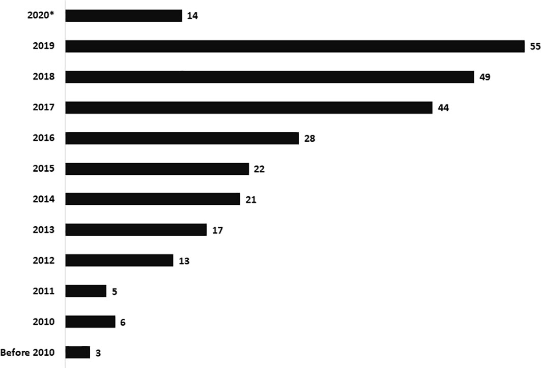
Figure 1

Male-female ratio was 196:81 (Table 1). Median [inter-quartile range (IQR)] age of onset of clinical symptoms and diagnosis was 2.5 months (1, 5) and 5 months (3.5, 8), respectively. Consanguinity was noted in 78 families (28.2%), and was noticeably more in Southern region (32.3%) of our country compared to Northern (22.4%). Family history of early childhood deaths was noted in 120 children (43.3%). Median (IQR) age at diagnosis in children who had a positive family history was 4.5 months (3, 6) compared to 6 months (4, 9) in children who did not have a family history, p<0.05 (Mann-Whitney U test).

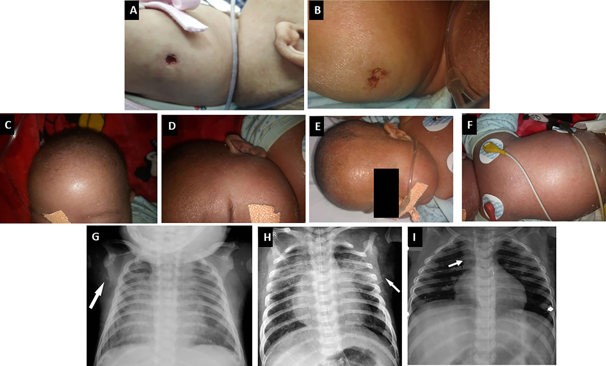
Opportunistic infections were the presenting manifestation in most patients. These included pneumonia (82%), diarrhoea (43.7%), oral thrush (18.4%), BCG site ulceration (17%), otitis media (12.6%), and meningitis (4%) (Figures 2, 3). Blood-culture proven septicemia was seen in 63 children (23%)—Candida sp. (16), Staphylococcus sp. (10), Escherichia coli (5), Acinetobacter sp. (5), Pseudomonas aeruginosa (8), Klebsiella pneumoniae (5), Enterococcus sp. (3), Enterobacter sp. (2), Streptococcus sp. (1), Pichia fermentans (1), Burkholderia cepacia (1), Chryseobacterium sp. (1), Bacillus subtilis (1), Citrobacter sp. (1), Moraxella sp. (1), Alcaligens faecalis (1), and Weisella confusa (1). Bacteria isolated from respiratory tract included Mycobacterium bovis (15), Klebsiella pneumoniae (5), P. aeruginosa (4), M. tuberculosis (3), atypical mycobacterium (1), E. coli (1), Staphylococcus aureus (1), and Acinetobacter sp. (1). Microbiology proven disseminated BCG infection was noted in 27 patients (9.7%). Apart from oral thrush and candidemia, other fungal infections noted were pneumonia due to Pneumocystis jirovecii (8), invasive aspergillosis (5), esophageal candidiasis (5), and pulmonary cryptcoccosis (1). Disseminated cytomegalovirus (CMV) infection was documented in 23 (8.3%) children and 6 amongst these had evidence of CMV retinitis. Intestinal lymphangiectasia due to CMV was noted on autopsy of a child with X-linked SCID (pt.8). Prolonged excretion of vaccine-derived poliovirus was documented in a child with leaky SCID at Mumbai (, ). Vaccine-associated paralytic poliovirus strain was also isolated in a child with RAG1 defect at Mumbai. He had presented with persistent diarrhea, developmental delay, and hypotonia.
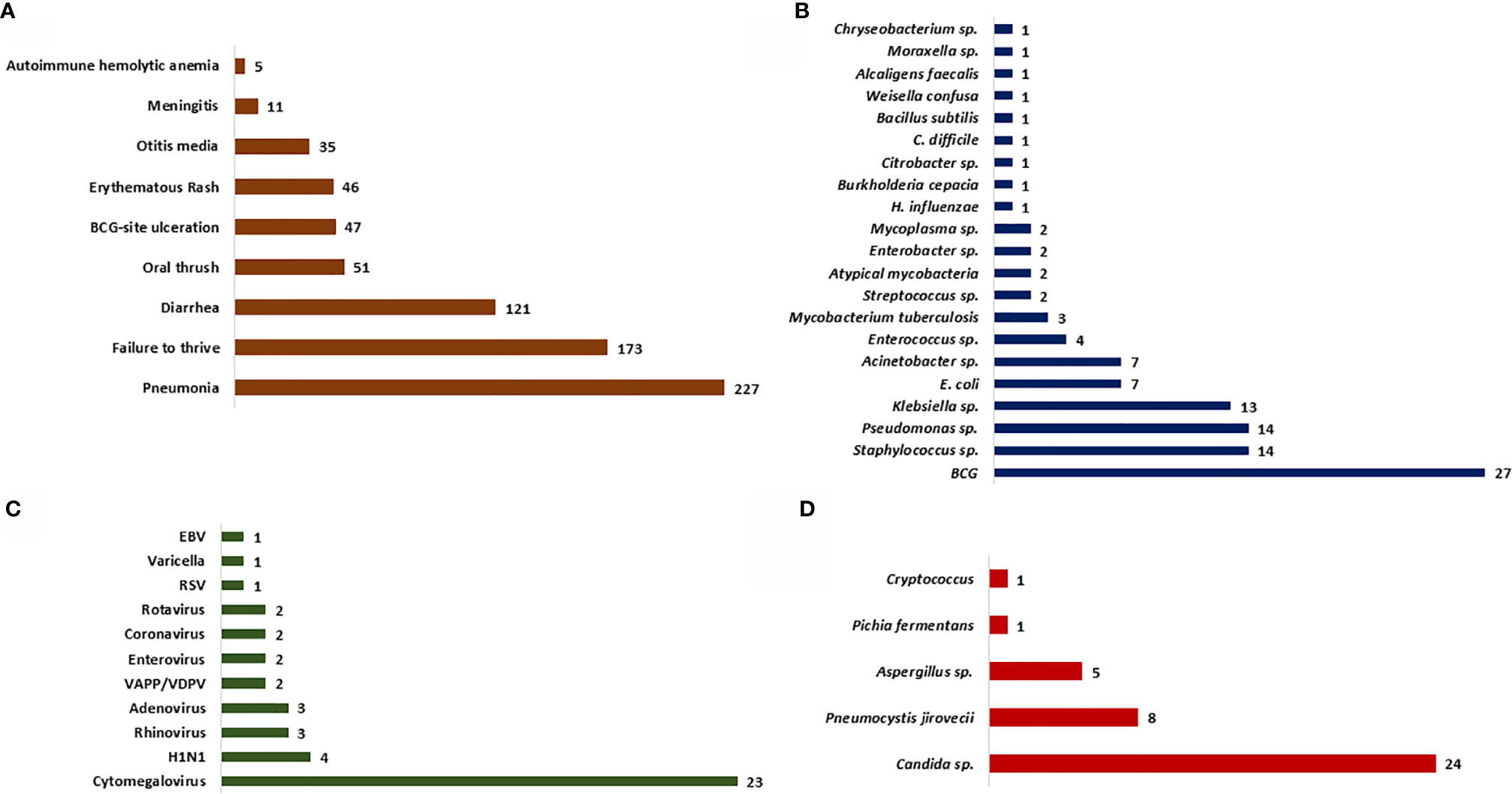
Figure 2

Figure 3

Clinical features of OS were seen in 33 children (11.9%)—classical OS in 11 and incomplete OS in 22 (Figure 3). Molecular defects associated with OS include RAG1 (7), RAG2 (5), ADA (2), NHEJ1 (1), IL2RG (1), JAK3 (1), STIM1 (1), CD3D (1), DCLRE1C (1), and RFXANK (1). Two children with IL2RG defect had features of engraftment of transplacental-acquired maternal T cells that mimicked clinical features of OS (Figure 4). Warm autoimmune hemolytic anemia (AIHA) requiring immunosuppressive medications was observed in 5 children. While anemia responded to intravenous (IV) methylprednisolone pulses in 2 patients (RAG1 and NHEJ1 defect each), pt.42 with STK4 defect received IV rituximab (375 mg/m2 2 doses) for control of AIHA and she did not have further relapse of AIHA for next 1.5 years. Transfusion-associated graft-vs-host reaction was documented in 4 patients (2 X-linked SCID; 2 AR-SCID); all had development of rash and transaminitis following transfusion of non-irradiated blood products. Four (4) children had features of hemophagocytic lymphohistiocytosis (HLH). Possible triggers for HLH included disseminated BCG (2) and H1N1 (1) infections. The child with SP110 defect did not have any identifiable trigger for HLH (pt.104). Hodgkin lymphoma and intra-cranial B cell lymphoma were noted in children with RAG1 and CORO1A defects, respectively.
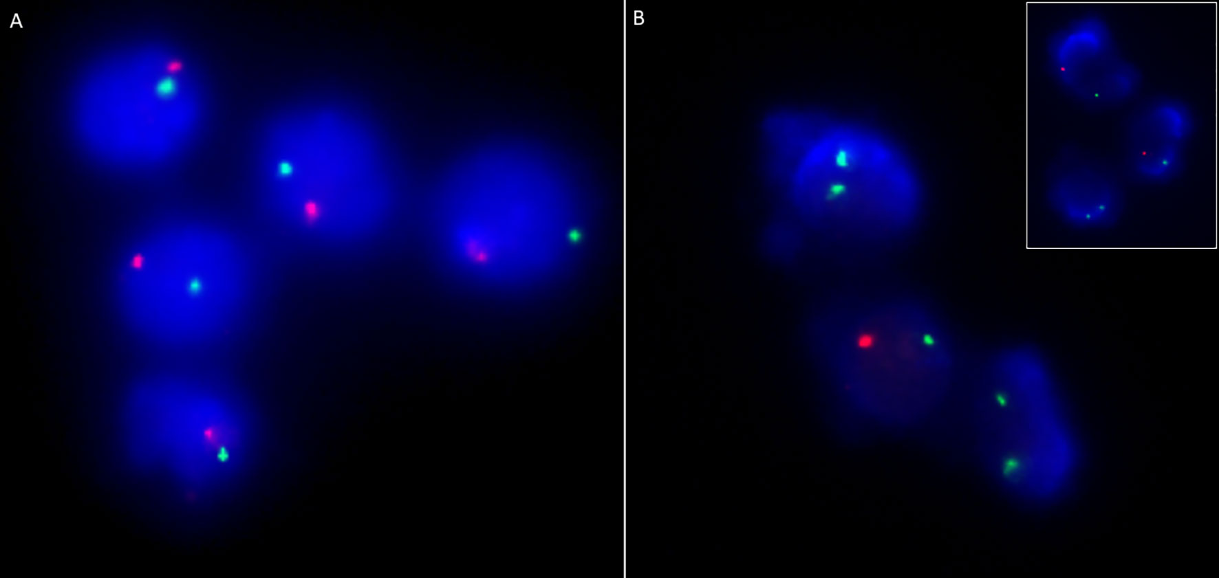
Figure 4

Four of 18 children with ADA defect were noted to have radiographic abnormalities—scapular spurring and flattening of lower end of scapula (Figure 3). Glomerular involvement was seen in 4 children—3 children with OS and 1 with atypical/leaky SCID. Nephrotic range proteinuria was noted in 3 patients and one child (pt.13) had features of mesangial sclerosis on autopsy. Another child (pt. 12) with OS had features of focal segmental glomerulosclerosis on autopsy. One child (pt.10) with IL7RA defect had features of distal renal tubular acidosis and nephrocalcinosis. This patient had deletion of exons 2–5 of CAPSL along with exon 4–8 deletion of IL7RA in chromosome 5p13.2. A child with PNP defect (pt.14) had evidence of horse-shoe kidney at autopsy ().

Median (IQR) absolute lymphocyte count (ALC) observed was 1.33 × 10⁹/L (0.6, 2.5). Normal ALC (≥ 3 × 10⁹/L) was observed in 51 children (18.4%)—of these 26 had OS, 2 had transplacental-acquired maternal T-cell engraftment, and 23 had leaky SCID/combined immunodeficiency. Eosinophilia was observed in 37 children, and 26 amongst these had features of OS. One child (Pt. 105) with RAG1 defect had unexplained monocytosis (2.7-3.0 × 10⁹/L) that resolved after HSCT. Results of immunoglobulin profile was available for 198 children. Fifty-five (55) children had normal or elevated levels of IgM levels—30 in SCID (14.2%), 7 in atypical SCID (41.2%), 8 in OS (30.8%), and 10 in CID (43.5%). We observed elevated levels of IgE in 12 children—8 had OS, 1 had eczema and STK4 defect, and 3 had unexplained eosinophilia.

Immunophenotyping by flow cytometry showed the following distribution: T-B-NK- (32), T-B+NK- (67), T-B+NK+ (33), T-B-NK+ (84). T+ SCID is observed in 20 children with OS—T+B-NK+ (17), T+B-NK- (2), T+B+NK+ (1) and 2 children with transplacental-acquired maternal T-cell engraftment—T+B+NK- (1), T+B-NK- (1). Genetic defects observed under each category are summarized (Supplementary Table 2). We observed decreased naïve (CD3+CD45RA+) and elevated memory (CD3+CD45RO+) CD3 lymphocytes in 24 children with OS. We noted elevated HLA-DR expression in CD3+ lymphocytes in 15 children with OS. CD132 expression by flow cytometry showed reduced expression in lymphocytes or monocytes in 8 children with suspected X-linked SCID (Table 2) (–). Levels of ADA and %dAXP were measured in 7 children with ADA SCID and 2 heterozygous carriers of ADA mutation (Table 3).
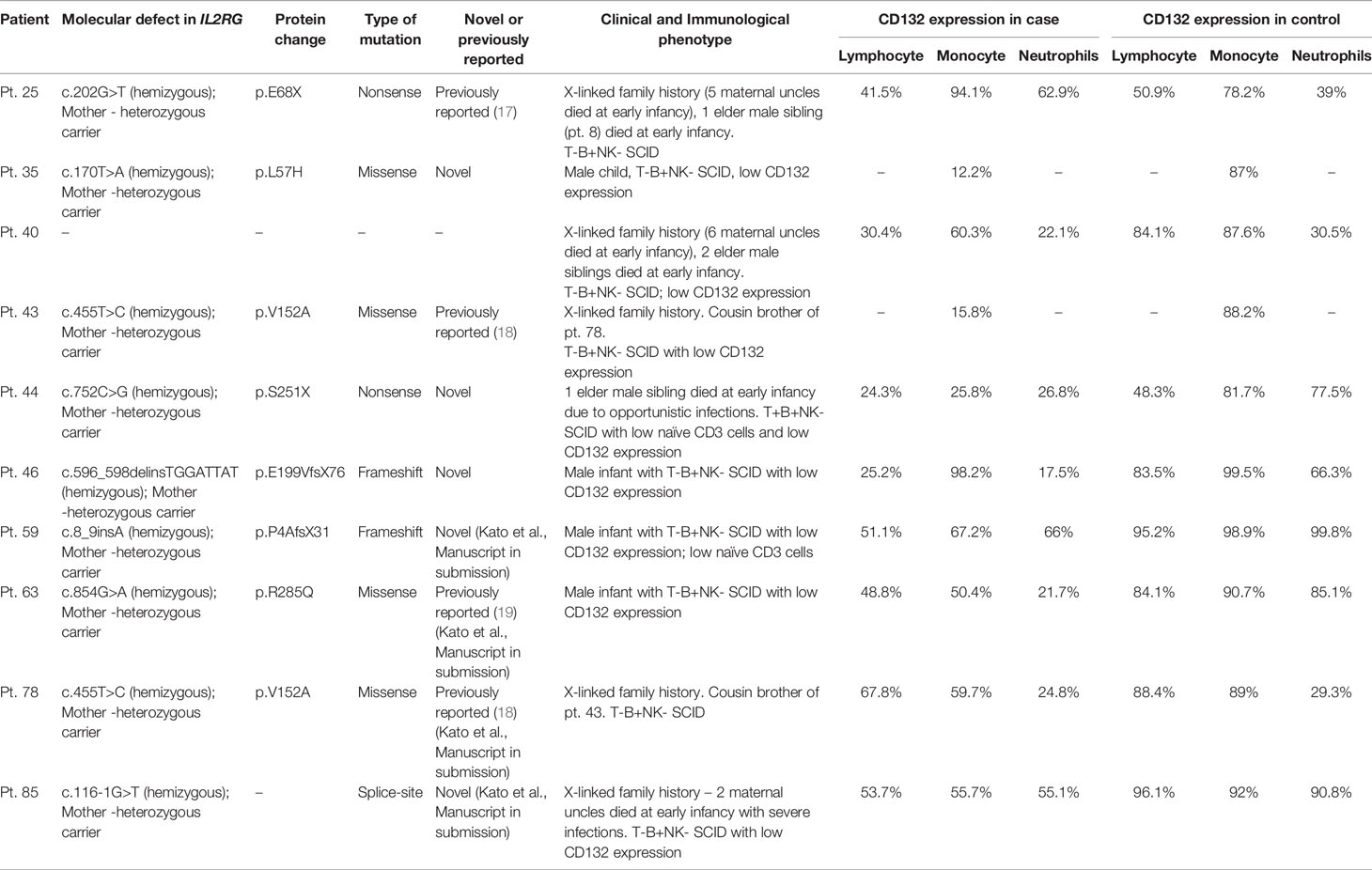
Table 2

PatientMolecular defect in IL2RGProtein changeType of mutationNovel or previously reportedClinical and Immunological phenotypeCD132 expression in caseCD132 expression in control
LymphocyteMonocyteNeutrophilsLymphocyteMonocyteNeutrophils
Pt. 25c.202G>T (hemizygous); Mother - heterozygous carrierp.E68XNonsensePreviously reported ()X-linked family history (5 maternal uncles died at early infancy), 1 elder male sibling (pt. 8) died at early infancy.
T-B+NK- SCID
41.5%94.1%62.9%50.9%78.2%39%
Pt. 35c.170T>A (hemizygous); Mother -heterozygous carrierp.L57HMissenseNovelMale child, T-B+NK- SCID, low CD132 expression–12.2%––87%–
Pt. 40––––X-linked family history (6 maternal uncles died at early infancy), 2 elder male siblings died at early infancy.
T-B+NK- SCID; low CD132 expression
30.4%60.3%22.1%84.1%87.6%30.5%
Pt. 43c.455T>C (hemizygous); Mother -heterozygous carrierp.V152AMissensePreviously reported ()X-linked family history. Cousin brother of pt. 78.
T-B+NK- SCID with low CD132 expression
–15.8%––88.2%–
Pt. 44c.752C>G (hemizygous); Mother -heterozygous carrierp.S251XNonsenseNovel1 elder male sibling died at early infancy due to opportunistic infections. T+B+NK- SCID with low naïve CD3 cells and low CD132 expression24.3%25.8%26.8%48.3%81.7%77.5%
Pt. 46c.596_598delinsTGGATTAT (hemizygous); Mother -heterozygous carrierp.E199VfsX76FrameshiftNovelMale infant with T-B+NK- SCID with low CD132 expression25.2%98.2%17.5%83.5%99.5%66.3%
Pt. 59c.8_9insA (hemizygous); Mother -heterozygous carrierp.P4AfsX31FrameshiftNovel (Kato et al., Manuscript in submission)Male infant with T-B+NK- SCID with low CD132 expression; low naïve CD3 cells51.1%67.2%66%95.2%98.9%99.8%
Pt. 63c.854G>A (hemizygous); Mother -heterozygous carrierp.R285QMissensePreviously reported () (Kato et al., Manuscript in submission)Male infant with T-B+NK- SCID with low CD132 expression48.8%50.4%21.7%84.1%90.7%85.1%
Pt. 78c.455T>C (hemizygous); Mother -heterozygous carrierp.V152AMissensePreviously reported () (Kato et al., Manuscript in submission)X-linked family history. Cousin brother of pt. 43. T-B+NK- SCID67.8%59.7%24.8%88.4%89%29.3%
Pt. 85c.116-1G>T (hemizygous); Mother -heterozygous carrier–Splice-siteNovel (Kato et al., Manuscript in submission)X-linked family history – 2 maternal uncles died at early infancy with severe infections. T-B+NK- SCID with low CD132 expression53.7%55.7%55.1%96.1%92%90.8%

CD132 expression by flow cytometry in children with X-linked SCID.
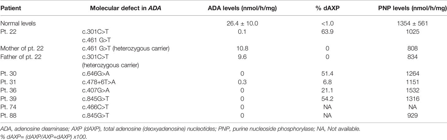
Table 3

PatientMolecular defect in ADAADA levels (nmol/h/mg)% dAXPPNP levels (nmol/h/mg)
Normal levels26.4 ± 10.0<1.01354 ± 561
Pt. 22c.301C>T
c.461 G>T
0.163.91025
Mother of pt. 22c.461 G>T (heterozygous carrier)10.80808
Father of pt. 22c.301C>T
(heterozygous carrier)
9.60834
Pt. 30c.646G>A051.41264
Pt. 31c.478+6T>A0.36.81151
Pt. 36c.407G>A021.11532
Pt. 39c.845G>T054.21316
Pt. 74c.466C>T0NANA
Pt. 88c.845G>T0NA929

Erythrocyte ADA levels and % dAXP measured in dried blood spots.

ADA, adenosine deaminase; AXP (dAXP), total adenosine (deoxyadenosine) nucleotides; PNP, purine nucleoside phosphorylase; NA, Not available.

% dAXP= (dAXP/AXP+dAXP) x100.

Molecular diagnosis was obtained in 162 patients—IL2RG (36), RAG1 (26), ADA (19), RAG2 (17), JAK3 (15), DCLRE1C (13), IL7RA (9), PNP (3), RFXAP (3), CIITA (2), RFXANK (2), NHEJ1 (2), CD3E (2), CD3D (2), RFX5 (2), ZAP70 (2), STK4 (1), CORO1A (1), STIM1 (1), PRKDC (1), AK2 (1), DOCK2 (1), and SP100 (1). Of the 176 molecular variants, 51 were identified to be novel in this study (Table 4, Supplementary Table 3) (, , –). A novel variant in RAG1 (c.1758_1760delinsGAATC) was identified in 2 unrelated North Indian families. Deletion of exons 1-3 (8947bp) in DCLRE1C was observed in 11 children (9 from North and 2 from South India). MLPA confirmed EX1_EX3del in DCLRE1C in 7 children from North India (Figure 5). Targeted clinical exome sequencing by NGS did not identify pathogenic variants in 25 patients. Whole exome sequencing was performed in 5 children, and pathogenic variants were detected in 2 amongst these (pt. 50 and 51).
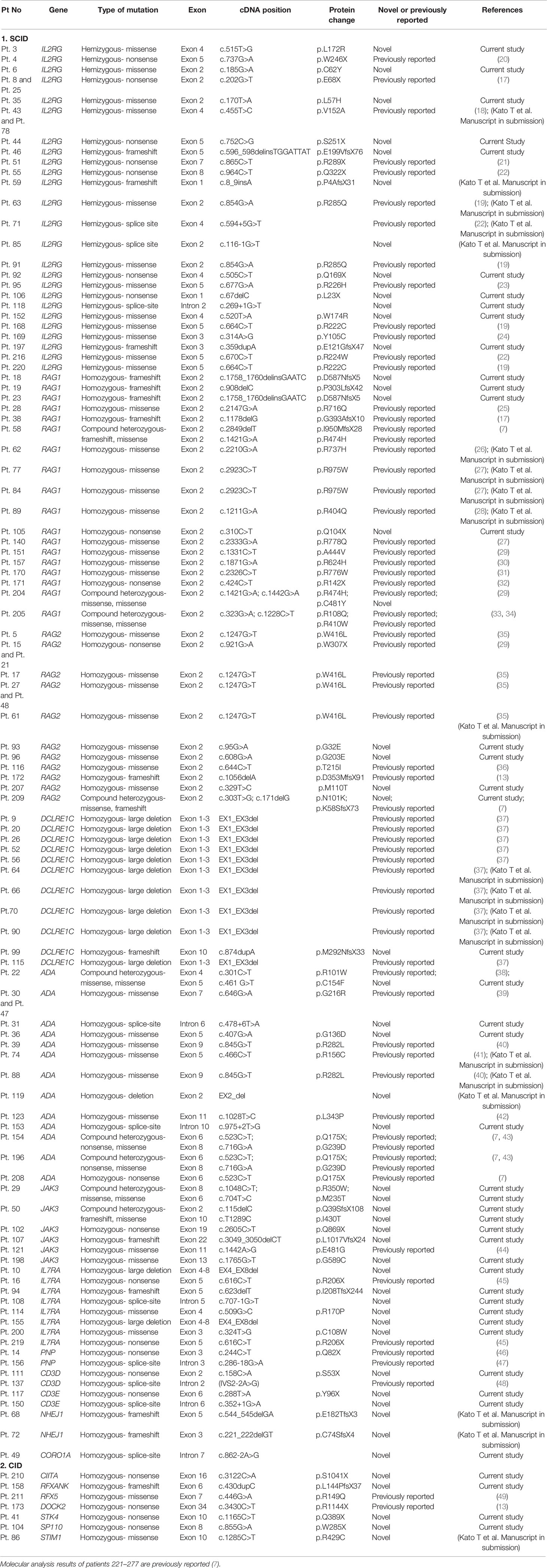
Table 4

Pt NoGeneType of mutationExoncDNA positionProtein changeNovel or previously reportedReferences
1. SCID
Pt. 3IL2RGHemizygous- missenseExon 4c.515T>Gp.L172RNovelCurrent study
Pt. 4IL2RGHemizygous- nonsenseExon 5c.737G>Ap.W246XPreviously reported ()
Pt. 6IL2RGHemizygous- missenseExon 2c.185G>Ap.C62YNovelCurrent study
Pt. 8 and Pt. 25IL2RGHemizygous- nonsenseExon 2c.202G>Tp.E68XPreviously reported ()
Pt. 35IL2RGHemizygous- missenseExon 2c.170T>Ap.L57HNovelCurrent study
Pt. 43 and Pt. 78IL2RGHemizygous- missenseExon 4c.455T>Cp.V152APreviously reported (); (Kato T et al. Manuscript in submission)
Pt. 44IL2RGHemizygous- nonsenseExon 5c.752C>Gp.S251XNovelCurrent Study
Pt. 46IL2RGHemizygous- frameshiftExon 5c.596_598delinsTGGATTATp.E199VfsX76NovelCurrent study
Pt. 51IL2RGHemizygous- nonsenseExon 7c.865C>Tp.R289XPreviously reported ()
Pt. 55IL2RGHemizygous- nonsenseExon 8c.964C>Tp.Q322XPreviously reported ()
Pt. 59IL2RGHemizygous- frameshiftExon 1c.8_9insAp.P4AfsX31Novel(Kato T et al. Manuscript in submission)
Pt. 63IL2RGHemizygous- missenseExon 2c.854G>Ap.R285QPreviously reported (); (Kato T et al. Manuscript in submission)
Pt. 71IL2RGHemizygous- splice siteExon 4c.594+5G>TPreviously reported (); (Kato T et al. Manuscript in submission)
Pt. 85IL2RGHemizygous- splice siteExon 2c.116-1G>TNovel(Kato T et al. Manuscript in submission)
Pt. 91IL2RGHemizygous- missenseExon 2c.854G>Ap.R285QPreviously reported ()
Pt. 92IL2RGHemizygous- nonsenseExon 4c.505C>Tp.Q169XNovelCurrent study
Pt. 95IL2RGHemizygous- missenseExon 5c.677G>Ap.R226HPreviously reported ()
Pt. 106IL2RGHemizygous- nonsenseExon 1c.67delCp.L23XNovelCurrent study
Pt. 118IL2RGHemizygous- splice-siteIntron 2c.269+1G>TNovelCurrent study
Pt. 152IL2RGHemizygous- missenseExon 4c.520T>Ap.W174RNovelCurrent study
Pt. 168IL2RGHemizygous- missenseExon 5c.664C>Tp.R222CPreviously reported ()
Pt. 169IL2RGHemizygous- missenseExon 3c.314A>Gp.Y105CPreviously reported ()
Pt. 197IL2RGHemizygous- frameshiftExon 3c.359dupAp.E121GfsX47NovelCurrent study
Pt. 216IL2RGHemizygous- missenseExon 5c.670C>Tp.R224WPreviously reported ()
Pt. 220IL2RGHemizygous- missenseExon 5c.664C>Tp.R222CPreviously reported ()
Pt. 18RAG1Homozygous- frameshiftExon 2c.1758_1760delinsGAATCp.D587NfsX5NovelCurrent study
Pt. 19RAG1Homozygous- frameshiftExon 2c.908delCp.P303LfsX42NovelCurrent study
Pt. 23RAG1Homozygous- frameshiftExon 2c.1758_1760delinsGAATCp.D587NfsX5NovelCurrent study
Pt. 28RAG1Homozygous- missenseExon 2c.2147G>Ap.R716QPreviously reported ()
Pt. 38RAG1Homozygous- frameshiftExon 2c.1178delGp.G393AfsX10Previously reported ()
Pt. 58RAG1Compound heterozygous- frameshift, missenseExon 2
Exon 2
c.2849delT
c.1421G>A
p.I950MfsX28
p.R474H
Previously reported
Previously reported
()
Pt. 62RAG1Homozygous- missenseExon 2c.2210G>Ap.R737HPreviously reported (); (Kato T et al. Manuscript in submission)
Pt. 77RAG1Homozygous- missenseExon 2c.2923C>Tp.R975WPreviously reported (); (Kato T et al. Manuscript in submission)
Pt. 84RAG1Homozygous- missenseExon 2c.2923C>Tp.R975WPreviously reported (); (Kato T et al. Manuscript in submission)
Pt. 89RAG1Homozygous- missenseExon 2c.1211G>Ap.R404QPreviously reported (); (Kato T et al. Manuscript in submission)
Pt. 105RAG1Homozygous- nonsenseExon 2c.310C>Tp.Q104XNovelCurrent study
Pt. 140RAG1Homozygous- missenseExon 2c.2333G>Ap.R778QPreviously reported ()
Pt. 151RAG1Homozygous- missenseExon 2c.1331C>Tp.A444VPreviously reported ()
Pt. 157RAG1Homozygous- missenseExon 2c.1871G>Ap.R624HPreviously reported ()
Pt. 170RAG1Homozygous- missenseExon 2c.2326C>Tp.R776WPreviously reported ()
Pt. 171RAG1Homozygous- nonsenseExon 2c.424C>Tp.R142XPreviously reported ()
Pt. 204RAG1Compound heterozygous- missense, missenseExon 2c.1421G>A; c.1442G>Ap.R474H;
p.C481Y
Previously reported;
Novel
()
Pt. 205RAG1Compound heterozygous- missense, missenseExon 2c.323G>A; c.1228C>Tp.R108Q;
p.R410W
Previously reported;
Previously reported
(, )
Pt. 5RAG2Homozygous- missenseExon 2c.1247G>Tp.W416LPreviously reported ()
Pt. 15 and Pt. 21RAG2Homozygous- nonsenseExon 2c.921G>Ap.W307XPreviously reported ()
Pt. 17RAG2Homozygous- missenseExon 2c.1247G>Tp.W416LPreviously reported ()
Pt. 27 and Pt. 48RAG2Homozygous- missenseExon 2c.1247G>Tp.W416LPreviously reported ()
Pt. 61RAG2Homozygous- missenseExon 2c.1247G>Tp.W416LPreviously reported ()
(Kato T et al. Manuscript in submission)
Pt. 93RAG2Homozygous- missenseExon 2c.95G>Ap.G32ENovelCurrent study
Pt. 96RAG2Homozygous- missenseExon 2c.608G>Ap.G203ENovelCurrent study
Pt. 116RAG2Homozygous- missenseExon 2c.644C>Tp.T215IPreviously reported ()
Pt. 172RAG2Homozygous- frameshiftExon 2c.1056delAp.D353MfsX91Previously reported ()
Pt. 207RAG2Homozygous- missenseExon 2c.329T>C p.M110TNovelCurrent study
Pt. 209RAG2Compound heterozygous- missense, frameshiftExon 2c.303T>G; c.171delGp.N101K;
p.K58SfsX73
Novel;
Previously reported
Current study;
()
Pt. 9DCLRE1CHomozygous- large deletionExon 1-3EX1_EX3delPreviously reported ()
Pt. 20DCLRE1CHomozygous- large deletionExon 1-3EX1_EX3delPreviously reported ()
Pt. 26DCLRE1CHomozygous- large deletionExon 1-3EX1_EX3delPreviously reported ()
Pt. 52DCLRE1CHomozygous- large deletionExon 1-3EX1_EX3delPreviously reported ()
Pt. 56DCLRE1CHomozygous- large deletionExon 1-3EX1_EX3delPreviously reported ()
Pt. 64DCLRE1CHomozygous- large deletionExon 1-3EX1_EX3delPreviously reported (); (Kato T et al. Manuscript in submission)
Pt. 66DCLRE1CHomozygous- large deletionExon 1-3EX1_EX3delPreviously reported (); (Kato T et al. Manuscript in submission)
Pt.70DCLRE1CHomozygous- large deletionExon 1-3EX1_EX3delPreviously reported (); (Kato T et al. Manuscript in submission)
Pt. 90DCLRE1CHomozygous- large deletionExon 1-3EX1_EX3delPreviously reported (); (Kato T et al. Manuscript in submission)
Pt. 99DCLRE1CHomozygous- frameshiftExon 10c.874dupAp.M292NfsX33NovelCurrent study
Pt. 115DCLRE1CHomozygous- large deletionExon 1-3EX1_EX3delPreviously reported ()
Pt. 22ADACompound heterozygous- missense, missenseExon 4
Exon 5
c.301C>T
c.461 G>T
p.R101W
p.C154F
Previously reported;
Novel
();
Current study
Pt. 30 and Pt. 47ADAHomozygous- missenseExon 7c.646G>Ap.G216RPreviously reported ()
Pt. 31ADAHomozygous- splice-siteIntron 6c.478+6T>ANovelCurrent study
Pt. 36ADAHomozygous- missenseExon 5c.407G>Ap.G136DNovelCurrent study
Pt. 39ADAHomozygous- missenseExon 9c.845G>Tp.R282LPreviously reported ()
Pt. 74ADAHomozygous- missenseExon 5c.466C>Tp.R156CPreviously reported (); (Kato T et al. Manuscript in submission)
Pt. 88ADAHomozygous- missenseExon 9c.845G>Tp.R282LPreviously reported (); (Kato T et al. Manuscript in submission)
Pt. 119ADAHomozygous- deletionExon 2EX2_delNovel(Kato T et al. Manuscript in submission)
Pt. 123ADAHomozygous- missenseExon 11c.1028T>Cp.L343PPreviously reported ()
Pt. 153ADAHomozygous- splice-siteIntron 10c.975+2T>GNovelCurrent study
Pt. 154ADACompound heterozygous- nonsense, missenseExon 6
Exon 8
c.523C>T;
c.716G>A
p.Q175X;
p.G239D
Previously reported; Previously reported (, )
Pt. 196ADACompound heterozygous- nonsense, missenseExon 6
Exon 8
c.523C>T;
c.716G>A
p.Q175X;
p.G239D
Previously reported;
Previously reported
(, )
Pt. 208ADAHomozygous- nonsenseExon 6c.523C>Tp.Q175XPreviously reported ()
Pt. 29JAK3Compound heterozygous- missense, missenseExon 8
Exon 6
c.1048C>T;
c.704T>C
p.R350W;
p.M235T
Novel
Novel
Current study
Current study
Pt. 50JAK3Compound heterozygous- frameshift, missenseExon 2
Exon 10
c.115delC
c.T1289C
p.Q39SfsX108
p.I430T
Novel
Novel
Current study
Current study
Pt. 102JAK3Homozygous- nonsenseExon 19c.2605C>Tp.Q869XNovelCurrent study
Pt. 107JAK3Homozygous- frameshiftExon 22c.3049_3050delCTp.L1017VfsX24NovelCurrent study
Pt. 121JAK3Homozygous- missenseExon 11c.1442A>Gp.E481GPreviously reported ()
Pt. 198JAK3Homozygous- missenseExon 13c.1765G>Tp.G589CNovelCurrent study
Pt. 10IL7RAHomozygous- large deletionExon 4-8EX4_EX8delNovelCurrent study
Pt. 16IL7RAHomozygous- nonsenseExon 5c.616C>Tp.R206XPreviously reported ()
Pt. 94IL7RAHomozygous- frameshiftExon 5c.623delTp.I208TfsX244NovelCurrent study
Pt. 108IL7RAHomozygous- splice-siteIntron 5c.707-1G>TNovelCurrent study
Pt. 114IL7RAHomozygous- missenseExon 4c.509G>Cp.R170PNovelCurrent study
Pt. 155IL7RAHomozygous- large deletionExon 4-8EX4_EX8delNovelCurrent study
Pt. 200IL7RAHomozygous- missenseExon 3c.324T>Gp.C108WNovelCurrent study
Pt. 219IL7RAHomozygous- nonsenseExon 5c.616C>Tp.R206XPreviously reported ()
Pt. 14PNPHomozygous- nonsenseExon 3c.244C>Tp.Q82XPreviously reported ()
Pt. 156PNPHomozygous- splice-siteIntron 3c.286-18G>APreviously reported ()
Pt. 111CD3DHomozygous- nonsenseExon 2c.158C>Ap.S53XNovelCurrent study
Pt. 137CD3DHomozygous- splice-siteIntron 2(IVS2-2A>G)Previously reported ()
Pt. 117CD3EHomozygous- nonsenseExon 6c.288T>Ap.Y96XNovelCurrent study
Pt. 150CD3EHomozygous- splice-siteIntron 6c.352+1G>ANovelCurrent study
Pt. 68NHEJ1Homozygous- frameshiftExon 5c.544_545delGAp.E182TfsX3Novel(Kato T et al. Manuscript in submission)
Pt. 72NHEJ1Homozygous- frameshiftExon 3c.221_222delGTp.C74SfsX4Novel(Kato T et al. Manuscript in submission)
Pt. 49CORO1AHomozygous- splice-siteIntron 7c.862-2A>GNovelCurrent study
2. CID
Pt. 210CIITAHomozygous- nonsenseExon 16c.3122C>Ap.S1041XNovelCurrent study
Pt. 158RFXANKHomozygous- frameshiftExon 6c.430dupCp.L144PfsX37NovelCurrent study
Pt. 211RFX5Homozygous- missenseExon 7c.446G>Ap.R149QPreviously reported ()
Pt. 173DOCK2Homozygous- nonsenseExon 34c.3430C>Tp.R1144XPreviously reported ()
Pt. 41STK4Homozygous- nonsenseExon 10c.1165C>Tp.Q389XNovelCurrent study
Pt. 104SP110Homozygous- nonsenseExon 8c.855G>Ap.W285XNovelCurrent study
Pt. 86STIM1Homozygous- missenseExon 10c.1285C>Tp.R429CNovel(Kato T et al. Manuscript in submission)

Molecular defects in genes associated with SCID/CID in our cohort.
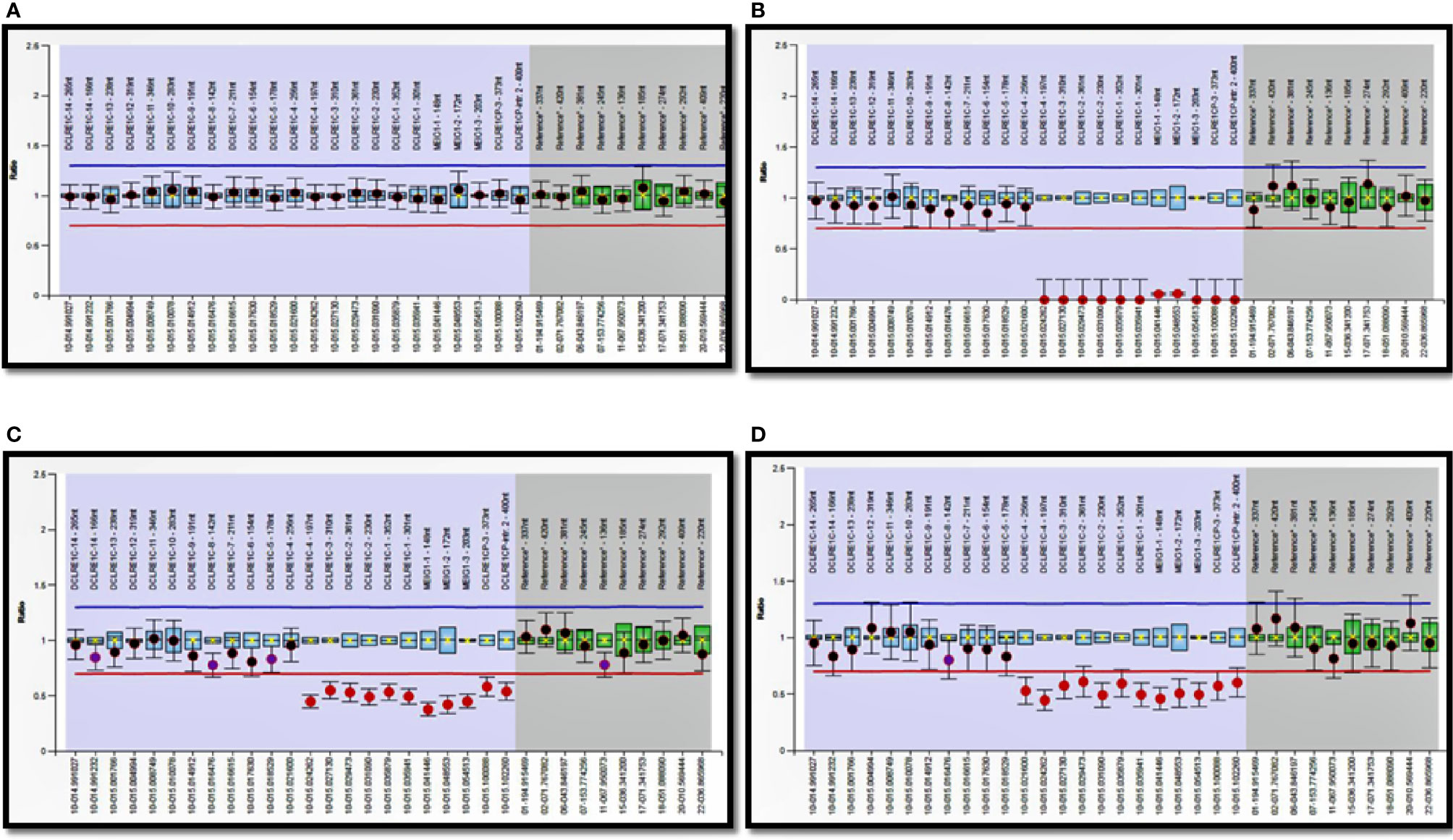
Molecular analysis results of patients 221–277 are previously reported ().

Figure 5

Majority of patients (n=198) in this cohort succumbed to overwhelming infections as HSCT could not be carried out in them (Figure 6). Twenty-three patients (8.3%) underwent hematopoietic stem cell transplantation (HSCT) and 11 are doing well post-HSCT. The centre at South India (Apollo Children’s Hospitals, Chennai) has performed HSCT for 32 children with SCID until now and 17 are alive and doing well on follow-up. However, only 4 children are included in this analysis, as flow cytometry and mutation details were not available for other children. Another centre in South India (Aster CMI Hospitals, Bengaluru) has carried out HSCT for 9 children with SCID in the last 3 years (Table 5).
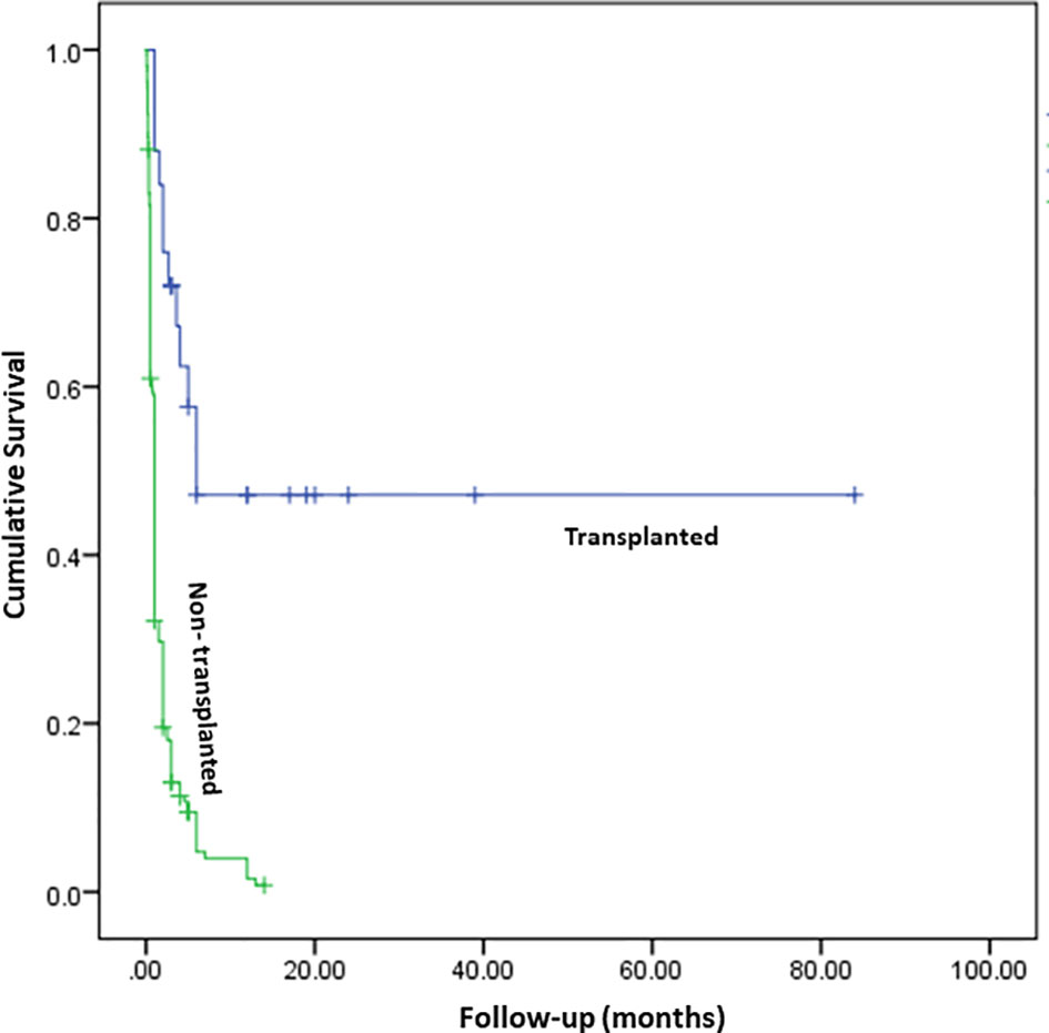
Figure 6

Table 5

PatientType of SCIDMolecular defectCentreAge at transplantationDonor characteristicsOutcome
Pt. 19T-B-NK+RAG1PGIMER, Chandigarh3.5 monthsFather who is a complete HLA match with childDeveloped BCG IRIS post-HSCT (D+90)- successfully treated with isoniazid, rifampicin, and ethambutol. He successfully engrafted and is currently doing well at 3rd year follow-up.
Pt. 31T-B-NK-ADADiagnosed at PGIMER, Chandigarh; transplanted at Apollo Hospitals, Chennai4 yearsFully matched unrelated donorEngrafted and doing well at 1st year follow-up.
Pt. 80T-B-NK+-PGIMER, Chandigarh18 monthsHaploidentical donorDeveloped graft failure. Underwent second transplant at 3 years (details not available).
Pt. 94T-B+NK+IL7RAAster CMI Hospitals, Bengaluru5 monthsHaploidentical (Mother)Delayed graft failure (6 months post-HSCT). Underwent second HSCT with father being donor. Successfully engrafted and doing well at 20 months follow-up.
Pt. 103T-B+NK+N.A.Aster CMI Hospitals, Bengaluru1.5 monthsHaploidentical (Mother)Failed engraftment. Underwent second HSCT with father being donor- successfully engrafted, however, died after 1.5 months due to fulminant sepsis.
Pt. 105T-B-NK+RAG1Aster CMI Hospitals, Bengaluru11 monthsHaploidentical (Mother)Successfully engrafted. Doing well 1.5 years post-HSCT.
Pt. 106T-B+NK-IL2RGAster CMI Hospitals, Bengaluru5 monthsHaploidentical (Mother)Successfully engrafted. Doing well 1.4 years post-HSCT.
Pt. 108T-B+NK-IL7RAAster CMI Hospitals, Bengaluru8.5 monthsFully matched siblingDied D+20 of HSCT- MDR Klebsiella sepsis.
Pt. 110T-B-NK+N.A.Aster CMI Hospitals, Bengaluru5 monthsHaploidentical (Mother)Successfully engrafted. Doing well 10 months post-HSCT.
Pt. 111T-B+NK+CD3DAster CMI Hospitals, Bengaluru6 monthsHaploidentical (Mother)Expired D+9 due to pulmonary haemorrhage.
Pt. 114T-B+NK+IL7RAster CMI Hospitals, Bengaluru9 monthsHaploidentical (Father)Developed severe gut GVHD and died D+60.
Pt. 115T-B-NK+DCLRE1CAster CMI Hospitals, Bengaluru7.5 monthsHaploidentical (Father)Successfully engrafted. Doing well 2 months post-HSCT.
Pt. 152T-B+NK-IL2RGAditya Birla Memorial Hospital, Pune5 monthsMatched family donorDoing well at 7th year follow-up.

Details of hematopoietic stem cell transplantation of 13 children with SCID.

Discussion

We describe the largest multi-centric cohort of patients with SCID from India. We included patients from 12 different tertiary care centers located in Northern, Southern, and Western parts of India. Patients from Eastern parts of India are usually referred to the centers located in other areas of India due to lack of availability of facilities for immunological investigations in that region. We witnessed an exponential rise in the number of cases with SCID after 2013 at multiple centers across India. We attribute this steady increase in cases to 2 factors—establishment of Indian Council of Medical Research Centers for Advanced Research in PIDs at PGIMER, Chandigarh (North India) and NIIH, Mumbai (West India) and expansion of laboratory facilities for pediatric immunology at other centers. The Pediatric Immunology and Bone Marrow Transplant Unit at Aster CMI Hospital, Bengaluru (South India) was established in 2017. Twenty-seven cases of SCID (Pt. 60–84) were diagnosed between 2017 and 2020, reflecting rise in awareness amongst referring pediatricians and better availability of diagnostic facilities at Bengaluru (South India).

Based on data from Sample Registration System of India, we estimated around 221 million live births from January 2011 to June 2020 (). An estimated 257 patients with SCID have been diagnosed in this time period, which suggests a rough incidence of SCID at 0.12 per 100,000 live births. Though we have included data from most of the centers that care for patients with SCID in India, the estimated incidence from this study may not reflect true incidence of the country because of retrospective nature of the study and some patients diagnosed at other centers may have been missed. Nation-wide registry for SCID is needed for an accurate estimation of incidence. Nevertheless, if we extrapolate our current data on to the U.S. incidence figures of SCID (i.e. 1:58,000 live births), estimated number of children with SCID in India would be around 3,822 during the period 2011–June 2020 (). Moreover, incidence of SCID in India is expected to be even higher than the U.S. considering high rates of consanguinity within the country. This suggests that though we have been increasingly diagnosing these children over the last few years, the diagnosis is still missed in almost 93% of these children. This is clearly unacceptable and mandates urgent intervention of health care professionals.

We observed a higher incidence of autosomal recessive forms of SCID (78.4%) compared to X-linked SCID. This is similar to reports from several other countries where consanguinity rates are high (Table 6) (, , –). Though consanguinity rate of 28.2% observed in our study is lower than that of Saudi Arabia and Iran, practice of endogamous and intra-community marriages is, perhaps, responsible for high proportion of autosomal recessive forms of SCID in India (, ). Median age at diagnosis of SCID in our study is 5 months. This is similar to reports from other countries such as China, Turkey, and U.S.A (Table 6). Children who had a family history of SCID had an earlier age of diagnosis (median:4.5 months) compared to children who did not have a suggestive family history (median:6 months). Our observation is similar to the report by Luk et al. that suggested the importance of family history for an early diagnosis of SCID ().

Table 6

Study (Year)PlaceNo. of patientsAge of onset and diagnosisClinical manifestationsMolecular defectsOutcome
Yao et al. ()Shanghai, China44;
Male: female – 40:4
Mean age of onset – 3.56 ± 3.91 months
Mean age at diagnosis – 7.1 ± 7.96 months
BCG-related complications noted in 14 children (31.8%). Three (3) had disseminated BCG infection. Two (2) had CMV infectionDefect in IL2RG noted in 25 children (56.8%).Mortality seen in 37 children (84%). Six (6) children underwent HSCT and 1 of them had survived.
Pasic et al. ()Serbia and Montenegro21Median age of onset – 2 monthsBCG-related complications in 41%. Pneumonia noted in 15 children (PJP- 5, CMV- 3, BCG-2, respiratory virus- 5). OS noted in 6 children.17 had proven molecular defect (81%). RAG1/2 commonest (12) followed by IL2RG (3), JAK3 (2), DCLRE1C (1)Mortality seen in 16 children (76.2%). Eight (8) children underwent HSCT and 5 of them survived.
Lee et al. ()South East Asia42;
Male: female – 30:12
Median age of onset and diagnosis – 2 and 4 months, respectively.BCG-related complications in 10 children (23.8%) – 6 had localized reaction; 3 had regional adenitis; 3 had disseminated BCGosis. Oral thrush (12), CMV (2), and PJP (2) are other documented infections. OS noted in 4 children.26 had proven molecular defect (61.9%). IL2RG commonest (19) followed by IL7RA (2), JAK3 (2), RAG1/2 (2), DCLRE1C (1)12 children underwent HSCT and 8 of them survived.
Abolhassani et al. ()Iran169;
Male: female – 96:73
Mean age of onset and diagnosis – 4.2 and 8.6 months, respectively.BCG-related complications noted in 23 (13.6%). Other infections noted are PJP (13), CMV (15), EBV (8), VDPV (6), Cryptococcus (6), and VZV (6). OS noted in 11 children.37 had proven molecular defect (21.9%). RAG1/2 commonest (19) followed by IL2RG (3), JAK3 (3), DCLRE1C (3), ADA (2), IL7RA (2), CD3E (1), CD3D (1), PRKDC (1), NHEJ1 (1), PTPRC (1)NA
Rozmus et al. ()Canada40Mean age at diagnosis – 4.2 months.Oral thrush (8), CMV (6), PJP (6), RSV (1), and adenovirus (1) are the infections noted.20 had proven molecular defect (50%). ADA commonest (10) followed by IL2RG (4), RAG1 (2), ZAP70 (2), and MHC Class II defects (2).Mortality observed in 12 children (30%). Fifteen (15) underwent HSCT and 10 of them survived.
Ikinciogullari et al. ()Turkey234 (transplanted patients);
Male: female – 145:89
Median age at diagnosis – 5 months.Infections noted are oral thrush (51.5%), CMV (13.5%), bacterial infections (7.4%), BCG-related complications (2.2%), and respiratory viruses (4.4%)42.3% had proven molecular defects – RAG1/2 (15.4%), JAK3 (6.8%), IL2RG (6%), DCLRE1C (5.6%)Survival at 20 years is 65.7%
Mazzucchelli et al. ()Brazil70;
Male: female – 49:21
Mean age of onset and diagnosis – 3.3 and 6.7 months, respectively.BCG-related complications seen in 39 children (55.7%) – disseminated form in 29 and localized in 10. Features of OS noted in 8 children.NAMortality seen in 35 patients (50%). Thirty (30) underwent HSCT and 18 of them survived.
de Pagter et al. ()Netherlands43Median delay in diagnosis in typical and atypical SCID – 2 and 27 months, respectively.Infections noted are bacterial sepsis (11), PJP (11), CMV (8), and BCGitis (6). AIHA seen in 5 children with atypical SCID.IL2RG (21%), RAG1 (21%), RAG2 (5%), ADA (12%), DCLRE1C (7%), PNP (7%), and IL7RA (5%)Mortality observed in 18 children (41.9%). Thirty-two (32) underwent HSCT and 24 of them survived. Two (2) underwent gene therapy and 1 survived.
Haddad et al. ()USA and Canada662 (transplanted patients);
Male: female – 471:191
Median age at diagnosis – 141.5 days (4.7 months)NAIL2RG (187), RAG1/2 (52), ADA (45), IL7RA (40), DCLRE1C (28), JAK3 (24), CD3 receptor defects (7), PNP (1), AK2 (1), CD45 (1)Survival is better in children transplanted less than 3.5 months. Survival at 10 years is 71% and is higher with matched sibling donors compared to other donor types.
Micho et al. ()Greece30;
Male: female – 19:11
Median age at diagnosis – 6.2 monthsNADCLRE1C (3), IL2RG (2), JAK3 (2), RAG1 (2), ADA (2), PNP (1)Mortality is observed in 15 children (50%). Twenty-two (22) underwent HSCT and 14 of them are doing well.
Aluri et al. ()India57;
Male: female – 40:17
Median age of onset and diagnosis – 2 and 5.1 months, respectivelyInfections observed include oral thrush (21%), BCG-related complications (12%), and PJP (1). OS noted in 4 children49 children had proven molecular defects (86%). RAG1/2 commonest (12), followed by JAK3 (9), IL2RG (9), MHC Class II defects (6), ADA (5), DCLRE1C (2), ZAP70 (2), IL7RA (1), PRKDC (1), PNP (1), and AK2 (1)Mortality observed in 47 children (82.5%). Four (4) underwent HSCT and none survived.
Present study (2020)India277 (23 CID, 254 SCID);
Male: female – 196:81
Median age of onset and diagnosis – 2.5 and 5 months, respectivelyBCG-related complications in 47 patients (17%) – localized form (20) and disseminated BCGosis (27). Other common infections include bacteria (72), CMV (23), Candida sp. (23), PJP (8), Aspergillus sp. (5), VAPP/VDPV (2). OS noted in 33 children. AIHA and lymphoreticular malignancy observed in 5 and 2 children, respectively.162 patients had proven molecular defects (58.5%) - RAG1/2 (43), IL2RG (36), ADA (19), JAK3 (15), DCLRE1C (13), IL7RA (9), PNP (3), CIITA (2), RFXAP (3), RFXANK (2), NHEJ1 (2), CD3E (2), CD3D (2), RFX5 (2), ZAP70 (2), STK4 (1), CORO1A (1), STIM1 (1), PRKDC (1), AK2 (1), DOCK2 (1), and SP100 (1)Mortality noted in 210 children (75.8%). Twenty-three (23) underwent HSCT and 11 of them are doing well.

Comparison of our cohort with published multicentric studies on SCID from other countries in last 10 years.

BCG, Bacillus Calmette-Guerin; CMV, Cytomegalovirus; HSCT, Hematopoietic stem cell transplantation; OS, Omenn syndrome; PJP, Pneumocystis jirovecii pneumonia; EBV, Epstein-Barr virus; VDPV, Vaccine-derived polio virus; VZV, Varicella zoster virus; AIHA, Autoimmune hemolytic anemia; VAPP, Vaccine-associated paralytic polio; CID, Combined Immune Deficiency; SCID, Severe Combined Immune Deficiency.

Opportunistic infections in SCID are life-threatening and must be identified and treated adequately before HSCT. We documented a higher incidence of microbiologically-proven infections in our cohort compared to a previous report published from India (). Amongst the bacterial infections, BCG was the commonest organism isolated. BCG-site ulceration has been noted in 47 children, however, disseminated BCGosis could be proven in 27 children only. BCG adenitis was noted in one child at D+90 post-HSCT as a part of immune reconstitution inflammatory syndrome. Lack of microbiological confirmation of BCG infection in many patients could have accounted for low rates of disseminated BCGosis in our cohort (Table 6) (, ). Infants with SCID who had received BCG vaccination and had not developed disseminated infection, are generally started on prophylactic medications—isoniazid and rifampicin at age-appropriate doses, that is generally continued until successful engraftment following HSCT.

Septicemia due to unusual organisms such as W. confusa and A. faecalis was also noted in our cohort. These are environmental bacteria and usually do not cause invasive infections in immunocompetent hosts. We also noted a high rate of disseminated CMV infection (8.3%) in our cohort. However, several amongst these were identified only on autopsy (). This underscores the importance of vigilant screening and preventive measures for CMV infection in children with SCID. Cytomegalovirus infection, in our cohort, was possibly transfusion-acquired as most of the children received blood transfusions that are not always leuko-depleted and screened for active CMV infection. Though many patients had clinical features suggestive of P. jirovecii pneumonia (tachypnea, hypoxemia, interstitial pneumonia) and were treated for the same, microbiological or histopathological confirmation was possible in only 8 of them.

Thirty-three children had features of OS in our cohort. One child with OS (pt.54) was being treated as severe eczema for 3 years with multiple topical and systemic immunosuppressive agents, and diagnosis of SCID was made only after he developed severe infections. This highlights the importance of early identification of clinical phenotype of OS based on clinical features (generalized erythematous rash with scaling and partial loss of scalp hairs and eyebrows) and referral for appropriate immunological workup. Twenty-eight (28) children with OS had normal or high ALC and 2 children with transplacental-acquired maternal T-cell engraftment had elevated ALC. Laboratory assay of naïve T cells, memory T cells, and HLA-DR expression in T lymphocytes necessary for the diagnosis of OS are currently being performed only in two centers (PGIMER, Chandigarh and NIIH, Mumbai).

Twenty-three children in our cohort who did not have OS had normal ALC (>3 × 10⁹/L). However, lymphocyte subsets and naïve T cell estimation revealed diagnosis of SCID in them, thereby highlighting the importance of clinical suspicion and immunological investigations in infants with severe and life-threatening infections even if ALC is normal. Expansion of B cells or NK cells, engraftment of transplacental-acquired maternal T cells, or partial genetic defects allowing selective clone of T cell expansion could be the possible reasons for normal ALC in SCID. Aluri et al. have previously highlighted the importance of assessment of naïve T helper and cytotoxic T cells in children with severe infections and normal ALC to characterise MHC class II and ZAP70 defects, respectively (). A child with IL7RA defect in our cohort had a T-B+NK- phenotype, similar to the report by Aluri et al. (). Also, two children with IL2RG defect had a T+B-NK- phenotype (1- OS, 1- transplacental-acquired maternal T cell engraftment). A possible explanation for low B cells is the depletion of B cells due to high inflammatory milieu secondary to OS and severe infections ().

CD132 expression by flow cytometry is currently carried out at only two centers—PGIMER, Chandigarh (North India) and NIIH, Mumbai (West India). At PGIMER, Chandigarh, we found low CD132 expression in lymphocytes by flow cytometry as an inexpensive and rapid method of confirmation of diagnosis of X-linked SCID in 7 children. Two (2) children with X-linked SCID and previously reported variants in IL2RG (pt. 25 and pt. 78) had a normal expression of CD132 in lymphocytes (Table 2). We could not assay phosphorylated STAT5 in activated T-cells by flow cytometry to determine the functionality of IL2Rγ in many patients due to absent or very low amounts of T cells, however, naïve T cells by flow cytometry and TREC levels by RT-PCR have been assayed in some of them (Table 2). Only a handful centers in India (e.g. PGIMER, Chandigarh, North India, and NIIH, Mumbai, West India) have the wherewithal to perform functional studies. Both the centers have performed flow cytometry tests for samples received from other centers, however, timely transportation of viable blood samples from far off places, especially during hot summers remains a significant problem (, ). Lack of state-of-the-art facilities to do functional assays in all patients with SCID is one of the limitations of our study. Establishment of more clinical immunology laboratories, training of necessary manpower, and improvement in existing laboratory services are needed to overcome these barriers (, ).

Genetic confirmation of diagnosis of SCID is necessary for identification of pattern of inheritance and genetic counselling of affected families. Eighty-two (82) patients did not undergo a molecular analysis for confirmation of diagnosis due to lack of easy access to molecular diagnostics and financial difficulties. With the establishment of commercial NGS laboratories and reduction in costs involved for genetic sequencing over last few years, NGS-based diagnostics have become feasible in India (, ). In-house NGS facility for molecular diagnosis of PID is currently available only at PGIMER, Chandigarh (North India) and Christian Medical College, Vellore (South India). Most of the patients with SCID present in a critically-ill state and convincing families for genetic studies is often challenging due to significant financial and social constraints. It must be noted that expenses for molecular diagnosis are borne by the families in India most of the times as it is not covered by state or insurance schemes. Despite these challenges, we have been able to perform genetic studies in 195 patients. Academic collaborations with institutes at Hong Kong, Japan, and USA helped the centre at PGIMER, Chandigarh (North India) to get free molecular diagnosis for the families who cannot afford for costly molecular tests. We prefer to store blood samples in terminally-ill patients and later call the family for counselling to undergo genetic tests, as confirmation of molecular diagnosis has helped the families to undergo antenatal testing in subsequent pregnancies.

Defects in RAG1/2 were found to be commonest in our cohort followed by IL2RG, DCLRE1C, and ADA. This is similar to the previous reports from Turkey, Iran, and Serbia (Table 6) (, , ). MHC Class II defect and defects in STIM1, DOCK2, SP110, ZAP70, and STK4 genes are categorized as combined immunodeficiencies as per the 2019 International Union of Immunological Societies Expert Committee classification of human inborn errors of immunity (IEI) (). However, we have included children with these defects in our cohort because they had severe infections from early infancy mimicking the clinical presentation of SCID ().

Clinical phenotype of patients with RAG1/2 defects in our cohort was very heterogenous. This included classical SCID, OS, atypical/leaky SCID phenotype, autoimmunity in form of AIHA, and development of hematological malignancy such as Hodgkin lymphoma. Wide spectrum of clinical manifestations could be due to difference in VDJ recombination activity or influence of other genetic or environmental factors (, ). Other reported clinical phenotypes in RAG1/2 such as cutaneous granulomas, CVID-like phenotype or elevated γδ T cells were not seen in our cohort.

Low or undetectable ADA levels and elevated %dAXP levels were seen in 7 and 5 children with ADA defect, respectively. We noted that %dAXP levels in 2 children (pt. 31 and 36) were lower compared to other 3 children. While pt.31 had a milder clinical phenotype, pt.36 had features of OS. This suggests that low levels of accumulation of dAXP with residual ADA activity in lymphocytes may play a role in development of restricted T-cell clones that could be responsible for partial immunity and development of OS ().

One child with ADA defect (pt. 36) had evidence of nephrotic syndrome along with OS. Renal abnormalities described with ADA defect (such as diffuse mesangial sclerosis) could result in nephrotic syndrome (). However, renal involvement in OS manifesting as nephrotic syndrome has also been previously reported (). We could not perform renal biopsy in this child due to severe ascites. Two other children with OS in our cohort also had renal involvement at autopsy—focal segmental glomerulosclerosis and mesangial sclerosis. Both of them also had severe infections—disseminated CMV in one and invasive aspergillosis in other. Whether the renal abnormalities are the result of genetic defect, inflammatory phenotype of OS, or severe infections is not clear and further research is needed in this regard.

Identification of radiosensitive forms of SCID is essential in B-NK+ SCID before HSCT as these children are prone to toxicity by chemotherapeutic drugs and radiation (). Amongst the radiosensitive forms of SCID, molecular defects are predominantly noted in DCLRE1C in our cohort. Moreover, only mutation in DCLRE1C observed in North Indian children (n=9) was EX1_EX3 del. Initial MLPA screening for DCLRE1C exon 1-3 deletion before NGS in children with B-NK+ SCID was found to be more cost-effective than subjecting these children to NGS without a MLPA screen. The former approach is preferred at Chandigarh (North India) because of two reasons—NGS can miss large deletions and patients identified to have EX1_EX3 del in DCLRE1C by MLPA do not need to undergo NGS that is four to five times more expensive than MLPA in India. We also describe molecular defects in STK4, CORO1A, CD3D, CD3E, and SP110 for the first time in India. Clinical phenotype of eczema, AIHA, and CD4 lymphopenia noted in STK4 defect (pt. 41) has been previously described (). Moshous et al. have described EBV-induced B cell lymphoma and naïve T-cell lymphopenia in patients with a hypomorphic missense variant in CORO1A (c.717G>A) (). Our patient (pt. 49) with a novel splice-site defect in CORO1A (c.862-2A>G) had CD3 and CD4 lymphopenia, and developed an intracranial B cell lymphoma at 3.5 years of age.

A significant proportion of children (n=254) could not be subjected to HSCT due to medical and social reasons and succumbed to the illness. Presence of fulminant infections at time of diagnosis and lack of financial support dissuaded many families to undergo a costly procedure like HSCT. At present, facilities for pediatric HSCT for IEI are available at very few centers in India. Two centers in India have carried out most of the transplants for SCID – Apollo Children’s Hospitals, Chennai (South India) and Aster CMI Hospitals, Bengaluru (South India). Establishment of such dedicated pediatric HSCT units and development of manpower for HSCT services across the country are the need of the hour to ensure easy access to these services for affected patients. Provision of financial support from the government to affected families to undergo HSCT will also be required for successful outcomes. Studies from Western countries have shown that children with SCID transplanted below the age of 3.5 months of age had a significantly better outcome compared to children who underwent transplantation later (). Though the age at diagnosis in our cohort is similar to countries where newborn screening has not been initiated, delayed referrals, presence of life-threatening infections at presentation, and lack of easy access to pediatric HSCT accounted for the unacceptable mortality rates in our cohort (Figure 6) (Table 6) (, ). We also realise that diagnosis of SCID is still being missed in most babies in India. Institution of universal newborn screening for SCID would provide more accurate estimates of incidence of SCID in our country and would also facilitate early diagnosis and treatment. However, financial implications and cost-effectiveness of implementing such a programme in a country as large, and as diverse, as India need to be worked out by health planners ().

To conclude, we describe the largest multicentric cohort of SCID from India and document several novel mutations. Number of children with molecular diagnosis and those who have undergone HSCT has increased significantly in last decade. However, we are only too aware of our limitations. Improvement in awareness amongst physicians and pediatricians, expansion of diagnostic laboratories, institution of newborn screening, development of pediatric HSCT services, and financial support to the families to undergo HSCT are essentially needed for a better diagnosis and outcome of affected patients in the country.

Statements

Data availability statement

The datasets presented in this study can be found in online repositories. The names of the repository/repositories and accession number(s) can be found in the article/ Supplementary Material.

Ethics statement

Ethical review and approval was not required for the study on human participants in accordance with the local legislation and institutional requirements. Written informed consent to participate in this study was provided by the participants’ legal guardian/next of kin. Written informed consent was obtained from the minor(s)’ legal guardian/next of kin for the publication of any potentially identifiable images or data included in this article.

Author contributions

PV, AS, AGum, JN, AJ, DS, AGup, AlK, MD, PT, VG, AP, SagB, SR, RC, MeS, DM, SarB, ArR, AA, FN, BJ, AM, HL, RU, RR, SanB, and SuS—Clinical management of patients; provided necessary clinical details for compilation. AmR, RK, MaS, AnK, BS, RM, KaS, AD, NJ, PK, MM, AV, KoS, SrS, YO, TK, KI, KC, DL, OO, SN, MH, and Y-LL—Laboratory work-up of patients; provided necessary laboratory results for compilation. KG—Provided necessary histopathology details. PV, RK, AS, AGum, MaS, AnK, and JN—Compiled the data and framed the initial draft and editing of manuscript. PV, RK—Literature search. PV, AmR, and SuS—Editing of manuscript at all stages of preparation and final approval. All authors contributed to the article and approved the submitted version.

Acknowledgments

The authors gratefully acknowledge the support provided by the Indian Council of Medical Research and Department of Health Research, Government of India; Foundation of Primary Immunodeficiency Diseases (FPID), United States of America; Prof. Sudhir Gupta, Professor of Medicine, Pathology & Laboratory Medicine, and Microbiology & Molecular Genetics, University of California at Irvine, Irvine, CA, United States of America.

The authors thankfully acknowledge Centre d’Etude des Déficits Immunitaires, Hôpital Necker-Enfants Malades, Paris, France for carrying out the molecular analysis for one of the patients (Case 152).

The authors also thankfully acknowledge Mr. Jitendra Kumar Shandilya, Ms. Jhumki Das, and Ms. Kanika Arora, PhD students in Allergy Immunology Unit, Advanced Pediatrics Centre, Post Graduate Institute of Medical Education and Research, Chandigarh, India for assisting in flow cytometry experiments.

Conflict of interest

The authors declare that the research was conducted in the absence of any commercial or financial relationships that could be construed as a potential conflict of interest.

Supplementary material

The Supplementary Material for this article can be found online at: https://www.frontiersin.org/articles/10.3389/fimmu.2020.619146/full#supplementary-material

References

  • 1

    KwanAAbrahamRSCurrierRBrowerAAndruszewskiKAbbottJKet al. Newborn screening for severe combined immunodeficiency in 11 screening programs in the United States. JAMA (2014) 312(7):729–38. doi: 10.1001/jama.2014.9132

  • 2

    Al-MousaHAl-DakheelGJabrAElbadaouiFAbouelhodaMBaigMet al. High Incidence of Severe Combined Immunodeficiency Disease in Saudi Arabia Detected Through Combined T Cell Receptor Excision Circle and Next Generation Sequencing of Newborn Dried Blood Spots. Front Immunol (2018) 9:782. doi: 10.3389/fimmu.2018.00782

  • 3

    van der BurgMMahlaouiNGasparHBPaiSY. Universal Newborn Screening for Severe Combined Immunodeficiency (SCID). Front Pediatr (2019) 7:373. doi: 10.3389/fped.2019.00373

  • 4

    FischerANotarangeloLDNevenBCavazzanaMPuckJM. Severe combined immunodeficiencies and related disorders. Nat Rev Dis Primers (2015) 1:15061. doi: 10.1038/nrdp.2015.61

  • 5

    TangyeSGAl-HerzWBousfihaAChatilaTCunningham-RundlesCEtzioniAet al. Human Inborn Errors of Immunity: 2019 Update on the Classification from the International Union of Immunological Societies Expert Committee. J Clin Immunol (2020) 40(1):24–64. doi: 10.1007/s10875-019-00737-x

  • 6

    ShahbaziZYazdaniRShahkaramiSShahbaziSHamidMSadeghi-ShabestariMet al. Genetic mutations and immunological features of severe combined immunodeficiency patients in Iran. Immunol Lett (2019) 216:70–8. doi: 10.1016/j.imlet.2019.10.001

  • 7

    AluriJDesaiMGuptaMDalviATeranceARosenzweigSDet al. Clinical, Immunological, and Molecular Findings in 57 Patients With Severe Combined Immunodeficiency (SCID) From India. Front Immunol (2019) 10:23. doi: 10.3389/fimmu.2019.00023

  • 8

    IkinciogullariACagdasDDoguFTugrulTKarasuGHaskologluSet al. Clinical Features and HSCT Outcome for SCID in Turkey. J Clin Immunol (2019) 39(3):316–23. doi: 10.1007/s10875-019-00610-x

  • 9

    DvorakCCHaddadEBuckleyRHCowanMJLoganBGriffithLMet al. The genetic landscape of severe combined immunodeficiency in the United States and Canada in the current era (2010-2018). J Allergy Clin Immunol (2019) 143(1):405–7. doi: 10.1016/j.jaci.2018.08.027

  • 10

    European Society for Immunodeficiencies. Registry Working Party Diagnosis Criteria (2019). Available at: https://esid.org/Working-Parties/Registry-Working-Party/Diagnosis-criteria (Accessed 10 October 2020).

  • 11

    RawatAAroraKShandilyaJVigneshPSuriDKaurGet al. Flow Cytometry for Diagnosis of Primary Immune Deficiencies-A Tertiary Center Experience From North India. Front Immunol (2019) 10:2111. doi: 10.3389/fimmu.2019.02111

  • 12

    MadkaikarMRShabrishSKulkarniMAluriJDalviAKelkarMet al. Application of Flow Cytometry in Primary Immunodeficiencies: Experience From India. Front Immunol (2019) 10:1248. doi: 10.3389/fimmu.2019.01248

  • 13

    ArunachalamAKMaddaliMAboobackerFNKorulaAGeorgeBMathewsVet al. Primary Immunodeficiencies in India: Molecular Diagnosis and the Role of Next-Generation Sequencing. J Clin Immunol (2020) 23. doi: 10.1007/s10875-020-00923-2

  • 14

    MohantyMCMadkaikarMRDesaiMAluriJVaroseSYTaurPet al. Natural Clearance of Prolonged VDPV Infection in a Child With Primary Immunodeficiency Disorder. Front Immunol (2019) 10:1567. doi: 10.3389/fimmu.2019.01567

  • 15

    MohantyMCMadkaikarMRDesaiMTaurPNalavadeUPSharmaDKet al. Poliovirus Excretion in Children with Primary Immunodeficiency Disorders, India. Emerg Infect Dis (2017) 23(10):1664–70. doi: 10.3201/eid2310.170724

  • 16

    GuptaKSinghSSharmaDSinghMPSinghM. An infant with repeated respiratory infections and failure to thrive. Indian Pediatr (2014) 51(10):819–26. doi: 10.1007/s13312-014-0509-6

  • 17

    LukADWLeePPMaoHChanKWChenXYChenTXet al. Family History of Early Infant Death Correlates with Earlier Age at Diagnosis But Not Shorter Time to Diagnosis for Severe Combined Immunodeficiency. Front Immunol (2017) 8:808. doi: 10.3389/fimmu.2017.00808

  • 18

    IzuharaKHeikeTOtsukaTYamaokaKMayumiMImamuraTet al. Signal transduction pathway of interleukin-4 and interleukin-13 in human B cells derived from X-linked severe combined immunodeficiency patients. J Biol Chem (1996) 271(2):619–22. doi: 10.1074/jbc.271.2.619

  • 19

    ClarkPALesterTGenetSJonesAMHendriksRLevinskyRJet al. Screening for mutations causing X-linked severe combined immunodeficiency in the IL-2R gamma chain gene by single-strand conformation polymorphism analysis. Hum Genet (1995) 96(4):427–32. doi: 10.1007/BF00191801

  • 20

    VihinenMArredondo-VegaFXCasanovaJLEtzioniAGilianiSHammarströmLet al. Primary immunodeficiency mutation databases. Adv Genet (2001) 43:103–88. doi: 10.1016/s0065-2660(01)43005-7

  • 21

    NoguchiMYiHRosenblattHMFilipovichAHAdelsteinSModiWSet al. Interleukin-2 receptor gamma chain mutation results in X-linked severe combined immunodeficiency in humans. Cell (1993) 73(1):147–57. doi: 10.1016/0092-8674(93)90167-o

  • 22

    PuckJMPepperAEHenthornPSCandottiFIsakovJWhitwamTet al. Mutation analysis of IL2RG in human X-linked severe combined immunodeficiency. Blood (1997) 89(6):1968–77. doi: 10.1182/blood.V89.6.1968

  • 23

    PepperAEBuckleyRHSmallTNPuckJM. Two mutational hotspots in the interleukin-2 receptor gamma chain gene causing human X-linked severe combined immunodeficiency. Am J Hum Genet (1995) 57(3):564–71.

  • 24

    PuckJM. IL2RGbase: a database of gamma c-chain defects causing human X-SCID. Immunol Today (1996) 17(11):507–11. doi: 10.1016/0167-5699(96)30062-5

  • 25

    National Center for Biotechnology Information. ClinVar; [VCV000304495.5]. Available at: https://www.ncbi.nlm.nih.gov/clinvar/variation/VCV000304495.5 (Accessed Oct. 10, 2020).

  • 26

    VillaASantagataSBozziFGilianiSFrattiniAImbertiLet al. Partial V(D)J recombination activity leads to Omenn syndrome. Cell (1998) 93(5):885–96. doi: 10.1016/s0092-8674(00)81448-8

  • 27

    SchuetzCHuckKGudowiusSMegahedMFeyenOHubnerBet al. An immunodeficiency disease with RAG mutations and granulomas. N Engl J Med (2008) 358(19):2030–8. doi: 10.1056/NEJMoa073966

  • 28

    NoordzijJGde Bruin-VersteegSVerkaikNSVossenJMde GrootRBernatowskaEet al. The immunophenotypic and immunogenotypic B-cell differentiation arrest in bone marrow of RAG-deficient SCID patients corresponds to residual recombination activities of mutated RAG proteins. Blood (2002) 100(6):2145–52. doi: 10.1182/blood.V100.6.2145

  • 29

    VillaASobacchiCNotarangeloLDBozziFAbinunMAbrahamsenTGet al. V(D)J recombination defects in lymphocytes due to RAG mutations: severe immunodeficiency with a spectrum of clinical presentations. Blood (2001) 97(1):81–8. doi: 10.1182/blood.v97.1.81

  • 30

    SchwarzKGaussGHLudwigLPannickeULiZLindnerDet al. RAG mutations in human B cell-negative SCID. Science (1996) 274(5284):97–9. doi: 10.1126/science.274.5284.97

  • 31

    XiaoZYannoneSMDunnECowanMJ. A novel missense RAG-1 mutation results in T-B-NK+ SCID in Athabascan-speaking Dine Indians from the Canadian Northwest Territories. Eur J Hum Genet (2009) 17(2):205–12. doi: 10.1038/ejhg.2008.150

  • 32

    KatoMKimuraHSekiMShimadaAHayashiYMorioTet al. Omenn syndrome–review of several phenotypes of Omenn syndrome and RAG1/RAG2 mutations in Japan. Allergol Int (2006) 55(2):115–9. doi: 10.2332/allergolint.55.115

  • 33

    National Center for Biotechnology Information. ClinVar; [VCV000968838.1]. Available at: https://www.ncbi.nlm.nih.gov/clinvar/variation/VCV000968838.1 (Accessed Oct. 11, 2020).

  • 34

    LeeYNFrugoniFDobbsKWalterJEGilianiSGenneryARet al. A systematic analysis of recombination activity and genotype-phenotype correlation in human recombination-activating gene 1 deficiency. J Allergy Clin Immunol (2014) 133(4):1099–108. doi: 10.1016/j.jaci.2013.10.007

  • 35

    SobacchiCMarrellaVRucciFVezzoniPVillaA. RAG-dependent primary immunodeficiencies. Hum Mutat (2006) 27(12):1174–84. doi: 10.1002/humu.20408

  • 36

    MeshaalSEl HawaryRElsharkawyMMousaRKFaridRJAbd ElazizDet al. Mutations in Recombination Activating Gene 1 and 2 in patients with severe combined immunodeficiency disorders in Egypt. Clin Immunol (2015) 158(2):167–73. doi: 10.1016/j.clim.2015.04.003

  • 37

    AlsmadiOAl-GhonaiumAAl-MuhsenSArnaoutRAl-DhekriHAl-SaudBet al. Molecular analysis of T-B-NK+ severe combined immunodeficiency and Omenn syndrome cases in Saudi Arabia. BMC Med Genet (2009) 10:116. doi: 10.1186/1471-2350-10-116

  • 38

    AkesonALWigintonDADusingMRStatesJCHuttonJJ. Mutant human adenosine deaminase alleles and their expression by transfection into fibroblasts. J Biol Chem (1988) 263(31):16291–6.

  • 39

    HirschhornRChakravartiVPuckJDouglasSD. Homozygosity for a newly identified missense mutation in a patient with very severe combined immunodeficiency due to adenosine deaminase deficiency (ADA-SCID). Am J Hum Genet (1991) 49(4):878–85.

  • 40

    HellaniAAlmassriNAbu-AmeroKK. A novel mutation in the ADA gene causing severe combined immunodeficiency in an Arab patient: a case report. J Med Case Rep (2009) 3:6799. doi: 10.1186/1752-1947-3-6799

  • 41

    HirschhornR. Identification of two new missense mutations (R156C and S291L) in two ADA- SCID patients unusual for response to therapy with partial exchange transfusions. Hum Mutat (1992) 1(2):166–8. doi: 10.1002/humu.1380010214

  • 42

    GnomAD browser. Single nucleotide variant: 20-43248990-A-G (GRCh37) (2020). Available at: https://gnomad.broadinstitute.org/variant/20-43248990-A-G?dataset=gnomad_r2_1 (Accessed 11 October 2020).

  • 43

    AdamsSPWilsonMHarbEFairbanksLXu-BayfordJBrownLet al. Spectrum of mutations in a cohort of UK patients with ADA deficient SCID: Segregation of genotypes with specific ethnicities. Clin Immunol (2015) 161(2):174–9. doi: 10.1016/j.clim.2015.08.001

  • 44

    CandottiFOakesSAJohnstonJAGilianiSSchumacherRFMellaPet al. Structural and functional basis for JAK3-deficient severe combined immunodeficiency. Blood (1997) 90(10):3996–4003. doi: 10.1182/blood.V90.10.3996

  • 45

    JoEKKookHUchiyamaTHakozakiIKimYOSongCHet al. Characterization of a novel nonsense mutation in the interleukin-7 receptor alpha gene in a Korean patient with severe combined immunodeficiency. Int J Hematol (2004) 80(4):332–5. doi: 10.1532/ijh97.04026

  • 46

    SomechRLevASimonAJHannaSEtzioniA. T- and B-cell defects in a novel purine nucleoside phosphorylase mutation. J Allergy Clin Immunol (2012) 130(2):539–42. doi: 10.1016/j.jaci.2012.03.038

  • 47

    MarkertMLFinkelBDMcLaughlinTMWatsonTJCollardHRMcMahonCPet al. Mutations in purine nucleoside phosphorylase deficiency. Hum Mutat (1997) 9(2):118–21. doi: 10.1002/(SICI)1098-1004(1997)9:2<118::AID-HUMU3>3.0.CO;2-5

  • 48

    TakadaHNomuraARoifmanCMHaraT. Severe combined immunodeficiency caused by a splicing abnormality of the CD3delta gene. Eur J Pediatr (2005) 164(5):311–4. doi: 10.1007/s00431-005-1639-6

  • 49

    NekrepNJabrane-FerratNWolfHMEiblMMGeyerMPeterlinBM. Mutation in a winged-helix DNA-binding motif causes atypical bare lymphocyte syndrome. Nat Immunol (2002) 3(11):1075–81. doi: 10.1038/ni840

  • 50

    Sample Registration System. Sample Registration System Statistical Report (2018). Available at: https://censusindia.gov.in/Vital_Statistics/SRS_Report_2018/SRS_Statistical_Report_2018.pdf (Accessed 11 October 2020).

  • 51

    YaoCMHanXHZhangYDZhangHJinYYCaoRMet al. Clinical characteristics and genetic profiles of 44 patients with severe combined immunodeficiency (SCID): report from Shanghai, China (2004-2011). J Clin Immunol (2013) 33(3):526–39. doi: 10.1007/s10875-012-9854-1

  • 52

    PasicSVujicDVeljkovićDSlavkovicBMostarica-StojkovicMMinicPet al. Severe combined immunodeficiency in Serbia and Montenegro between years 1986 and 2010: a single-center experience. J Clin Immunol (2014) 34(3):304–8. doi: 10.1007/s10875-014-9991-9

  • 53

    LeePPChanKWChenTXJiangLPWangXCZengHSet al. Molecular diagnosis of severe combined immunodeficiency–identification of IL2RG, JAK3, IL7R, DCLRE1C, RAG1, and RAG2 mutations in a cohort of Chinese and Southeast Asian children. J Clin Immunol (2011) 31(2):281–96. doi: 10.1007/s10875-010-9489-z

  • 54

    AbolhassaniHChouJBainterWPlattCDTavassoliMMomenTet al. Clinical, immunologic, and genetic spectrum of 696 patients with combined immunodeficiency. J Allergy Clin Immunol (2018) 141(4):1450–8. doi: 10.1016/j.jaci.2017.06.049

  • 55

    RozmusJJunkerAThibodeauMLGrenierDTurveySEYacoubWet al. Severe combined immunodeficiency (SCID) in Canadian children: a national surveillance study. J Clin Immunol (2013) 33(8):1310–6. doi: 10.1007/s10875-013-9952-8

  • 56

    MazzucchelliJTBonfimCCastroGGCondino-NetoAACostaNMCunhaLet al. Severe combined immunodeficiency in Brazil: management, prognosis, and BCG-associated complications. J Investig Allergol Clin Immunol (2014) 24(3):184–91.

  • 57

    de PagterAPBrediusRGKuijpersTWTramperJvan der BurgMvan MontfransJet al. Overview of 15-year severe combined immunodeficiency in the Netherlands: towards newborn blood spot screening. Eur J Pediatr (2015) 174(9):1183–8. doi: 10.1007/s00431-015-2518-4

  • 58

    HaddadELoganBRGriffithLMBuckleyRHParrottREProckopSEet al. SCID genotype and 6-month posttransplant CD4 count predict survival and immune recovery. Blood (2018) 132(17):1737–49. doi: 10.1182/blood-2018-03-840702

  • 59

    MichosATzanoudakiMVillaAGilianiSChrousosGKanariouM. Severe combined immunodeficiency in Greek children over a 20-year period: rarity of γc-chain deficiency (X-linked) type. J Clin Immunol (2011) 31(5):778–83. doi: 10.1007/s10875-011-9564-0

  • 60

    GuptaKRawatAAgrawalPJindalANadaRSaikiaBet al. Infectious and non-infectious complications in primary immunodeficiency disorders: an autopsy study from North India. J Clin Pathol (2018) 71(5):425–35. doi: 10.1136/jclinpath-2017-204708

  • 61

    CainDKondoMChenHKelsoeG. Effects of acute and chronic inflammation on B-cell development and differentiation. J Invest Dermatol (2009) 129(2):266–77. doi: 10.1038/jid.2008.286

  • 62

    DelmonteOMSchuetzCNotarangeloLD. RAG Deficiency: Two Genes, Many Diseases. J Clin Immunol (2018) 38(6):646–55. doi: 10.1007/s10875-018-0537-4

  • 63

    FlinnAMGenneryAR. Adenosine deaminase deficiency: a review. Orphanet J Rare Dis (2018) 13(1):65. doi: 10.1186/s13023-018-0807-5

  • 64

    WhitmoreKVGasparHB. Adenosine Deaminase Deficiency - More Than Just an Immunodeficiency. Front Immunol (2016) 7:314. doi: 10.3389/fimmu.2016.00314

  • 65

    RybojadMCambiaghiSMoraillonIVignon-PennmenMDMorelPBaudoinVet al. Omenn’s reticulosis associated with the nephrotic syndrome. Br J Dermatol (1996) 135(1):124–7. doi: 10.1046/j.1365-2133.1996.d01-948.x

  • 66

    DvorakCCCowanMJ. Radiosensitive severe combined immunodeficiency disease. Immunol Allergy Clin North Am (2010) 30(1):125–42. doi: 10.1016/j.iac.2009.10.004

  • 67

    HalacliSOAyvazDCSun-TanCErmanBUzEYilmazDYet al. STK4 (MST1) deficiency in two siblings with autoimmune cytopenias: A novel mutation. Clin Immunol (2015) 161(2):316–23. doi: 10.1016/j.clim.2015.06.010

  • 68

    MoshousDMartinECarpentierWLimACallebautICanioniDet al. Whole-exome sequencing identifies Coronin-1A deficiency in 3 siblings with immunodeficiency and EBV-associated B-cell lymphoproliferation. J Allergy Clin Immunol (2013) 131(6):1594–603. doi: 10.1016/j.jaci.2013.01.042

  • 69

    LeungDChuaGTMondragonAVZhongYNguyen-Ngoc-QuynhLImaiKet al. Current Perspectives and Unmet Needs of Primary Immunodeficiency Care in Asia Pacific. Front Immunol (2020) 11:1605. doi: 10.3389/fimmu.2020.01605

Summary

Keywords

severe combined immune deficiency, India, hematopoietic stem cell transplantation, newborn screening, BCG

Citation

Vignesh P, Rawat A, Kumrah R, Singh A, Gummadi A, Sharma M, Kaur A, Nameirakpam J, Jindal A, Suri D, Gupta A, Khadwal A, Saikia B, Minz RW, Sharma K, Desai M, Taur P, Gowri V, Pandrowala A, Dalvi A, Jodhawat N, Kambli P, Madkaikar MR, Bhattad S, Ramprakash S, CP R, Jayaram A, Sivasankaran M, Munirathnam D, Balaji S, Rajendran A, Aggarwal A, Singh K, Na F, George B, Mehta A, Lashkari HP, Uppuluri R, Raj R, Bartakke S, Gupta K, Sreedharanunni S, Ogura Y, Kato T, Imai K, Chan KW, Leung D, Ohara O, Nonoyama S, Hershfield M, Lau Y-L and Singh S (2021) Clinical, Immunological, and Molecular Features of Severe Combined Immune Deficiency: A Multi-Institutional Experience From India. Front. Immunol. 11:619146. doi: 10.3389/fimmu.2020.619146

Received

19 October 2020

Accepted

17 December 2020

Published

08 February 2021

Volume

11 - 2020

Edited by

Sudhir Gupta, University of California, Irvine, United States

Reviewed by

Javier Chinen, Baylor College of Medicine, United States; Raz Somech, Sheba Medical Center, Israel

Updates

Copyright

*Correspondence: Amit Rawat,

†These authors share first authorship

This article was submitted to Primary Immunodeficiencies, a section of the journal Frontiers in Immunology

Disclaimer

All claims expressed in this article are solely those of the authors and do not necessarily represent those of their affiliated organizations, or those of the publisher, the editors and the reviewers. Any product that may be evaluated in this article or claim that may be made by its manufacturer is not guaranteed or endorsed by the publisher.

Outline

Figures

Cite article

Copy to clipboard


Export citation file


Share article

Article metrics