 Gabriela H. Da Silva1
 Lidiane S. Franqui1,2
 Romana Petry1,3
 Marcella T. Maia1Leandro C. Fonseca4Adalberto Fazzio1,3Oswaldo L. Alves4*
 Diego Stéfani T. Martinez1,2*
- 1Brazilian Nanotechnology National Laboratory (LNNano), Brazilian Center for Research in Energy and Materials (CNPEM), Campinas, Brazil
- 2School of Technology, University of Campinas (Unicamp), Limeira, Brazil
- 3Center of Natural and Human Sciences, Federal University of ABC (UFABC), Santo Andre, Brazil
- 4NanoBioss Laboratory and Solid State Chemistry Laboratory (LQES), Institute of Chemistry, University of Campinas (Unicamp), Campinas, Brazil
Two-dimensional (2D) materials have emerged as an important class of nanomaterials for technological innovation due to their remarkable physicochemical properties, including sheet-like morphology and minimal thickness, high surface area, tuneable chemical composition, and surface functionalization. These materials are being proposed for new applications in energy, health, and the environment; these are all strategic society sectors toward sustainable development. Specifically, 2D materials for nano-imaging have shown exciting opportunities in in vitro and in vivo models, providing novel molecular imaging techniques such as computed tomography, magnetic resonance imaging, fluorescence and luminescence optical imaging and others. Therefore, given the growing interest in 2D materials, it is mandatory to evaluate their impact on the immune system in a broader sense, because it is responsible for detecting and eliminating foreign agents in living organisms. This mini-review presents an overview on the frontier of research involving 2D materials applications, nano-imaging and their immunosafety aspects. Finally, we highlight the importance of nanoinformatics approaches and computational modeling for a deeper understanding of the links between nanomaterial physicochemical properties and biological responses (immunotoxicity/biocompatibility) towards enabling immunosafety-by-design 2D materials.
Introduction
Two-dimensional (2D) materials constitutes an emerging class of nanomaterials, characterized mainly by their high surface-area-to-mass ratio due to a sheet-like morphology; responsible for their outstanding physicochemical properties (e.g., electronic, optical, mechanical, and magnetic) with a currently leading position in materials science and technology (1, 2). Since the pioneering work of Novoselov et al. (3) in 2004, several 2D materials have been produced for many applications in energy, catalysis, composites, sensors, biomedicine, agriculture, and environmental remmediation (4–7).
Beyond graphene-based materials (GBMs), other 2D materials have also emerged, by replacing carbon elements for other heteroatoms (P, B, O, and N) (8). Black phosphorus (BP), transition metal dichalcogenides (TMDs), transition metal carbides, nitrides, and carbonitrides (MXenes), layered double hydroxides (LDHs), antimonenes (AM), boron nitride nanosheets (BNNs) are the most common graphene analogs under investigation (9–17).
Among several applications, 2D materials have attracted special interest to be applied in the bioimaging field because of their high electrical and thermal conductivity, high degree of anisotropy, exceptional mechanical strength, and unique optical properties (18). Due to such properties, 2D materials have been developed to be applied in molecular imaging techniques, such as computed tomography (CT), magnetic resonance imaging (MRI), optical imaging (fluorescence and luminescence), and nuclear imaging including positron emission tomography (PET) and single photon emission computed tomography (SPECT) (19). Besides, 2D materials allow multimodal imaging by providing a variety of properties useful for more than one imaging technique and/or because of their facility to combine them to form nanocomposites and hybrid materials (20). Given the applicability and growing interests in 2D materials, unveiling their impact on the immune system is a key step towards safe use and responsible innovation (21, 22). These materials’ intrinsic characteristics, such as chemical composition, surface chemistry, functionalization, morphology, lateral size, purity, and crystallinity are directed related to their degradability, dispersion stability, and protein corona profile; hence, their adverse effects in a biological system (23–26). Such parameters modulate the biotransformation and biodistribution of 2D materials under in vitro and in vivo models, influencing their interaction with the immune system, fate, and toxicological profile (27–30).
Biocompatibility, biodegradability, and eliciting an adequate biological effect in the organisms are crucial to the applicability of 2D materials (22, 24, 31). Indeed, the complexity of toxicokinetic and toxicodynamic events of 2D materials under physiological conditions associated with a lack of harmonized protocols for experimental research represents majors challenges for clinical translation and safety regulation involving these emerging materials (32–35). Therefore, combining systems toxicology and nanoinformatics is a foremost strategy in the integration of 2D material design on a safe and sustainable basis (36–38).
In this mini-review, we present the recent advances involving 2D materials, nano-imaging, and immunosafety. Briefly, the main findings associated with the adverse immunological effects were shown in in vitro and in vivo models. Finally, we highlight the great potential of nanoinformatics approaches towards immunosafety-by-design 2D materials (Figure 1).
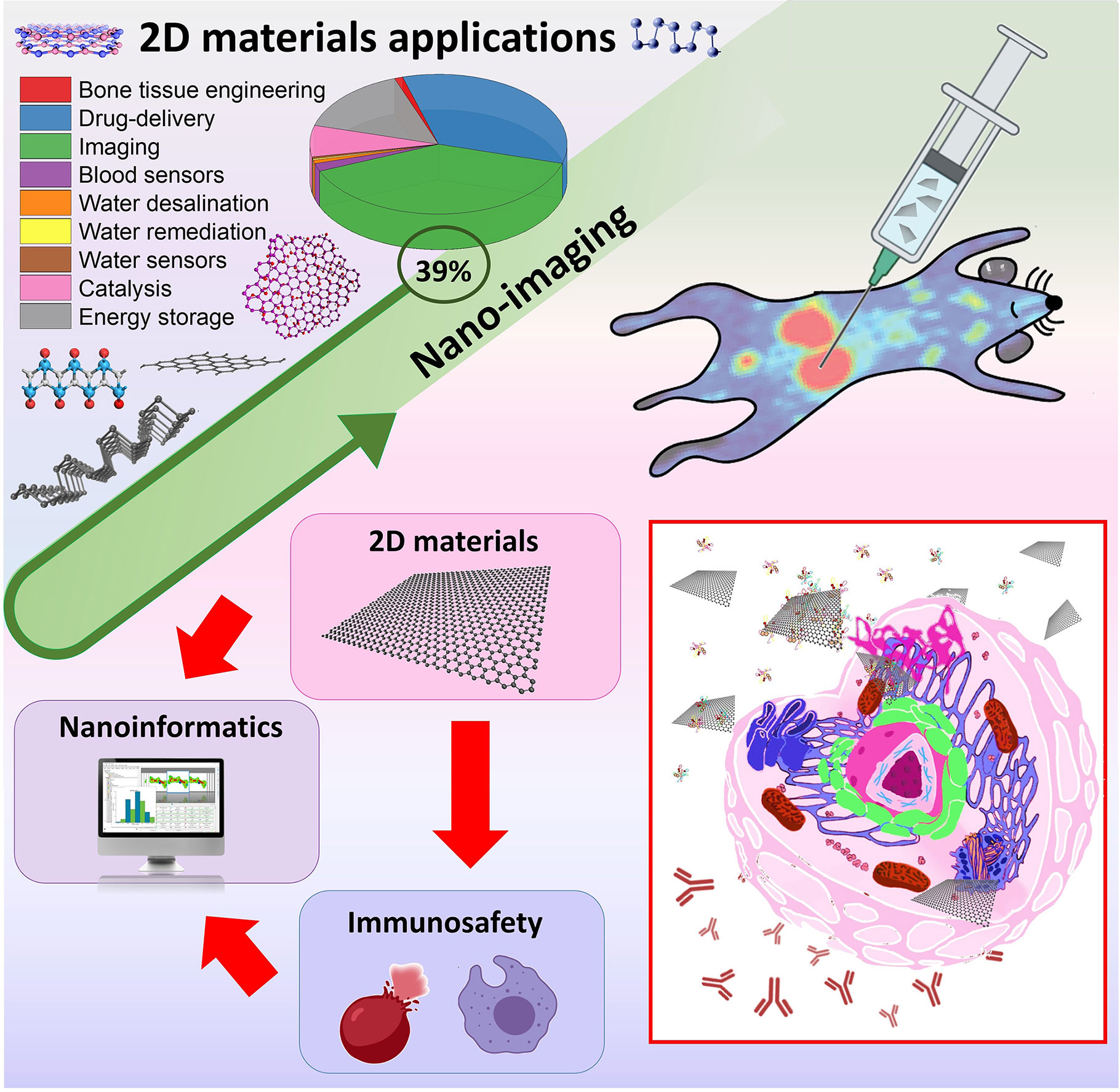
Figure 1 Two-dimensional materials applications, nano-imaging and their links with immunosafety and nanoinformatics approaches.
Technological Applications And Innovation Of 2d Materials
A literature review on the Web of Science™ database was performed, considering articles published from 2000 to 2021 (25/03/2021), and over these last 20 years, many 2D materials have been synthesized as exemplified in Figure 2A. The number of publications of 2D materials and their applications is growing, in which nano-imaging and drug release systems stand out and are present mostly in the health sector (Figures 2B, D). For energy application, the structural and electronic properties of 2D materials have been shown to improve the energy accumulation in devices such as lithium-ion, metal-air batteries (LIBs) (9, 39, 49, 50) and electrochemical devices (51, 52). Moreover, these 2D materials are of particular interest as catalysts and nanoscale substrates, replacing transition, or noble metals normally used to catalyze an acid-basic reaction, producing metal free-catalysts (53, 54). In environment, the 2D materials have been used as adsorbents for removing pollutants to treat contaminated water (55–57). Their atomic thickness and antibacterial activity contribute to superior water permeability and anti-fouling capacity in the development of membranes for desalination (58–62) and cleaning purposes (63–65). Sensing has covered both environmental and health sectors, contributing to the detection and monitoring of traces of pollutants (66, 67) and blood biomarkers (68–71). The thin structure, large surface area, chemical modifications and quenching ability of 2D materials provide high sensitivity, durability, stability, selectivity, and conductivity for sensors and biosensors (72–82).
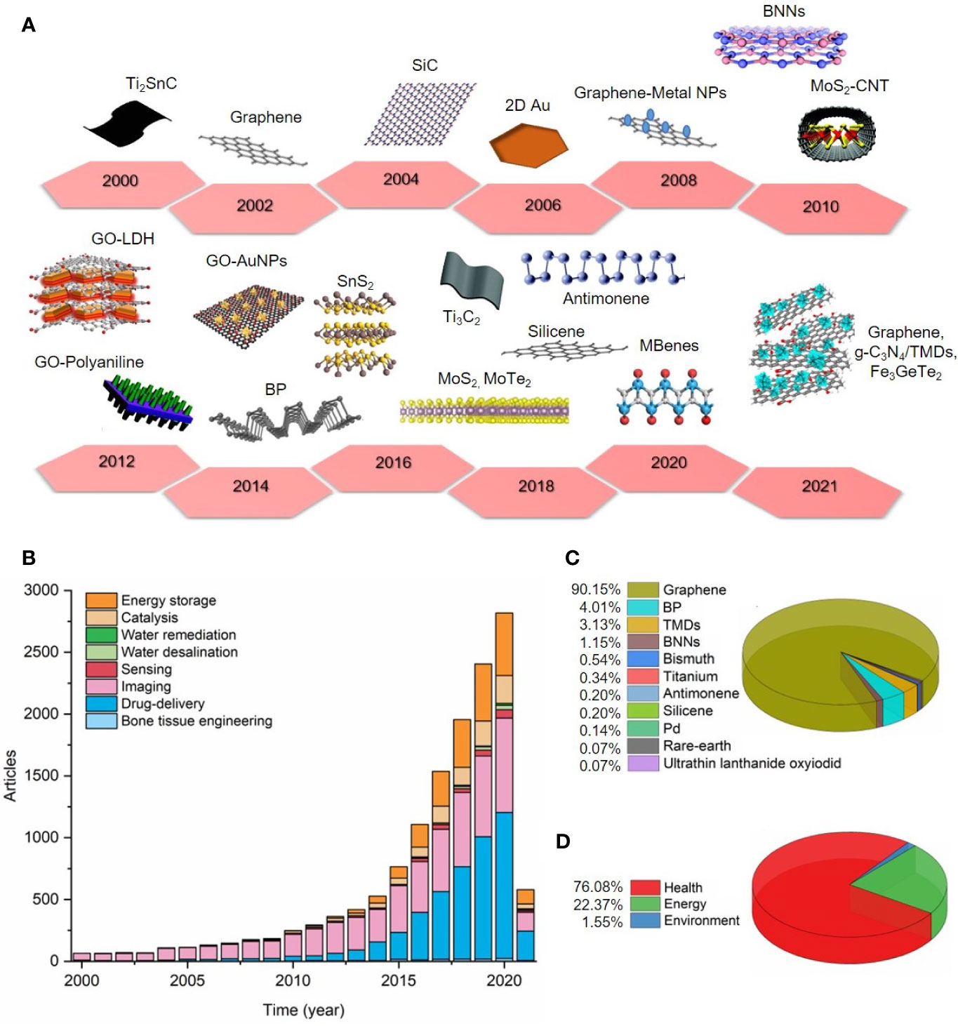
Figure 2 The data obtained previously was organized into the following sectors: health (bone tissue engineering, drug delivery, imaging, sensing blood markers), energy (catalysis and energy storage), and environment (water remediation and desalination, and water sensing contaminants). (A) Timeline showing examples of 2D materials produced over the period established (from 2000 to 2021). (B) Number of articles from 2000 to 2021 (25/03/2021) (C) 2D materials used in nano-imaging applications (see supporting information) (D) Percentage of 2D materials applied in health, energy and environment sectors.
Considering biomedical applications, 2D materials have been applied in bone tissue engineering, conferring improved mechanical characteristics and great osteoconductivity for scaffold design (83–87). However, due to the higher surface area of 2D materials and distinguish light-material interactions, research has mostly given attention to their usefulness in nano-imaging and therapeutics (theranostics) (88) (Figures 2B, C), including early detection, monitoring, and treatment of diseases, which are the main examples described in this mini-review (89). For example, in cancer, malign tumors are sensitive to heating when compared to healthy tissues. Graphene oxide decorated with gold nanoparticles (GO-AuNPs), TMDs (MoS2, WS2), and MXenes (MoC2, Ti3C2) have shown effective agents in photothermal and photodynamic therapy for inducing tumor necrosis (40, 41, 90–92). 2D materials have been successfully modified with numerous polymers to enhance their cytocompatibility and dispersibility (90) and used as nanoplatforms carrying active molecules or imaging agents to improve their biological function (93) and clinical visualization for imaging-drug delivery guiding (12, 94). MoS2 and BNNs have been employed as effective fluorescence quenchers and associated with aptamers, substituting antibody-based therapy (69, 95–97). Compared to the other 2D inorganic materials, and in addition to the previous features, the ultrathin structure of the BP nanosheets results in an exceptional biodegradability in physiological media it shows promising in theranostics (98, 99). Magnetic nanoparticles have been used as contrast agents and incorporated into 2D materials in MRI, in place of conventional ones (100, 101). In this respect, 2D magnetic materials production can be very useful for accurate bioimaging and therapy of diseases in vivo using MRI and CT techniques (10, 102).
2D Materials And The Immune System: Adverse Effects In In Vitro And In Vivo Models
As far as it is known, 2D materials have proven their significance and innovation perspective in almost all industrial areas and sectors, making it imperative to assess their environmental health risks and safety aspects (24, 103–105). However, toxicological studies, including immunotoxicity, are still in their infancy for GBMs and 2D inorganic materials (31). Table 1 is an extensive literature revision reporting major findings of 2D materials and their adverse effects in the immunological system considering in vitro and in vivo models. The terms used for the literature research is detailed in the supplementary material.

Table 1 Relevant studies addressing the adverse immunological effects of 2D materials in in vitro and in vivo models from 2000 to 2021.
Studies have demonstrated that 2D materials can induce immunological system activation with a consequent induction of an inflammatory response (145). This immunological system activation showed itself to be dependent of the 2D materials’ physicochemical properties, such as size (106–109, 144), surface chemistry (114, 115, 123), number of layers, shape (118, 119), and functionalization (109, 112, 114, 128, 135, 139). For example, Yue et al. (106) demonstrated that larger graphene oxide (GO) (2 µm) has induced a higher immunological activation than smaller GO (350 nm) both in vitro (peritoneal macrophages) and in vivo (C57BL/6 mice). Similarly, Ma et al. (107) showed a lateral-size-dependent pro-inflammatory effect of GO under in vitro and in vivo conditions, wherein the largest GO (L-GO; 750–1300 nm) elicit higher inflammatory response than smallest GO (S-GO; 50–350 nm). Moreover, the mechanism of inflammation has also differed according to the lateral size, with L-GO being more prone to plasma membrane adsorption and the toll-like receptors (TLRs) and nuclear factor-κB (NF-κB) pathways activation, whereas S-GO was mostly taken up by macrophages. In another study that investigated the effects of small GO (S-GO < 1 µm) and large GO (L-GO, 1–10 µm) on human peripheral immune cells, it was found that the S-GO has a more significant impact on the upregulation of critical genes implicated in immune responses and the release of cytokines IL1β and TNFα compared to L-GO (108). However, it is important to clarify here that the S-GO in this study presented similar lateral size of the L-GO in the previous studies cited, which means that all these studies are in agreement, and we may erroneously interpret them because attention to the lateral size was not devoted. Indeed, a nomenclature harmonization of GBMs is urgently needed to allow a clear understanding on the impacts of GBM physicochemical properties on their biocompatibility.
Besides to assess the effect of lateral size, Duarte and coworkers (109) investigated the impacts of two different surfaces functionalization: pegylated graphene oxide (GO-PEG, 200–500 nm) and flavin mononucleotide-stabilized pristine graphene with two different sizes (200–400 nm and 100–200 nm). Their results showed that the cellular uptake of GBMs was mainly influenced by their lateral size, with smaller particles showing greater internalization, while the inflammatory response depended also on the type of functionalization, with GO-PEG showing the lower pro-inflammatory potential. This study corroborates in number previous ones that also showed an increased biocompatibility of GO due to the pegylation (GO-PEG) (110, 111). Similarly, Xie et al. (139) studied PEG coated 2D titanium nanosheets (TiNS-PEG) and reported no indication of inflammation and other negative impacts. Moreover, the material was promising for photothermal tumor therapy and presented a high contrast for in vivo imaging. However, Gu et al. (129) found that MoS2 and PEGylated MoS2 induced a robust macrophage immune response, with PEG-MoS2 eliciting stronger cytokine secretion than the pristine MoS2. By performing molecular dynamics simulations, they demonstrated that small MoS2 nanoflakes can penetrate the macrophage membrane, and that the PEG chain on PEG-MoS2 lead to a prolonged passage throughout the membrane. Such a result might explain why PEG-MoS2 triggers sustained more stimulation of macrophages than pristine MoS2.
Other types of functionalization have also been studied in respect to their biocompatibility to immune cells. For instance, Zhi et al. (112) reported that the polyvinylpyrrolidone (PVP) coating of GO has exhibited lower immunogenicity when compared with pristine GO in relation to the inducing differentiation and maturation of dendritic cells (DCs), provoking a delaying in apoptotic process of T lymphocytes and the anti-phagocytosis ability against macrophages.
Surface chemistry has also been shown to influence on the immunotoxicity of 2D materials. Gurunathan et al. (114) reported that both GO and reduced GO (rGO) induced a dose-dependent loss of cell viability and proliferation, cell membrane damage, a loss of mitochondrial membrane potential, a decreased level of ATP, a redox imbalance, and an increased secretion of various cytokines and chemokines (IL1-β, TNF-α, GM-CSF, IL-6, IL-8, and MCP-1) by THP-1 cells. However, to all these toxic effects the rGO presented a significantly worse response compared to GO. In a previous study, Yan et al. (115) showed that different oxidation degrees resulted in the toxicity of monocytes via different signaling pathways, with GO nanoplatelets (GONPs) inducing the expression of antioxidative enzymes and inflammatory factors, whereas the reduced GO nanoplatelets (rGONPs) activated the NF-кB pathway. The contradictory results between these two studies, in relation to cytokine and chemokine expression, may be due to differences in the GBMs studied (i.e. GO sheets versus GO nanoplatelets), and they raise the need for further investigation concerning the effects of the oxidative degree of GBMs on immune cells.
In order to investigate the pristine graphene effects in vitro (THP-1 cell line) and in vivo (C57BL/6 strain mice), Schinwald et al. (118) have assessed the impacts of the shape of graphene nanoplatelets (GNPs) on their inflammatory potential. This large few-layer graphene presented as inflammogenic both in vitro and in vivo, which was attributed to its large size that led to frustrated phagocytosis. The authors highlighted that the potential hazard of GNPs could be minimized by producing GNPs small enough to be phagocytosed by macrophages. Moreover, the number of GO layers has been shown to affect its immunotoxicity, in which single-layer GO (SLGO) caused a more pronounced decrease in cell viability due to membrane damage of THP-1 cells, while multi-layer GO (MLGO) induced higher reactive oxygen species (ROS) and IL-1β production, leading to necrosis and apoptosis (120). In addition, the histological animal analysis revealed that SLGO and MLGO induced acute and chronic damage to the lungs and kidneys in the presence or absence of Pluronic F-127 (120).
Another important parameter, when approaching nanomaterial biosafety, is colloidal stability. Aggregation can influence the immunological response as observed by Wang et al. (127), when compared the toxicological profile of 2D MoS2 versus aggregated MoS2 in lung cells and mice. In their in vitro evaluation, in THP-1 and BEAS-2B cells, they found that aggregated MoS2 induces strong proinflammatory and profibrogenic responses, while 2D MoS2 have little or no effect. In agreement with in vitro results, an acute toxicity study in vivo showed that aggregated MoS2 induced an acute lung inflammation, while 2D MoS2 had no or a slight effect.
To increase the stability of 2D materials, studies have shown that proteins can be used as a dispersant agent. Lin et al. (142) studied silicene nanosheets modified with a bovine albumin serum protein corona (SNSs-BSA) and observed a significant increase in the colloidal stability in several physiological media (0.9% saline, phosphate buffered saline and Dulbecco’s modified Eagle medium). Furthermore, SNSs-BSA did not cause significant toxicity in vitro neither significant acute toxicity in vivo. Only meaningless hematological changes were observed during the treatment duration, and no significant inflammation or infection were caused by the SNSs-BSA.
It is imperative that in a physiological environment, the nanomaterials will interact with biomolecules, forming a complex biomolecular corona. Those biomolecules (e.g., proteins, lipids, carbohydrates) can change the identity of the nanomaterials and influence their interaction with biological systems, causing an increase or decrease in internalization, toxicity, and biocompatibility as well as in colloidal stability over time. Thus, the biotransformation of nanomaterials in a physiological environment is an important parameter to be studied (146). The most common and highly studied component of biomolecular corona is the protein corona. In this sense, Mo et al. (132) studied the effect of the human plasma protein corona on the cytotoxicity of BP nanosheets and BP quantum dots (BPQDs) observing a reduction in cell viability for both nanomaterials when coated with proteins. However, protein corona facilitated BP nanosheet internalization and induced an increase in inflammatory cytokines (IL-1β, IL-6, IL-8 and IFN-γ) and in ROS generation. Besides, it was observed that protein corona coated BP caused an induction on the nitric oxide (NO) and tumour necrosis factor. Further, Mo et al. (133) studied the effect of the human plasma protein corona in BP toxicity, and observed an increased macrophage polarization due to the adsorption of opsonins present in the plasma, increasing the uptake of BP and the interaction with stromal interaction molecule 2 (STIM2) protein facilitating Ca2+ influx.
Similarly, Han et al. (126) studied the effect of plasma corona-coated 2D monoelemental nanosheets and observed that the protein corona decreases cytotoxicity and cell membrane damage for borophene, phosphorene, and graphene nanosheets. The corona coating induced the secretion of inflammatory cytokines (IL-1β, IL-6, IL-8, and IFN-γ) for all three materials. Also, for BNNs, it was observed an increase in cellular uptake when the material was coronated, and therefore, the corona may promote phagocytosis. Baimanov et al. (31) also investigated the effect of four different blood protein coronas (human serum albumin (HSA), transferrin (Tf), fibrinogen (Fg), and immunoglobulin G (IgG) corona) on cell viability, uptake, and pro-inflammatory effects of MoS2 nanosheets (NSs) in the macrophages cell line. Their results demonstrate that blood proteins contribute to uptake and inflammatory effects, as protein coated MoS2 NSs increase cell viability and decrease cytoplasmic membrane damage when compared to non-coated MoS2 NSs. Besides, it was observed that the type of protein influences cytokine secretion, as IgG-coated MoS2 NSs causes more inflammatory cytokine secretion (TNF-α, IL-6 and IL-1β). The highest proportion of β-sheets on IgG led to fewer secondary structure changes on MoS2 NSs, facilitating uptake and producing a stronger pro-inflammatory response in macrophages due to the recognition of an MoS2 NSs−IgG complex by Fc gamma receptors and the subsequent activation of the NF-κB pathways. Another interesting finding is that in a serum-containing medium, cellular uptake of MoS2 NSs−protein complexes was higher than that in a serum-free medium. Also, the MoS2 NSs−Fg, and MoS2 NSs–serum complexes had similar results in serum-free conditions and different results in a serum-containing medium, suggesting the formation of the protein corona layer above the previously formed MoS2 NSs−protein complexes. Those studies can help to elucidate the mechanisms in which protein corona can affects the toxicity of 2D materials.
One important ability of the immune system is the innate immune memory, where cells from the innate immune system react to secondary stimulus, which mostly includes an increased or decreased production of inflammation-related factors (147). With regard to 2D materials studies, there is yet a little research on this topic. Liu et al. (148) functionalize GO with lentinan (LNT) and observed that GO-LNT was able to promote macrophage activation by NF-κB and TLR signaling pathway, as well as enhance antigen protein processing after initial contact with macrophage. Moreover, the efficiency of this material was investigated, as a vaccine adjuvant for ovalbumin (OVA), in this sense GO-LNT induced robust long-term OVA-specific antibody responses due to the prolonged release of OVA. Besides this, GO-LNT was able to sustain a long-term immune response because it facilitated the uptake and slowed the release rate of antigen in macrophage. Further, Lebre et al. (149), demonstrated that pristine graphene can promote the innate immune training, enhancing the secretion of IL-6 and TNF-α and a decrease in IL-10 after toll-like receptor ligand stimulation 5 days after graphene exposure, indicating that pristine graphene can activate the immune innate memory.
Immune cells, such as macrophages and neutrophils, are one of the first line of defense of the immune system; they are capable of engulf the foreign material (or pathogen), degrading it and producing cytokines to enhanced the immune response (150). The uptake of 2D materials by immune system cells have been reported in various studies (31, 109, 115, 126, 132); however, there are few studies that address the degradation of those materials after internalization. Mukherjee et al. (151) studied the degradation of large and small GO by neutrophils and observed that not only both GO be degraded by neutrophils but also that the product of the degradation was non-toxic to human cells. Similarly, Moore et al. (152) studied the degradation of few-layer MoS2 in human macrophage-like cells and observed that internalization occurred following 4 h of exposure and after 24 h the in vitro degradation of the material was confirmed, which occurred within lipidic vesicles and associated with enzymatic regions containing lysozyme.
As presented above, 2D nanomaterials may have an inflammogenic potential and immunotoxicity, which may impair their successful clinical translation; however, the immunological system activation can also be useful for theragnostic purposes. This application uses the immune responses to protect the body and eliminate cancer cells. The advantage of immunotherapy is that it engages the immune system to kill tumor cells without damaging healthy cells, additionally, it may induce immunological memory, causing long-lasting protection (153).
Nanoinformatics Approaches Toward Immunosafety-by-Design
In materials science, theory, computational modeling and informatics have a substantial role in accelerating and discovering new materials with interesting properties and applications (154–156). Due to the growing interest in 2D nanomaterials, computational approaches are extensively used in the discovery, development and application of these materials by detailed study of their structure/property relationships (156–158).
The nano-bio interface phenomena are directly related to the physicochemical properties of nanomaterials. However, tracing general correlations and delineating predictive models of nanomaterials biological effects remains challenging. Some issues include the complexity of nano-bio interactions, nanomaterials structural heterogeneity, lack of standard methodologies, absence of systematic studies and low-quality nanomaterial characterization (159–161). In this context, computational methods have been incorporated into the nanotoxicology field to support the understanding of the nano-bio interface to enable the development of safe-by-design principles applied to nanomaterials (162, 163). Theoretical modeling (i.e., molecular dynamics, density functional theory) enables precise control of critical parameters of the nanomaterials surface to study their individual effects in nano-bio interactions, providing mechanistic knowledge (164–166). On the other hand, machine learning (ML) techniques are used to assess datasets of nanomaterials biological outcomes in order to find patterns and correlations between physicochemical properties and biological effects, often undetectable through other types of analysis (167–169).
Applications of data-driven strategies include data filling, grouping, and predictive modeling. Quantitative nanostructure–activity relationships (QNAR) consist of the main strategy to delineate prediction models based on correlations between nanomaterial structural characteristics to their properties and biological activities (170, 171). It is based on the assumption that nanomaterials in their properties present similar biological effects. Diverse algorithms can be used in QNAR models, including support vector machine (172), artificial neural network (173), and decision trees (174), among others, and depending of the level of algorithms interpretability may enable the outline of causal relationships.
The scarcity of quality data and comprehensive databases is the major bottleneck in the application of ML to predict nanomaterials immune reactions (175, 176). Data-driven strategies have been making important advances in modeling biological phenomena that have potential usage to evaluate nano-immune interactions, such as predicting biomolecular corona compositions (177–181), and nanomaterials and cell interactions (e.g., cell uptake, cytotoxicity, membrane integrity, oxidative stress) (182–185). Furthermore, the exploration of omics approaches (e.g., genomics, transcriptomics, and metabolomics) has promoting the development of ML models to process the complex data generated by these techniques and enables a better understanding of the molecular mechanisms of nanomaterials adverse effects in a systemic context, defining and predicting adverse outcome pathways (186–189). The omics’ potential of data generation is demonstrated by Kinaret et al. (190), who were able to connect immune responses to observed transcriptomic alterations in mouse airway exposed to 28 engineered nanomaterials. Together with cytological and histological analyses (imaging processing), they generated an extensive in vivo data set of nanomaterial adverse effects.
Allied with quality data infrastructure and processing, computational methods are sizeable to deal with complexity of nano-bio interface to assess and model the toxicity of nanomaterials in a variety of environments (163, 191–194). To support safe-by-design approaches, international efforts have been made to provide data integration and sharing, modeling tools, standard protocols, and ontologies, to ensure Findable, Accessible, Interoperate, and Reusable (FAIR) data (195, 196). For example, European projects, such as NanosolveIT and NanoCommons, and more recently CompSafeNano are initiatives facing on this direction (164, 165, 197, 198). In accordance with these initiatives, Gazzi et al. (199) recently presented the nanoimmunity-by-design concept developed inside G-IMMUNOMICS and CARBO-IMmap projects, which aim to bridge the knowledge gaps in the immune characterization of carbon-based materials, integrating data-driven methodologies which are extendable to other 2D materials.
Conclusions And Future Perspectives
Two-dimensional materials are key elements for nanoscience and innovation in energy, health, and the environment. This can lead to a broad range of technological applications, especially nano-imaging, which has been growing exponentially in recent years. The wide number of 2D materials with different physicochemical properties make immunotoxicity and safety evaluation a challenge. There are therefore still gaps and controversial data in the literature. For example, within the same material category (i.e., graphene oxide) different properties were observed that might affect immunological and toxicological responses. It is imperative to evaluate the biological effects of biomolecular corona formation on 2D materials at nanobiointerfaces. Only by the identification of these material properties (intrinsic and extrinsic) and an integrated understanding on how they may influence its immunological response, we can manage immunotoxicity/biocompatibility and then benefit from their unique properties for many applications. Furthermore, it is very important to highlight the critical influence of endotoxin contamination prior immunological studies and toxicity testing. Special attention on this topic will avoid misinterpretation of immunosafety results involving 2D materials (148). In addition, it is important to advance in the understanding of the links between nanomaterials and the immune system across environmental species; this being a future challenge for immunosafety research associated with 2D materials (200). Nanoinformatics and computational modeling will have a decisive role on immunotoxicological studies with nanomaterials toward the practical implementation of immunosafety-by-design. However, it is very important to develop harmonized protocols, ontologies, and public databases to facilitate and promote a global research community for the collaboration and an exchange of knowledge in this field, focusing efforts on FAIR data principles.
Author Contributions
All authors listed have made a substantial, direct and intellectual contribution to the work, and approved it for publication. GS and LFr: literature research, data curation, writing, and editing. RP, LFo, and MM: literature research and writing. DM, AF, and OA: funding acquisition, supervision, project administration, and writing. All authors contributed to the article and approved the submitted version.
Funding
This work was funded by the Sao Paulo Research Foundation (FAPESP, grant no. 18/25103-0; 17/02317-2; 14/50906-9), the National Council for Scientific and Technological Development (CNPq, grant no. 315575/2020-4; 301358/2020-6), and the Coordination for the Improvement of Higher Education Personnel (CAPES, Finance code 001).
Conflict of Interest
The authors declare that the research was conducted in the absence of any commercial or financial relationships that could be construed as a potential conflict of interest.
Acknowledgments
The authors thank the National System of Laboratories in Nanotechnologies (SisNANO/MCTI), the National Institute for Research and Development in Complex Functional Materials (INCT-Inomat) and the National Institute for Research and Development in Carbon nanomaterials (INCT-NanoCarbono).
Supplementary Material
The Supplementary Material for this article can be found onlineat: https://www.frontiersin.org/articles/10.3389/fimmu.2021.689519/full#supplementary-material
References
1. Nicolosi V, Chhowalla M, Kanatzidis MG, Strano MS, Coleman JN. Liquid Exfoliation of Layered Materials. Sci (80- ) (2013) 340:1226419–1226419. doi: 10.1126/science.1226419
2. Hu T, Mei X, Wang Y, Weng X, Liang R, Wei M. Two-Dimensional Nanomaterials: Fascinating Materials in Biomedical Field. Sci Bull (2019) 64:1707–27. doi: 10.1016/j.scib.2019.09.021
3. Novoselov KS, Geim AK, Morozov SV, Jiang D, Zhang Y, Dubonos SV, et al. Electric Field in Atomically Thin Carbon Films. Sci (80- ) (2004) 306:666–9. doi: 10.1126/science.1102896
4. Hao S, Zhao X, Cheng Q, Xing Y, Ma W, Wang X, et al. A Mini Review of the Preparation and Photocatalytic Properties of Two-Dimensional Materials. Front Chem (2020) 8:582146. doi: 10.3389/fchem.2020.582146
5. Mohammadpour Z, Majidzadeh-a K. Applications of Two-Dimensional Nanomaterials in Breast Cancer Theranostics. ACS Biomater Sci Eng (2020) 6(4):1852–73. doi: 10.1021/acsbiomaterials.9b01894
6. Rohaizad N, Mayorga-Martinez CC, Fojtů M, Latiff NM, Pumera M. Two-Dimensional Materials in Biomedical, Biosensing and Sensing Applications. Chem Soc Rev (2021) 50:619–57. doi: 10.1039/d0cs00150c
7. Bolotsky A, Butler D, Dong C, Gerace K, Glavin NR. Two-Dimensional Materials in Biosensing and Healthcare: From In Vitro Diagnostics to Optogenetics and Beyond. ACS Nano (2019) 13:9781–810. doi: 10.1021/acsnano.9b03632
8. Samorì P, Feng X, Palermo V. Introduction to ‘Chemistry of 2D Materials: Graphene and Beyond.’. Nanoscale (2020) 12:24309–10. doi: 10.1039/d0nr90263b
9. Ostadhossein A, Guo J, Simeski F, Ihme M. Functionalization of 2D Materials for Enhancing OER/ORR Catalytic Activity in Li–oxygen Batteries. Commun Chem (2019) 2:1–11. doi: 10.1038/s42004-019-0196-2
10. Och M, Martin M, Dlubak B, Mattevi C, Martin M, Martin M. Synthesis of Emerging 2D Layered Magnetic Materials. Nanoscale (2021) 13:2157–80. doi: 10.1039/d0nr07867k
11. Tao W, Ji X, Zhu X, Li L, Wang J, Zhang Y, et al. Two-Dimensional Antimonene-Based Photonic Nanomedicine for Cancer Theranostics. Adv Mater (2018) 30:1–11. doi: 10.1002/adma.201802061
12. Tao W, Zhu X, Yu X, Zeng X, Xiao Q, Zhang X, et al. Black Phosphorus Nanosheets as a Robust Delivery Platform for Cancer Theranostics. Adv Mater (2017) 29:1–9. doi: 10.1002/adma.201603276
13. Ares P, Palacios JJ, Abellán G, Gómez-Herrero J, Zamora F. Recent Progress on Antimonene: A New Bidimensional Material. Adv Mater (2018) 30:1–27. doi: 10.1002/adma.201703771
14. Wang H, Wang X, Xia F, Wang L, Jiang H, Xia Q, et al. Black Phosphorus Radio-Frequency Transistors. Nano Lett (2014) 14:6424–9. doi: 10.1021/nl5029717
15. Ugeda MM, Bradley AJ, Shi SF, Da Jornada FH, Zhang Y, Qiu DY, et al. Giant Bandgap Renormalization and Excitonic Effects in a Monolayer Transition Metal Dichalcogenide Semiconductor. Nat Mater (2014) 13:1091–5. doi: 10.1038/nmat4061
16. Huang C, Wu S, Sanchez AM, Peters JJP, Beanland R, Ross JS, et al. Lateral Heterojunctions Within Monolayer MoSe2-WSe2 Semiconductors. Nat Mater (2014) 13:1096–101. doi: 10.1038/nmat4064
17. Abo El-Reesh GY, Farghali AA, Taha M, Mahmoud RK. Novel Synthesis of Ni/Fe Layered Double Hydroxides Using Urea and Glycerol and Their Enhanced Adsorption Behavior for Cr(VI) Removal. Sci Rep (2020) 10:1–20. doi: 10.1038/s41598-020-57519-4
18. Zhu C, Du D, Lin Y. Graphene and Graphene-Like 2D Materials for Optical Biosensing and Bioimaging: A Review. 2D Mater (2015) 2:32004. doi: 10.1088/2053-1583/2/3/032004
19. Eom S, Choi G, Nakamura H, Choy J-H. 2-Dimensional Nanomaterials With Imaging and Diagnostic Functions for Nanomedicine; A Review. Bull Chem Soc Jpn (2020) 93:1–12. doi: 10.1246/bcsj.20190270
20. Wang X, Cheng L. Multifunctional Two-Dimensional Nanocomposites for Photothermal-Based Combined Cancer Therapy. Nanoscale (2019) 11:15685–708. doi: 10.1039/C9NR04044G
21. Cai X, Liu X, Jiang J, Gao M, Wang W, Zheng H, et al. Molecular Mechanisms, Characterization Methods, and Utilities of Nanoparticle Biotransformation in Nanosafety Assessments. Small (2020) 1907663:1–19. doi: 10.1002/smll.201907663
22. Fadeel B, Bussy C, Merino S, Vázquez E, Flahaut E, Mouchet F, et al. Safety Assessment of Graphene-Based Materials: Focus on Human Health and the Environment. ACS Nano (2018) 12:10582–620. doi: 10.1021/acsnano.8b04758
23. Chetwynd AJ, Wheeler KE, Lynch I. Best Practice in Reporting Corona Studies: Minimum Information About Nanomaterial Biocorona Experiments (Minbe). Nano Today (2019) 28:100758. doi: 10.1016/j.nantod.2019.06.004
24. Ma B, Martín C, Kurapati R, Bianco A. Degradation-by-Design: How Chemical Functionalization Enhances the Biodegradability and Safety of 2D Materials. Chem Soc Rev (2020) 49:6224–47. doi: 10.1039/c9cs00822e
25. Ganguly P, Breen A, Pillai SC. Toxicity of Nanomaterials: Exposure, Pathways, Assessment, and Recent Advances. ACS Biomater Sci Eng (2018) 4:2237–75. doi: 10.1021/acsbiomaterials.8b00068
26. Orecchioni M, Bedognetti D, Newman L, Fuoco C, Spada F, Hendrickx W, et al. Single-Cell Mass Cytometry and Transcriptome Profiling Reveal the Impact of Graphene on Human Immune Cells. Nat Commun (2017) 8(1):1–14. doi: 10.1038/s41467-017-01015-3
27. Kämpfer AAM, Busch M, Schins RPF. Advanced In Vitro Testing Strategies and Models of the Intestine for Nanosafety Research. Chem Res Toxicol (2020) 33:1163–78. doi: 10.1021/acs.chemrestox.0c00079
28. Cronin JG, Jones N, Thornton CA, Jenkins GJS, Doak SH, Clift MJD. Nanomaterials and Innate Immunity: A Perspective of the Current Status in Nanosafety. Chem Res Toxicol (2020) 33:1061–73. doi: 10.1021/acs.chemrestox.0c00051
29. Franqui LS, De Farias MA, Portugal RV, Costa CAR, Domingues RR, Souza Filho AG, et al. Interaction of Graphene Oxide With Cell Culture Medium: Evaluating the Fetal Bovine Serum Protein Corona Formation Towards In Vitro Nanotoxicity Assessment and Nanobiointeractions. Mater Sci Eng C (2019) 100:363–77. doi: 10.1016/J.MSEC.2019.02.066
30. Cao M, Cai R, Zhao L, Guo M, Wang L, Wang Y, et al. Molybdenum Derived From Nanomaterials Incorporates Into Molybdenum Enzymes and Affects Their Activities In Vivo. Nat Nanotechnol (2021) 1–9. doi: 10.1038/s41565-021-00856-w
31. Baimanov D, Wu J, Chu R, Cai R, Wang B, Cao M, et al. Immunological Responses Induced by Blood Protein Coronas on Two-Dimensional Mos 2 Nanosheets. ACS Nano (2020) 14:5529–42. doi: 10.1021/acsnano.9b09744
32. Ede JD, Lobaskin V, Vogel U, Lynch I, Halappanavar S, Doak SH, et al. Translating Scientific Advances in the AOP Framework to Decision Making for Nanomaterials. Nanomaterials (2020) 10:1–22. doi: 10.3390/nano10061229
33. Chen C, Leong DT, Lynch I. Rethinking Nanosafety: Harnessing Progress and Driving Innovation. Small (2020) 16:2–5. doi: 10.1002/smll.202002503
34. Miernicki M, Hofmann T, Eisenberger I, Von Der KF, Praetorius A. Legal and Practical Challenges in Classifying Nanomaterials According to Regulatory Definitions. Nat Nanotechnol (2019) 14(3):208–16. doi: 10.1038/s41565-019-0396-z
35. Shatkin JA. The Future in Nanosafety. Nano Lett (2020) 20:1479–80. doi: 10.1021/acs.nanolett.0c00432
36. Varsou D, Afantitis A, Tsoumanis A, Melagraki G, Sarimveis H, Valsamijones E. Nanoscale Advances A Safe-by-Design Tool for Functionalised Nanomaterials Through the Enalos Nanoinformatics. Nanoscale Adv (2019) 1:706–18. doi: 10.1039/c8na00142a
37. Singh AV, Maharjan RS, Kanase A, Siewert K, Rosenkranz D, Singh R, et al. Machine-Learning-Based Approach to Decode the Influence of Nanomaterial Properties on Their Interaction With Cells. ACS Appl Mater Interfaces (2021) 13:1943–55. doi: 10.1021/acsami.0c18470
38. Sturla SJ, Boobis AR, FitzGerald RE, Hoeng J, Kavlock RJ, Schirmer K, et al. Systems Toxicology: From Basic Research to Risk Assessment. Chem Res Toxicol (2014) 27:314–29. doi: 10.1021/tx400410s
39. Li B, Wu Y, Li N, Chen X, Zeng X, Arramel, et al. Single-Metal Atoms Supported on MBenes for Robust Electrochemical Hydrogen Evolution. ACS Appl Mater Interfaces (2020) 12:9261–7. doi: 10.1021/acsami.9b20552
40. Wang Y, Polavarapu L, Liz-Marzán LM. Reduced Graphene Oxide-Supported Gold Nanostars for Improved SERS Sensing and Drug Delivery. ACS Appl Mater Interfaces (2014) 6:21798–805. doi: 10.1021/am501382y
41. Yin W, Yan L, Yu J, Tian G, Zhou L, Zheng X, et al. High-Throughput Synthesis of Single-Layer MoS2 Nanosheets as a Near-Infrared Photothermal-Triggered Drug Delivery for Effective Cancer Therapy. ACS Nano (2014) 8:6922–33. doi: 10.1021/nn501647j
42. Garcia-Gallastegui A, Iruretagoyena D, Gouvea V, Mokhtar M, Asiri AM, Basahel SN, et al. Graphene Oxide as Support for Layered Double Hydroxides: Enhancing the CO2 Adsorption Capacity. Chem Mater (2012) 24:4531–9. doi: 10.1021/cm3018264
43. Warner JH, Rümmeli MH, Bachmatiuk A, Büchner B. Atomic Resolution Imaging and Topography of Boron Nitride Sheets Produced by Chemical Exfoliation. ACS Nano (2010) 4:1299–304. doi: 10.1021/nn901648q
44. Huang Y, Sutter E, Sadowski JT, Cotlet M, Monti OLA, Racke DA, et al. Tin Disulfide-an Emerging Layered Metal Dichalcogenide Semiconductor: Materials Properties and Device Characteristics. ACS Nano (2014) 8:10743–55. doi: 10.1021/nn504481r
45. Naguib M, Mashtalir O, Carle J, Presser V, Lu J, Hultman L, et al. Two-Dimensional Transition Metal Carbides. ACS Nano (2012) 6:1322–31. doi: 10.1021/nn204153h
46. Shwetharani R, Kapse S, Thapa R, Nagaraju DH, Balakrishna RG. Dendritic Ferroselite (FeSe2) With 2D Carbon-Based Nanosheets of rGO and G-C3n4as Efficient Catalysts for Electrochemical Hydrogen Evolution. ACS Appl Energy Mater (2020) 3:12682–91. doi: 10.1021/acsaem.0c02619
47. Wang Z, Li H, Liu Z, Shi Z, Lu J, Suenaga K, et al. Mixed Low-Dimensional Nanomaterial: 2D Ultranarrow MoS2 Inorganic Nanoribbons Encapsulated in quasi-1D Carbon Nanotubes. J Am Chem Soc (2010) 132:13840–7. doi: 10.1021/ja1058026
48. Wang L, Ye Y, Lu X, Wen Z, Li Z, Hou H, et al. Hierarchical Nanocomposites of Polyaniline Nanowire Arrays on Reduced Graphene Oxide Sheets for Supercapacitors. Sci Rep (2013) 3:5019–26. doi: 10.1038/srep03568
49. Li S, Liu Y, Zhao X, Shen Q, Zhao W, Tan Q, et al. Sandwich-Like Heterostructures of MoS2/Graphene With Enlarged Interlayer Spacing and Enhanced Hydrophilicity as High-Performance Cathodes for Aqueous Zinc-Ion Batteries. Adv Mater (2021) 2007480:1–9. doi: 10.1002/adma.202007480
50. Wang C, Yu X, Park HS. Boosting Redox-Active Sites of 1T MoS2Phase by Phosphorus-Incorporated Hierarchical Graphene Architecture for Improved Li Storage Performances. ACS Appl Mater Interfaces (2020) 12:51329–36. doi: 10.1021/acsami.0c12414
51. Vaghasiya JV, Mayorga-Martinez CC, Sofer Z, Pumera M. Mxene-Based Flexible Supercapacitors: Influence of an Organic Ionic Conductor Electrolyte on the Performance. ACS Appl Mater Interfaces (2020) 12:53039–48. doi: 10.1021/acsami.0c12879
52. Shen J, Chen X, Wang P, Zhou F, Lu L, Wang R, et al. Electrochemical Performance of Zinc Carbodiimides Based Porous Nanocomposites as Supercapacitors. Appl Surf Sci (2021) 541:148355. doi: 10.1016/j.apsusc.2020.148355
53. Chen LX, Chen W, Jiang M, Lu Z, Gao C. Insights on the Dual Role of Two-Dimensional Materials as Catalysts and Supports for Energy and Environmental Catalysis. J Mater Chem A (2021) 9:2018–42. doi: 10.1039/d0ta08649e
54. Deng D, Novoselov KS, Fu Q, Zheng N, Tian Z, Bao X. Catalysis With Two-Dimensional Materials and Their Heterostructures. Nat Nanotechnol (2016) 11:218–30. doi: 10.1038/nnano.2015.340
55. Huang H, Wang Y, Zhang Y, Niu Z, Li X. Amino-Functionalized Graphene Oxide for Cr(VI), Cu(Ii), Pb(II) and Cd(II) Removal From Industrial Wastewater. Open Chem (2020) 18:97–107. doi: 10.1515/chem-2020-0009
56. Ahmad H, Huang Z, Kanagaraj P, Liu C. Separation and Preconcentration of Arsenite and Other Heavy Metal Ions Using Graphene Oxide Laminated With Protein Molecules. J Hazard Mater (2020) 384:121479. doi: 10.1016/j.jhazmat.2019.121479
57. Li DO, Gilliam MS, Debnath A, Chu XS, Yousaf A, Green AA, et al. Interaction of Pb2+ Ions in Water With Two-Dimensional Molybdenum Disulfide. J Phys Mater (2020) 3:024007. doi: 10.1088/2515-7639/ab7ab3
58. Oliveira NC, Maia MT, Noronha VT, Petry R, Aquino YMLO, Paula AJ. Nanomaterials for Desalination. Elsevier Inc (2019) 227–62. doi: 10.1016/B978-0-12-814829-7.00006-9
59. Liu G, Shen J, Liu Q, Liu G, Xiong J, Yang J, et al. Ultrathin Two-dimensional Mxene Membrane for Pervaporation Desalination. J Memb Sci (2017) 548:548–58. doi: 10.1016/j.memsci.2017.11.065
60. Safaei J, Xiong P, Wang G. Progress and Prospects of Two-Dimensional Materials for Membrane- Based Water Desalination. Mater Today Adv (2020) 8:100108. doi: 10.1016/j.mtadv.2020.100108
61. Heiranian M, Farimani AB, Aluru NR. Water Desalination With a Single-Layer MoS2 Nanopore. Nat Commun (2015) 6(1):1–6. doi: 10.1038/ncomms9616
62. Caglar M, Silkina I, Brown BT, Thorneywork AL, Burton OJ, Babenko V, et al. Tunable Anion-Selective Transport Through Monolayer Graphene and Hexagonal Boron Nitride. ACS Nano (2020) 14:2729–38. doi: 10.1021/acsnano.9b08168
63. Ye S, Wang B, Pu Z, Liu T, Feng Y, Han W, et al. Flexible and Robust Porous Thermoplastic Polyurethane / Reduced Graphene Oxide Monolith With Special Wettability for Continuous Oil / Water Separation in Harsh Environment. Sep Purif Technol (2021) 266:118553. doi: 10.1016/j.seppur.2021.118553
64. Li Q, Zhang N, Yang Y, Wang G, Ng DHL. High Efficiency Photocatalysis for Pollutant Degradation With MoS2/ C3n4 Heterostructures. Langmuir (2014) 30:8695–972. doi: 10.1021/la502033t
65. Online VA, Yang X, Li J, Liang T, Zhang Y, Chen H, et al. Antibacterial Activity of Two-Dimensional MoS 2 Sheets. Nanoscale (2014) 6:10126–33. doi: 10.1039/c4nr01965b
66. Guo X, Yue G, Huang J, Liu C, Zeng Q, Wang L. Label-Free Simultaneous Analysis of Fe (III) and Ascorbic Acid Using Fluorescence Switching of Ultrathin Graphitic Carbon Nitride Nanosheets. ACS Appl Mater Interfaces (2018) 10:26118–27. doi: 10.1021/acsami.8b10529
67. Huang H, Chen R, Ma J, Yan L, Zhao Y, Wang Y, et al. Graphitic Carbon Nitride Solid Nanofilms for Selective and Recyclable Sensing of Cu2+ and Ag+ in Water and Serum. Chem Commun (2014) 50:15415–8. doi: 10.1039/c4cc06659f
68. Ou JZ, Chrimes AF, Wang Y, Tang S, Strano MS, Kalantar-zadeh K. Ion-Driven Photoluminescence Modulation of Quasi-Two- Dimensional MoS 2 Nano Fl Akes for Applications in Biological Systems. Nano Lett (2014) 14:857–63. doi: 10.1021/nl4042356
69. Deng H, Yang X, Gao Z. MoS2 Nanosheets as an Effective Fluorescence Quencher for DNA Methyltransferase Activity Detection. Analyst (2015) 140:3210–5. doi: 10.1039/c4an02133a
70. Wang L, Wang Y, Wong JI, Palacios T, Kong J, Yang HY. Functionalized MoS2 nanosheet‐based field‐effect biosensor for label‐free sensitive detection of cancer marker proteins in solution.. Small (2014) 10(6):1101–5. doi: 10.1002/smll.201302081
71. Lee KT, Liang YC, Lin HH, Li CH, Lu SY. Exfoliated SnS2 Nanoplates for Enhancing Direct Electrochemical Glucose Sensing. Electrochim Acta (2016) 219:241–50. doi: 10.1016/j.electacta.2016.10.003
72. Singh C, Ali A, Kumar V, Ahmad R, Sumana G. Chemical Functionalized MoS2 Nanosheets Assembled Microfluidic Immunosensor for Highly Sensitive Detection of Food Pathogen. Sensors Actuators B Chem (2018) 259:1090–8. doi: 10.1016/j.snb.2017.12.094
73. Elumalai S, Mani V, Jeromiyas N, Ponnusamy VK, Yoshimura M. A Composite Film Prepared From Titanium Carbide Ti3C2Tx (Mxene) and Gold Nanoparticles for Voltammetric Determination of Uric Acid and Folic Acid. Microchim Acta (2020) 187:1–10. doi: 10.1007/s00604-019-4018-0
74. Hernández-Sánchez D, Villabona-Leal G, Saucedo-Orozco I, Bracamonte V, Pérez E, Bittencourt C, et al. Stable Graphene Oxide-Gold Nanoparticle Platforms for Biosensing Applications. Phys Chem Chem Phys (2018) 20:1685–92. doi: 10.1039/c7cp04817c
75. Ji J, Wen J, Shen Y, Lv Y, Chen Y, Liu S, et al. Simultaneous Noncovalent Modification and Exfoliation of 2D Carbon Nitride for Enhanced Electrochemiluminescent Biosensing. J Am Chem Soc (2017) 139:11698–701. doi: 10.1021/jacs.7b06708
76. Lei Y, Butler D, Lucking MC, Zhang F, Xia T, Fujisawa K, et al. Single-Atom Doping of MoS2 With Manganese Enables Ultrasensitive Detection of Dopamine: Experimental and Computational Approach. Sci Adv (2020) 6:1–9. doi: 10.1126/sciadv.abc4250
77. Fu Y, Zhang Y, Zheng S, Jin W. Bifunctional Electrochemical Detection of Organic Molecule and Heavy Metal At Two-Dimensional Sn-In2S3 Nanocomposite. Microchem J (2020) 159:105454. doi: 10.1016/j.microc.2020.105454
78. Peng Y, Zhou J, Song X, Pang K, Samy A, Hao Z, et al. A Flexible Pressure Sensor With Ink Printed Porous Graphene for Continuous Cardiovascular Status Monitoring. Sensors (Switzerland) (2021) 21:1–12. doi: 10.3390/s21020485
79. Ramalingam S, Elsayed A, Singh A. An Electrochemical Microfluidic Biochip for the Detection of Gliadin Using MoS2/graphene/gold Nanocomposite. Microchim Acta (2020) 187(12):1–11. doi: 10.1007/s00604-020-04589-w
80. Moghzi F, Soleimannejad J, Sañudo EC, Janczak J. Dopamine Sensing Based on Ultrathin Fluorescent Metal-Organic Nanosheets. ACS Appl Mater Interfaces (2020) 12:44499–507. doi: 10.1021/acsami.0c13166
81. Noronha VT, Aquino YMLO, Maia MT, Freire RM. Sensing of Water Contaminants: From Traditional to Modern Strategies Based on Nanotechnology. Elsevier Inc (2019) 109–50. doi: 10.1016/B978-0-12-814829-7.00003-3
82. Kokulnathan T, Kumar EA, Wang TJ. Design and in Situ Synthesis of Titanium Carbide/Boron Nitride Nanocomposite: Investigation of Electrocatalytic Activity for the Sulfadiazine Sensor. ACS Sustain Chem Eng (2020) 8:12471–81. doi: 10.1021/acssuschemeng.0c03281
83. Purohit SD, Singh H, Bhaskar R, Yadav I, Bhushan S. Fabrication of Graphene Oxide and Nanohydroxyapatite Reinforced Gelatin – Alginate Nanocomposite Scaffold for Bone Tissue Regeneration. Front Mater (2020) 7:1–10. doi: 10.3389/fmats.2020.00250
84. Ramani D, Sastry TP. Bacterial Cellulose-Reinforced Hydroxyapatite Functionalized Graphene Oxide: A Potential Osteoinductive Composite. Cellulose (2014) 21:3585–95. doi: 10.1007/s10570-014-0313-4
85. Fu Y, Zhang JB, Lin H, Mo A. 2D Titanium Carbide(Mxene) Nanosheets and 1D Hydroxyapatite Nanowires Into Free Standing Nanocomposite Membrane: In Vitro and In Vivo Evaluations for Bone Regeneration. Mater Sci Eng C (2021) 118:111367. doi: 10.1016/j.msec.2020.111367
86. Liu X, George MN, Li L, Gamble D, Miller AL, Gaihre B, et al. Injectable Electrical Conductive and Phosphate Releasing Gel With Two-Dimensional Black Phosphorus and Carbon Nanotubes for Bone Tissue Engineering. ACS Biomater Sci Eng (2020) 6:4653–65. doi: 10.1021/acsbiomaterials.0c00612
87. Liu H, Yang G, Yin H, Wang Z, Chen C, Liu Z, et al. In Vitro and In Vivo Osteogenesis Up-Regulated by Two-Dimensional Nanosheets Through a Macrophage-Mediated Pathway. Biomater Sci (2021) 9:780–94. doi: 10.1039/d0bm01596b
88. Le HuX, Kwon N, Yan KC, Sedgwick AC, Chen GR, He XP, et al. Bio-Conjugated Advanced Materials for Targeted Disease Theranostics. Adv Funct Mater (2020) 30:1–25. doi: 10.1002/adfm.201907906
89. Ji X, Kong N, Wang J, Li W, Xiao Y, Gan ST, et al. A Novel Top-Down Synthesis of Ultrathin 2d Boron Nanosheets for Multimodal Imaging-Guided Cancer Therapy. Adv Mater (2018) 30:1803031. doi: 10.1002/adma.201803031
90. Feng W, Wang R, Zhou Y, Ding L, Gao X, Zhou B, et al. Ultrathin Molybdenum Carbide MXene With Fast Biodegradability for Highly Efficient Theory-Oriented Photonic Tumor Hyperthermia. Adv Funct Mater (2019) 29:1–15. doi: 10.1002/adfm.201901942
91. Lin H, Wang X, Yu L, Chen Y, Shi J. Two-Dimensional Ultrathin Mxene Ceramic Nanosheets for Photothermal Conversion. Nano Lett (2017) 17:384–91. doi: 10.1021/acs.nanolett.6b04339
92. Murugan C, Sharma V, Murugan RK, Malaimegu G, Sundaramurthy A. Two-Dimensional Cancer Theranostic Nanomaterials: Synthesis, Surface Functionalization and Applications in Photothermal Therapy. J Control Release (2019) 299:1–20. doi: 10.1016/j.jconrel.2019.02.015
93. Chen L, Chen C, Chen W, Li K, Chen X, Tang X, et al. Biodegradable Black Phosphorus Nanosheets Mediate Specific Delivery of hTERT siRNA for Synergistic Cancer Therapy. ACS Appl Mater Interfaces (2018) 10:21137–48. doi: 10.1021/acsami.8b04807
94. Kang Y, Ji X, Li Z, Su Z, Zhang S. Boron-Based Nanosheets for Combined Cancer Photothermal and Photodynamic Therapy. J Mater Chem B (2020) 8:4609–19. doi: 10.1039/d0tb00070a
95. Sekhon SS, Kaur P, Kim Y-H, Sekhon SS. 2D Graphene Oxide – Aptamer Conjugate Materials for Cancer Diagnosis. Nat Partn Journals 2D Mater Appl (2021) 5(1):1–19. doi: 10.1038/s41699-021-00202-7
96. Zhan Y, Yan J, Wu M, Guo L, Lin Z, Qiu B, et al. Wong K Yin. Boron Nitride Nanosheets as a Platform for Fluorescence Sensing. Talanta (2017) 174:365–71. doi: 10.1016/j.talanta.2017.06.032
97. Wang Q, Wang W, Lei J, Xu N, Gao F, Ju H. Fluorescence Quenching of Carbon Nitride Nanosheet Through its Interaction With DNA for Versatile Fluorescence Sensing. Anal Chem (2013) 85:12182–8. doi: 10.1021/ac403646n
98. Qian X, Gu Z, Chen Y. Two-Dimensional Black Phosphorus Nanosheets for Theranostic Nanomedicine. Mater Horizons (2017) 4:800–16. doi: 10.1039/c7mh00305f
99. Yang X, Wang D, Shi Y, Zou J, Zhao Q, Zhang Q, et al. Black Phosphorus Nanosheets Immobilizing Ce6 for Imaging-Guided Photothermal/Photodynamic Cancer Therapy. ACS Appl Mater Interfaces (2018) 10:12431–40. doi: 10.1021/acsami.8b00276
100. Zhang N, Wang Y, Zhang C, Fan Y, Li D, Cao X, et al. Theranostics LDH-stabilized Ultrasmall Iron Oxide Nanoparticles as a Platform for Hyaluronidase-Promoted MR Imaging and Chemotherapy of Tumors. Theranostics (2020) 10(6):2791–802. doi: 10.7150/thno.42906
101. Gonzalez-Rodriguez R, Campbell E, Naumov A. Multifunctional Graphene Oxide/Iron Oxide Nanoparticles for Magnetic Targeted Drug Delivery Dual Magnetic Resonance/ Fluorescence Imaging and Cancer Sensing. PloS One (2019) 14:1–19. doi: 10.1371/journal.pone.0217072
102. Guo Jj, Xia Ql, Wang Xg, Nie Yz, Xiong R, Guo Gh. Temperature and Thickness Dependent Magnetization Reversal in 2D Layered Ferromagnetic Material Fe3GeTe2. J Magn Magn Mater (2021) 527:167719. doi: 10.1016/j.jmmm.2020.167719
103. Xu Z, Lu J, Zheng X, Chen B, Luo Y, Nauman M, et al. A Critical Review on the Applications and Potential Risks of Emerging MoS2 Nanomaterials. J Hazard Mater (2020) 399:123057. doi: 10.1016/j.jhazmat.2020.123057
104. Wang Z, Zhu W, Qiu Y, Yi X, Von Dem Bussche A, Kane A, et al. Biological and Environmental Interactions of Emerging Two-Dimensional Nanomaterials. Chem Soc Rev (2016) 45:1750–80. doi: 10.1039/c5cs00914f
105. Zhou X, Sun H, Bai X. Two-Dimensional Transition Metal Dichalcogenides: Synthesis, Biomedical Applications and Biosafety Evaluation. Front Bioeng Biotechnol (2020) 8:236. doi: 10.3389/fbioe.2020.00236
106. Yue H, Wei W, Yue Z, Wang B, Luo N, Gao Y, et al. The Role of the Lateral Dimension of Graphene Oxide in the Regulation of Cellular Responses. Biomaterials (2012) 33:4013–21. doi: 10.1016/j.biomaterials.2012.02.021
107. Ma J, Liu R, Wang X, Liu Q, Chen Y, Valle RP, et al. Crucial Role of Lateral Size for Graphene Oxide in Activating Macrophages and Stimulating Pro-Inflammatory Responses in Cells and Animals. ACS Nano (2015) 9:10498–515. doi: 10.1021/acsnano.5b04751
108. Orecchioni M, Jasim DA, Pescatori M, Manetti R, Fozza C, Sgarrella F, et al. Molecular and Genomic Impact of Large and Small Lateral Dimension Graphene Oxide Sheets on Human Immune Cells From Healthy Donors. Adv Healthc Mater (2016) 5:276–87. doi: 10.1002/adhm.201500606
109. Cicuéndez M, Fernandes M, Ayán-Varela M, Oliveira H, Feito MJ, Diez-Orejas R, et al. Macrophage Inflammatory and Metabolic Responses to Graphene-Based Nanomaterials Differing in Size and Functionalization. Colloids Surf B Biointerfaces (2020) 186:110709. doi: 10.1016/j.colsurfb.2019.110709
110. Feito MJ, Diez-Orejas R, Cicuéndez M, Casarrubios L, Rojo JM, Portolés MT. Characterization of M1 and M2 Polarization Phenotypes in Peritoneal Macrophages After Treatment With Graphene Oxide Nanosheets. Colloids Surf B Biointerfaces (2019) 176:96–105. doi: 10.1016/j.colsurfb.2018.12.063
111. Feito MJ, Vila M, Matesanz MC, Linares J, Gonçalves G, Marques PAAP, et al. In Vitro Evaluation of Graphene Oxide Nanosheets on Immune Function. J Colloid Interface Sci (2014) 432:221–8. doi: 10.1016/j.jcis.2014.07.004
112. Zhi X, Fang H, Bao C, Shen G, Zhang J, Wang K, et al. The Immunotoxicity of Graphene Oxides and the Effect of PVP-Coating. Biomaterials (2013) 34:5254–61. doi: 10.1016/j.biomaterials.2013.03.024
113. Xu M, Zhu J, Wang F, Xiong Y, Wu Y, Wang Q, et al. Improved In Vitro and In Vivo Biocompatibility of Graphene Oxide Through Surface Modification: Poly(Acrylic Acid)-Functionalization is Superior to Pegylation. ACS Nano (2016) 10:3267–81. doi: 10.1021/acsnano.6b00539
114. Gurunathan S, Kang M-H, Jeyaraj M, Kim J-H. Differential Immunomodulatory Effect of Graphene Oxide and Vanillin-Functionalized Graphene Oxide Nanoparticles in Human Acute Monocytic Leukemia Cell Line (Thp-1). Int J Mol Sci (2019) 20:247. doi: 10.3390/ijms20020247
115. Yan J, Chen L, Huang C-C, Lung S-CC, Yang L, Wang W-C, et al. Consecutive Evaluation of Graphene Oxide and Reduced Graphene Oxide Nanoplatelets Immunotoxicity on Monocytes. Colloids Surf B Biointerfaces (2017) 153:300–9. doi: 10.1016/j.colsurfb.2017.02.036
116. Zhou H, Zhao K, Li W, Yang N, Liu Y, Chen C, et al. The Interactions Between Pristine Graphene and Macrophages and the Production of Cytokines/Chemokines Via TLR- and NF-κb-Related Signaling Pathways. Biomaterials (2012) 33:6933–42. doi: 10.1016/j.biomaterials.2012.06.064
117. Li Y, Liu Y, Fu Y, Wei T, Le Guyader L, Gao G, et al. The Triggering of Apoptosis in Macrophages by Pristine Graphene Through the MAPK and TGF-beta Signaling Pathways. Biomaterials (2012) 33:402–11. doi: 10.1016/j.biomaterials.2011.09.091
118. Schinwald A, Murphy FA, Jones A, MacNee W, Donaldson K. Graphene-Based Nanoplatelets: A New Risk to the Respiratory System as A Consequence of Their Unusual Aerodynamic Properties. ACS Nano (2012) 6:736–46. doi: 10.1021/nn204229f
119. Park E-J, Lee SJ, Lee K, Choi YC, Lee B-S, Lee G-H, et al. Pulmonary Persistence of Graphene Nanoplatelets may Disturb Physiological and Immunological Homeostasis. J Appl Toxicol (2017) 37:296–309. doi: 10.1002/jat.3361
120. Cho YC, Pak PJ, Joo YH, Lee H-S, Chung N. In Vitro and In Vivo Comparison of the Immunotoxicity of Single- and Multi-Layered Graphene Oxides With or Without Pluronic F-127. Sci Rep (2016) 6:38884. doi: 10.1038/srep38884
121. Lin Y, Zhang Y, Li J, Kong H, Yan Q, Zhang J, et al. Blood Exposure to Graphene Oxide May Cause Anaphylactic Death in Non-Human Primates. Nano Today (2020) 35:100922. doi: 10.1016/j.nantod.2020.100922
122. de Luna LAV, Zorgi NE, de Moraes ACM, da Silva DS, Consonni SR, Giorgio S, et al. In Vitro Immunotoxicological Assessment of A Potent Microbicidal Nanocomposite Based on Graphene Oxide and Silver Nanoparticles. Nanotoxicology (2019) 13:189–203. doi: 10.1080/17435390.2018.1537410
123. Gong F, Chen M, Yang N, Dong Z, Tian L, Hao Y, et al. Bimetallic Oxide Fewo X Nanosheets as Multifunctional Cascade Bioreactors for Tumor Microenvironment-Modulation and Enhanced Multimodal Cancer Therapy. Adv Funct Mater (2020) 30:2002753. doi: 10.1002/adfm.202002753
124. Fang X, Wu X, Li Z, Jiang L, Lo W, Chen G, et al. Biomimetic Anti-PD-1 Peptide-Loaded 2d FePSe 3 Nanosheets for Efficient Photothermal and Enhanced Immune Therapy With Multimodal Mr/Pa/Thermal Imaging. Adv Sci (2021) 8:2003041. doi: 10.1002/advs.202003041
125. Liu X, Yan B, Li Y, Ma X, Jiao W, Shi K, et al. Graphene Oxide-Grafted Magnetic Nanorings Mediated Magnetothermodynamic Therapy Favoring Reactive Oxygen Species-Related Immune Response for Enhanced Antitumor Efficacy. ACS Nano (2020) 14:1936–50. doi: 10.1021/acsnano.9b08320
126. Han M, Zhu L, Mo J, Wei W, Yuan B, Zhao J, et al. Protein Corona and Immune Responses of Borophene: A Comparison of Nanosheet–Plasma Interface With Graphene and Phosphorene. ACS Appl Bio Mater (2020) 3:4220–9. doi: 10.1021/acsabm.0c00306
127. Wang X, Mansukhani ND, Guiney LM, Ji Z, Chang CH, Wang M, et al. Differences in the Toxicological Potential of 2D Versus Aggregated Molybdenum Disulfide in the Lung. Small (2015) 11:5079–87. doi: 10.1002/smll.201500906
128. Kurapati R, Muzi L, de Garibay APR, Russier J, Voiry D, Vacchi IA, et al. Enzymatic Biodegradability of Pristine and Functionalized Transition Metal Dichalcogenide MoS 2 Nanosheets. Adv Funct Mater (2017) 27:1605176. doi: 10.1002/adfm.201605176
129. Gu Z, Chen SH, Ding Z, Song W, Wei W, Liu S, et al. The Molecular Mechanism of Robust Macrophage Immune Responses Induced by PEGylated Molybdenum Disulfide. Nanoscale (2019) 11:22293–304. doi: 10.1039/C9NR04358F
130. Han Q, Wang X, Jia X, Cai S, Liang W, Qin Y, et al. Cpg Loaded MoS 2 Nanosheets as Multifunctional Agents for Photothermal Enhanced Cancer Immunotherapy. Nanoscale (2017) 9:5927–34. doi: 10.1039/C7NR01460K
131. Deng L, Pan X, Zhang Y, Sun S, Lv L, Gao L, et al. Immunostimulatory Potential of MoS2 Nanosheets: Enhancing Dendritic Cell Maturation, Migration and T Cell Elicitation. Int J Nanomed (2020) 15:2971–86. doi: 10.2147/IJN.S243537
132. Mo J, Xie Q, Wei W, Zhao J. Revealing the Immune Perturbation of Black Phosphorus Nanomaterials to Macrophages by Understanding the Protein Corona. Nat Commun (2018) 9:2480. doi: 10.1038/s41467-018-04873-7
133. Mo J, Xu Y, Wang X, Wei W, Zhao J. Exploiting the Protein Corona: Coating of Black Phosphorus Nanosheets Enables Macrophage Polarization Via Calcium Influx. Nanoscale (2020) 12:1742–8. doi: 10.1039/C9NR08570J
134. Raucci MG, Fasolino I, Caporali M, Serrano-Ruiz M, Soriente A, Peruzzini M, et al. Exfoliated Black Phosphorus Promotes In Vitro Bone Regeneration and Suppresses Osteosarcoma Progression Through Cancer-Related Inflammation Inhibition. ACS Appl Mater Interfaces (2019) 11:9333–42. doi: 10.1021/acsami.8b21592
135. Su Y, Wang T, Su Y, Li M, Zhou J, Zhang W, et al. A Neutrophil Membrane-Functionalized Black Phosphorus Riding Inflammatory Signal for Positive Feedback and Multimode Cancer Therapy. Mater Horizons (2020) 7:574–85. doi: 10.1039/C9MH01068H
136. Zhao H, Chen H, Guo Z, Zhang W, Yu H, Zhuang Z, et al. In Situ Photothermal Activation of Necroptosis Potentiates Black Phosphorus-Mediated Cancer Photo-Immunotherapy. Chem Eng J (2020) 394:124314. doi: 10.1016/j.cej.2020.124314
137. Song S-S, Xia B-Y, Chen J, Yang J, Shen X, Fan S-J, et al. Two Dimensional TiO 2 Nanosheets: In Vivo Toxicity Investigation. RSC Adv (2014) 4:42598–603. doi: 10.1039/C4RA05953K
138. Tang W, Dong Z, Zhang R, Yi X, Yang K, Jin M, et al. Multifunctional Two-Dimensional Core–Shell MXene@Gold Nanocomposites for Enhanced Photo–Radio Combined Therapy in the Second Biological Window. ACS Nano (2019) 13:284–94. doi: 10.1021/acsnano.8b05982
139. Xie Z, Chen S, Duo Y, Zhu Y, Fan T, Zou Q, et al. Biocompatible Two-Dimensional Titanium Nanosheets for Multimodal Imaging-Guided Cancer Theranostics. ACS Appl Mater Interfaces (2019) 11:22129–40. doi: 10.1021/acsami.9b04628
140. Hao J, Song G, Liu T, Yi X, Yang K, Cheng L, et al. In Vivo Long-Term Biodistribution, Excretion, and Toxicology of PEGylated Transition-Metal Dichalcogenides Ms 2 (M = Mo, W, Ti) Nanosheets. Adv Sci (2017) 4:1600160. doi: 10.1002/advs.201600160
141. Qian X, Shen S, Liu T, Cheng L, Liu Z. Two-Dimensional TiS 2 Nanosheets for In Vivo Photoacoustic Imaging and Photothermal Cancer Therapy. Nanoscale (2015) 7:6380–7. doi: 10.1039/C5NR00893J
142. Lin H, Qiu W, Liu J, Yu L, Gao S, Yao H, et al. Silicene: Wet-Chemical Exfoliation Synthesis and Biodegradable Tumor Nanomedicine. Adv Mater (2019) 31:1903013. doi: 10.1002/adma.201903013
143. Xie H, Li Z, Sun Z, Shao J, Yu X-F, Guo Z, et al. Metabolizable Ultrathin Bi 2 Se 3 Nanosheets in Imaging-Guided Photothermal Therapy. Small (2016) 12:4136–45. doi: 10.1002/smll.201601050
144. Chen M, Chen S, He C, Mo S, Wang X, Liu G, et al. Safety Profile of Two-Dimensional Pd Nanosheets for Photothermal Therapy and Photoacoustic Imaging. Nano Res (2017) 10:1234–48. doi: 10.1007/s12274-016-1349-6
145. Orecchioni M, Ménard-Moyon C, Delogu LG, Bianco A. Graphene and the Immune System: Challenges and Potentiality. Adv Drug Delivery Rev (2016) 105:163–75. doi: 10.1016/j.addr.2016.05.014
146. Corbo C, Molinaro R, Parodi A, Toledano Furman NE, Salvatore F, Tasciotti E. The Impact of Nanoparticle Protein Corona on Cytotoxicity, Immunotoxicity and Target Drug Delivery. Nanomedicine (2016) 11:81–100. doi: 10.2217/nnm.15.188
147. Italiani P, Della Camera G, Boraschi D. Induction of Innate Immune Memory by Engineered Nanoparticles in Monocytes/Macrophages: From Hypothesis to Reality. Front Immunol (2020) 11:566309. doi: 10.3389/fimmu.2020.566309
148. Liu Z, He J, Zhu T, Hu C, Bo R, Wusiman A, et al. Lentinan-Functionalized Graphene Oxide Is an Effective Antigen Delivery System That Modulates Innate Immunity and Improves Adaptive Immunity. ACS Appl Mater Interfaces (2020) 12:39014–23. doi: 10.1021/acsami.0c12078
149. Lebre F, Boland JB, Gouveia P, Gorman AL, Lundahl MLE, I Lynch R, et al. Pristine Graphene Induces Innate Immune Training. Nanoscale (2020) 12:11192–200. doi: 10.1039/C9NR09661B
150. Su Y, Gao J, Kaur P, Wang Z. Neutrophils and Macrophages as Targets for Development of Nanotherapeutics in Inflammatory Diseases. Pharmaceutics (2020) 12:1222. doi: 10.3390/pharmaceutics12121222
151. Mukherjee SP, Gliga AR, Lazzaretto B, Brandner B, Fielden M, Vogt C, et al. Graphene Oxide is Degraded by Neutrophils and the Degradation Products are non-Genotoxic. Nanoscale (2018) 10:1180–8. doi: 10.1039/C7NR03552G
152. Moore C, Harvey A, Coleman JN, Byrne HJ, McIntyre J. In Vitro Localisation and Degradation of Few-Layer MoS 2 Submicrometric Plates in Human Macrophage-Like Cells: A Label Free Raman Micro-Spectroscopic Study. 2D Mater (2020) 7:025003. doi: 10.1088/2053-1583/ab5d98
153. Grimaldi AM, Incoronato M, Salvatore M, Soricelli A. Nanoparticle-Based Strategies for Cancer Immunotherapy and Immunodiagnostics. Nanomedicine (2017) 12:2349–65. doi: 10.2217/nnm-2017-0208
154. De Pablo JJ, Jones B, Kovacs CL, Ozolins V, Ramirez AP. The Materials Genome Initiative, the Interplay of Experiment, Theory and Computation. Curr Opin Solid State Mater Sci (2014) 18:99–117. doi: 10.1016/j.cossms.2014.02.003
155. Schmidt J, Marques MRG, Botti S, Marques MAL. Recent Advances and Applications of Machine Learning in Solid-State Materials Science. NPJ Comput Mater (2019) 5:83. doi: 10.1038/s41524-019-0221-0
156. Schleder GR, Padilha ACM, Acosta CM, Costa M, Fazzio A. From DFT to Machine Learning: Recent Approaches to Materials Science–a Review. J Phys Mater (2019) 2:032001. doi: 10.1088/2515-7639/ab084b
157. Liu C, Chen H, Wang S, Liu Q, Jiang Y-G, Zhang DW, et al. Two-Dimensional Materials for Next-Generation Computing Technologies. Nat Nanotechnol (2020) 15:545–57. doi: 10.1038/s41565-020-0724-3
158. Tawfik SA, Isayev O, Stampfl C, Shapter J, Winkler DA, Ford MJ. Efficient Prediction of Structural and Electronic Properties of Hybrid 2d Materials Using Complementary DFT and Machine Learning Approaches. Adv Theory Simul (2019) 2:1800128. doi: 10.1002/adts.201800128
159. Giusti A, Atluri R, Tsekovska R, Gajewicz A, Apostolova MD, Battistelli CL, et al. Nanomaterial Grouping: Existing Approaches and Future Recommendations. NanoImpact (2019) 16:100182. doi: 10.1016/j.impact.2019.100182
160. Basei G, Hristozov D, Lamon L, Zabeo A, Jeliazkova N, Tsiliki G, et al. Making Use of Available and Emerging Data to Predict the Hazards of Engineered Nanomaterials by Means of in Silico Tools: A Critical Review. NanoImpact (2019) 13:76–99. doi: 10.1016/j.impact.2019.01.003
161. Karcher S, Willighagen EL, Rumble J, Ehrhart F, Evelo CT, Fritts M, et al. Integration Among Databases and Data Sets to Support Productive Nanotechnology: Challenges and Recommendations. NanoImpact (2018) 9:85–101. doi: 10.1016/j.impact.2017.11.002
162. Lynch I, Afantitis A, Leonis G, Melagraki G, Valsami-Jones E. Strategy for Identification of Nanomaterials’ Critical Properties Linked to Biological Impacts: Interlinking of Experimental and Computational Approaches. InAdvances in QSAR Modeling 2017. (pp. 385–424). Springer, Cham.
163. Singh AV, Rosenkranz D, Ansari MHD, Singh R, Kanase A, Singh SP, et al. Artificial Intelligence and Machine Learning Empower Advanced Biomedical Material Design to Toxicity Prediction. Adv Intell Syst (2020) 2:2000084. doi: 10.1002/aisy.202000084
164. Afantitis A, Melagraki G, Isigonis P, Tsoumanis A, Varsou DD, Valsami-Jones E, et al. Nanosolveit Project: Driving Nanoinformatics Research to Develop Innovative and Integrated Tools for in Silico Nanosafety Assessment. Comput Struct Biotechnol J (2020) 18:583–602. doi: 10.1016/j.csbj.2020.02.023
165. Haase A, Klaessig F. Eu US Roadmap Nanoinformatics 2030. EU Nanosafety Clust (2018), 0–127. doi: 10.5281/zenodo.1486012
166. Cui Q, Hernandez R, Mason SE, Frauenheim T, Pedersen JA, Geiger F. Sustainable Nanotechnology: Opportunities and Challenges for Theoretical/Computational Studies. J Phys Chem B (2016) 120:7297–306. doi: 10.1021/acs.jpcb.6b03976
167. Winkler DA. Role of Artificial Intelligence and Machine Learning in Nanosafety. Small (2020) 16:2001883. doi: 10.1002/smll.202001883
168. Lynch I, Afantitis A, Exner T, Himly M, Lobaskin V, Doganis P, et al. Can An Inchi for Nano Address the Need for a Simplified Representation of Complex Nanomaterials Across Experimental and Nanoinformatics Studies? Nanomaterials (2020) 10:1–44. doi: 10.3390/nano10122493
169. Rajan K. Nanoinformatics: data-driven materials design for health and environmental needs. InNanotechnology Environmental Health and Safety 2014 Jan 1 (pp. 173–198). William Andrew Publishing.
170. Muratov EN, Bajorath J, Sheridan RP, Tetko IV, Filimonov D, Poroikov V, et al. QSAR Without Borders. Chem Soc Rev (2020) 49:3525–64. doi: 10.1039/d0cs00098a
171. Winkler DA. Recent Advances, and Unresolved Issues, in the Application of Computational Modelling to the Prediction of the Biological Effects of Nanomaterials. Toxicol Appl Pharmacol (2016) 299:96–100. doi: 10.1016/j.taap.2015.12.016
172. Trinh TX, Ha MK, Choi JS, Byun HG, Yoon TH. Curation of Datasets, Assessment of Their Quality and Completeness, and nanoSAR Classification Model Development for Metallic Nanoparticles. Environ Sci Nano (2018) 5:1902–10. doi: 10.1039/c8en00061a
173. Choi J-S, Ha MK, Trinh TX, Yoon TH, Byun H-G. Towards a Generalized Toxicity Prediction Model for Oxide Nanomaterials Using Integrated Data From Different Sources. Sci Rep (2018) 8:6110. doi: 10.1038/s41598-018-24483-z
174. Gajewicz A, Puzyn T, Odziomek K, Urbaszek P, Haase A, Riebeling C, et al. Decision Tree Models to Classify Nanomaterials According to the DF4nanoGrouping Scheme. Nanotoxicology (2018) 12:1–17. doi: 10.1080/17435390.2017.1415388
175. Feng R, Yu F, Xu J, Hu X. Knowledge Gaps in Immune Response and Immunotherapy Involving Nanomaterials: Databases and Artificial Intelligence for Material Design. Biomaterials (2021) 266:120469. doi: 10.1016/j.biomaterials.2020.120469
176. Burello E. A Mechanistic Model for Predicting Lung Inflammogenicity of Oxide Nanoparticles. Toxicol Sci (2017) 159:339–53. doi: 10.1093/toxsci/kfx136
177. Ban Z, Yuan P, Yu F, Peng T, Zhou Q, Hu X. Machine Learning Predicts the Functional Composition of the Protein Corona and the Cellular Recognition of Nanoparticles. Proc Natl Acad Sci U.S.A. (2020) 117:10492–9. doi: 10.1073/pnas.1919755117
178. Quan X, Liu J, Zhou J. Multiscale Modeling and Simulations of Protein Adsorption: Progresses and Perspectives. Curr Opin Colloid Interface Sci (2019) 41:74–85. doi: 10.1016/j.cocis.2018.12.004
179. Duan Y, Coreas R, Liu Y, Bitounis D, Zhang Z, Parviz D, et al. Prediction of Protein Corona on Nanomaterials by Machine Learning Using Novel Descriptors. NanoImpact (2020) 17:100207. doi: 10.1016/j.impact.2020.100207
180. Alsharif SA, Power D, Rouse I, Lobaskin V. In Silico Prediction of Protein Adsorption Energy on Titanium Dioxide and Gold Nanoparticles. Nanomaterials (2020) 10:1–21. doi: 10.3390/nano10101967
181. Findlay MR, Freitas DN, Mobed-Miremadi M, Wheeler KE. Machine Learning Provides Predictive Analysis Into Silver Nanoparticle Protein Corona Formation From Physicochemical Properties. Environ Sci Nano (2018) 5:64–71. doi: 10.1039/C7EN00466D
182. Le TC, Yin H, Chen R, Chen Y, Zhao L, Casey PS, et al. An Experimental and Computational Approach to the Development of ZnO Nanoparticles That are Safe by Design. Small (2016) 12:3568–77. doi: 10.1002/smll.201600597
183. Mikolajczyk A, Gajewicz A, Mulkiewicz E, Rasulev B, Marchelek M, Diak M, et al. Nano-QSAR Modeling for Ecosafe Design of Heterogeneous TiO 2 -Based Nano-Photocatalysts. Environ Sci Nano (2018) 5:1150–60. doi: 10.1039/C8EN00085A
184. Puzyn T, Rasulev B, Gajewicz A, Hu X, Dasari TP, Michalkova A, et al. Using nano-QSAR to Predict the Cytotoxicity of Metal Oxide Nanoparticles. Nat Nanotechnol (2011) 6:175–8. doi: 10.1038/nnano.2011.10
185. Wang W, Sedykh A, Sun H, Zhao L, Russo DP, Zhou H, et al. Predicting Nano-Bio Interactions by Integrating Nanoparticle Libraries and Quantitative Nanostructure Activity Relationship Modeling. ACS Nano (2017) 11:12641–9. doi: 10.1021/acsnano.7b07093
186. Martins C, Dreij K, Costa PM. The State-of-the Art of Environmental Toxicogenomics: Challenges and Perspectives of “Omics” Approaches Directed to Toxicant Mixtures. Int J Environ Res Public Health (2019) 16:1–16. doi: 10.3390/ijerph16234718
187. Peng T, Wei C, Yu F, Xu J, Zhou Q, Shi T, et al. Predicting Nanotoxicity by an Integrated Machine Learning and Metabolomics Approach. Environ Pollut (2020) 267:115434. doi: 10.1016/j.envpol.2020.115434
188. Serra A, Fratello M, Cattelani L, Liampa I, Melagraki G, Kohonen P, et al. Transcriptomics in Toxicogenomics, Part III: Data Modelling for Risk Assessment. Nanomaterials (2020) 10:708. doi: 10.3390/nano10040708
189. Ahmad F, Mahmood A, Muhmood T. Machine Learning-Integrated Omics for the Risk and Safety Assessment of Nanomaterials. Biomater Sci (2021) 9:1598–608. doi: 10.1039/D0BM01672A
190. Kinaret PAS, Ndika J, Ilves M, Wolff H, Vales G, Norppa H, et al. Toxicogenomic Profiling of 28 Nanomaterials in Mouse Airways. Adv Sci (2021) 2004588:2004588. doi: 10.1002/advs.202004588
191. Singh AV, Ansari MHD, Rosenkranz D, Maharjan RS, Kriegel FL, Gandhi K, et al. Artificial Intelligence and Machine Learning in Computational Nanotoxicology: Unlocking and Empowering Nanomedicine. Adv Healthc Mater (2020) 9:1901862. doi: 10.1002/adhm.201901862
192. Murugadoss S, Das N, Godderis L, Mast J, Hoet PH, Ghosh M. Identifying Nanodescriptors to Predict the Toxicity of Nanomaterials: A Case Study on Titanium Dioxide. Environ Sci Nano (2021) 8(2):580–90. doi: 10.1039/D0EN01031F
193. Papadiamantis AG, Jänes J, Voyiatzis E, Sikk L, Burk J, Burk P, et al. Predicting Cytotoxicity of Metal Oxide Nanoparticles Using Isalos Analytics Platform. Nanomaterials (2020) 10:1–19. doi: 10.3390/nano10102017
194. Milosevic A, Romeo D, Wick P. Understanding Nanomaterial Biotransformation: An Unmet Challenge to Achieving Predictive Nanotoxicology. Small (2020) 1907650. doi: 10.1002/smll.201907650
195. Papadiamantis AG, Klaessig FC, Exner TE, Hofer S, Hofstaetter N, Himly M, et al. Metadata Stewardship in Nanosafety Research: Community-Driven Organisation of Metadata Schemas to Support Fair Nanoscience Data. Nanomaterials (2020) 10:2033. doi: 10.3390/nano10102033
196. Martinez DST, Da Silva GH, de Medeiros AMZ, Khan LU, Papadiamantis AG, Lynch I. Effect of the Albumin Corona on the Toxicity of Combined Graphene Oxide and Cadmium to Daphnia Magna and Integration of the Datasets Into the NanoCommons Knowledge Base. Nanomaterials (2020) 10:1936. doi: 10.3390/nano10101936
197. Worth A, Aschberger K, Asturiol D, Bessems J, Gerloff K, Graepel R, et al. Evaluation of the Availability and Applicability of Computational Approaches in the Safety Assessment of Nanomaterials. Publications Office of the European Union, Luxembourg (2017). doi: 10.2760/248139
198. Varsou DD, Afantitis A, Tsoumanis A, Melagraki G, Sarimveis H, Valsami-Jones E, et al. A Safe-by-Design Tool for Functionalised Nanomaterials Through the Enalos Nanoinformatics Cloud Platform. Nanoscale Adv (2019) 1:706–18. doi: 10.1039/c8na00142a
199. Gazzi A, Fusco L, Orecchioni M, Ferrari S, Franzoni G, Yan JS, et al. Graphene, Other Carbon Nanomaterials and the Immune System: Toward Nanoimmunity-by-Design. J Phys Mater (2020) 3:034009. doi: 10.1088/2515-7639/ab9317
Keywords: nanomaterials, bioimaging, immunotoxicity, nanobiotechnology, nanosafety
Citation: Da Silva GH, Franqui LS, Petry R, Maia MT, Fonseca LC, Fazzio A, Alves OL and Martinez DST (2021) Recent Advances in Immunosafety and Nanoinformatics of Two-Dimensional Materials Applied to Nano-imaging. Front. Immunol. 12:689519. doi: 10.3389/fimmu.2021.689519
Received: 01 April 2021; Accepted: 10 May 2021;
Published: 03 June 2021.
Edited by:
Diana Boraschi, Shenzhen Institutes of Advanced Technology, Chinese Academy of Sciences (CAS), ChinaReviewed by:
Paola Italiani, National Research Council (CNR), ItalyMariusz Piotr Madej, OcellO B.V., Netherlands
Copyright © 2021 Da Silva, Franqui, Petry, Maia, Fonseca, Fazzio, Alves and Martinez. This is an open-access article distributed under the terms of the Creative Commons Attribution License (CC BY). The use, distribution or reproduction in other forums is permitted, provided the original author(s) and the copyright owner(s) are credited and that the original publication in this journal is cited, in accordance with accepted academic practice. No use, distribution or reproduction is permitted which does not comply with these terms.
*Correspondence: Oswaldo L. Alves, aXpvbGFzQHVuaWNhbXAuYnI=; Diego Stéfani T. Martinez, ZGllZ28ubWFydGluZXpAbG5uYW5vLmNucGVtLmJy