 Haiying Liu
 Liqing Liang2†
 Congfu Huang
- 1Department of Pediatrics, Shenzhen Maternity and Child Healthcare Hospital, Southern Medical University, Shenzhen, China
- 2Department of Pediatrics, Longgang District Maternity & Child Healthcare Hospital of Shenzhen City (Longgang Maternity and Child Institute of Shantou University Medical College), Shenzhen, China
- 3Department of Pediatrics, Shenzhen Fourth People’s Hospital (Sami Medical Center), Shenzhen, China
Background: The comorbidity of allergic rhinitis (AR) and functional constipation (FC), termed ARFC, implies shared gut–immune pathways. Although bacterial dysbiosis has been implicated, the role of the gut mycobiota (fungal community) in this specific comorbidity remains unexplored.
Methods: This pilot case-control study characterized the gut mycobiota in 19 ARFC and 17 healthy control (HC) children aged 3–6 years using metagenomic sequencing. Fungal community structure, taxonomic composition, and correlations with IgE levels were analyzed. Cross-kingdom bacterial–fungal interaction networks were constructed, and functional potential was predicted.
Results: Alpha diversity was comparable, whereas beta diversity revealed significant structural shifts in the ARFC gut mycobiota. Key immunomodulatory fungi, including Cenococcum, Dentiscutata, Ambispora, and Saccharomyces, were markedly depleted in ARFC. These taxa served as top discriminators in random forest models and exhibited significant inverse correlations with total and allergen-specific IgE levels. Cross-kingdom network analysis identified dramatic ecological restructuring: the HC network was characterized by prevalent competitive interactions, whereas the ARFC network shifted exclusively to positive correlations, a state termed “co-dysbiosis.” No significant differences were observed in predicted KEGG functional pathways.
Conclusion: This study provides the first evidence that gut mycobiota dysbiosis—marked by depletion of immunoregulatory fungi and an ecological shift toward cooperative interkingdom interactions (“co-dysbiosis”)—is associated with IgE sensitization in ARFC children. These findings position the gut mycobiota as a novel element of the gut–nose axis in allergic disease, warranting further investigation.
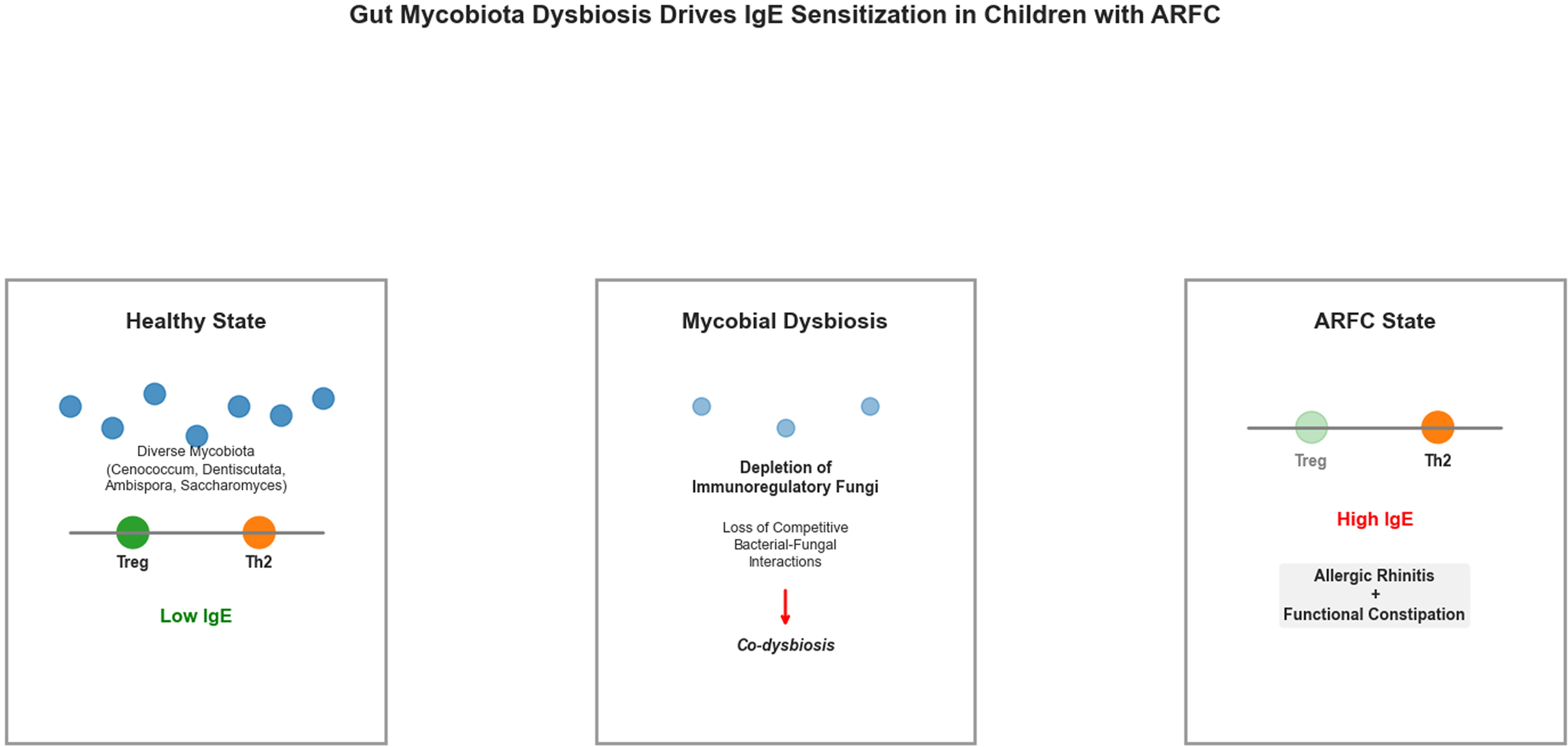
Graphical Abstract.
Proposed mechanism linking gut mycobiota dysbiosis to IgE sensitization in children with comorbid allergic rhinitis and functional constipation (ARFC).
This model hypothesizes how gut mycobiota dysbiosis may contribute to IgE sensitization in ARFC. In the healthy state (left), a diverse mycobiota, including genera such as Cenococcum, Dentiscutata, Ambispora, and Saccharomyces, may support immune homeostasis by promoting regulatory T cell (Treg) responses and suppressing IgE production. In ARFC (right), depletion of these fungi is proposed to disrupt immunoregulatory pathways (e.g., Dectin-1 signaling, SCFA production), leading to a predominant Th2 response, elevated IgE, and clinical manifestations.
1 Introduction
Allergic rhinitis (AR) ranks among the most prevalent chronic inflammatory diseases in pediatric populations, with a steadily increasing global prevalence that significantly impairs quality of life and disrupts sleep (1, 2). Clinically, AR is frequently comorbid with functional constipation (FC)—a gastrointestinal disorder characterized by difficult and infrequent bowel movements—due to notable epidemiological links (3, 4). Evidence from population-based cohorts suggests constipation may be a risk factor for subsequent AR development (3), potentially exacerbating allergic symptoms (5). This clinical synergy indicates shared pathophysiology mediated through the gut–microbiota–host immune axis, as evidenced by bacterial dysbiosis in both conditions (6–8). However, the high impact of ARFC comorbidity on pediatric quality of life contrasts with scarce research on its shared mechanisms. Current management often addresses each condition in isolation, highlighting an unmet need for integrated approaches.
Dysbiosis of the bacterial microbiota is well-documented in isolated AR and constipation, implicating roles in immune regulation, barrier integrity, and immunomodulatory metabolite production (e.g., short-chain fatty acids) (6–10). In contrast, the gut mycobiota—the fungal component—remains understudied (11, 12). Recent research illuminates fungi as active immunomodulators via Dectin-1 and CARD9 signaling (13–15), often through interactions with bacteria (16) and host immune cells (e.g., CX3CR1+ mononuclear phagocytes) (17). Genomic resources highlight fungal diversity and disease associations (18), yet their role in ARFC remains unknown.
Fungi engage host pattern-recognition receptors (e.g., Dectin-1, TLRs), initiating cascades such as the SYK–CARD9 axis, which regulates antifungal immunity and allergic inflammation (13, 14, 19). The “gut–fungi–lung axis” demonstrates intestinal fungal influence on remote mucosal sites via systemic immune activation (20, 21), with evidence supporting gut-lung crosstalk in asthma (22). For instance, gut-resident CX3CR1+ phagocytes respond to fungal dysbiosis, aggravating allergic airway disease (20). Similarly, expansion of Wallemia mellicola enhances allergic airway severity in mice (23). Building on the “gut–fungi–lung axis” (20, 21, 24) and gut-nose communication in allergy (25–27), a “gut–fungi–nose axis” is hypothesized to underlie ARFC comorbidity, supported by fungal associations with immune-mediated conditions (e.g., asthma, inflammatory bowel disease) (14, 19, 28).
Significant knowledge gaps persist. First, while fungi have been examined in isolated AR or FC (19, 29), the gut mycobiota in ARFC children remains uncharacterized. Second, the potential for fungal dysbiosis to disrupt immune homeostasis—via mechanisms like barrier impairment and SYK–CARD9 activation (30, 31)—is unexplored in this context.
Therefore, this pilot study characterized the gut mycobiota in ARFC children versus healthy controls (HC) using metagenomic sequencing, testing the hypothesis that its composition correlates with IgE-mediated sensitization. To our knowledge, this is the first mycobiota profiling in ARFC comorbidity, aiming to generate hypotheses about the gut–fungi–nose axis.
2 Materials and methods
2.1 Study participants and sample collection
This case-control study was conducted in the Department of Pediatrics at Longgang District Maternity & Child Healthcare Hospital of Shenzhen between June 2024 and June 2025. The study protocol was approved by the Institutional Ethics Committee (Approval No: KYXMLL-01-CZGC-14-2-1), and written informed consent was obtained from guardians. Thirty-six children aged 3–6 years were enrolled (19 ARFC, 17 HC). FC diagnosis followed Rome IV criteria (32), including symptoms persisting ≥1 month. AR diagnosis adhered to Chinese Guidelines (2022) (33). Demographic characteristics are summarized in Table 1. No significant differences in age (unpaired t-test) or gender distribution (χ² test) were observed (p > 0.05). HC children had no history of allergic diseases, infections, or antibiotic use within one month prior. Exclusion criteria included chronic comorbidities and recent probiotic/immunomodulator use. Fecal samples were collected and stored at −80°C.
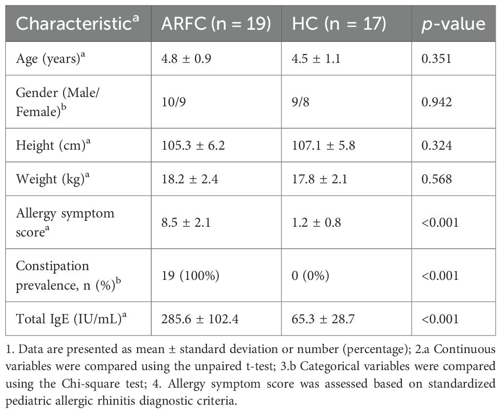
Table 1. Demographic and clinical characteristics of children with allergic rhinitis and functional constipation (ARFC) and healthy controls (HC).
2.2 Sample handling and storage
Fecal samples were collected using sterile swabs, flash-frozen in liquid nitrogen, and stored at −80°C within 30 minutes. Transportation occurred on dry ice within 2 hours, with processing completed within 24 hours. Samples were lyophilized and homogenized prior to DNA extraction.
2.3 Serum IgE measurement
Venous blood was collected at enrollment. Serum was separated by centrifugation (3000 rpm, 10 min) and stored at −80°C.
Total IgE: Quantified via Human IgE ELISA Kit (Elabscience, Catalog No. E-EL-H0104c) per manufacturer instructions. Duplicate assays expressed IgE in IU/mL (detection limit: 0.1 IU/mL; CV < 8%).
Allergen-specific IgE: Quantified against pediatric allergens (e.g., dust mites, shrimp) via AllergyScreen® microarray (Mediwiss Analytic GmbH), based on immunoCAP® FEIA. Sensitization threshold: ≥0.35 kUa/L. Manufacturer controls were included.
2.4 Metagenomic sequencing and fungal community analysis
2.4.1 DNA extraction and quality control
Total genomic DNA was extracted from 0.2 g stool using FastPure Stool DNA Isolation Kit (MJYH). Concentration and purity were assessed via Synergy HTX reader (BioTek) and NanoDrop 2000 (Thermo Fisher). Samples with DNA ≥ 20 ng/μL and A260/A280 1.8–2.0 were retained; integrity was confirmed by 1% agarose gel electrophoresis.
2.4.2 Library preparation and sequencing
Qualified DNA (100 ng) was fragmented to ~350 bp via Covaris M220 ultrasonicator. Paired-end libraries were prepared with NEXTFLEX Rapid DNA-Seq Kit (Bioo Scientific). Shotgun sequencing was performed on Illumina NovaSeq X Plus (Majorbio Bio-Pharm Technology Co.), yielding ≥10 Gb/sample.
2.4.3 Sequence preprocessing and fungal contig identification
Raw data were processed on Majorbio Cloud Platform. Adapters and low-quality reads were removed using fastp (v0.23.0). Human reads (GRCh38) were excluded via BWA (v0.7.17). High-quality reads were assembled de novo with MEGAHIT (v1.1.2), retaining contigs ≥ 300 bp.
Fungal contigs were classified using VirSorter2 (v2.2.4; categories 1,2,4,5) or DeepVirFinder (v1.0; score >0.9, p<0.05). Contig quality was assessed with CheckV (v1.0.1; completeness ≥50%, contamination ≤10%). Host/bacterial sequences were removed via Bowtie2 (v2.4.5) against RefSeq genomes. Statistics are in Supplementary Table S1.
2.4.4 Taxonomic annotation and abundance profiling
Open reading frames (ORFs) were predicted via Prodigal (v2.6.3), retaining ≥100-bp sequences. A non-redundant gene catalog was constructed with CD-HIT (v4.6.1; 90% identity/coverage). Gene abundance was estimated by mapping reads to the catalog via SOAPaligner (v2.21; 95% identity). Taxonomic annotation aligned genes against NCBI NR fungi database using DIAMOND (v2.0.13; e-value ≤1e−5). Dietary contigs were excluded.
2.4.5 Functional annotation
Functional potential was assessed by aligning the gene catalog against KEGG Orthology (KO) via DIAMOND (e-value ≤1e−5). Abundances were summarized at Level 3 pathways.
2.5 Statistical analysis
All statistical and bioinformatic analyses were conducted in R (version 4.3.1). Alpha diversity indices (Chao1 and Shannon) were compared between groups using the Wilcoxon rank-sum test. Beta diversity was assessed based on Bray–Curtis distances and visualized via principal coordinate analysis (PCoA). Permutational multivariate analysis of variance (PERMANOVA) with 9999 permutations was used to examine group differences in fungal community structure, and Procrustes analysis was applied to assess overall configuration similarity.
Differential abundance of fungal taxa across taxonomic levels (phylum, family, genus) was assessed using linear discriminant analysis effect size (LEfSe). A threshold of LDA score > 2 and FDR-adjusted p-value < 0.05 was applied to define statistically significant taxa.
A random forest model implemented via the randomForest package was used to identify key fungal genera and clinical variables discriminating between ARFC and HC participants. The model was trained using 10-fold cross-validation, and variable importance was evaluated based on the MeanDecreaseGini metric. Model performance was assessed using the area under the receiver operating characteristic curve (AUC) and out-of-bag (OOB) error estimation.
Associations between fungal genera and clinical variables (total IgE and allergen-specific IgE levels) were examined using Spearman’s rank correlation. To assess the functional potential of the gut mycobiota, quality-filtered reads were aligned against the KEGG Orthology (KO) database using Diamond (v2.1.8) with an e-value threshold of 1e−5. KO abundances were summarized at Level 3 pathway categories. Differential abundance of KEGG pathways between groups was assessed using the ReporterScore algorithm. Pathways with an absolute ReporterScore > |1.5| and a corrected significance designation (“yes”) were considered significantly enriched. Pathways labeled “ns” (not significant) were retained for exploratory interpretation.
2.6 Bacterial–fungal interaction network analysis
To elucidate the ecological relationships between gut bacteria and fungi, cross-kingdom interaction networks were constructed separately for the ARFC and HC groups. To ensure maximum comparability and integration, bacterial abundance profiles were directly derived from the identical whole-genome shotgun metagenomic sequencing dataset used for mycobiota characterization. This approach guarantees that all downstream correlations are inferred from the same biological sample aliquots and sequencing runs, providing a unified basis for interkingdom network analysis.
Network Construction: For each group, pairwise Spearman’s rank correlation coefficients (ρ) were calculated between the relative abundances of all bacterial genera and fungal genera. Only correlations with an absolute coefficient (|ρ|) ≥ 0.6 and a p-value < 0.05 after false discovery rate (FDR) correction using the Benjamini–Hochberg method were considered statistically significant and retained for network construction. This stringent threshold was chosen to focus on strong and robust inter-kingdom associations.
Network Visualization and Topological Analysis: The resulting correlation matrices were imported into Gephi (version 0.10) for visualization and topological analysis. The following conventions were applied: ①Nodes represent microbial genera; ②Node size is proportional to the mean relative abundance of the corresponding genus within the group; ③Node color and shape encode kingdom: bacterial genera are depicted as yellow circles, and fungal genera as green squares; ④Edges represent statistically significant correlations; ⑤Edge color indicates correlation direction: red edges denote positive correlations (ρ > 0), and blue edges denote negative correlations (ρ < 0).
Interpretation of Ecological States: The term “co-dysbiosis” is proposed to describe the network state observed in ARFC, characterized by a predominance of positive correlations and a loss of competitive (negative) interactions. This concept is used to hypothesize an ecological shift from a homeostatic, antagonism-rich network (HC) toward a simplified, cooperation-dominant network (ARFC), which may reflect reduced ecological resilience and facilitate disease-associated community consolidation.
3 Results
3.1 Altered gut fungal community structure without significant diversity changes in ARFC children
No significant alpha diversity differences were observed (Chao1, p = 0.401; Figure 1A). However, PCoA revealed partial structural separation (Bray–Curtis; Figure 1B). PERMANOVA was non-significant (R² = 0.0441, p = 0.88), likely due to inter-individual variation and limited sample size. HC contained 98 unique fungal OTUs versus one in ARFC (Figure 1C). Procrustes analysis showed no configuration difference (M² = 0.991, R = 0.0949, p = 0.233; Figure 1D).
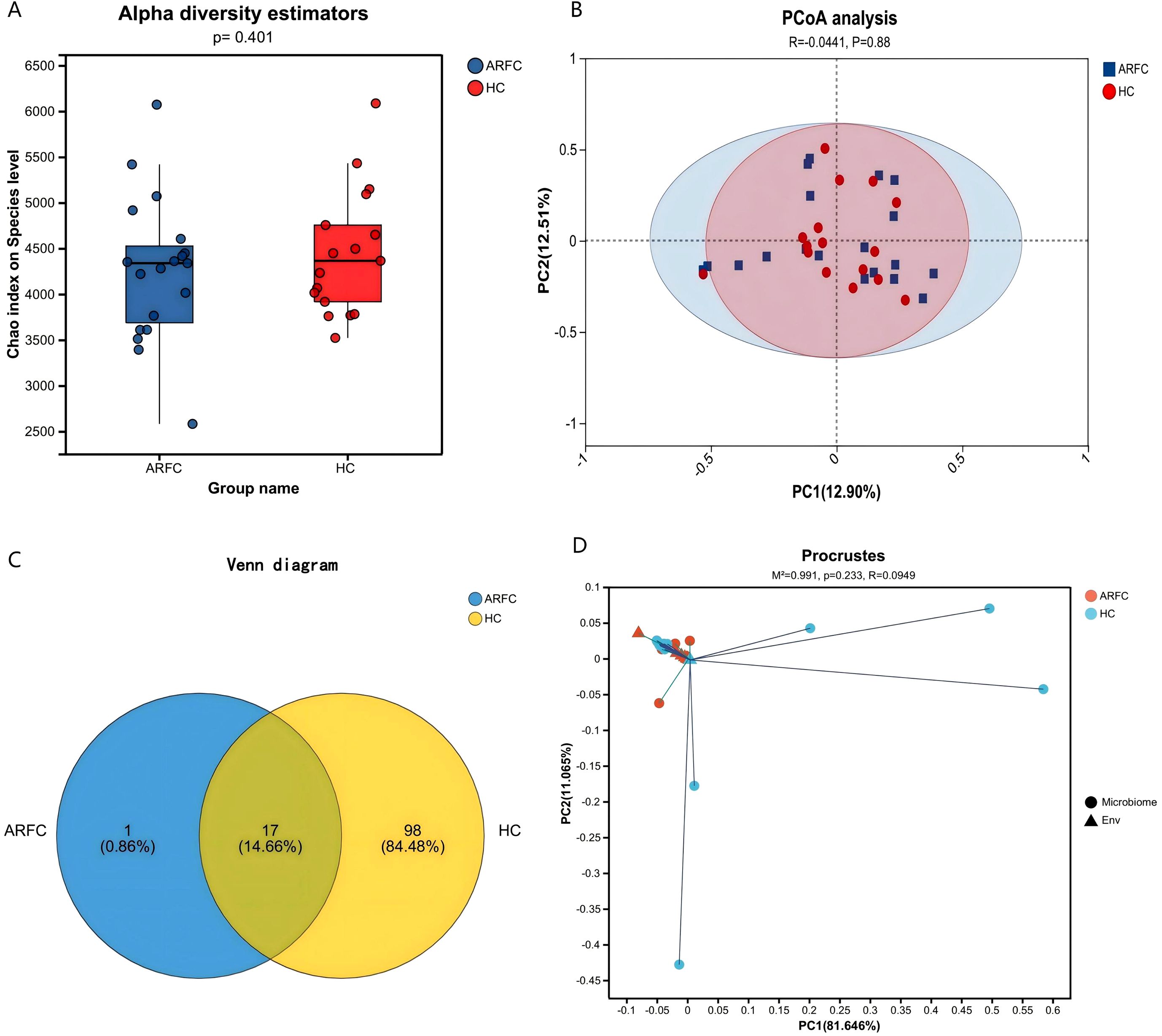
Figure 1. Gut fungal community diversity in children with allergic rhinitis and functional constipation (ARFC) and healthy controls (HC). (A) Chao1 index showing no significant difference in fungal richness between groups (Wilcoxon test, p = 0.401). (B) Principal coordinate analysis (PCoA) based on Bray–Curtis distances illustrating structural separation between ARFC and HC groups. (C) Venn diagram depicting the number of unique and shared fungal operational taxonomic units (OTUs) between groups. (D) Procrustes analysis indicating no significant overall configuration difference (M² = 0.991, p = 0.233). Sample sizes: n = 19 ARFC, n = 17 HC.
3.2 Altered Fungal composition and multi-level biomarkers in ARFC
No phylum-level differences occurred (p > 0.05). At the family level, Gigasporaceae (HC: 3.85 ± 4.41% vs. ARFC: 1.49 ± 5.43%; p = 0.015) and Gloniaceae (HC: 1.74 ± 3.27% vs. ARFC: 0.30 ± 1.31%; p = 0.046) were enriched in HC (Supplementary Table S2; Figure 2A).
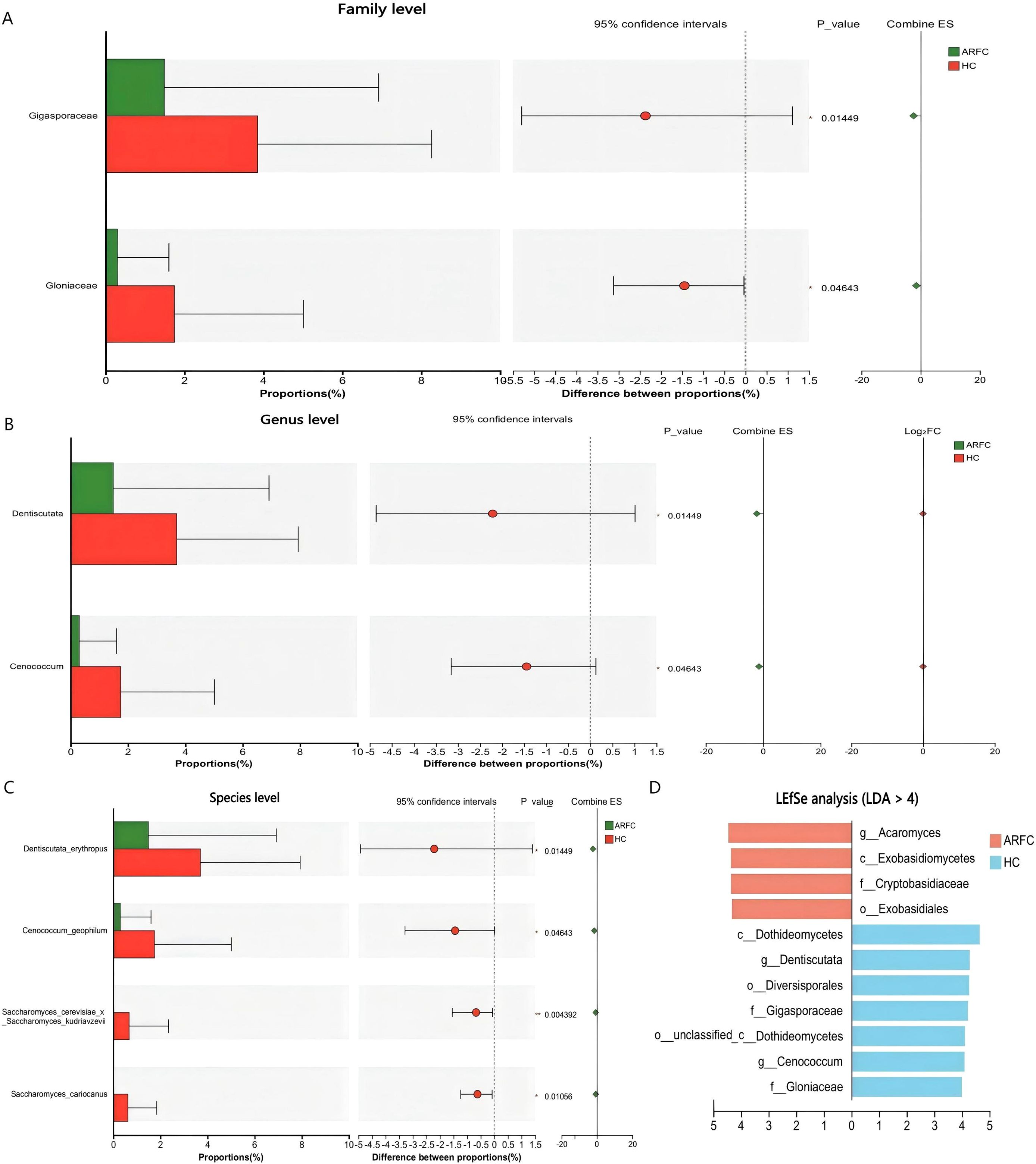
Figure 2. Differential abundance of fungal taxa at the family and genus levels between ARFC and HC groups. (A) Bar plot showing relative abundance of fungal families with significant differences (Wilcoxon test, p < 0.05). (B) Relative abundance of fungal genera significantly enriched in HC. (C) Box plots of selected fungal species exclusively detected in HC. (D) Linear discriminant analysis effect size (LEfSe) identifying fungal taxa enriched in HC (LDA score > 4). Data are presented as mean ± SD; p-values indicate statistical significance.
At the genus level, Dentiscutata (HC: 3.70 ± 4.24% vs. ARFC: 1.49 ± 5.43%; p = 0.015) and Cenococcum (HC: 1.74 ± 3.27% vs. ARFC: 0.30 ± 1.31%; p = 0.046) were depleted in ARFC (Supplementary Table S3; Figure 2B). Species-level depletion included Saccharomyces cerevisiae x S. kudriavzevii (HC: 0.67 ± 1.67% vs. ARFC: 0%; p = 0.004) and S. cariocanus(HC: 0.62 ± 1.22% vs. ARFC: 0%; p = 0.011) (Supplementary Table S4; Figure 2C).
LEfSe identified HC-enriched biomarkers (LDA >4): Glomeromycota taxa (e.g., Dentiscutata) and Ascomycota taxa (e.g., Cenococcum). ARFC showed enrichment in Basidiomycota classes (Figure 2D).
3.3 Key discriminators in random forest model
The random forest model confirmed depleted genera (Cenococcum, Dentiscutata, Ambispora) as top discriminators (Figure 3). Total IgE was the most predictive clinical variable (MeanDecreaseGini = 15.5). Fungi outperformed some allergen-specific IgEs (e.g., Cenococcum importance = 3.95). Depleted genera (e.g., Trichoderma) exhibited negative importance scores.
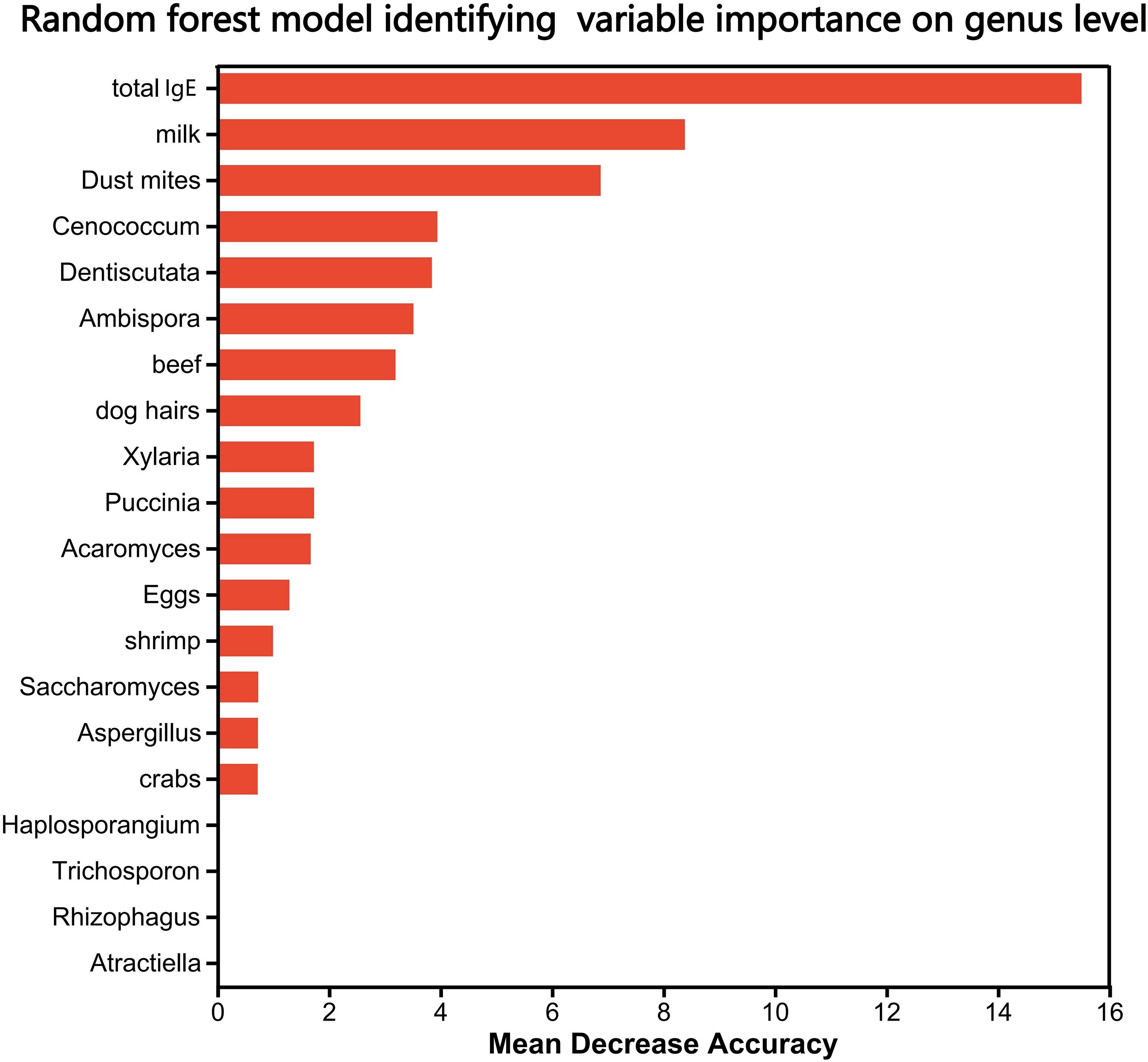
Figure 3. Random forest model identifying key fungal and clinical discriminators between ARFC and HC groups. Variable importance is ranked by MeanDecreaseGini. Total IgE was the most predictive clinical variable. Fungal genera including Cenococcum, Dentiscutata, and Ambispora exhibited high discriminatory power. Model performance was evaluated using 10-fold cross-validation (AUC and OOB error provided in methods).
3.4 Correlations between gut fungi and immunological parameters
Spearman correlation analysis revealed significant associations between gut fungal genera and immunological parameters (Figure 4): 1) Negative correlations: The abundances of Saccharomyces (ρ = -0.39), Ambispora (ρ = -0.40), Dentiscutata (ρ = -0.43), Patellaria (ρ = -0.37), Acaromyces (ρ = -0.34) and Cenococcum (ρ = -0.40) were significantly inversely correlated with total serum IgE, and specific IgE levels against house dust mites (Ambispora ρ = -0.42). 2) Positive correlation: The abundance of Paramyrothecium showed a positive correlation with specific IgE against shrimp (ρ = 0.50) and crab (ρ = 0.49); Tulasnellaceae with specific IgE against dust mites (ρ = 0.37) and eggs (ρ = 0.47). 3) No significant correlation: Most low-abundance fungal genera showed no significant correlation with the measured IgE levels.
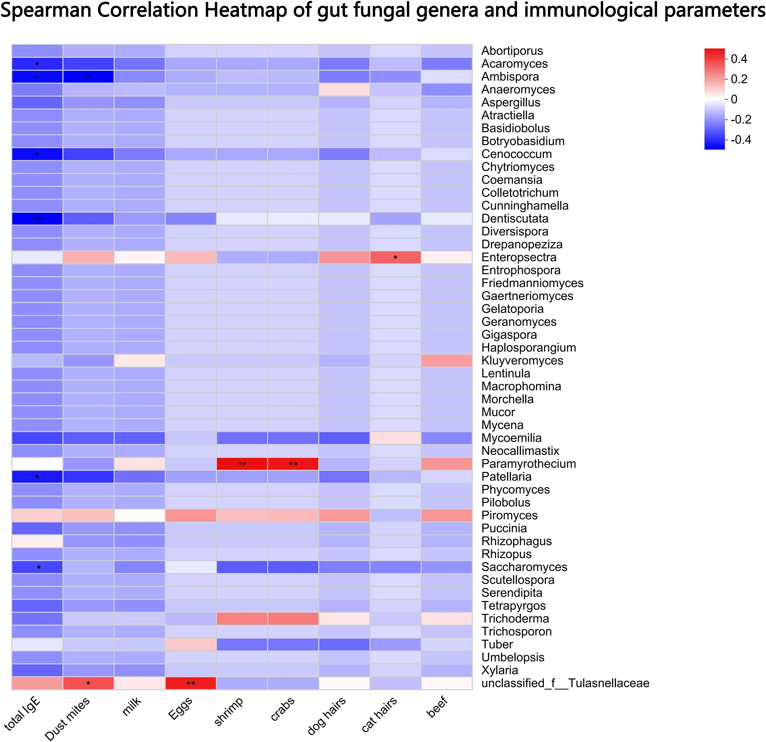
Figure 4. Spearman correlation analysis between gut fungal genera and immunological parameters. Heatmap displays correlation coefficients (ρ) between fungal abundances and total or allergen-specific IgE levels. Significant negative correlations were observed for Saccharomyces, Ambispora, Dentiscutata, and Cenococcum with total IgE and house dust mite-specific IgE. Only correlations with p < 0.05 are shown. * p < 0.05, ** p < 0.01.
3.5 No significant differences in predicted fungal functional profiles
Comparative analysis of the gut mycobiota’s functional potential via KEGG pathway enrichment showed no significant differences between the ARFC and HC groups at either Level 1, Level 2 and Level 3 categories (Figure 5), indicating that despite structural differences, the overall functional profile of the gut fungi remained relatively conserved in children with ARFC.
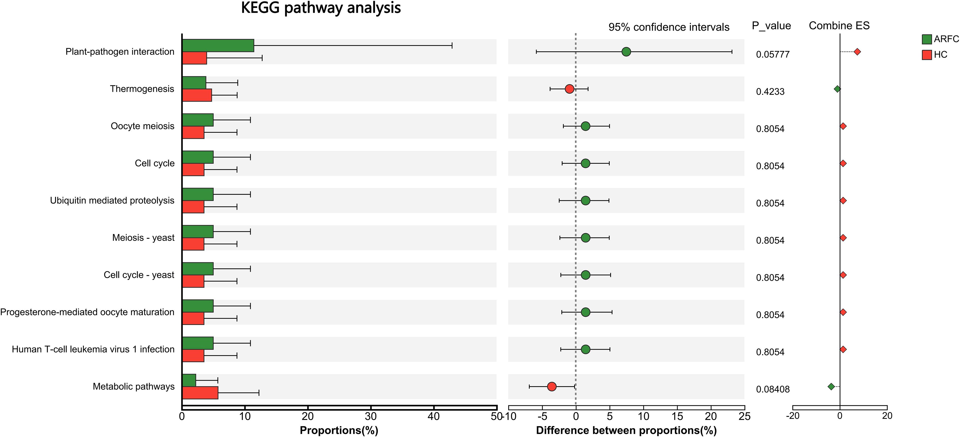
Figure 5. Predicted functional potential of the gut mycobiota based on KEGG pathway analysis. No significant differences were observed between ARFC and HC groups at any KEGG pathway level (Levels 1–3), as assessed by the ReporterScore algorithm (absolute score < |1.5|, not significant). Bar plots represent the relative abundance of KEGG pathways in each group.
3.6 Comparative analysis of gut bacteriome-mycobiome interaction networks in ARFC and HC children
To elucidate the potential impact of the ARFC condition on the ecological relationships within the gut ecosystem, we constructed and compared bacterial–fungal correlation networks for the ARFC and HC groups. Bacterial abundance data were derived from the same metagenomic sequencing dataset used for mycobiota analysis, allowing integrated cross-kingdom network inference. This comparative analysis revealed profound differences in the structure and composition of the interkingdom networks between the two groups (Figure 6).
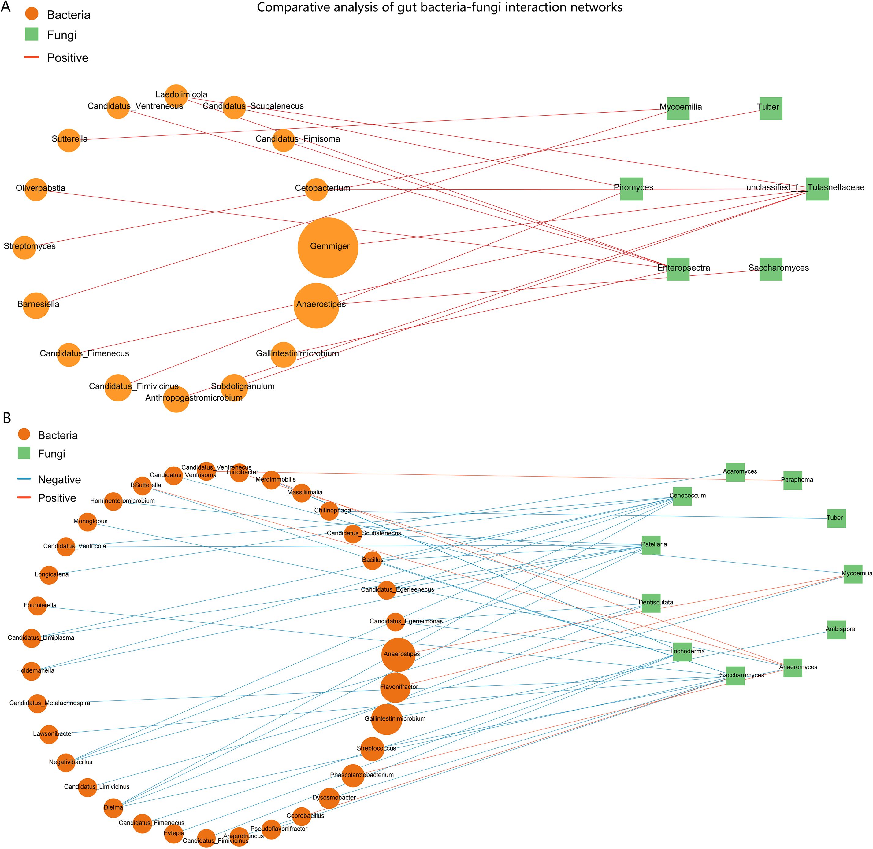
Figure 6. Comparative analysis of gut bacteria-fungi interaction networks in HC and ARFC children. Networks depict significant Spearman correlations (|r| ≥ 0.6, p < 0.05). Nodes: yellow circles (bacterial genera), green squares (fungal genera). Edges: red (positive correlations), blue (negative correlations). (A) HC network is characterized by numerous negative correlations, suggesting competitive interactions. (B) ARFC network is exclusively positive, indicating a shift toward cooperative interkingdom structure and potential “co-dysbiosis.”.
3.6.1 Network in HC is characterized by prevalent negative interactions
The HC group network was complex and exhibited a high degree of negative correlations, which constituted the majority (44 out of 51 significant correlations, 86.3%) of all interactions. This suggests a network structure potentially dominated by competitive or antagonistic relationships under homeostasis. Notably, we screened the fungal contigs for putative antibiotic biosynthesis gene clusters (BGCs) using antiSMASH fungal mode (v7.0), which identified several biosynthetic pathways in fungal genera such as Cenococcum and Dentiscutata that are known to produce antifungal or antibacterial metabolites. These functional annotations lend ecological plausibility to the observed negative correlations, suggesting that fungal-derived antimicrobial activity may contribute to the competitive bacterial–fungal interactions observed in healthy controls. Key fungal genera, including Saccharomyces, Patellaria, Cenococcum, and Dentiscutata, acted as central hubs with multiple negative bacterial partners. For instance, the bacterium Bacillus was negatively correlated with Saccharomyces (r = -0.75, p = 0.0005), Patellaria, and Cenococcum. Similarly, the bacterium Dielma displayed significant negative correlations with four different fungal genera (Saccharomyces, Dentiscutata, Patellaria, Cenococcum). A limited number of positive correlations were observed in the HC network, such as between Flavonifractor and Mycoemilia (r = 0.74, p = 0.0006).
3.6.2 Network in ARFC patients shifts towards an exclusively positive topology
In stark contrast, the interaction network in the ARFC group was exclusively composed of positive correlations (17 out of 17, 100%). This unanimous shift towards cooperative interactions indicates a fundamental restructuring of the gut ecosystem in ARFC. The ARFC network was less complex and centered around fewer fungal hubs. The unclassified Tulasnellaceae and the genus Enteropsectra emerged as the core nodes, forming positive correlations with seven and six bacterial partners, respectively. Notably, several bacterial genera involved in these cooperative clusters, such as Anaerostipes and Subdoligranulum, are known short-chain fatty acid (SCFA) producers. For example, Anaerostipes was positively correlated with Saccharomyces (r = 0.71, p = 0.0007) in ARFC, whereas it was linked to Mycoemilia (r = 0.66, p = 0.0039) in HC.
3.6.3 Comparative summary
The direct comparison highlights a dramatic loss of negative interactions and a simplification of network architecture in ARFC patients. The HC network’s structure, rich in negative correlations, may reflect a robust, homeostatic ecosystem where microbial populations check and balance each other. Conversely, the ARFC network’s shift to a uniformly positive correlation profile suggests a state of “co-dysbiosis,” where formerly competitive relationships are replaced by cooperative ones, potentially facilitating the consolidation of a disease-associated microbial community and contributing to the pathophysiology of both allergic inflammation and gastrointestinal dysmotility.
4 Discussion
This study provides the first characterization of the gut mycobiota in ARFC children, revealing a significant depletion of specific immunomodulatory fungi—including Cenococcum, Dentiscutata, Ambispora, and Saccharomyces—in children with ARFC. These fungal taxa served as top discriminators in our machine learning model and exhibited significant inverse correlations with both total and allergen-specific IgE levels. Moreover, cross-kingdom network analysis uncovered a profound ecological restructuring in ARFC, characterized by a shift from a homeostatic, competition-rich network in healthy controls to an exclusively cooperative state. This unanimous shift towards positive correlations may reflect a loss of ecological resilience. Competitive interactions are fundamental for maintaining community stability and resisting perturbation (34, 35). In microbial ecology, such simplified, cooperation-dominant networks have been associated with ecosystem instability and reduced functional redundancy (36). We therefore propose the term “co-dysbiosis” to describe this altered state, which emphasizes the concurrent disruption of bacterial-fungal interkingdom interactions and the loss of competitive microbial relationships that typify a healthy gut ecosystem. We hypothesize that this simplified, cooperation-dominant network may represent a loss of ecological resilience, a concept that warrants validation in future, larger-scale ecological studies.
The depletion of these fungi likely contributes to ARFC pathogenesis via disrupted immune regulation. Fungi such as Saccharomyces and Cenococcum engage with host pattern-recognition receptors like Dectin-1, influencing the Th2/regulatory T cell (Treg) balance and IgE production (14, 19, 37). Their loss, combined with the observed “co-dysbiosis”, may impair the production of key immunomodulatory metabolites. For instance, our findings suggest that Saccharomyces species, which were depleted in ARFC, may contribute to intestinal barrier function and immune modulation, as previous studies have implicated this genus in metabolite production and Treg induction (14, 37). Similarly, the reduction in Glomeromycota fungi (e.g., Dentiscutata, Ambispora) could plausibly diminish the production of aryl hydrocarbon receptor (AhR) ligands, which are crucial for maintaining immune tolerance (13, 16). However, direct evidence for these specific mechanisms in our cohort requires further validation. Based on our correlations, we speculate that the depletion of these immunomodulatory fungi, coupled with the observed “co-dysbiosis”, may collectively disrupt systemic immune homeostasis, thereby potentially facilitating IgE sensitization in ARFC. Direct experimental evidence for this causal pathway is needed to confirm this model.
The significant inverse correlations between genera such as Saccharomyces, Ambispora, Dentiscutata, and Cenococcum with IgE levels support their immunoregulatory potential. For instance, Saccharomyces species are known to enhance short-chain fatty acid (SCFA) production, which promotes Treg differentiation and suppresses IgE responses (37, 38). Similarly, Glomeromycota-related taxa (e.g., Dentiscutata, Ambispora) may contribute to the production of aryl hydrocarbon receptor (AhR) ligands, which are crucial for maintaining immune tolerance and restraining Th2-driven IgE production (13, 16).
Beyond metabolite-mediated effects, fungi including Cenococcum can directly engage host pattern-recognition receptors such as Dectin-1. This interaction modulates the SYK–CARD9 signaling axis, which plays a fundamental role in shaping the Th2/Treg balance and controlling IgE production (28, 30). Although we did not directly measure SCFAs or AhR activity, the consistent depletion of these immunoregulatory fungi aligns with their established roles in systemic immune homeostasis, providing a mechanistic basis for the elevated IgE sensitization observed in ARFC children.
Although we found no significant differences in the predicted functional potential (KEGG pathways) of the gut mycobiota, this absence of metabolic signals should be interpreted cautiously. Functional redundancy within the fungal community or limitations in current fungal functional databases (12, 31, 39) may obscure genuine metabolic differences. Importantly, the structural and ecological alterations we observed could exert critical immunologic effects through non-metabolic mechanisms, particularly via direct fungal-host interactions through pattern-recognition receptors (28, 30). Fungal community structure, particularly the depletion of immunomodulatory taxa, can influence host immunity via pattern-recognition receptors (e.g., Dectin-1, TLRs) and downstream signaling cascades such as the SYK–CARD9 axis, which regulates inflammasome activation, Th2/Treg balance, and IgE production (19, 28, 30). This is supported by studies in other immune-mediated conditions where fungal dysbiosis alters mucosal immunity without concomitant shifts in metabolic pathways (40, 41). Thus, while functional redundancy or database limitations may obscure metabolic differences, the immunologic impact of structural dysbiosis remains evident.
Our comparative bacterial-fungal network analysis further underscores a fundamental ecological restructuring in ARFC. The healthy control network was characterized by numerous negative correlations, suggesting a stable, competitive ecosystem that may help maintain microbial diversity and prevent pathogen expansion (6, 34). In stark contrast, the ARFC network exhibited an exclusively positive correlation profile, indicating a collapse of these competitive restraints. This unanimous shift towards positive correlations may reflect a loss of ecological resilience, as competitive interactions are fundamental for maintaining community stability and resisting perturbation (35). In microbial ecology, such simplified, cooperation-dominant networks have been associated with ecosystem instability and reduced functional redundancy (36). This simplified, cooperative state, centered around new hub taxa like Tulasnellaceae, may facilitate the persistence of a disease-associated microbial community.
We propose the term “co-dysbiosis” to describe this altered state, which emphasizes the concurrent disruption of bacterial–fungal interkingdom interactions and the loss of competitive microbial relationships that typify a healthy gut ecosystem (34, 42). Importantly, while our bacterial data were derived from metagenomic reads, the consistent and dramatic shift from competitive to exclusively cooperative correlations suggests a fundamental ecological reorganization in ARFC. This simplified, cooperation-dominant network may reflect a loss of ecological resilience, a concept that warrants validation in future, larger-scale studies with matched multi-kingdom profiling (35, 36). Additionally, the identification of antibiotic biosynthesis gene clusters (BGCs) in fungal taxa such as Cenococcum and Dentiscutata—which were depleted in ARFC—suggests that these fungi may produce metabolites capable of modulating bacterial communities (43, 44). The loss of such fungi could therefore contribute to a “double dysbiosis”: not only a reduction in immunoregulatory fungi but also a disruption in fungal-mediated bacterial inhibition. This may further destabilize the gut ecosystem, shifting from a competitive, homeostatic network to a cooperative, dysbiotic state characterized by reduced microbial antagonism and potentially facilitating the emergence of disease-associated consortia (45, 46). This hypothesis aligns with the observed simplification of the ARFC network and underscores the interconnectedness of bacterial and fungal dysbiosis in ARFC pathogenesis.
In addition to immune dysregulation, the observed fungal dysbiosis and “co-dysbiosis” may also contribute to the gastrointestinal symptoms of ARFC, particularly constipation. Our network analysis revealed that in ARFC, several short-chain fatty acid (SCFA)-producing bacterial genera (e.g., Anaerostipes, Subdoligranulum) formed cooperative clusters with fungi such as Tulasnellaceae and Enteropsectra. While SCFAs like butyrate are generally promotive of gut motility, their net effect depends on the broader ecological context and interacting microbial consortia (47). The shift from a competitive to an exclusively cooperative network (“co-dysbiosis”) may alter the overall metabolic output of the gut ecosystem. This could facilitate the overgrowth or altered activity of bacterial taxa previously linked to constipation, such as specific Clostridium clusters involved in bile acid transformation (48) or mucin-degrading bacteria that affect mucosal turnover and stool consistency (49). For instance, reductions in Bacteroides and increases in certain Firmicutes have been associated with slowed colonic transit (49). The loss of fungal-derived inhibitory metabolites (e.g., from depleted Cenococcum) might remove a natural check on such bacteria, thereby contributing to dysmotility. Thus, “co-dysbiosis” may represent an ecological driver linking mycobiota alterations to both immune (IgE sensitization) and motor (constipation) components of ARFC. Furthermore, diet—a key modulator of gut microbiota—was not systematically assessed in this pilot study, but future investigations should evaluate dietary patterns (e.g., fiber intake, fermentable carbohydrates) as potential confounders or effect modifiers linking mycobiota dysbiosis, bacterial community structure, and stool characteristics in ARFC (50, 51).
In summary, this study provides the first multi-faceted characterization of gut mycobiota dysbiosis in children with ARFC, encompassing the depletion of specific immunomodulatory fungi and a profound ecological restructuring of bacterial-fungal networks from a competitively dominant state to an exclusively cooperative one, termed “co-dysbiosis.” These alterations are significantly correlated with elevated serum IgE levels. Based on these interconnected findings, we propose an integrative mechanistic hypothesis (as summarized in the Graphical Abstract): In the healthy state, a diverse mycobiota, including genera such as Cenococcum and Dentiscutata, contributes to immune homeostasis by engaging host pattern-recognition receptors (e.g., Dectin-1) and facilitating the production of immunomodulatory metabolites (e.g., SCFAs), thereby suppressing IgE responses. In ARFC, the depletion of these protective fungi, coupled with the simplified, cooperation-dominant network architecture of “co-dysbiosis,” collectively impairs intestinal immunoregulation. This disruption may facilitate a Th2-skewed immune response and IgE sensitization via the putative “gut–fungi–nose axis,” ultimately contributing to the comorbid clinical manifestations of allergic rhinitis and constipation. While this hypothesis requires validation in future studies, it offers a novel, mycobiota-centric framework for understanding ARFC pathogenesis.
Several limitations should be acknowledged. First, the cross-sectional design precludes causal inference about the relationship between mycobiota alterations and disease development. Second, the sample size, appropriate for a pilot investigation, inevitably limits the statistical power for detecting subtle associations. However, this design was instrumental in generating the first high-dimensional map of the mycobiota in ARFC and identifying robust, large-effect-size signals (e.g., the depletion of key genera and the striking network shift) that provide a clear rationale and specific hypotheses for future, larger-scale studies. Third, our metagenomic approach for mycobiota characterization employed VirSorter2 and DeepVirFinder to maximize sensitivity for detecting diverse fungal sequences. While we applied stringent post-hoc filters (e.g., CheckV, alignment against host and bacterial genomes), this method may carry a risk of misclassifying non-fungal elements. Future studies should validate these findings using complementary approaches such as ITS amplicon sequencing or fungal-specific marker gene analysis. Fourth, while bacterial abundance data were extracted from the same metagenomic libraries for network construction—ensuring sample-level integration—the sequencing depth and analytical pipeline were primarily optimized for fungal sequence recovery. Consequently, metagenomic-based bacterial profiling, while providing a direct cross-kingdom dataset, may offer lower taxonomic resolution for rare or low-abundance bacterial taxa compared to targeted 16S rRNA gene sequencing. This could potentially affect the detection of subtle interkingdom associations involving these minority bacterial populations. Future studies employing matched, deep shotgun sequencing optimized for both kingdoms, or complementary 16S rRNA sequencing, would help validate the observed network topology and better resolve the specific bacterial partners within the “co-dysbiosis” state. Fifth, we did not quantify fungal biomass using methods such as qPCR targeting fungal rRNA genes, which would have helped confirm that the lack of alpha diversity differences reflects biological reality rather than technical variation in sequencing depth. Finally, taxonomic annotation remains constrained by the underrepresentation of fungal genomes in public databases, particularly for environmental and plant-associated fungi that may transiently inhabit the human gut.
Future studies should employ larger, longitudinal cohorts to validate these findings and explore causality. Integrated multi-omics approaches—combining metagenomics, metatranscriptomics, and metabolomics—are needed to elucidate the functional mechanisms of specific fungi and their interactions with bacteria. Furthermore, in vivo and in vitro models are essential to confirm the immunomodulatory roles of the depleted fungi and their potential as therapeutic targets (8, 19).
Our pilot study unveils a multi-faceted dysbiosis of the gut mycobiota in ARFC, encompassing taxonomic depletion, profound ecological restructuring, and significant immunological correlations. To synthesize these interconnected findings and their broader implications, we present a conceptual summary in Table 2.
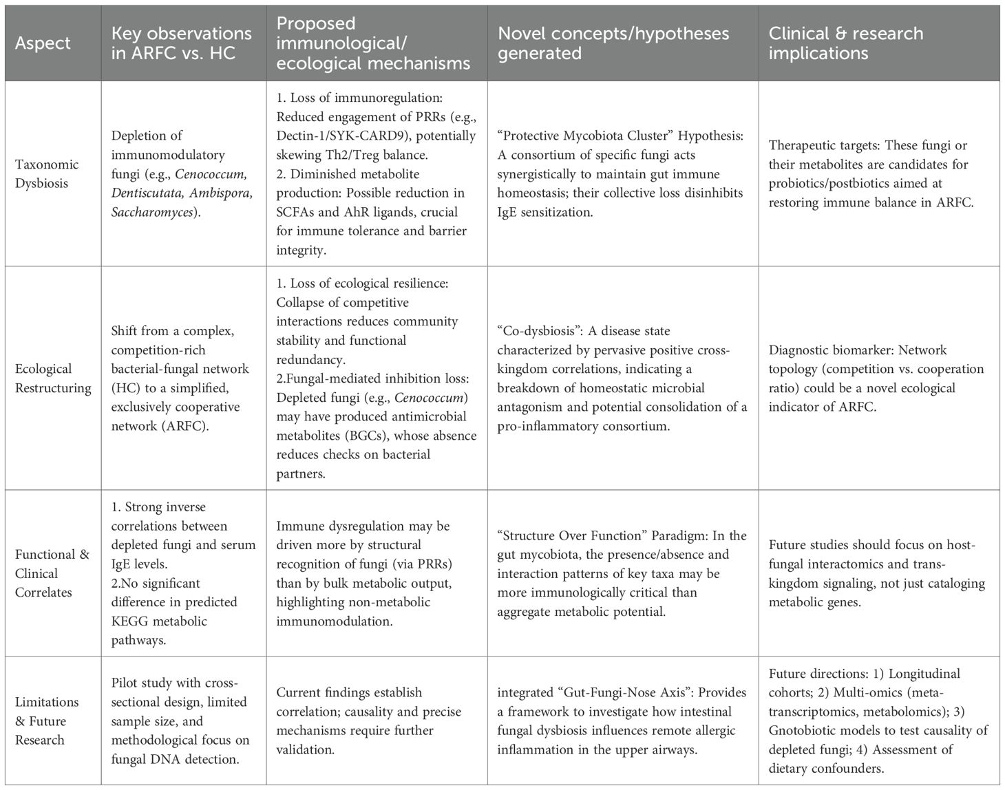
Table 2. Conceptual summary of gut mycobiota alterations, ecological restructuring, and immunological correlates in children with comorbid allergic rhinitis and functional constipation (ARFC).
5 Conclusion
This pilot study demonstrates gut mycobiota dysbiosis in ARFC children, marked by depletion of beneficial fungi and a shift to “co-dysbiosis.” These alterations correlate with IgE sensitization and may underlie gut–nose axis dysregulation. Despite structural changes, functional metabolic potential was conserved. The mycobiota emerges as a critical element in ARFC pathogenesis, warranting larger-scale validation.
Data availability statement
The raw metagenomic sequencing data generated in this study have been deposited in the NCBI Sequence Read Archive (SRA) under accession number PRJNA1279672. These data will be made publicly accessible upon publication via the following link: https://www.ncbi.nlm.nih.gov/sra/PRJNA1279672.
Ethics statement
The studies involving humans were approved by Longgang District Maternity & Child Healthcare Hospital. The studies were conducted in accordance with the local legislation and institutional requirements. Written informed consent for participation in this study was provided by the participants’ legal guardians/next of kin.
Author contributions
HL: Data curation, Investigation, Project administration, Resources, Software, Writing – original draft, Writing – review & editing. LL: Formal Analysis, Methodology, Resources, Supervision, Validation, Writing – review & editing. CW: Formal Analysis, Resources, Supervision, Validation, Visualization, Writing – review & editing. RL: Conceptualization, Data curation, Investigation, Methodology, Software, Writing – review & editing. QL: Data curation, Formal Analysis, Resources, Validation, Visualization, Writing – review & editing. CH: Data curation, Funding acquisition, Methodology, Project administration, Resources, Writing – original draft, Writing – review & editing.
Funding
The author(s) declared that financial support was received for this work and/or its publication. This research was supported by the Research Initiation Fund of Longgang District Maternity and Child Healthcare Hospital of Shenzhen City (Grant No: Y2024011), the Longgang District Science and Technology Innovation Bureau (Grants No: LGWJ2023-038 and LGWJ20230-072), Sanming Project of Medicine in Shenzhen (No. SZSM202311021), and the Key Medical Disciplines Program of Longgang District.
Acknowledgments
We sincerely thank all the children and their families for participating in this study. We also extend our gratitude to the medical staff at Longgang District Maternity and Child Healthcare Hospital for their assistance in clinical evaluations and sample collection. Additionally, we acknowledge the researchers who have publicly shared data and tools that facilitated this work.
Conflict of interest
The authors declared that this work was conducted in the absence of any commercial or financial relationships that could be construed as a potential conflict of interest.
Generative AI statement
The author(s) declared that generative AI was used in the creation of this manuscript. As part of our manuscript preparation process, the authors acknowledge the use of DeepSeek, a generative AI tool, to assist in the literature review process and in refining certain textual elements of this manuscript. Specifically, DeepSeek was utilized to support the initial screening and summarization of a subset of relevant publications, and to aid in the linguistic polishing and clarity of expression in several sections. All AI-generated content or suggestions were critically reviewed, verified, and substantively edited by the authors, who assume full responsibility for the accuracy, integrity, and scientific validity of the final work. This use of AI tools aligns with the journal’s policy on transparent reporting of manuscript preparation assistance.
Any alternative text (alt text) provided alongside figures in this article has been generated by Frontiers with the support of artificial intelligence and reasonable efforts have been made to ensure accuracy, including review by the authors wherever possible. If you identify any issues, please contact us.
Publisher’s note
All claims expressed in this article are solely those of the authors and do not necessarily represent those of their affiliated organizations, or those of the publisher, the editors and the reviewers. Any product that may be evaluated in this article, or claim that may be made by its manufacturer, is not guaranteed or endorsed by the publisher.
Supplementary material
The Supplementary Material for this article can be found online at: https://www.frontiersin.org/articles/10.3389/fimmu.2025.1745580/full#supplementary-material
References
1. Yamaguchi T, Nomura A, Matsubara A, Hisada T, Tamada Y, Mikami T, et al. Effect of gut microbial composition and diversity on major inhaled allergen sensitization and onset of allergic rhinitis. Allergol Int. (2023) 72:135–42. doi: 10.1016/j.alit.2022.06.005
2. Kallio S, Jian C, Korpela K, Kukkonen AK, Salonen A, Savilahti E, et al. Early-life gut microbiota associates with allergic rhinitis during 13-year follow-up in a Finnish probiotic intervention cohort. Microbiol Spectr. (2024) 12:e0413523. doi: 10.1128/spectrum.04135-23
3. Wu MC, Jan MS, Chiou JY, Wang YH, and Wei JC. Constipation might be associated with risk of allergic rhinitis: A nationwide population-based cohort study. PloS One. (2020) 15:e0239723. doi: 10.1371/journal.pone.0239723
4. Lee MH, Wu MC, Wang YH, and Wei JC. Maternal constipation is associated with allergic rhinitis in the offspring: A nationwide retrospective cohort study. PloS One. (2023) 18:e0292594. doi: 10.1371/journal.pone.0292594
5. Ho SW, Lin CP, and Ku MS. The impact of allergic rhinitis on gastrointestinal disorders among young adults. J Eval Clin Pract. (2020) 26:242–7. doi: 10.1111/jep.13108
6. Zhou MS, Zhang B, Gao ZL, and Zheng RP. Altered diversity and composition of gut microbiota in patients with allergic rhinitis. Microb Pathog. (2021) 161:105272. doi: 10.1016/j.micpath.2021.105272
7. Zhang S, Wang R, Li D, Zhao L, and Zhu L. Role of gut microbiota in functional constipation. Gastroenterol Rep (Oxf). (2021) 9:392–401. doi: 10.1093/gastro/goab035
8. Wang C, Liu H, Li X, Kong W, Wu H, and Huang C. Multiomics technology reveals the changes in gut microbiota to stimulate aromatic amino acid metabolism in children with allergic rhinitis and constipation. Front Allergy. (2025) 6:1562832. doi: 10.3389/falgy.2025.1562832
9. Salgaço MK, Perina NP, Tomé TM, Mosquera EMB, Lazarini T, Sartoratto A, et al. Probiotic infant cereal improves children’s gut microbiota: Insights using the Simulator of Human Intestinal Microbial Ecosystem (SHIME®). Food Res Int. (2021) 143:110292. doi: 10.1016/j.foodres.2021.110292
10. Chiu CY, Cheng ML, Chiang MH, Kuo YL, Tsai MH, Chiu CC, et al. Gut microbial-derived butyrate is inversely associated with IgE responses to allergens in childhood asthma. Pediatr Allergy Immunol. (2019) 30:689–97. doi: 10.1111/pai.13096
11. Botschuijver S, Roeselers G, Levin E, Jonkers DM, Welting O, Heinsbroek SEM, et al. Intestinal fungal dysbiosis is associated with visceral hypersensitivity in patients with irritabl e bowel syndrome and rats. Gastroenterology. (2017) 153:1026–39. doi: 10.1053/j.gastro.2017.06.004
12. Iliev ID and Cadwell K. Effects of intestinal fungi and viruses on immune responses and inflammatory bowel diseases. Gastroenterology. (2021) 160:1050–66. doi: 10.1053/j.gastro.2020.06.100
13. Huang H, Wang Q, Yang Y, Zhong W, He F, Li J, et al. The mycobiome as integral part of the gut microbiome: crucial role of symbiotic fungi in health and disease. Gut Microbes. (2024) 16:2440111. doi: 10.1080/19490976.2024.2440111
14. Wu X, Xia Y, He F, Zhu C, and Ren W. Intestinal mycobiota in health and diseases: from a disrupted equilibrium to clinical opportunities. Microbiome. (2021) 9:60. doi: 10.1186/s40168-021-01024-x
15. Li XV, Leonardi I, and Iliev ID. Gut mycobiota in immunity and inflammatory disease. Immunity. (2019) 50:1365–79. doi: 10.1016/j.immuni.2019.05.023
16. Zhang F, Aschenbrenner D, Yoo JY, and Zuo T. The gut mycobiome in health, disease, and clinical applications in association with the gut bacterial microbiome assembly. Lancet Microbe. (2022) 3:e969–83. doi: 10.1016/s2666-5247(22)00203-8
17. Leonardi I, Li X, Semon A, Li D, Doron I, Putzel G, et al. CX3CR1(+) mononuclear phagocytes control immunity to intestinal fungi. Science. (2018) 359:232–6. doi: 10.1126/science.aao1503
18. Yan Q, Li S, Yan Q, Huo X, Wang C, Wang X, et al. A genomic compendium of cultivated human gut fungi characterizes the gut mycobiome and its relevance to common diseases. Cell. (2024) 187:2969–2989.e2924. doi: 10.1016/j.cell.2024.04.043
19. Glatthardt T, van Tilburg Bernardes E, and Arrieta MC. The mycobiome in atopic diseases: Inducers and triggers. J Allergy Clin Immunol. (2023) 152:1368–75. doi: 10.1016/j.jaci.2023.10.006
20. Li X, Leonardi I, Semon A, Doron I, Gao IH, Putzel GG, et al. Response to fungal dysbiosis by gut-resident CX3CR1(+) mononuclear phagocytes aggravates allergic airway disease. Cell Host Microbe. (2018) 24:847–856.e844. doi: 10.1016/j.chom.2018.11.003
21. Wang Y, He F, Liu B, Wu X, Han Z, Wang X, et al. Interaction between intestinal mycobiota and microbiota shapes lung inflammation. Imeta. (2024) 3:e241. doi: 10.1002/imt2.241
22. Barcik W, Boutin RCT, Sokolowska M, and Finlay BB. The role of lung and gut microbiota in the pathology of asthma. Immunity. (2020) 52:241–55. doi: 10.1016/j.immuni.2020.01.007
23. Skalski JH, Limon JJ, Sharma P, Gargus MD, Nguyen C, Tang J, et al. Expansion of commensal fungus Wallemia mellicola in the gastrointestinal mycobiota enhances the severity of allergic airway disease in mice. PloS Pathog. (2018) 14:e1007260. doi: 10.1371/journal.ppat.1007260
24. Shibuya A and Shibuya K. Exploring the gut fungi-lung allergy axis. Cell Host Microbe. (2018) 24:755–7. doi: 10.1016/j.chom.2018.11.012
25. Zhu L, Wu Y, Lin C, Tang L, Yu B, Wan W, et al. Dynamic microbial shifts and signatures of long-term remission in allergic rhinitis after an herbal formula treatment. Front Immunol. (2021) 12:774966. doi: 10.3389/fimmu.2021.774966
26. Zhang P, Zhou X, Tan H, Jian F, Jing Z, Wu H, et al. Microbial signature of intestine in children with allergic rhinitis. Front Microbiol. (2023) 14:1208816. doi: 10.3389/fmicb.2023.1208816
27. Hu Y, Zhang R, Li J, Wang H, Wang M, Ren Q, et al. Association between gut and nasal microbiota and allergic rhinitis: A systematic review. J Asthma Allergy. (2024) 17:633–51. doi: 10.2147/jaa.S472632
28. Kanj AN, Kottom TJ, Schaefbauer KJ, Choudhury M, Limper AH, and Skalski JH. Dysbiosis of the intestinal fungal microbiota increases lung resident group 2 innate lymphoid cells and is associated with enhanced asthma severity in mice and humans. Respir Res. (2023) 24:144. doi: 10.1186/s12931-023-02422-5
29. Zheng F, Yang Y, Lu G, Tan JS, Mageswary U, Zhan Y, et al. Metabolomics insights into gut microbiota and functional constipation. Metabolites. (2025) 15:269. doi: 10.3390/metabo15040269
30. Malik A, Sharma D, Malireddi RKS, Guy CS, Chang TC, Olsen SR, et al. SYK-CARD9 signaling axis promotes gut fungi-mediated inflammasome activation to restrict colitis and colon cancer. Immunity. (2018) 49:515–530.e515. doi: 10.1016/j.immuni.2018.08.024
31. Li J, Chen D, Yu B, He J, Zheng P, Mao X, et al. Fungi in gastrointestinal tracts of human and mice: from community to functions. Microb Ecol. (2018) 75:821–9. doi: 10.1007/s00248-017-1105-9
32. Hyams JS, Di Lorenzo C, Saps M, Shulman RJ, Staiano A, and van Tilburg M. Functional disorders: children and adolescents. Gastroenterology. (2016) S0016-5085(16)00181–5. doi: 10.1053/j.gastro.2016.02.015
33. Cheng L. Revising allergic rhinitis guidelines to standardize clinical diagnosis and treatment. Zhonghua Er Bi Yan Hou Tou Jing Wai Ke Za Zhi. (2022) 57:413–7. doi: 10.3760/cma.j.cn115330-20220325-00134
34. Maas E, Penders J, and Venema K. Fungal-bacterial interactions in the human gut of healthy individuals. J Fungi (Basel). (2023) 9:139. doi: 10.3390/jof9020139
35. Coyte KZ, Schluter J, and Foster KR. The ecology of the microbiome: Networks, competition, and stability. Science. (2015) 350:663–6. doi: 10.1126/science.aad2602
36. Faust K and Raes J. Microbial interactions: from networks to models. Nat Rev Microbiol. (2012) 10:538–50. doi: 10.1038/nrmicro2832
37. Wang H, Wu H, Li KD, Wang YY, Huang RG, Du YJ, et al. Intestinal fungi and systemic autoimmune diseases. Autoimmun Rev. (2023) 22:103234. doi: 10.1016/j.autrev.2022.103234
38. Zhang H, Wei Y, Jia H, Chen D, Tang X, Wang J, et al. Immune activation of characteristic gut mycobiota Kazachstania pintolopesii on IL-23/IL-17R signaling in ankylosing spondylitis. Front Cell Infect Microbiol. (2022) 12:1035366. doi: 10.3389/fcimb.2022.1035366
39. Narunsky-Haziza L, Sepich-Poore GD, Livyatan I, Asraf O, Martino C, Nejman D, et al. Pancancer analyses reveal cancer-type-specific fungal ecologies and bacteriome interactions. Cell. (2022) 185:3789–3806.e17. doi: 10.1016/j.cell.2022.09.005
40. Carlson SL, Mathew L, Savage M, Kok K, Lindsay JO, Munro CA, et al. Mucosal immunity to gut fungi in health and inflammatory bowel disease. J Fungi (Basel). (2023) 9:1105. doi: 10.3390/jof9111105
41. Kiran NS, Chatterjee A, Yashaswini C, Deshmukh R, Alsaidan OA, Bhattacharya S, et al. The gastrointestinal mycobiome in inflammation and cancer: unraveling fungal dysbiosis, pathogenesis, and therapeutic potential. Med Oncol. (2025) 42:195. doi: 10.1007/s12032-025-02761-x
42. Sam QH, Chang MW, and Chai LY. The fungal mycobiome and its interaction with gut bacteria in the host. Int J Mol Sci. (2017) 18:330. doi: 10.3390/ijms18020330
43. Baral B, Akhgari A, and Metsä-Ketelä M. Activation of microbial secondary metabolic pathways: Avenues and challenges. Synth Syst Biotechnol. (2018) 3:163–78. doi: 10.1016/j.synbio.2018.09.001
44. Nenciarini S and Cavalieri D. Immunomodulatory potential of fungal extracellular vesicles: insights for therapeutic applications. Biomolecules. (2023) 13:1487. doi: 10.3390/biom13101487
45. Tipton L, Müller CL, Kurtz ZD, Huang L, Kleerup E, Morris A, et al. Fungi stabilize connectivity in the lung and skin microbial ecosystems. Microbiome. (2018) 6:12. doi: 10.1186/s40168-017-0393-0
46. van Tilburg Bernardes E, Pettersen VK, Gutierrez MW, Laforest-Lapointe I, Jendzjowsky NG, Cavin JB, et al. Intestinal fungi are causally implicated in microbiome assembly and immune development in mice. Nat Commun. (2020) 11:2577. doi: 10.1038/s41467-020-16431-1
47. Saito Y, Sato T, Nomoto K, and Tsuji H. Identification of phenol- and p-cresol-producing intestinal bacteria by using media supplemented with tyrosine and its metabolites. FEMS Microbiol Ecol. (2018) 94:fiy125. doi: 10.1093/femsec/fiy125
48. Zhao L, Yang W, Chen Y, Huang F, Lu L, Lin C, et al. A Clostridia-rich microbiota enhances bile acid excretion in diarrhea-predominant irrita ble bowel syndrome. J Clin Invest. (2020) 130:438–50. doi: 10.1172/JCI130976
49. Roager HM, Hansen LBS, Bahl MI, Frandsen HL, Carvalho V, Gøbel RJ, et al. Colonic transit time is related to bacterial metabolism and mucosal turnover in the gut. Nat Microbiol. (2016) 1:16093. doi: 10.1038/nmicrobiol.2016.93
50. Sonnenburg ED, Smits SA, Tikhonov M, Higginbottom SK, Wingreen NS, and Sonnenburg JL. Diet-induced extinctions in the gut microbiota compound over generations. Nature. (2016) 529:212–5. doi: 10.1038/nature16504
Keywords: allergic rhinitis, children, co-dysbiosis, cross-kingdom network, functional constipation, fungal dysbiosis, gut mycobiota, IgE
Citation: Liu H, Liang L, Wang C, Luo R, Luo Q and Huang C (2026) Gut mycobiota dysbiosis and an emergent state of “co-dysbiosis” are associated with IgE sensitization in children with comorbid allergic rhinitis and constipation. Front. Immunol. 16:1745580. doi: 10.3389/fimmu.2025.1745580
Received: 13 November 2025; Accepted: 30 December 2025; Revised: 26 December 2025;
Published: 23 January 2026.
Edited by:
Hector Mora Montes, University of Guanajuato, MexicoReviewed by:
Bernardo Franco, University of Guanajuato, MexicoLaura Garcia-Carnero, University of São Paulo, Brazil
Copyright © 2026 Liu, Liang, Wang, Luo, Luo and Huang. This is an open-access article distributed under the terms of the Creative Commons Attribution License (CC BY). The use, distribution or reproduction in other forums is permitted, provided the original author(s) and the copyright owner(s) are credited and that the original publication in this journal is cited, in accordance with accepted academic practice. No use, distribution or reproduction is permitted which does not comply with these terms.
*Correspondence: Congfu Huang, NzgzMzM3NTVAcXEuY29t
†These authors have contributed equally to this work