Abstract
Antiphospholipid Syndrome (APS) is a systemic autoimmune disorder characterized by persistent antiphospholipid antibodies, associated with thrombosis or adverse pregnancy outcomes. Although non-thrombotic manifestations are less common than thrombotic events, they play an increasingly important role in diagnosis and disease progression, particularly in pediatric APS, and may interact with each other. Early recognition and management of these symptoms are crucial for patient prognosis. We present a case of 13-year-old male child presenting as autoimmune hemolytic anemia (AIHA) and myocarditis, with positive antiphospholipid antibodies (aPL). The patient showed significant improvement after combination therapy with corticosteroids, prophylactic low molecular weight heparin and aspirin. In addition, we conducted a comprehensive literature review on APS in conjunction with hemolytic anemia and cardiac complications, and found that in 171 APS cases with hemolytic anemia, 35 of them had cardiac complications. There were 8 cases of myocardial infarction (8/171,4.7%) and 3 cases of myocarditis (3/171, 1.8%). Compared with AIHA, the incidence of cardiopathy was significantly higher in microvascular hemolytic anemia (MAHA) (p = 0.020). Among 13 cases that recorded the time window from hemolytic anemia to cardiac symptoms, 92.3% of them developed cardiac complications within one year after the onset of hemolytic anemia, typically within a median interval of 3 months (ranging from 5 days to 2 years). Notably, 28.6% of the 35 cases reviewed involved children under 18, with nearly half presenting hemolytic anemia as the initial symptom. This case underscores APS patients with hemolytic anemia, particularly MAHA, require early intervention and timely cardiac follow-up. Given the current lack of definitive classification and treatment criteria for pediatric APS, future guidelines should incorporate the significance of non-thrombotic manifestations and emphasize early management strategies.
1 Introduction
Antiphospholipid syndrome (APS) is a systemic autoimmune disease characterized by recurrent vascular thrombosis or pregnancy complications, with persistent positivity for antiphospholipid antibodies (aPL) (1). It can present as a primary condition, called primary APS or can be secondary to other autoimmune diseases, commonly seen in systemic lupus erythematosus (SLE) (2, 3). Pediatric antiphospholipid syndrome (Ped-APS) is relatively rare, and the prevalence of secondary APS is slightly higher than that of primary APS (4, 5).
In addition to the common arterial and venous thrombosis, APS can present as microthrombosis or a variety of non-thrombotic manifestations (6), such as thrombocytopenia, autoimmune hemolytic anemia (AIHA), cardiac valve disease, livedo reticularis, kidney disease, chorea, migraine, and cognitive dysfunction (7, 8), and these manifestations may interact with each other (9). AIHA is typically caused by immune-mediated destruction of red blood cells (10). Notably, patients who initially present with hemolytic anemia have a significantly increased incidence of subsequent serious events (11). Myocarditis is less common in APS but may cause severe consequences, potentially resulting from microvascular thrombosis or direct immune-mediated myocardial injury (12, 13). It is particularly important to note that Ped-APS is not common, there is no pediatric-specific classification criteria, and diagnosis often relies on “adult criteria and the SHARE Initiative” (14, 15). However, compared to adults, Ped-APS is more likely to present with non-thrombotic symptoms, sometimes even as the initial symptom, which can be easily overlooked and lead to delayed diagnosis (4, 16). Therefore, early recognition and appropriate anticoagulation therapy are crucial for preventing the morbidity and mortality associated with APS.
Herein, we present a case of a 13-year-old male who was diagnosed with primary APS associated with AIHA and myocarditis. Additionally, we conducted a comprehensive literature review on APS in conjunction with hemolytic anemia and cardiac complications, aiming to explore their clinical features and to investigate any potential associations between hemolytic anemia and cardiac symptoms in the context of APS. This review seeks to enhance the understanding of the diagnosis and management of these associated complications.
2 Case presentation
A 13-year-old male patient presented to our department with symptoms of fever and fatigue for 5 days. Blood tests at a local hospital indicated a hemoglobin (Hb) level of 43 g/L (normal 120-165g/L). The child had no respiratory or gastrointestinal symptoms, no epistaxis, no gross hematuria, and no melena. His Hb level was normal one month prior to admission. He had no previous medical problems with no family history of genetic diseases.
Laboratory tests showed a significant decrease in Hb to 37 g/L and an elevated reticulocyte count of 93.5 × 10⁹/L (normal 24-84 × 109/L). Direct Coombs test revealed positivity for C3 and polyspecific antibodies. Activated partial thromboplastin time (APTT) was significantly prolonged at 58.6 seconds (normal 25–35 seconds) and could not be corrected. The platelet count dropped to 80 × 109/L. The lupus anticoagulant (LAC) assay detected a positive result. Additionally, standardized enzyme-linked immunosorbent assay (ELISA) testing revealed moderate or high positivity for both anticardiolipin (aCL) and anti-β2-glycoprotein I (anti-β2GPI) antibodies (IgG and IgM). Rheumatological and immunological tests, including antinuclear antibody, anti-double-stranded DNA and anti-Smith antibodies, were both negative, with normal C3 and C4 levels. No evidence of thrombosis.
Based on these findings, despite the presence of three positive aPL, the absence of clinical symptoms meeting APS classification criteria led us to provide personalized treatment for the patient. The patient was initiated on a combination therapy including prednisone (20 mg per dose, three times daily) and low-molecular weight heparin (4100 IU nadroparin calcium, once daily). Two weeks later, Hb levels rapidly achieved to normal (Figure 1A). One month later, prophylactic low molecular weight heparin was discontinued, and the dose of corticosteroid was gradually tapered and discontinued after two months.
Figure 1

(A) The line chart shows the hemoglobin (Hb) changes of the patient during the whole course of treatment. The patient presented with severe anemia upon initial admission, and corticosteroid treatment quickly corrected the anemia. ★ The patient was transfused with Leukocyte-reduced Red Blood Cells suspension on May 6, May 8, and May 12, and Hb briefly increased after infusion. ▴ On May 15, the patient was prescribed prednisone and prophylactic low molecular weight heparin therapy. ♦ On May 21, the patient was discharged and prescribed oral prednisone following discharge. ▾ On July 2, the patient discontinued corticosteroids. (B) The line chart shows changes in troponin I (TnI) and high-sensitivity troponin T (hs-TnT) in patients throughout the course of treatment. When the patient was admitted to hospital for the second time on July 2, the laboratory examination showed that TnI and hs-TnT were particular higher than normal. With the onset of treatment, the patient’s TnI and hs-TnT decreased to normal. ★ On July 6, the patient was prescribed corticosteroid, aspirin, prophylactic low molecular weight heparin therapy, TnI and hs-TnT decreased significantly after drug administration. ▴ On July 18, the patient was discharged, and continued with oral prednisone and aspirin.
However, during the follow-up two weeks later, it was found that the troponin I (TnI) level increased to 1.822 ng/mL (normal <0.026 ng/mL), and the high-sensitivity troponin T (hs-TnT) level increased to 0.119 ng/L (normal <0.1 ng/L) (Figure 1B). Electrocardiogram (ECG) indicated sinus arrhythmia. There were no symptoms of chest discomfort, pain, palpitations, or fatigue. Pathogen detection was negative, ruling out bacterial or viral infections as causes of myocarditis. Subsequent laboratory evaluations showed another significant prolongation of APTT, with LAC was still positive, and according to ELISA, aCL and anti-β2GPl (lgG and lgM) still showing moderate to high positivity. Imaging studies, including echocardiography and multisite CTA, showed no thrombotic lesions. Cardiovascular magnetic resonance (CMR) with contrast suggested myocarditis with myocardial injury, myocardial edema and pericardial effusion (Figures 2A–D). We initiated corticosteroid treatment again, in combination with prophylactic aspirin for antiplatelet aggregation and low molecular weight heparin therapy. As shown in Figure 1B, the patient’s clinical indicators showed improvement.
Figure 2
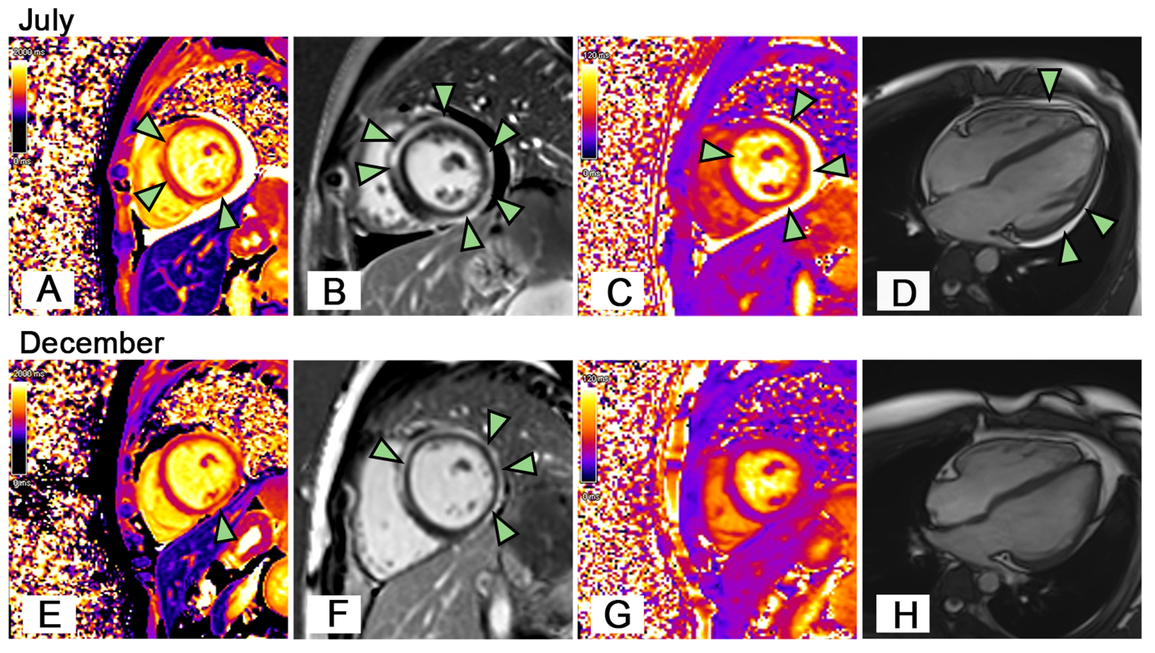
Cardiac Magnetic Resonance Imaging. On July (A) The patient’s short-axis view showed local increases in T1 values in the anterior septum and inferior wall of the left ventricle (arrowheads). (B) The patient’s LGE image showed delayed subepicardial enhancement of the left ventricle, including the anterior septum, anterior wall, lateral wall, posterior wall, and inferior wall (arrowheads). (C) The patient’s short-axis view showed increased T2 values in the left ventricular myocardium (edema), especially in the anterior septum, inferior, posterior, and lateral walls (arrowheads). (D) The patient had pericardial effusion (arrowheads). On December (E) The T1 value of the anterior septum of the left ventricle decreased to normal, while the local T1 value of the inferior wall was still increased (arrowhead). (F) LGE image showed the delayed enhancement of the left ventricular subepicardium persisted (arrowheads). (G) The overall left ventricular T2 value decreased to normal. (H) The pericardial effusion disappeared.
At a follow-up after 5 months, CMR revealed complete resolution of pericardial effusion and myocardial edema (Figures 2E–H). Subsequent laboratory tests showed Hb levels were normal, Coombs test was negative, and cardiac biomarkers including TnI, hs-TnT, CK-MB, and NT-Pro BNP returned to normal. The aPL remained consistently positive. The patient continued to take a low dose of corticosteroids and aspirin. During our close follow-up, the patient remained in good health.
3 Literature review
We conducted a comprehensive search of the literatures related to “antiphospholipid syndrome” and “hemolytic anemia” published before March 13, 2025 in PubMed, Web of Science and Embase databases. We collected cases with complete clinical data and a confirmed diagnosis of APS accompanied by hemolytic anemia, while excluding those with congenital hereditary hemolytic disorders and studies that did not specify the presence of hemolytic anemia at the individual patient level. The literature screening process is detailed in Figure 3. Of these studies, two were retrospective analyses and the others were case reports. In the included literature, we further screened for cases where cardiac complications were clearly identified. Data extracted included the age of onset, gender, cardiac clinical manifestations, classification of hemolytic anemia, and laboratory test results for aPL (Supplementary Table 1).
Figure 3
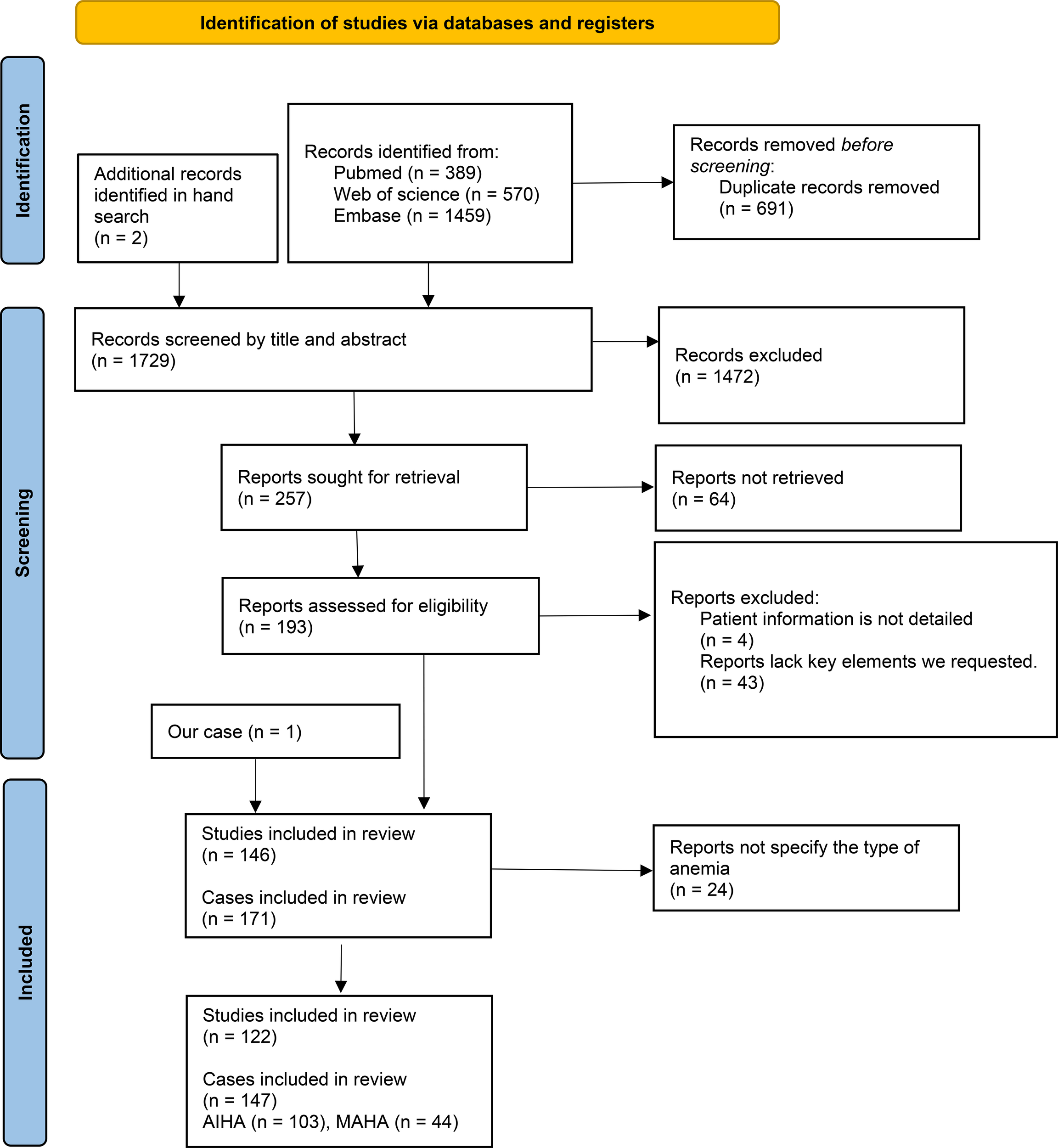
PRISMA flow diagram for the study.
The initial literature search yielded 146 results, including our case, resulting in a total of 171 patients after the removal of duplicates. To investigate the likelihood of cardiac complications in patients with APS associated with different types of anemia, the anemias were ultimately categorized into autoimmune hemolytic anemia (AIHA) (n = 103) and microangiopathic hemolytic anemia (MAHA) (n = 44), with 24 cases having an unspecified type of anemia. Specific information is provided in Supplementary Table 2. Manual screening identified 35 patients with APS associated with hemolytic anemia and cardiac disease, along with our reported case, were included in Supplementary Table 1 for statistical analysis.
All 147 cases of APS with definite hemolytic anemia, we found that MAHA is associated with a significantly higher incidence of cardiac complications compared to AIHA (36.4% vs. 18.4%, p = 0.020) (see Table 1).
Table 1
| Cardiac complications | AIHA (n, %) | MAHA (n, %) | P* |
|---|---|---|---|
| Yes | 19 (18.4) | 16 (36.4) | 0.020 |
| No | 84 (81.6) | 28 (63.6) |
Differences in cardiac complications among antiphospholipid syndrome patients with various type of hemolytic anemia.
*Chi-square test. p<0.05.
Among 35 APS patients who had hemolytic anemia and cardiac disease, the median age of onset was 32 years (ranging from 7 to 72 years, Supplementary Table 1). 10 patients were under the age of 18, accounting for 28.6% of the total. Males were 25.7% (9/35), while females accounted for 74.3% (26/35). Among the 35 APS patients, 13 cases reported the time window from hemolytic anemia to the onset of cardiac complications, with a median interval of 3 months (ranging from 5 days to 2 years). Among these 13 cases, 12 (92.3%) experienced cardiac complications within one year. The median age of these 13 patients was 14 years (with a range of 7 to 54 years), including 7 patients (53.8%) under the age of 18.
Notably, among 35 APS patients with both hemolytic anemia and cardiac disease, myocardial complications accounted for 62.9% (22/35), valvular disease for 25.7% (9/35), and intracardiac thrombosis for 14.3% (5/35). Among the myocardial complications, there were 8 cases of myocardial infarction, 6 cases of heart failure, 3 cases of cardiomyopathy, and 3 cases of myocarditis.
4 Discussion
We report a child presenting with AIHA, myocarditis, and positive aPLs, who was diagnosed with suspected APS. The patient’s ECG did not show ST segment changes, and both echocardiography and CTA did not reveal any thrombi. However, CMR indicated myocarditis with myocardial injury, myocardial edema, and pericardial effusion. Given the presence of prolonged APTT and multiple aPL, we strongly suspected APS. After initiating treatment with corticosteroids and prophylactic low molecular weight heparin for anticoagulation, the patient’s abnormal parameters improved, and subsequent follow-up did not reveal any thrombotic manifestations. Notably, this case is particularly interesting because, based on the limited diagnostic findings available, the cardiac involvement in this APS patient likely differs from typical arterial thrombotic features (17) associated with APS. It is more likely to be a non-thrombotic form of myocardial disease. However, we acknowledge that since no biopsy was performed, we cannot definitively rule out the possibility of microangiopathic lesions. To our knowledge, this is the first reported case of myocarditis in a child with primary APS. This finding broadens the understanding of multi-organ manifestations in APS and highlights myocarditis as a potential rare yet important clinical phenotype.
APS is known for its thrombotic clinical manifestations, and hemolytic anemia, considered a non-thrombotic manifestation of APS, may influence cardiac complications through several mechanisms, including the onset of myocardial ischemia, an increase in cardiac workload, the activation of inflammatory responses, and the hypercoagulable state (10, 18–23). From our analysis of 171 APS patients with hemolytic anemia, we found that 4.7% (8/171) experienced myocardial infarction, which is significantly higher than the reported incidence of first-onset myocardial infarction in the general APS patient cohorts studied by Hui Shi (1.2%) (24) and Cervera (2.8%) (25). Additionally, 1.8% (3/171) experienced myocarditis, also exceeding the reported incidence of myocarditis in the general APS patient cohort of the Yuan Zhao’s study (0.9%) (26). This phenomenon suggests that hemolytic anemia may be associated with an increased risk of cardiac complications in APS patients. Furthermore, in our analysis, MAHA is more likely to be associated with cardiac complications compared to AIHA (P = 0.020) (Table 1). This increased risk may be related to the fact that nearly half of these 35 cases evolved into CAPS, where the underlying mechanism includes extensive microvascular and macrovascular changes due to intravascular thrombosis (27). This situation is more likely to present clinical manifestations of both hemolytic anemia and cardiac complications compared to classical APS. Additionally, MAHA often leads to mechanical damage to erythrocytes from thrombus formation in micro-vessels. These microthrombi can not only destroy erythrocytes but may also occlude cardiac micro-vessels, potentially resulting in myocardial ischemia or infarction (28, 29). However, it is important to note that the articles included in our review are primarily case reports. Case reports often describe more severe conditions, which may lead to publication bias; consequently, the absolute frequencies presented here could overestimate the actual prevalence of complications in the general population.
In the analysis of the 13 cases that reported the time window from hemolytic anemia to the onset of cardiac complications (Supplementary Table 1), we observed a wide variation in the time window, ranging from several days to several years, with a median interval of 3 months. Notably, 92.3% (12 cases) experienced the onset of cardiac complications within one year, with some patients even undergoing anticoagulant therapy during this period. Therefore, for APS patients with concomitant hemolytic anemia, clinicians need to be aware of the potential for cardiac complications. Notably, the majority of these cases were children (53.8%), highlighting the need for special attention to the characteristics of disease progression in pediatric patients.
As shown in Supplementary Table 1, among the 35 cases of hemolytic anemia associated with cardiac complications, 28.6% were children under the age of 18, with nearly half of these pediatric patients (4/10) presenting with hemolytic anemia as the initial symptom. Interestingly, similar to the cases we reported, 12 cases exhibiting hemolytic anemia as the initial symptom were identified in Supplementary Table 1. These data add to the evidence that Ped-APS patients may present with non-thrombotic manifestations. Currently, there are no independent classification standards for Ped-APS, and classification criteria still relies on adult criteria (30). However, the clinical presentations of APS in children are different from those in adults, as many traditional thrombotic risk factors, such as smoking and hypertension, are rare in pediatric populations (4). In contrast, non-thrombotic manifestations, such as hematological abnormalities, microvascular lesions, and neurological symptoms, are more prevalent in Ped-APS than in adults, and their importance cannot be ignored, which may be an important marker of early disease (4, 31, 32).
Regarding the therapy of APS, our patient received corticosteroids as the first-line treatment for AIHA in the early stage of the disease (33). Laboratory tests revealed triple-positive aPL profiles, accompanied by prolonged APTT, indicating a high risk of APS (34). Although no thrombotic events were observed and the patient did not meet the classification criteria for APS, we administered anticoagulant therapy (prophylactic low molecular weight heparin). Once the patient’s Hb level returned to normal, we discontinued corticosteroids. However, shortly after drug withdrawal, the patient developed asymptomatic myocarditis, and it was highly suspected to be APS. While the primary treatment for APS usually does not focus primarily on corticosteroids, the presence of myocarditis as a significant clinical manifestation prompted us to reinitiate corticosteroids, along with prophylactic low molecular weight heparin for anticoagulation (35). Additionally, aspirin was included for antiplatelet therapy to prevent thrombosis. The patient responded well to this treatment plan. From this case, it is evident that in managing Ped-APS, it is crucial to emphasize not only the necessary anti-thrombotic therapy but also the treatment of non-thrombotic clinical manifestations. Their potential application in pediatric patients deserves further exploration (36). Therefore, based on the existing evidence, we advocate that future Ped-APS treatment guidelines should thoroughly address treatment regimens specifically targeting non-thrombotic symptoms.
5 Conclusion
In conclusion, APS patients presenting with hemolytic anemia, especially MAHA, may be at an elevated risk for complications such as myocardial infarction and myocarditis. Therefore, it may be beneficial to maintain heightened vigilance for cardiac complications in APS patients, particularly those with concurrent hemolytic anemia. Prompt cardiac evaluation is advised at initial presentation and during subsequent follow-up. Furthermore, although Ped-APS is rare, it is essential to enhance awareness of this condition in children, particularly regarding non-thrombotic manifestations such as hemolytic anemia and myocarditis, which may even present as initial symptoms of APS. It may be beneficial to give more attention to non-thrombotic manifestations, particularly when developing criteria for children.
Statements
Data availability statement
The original contributions presented in the study are included in the article/Supplementary Material. Further inquiries can be directed to the corresponding author.
Ethics statement
Written informed consent was obtained from the minor(s)’ legal guardian/next of kin for the publication of any potentially identifiable images or data included in this article. Written informed consent was obtained from the participant/patient(s) for the publication of this case report.
Author contributions
WC: Conceptualization, Investigation, Formal Analysis, Data curation, Writing – original draft, Visualization, Project administration. YS: Formal Analysis, Data curation, Writing – original draft, Visualization, Investigation. DZ: Data curation, Writing – original draft, Formal Analysis. MV: Writing – original draft. GW: Writing – original draft, Visualization. XQW: Validation, Writing – review & editing. MS: Writing – review & editing. XHW: Writing – review & editing. XZ: Writing – review & editing. HJ: Project administration, Conceptualization, Writing – review & editing, Supervision, Validation.
Funding
The author(s) declared that financial support was received for this work and/or its publication. This study was supported by a grant from Natural Science Foundation of China (No.81300130), Basic Research Projects of Liaoning Province (JYTMS20230073), Technology Research and Development Program Project of Liaoning Province (2024JH2/102600318).
Conflict of interest
The author(s) declared that this work was conducted in the absence of any commercial or financial relationships that could be construed as a potential conflict of interest.
Generative AI statement
The author(s) declared that generative AI was not used in the creation of this manuscript.
Any alternative text (alt text) provided alongside figures in this article has been generated by Frontiers with the support of artificial intelligence and reasonable efforts have been made to ensure accuracy, including review by the authors wherever possible. If you identify any issues, please contact us.
Publisher’s note
All claims expressed in this article are solely those of the authors and do not necessarily represent those of their affiliated organizations, or those of the publisher, the editors and the reviewers. Any product that may be evaluated in this article, or claim that may be made by its manufacturer, is not guaranteed or endorsed by the publisher.
Supplementary material
The Supplementary Material for this article can be found online at: https://www.frontiersin.org/articles/10.3389/fimmu.2026.1724748/full#supplementary-material
References
1
Hughes GR . Thrombosis, abortion, cerebral disease, and the lupus anticoagulant. Br Med J (Clin Res Ed). (1983) 287:1088–9. doi: 10.1136/bmj.287.6399.1088
2
Gezer S . Antiphospholipid syndrome. Dis Mon. (2003) 49:696–741. doi: 10.1016/j.disamonth.2003.10.001
3
Harris EN Pierangeli SS . Primary, Secondary, Catastrophic Antiphospholipid Syndrome: is there a difference? Thromb Res. (2004) 114:357–61. doi: 10.1016/j.thromres.2004.08.003
4
Bitsadze V Khizroeva J Lazarchuk A Salnikova P Yagubova F Tretyakova M et al . Pediatric antiphospholipid syndrome: is it the same as an adult? J Matern Fetal Neonatal Med. (2024) 37:2390637. doi: 10.1080/14767058.2024.2390637
5
Tarango C Palumbo JS . Antiphospholipid syndrome in pediatric patients. Curr Opin Hematol. (2019) 26:366–71. doi: 10.1097/moh.0000000000000523
6
Garcia D Erkan D . Diagnosis and management of the antiphospholipid syndrome. N Engl J Med. (2018) 378:2010–21. doi: 10.1056/NEJMra1705454
7
Miyakis S Lockshin MD Atsumi T Branch DW Brey RL Cervera R et al . International consensus statement on an update of the classification criteria for definite antiphospholipid syndrome (APS). J Thromb Haemost. (2006) 4:295–306. doi: 10.1111/j.1538-7836.2006.01753.x
8
Xourgia E Tektonidou MG . Management of non-criteria manifestations in antiphospholipid syndrome. Curr Rheumatol Rep. (2020) 22:51. doi: 10.1007/s11926-020-00935-2
9
Krause I Leibovici L Blank M Shoenfeld Y . Clusters of disease manifestations in patients with antiphospholipid syndrome demonstrated by factor analysis. Lupus. (2007) 16:176–80. doi: 10.1177/0961203306075977
10
Ames PRJ Merashli M Bucci T Pastori D Pignatelli P Arcaro A et al . Antiphospholipid antibodies and autoimmune haemolytic anaemia: A systematic review and meta-analysis. Int J Mol Sci. (2020) 21:4120. doi: 10.3390/ijms21114120
11
Tektonidou MG Ioannidis JP Boki KA Vlachoyiannopoulos PG Moutsopoulos HM . Prognostic factors and clustering of serious clinical outcomes in antiphospholipid syndrome. Qjm. (2000) 93:523–30. doi: 10.1093/qjmed/93.8.523
12
Cheng CY Baritussio A Giordani AS Iliceto S Marcolongo R Caforio ALP . Myocarditis in systemic immune-mediated diseases: Prevalence, characteristics and prognosis. A systematic review. Autoimmun Rev. (2022) 21:103037. doi: 10.1016/j.autrev.2022.103037
13
Zhang L Zhu YL Li MT Gao N You X Wu QJ et al . Lupus myocarditis: A case-control study from China. Chin Med J (Engl). (2015) 128:2588–94. doi: 10.4103/0366-6999.166029
14
Groot N de Graeff N Avcin T Bader-Meunier B Brogan P Dolezalova P et al . European evidence-based recommendations for diagnosis and treatment of childhood-onset systemic lupus erythematosus: the SHARE initiative. Ann Rheum Dis. (2017) 76:1788–96. doi: 10.1136/annrheumdis-2016-210960
15
Zheng J Wei ZY Lin SC Wang Y Fang X . Antiphospholipid syndrome onset with hemolytic anemia and accompanied cardiocerebral events: a case report. Front Pediatr. (2024) 12:1370285. doi: 10.3389/fped.2024.1370285
16
Avcin T Cimaz R Silverman ED Cervera R Gattorno M Garay S et al . Pediatric antiphospholipid syndrome: clinical and immunologic features of 121 patients in an international registry. Pediatrics. (2008) 122:e1100–1107. doi: 10.1542/peds.2008-1209
17
Puntmann VO Valbuena S Hinojar R Petersen SE Greenwood JP Kramer CM et al . Society for Cardiovascular Magnetic Resonance (SCMR) expert consensus for CMR imaging endpoints in clinical research: part I - analytical validation and clinical qualification. J Cardiovasc Magn Reson. (2018) 20:67. doi: 10.1186/s12968-018-0484-5
18
Abou-Ismail MY Kapoor S Citla D Sridhar Nayak L Ahuja S . Thrombotic microangiopathies: An illustrated review. Res Pract Thromb Haemost. (2022) 6:e12708. doi: 10.1002/rth2.12708
19
Palmer D Seviar D . How to approach haemolysis: Haemolytic anaemia for the general physician. Clin Med (Lond). (2022) 22:210–3. doi: 10.7861/clinmed.2022-0142
20
Dimitrov JD Roumenina LT Perrella G Rayes J . Basic mechanisms of hemolysis-associated thrombo-inflammation and immune dysregulation. Arterioscler Thromb Vasc Biol. (2023) 43:1349–61. doi: 10.1161/atvbaha.123.318780
21
Pires IS Berthiaume F Palmer AF . Engineering therapeutics to detoxify hemoglobin, heme, and iron. Annu Rev BioMed Eng. (2023) 25:1–21. doi: 10.1146/annurev-bioeng-081622-031203
22
Dutra FF Bozza MT . Heme on innate immunity and inflammation. Front Pharmacol. (2014) 5:115. doi: 10.3389/fphar.2014.00115
23
Ataga KI . Hypercoagulability and thrombotic complications in hemolytic anemias. Haematologica. (2009) 94:1481–4. doi: 10.3324/haematol.2009.013672
24
Shi H Teng JL Sun Y Wu XY Hu QY Liu HL et al . Clinical characteristics and laboratory findings of 252 Chinese patients with anti-phospholipid syndrome: comparison with Euro-Phospholipid cohort. Clin Rheumatol. (2017) 36:599–608. doi: 10.1007/s10067-017-3549-1
25
Cervera R Piette JC Font J Khamashta MA Shoenfeld Y Camps MT et al . Antiphospholipid syndrome: clinical and immunologic manifestations and patterns of disease expression in a cohort of 1,000 patients. Arthritis Rheumatol. (2002) 46:1019–27. doi: 10.1002/art.10187
26
Zhao Y Qi W Huang C Zhou Y Wang Q Tian X et al . Serum calprotectin as a potential predictor of microvascular manifestations in patients with antiphospholipid syndrome. Rheumatol Ther. (2023) 10:1769–83. doi: 10.1007/s40744-023-00610-9
27
Salter BM Crowther MA . Catastrophic antiphospholipid syndrome: a CAPS-tivating hematologic disease. Hematol Am Soc Hematol Educ Program. (2024) 2024:214–21. doi: 10.1182/hematology.2024000544
28
Peigne V Perez P Resche Rigon M Mariotte E Canet E Mira JP et al . Causes and risk factors of death in patients with thrombotic microangiopathies. Intensive Care Med. (2012) 38:1810–7. doi: 10.1007/s00134-012-2638-5
29
Fox LC Cohney SJ Kausman JY Shortt J Hughes PD Wood EM et al . Consensus opinion on diagnosis and management of thrombotic microangiopathy in Australia and New Zealand. Intern Med J. (2018) 48:624–36. doi: 10.1111/imj.13804
30
Barbhaiya M Zuily S Naden R Hendry A Manneville F Amigo MC et al . The 2023 ACR/EULAR antiphospholipid syndrome classification criteria. Arthritis Rheumatol. (2023) 75:1687–702. doi: 10.1002/art.42624
31
Morán-Álvarez P Andreu-Suárez Á Caballero-Mota L Gassiot-Riu S Berrueco-Moreno R Calzada-Hernández J et al . Non-criteria manifestations in the presence of antiphospholipid antibodies in a paediatric cohort. Rheumatol (Oxford). (2022) 61:4465–71. doi: 10.1093/rheumatology/keac070
32
Miyamae T Kawabe T . Non-criteria manifestations of juvenile antiphospholipid syndrome. J Clin Med. (2021) 10:1240. doi: 10.3390/jcm10061240
33
Jäger U Barcellini W Broome CM Gertz MA Hill A Hill QA et al . Diagnosis and treatment of autoimmune hemolytic anemia in adults: Recommendations from the First International Consensus Meeting. Blood Rev. (2020) 41:100648. doi: 10.1016/j.blre.2019.100648
34
Devreese KMJ Ortel TL Pengo V de Laat B . Laboratory criteria for antiphospholipid syndrome: communication from the SSC of the ISTH. J Thromb Haemost. (2018) 16:809–13. doi: 10.1111/jth.13976
35
Putschoegl A Auerbach S . Diagnosis, evaluation, and treatment of myocarditis in children. Pediatr Clin North Am. (2020) 67:855–74. doi: 10.1016/j.pcl.2020.06.013
36
Islabão AG Trindade VC da Mota LMH Andrade DCO Silva CA . Managing antiphospholipid syndrome in children and adolescents: current and future prospects. Paediatr Drugs. (2022) 24:13–27. doi: 10.1007/s40272-021-00484-w
Summary
Keywords
antiphospholipid syndrome, case report, hemolytic anemia, myocarditis, pediatric immunology
Citation
Cui W, Sun Y, Zhao D, Varkani MN, Wang G, Wen X, Shi M, Wang X, Zhang X and Jiang H (2026) Don’t neglect the non-thrombotic manifestations of antiphospholipid syndrome in children – autoimmune hemolytic anemia and myocarditis: a case report and literature review. Front. Immunol. 17:1724748. doi: 10.3389/fimmu.2026.1724748
Received
14 October 2025
Revised
11 December 2025
Accepted
16 January 2026
Published
30 January 2026
Volume
17 - 2026
Edited by
Danieli Castro Oliveira De Andrade, University of São Paulo, Brazil
Reviewed by
Jacqueline A Madison, University of Michigan, United States
Ricardo Krieger Azzolini, University of São Paulo, Brazil
Updates
Copyright
© 2026 Cui, Sun, Zhao, Varkani, Wang, Wen, Shi, Wang, Zhang and Jiang.
This is an open-access article distributed under the terms of the Creative Commons Attribution License (CC BY). The use, distribution or reproduction in other forums is permitted, provided the original author(s) and the copyright owner(s) are credited and that the original publication in this journal is cited, in accordance with accepted academic practice. No use, distribution or reproduction is permitted which does not comply with these terms.
*Correspondence: Hongkun Jiang, hkjiang@cmu.edu.cn
†These authors have contributed equally to this work
Disclaimer
All claims expressed in this article are solely those of the authors and do not necessarily represent those of their affiliated organizations, or those of the publisher, the editors and the reviewers. Any product that may be evaluated in this article or claim that may be made by its manufacturer is not guaranteed or endorsed by the publisher.