 Wei-Jun Li
 Xiao-Zhen Li
- Department of Plant Protection, College of Agronomy, Jiangxi Agricultural University, Nanchang, China
Background: Different insect tissues represent heterogeneous niches with distinct physiological and biochemical characteristics, and therefore host different bacterial communities.
Methods: In this study, those overlapping and specific bacterial communities in the female gut (fG), male gut (mG), female reproductive system (fR), and male reproductive system (mR) of Bactrocera dorsalis (Hendel) adults were determined by high-throughput sequencing targeting 16S rRNA gene.
Results: The richness of bacterial taxa based on OTU was higher in fR compared to the other three tissues. Among the 29 identified bacterial phyla, Pseudomonadota, Bacillota, and Bacteroidota were predominant, while among the 48 identified genera, Enterobacter, Kluyvera, Asticcacaulis, Mesorhizobium, and Serratia were common in the four tissues. fG harbored specific bacterial genus Morganella, mG harbored specific bacterial genera Vagococcus, Lactobacillus, Lactococcus, Lactobacillales, and Bacilli, fR harbored specific bacterial genera Blastomonas, Ralstonia and Providencia, and mR harbored specific bacterial genera Sphingobacteriia, Asticcacaulis, Caulobacter, Caulobacterales, Bradyrhizobium, and Luteimonas. In the 35 annotated KEGG pathways, high-abundance bacterial taxa were mainly enriched in these pathways of membrane transport, carbohydrate metabolism, amino acid metabolism, replication and repair, and energy metabolism, while low-abundance bacterial taxa were involved in these pathways of cardiovascular diseases, circulatory system, and excretory system. The abundances of the 5 pathways associated with cardiovascular diseases, circulatory system, excretory system, membrane transport, and polysaccharide biosynthesis and metabolism exhibited greater variations among fG, mG, fR, and mR. Among them, the two pathways abundances of cardiovascular disease and circulatory system were higher in the reproductive system, whereas the other three pathways abundances were higher in the female gut.
Conclusion: Our study revealed the abundance, composition and function of overlapping and specific bacterial communities in the gut and reproductive system of B. dorsalis, providing valuable information for inhibiting the occurrence of B. dorsalis by interfering with these functional bacterial communities in tissues.
1 Introduction
Insects often coexist with symbiotic bacteria living inside their bodies (Amdam, 2012; Shi W. et al., 2012). Of special interest are insect-associated bacterial communities, some being ubiquitous across different insect tissues, others being specific to particular tissues (de Bekker et al., 2013; Santos et al., 2023). For example, Enterobacter, Kluyvera, and Asticcacaulis are very common in the gut and reproductive system of insects, such as Phlebotomus papatasi (Scopoli) (Maleki-Ravasan et al., 2014), Ceratitis capitata (Wiedemann) (Ben-Yosef et al., 2008), and Bactrocera cacuminata (Hering) (Thaochan et al., 2010). In contrast, Wolbachia strains wCer1 and wCer2 are gram-negative bacteria that exhibit significant tissue specificity, primarily residing in the reproductive system of insects (Riegler and Stauffer, 2002). Insect-associated bacteria may establish a mutualistic symbiotic relationship with insects (Adair and Douglas, 2017), where insects provide space and nutrient for their associated bacteria, while the bacteria support insect growth and health by aiding insects in obtaining nutrients or resisting pathogens (Kandasamy et al., 2022). An example of such a mutualistic relationship is that two lactic acid-producing bacteria, Enterococcus casseliflavus and Lactococcus lactis, may increase the resistance of Bactrocera dorsalis (Hendel) to β-cypermethrin (Zeng et al., 2024).
Different insect tissues exhibit physiological and functional differences. For example, insect guts may contain specific substances, such as digestive enzymes, proteins, and vitamins, which aid in the degradation and absorption of dietary ingredients (Douglas, 2009); and insect reproductive systems contain nutrients and enzymes related to the formation and maturation of eggs or sperms, helping insects to reproduce offspring (Gavriel et al., 2011). The different tissues of insects represent heterogeneous niches with distinct physiological characteristics, and may thus host their own specific bacterial communities (Santos et al., 2023). This heterogeneity may drive the adaptive evolutionary and tissue-specific distribution of insect-associated bacterial communities. Meanwhile, insect-associated bacteria may also require localization in specific insect tissues to function optimally (Feldhaar and Gross, 2009; Lu et al., 2016).
Bacterial communities in different insect tissues have received considerable attentions. We retrieved over 50 articles from PubMed database, exploring the diversity of bacteria in the gut of insects, such as Laodelphax striatellus (Fallen) (Zhang et al., 2020), Recilia dorsalis (Motschulsky) (Huang et al., 2023), Schistocerca gregaria Forskål (Dillon et al., 2010), Sogatella furcifera (Horvath) (Bing et al., 2020), and Zeugodacus cucurbitae (Coquillett) (Lakshmi et al., 2023), and six articles retailing the bacterial communities in the reproductive system of insects, such as B. dorsalis (Shi Z. et al., 2012), B. minax (Enderlein) (Wang A. L. et al., 2014), and Blattella germanica L. (Kakumanu et al., 2018). However, few studies were conducted on those overlapping and unique bacterial communities in specific insect tissues. Here, we only found 2 similar articles: one recording the overlapping bacteria between the gut and feces of B. germanica (Kakumanu et al., 2018), and the other one determining the overlapping microbiota between the intestinal and reproductive tract of chickens (Shterzer et al., 2020). Studying those overlapping and specific bacterial communities in various insect tissues may offer insights into the intrinsic relationship between bacterial communities and the function of tissues associated with bacteria.
Bactrocera dorsalis (Hendel), also known as the oriental fruit fly, is a highly invasive pest of fruits and vegetables, infesting a wide range of crops including citrus, mango, carambola, and guava (Wang H. et al., 2014; Li X. Z. et al., 2024). Due to difference in physiological and biochemical characteristics, the gut and reproductive system of B. dorsalis adults may harbor their specific bacterial communities, in addition to sharing some common bacterial communities (de Bekker et al., 2013). Phylogenetic analyses have shown that the gut of B. dorsalis harbors bacterial genera, such as Enterobacter, Citrobacter, Klebsiella, Pantoea, Pectobacterium, and Serratia (Gwokyalya et al., 2024; Yong et al., 2017; Liu et al., 2016; Behar et al., 2008), whereas its reproductive system contains bacterial species, namely Enterobacter sakazakii, Klebsiella oxytoca, Klebsiella pneumoniae, Raoultella terrigena, and Enterobacter amnigenus (Shi Z. et al., 2012). However, the overlapping and specific bacterial communities, particularly the functional pathway underlying their enrichment, in the gut and reproductive system of B. dorsalis still remained an area ripe for further exploration.
This study aimed to elucidate the overlapping and specific bacterial communities present in the gut and reproductive system of male and female B. dorsalis. The gut and reproductive system was chosen here because they are the main functional tissues of B. dorsalis and the optimal ecological niches for the development and proliferation of bacteria (He et al., 2022; Yin et al., 2024). Due to the differences in physiological functions and nutritional components among different tissues (Douglas, 2009; Gavriel et al., 2011), we hypothesized that there would also be some variations in bacterial taxa, as well as the abundance and functions of bacteria communities within each tissue. Our study would provide a comprehensive catalog of bacterial taxa present in B. dorsalis different tissues, contributing to functional study of important bacteria.
2 Materials and methods
2.1 Sample preparation
The laboratory population of B. dorsalis was derived from 50 to 100 infested navel oranges (Citrus sinensis Osbeck cv. Newhall) collected from an orchard (115°65′E, 25°97’N) in Ganzhou, Jiangxi Province, China in late October, 2018. Larvae from these infested oranges were isolated, and subsequently fed with an artificial diet consisting of wheat bran (300 g), beer yeast (60 g), white sugar (40 g), potassium sorbate (0.8 g), methyl-p-hydroxybenzoate (0.8 g), and water (1,000 mL) until pupation. Pupae were kept in a plastic tray containing 5 cm of loose wet soil (approximately 400 cm3) until adults emerged. Newly emerged adults were separated by sex, and housed separately in an insect-rearing box (78 × 50 × 55 cm), where they were provided with a diet consisting of beer yeast, diluted honey (1%), and water (Li W. J. et al., 2024). During the feeding process, the B. dorsalis population was maintained at 26.5 ± 2°C with 75 ± 5% relative humidity under a 14 h: 10 h (light: dark) photoperiod cycle.
Before sampling, 5-day-old virgin flies were starved for 12 h to eliminate resident bacterial communities retained in their guts and reproductive systems (Andongma et al., 2015). Flies were then surface-sterilized with a 75% ethanol solution for 2–3 min, followed by three rinses in sterile water before dissection. These flies were carefully dissected using a blade on a plate containing 3 mL sterile phosphate-buffered saline (PBS, pH 7.4) underneath a stereomicroscope (CNoptec, Chongqing, China). The female gut (fG), male gut (mG), female reproductive system (fR), and male reproductive system (mR), obtained through dissection, were transferred separately into 2 mL centrifuge tubes each containing 1 ml of extract solution [TIANamp Genomic DNA Kit (TIANGEN, Beijing, China)]. These tissues/samples were then homogenized. Each tissue/sample type comprised three biological replicates, with each replicate consisting of 30 guts or reproductive systems. All samples were snap-frozen in liquid nitrogen, and subsequently stored at −80°C until DNA extraction.
2.2 DNA extraction and PCR amplification
DNA was extracted from the above-mentioned frozen homogenized samples using the TIANamp Genomic DNA Kit (TIANGEN, Beijing, China), following the manufacturer’s protocol. The integrity of the extracted DNA was verified by 1.5% agarose gel electrophoresis, and its concentration was determined by measuring the absorbance at 260 nm using a Nanodrop spectrophotometer (Thermo Fisher Scientific, Madison, WI, USA). For taxonomical profiling of bacteria, the universal primers, forward 338F (5′-ACTCCTACGGGAGGCAGCAG-3′) and reverse 806R (5′-TACHVGGGTWTCTAAT-3′) (Johnson et al., 2019), were used to amplify the V3–V4 hypervariable regions of the bacterial 16S rDNA gene. Both the forward and reverse primers were tagged with linker sequences, pad and Illumina adapter. These primers were designed to incorporate an 8-nucleotide (nt) barcode sequence to accommodate multiple samples. Polymerase chain reaction (PCR) was performed in a total reaction volume of 12.5 μL per sample. Each 12.5 μL reaction mixture comprised 0.5 μL of extracted DNA template, 0.25 μL of each forward and reverse primer, 5 μL of 2 × Hot Start PCR Master Mix (Invitrogen) and 6.5 μL of ddH2O. The PCR cycling conditions were as follows: initial denaturation at 95°C for 5 min, followed by 30 amplification cycles at 94°C for 30 s, annealing 56°C for 30 s, extension at 72°C for 60 s, and a final extension step at 72°C for 10 min.
2.3 High-throughput sequencing and bioinformatics analysis
The PCR amplification products were verified by 1.5% agarose gel electrophoresis, purified using the AMPure XT beads kit (Beckman Coulter, Beverly), CA, United States), and quantified using the Library Quantification Kit for Illumina (Kapa, Woburn, MA, United States). The length distribution of the DNA fragments was analyzed using the Agilent 2100 Bioanalyzer (Agilent, Palo Alto, CA, United States). The purified 16S amplicons from each sample was pooled in equimolar amounts and subjected to emulsion PCR to generate amplicon libraries (Andongma et al., 2015). These libraries were paired-end sequenced (PE250) using the Illumina MiSeq sequencer (Illumina, San Diego, CA, United States) on the Illumina MiseqTM 2500 platform at Sangon Biotech (Shanghai, China), following the standard protocols.
After sequencing all samples, the raw paired-end reads of each sample replicate were merged using FLASH v3.3, with a minimum overlap of 10 bp defined to obtain raw tags (Magoč and Salzberg, 2011). Paired-end sequences were then trimmed to remove mismatches in the barcode, more than two primer mismatches, homopolymers shorter than 200 bp or longer than 8 bases, and chimera sequences. These were done using the script Reads_Quality_Length_distribution.pl in Trimmomatic v3.0 (Bolger et al., 2014) and the UCHIME v4.2.4 (Edgar et al., 2011). Quality-filtered and trimmed sequences were analyzed using the quality control process of FastQC v0.11.5 (Brown et al., 2017). Operational Taxonomic Unit (OTU) was generated using an open-reference OTU-picking strategy, based on the Usearch v5.2.0 algorithm with a 97% similarity cutoff level (Edgar, 2010). OTUs with low abundance (with a minimum combined abundance threshold of 50) and OTUs belonging to host DNA were removed. The remaining OTUs were identified using UPARSE software v7.0.1001 (Edgar, 2013), and classified into various taxa at the phylum, class, and genus levels against SILVA database (Release138.1) using the RDP classifier v2.12 (Quast et al., 2013). Full-length 16S rRNA sequences were analyzed using SnapGene v4.3.6, and the resulting contigs for each tissue DNA sample were then compared with a corresponding database from NCBI using BLAST to identify bacterial species (Wang A. L. et al., 2014). We counted the numbers of representative reads for each OTU determined at 3% dissimilarity level at each taxonomic level, and calculated the proportion of each group within samples.
2.4 Diversity and function of bacterial community
The OTU abundance statistics table for each sample was obtained by comparing all tags against OTUs using the USEARCH_global (Edgar, 2010). The alpha diversities of bacterial communities were calculated using Mothur v1.31.2 (Schloss et al., 2009), based on both phylogenetic distances and non-phylogenetic metrics, and beta diversities were estimated using QIIME v1.8.0 based on the weighted UniFrac distance metrics (Lozupone and Knight, 2005; Caporaso et al., 2010). Principal co-ordinates analyses (PCoA) were conducted using QIIME v1.8.0 to assess the dissimilarities in bacterial community composition among tissues. The homology relationships of bacterial 16S rRNA gene sequences in the samples, when compared against sequences held in the GenBank database, were identified using the BLAST algorithm. The closest relatives, based on percent homology, were used to report taxonomic affiliations at the phylum, genus, and species levels (Altschul et al., 1990). LEfSe clustering and Linear Discriminant Analysis (LDA) was performed using LEfSe v1.1.0 to identify specific and dominant bacteria communities (Segata et al., 2011). We employed the PICRUSt2 (Phylogenetic Investigation of Communities by Reconstruction of Unobserved States) algorithm to extrapolate the potential function of those bacteria communities from different samples by comparing them with reference sequences in the KEGG database (Langille et al., 2013). During this process, we also assessed the abundance of KEGG pathways by assigning sequences to specific KEGG Orthology (KO) numbers, based on the 16S rRNA gene sequence data of OTUs.
2.5 Data statistics
The Chao1, ACE, Shannon, and Simpson indices of bacterial community in various tissues of B. dorsalis were calculated using Mothur v1.31.2 (Schloss et al., 2009). The significance of the diversity indices (means ± SE) among tissues were evaluated using SPSS v19.0 (IBM, Armonk, NY, United States) through one-way analysis of variance (ANOVA), followed by Tukey’s honest significant difference (HSD) test (P < 0.05). PCoA patterns were visualized using QIIME v1.8.0 (Gu F. et al., 2024), and circular cladogram were created using R package v3.4.1 with default parameters (Segata et al., 2011). Other related graphics were drawn using GraphPad Prism v8.0.1 (Swift, 1997).
3 Results
3.1 General feature of 16S rRNA amplicon sequence
Bacterial communities in the fG, mG, fR and mR of B. dorsalis adults were quantified by high-throughput sequencing targeting 16S rRNA gene. After removing pair-end mismatches, homopolymers, and chimeric sequences, we obtained a total of 363,143 high-quality clean reads, with an average read length of 420.50 bp, spanning the V3–V4 variable regions of the 16S rDNA. These clean reads were distributed among the four types of tissues as follows: fG (11.94%), mG (11.71%), fR (35.62%), and mR (40.73%) (Table 1). All of these sequence data generated have been deposited in the NCBI system1 under the accession number: PRJNA1122372.
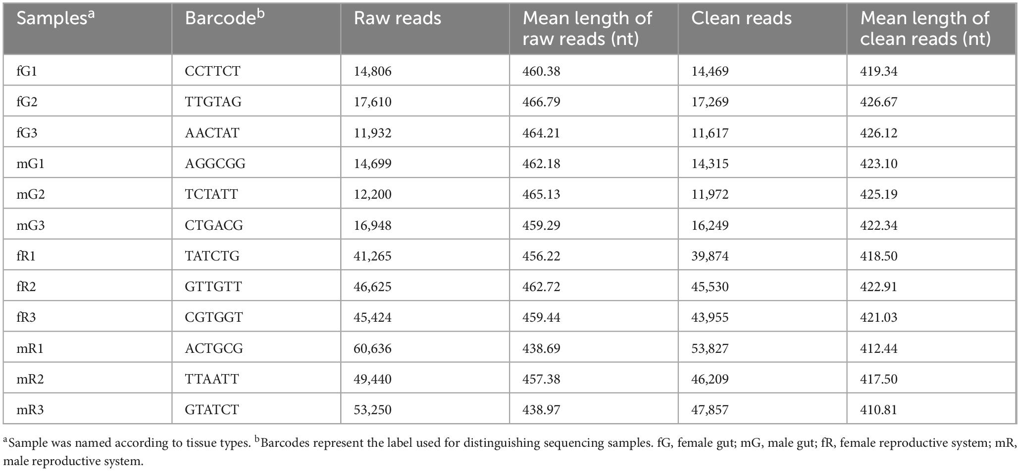
Table 1. Statistical analysis of 16S rDNA amplicon sequence.
The 363,143 clear reads mentioned-above were binned to 3,382 OTUs at a 97% similarity threshold (Supplementary Table S1). Among them, 941 OTUs were from fG, 1,396 OTUs from mG, 1,609 OTUs from fR, and 1,653 OTUs from mR. Obviously, the number of OTUs in the reproductive system, especially in the male reproductive system, was higher than those in the gut (F = 11.77; df = 3, 8; P < 0.01) (Figure 1A). Venn diagram showed that the number of specific OTUs in fR (583) and mR (726) was higher than that in fG (147) and mG (475). There were 379 OTUs overlapped between fG and mG, 609 OTUs overlapped between fR and mR, 608 OTUs overlapped between fG and fR, and 533 OTUs overlapped between mG and mR. The four tissues/samples (fG, mG, fR, and mR) had 200 shared OTUs (Figure 1B).
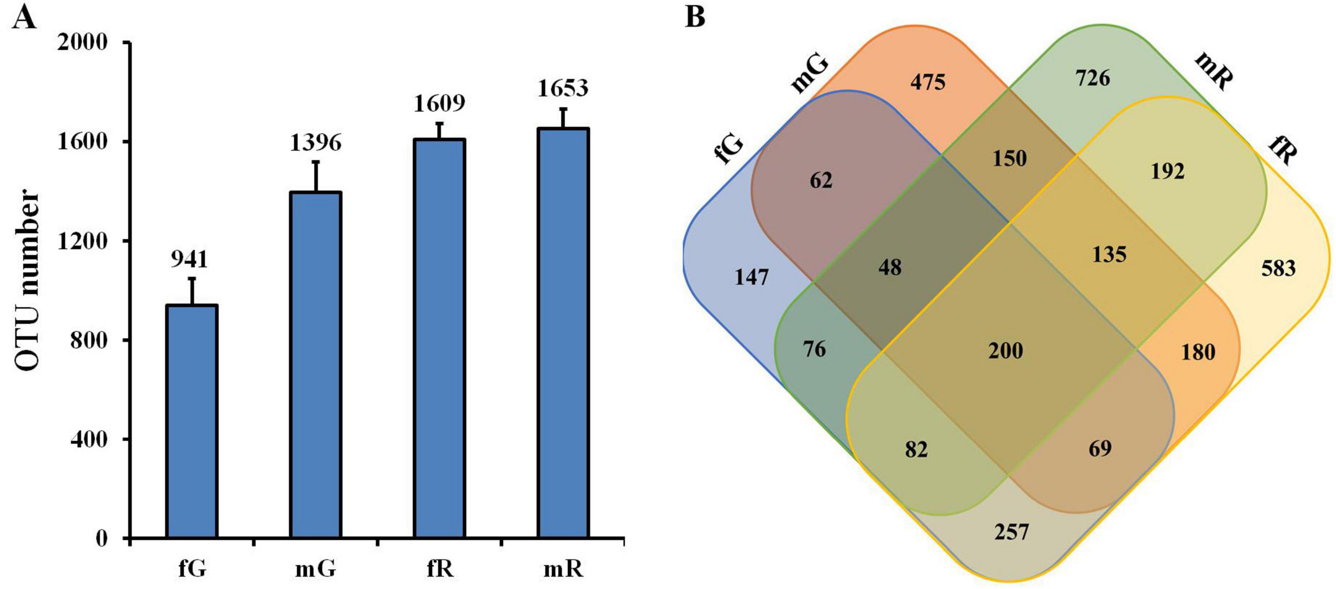
Figure 1. Comparisons of bacterial community based on the OTUs in the fG, mG, fR, and mR of B. dorsalis. (A) OTU number in each tissue. (B) Venn diagram shows those shared and specific OTUs among the four tissues. OTUs were defined based on 3% sequence divergence.
3.2 Diversity characteristics of bacterial community
The Chao1, ACE, Shannon, and Simpson indices for the bacterial community in fG, mG, fR, and mR, based on OTUs, were estimated using the phylogenetic distances and non-phylogenetic metrics. Both the Chao1 and ACE indices showed that the richness of the bacterial community in fR was higher than those in fG (Chao1: n = 3, p = 0.026; ACE: n = 3, p = 0.048) and mG (Chao1: n = 3, p = 0.045; ACE: n = 3, p = 0.024). The Shannon (n = 3, p = 0.226) and Simpson (n = 3, p = 0.307) indices suggested that there were no significant differences in the taxonomic diversity of the bacterial community among the fG, mG, fR and mR of B. dorsalis (Table 2).
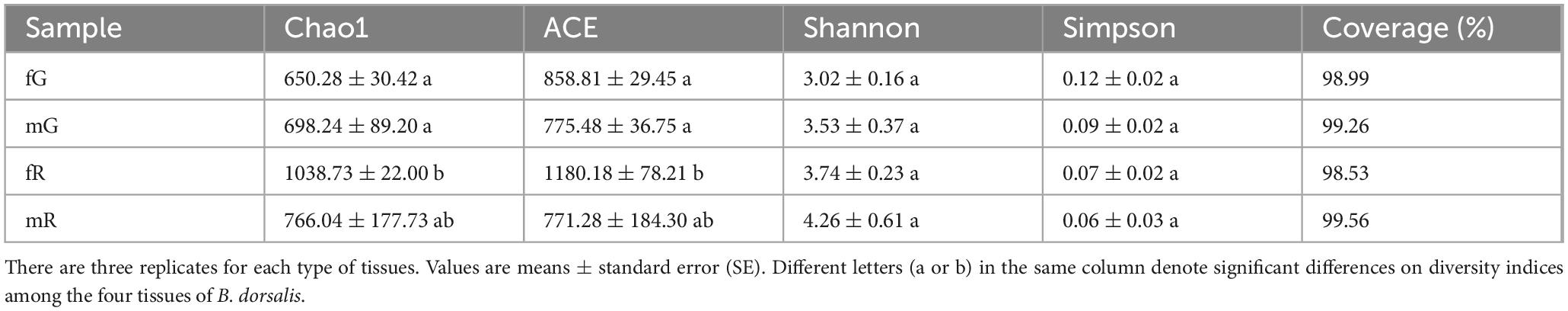
Table 2. Bacterial diversity indices estimated for the four tissues of B. dorsalis adults.
PCoA yielded two main axes, which represented 72.80% of the total variation in bacterial community composition. In this PCoA pattern, we observed a considerable dissimilarity in the composition of bacterial community among fG, mG, fR, and mR, and an obvious clustering of three biological replicates within each specific tissue. By comparison, we also found that the composition of bacterial community between fR and mR was similar, whereas that between fG and mG was distant (Figure 2).
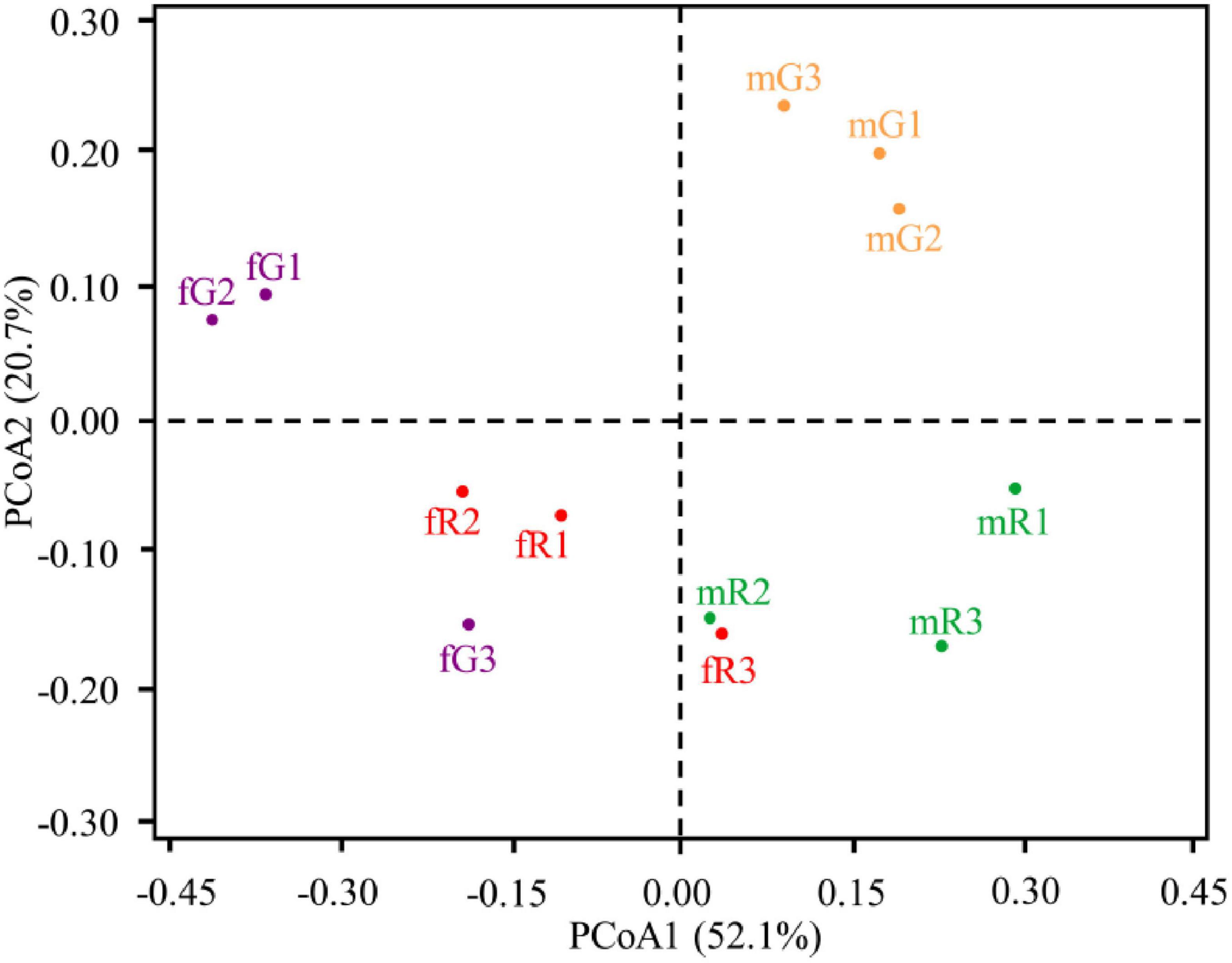
Figure 2. Principal co-ordinates analysis (PCoA) illustrating differences in the taxonomic composition of bacterial communities among independent replicates of four tissue samples (fG, mG, fR, and mR). Dissimilarity in bacterial community composition is based on the weighted UniFrac distance metrics. Percentage of variation explained by each principle component is indicated on axis.
3.3 Identification of bacterial community
3.3.1 Bacterial phyla
We identified a total of 29 bacterial phyla, including Pseudomonadota, Bacillota, Bacteroidota, Euryarchaeota, Actinobacteria, Acidobacteria, Planctomycetes, Deinococcus-Thermus, Chloroflexi, Gemmatimonadetes, Verrucomicrobia, and others, in addition to some unknown taxa. The three bacterial phyla Pseudomonadota, Bacillota, and Bacteroidota were highly abundant in the fG, mG, fR, and mR of B. dorsalis. Among them, Pseudomonadota was the most abundant phylum, with relative abundances of 94.99% in fG, 86.85% in fR, 58.69% in mG, and 60.58% in mR. The abundances of Bacillota and Bacteroidota were relatively high, particularly in mG (Bacillota: 34.43%; Bacteroidota: 3.91%) and mR (Bacillota: 20.34%; Bacteroidota: 5.48%). Notably, high abundances of Euryarchaeota (4.05%), Actinobacteria (3.79%), and Acidobacteria (1.92%) were observed only in mR (Figure 3A).
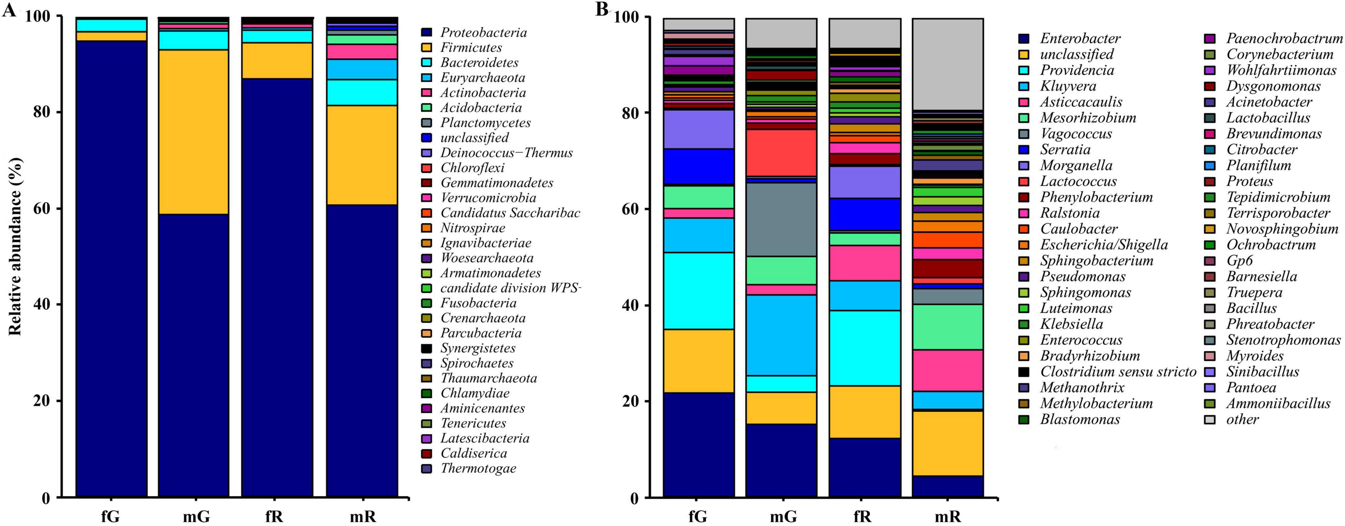
Figure 3. Taxonomic classification of bacterial communities associated with the different tissues of B. dorsalis adults. (A) Relative abundance of bacteria at phylum level. (B) Relative abundance of top 49 bacteria at genus level. The term “other” includes all un-annotated bacterial communities.
3.3.2 Bacterial genera
We identified 48 genera with relatively high abundance, including Enterobacter, Providencia, Kluyvera, Asticcacaulis, Mesorhizobium, Vagococcus, Serratia, Morganella, Lactococcus, Phenylobacterium, Ralstonia, Caulobacter, Escherichia, and Sphingobacterium, and others. In fG, the five most abundant genera were Enterobacter (21.77%), Providencia (16.06%), an unidentified genus (13.71%), Morganella (7.91%), and Serratia (6.86%). In mG, the five most abundant genera were Kluyvera (16.67%), Vagococcus (15.82%), Enterobacter (14.98%), Lactococcus (10.02%), and Mesorhizobium (6.22%). In fR, the five most abundant genera were Providencia (15.30%), Enterobacter (11.83%), an unidentified genus (11.39%), Asticcacaulis (7.28%), and Serratia (6.96%). In mR, the five most abundant genera were an unidentified genus (13.14%), Mesorhizobium (9.04%), Asticcacaulis (8.75%), Enterobacter (4.26%), and Phenylobacterium (2.84%) (Figure 3B). In addition, we did not detect Novosphingobium, Rhizobium, and Gaiella in fG, Paenochrobactrum, Wohlfahrtiimonas, and Lampropedia in mG, Comamonas, Prolixibacter, and Tissierella in fR, and Gaiella, Bdellovibrio, and Parasegetibacter in mR.
3.3.3 Bacterial species
The top 10 bacterial species were Enterobacter ludwigii, Enterococcus faecalis, Citrobacter freundii, Gibbsiella quercinecans, Citrobacter murliniae, Klebsiella pneumoniae, Lactococcus lactis, Citrobacter koseri, Gluconobacter oxydans, and Stenotrophomonas maltophilia. Among them, Citrobacter murliniae was the most abundant species in fG, with a relative abundance of 0.34%. In mG, Enterococcus faecalis was the most abundant species, with a relative abundance of 1.61%. Enterobacter ludwigii was the most abundant species in both fR and mR with relative abundances of 2.62 and 4.80%, respectively. Notably, Gibbsiella quercinecans was absent in both fG and mG (Table 3).
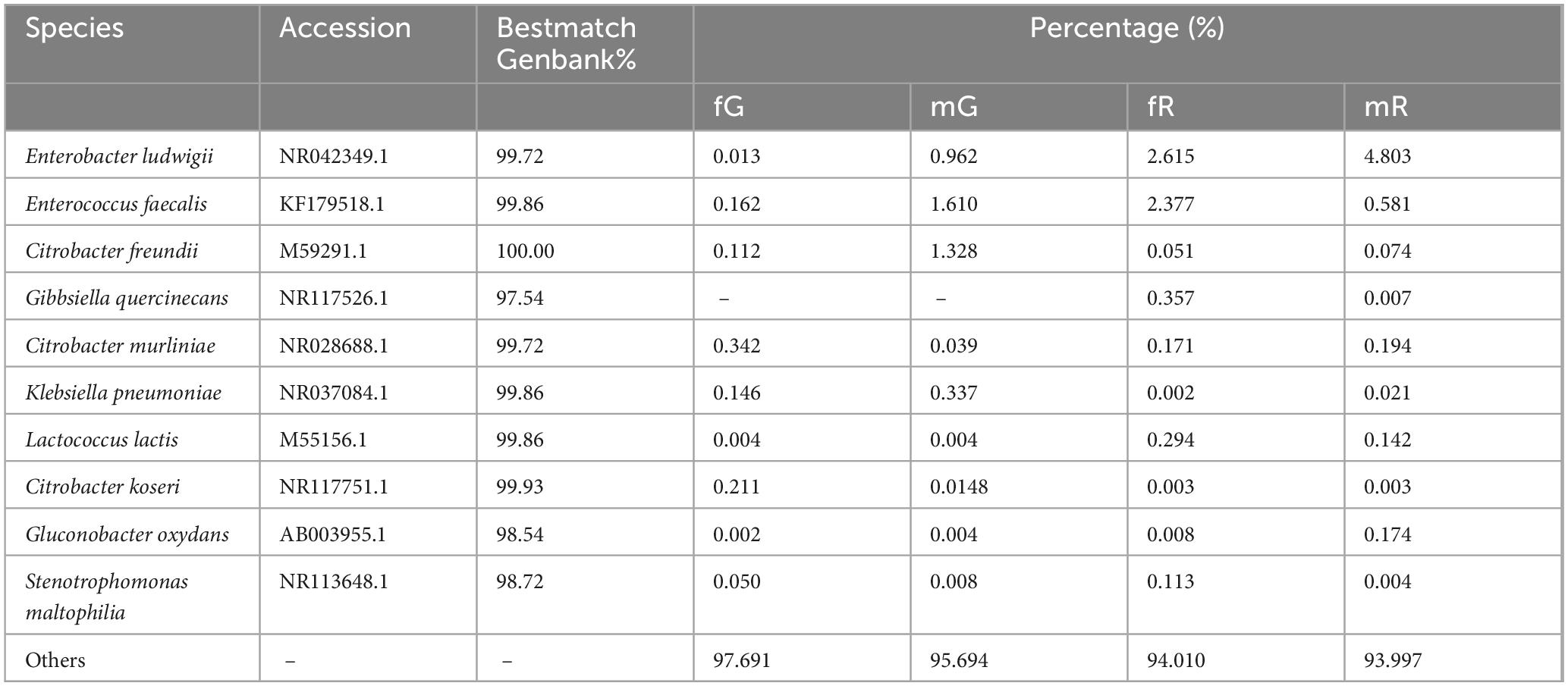
Table 3. Abundance of top 10 bacteria species in the fG, mG, fR, and mR of B. dorsalis.
3.3.4 Specific and dominant bacterial taxa
Specific bacterial taxa in the fG, mG, fR, and mR of B. dorsalis were detected using LEfSe analysis. The circular cladogram showed that fG harbored specific bacterial taxa, including the class Gammaproteobacteria, the order Enterobacteriales, the family Enterobacteriaceae, the genus Morganella; mG harbored specific bacterial taxa, including the families Enterococcaceae, Lactobacillaceae, and Streptococcaceae, the genera Vagococcus, Lactobacillus, Lactococcus, Lactobacillales and Bacilli; fR harbored specific bacterial taxa, including the family Burkholderiaceae, the genera Blastomonas, Ralstonia and Providencia; mR harbored specific bacterial taxa, including the order Sphingobacteriales, the families Caulobacteraceae and Bradyrhizobiaceae, the genera Sphingobacteriia, Asticcacaulis, Caulobacter, Caulobacterales, Bradyrhizobium, and Luteimonas (Figure 4A).
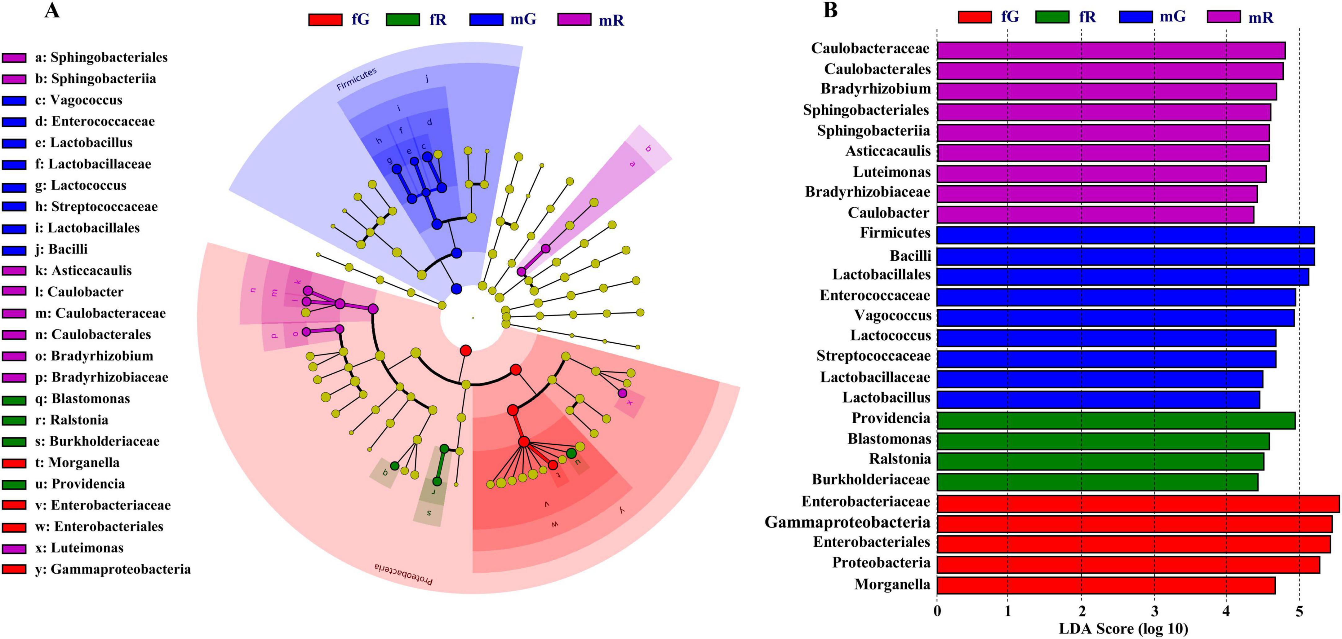
Figure 4. Diagram of specific and dominant bacterial taxa based on LEfSe analysis. (A) Circular cladogram starting from the innermost circle: phylum, class, order, family and genus. Each node represents a taxa and the larger the node, the higher the abundance of that taxa. Yellow nodes represent those bacterial taxa that there are no significant differences among different tissues. The other color nodes, such as red nodes, represent those bacterial taxa that there are significant differences among different tissues. Names of specific bacterial taxa are showed in the legend on the left. (B) Bar chart created based on the LDA score. Four colors (purple, blue, green and red) in this chart are used to distinguish those bacterial taxa in fG, mG, fR, and mR, respectively. The larger the LDA score, the greater the difference, indicating that the abundance in this bacterial taxon is higher than those in other bacterial taxa.
The dominant bacterial taxa in the fG, mG, fR, and mR of B. dorsalis were also determined based on the LDA score. The LDA analysis showed that there were 5 bacterial taxa in fG, 9 in mG, 4 in fR, and 9 in mR, each with LDA scores > 4. Among them, there were 3 dominant phyla (Bacillota, Gammaproteobacteria, and Pseudomonadota), 1 dominant class (Bacilli), 2 dominant Orders (Lactobacillales and Enterobacteriales), and 1 dominant family (Enterobacteriaceae) in mG or fG, each with LDA scores > 5 (Figure 4B).
3.4 Function annotation of bacterial community
KEGG pathway analysis showed that the bacterial communities from the fG, mG, fR, and mR of B. dorsalis were annotated as being associated with 6 primary pathways, as organismal systems, metabolism, human diseases, genetic information processing, environmental information processing, and cellular processes. Among them, the metabolism pathway contained 12 subcategories, the organismal systems pathway contained 7 subcategories, while the cellular processes pathway, and environmental information processing pathway each contained only 3 subcategories. In these subcategories pathways, high-abundance bacterial communities (with relative abundance > 20) were enriched in the 5 pathways (subcategories): membrane transport, carbohydrate metabolism, amino acid metabolism, replication and repair, and energy metabolism. Low-abundance bacterial taxa (with relative abundance < 14) were mainly involved in the 3 pathways (subcategories): cardiovascular diseases, circulatory system, and excretory system (Figure 5).

Figure 5. Function classification and annotation of bacterial communities from the fG, mG, fR, and mR of B. dorsalis based on KEGG pathway analysis.
The differences in the relative abundance of 35 pathways (subcategories) enriched by bacterial communities were revealed. The relative abundances of the 5 pathways associated with cardiovascular diseases, circulatory system, excretory system, membrane transport, and glycan biosynthesis and metabolism exhibited greater variations among the four tissues of B. dorsalis. Among them, the abundances of the 2 pathways related to cardiovascular diseases and circulatory system were higher in the reproductive system (fR: 12.62 and mR: 13.41 for cardiovascular diseases; fR: 12.91 and mR: 13.79 for circulatory system), compared to the gut (fG: 11.72 and mG: 11.64 for cardiovascular diseases; fG: 12.47 and mG: 12.42 for circulatory system). The abundances of the other 3 pathways were all highest in fG, with values of 13.73 for excretory system, 22.87 for membrane transport, and 19.89 for glycan biosynthesis and metabolism among the four tissues. In addition, the relative abundances of the remained 30 pathways exhibited relatively minor variations among the four tissues of B. dorsalis (Figure 5).
Several differences in the absolute abundance of KEGG pathway related to the fG, mG, fR, and mR of B. dorsalis were revealed. Among the 6 pathways depicted in Figure 6, the absolute abundance of the pathway related to carbohydrate metabolism was highest (> 2.8 × 10⁶), while that related to biosynthesis of other secondary metabolites was lowest (< 3.1 × 10⁵). Among the fG, mG, fR, and mR of B. dorsalis, the abundances of pathways related to carbohydrate metabolism, enzyme families, glycan biosynthesis and metabolism, as well as metabolism of cofactors and vitamins were higher in fG compared to those in mR, and the abundances of pathways related to metabolism of other amino acids, and biosynthesis of other secondary metabolites were also higher in fG compared to those in mG (Figure 6).
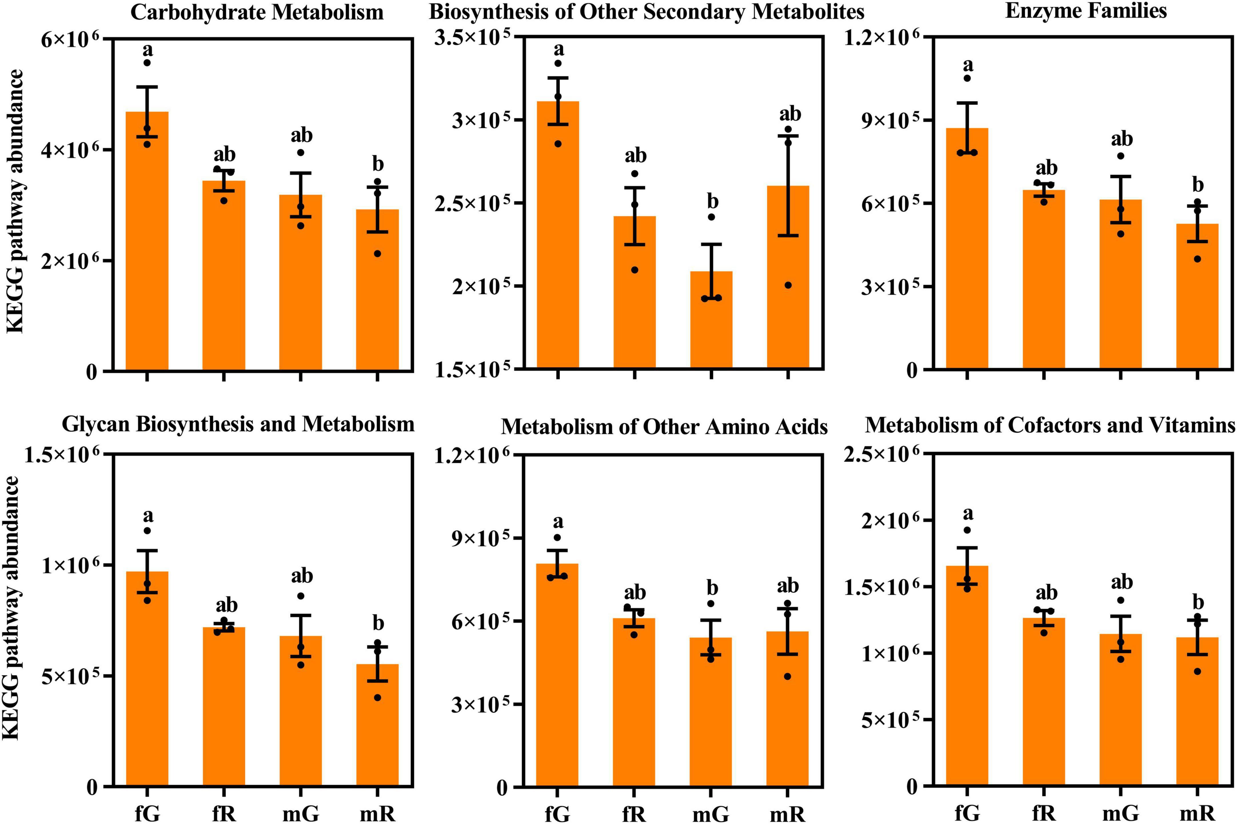
Figure 6. Comparisons of KEGG pathway absolute abundance among the fG, mG, fR, and mR of B. dorsalis. Values are means ± standard error (SE). Different letters (a or b) above the different column denote significant differences on KEGG pathway abundance.
4 Discussion
Different insect tissues harbor tissue-specific bacterial communities, as well as common or ubiquitous bacterial communities (Santos et al., 2023). Several studies have explored these bacterial communities in insect tissues, focusing on their richness and diversity (Jones et al., 2011; Chen et al., 2017; Kumar et al., 2023). The type and structure of bacteria present in insect tissues are closely linked to the complex physiological and biochemical characteristics of specific tissues (Collins and Patterson, 2020; Naveed et al., 2024). The study conducted here represents the most comprehensive exploration undertaken to-date of the overlapping and specific bacteria communities in the gut and reproductive system of B. dorsalis adults. Our results are instrumental in understanding the intrinsic relationship between the structure of bacterial communities and the function of tissues associated with these bacteria.
The removal of pair-end mismatches, homopolymers, and chimeric sequences ensured the quality of 16S rRNA gene sequence we obtained, which was beneficial for identifying the common and specific bacterial taxa and analyzing the functional pathway of bacteria enrichment. Meanwhile, over 30,000 clean reads per biological replicate were generated, providing sufficient RNA-seq data for analyzing the richness and diversity of bacterial taxa based on OTUs within B. dorsalis tissues. The higher numbers of clean reads and OTUs in fR and mR than in fG and mG (Table 1; Figure 1) indicated a greater diverse of bacterial taxa in the reproductive system of B. dorsalis.
Our study demonstrated that the richness indices of bacterial taxa in the reproductive system, particularly in the female reproductive system, were higher than those in the gut of B. dorsalis (Table 2). This finding is credible, because the environment of reproductive system is relatively stable, which provides more suitable living condition for its bacterial communities (Huang et al., 2010). Moreover, B. dorsalis adults were starved for 12 h before sampling, which eliminated a large number of bacteria via feces, particularly those retained in their guts (Andongma et al., 2015). The Venn diagram illustrated that the reproductive systems of B. dorsalis harbored a greater number of specific bacterial taxa compared to their guts. Our explanation is that the physiological and biochemical processes of the reproductive system are more diverse and complex, necessitating the participation of a greater variety of bacteria communities (Ben-Yosef et al., 2008). A study by Wang A. L. et al. (2014) also confirmed that the reproductive system of B. minax, a tephritid species closely related genetically to B. dorsalis, harbored more diverse and specific bacterial taxa than its gut.
We found that the bacterial community in different tissues of B. dorsalis was dominated by Pseudomonadota, followed by Bacillota and Bacteroidota. Other studies have also reported an abundance of Pseudomonadota in various insects, including B. minax (Wang A. L. et al., 2014), Kerria lacca (Kerr) (Kandasamy et al., 2022), and Z. cucurbitae (Lakshmi et al., 2023). On the contrary, bacterial communities associated with certain insects, such as Apis mellifera L. (Mohr and Tebbe, 2006) and Coptotermes formosanus Shiraki (Xiang et al., 2012), are more commonly dominated by Bacillota and Bacteroidota. Within the Pseudomonadota, members of the Enterobacteriaceae family constitute the majority of the bacterial community, which is also predominant in the guts of C. capitata (Marchini et al., 2002) and Drosophila immigrans (Berlin) (Jones et al., 2013). The wide distribution of Enterobacteriaceae suggested its important role in insect tissues. A study by Gu J. et al. (2024) revealed that some bacterial species within Enterobacteriaceae promoted the growth and development of B. dorsalis larvae through the vitamin B6 biosynthesis pathway. Furthermore, researches have shown that Enterobacteriaceae within insects may indirectly contribute to host adaptation, energy metabolism, and food digestion (Martinez et al., 2019; Wang et al., 2020; Zou et al., 2020).
The genera Enterobacter, Kluyvera, Asticcacaulis, Mesorhizobium, and Serratia were commonly found in the gut and reproductive system of B. dorsalis. Research has confirmed their widespread presence in the tissues of other insects, such as C. capitata (Behar et al., 2008), P. papatasi (Maleki-Ravasan et al., 2014), and Bombyx mori (Chen et al., 2018). These genera may play important roles in specific insect tissues. For instance, Enterobacter in the gut of Helicoverpa armigera (Hübner) is associated with the digestion and absorption of cellulosic food materials (Priya et al., 2012).
LEfSe analysis showed that the different tissues of B. dorsalis harbored their specific bacterial communities. Our study demonstrated that the gut of female B. dorsalis contained four specific bacterial taxa, namely, the class Gammaproteobacteria, the order Enterobacteriales, the family Enterobacteriaceae, and the genus Morganella. The guts of other fly species, such as C. capitata (Marchini et al., 2002) and D. immigrans (Jones et al., 2013) also harbored some members of Gammaproteobacteria. Furthermore, an increase in the abundance of Gammaproteobacteria in S. gregaria may enhance its immune defense against intestinal pathogens (Dillon et al., 2010). We also found that female adults of B. dorsalis harbored abundant members of the genus Providencia, whereas it had not been observed in male adults. A study by Roque-Romero et al. (2020) confirmed the presence of Providencia in female Anastrepha obliqua (Macquart) as well. Furthermore, male A. obliqua that were fed with food containing Providencia rettgeri became more attractive to female adults of the same species, suggesting that Providencia may be associated with the courtship and mating behavior of female insects. Unfortunately, the roles of many tissue-specific bacterial communities in insects remain poorly understood. A possible reason is that the abundance of these bacteria in insect tissues is low and often overlooked.
The top 10 bacterial species, including Enterobacter ludwigii, Enterococcus faecalis, Citrobacter freundii, Lactococcus lactis and others, were identified by the BLAST algorithm. These species are common in many insect tissues, and their functions have also been revealed in some insect species (Priyadarsini et al., 2020). For example, Romero et al. (2006) demonstrated that Citrobacter freundii can stimulate oviposition in Stomoxys calcitrans L. (a fly species), and contribute to the development and reproduction of this fly species. Cheng et al. (2017) revealed that C. freundii may degrade trichlorphon (an organophosphate insecticide) into dimethyl phosphite and chloral hydrate, thereby enhancing the resistance of B. dorsalis to trichlorphon. Gwokyalya et al. (2023) found that L. lactis may improve the parasitic performance of parasitoids (Fopius arisanus and Diachasmimorpha longicaudata) against B. dorsalis, underscoring the critical role of bacterial symbionts in controlling this pest.
KEGG pathway analysis showed that the function of bacterial communities from the gut and reproductive system of B. dorsalis were mainly enriched in metabolism pathways, particularly those associated with carbohydrate metabolism, amino acid metabolism, and energy metabolism. This finding indicated that the majority of bacteria communities in the gut and the reproductive system of B. dorsalis are involved in the production, transformation, and utilization of food nutrients, thereby supporting their growth, development, and reproduction (Huang et al., 2017). Similar phenomena have also been demonstrated in other insect species, such as Bombyx mori L. and Plutella xylostella L. (Chen et al., 2017). By checking each of the 35 pathways annotated, we found that the abundances of the 2 pathways related to cardiovascular diseases and circulatory system were higher in the reproductive system compared to the gut. This phenomenon may be related to the reproduction of offspring in B. dorsalis. Among them, the function of the cardiovascular diseases pathway was likely to prevent genetically related disorders occurring in offspring, whereas that of the circulatory system pathway supported the production of robust offspring (Chen et al., 2024; Jama et al., 2024). The abundances of the 3 pathways associated with excretory system, membrane transport, and glycan biosynthesis and metabolism were all highest in the female gut (fG) of B. dorsalis among its four tissues. Clearly, the functions of these pathways are intricately tied to the absorption, utilization, and excretion of nutrients in B. dorsalis via its gut (Hu et al., 2011). Consequently, one plausible explanation for the high abundance in fG was that female B. dorsalis required a larger amount of food consumption to cope with more intense reproductive activity (Huang et al., 2017).
In summary, our research conducted here revealed the overlapping and specific bacterial communities between the gut and reproductive system of female and male B. dorsalis. Our study demonstrated that the reproductive system harbored more diverse and specific bacterial communities compared to the gut, whereas the female gut exhibited a higher abundance of function pathways than other tissues. Future studies should focus on revealing the roles of specific bacteria genera/species and identifying whether these bacteria could serve as targets for controlling B. dorsalis.
5 Conclusion
This study characterized the overlapping and specific bacterial communities in the gut and reproductive system of female and male B. dorsalis. Compared with guts, reproductive systems have more diverse and specific bacterial taxa based on OTUs. In bacterial communities, Enterobacter, Kluyvera, Asticcacaulis and Mesorhizobium were common, while Gammaproteobacteria, Vagococcus, Providencia and Sphingobacteriia exhibited tissue specificity. High-abundance bacterial communities were enriched in these function pathways of membrane transport, carbohydrate metabolism, amino acid metabolism, replication and repair, and energy metabolism. The abundance of pathway related to carbohydrate metabolism was highest in the gut of female adults, compared with other pathways. Our research demonstrated that the bacterial communities vary across different tissues of B. dorsalis, providing clues that elucidate the intrinsic relationship between these bacterial communities and the function of bacteria-associated tissues.
Data availability statement
The original contributions presented in the study are publicly available. This data can be found here: https://www.ncbi.nlm.nih.gov/, accession number: PRJNA1122372.
Author contributions
W-JL: Data curation, Formal Analysis, Software, Visualization, Writing – original draft. K-PH: Investigation, Methodology, Validation, Writing – original draft. XZ: Investigation, Validation, Writing – original draft. S-LS: Investigation, Methodology, Writing – original draft. C-KX: Data curation, Formal Analysis, Writing – original draft. Q-XX: Formal Analysis, Investigation, Writing – original draft. X-ZL: Conceptualization, Funding acquisition, Project administration, Supervision, Visualization, Writing – review & editing, Resources, Writing – original draft.
Funding
The author(s) declare that financial support was received for the research and/or publication of this article. This work was funded in part by the National Natural Science Foundation of China (32360713, 32202298, 31560516) and the Foundation Project of Jiangxi Educational Committee (GJJ210443).
Conflict of interest
The authors declare that the research was conducted in the absence of any commercial or financial relationships that could be construed as a potential conflict of interest.
Generative AI statement
The authors declare that no Generative AI was used in the creation of this manuscript.
Publisher’s note
All claims expressed in this article are solely those of the authors and do not necessarily represent those of their affiliated organizations, or those of the publisher, the editors and the reviewers. Any product that may be evaluated in this article, or claim that may be made by its manufacturer, is not guaranteed or endorsed by the publisher.
Supplementary material
The Supplementary Material for this article can be found online at: https://www.frontiersin.org/articles/10.3389/fmicb.2025.1567154/full#supplementary-material
Supplementary Table S1 | Information on the total OTUs within the gut and reproductive system of B. dorsalis.
Footnotes
References
Adair, K. L., and Douglas, A. E. (2017). Making a microbiome: The many determinants of host-associated microbial community composition. Curr. Opin. Microbiol. 35, 23–29. doi: 10.1016/j.mib.2016.11.002
Altschul, S. F., Gish, W., Miller, W., Myers, E. W., and Lipman, D. J. (1990). Basic local alignment search tool. J. Mol. Biol. 215, 403–410. doi: 10.1016/S0022-2836(05)80360-2
Amdam, G. V. (2012). Paratransgenesis: An approach to improve colony health and molecular insight in honey bees (Apis mellifera)? Integr. Comp. Biol. 52, 89–99. doi: 10.1093/icb/ics089
Andongma, A. A., Wan, L., Dong, Y. C., Li, P., Desneux, N., White, J. A., et al. (2015). Pyrosequencing reveals a shift in symbiotic bacteria populations across life stages of Bactrocera dorsalis. Sci. Rep. 5:9470. doi: 10.1038/srep09470
Behar, A., Yuval, B., and Jurkevitch, E. (2008). Gut bacterial communities in the Mediterranean fruit fly (Ceratitis capitata) and their impact on host longevity. J. Insect Physiol. 54, 1377–1383. doi: 10.1016/j.jinsphys.2008.07.011
Ben-Yosef, M., Jurkevitch, E., and Yuval, B. (2008). Effect of bacteria on nutritional status and reproductive success of the Mediterranean fruit fly Ceratitis capitata. Physiol. Entomol. 33, 145–154. doi: 10.1111/j.1365-3032.2008.00617.x
Bing, X. L., Zhao, D. S., Peng, C. W., Huang, H. J., and Hong, X. Y. (2020). Similarities and spatial variations of bacterial and fungal communities in field rice planthopper (Hemiptera: Delphacidae) populations. Insect Sci. 27, 947–963. doi: 10.1111/1744-7917.12782
Bolger, A. M., Lohse, M., and Usadel, B. (2014). Trimmomatic: A flexible trimmer for Illumina sequence data. Bioinformatics 30, 2114–2120. doi: 10.1093/bioinformatics/btu170
Brown, J., Pirrung, M., and McCue, L. A. (2017). FQC dashboard: Integrates FastQC results into a web-based, interactive, and extensible FASTQ quality control tool. Bioinformatics 33, 3137–3139. doi: 10.1093/bioinformatics/btx373
Caporaso, J. G., Kuczynski, J., Stombaugh, J., Bittinger, K., Bushman, F. D., Costello, E. K., et al. (2010). QIIME allows analysis of high-throughput community sequencing data. Nat. Methods 7, 335–336. doi: 10.1038/nmeth.f.303
Chen, B. S., Lu, X. M., and Shao, Y. Q. (2017). Diversity of the gut microbiota in lepidopteran insects and their interaction with hosts. Acta Entomol. Sin. 60, 710–722. doi: 10.16380/j.kcxb.2017.06.011
Chen, B., Du, K., Sun, C., Vimalanathan, A., Liang, X., Li, Y., et al. (2018). Gut bacterial and fungal communities of the domesticated silkworm (Bombyx mori) and wild mulberry-feeding relatives. ISME J. 12, 2252–2262. doi: 10.1038/S41396-018-0174-1
Chen, Y., Chen, Y., Li, Y., Du, E., Sun, Z., Lu, Z., et al. (2024). Comparative study of the gut microbial community structure of Spodoptera frugiperda and Spodoptera literal (Lepidoptera). PeerJ 12:e17450. doi: 10.7717/peerj.17450
Cheng, D., Guo, Z., Riegler, M., Xi, Z., Liang, G., and Xu, Y. (2017). Gut symbiont enhances insecticide resistance in a significant pest, the oriental fruit fly Bactrocera dorsalis (Hendel). Microbiome 5:13. doi: 10.1186/s40168-017-0236-z
Collins, S. L., and Patterson, A. D. (2020). The gut microbiome: An orchestrator of xenobiotic metabolism. Acta Pharm. Sin. B 10, 19–32. doi: 10.1016/j.apsb.2019.12.001
de Bekker, C., Smith, P. B., Patterson, A. D., and Hughes, D. P. (2013). Metabolomics reveals the heterogeneous secretome of two entomopathogenic fungi to ex vivo cultured insect tissues. PLoS One 8:e70609. doi: 10.1371/journal.pone.0070609
Dillon, R. J., Webster, G., Weightman, A. J., and Keith Charnley, A. (2010). Diversity of gut microbiota increases with aging and starvation in the desert locust. Antonie Van Leeuwenhoek Int. J. 97, 69–77. doi: 10.1007/s10482-009-9389-5
Douglas, A. E. (2009). The microbial dimension in insect nutritional ecology. Funct. Ecol. 23, 38–47. doi: 10.1111/j.1365-2435.2008.01442.x
Edgar, R. C. (2010). Search and clustering orders of magnitude faster than BLAST. Bioinformatics 26, 2460–2461. doi: 10.1093/bioinformatics/btq461
Edgar, R. C. (2013). UPARSE: Highly accurate OTU sequences from microbial amplicon reads. Nat. Methods 10, 996–998. doi: 10.1038/NMETH.2604
Edgar, R. C., Haas, B. J., Clemente, J. C., Quince, C., and Knight, R. (2011). UCHIME improves sensitivity and speed of chimera detection. Bioinformatics 27, 2194–2200. doi: 10.1093/bioinformatics/btr381
Feldhaar, H., and Gross, R. (2009). Insects as hosts for mutualistic bacteria. Int. J. Med. Microbiol. 299, 1–8. doi: 10.1016/j.ijmm.2008.05.010
Gavriel, S., Jurkevitch, E., Gazit, Y., and Yuval, B. (2011). Bacterially enriched diet improves sexual performance of sterile male Mediterranean fruit flies. J. Appl. Entomol. 135, 564–573. doi: 10.1111/j.1439-0418.2010.01605.x
Gu, F., He, W., Zhu, D., Zeng, Q., Li, X., Xiao, S., et al. (2024). Genome-wide comparative analysis of CC1 Staphylococcus aureus between colonization and infection. Eur. J. Med. Res. 29:474. doi: 10.1186/s40001-024-02076-z
Gu, J., Yao, Z., Lemaitre, B., Cai, Z., Zhang, H., and Li, X. (2024). Intestinal commensal bacteria promote Bactrocera dorsalis larval development through the vitamin B6 synthesis pathway. Microbiome 12, 1–15. doi: 10.1186/s40168-024-01931-9
Gwokyalya, R., Herren, J. K., Weldon, C. W., Ndlela, S., Gichuhi, J., Ongeso, N., et al. (2024). Shaping the microbial landscape: Parasitoid-driven modifications of Bactrocera dorsalis Microbiota. Microb. Ecol. 87:81. doi: 10.1007/s00248-024-02393-0
Gwokyalya, R., Weldon, C. W., Herren, J. K., Gichuhi, J., Makhulu, E. E., Ndlela, S., et al. (2023). Friend or foe: Symbiotic bacteria in Bactrocera dorsalis – parasitoid associations. Biology 12:274. doi: 10.3390/biology12020274
He, M. Y., Chen, H. M., Yang, X. R., Gao, Y., Lu, Y. Y., and Cheng, D. F. (2022). Gut bacteria induce oviposition preference through ovipositor recognition in fruit fly. Commun. Biol. 5:973. doi: 10.1038/s42003-022-03947-z
Hu, L. L., Chen, C., Huang, T., Cai, Y. D., and Chou, K. C. (2011). Predicting biological functions of compounds based on chemical-chemical interactions. PLoS One 6:e29491. doi: 10.1371/journal.pone.0029491
Huang, Q., Shan, H. W., Chen, J. P., and Wu, W. (2023). Diversity and dynamics of bacterial communities in the digestive and excretory systems across the life cycle of leafhopper, Recilia dorsalis. Insects 14:545. doi: 10.3390/insects14060545
Huang, S. W., Zhang, H. Y., Marshall, S., and Jackson, T. A. (2010). The scarab gut: A potential bioreactor for bio-fuel production. Insect Sci. 17, 175–183. doi: 10.1111/j.1744-7917.2010.01320.x
Huang, Y., Ao, Y., and Jiang, M. (2017). Reproductive plasticity of an invasive insect pest, rice water weevil (Coleoptera: Curculionidae). J. Econ. Entomol. 110, 2381–2387. doi: 10.1093/jee/tox274
Jama, H. A., Dona, M. S. I., Dinakis, E., Nakai, M., Paterson, M. R., Shihata, W. A., et al. (2024). Maternal diet and gut microbiota influence predisposition to cardiovascular disease in offspring. Circ. Res. 135:3. doi: 10.1161/CIRCRESAHA.124.324614
Johnson, J. S., Spakowicz, D. J., Hong, B. Y., Petersen, L. M., Demkowicz, P., Chen, L., et al. (2019). Evaluation of 16S rRNA gene sequencing for species and strain-level microbiome analysis. Nat. Commun. 10:5029. doi: 10.1038/s41467-019-13036-1
Jones, R. T., Bressan, A., Greenwell, A. M., and Fierer, N. (2011). Bacterial communities of two parthenogenetic Aphid species cocolonizing two host plants across the Hawaiian Islands. Appl. Environ. Microb. 77, 8345–8349. doi: 10.1128/AEM.05974-11
Jones, R. T., Sanchez, L. G., and Fierer, N. (2013). A cross-taxon analysis of insect-associated bacterial diversity. PLoS One 8:e61218. doi: 10.1371/journal.pone.0061218
Kakumanu, M. L., Maritz, J. M., Carlton, J. M., and Schal, C. (2018). Overlapping community compositions of gut and fecal microbiomes in lab-reared and field-collected German cockroaches. Appl. Environ. Microbiol. 84:e01037–18. doi: 10.1128/AEM.01037-18
Kandasamy, T., Ekbal, S., Kumari, K., Lohot, V. D., Mohanasundaram, A., and Sharma, K. K. (2022). Unraveling bacterial diversity of the Indian Lac Insect Kerria lacca (Kerr) using next generation sequencing. Int. J. Trop. Insect Sci. 42, 2365–2372. doi: 10.1007/s42690-022-00758-x
Kumar, S. S., Tamoghna, S., and Kiran, K. (2023). Diversity of bacterial communities associated with the gut of the fall armyworm, Spodoptera frugiperda (J. E. Smith) (Lepidoptera: Noctuidae) in Eastern India. Phytoparasitica 51, 447–460. doi: 10.1007/s12600-023-01077-8
Lakshmi, B. C. M., Panduranga, G. S., Rajesh, A., Reddy, P. K., and Manjula, K. (2023). Isolation and characterization of bacterial gut symbionts from irradiated, wild and lab reared males of melon fly, Zeugodacus cucurbitae (Coquillett). J. Entomol. Res. Soc. 25, 351–362. doi: 10.51963/jers.2023.93
Langille, M. G. I., Zaneveld, J., Caporaso, J. G., McDonald, D., Knights, D., Reyes, J. A., et al. (2013). Predictive functional profiling of microbial communities using 16S rRNA marker gene sequences. Nat. Biotechnol. 31, 814–821. doi: 10.1038/nbt.2676
Li, W. J., Xu, C. K., Ong, S. Q., Majid, A. H. A., Wang, J. G., and Li, X. Z. (2024). Comparative analyses of the transcriptome among three development stages of Zeugodacus tau larvae (Diptera: Tephritidae). Comp. Biochem. Physiol Part D Genom. Proteomics 52:101333. doi: 10.1016/j.cbd.2024.101333
Li, X. Z., Wang, G. L., Wang, C. L., Li, W. J., Lu, T., Ge, Y. G., et al. (2024). Long-term monitoring of Bactrocera and Zeugodacus spp. (Diptera: Tephritidae) in China and evaluation of different control methods for Bactrocera dorsalis (Hendel). Crop Prot. 182:106708. doi: 10.1016/j.cropro.2024.106708
Liu, L. J., Martinez-Sañudo, I., Mazzon, L., Prabhakar, C. S., Girolami, V., Deng, Y. L., et al. (2016). Bacterial communities associated with invasive populations of Bactrocera dorsalis (Diptera: Tephritidae) in China. Bull. Entomol. Res. 106, 718–728. doi: 10.1017/S0007485316000390
Lozupone, C., and Knight, R. (2005). UniFrac: A new phylogenetic method for comparing microbial communities. Appl. Environ. Microbiol. 71, 8228–8235. doi: 10.1128/AEM.71.12.8228-8235.2005
Lu, M., Hulcr, J., and Sun, J. (2016). The role of symbiotic microbes in insect invasions. Annu. Rev. Ecol. Evol. Sci. 47, 487–505. doi: 10.1146/annurev-ecolsys-121415-032050
Magoč, T., and Salzberg, S. L. (2011). FLASH: Fast length adjustment of short reads to improve genome assemblies. Bioinformatics 27, 2957–2963. doi: 10.1093/bioinformatics/btr507
Maleki-Ravasan, N., Oshaghi, M. A., Hajikhani, S., Saeidi, Z., Akhavan, A. A., Gerami-Shoar, M., et al. (2014). Aerobic microbial community of insectary population of Phlebotomus papatasi. J. Arthropod Borne Dis. 8, 69–81. doi: 10.1017/S0022149X13000060
Marchini, D., Rosetto, M., Dallai, R., and Marri, L. (2002). Bacteria associated with the oesophageal bulb of the medfly Ceratitis capitata (Diptera:Tephritidae). Curr. Microbiol. 44, 120–124. doi: 10.1007/s00284-001-0061-1
Martinez, A. J., Onchuru, T. O., Ingham, C. S., Sandoval-Calderón, M., Salem, H., Deckert, J., et al. (2019). Angiosperm to Gymnosperm host-plant switch entails shifts in microbiota of the Welwitschia bug, Probergrothius angolensis (Distant, 1902). Mol. Ecol. 28, 5172–5187. doi: 10.1111/mec.15281
Mohr, K. I., and Tebbe, C. C. (2006). Diversity and phylotype consistency of bacteria in the guts of three bee species (Apoidea) at an oilseed rape field. Environ. Microbiol. 8, 258–272. doi: 10.1111/j.1462-2920.2005.00893.x
Naveed, W. A., Liu, Q., Lu, C., and Huang, X. (2024). Symbiotic bacterial communities of insects feeding on the same plant lineage: Distinct composition but congruent function. Insects 15:187. doi: 10.3390/insects15030187
Priya, N. G., Ojha, A., Kajla, M. K., Raj, A., and Rajagopal, R. (2012). Host plant induced variation in gut bacteria of Helicoverpa armigera. PLoS One 7:e30768. doi: 10.1371/journal.pone.0030768
Priyadarsini, S., Mukherjee, S., Samikshya, S. N., Bhanja, A., and Mishra, M. (2020). Dietary infection of Enterobacter ludwigii causes fat accumulation and resulted in the diabetes-like condition in Drosophila melanogaster. Micrib. Pathog. 149:104276. doi: 10.1016/j.micpath.2020.104276
Quast, C., Pruesse, E., Yilmaz, P., Gerken, J., Schweer, T., Yarza, P., et al. (2013). The SILVA ribosomal RNA gene database project: Improved data processing and web-based tools. Nucleic Acids Res. 41, 590–596. doi: 10.1093/nar/gks1219
Riegler, M., and Stauffer, C. (2002). Wolbachia infections and superinfections in cytoplasmically incompatible populations of the European cherry fruit fly Rhagoletis cerasi (Diptera, Tephritidae). Mol. Ecol. 11, 2425–2434. doi: 10.1046/j.1365-294X.2002.01614.x
Romero, A., Broce, A., and Zurek, L. (2006). Role of bacteria in the oviposition behaviour and larval development of stable flies. Med. Vet. Entomol. 20, 115–121. doi: 10.1111/j.1365-2915.2006.00602.x
Roque-Romero, L., Hernández, E., Aceituno-Medina, M., Ventura, C., Toledo, J., and Malo, E. A. (2020). Attractiveness and sexual competitiveness of Anastrepha obliqua males (Diptera: Tephritidae) fed on a diet enriched with Providencia rettgeri. Front. Microbiol. 11:1777. doi: 10.3389/fmicb.2020.01777
Santos, N. A., Carvalho, V. R., Souza-Neto, J. A., Alonso, D. P., Ribolla, P. E., Medeiros, J. F., et al. (2023). Bacterial microbiota from lab-reared and field-captured Anopheles darlingi midgut and salivary gland. Microorganisms 11:1145. doi: 10.3390/microorganisms11051145
Schloss, P. D., Westcott, S. L., Ryabin, T., Hall, J. R., Hartmann, M., Hollister, E. B., et al. (2009). Introducing mothur: Open-source, platform-independent, community-supported software for describing and comparing microbial communities. Appl. Environ. Microbiol. 75, 7537–7541. doi: 10.1128/AEM.01541-09
Segata, N., Izard, J., Waldron, L., Gevers, D., Miropolsky, L., Garrett, W. S., et al. (2011). Metagenomic biomarker discovery and explanation. Genome Biol. 12:R60. doi: 10.1186/gb-2011-12-6-r60
Shi, W., Kerdelhué, C., and Ye, H. (2012). Genetic structure and inferences on potential source areas for Bactrocera dorsalis (Hendel) based on mitochondrial and microsatellite markers. PLoS One 7:e37083. doi: 10.1371/journal.pone.0037083
Shi, Z., Wang, L., and Zhang, H. (2012). Low diversity bacterial community and the trapping activity of metabolites from cultivable bacteria species in the female reproductive system of the oriental fruit fly, Bactrocera dorsalis Hendel (Diptera: Tephritidae). Int. J. Mol. Sci. 13, 6266–6278. doi: 10.3390/ijms13056266
Shterzer, N., Rothschild, N., Sbehat, Y., Stern, E., Nazarov, A., and Mills, E. (2020). Large overlap between the intestinal and reproductive tract microbiomes of chickens. Front. Microbiol. 11:1508. doi: 10.3389/fmicb.2020.01508
Swift, M. L. (1997). Graphpad prism, data analysis, and scientific graphing. J. Chem. Inform. Model. 37, 411–412. doi: 10.1021/ci960402j
Thaochan, N., Drew, R. A. I., Hughes, J. M., Vijaysegaran, S., and Chinajariyawong, A. (2010). Alimentary tract Bacteria isolated and identified with API-20E and molecular cloning techniques from Australian tropical fruit flies, Bactrocera cacuminata and B. tryoni. J. Iinsect Sci. 1:131. doi: 10.1673/031.010.13101
Wang, A. L., Yao, Z. C., Zheng, W. W., and Zhang, H. Y. (2014). Bacterial communities in the gut and reproductive organs of Bactrocera minax (Diptera: Tephritidae) based on 454 pyrosequencing. PLoS ONE 9:e106988. doi: 10.1371/journal.pone.0106988
Wang, H., Jin, L., Peng, T., Zhang, H., Chen, Q., and Hua, Y. (2014). Identification of cultivable bacteria in the intestinal tract of Bactrocera dorsalis from three different populations and determination of their attractive potential. Pest Manag. Sci. 70, 80–87. doi: 10.1002/ps.3528
Wang, Z. L., Wang, T. Z., Zhu, H. F., Pan, H. B., and Yu, X. P. (2020). Diversity and dynamics of microbial communities in brown planthopper at different developmental stages revealed by high-throughput amplicon sequencing. Insect Sci. 27, 883–894. doi: 10.1111/1744-7917.12729
Xiang, H., Xie, L., Zhang, J., Long, Y. H., Liu, N., Huang, Y. P., et al. (2012). Intracolonial differences in gut bacterial community between worker and soldier castes of Coptotermes formosanus. Insect Sci. 19, 86–95. doi: 10.1111/j.1744-7917.2011.01435.x
Yin, P. K., Xiao, H., Yang, Z. B., Yang, D. S., and Yang, Y. H. (2024). Shotgun metagenomics reveals the gut microbial diversity and functions in Vespa mandarinia (Hymenoptera: Vespidae) at multiple life stages. Front. Microbiol. 15:1288051. doi: 10.3389/fmicb.2024.1288051
Yong, H. S., Song, S. L., Chua, K. O., and Lim, P. E. (2017). Microbiota associated with Bactrocera carambolae and B. dorsalis (Insecta: Tephritidae) revealed by next-generation sequencing of 16S rRNA gene. Elsevier 11, 189–196. doi: 10.1016/j.mgene.2016.10.009
Zeng, T., Fu, Q., Luo, F., Dai, J., Fu, R., Qi, Y., et al. (2024). Lactic acid bacteria modulate the CncC pathway to enhance resistance to β-cypermethrin in the oriental fruit fly. ISME J. 18:wrae058. doi: 10.1093/ismejo/wrae058
Zhang, X., Li, T. P., Zhou, C. Y., Zhao, D. S., Zhu, Y. X., Bing, X. L., et al. (2020). Antibiotic exposure perturbs the bacterial community in the small brown planthopper Laodelphax striatellus. Insect Sci. 27, 895–907. doi: 10.1111/1744-7917.12675
Keywords: Bactrocera dorsalis, gut, reproductive system, bacterial diversity, specific bacterial communities, function annotation
Citation: Li W-J, Hu K-P, Zhong X, Song S-L, Xu C-K, Xie Q-X and Li X-Z (2025) Overlapping and specific bacterial communities in the gut and reproductive system of Bactrocera dorsalis (Hendel) (Diptera: Tephritidae) adults. Front. Microbiol. 16:1567154. doi: 10.3389/fmicb.2025.1567154
Received: 26 January 2025; Accepted: 12 May 2025;
Published: 02 June 2025.
Edited by:
Decai Jin, Chinese Academy of Sciences (CAS), ChinaReviewed by:
Rehemah Gwokyalya, International Centre of Insect Physiology and Ecology (ICIPE), KenyaXiaoxue Li, Huazhong Agricultural University, China
Copyright © 2025 Li, Hu, Zhong, Song, Xu, Xie and Li. This is an open-access article distributed under the terms of the Creative Commons Attribution License (CC BY). The use, distribution or reproduction in other forums is permitted, provided the original author(s) and the copyright owner(s) are credited and that the original publication in this journal is cited, in accordance with accepted academic practice. No use, distribution or reproduction is permitted which does not comply with these terms.
*Correspondence: Xiao-Zhen Li, bHh6aWl6QDE2My5jb20=